l1interactionwithankyrinregulatesmediolateral ... · understood axon guidance pathway. ... mouse l1...
TRANSCRIPT
Development/Plasticity/Repair
L1 Interaction with Ankyrin Regulates MediolateralTopography in the Retinocollicular Projection
Mona Buhusi, Monika C. Schlatter, Galina P. Demyanenko, Randy Thresher, and Patricia F. ManessDepartment of Biochemistry, University of North Carolina School of Medicine, Chapel Hill, North Carolina 27599
Dynamic modulation of adhesion provided by anchorage of axonal receptors with the cytoskeleton contributes to attractant or repellentresponses that guide axons to topographic targets in the brain. The neural cell adhesion molecule L1 engages the spectrin-actin cytoskel-eton through reversible linkage of its cytoplasmic domain to ankyrin. To investigate a role for L1 association with the cytoskeleton intopographic guidance of retinal axons to the superior colliculus, a novel mouse strain was generated by genetic knock-in that expressesan L1 point mutation (Tyr1229His) abolishing ankyrin binding. Axon tracing revealed a striking mistargeting of mutant ganglion cellaxons from the ventral retina, which express high levels of ephrinB receptors, to abnormally lateral sites in the contralateral superiorcolliculus, where they formed multiple ectopic arborizations. These axons were compromised in extending interstitial branches in themedial direction, a normal response to the high medial to low lateral SC gradient of ephrinB1. Furthermore, ventral but not dorsalL1(Y1229H) retinal cells were impaired for ephrinB1-stimulated adhesion through �1 integrins in culture. The retinocollicular pheno-type of the L1(Tyr1229His) mutant provides the first evidence that L1 regulates topographic mapping of retinal axons through adhesionmediated by linkage to the actin cytoskeleton and functional interaction with the ephrinB/EphB targeting system.
Key words: L1; retinocollicular; ankyrin; ephrinB; axon guidance; cell adhesion
IntroductionNavigation of axons to their synaptic targets and formation oftopographic maps are orchestrated by the action of cell adhesionmolecules, attractant and repellent ligands, and growth coneguidance receptors (Maness and Schachner, 2007). An importantchallenge is to identify functional interactions between adhesionmolecules and specific guidance receptors to better comprehendthe mechanism of axon targeting and map formation. The pro-jection of retinal ganglion cell (RGC) axons to their targets in thesuperior colliculus (SC) to form a topographic map is the bestunderstood axon guidance pathway. Ephrin ligands and theirreceptors, Eph tyrosine kinases, are pivotal axon guidance mole-cules specifying the retinocollicular map (Drescher et al., 1995;Birgbauer et al., 2000; Feldheim et al., 2000, 2004; Mann et al.,2004). Countergradients of A-class ephrins in the SC and theirEphA receptors on RGC axons contribute to mapping of axonsfrom the temporonasal axis of the retina to coordinates along theanteroposterior axis of the SC, whereas countergradients ofB-class ephrins in the SC and their ephrinB (EphB) receptors onRGC axons map the dorsoventral axis of the retina to the medio-lateral axis of the SC (Lemke and Reber, 2005; McLaughlin and
O’Leary, 2005; Flanagan, 2006). Depending on their concentra-tion, ephrins can exert both positive and negative effects on axongrowth, ranging from complete inhibition to growth promotion(Hansen et al., 2004), so that the final map position of an axonterminal is the point where positive and negative forces balanceout (Flanagan, 2006). However, this model does not fully accountfor the retinocollicular map, as the phenotypes of animals withephrin or Eph loss-of-function mutations are not fully penetrant.Previous studies show that Wnt/Ryk signaling is another contrib-utor to mediolateral targeting of RGC axons providing a coun-terbalancing force in ephrinB-mediated medial attraction(Schmitt et al., 2006) and other modulators are still to beuncovered.
L1 is a neural cell adhesion molecule expressed on developingretinal axons and their growth cones, known to participate inretinocollicular guidance. In L1 deficient mice, temporal retinalaxons exhibit targeting defects along the anteroposterior axis ofthe SC (Demyanenko and Maness, 2003), similar to EphrinA2/A5 null mutants (Feldheim et al., 2000), although targetingalong the mediolateral axis is affected to a lesser extent. Thus, L1might interact functionally with EphA and EphB receptors tocontrol retinal axon targeting along both axes, providing the ad-hesion force needed for guidance.
Attractant and repellent axon guidance is regulated by dy-namics in the actin and microtubule cytoskeleton, which mayinfluence adhesion (Kalil and Dent, 2005). L1 is able to serve as amechanical integrator of traction forces at the membranethrough anchorage to the actin cytoskeleton (Gil et al., 2003). L1cooperate with �1 integrins in potentiating cell adhesion andmigration (Felding-Habermann et al., 1997; Silletti et al., 2000;Thelen et al., 2002; Panicker et al., 2006). The extracellular do-
Received Aug. 6, 2007; revised Sept. 25, 2007; accepted Oct. 22, 2007.This work was supported by National Science Foundation Grants 0618176, National Institutes of Health Grant
R0149109, and Postdoctoral Fellowships from the Swiss National Science Foundation and Roche Research Founda-tion (M.C.S.). We are grateful to Dr. Elena Pasquale for helpful discussions.
Correspondence should be addressed to Patricia F. Maness, Department of Biochemistry, University of NorthCarolina School of Medicine, CB 7260, Chapel Hill, NC 27599. E-mail: [email protected].
M. Buhusi’s present address: Department of Neurosciences, Medical University of South Carolina, Charleston, SC29425.
DOI:10.1523/JNEUROSCI.3573-07.2008Copyright © 2008 Society for Neuroscience 0270-6474/08/280177-12$15.00/0
The Journal of Neuroscience, January 2, 2008 • 28(1):177–188 • 177

main of L1 can mediate cell-to-matrix or cell-to-cell adhesionthrough cis-interactions with �1-integrins or trans homophilicbinding, whereas the L1 cytoplasmic domain can stabilize adhe-sive interactions by transmitting force from the membrane to thecell through its ability to link to the cytoskeleton. Indeed, the L1cytoplasmic domain provides linkage to the actin cytoskeletonthrough ankyrin and ezrin-radixin-moesin binding (Dickson etal., 2002). A conserved sequence in the cytoplasmic domain of L1cell adhesion molecules (L1CAMs) (SFIGQY) reversibly bindsankyrin, a spectrin adapter that couples L1 to the subcorticalactin cytoskeleton (Davis and Bennett, 1994). L1CAM linkage toankyrin is modulated by phosphorylation on the tyrosine 1229 res-idue of the SFIGQY motif by an unknown kinase (Jenkins et al.,2001). A mutation of tyrosine 1229 to histidine interferes withankyrin binding (Needham et al., 2001). The importance L1–ankyrin binding in human neurodevelopment is underscored bythe fact that the Y1229H mutation in L1 is a pathological muta-tion underlying the L1 syndrome of mental retardation (Ken-wrick et al., 2000).
To probe the role of L1 anchorage to the cytoskeleton in ax-onal guidance in vivo, we generated a novel mouse line in whichthe L1cam gene was replaced with the L1(Y1229H) mutation bygenetic knock-in. Analysis of the retinocollicular map ofL1(Y1229H) mutant mice revealed a phenotype with striking me-diolateral mapping errors and misdirected interstitial branchingof RGC axons. This phenotype was different from that of the L1total knock-out (Demyanenko and Maness, 2003), and strikinglyresembled the map of EphB2/EphB3-deficient mice (Hindges etal., 2002). Loss of ankyrin binding resulted in decreased integrin-dependent adhesion and ephrinB1 responsiveness in ventral butnot dorsal retinal cells in culture, correlating with EphB receptorexpression. The retinocollicular phenotype of the L1(Y1229H)mutant mice provides the first example of a point mutation in anadhesion molecule that disrupts topographic axon targeting.
Materials and MethodsAll animals were used according to University of North Carolina atChapel Hill Institutional Animal Care and Use Committee policies andin accordance with National Institutes of Health guidelines.Generation of L1(Y1229H) mutant mice. The genomic sequence of themouse L1 gene was obtained from BACs (bacterial artificial chromo-somes) 309B4 and 567G9 from a 129Sv embryonic stem (ES) cell library(a gift from Dr. Gaiping Wen, Institute for Molecular Biology, Jena,Germany). A fragment (1.6 kb) containing exon 28 encoding part of theL1 cytoplasmic domain together with the 3� untranslated region of themouse L1 gene was transferred to pBluescript (Stratagene, La Jolla, CA)and modified by Quikchange mutagenesis using the following primers:Y1229H-FP, 5�-TTTCATCGGCCAGCACTCGGGCAAGAAAGAGAA-GGAGGC-3� and Y1229H-RP 5�-GCCTCCTTCTCTTTCTTGCCCG-AGTGCTGGCCGATGAAAG-3�. The mutation was confirmed by se-quencing and the 3� L1 fragment was transferred to a targeting vector (OSdup/del; University of North Carolina Animal Models Facility). Anotherfragment (5.6 kb) of the mouse L1 genomic sequence consisting of exons15–27 was also inserted into this plasmid to serve as the 5� arm of homol-ogy (see Fig. 1 A). The plasmid was electroporated into ES cells, whichwere further cultured under G418 selection medium. Three hundredclones were analyzed for homologous recombination by PCR and twoPCR-positive clones were confirmed using Southern blotting. For PCRscreening, we used a forward primer in 3� region of the L1 genomicsequence, outside the fragment used for targeting (5�-GATAACATGA-CATCAGGTACAACATACAGG-3�) and a reverse primer in the neomy-cin resistance gene (5�-CTATTCGGCTATGACTGGGCACAACAG-3�).For Southern analysis, genomic DNA from the PCR-positive clones wasdigested with XbaI and PacI (site introduced through recombination),and a radioactively labeled probe (exons 16 –19) was used to detect the
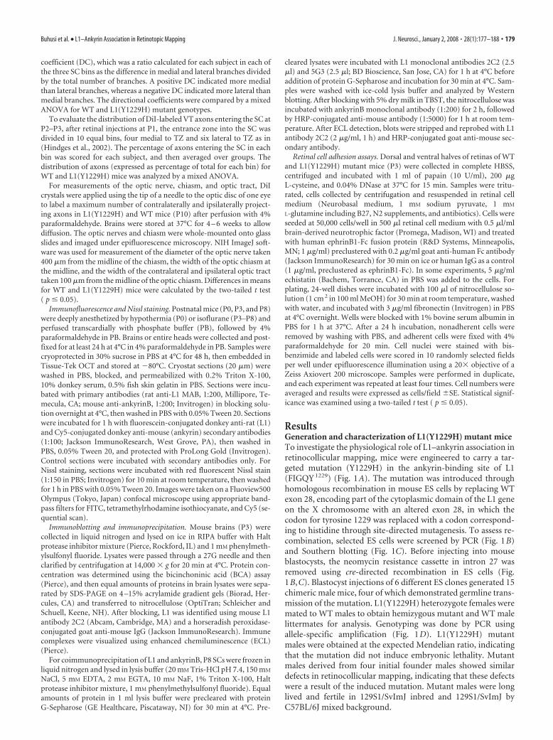
fragment corresponding to the L1 sequence. This probe detected a wild-type (WT) band of 10 kb in parental ES cells and a targeted (neomycin�)band of 7 kb in the PCR-positive clones (see Fig. 1C). In a second step, theneomycin-resistance gene in intron 27 was eliminated from the genomicDNA by cre-mediated recombination in ES cells, and selected clones wereretested by Southern blotting and allele-specific PCR (using primers thatrecognize either the WT or the mutant sequence in exon 28: EX28WT5�-TCTTTCATCGGCCAGCACTCG-3� and EX28MUT 5�-CTCTTT-CATCGGCCAGTACAG-3�, and a primer annealing to a sequence inintron 27 ACROSSL1RP 5�-AATAAAGAGGGTAGCGGGTGGTAAG-3�) (see Fig. 1 B). The WT set of primers amplified a 450 bp fragmentfrom only the parental ES cells, whereas the MUT set of primers ampli-fied a 1600 bp fragment from the neomycin-positive mutant clone and a550 bp from the final neomycin-negative clone, and did not anneal to theparental ES cell DNA. A PCR using primers on both sides of the neomy-cin cassette (AcrossL1FP in the 3� region of the L1 gene and AcrossL1RPin intron 27) confirmed the excision of the neomycin cassette (see Fig.1 B) by cre recombination. After the cre-mediated excision of the neomy-cin gene, the Southern blot probe recognized a targeted (neomycin�)band of �6 kb (see Fig. 1C).
Mouse blastocysts were injected with six different selected ES clonesand transferred to pseudopregnant mice in the University of North Caro-lina Animal Models Facility. Fifteen chimeric male mice were obtained,four of which demonstrated germline transmission. Male chimeras weremated with WT 129S1/SvImJ mice to generate isogenic L1(Y1229H)heterozygous females. After genotyping female offspring by allele-specific PCR (see Fig. 1 D), the L1(Y1229H) heterozygotes were mated toWT males to obtain male L1 mutants [L1(Y1229H)/Y; 50% of maleoffspring] and control littermates for analysis. To show whether geneticbackground influences phenotype, heterozygous females were back-crossed to C57BL/6J to create a nearly congenic line. L1(Y1229H) mutantmales were obtained at the expected Mendelian ratio, indicating that themutation does not induce embryonic lethality. Mutant males were longlived and fertile in 129S1/SvImJ inbred and 129S1/SvImJ by C57BL/6Jmixed backgrounds.
Axon tracing and analysis. Axonal tracing was performed essentially asdescribed previously (Simon and O’Leary, 1992; Demyanenko and Ma-ness, 2003). L1(Y1229H) mutant males and WT littermates at P2–P3 andP10 –P12 were anesthetized and anterograde tracing was performed byfocal injection of DiI (Invitrogen, Carlsbad, CA) as an 10% solution indimethylformamide or dimethylsulfoxide into the peripheral region ofthe retina using a Picospritzer II (General Valve, Fairfield, NJ) and cap-illary glass micropipettes (tip internal diameter, �40 �m). After 48 h,mice were deeply anesthetized and perfused transcardially with 4% para-formaldehyde in 0.1 M phosphate buffer (PB), pH 7.4. Before removingthe retina, the eye was visually inspected to identify the injection site andassess its position in the required quadrant relative to the extraocularmuscles (lateral and inferior recti). The eyes were then removed andincisions were made to demarcate the four quadrants of the retina. Toverify the injection sites and analyze the projection areas, injected retinasas well as superior and inferior colliculi were whole-mounted onto glassslides and examined by epifluorescence and confocal microscopy (Uni-versity of North Carolina Microscopy Services; LSM 5; Zeiss,Oberkochen, Germany) using appropriate filters. The injection sites cov-ered 3–5% of the retina. The boundaries of the SC and IC were deter-mined by their characteristic shape and location. Termination zones(TZs) were verified by their branched appearance at high magnification.For analysis, images of the SC were acquired at the same magnification,and the locations of the TZs were scored along the mediolateral (x) andanteroposterior ( y) axes. The anteromedial corner of the SC was consid-ered as the zero point (x � 0; y � 0).
To quantitatively evaluate branch orientation of ventrotemporal (VT)axons in L1(Y1229H) and WT mice at P2–P3, the SC was divided intothree bins along the mediolateral axis, relative to the emerging TZ: lateralto TZ, within TZ, and medial to TZ, as described previously (Hindges etal., 2002). Only mutants with one normally positioned TZs were in-cluded in this analysis. The total number of labeled axons and brancheswas counted in confocal z-stacks, and medial or lateral branch orienta-tion was recorded for each bin. The results were expressed as a directional
178 • J. Neurosci., January 2, 2008 • 28(1):177–188 Buhusi et al. • L1–Ankyrin Association in Retinotopic Mapping
coefficient (DC), which was a ratio calculated for each subject in each ofthe three SC bins as the difference in medial and lateral branches dividedby the total number of branches. A positive DC indicated more medialthan lateral branches, whereas a negative DC indicated more lateral thanmedial branches. The directional coefficients were compared by a mixedANOVA for WT and L1(Y1229H) mutant genotypes.
To evaluate the distribution of DiI-labeled VT axons entering the SC atP2–P3, after retinal injections at P1, the entrance zone into the SC wasdivided in 10 equal bins, four medial to TZ and six lateral to TZ as in(Hindges et al., 2002). The percentage of axons entering the SC in eachbin was scored for each subject, and then averaged over groups. Thedistribution of axons (expressed as percentage of total for each bin) forWT and L1(Y1229H) mice was analyzed by a mixed ANOVA.
For measurements of the optic nerve, chiasm, and optic tract, DiIcrystals were applied using the tip of a needle to the optic disc of one eyeto label a maximum number of contralaterally and ipsilaterally project-ing axons in L1(Y1229H) and WT mice (P10) after perfusion with 4%paraformaldehyde. Brains were stored at 37°C for 4 – 6 weeks to allowdiffusion. The optic nerves and chiasm were whole-mounted onto glassslides and imaged under epifluorescence microscopy. NIH ImageJ soft-ware was used for measurement of the diameter of the optic nerve taken400 �m from the midline of the chiasm, the width of the optic chiasm atthe midline, and the width of the contralateral and ipsilateral optic tracttaken 100 �m from the midline of the optic chiasm. Differences in meansfor WT and L1(Y1229H) mice were calculated by the two-tailed t test( p � 0.05).
Immunofluorescence and Nissl staining. Postnatal mice (P0, P3, and P8)were deeply anesthetized by hypothermia (P0) or isoflurane (P3–P8) andperfused transcardially with phosphate buffer (PB), followed by 4%paraformaldehyde in PB. Brains or entire heads were collected and post-fixed for at least 24 h at 4°C in 4% paraformaldehyde in PB. Samples werecryoprotected in 30% sucrose in PBS at 4°C for 48 h, then embedded inTissue-Tek OCT and stored at �80°C. Cryostat sections (20 �m) werewashed in PBS, blocked, and permeabilized with 0.2% Triton X-100,10% donkey serum, 0.5% fish skin gelatin in PBS. Sections were incu-bated with primary antibodies (rat anti-L1 MAB, 1:200, Millipore, Te-mecula, CA; mouse anti-ankyrinB, 1:200; Invitrogen) in blocking solu-tion overnight at 4°C, then washed in PBS with 0.05% Tween 20. Sectionswere incubated for 1 h with fluorescein-conjugated donkey anti-rat (L1)and Cy5-conjugated donkey anti-mouse (ankyrin) secondary antibodies(1:100; Jackson ImmunoResearch, West Grove, PA), then washed inPBS, 0.05% Tween 20, and protected with ProLong Gold (Invitrogen).Control sections were incubated with secondary antibodies only. ForNissl staining, sections were incubated with red fluorescent Nissl stain(1:150 in PBS; Invitrogen) for 10 min at room temperature, then washedfor 1 h in PBS with 0.05% Tween 20. Images were taken on a Fluoview500Olympus (Tokyo, Japan) confocal microscope using appropriate band-pass filters for FITC, tetramethylrhodamine isothiocyanate, and Cy5 (se-quential scan).
Immunoblotting and immunoprecipitation. Mouse brains (P3) werecollected in liquid nitrogen and lysed on ice in RIPA buffer with Haltprotease inhibitor mixture (Pierce, Rockford, IL) and 1 mM phenylmeth-ylsulfonyl fluoride. Lysates were passed through a 27G needle and thenclarified by centrifugation at 14,000 � g for 20 min at 4°C. Protein con-centration was determined using the bicinchoninic acid (BCA) assay(Pierce), and then equal amounts of proteins in brain lysates were sepa-rated by SDS-PAGE on 4 –15% acrylamide gradient gels (Biorad, Her-cules, CA) and transferred to nitrocellulose (OptiTran; Schleicher andSchuell, Keene, NH). After blocking, L1 was identified using mouse L1antibody 2C2 (Abcam, Cambridge, MA) and a horseradish peroxidase-conjugated goat anti-mouse IgG (Jackson ImmunoResearch). Immunecomplexes were visualized using enhanced chemiluminescence (ECL)(Pierce).
For coimmunoprecipitation of L1 and ankyrinB, P8 SCs were frozen inliquid nitrogen and lysed in lysis buffer (20 mM Tris-HCl pH 7.4, 150 mM
NaCl, 5 mM EDTA, 2 mM EGTA, 10 mM NaF, 1% Triton X-100, Haltprotease inhibitor mixture, 1 mM phenylmethylsulfonyl fluoride). Equalamounts of protein in 1 ml lysis buffer were precleared with proteinG-Sepharose (GE Healthcare, Piscataway, NJ) for 30 min at 4°C. Pre-
cleared lysates were incubated with L1 monoclonal antibodies 2C2 (2.5�l) and 5G3 (2.5 �l; BD Bioscience, San Jose, CA) for 1 h at 4°C beforeaddition of protein G-Sepharose and incubation for 30 min at 4°C. Sam-ples were washed with ice-cold lysis buffer and analyzed by Westernblotting. After blocking with 5% dry milk in TBST, the nitrocellulose wasincubated with ankyrinB monoclonal antibody (1:200) for 2 h, followedby HRP-conjugated anti-mouse antibody (1:5000) for 1 h at room tem-perature. After ECL detection, blots were stripped and reprobed with L1antibody 2C2 (2 �g/ml, 1 h) and HRP-conjugated goat anti-mouse sec-ondary antibody.
Retinal cell adhesion assays. Dorsal and ventral halves of retinas of WTand L1(Y1229H) mutant mice (P3) were collected in complete HBSS,centrifuged and incubated with 1 ml of papain (10 U/ml), 200 �gL-cysteine, and 0.04% DNase at 37°C for 15 min. Samples were tritu-rated, cells collected by centrifugation and resuspended in retinal cellmedium (Neurobasal medium, 1 mM sodium pyruvate, 1 mM
L-glutamine including B27, N2 supplements, and antibiotics). Cells wereseeded at 50,000 cells/well in 500 �l retinal cell medium with 0.5 �l/mlbrain-derived neurotrophic factor (Promega, Madison, WI) and treatedwith human ephrinB1-Fc fusion protein (R&D Systems, Minneapolis,MN; 1 �g/ml) preclustered with 0.2 �g/ml goat anti-human Fc antibody(Jackson ImmunoResearch) for 30 min on ice or human IgG as a control(1 �g/ml, preclustered as ephrinB1-Fc). In some experiments, 5 �g/mlechistatin (Bachem, Torrance, CA) in PBS was added to the cells. Forplating, 24-well dishes were incubated with 100 �l of nitrocellulose so-lution (1 cm 2 in 100 ml MeOH) for 30 min at room temperature, washedwith water, and incubated with 3 �g/ml fibronectin (Invitrogen) in PBSat 4°C overnight. Wells were blocked with 1% bovine serum albumin inPBS for 1 h at 37°C. After a 24 h incubation, nonadherent cells wereremoved by washing with PBS, and adherent cells were fixed with 4%paraformaldehyde for 20 min. Cell nuclei were stained with bis-benzimide and labeled cells were scored in 10 randomly selected fieldsper well under epifluorescence illumination using a 20� objective of aZeiss Axiovert 200 microscope. Samples were performed in duplicate,and each experiment was repeated at least four times. Cell numbers wereaveraged and results were expressed as cells/field �SE. Statistical signif-icance was examined using a two-tailed t test ( p � 0.05).
ResultsGeneration and characterization of L1(Y1229H) mutant miceTo investigate the physiological role of L1–ankyrin association inretinocollicular mapping, mice were engineered to carry a tar-geted mutation (Y1229H) in the ankyrin-binding site of L1(FIGQY 1229) (Fig. 1A). The mutation was introduced throughhomologous recombination in mouse ES cells by replacing WTexon 28, encoding part of the cytoplasmic domain of the L1 geneon the X chromosome with an altered exon 28, in which thecodon for tyrosine 1229 was replaced with a codon correspond-ing to histidine through site-directed mutagenesis. To assess re-combination, selected ES cells were screened by PCR (Fig. 1B)and Southern blotting (Fig. 1C). Before injecting into mouseblastocysts, the neomycin resistance cassette in intron 27 wasremoved using cre-directed recombination in ES cells (Fig.1B,C). Blastocyst injections of 6 different ES clones generated 15chimeric male mice, four of which demonstrated germline trans-mission of the mutation. L1(Y1229H) heterozygote females weremated to WT males to obtain hemizygous mutant and WT malelittermates for analysis. Genotyping was done by PCR usingallele-specific amplification (Fig. 1D). L1(Y1229H) mutantmales were obtained at the expected Mendelian ratio, indicatingthat the mutation did not induce embryonic lethality. Mutantmales derived from four initial founder males showed similardefects in retinocollicular mapping, indicating that these defectswere a result of the induced mutation. Mutant males were longlived and fertile in 129S1/SvImJ inbred and 129S1/SvImJ byC57BL/6J mixed background.
Buhusi et al. • L1–Ankyrin Association in Retinotopic Mapping J. Neurosci., January 2, 2008 • 28(1):177–188 • 179
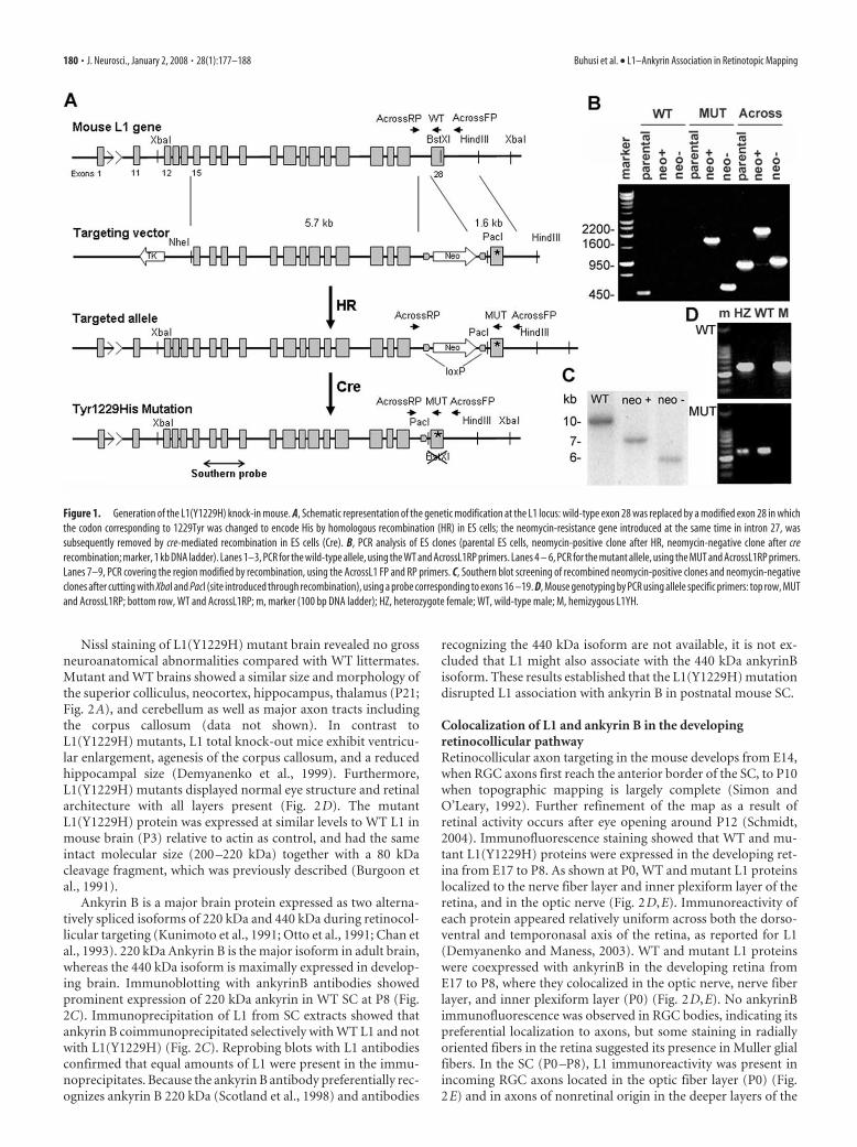
Nissl staining of L1(Y1229H) mutant brain revealed no grossneuroanatomical abnormalities compared with WT littermates.Mutant and WT brains showed a similar size and morphology ofthe superior colliculus, neocortex, hippocampus, thalamus (P21;Fig. 2A), and cerebellum as well as major axon tracts includingthe corpus callosum (data not shown). In contrast toL1(Y1229H) mutants, L1 total knock-out mice exhibit ventricu-lar enlargement, agenesis of the corpus callosum, and a reducedhippocampal size (Demyanenko et al., 1999). Furthermore,L1(Y1229H) mutants displayed normal eye structure and retinalarchitecture with all layers present (Fig. 2D). The mutantL1(Y1229H) protein was expressed at similar levels to WT L1 inmouse brain (P3) relative to actin as control, and had the sameintact molecular size (200 –220 kDa) together with a 80 kDacleavage fragment, which was previously described (Burgoon etal., 1991).
Ankyrin B is a major brain protein expressed as two alterna-tively spliced isoforms of 220 kDa and 440 kDa during retinocol-licular targeting (Kunimoto et al., 1991; Otto et al., 1991; Chan etal., 1993). 220 kDa Ankyrin B is the major isoform in adult brain,whereas the 440 kDa isoform is maximally expressed in develop-ing brain. Immunoblotting with ankyrinB antibodies showedprominent expression of 220 kDa ankyrin in WT SC at P8 (Fig.2C). Immunoprecipitation of L1 from SC extracts showed thatankyrin B coimmunoprecipitated selectively with WT L1 and notwith L1(Y1229H) (Fig. 2C). Reprobing blots with L1 antibodiesconfirmed that equal amounts of L1 were present in the immu-noprecipitates. Because the ankyrin B antibody preferentially rec-ognizes ankyrin B 220 kDa (Scotland et al., 1998) and antibodies
recognizing the 440 kDa isoform are not available, it is not ex-cluded that L1 might also associate with the 440 kDa ankyrinBisoform. These results established that the L1(Y1229H) mutationdisrupted L1 association with ankyrin B in postnatal mouse SC.
Colocalization of L1 and ankyrin B in the developingretinocollicular pathwayRetinocollicular axon targeting in the mouse develops from E14,when RGC axons first reach the anterior border of the SC, to P10when topographic mapping is largely complete (Simon andO’Leary, 1992). Further refinement of the map as a result ofretinal activity occurs after eye opening around P12 (Schmidt,2004). Immunofluorescence staining showed that WT and mu-tant L1(Y1229H) proteins were expressed in the developing ret-ina from E17 to P8. As shown at P0, WT and mutant L1 proteinslocalized to the nerve fiber layer and inner plexiform layer of theretina, and in the optic nerve (Fig. 2D,E). Immunoreactivity ofeach protein appeared relatively uniform across both the dorso-ventral and temporonasal axis of the retina, as reported for L1(Demyanenko and Maness, 2003). WT and mutant L1 proteinswere coexpressed with ankyrinB in the developing retina fromE17 to P8, where they colocalized in the optic nerve, nerve fiberlayer, and inner plexiform layer (P0) (Fig. 2D,E). No ankyrinBimmunofluorescence was observed in RGC bodies, indicating itspreferential localization to axons, but some staining in radiallyoriented fibers in the retina suggested its presence in Muller glialfibers. In the SC (P0 –P8), L1 immunoreactivity was present inincoming RGC axons located in the optic fiber layer (P0) (Fig.2E) and in axons of nonretinal origin in the deeper layers of the
Figure 1. Generation of the L1(Y1229H) knock-in mouse. A, Schematic representation of the genetic modification at the L1 locus: wild-type exon 28 was replaced by a modified exon 28 in whichthe codon corresponding to 1229Tyr was changed to encode His by homologous recombination (HR) in ES cells; the neomycin-resistance gene introduced at the same time in intron 27, wassubsequently removed by cre-mediated recombination in ES cells (Cre). B, PCR analysis of ES clones (parental ES cells, neomycin-positive clone after HR, neomycin-negative clone after crerecombination; marker, 1 kb DNA ladder). Lanes 1–3, PCR for the wild-type allele, using the WT and AcrossL1RP primers. Lanes 4 – 6, PCR for the mutant allele, using the MUT and AcrossL1RP primers.Lanes 7–9, PCR covering the region modified by recombination, using the AcrossL1 FP and RP primers. C, Southern blot screening of recombined neomycin-positive clones and neomycin-negativeclones after cutting with XbaI and PacI (site introduced through recombination), using a probe corresponding to exons 16 –19. D, Mouse genotyping by PCR using allele specific primers: top row, MUTand AcrossL1RP; bottom row, WT and AcrossL1RP; m, marker (100 bp DNA ladder); HZ, heterozygote female; WT, wild-type male; M, hemizygous L1YH.
180 • J. Neurosci., January 2, 2008 • 28(1):177–188 Buhusi et al. • L1–Ankyrin Association in Retinotopic Mapping
SC (asterisk) (Lyckman et al., 2000). AnkyrinB colocalized withL1 in axons at these locations (Fig. 2E, overlay) and appeareduniform across the lateromedial axis of the SC. L1(Y1229H)showed a similar distribution and colocalization with ankyrinB inthe SC (Fig. 2E, overlay).
Countergradients of EphB receptors along the dorsoventralaxis of the retina (high ventral to low dorsal) and ephrinB1 alongthe mediolateral axis of the SC (high medial to low lateral) estab-lish topographically appropriate projections across the mediolat-eral SC axis (Hindges et al., 2002). EphrinB/EphB interactions
during retinocollicular mapping do notresult in an inhibitory, repellent responseof growth cones, but instead appear to me-diate attraction of interstitial axonbranches up the ephrinB1 gradient of theSC (Hindges et al., 2002). L1(Y1229H)mutant mice (P0) showed no difference inthe level or pattern of immunofluores-cence staining of EphB2 in the retina orephrinB1 in the SC compared with WT(data not shown), with the same gradedpattern as reported previously (Hindges etal., 2002).
Retinotopic mapping defects inL1(Y1229H) miceL1 knock-out mice show major defects intargeting of temporal RGC axons along theanteroposterior axis, resulting in over-shooting the correct TZ as well as lateraldisplacement (Demyanenko and Maness,2003). To investigate the effects of disrupt-ing the L1–ankyrin interaction on axonalgrowth and guidance, the topographicprojections of RGC axons in the SC ofL1(Y1229H) mice were analyzed at stageswhen the basic topography of the retino-collicular map resembles its mature form(P10 –P12). Focal injections of the axonaltracer DiI were made into the retina of live,anesthetized mice at P8 –P10, and labeledRGC projections in the SC were analyzed2 d later. Analyses were restricted to ani-mals in which retinal whole mounts dis-played a single DiI injection of appropriatesize (3–5% of the retina) and positioningrelative to the insertions of the extraocularmuscles.
During development of the topo-graphic map, temporal RGC axons projectto the contralateral SC, whereas nasal ax-ons project to the posterior SC by mecha-nisms requiring ephrinA/EphA receptorsignaling (Feldheim et al., 2000, 2004).Thus, altered anteroposterior mapping ismost evident in the projection of temporalaxons, although deviation along the me-diolateral axis would also be observed. Asexpected, in 10 of 10 WT mice focal DiIinjections into the midtemporal (T) retinalabeled a single dense TZ in the middle an-terior region of the contralateral SC (Fig.3A,B). In contrast, similar injections into
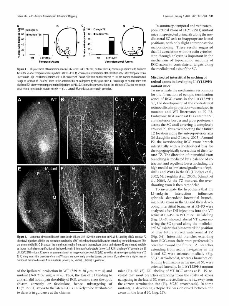
the mid-temporal retina of L1(Y1229H) mutants identified to-pographic targeting errors along the mediolateral axis of the con-tralateral SC. In 22 of 28 injections, mutant axons terminated atabnormally lateral sites, mostly as multiple ectopic terminationzones (eTZs) that appeared to be connected to a correct TZ in themidanterior SC (8 of 28 cases) (Fig. 3D–F). Alternatively, mutanttemporal axons terminated as a single laterally displaced TZ (14of 28 cases) (Fig. 3G). The percentage of cases with abnormalmediolateral positioning in the mutant mice was significantlydifferent from WT (�2 � 18.66; p � 0.0001) (Fig. 4). In mutants,
Figure 2. L1 expression and colocalization with ankyrinB in brain and retina of WT and L1(Y1229H) mice. A, Nissl staining of WTand L1(Y1229H) mice at P21 (strain 129). C, Cortex; HC, hippocampus; TH, thalamus; LV, lateral ventricle; cc, corpus callosum. B,Western blot analysis of P3 brain lysates (45 �g) from WT (lane 1) and mutant (lane 2) mice using an L1 antibody. C, Coimmu-noprecipitation of ankyrinB with L1 from WT mouse superior colliculus lysates (P8). No ankyrinB coimmunoprecipitated withnonimmune mouse IgG or with L1(Y12229H) protein. D, Immunofluorescence localization of L1 and ankyrinB in sagittal sectionsof the P0 mouse retina. E, Localization of L1 and ankyrinB in sagittal sections of the P0 mouse retina (high magnification) and incoronal sections of the superior colliculus in the anterior third of the anteroposterior axis: L1, green; ankyrinB, red; Nissl stain,pseudocolored blue. NFL, Nerve fiber layer; GCL, ganglion cell layer; IPL, inner plexiform layer; D, dorsal; V, ventral; ON, optic nerve;ONL, optic nerve layer in the SC. F, Optic chiasm visualized by DiI tracing from the left retina. Contralateral (CL) and ipsilateral (IL)projections in WT and L1(Y1229H) littermates were of similar sizes.
Buhusi et al. • L1–Ankyrin Association in Retinotopic Mapping J. Neurosci., January 2, 2008 • 28(1):177–188 • 181

TZs or eTZs were often slightly malposi-tioned posteriorly along the anteroposte-rior axis of the SC (Figs. 3F,G, 4) but notto the same extent seen in L1 null mutantmice (Demyanenko and Maness, 2003).To more critically assess the mediolateralmisplacement of TZs in mutant mice, VTretinal injections of DiI were performed inmutants and WT littermates. Because VTaxons express high levels of EphB recep-tors and normally project to the anterome-dial corner of the SC, their attractive re-sponse to the high medial to low lateralephrinB1 gradient in the SC renders themespecially sensitive to deficiencies in me-diolateral targeting, which is regulated byephrinB1/EphB receptor signaling(Hindges et al., 2002). In seven of sevenWT mice, VT retinal injections resulted inlabeling a single well defined TZ in the an-teromedial SC (Fig. 3H, I). In contrast, VTaxons of 10/15 mutants projected to ab-normally lateral SC sites (Fig. 3J–L). In 8 of15 cases, mutant VT axons formed multi-ple ectopic TZs in addition to a correctlypositioned TZ, whereas in 2 of 15 cases asingle laterally displaced TZ was observed.The percentage of cases with abnormal tar-geting of VT axons in L1(Y1229H) mutantmice was significantly different from WT(�2 � 8.55; p � 0.005) (Fig. 4). VT axonsof L1(Y1229H) mice rarely terminatedposteriorly along the anteroposterior SCaxis, indicating that L1–ankyrin interac-tions play a more significant role in medio-lateral rather than anteroposterior target-ing of these axons.
The projection of RGC axons from thedorsal or nasal retina was not perturbed inL1(Y1229H) mutants. Dorsal retinal injec-tions in L1(Y1229H) mice resulted in la-beling of a single TZ within the lateral SClocated midway along the rostrocaudalaxis (Fig. 3O,P) (n � 2), which occurred atthe same location in WT mice (data notshown). Nasal retinal injections inL1(Y1229H) mice resulted in labeling of asingle TZ in the mid-posterior SC (n � 8)(Fig. 3M,N) at the same location as in WTmice (data not shown). Thus, mapping of RGC axons from thedorsal and nasal retina appeared to be relatively unaffected by theankyrin binding site mutation in L1. It should be added thatimmunofluorescence staining for EphB2 in the retina and eph-rinB1 in the SC of L1(Y1229H) and WT mice (P0) showed nodifferences in level of expression (data not shown).
An EphB1-expressing subset of VT axons normally projectipsilaterally because of ephrinB2/EphB1-dependent axon diver-gence at the optic chiasm (Williams et al., 2003). To determinewhether L1Y1229H) RGC axons were impaired for guidance atthe optic chiasm, DiI crystals were inserted onto the optic disk ofone eye to label contralaterally and ipsilaterally projecting axonsin mutant and WT littermates (P10). As shown in whole mounts,most labeled RGC axons in the optic nerve crossed the midline at
the optic chiasm to the contralateral optic tract and a small ipsi-lateral projection was observed in both WT and L1(Y1229H)mutants (Fig. 2F). RGC axons in the optic nerve and optic tractexhibited a similar degree of fasciculation in WT and mutants.Direct measurement of the diameter of the optic nerve at a posi-tion 400 �m from the midline of the optic chiasm showed nosignificant differences (t test, two-tailed) in means for WT (338 �27 �m; n � 4) and L1(Y1229H) mutants (402 � 24 �m; n � 6;p � 0.05). The mean width of the optic chiasm measured at themidline was also not significantly different for WT (873 � 37 �m;n � 4) and L1(Y1229H) mutants (920 � 74 �m; n � 6). Themean width of the optic tract measured 100 �m posterior to themidline was also the same for WT (645 � 76 �m; n � 4) andL1(Y1229H) mutants (691 � 82 �m; n � 6), as was the diameter
Figure 3. Defects in retinocollicular mapping in L1(Y1229H) mutant mice. A, B, Injection of DiI into the peripheral temporalretina (shown in flatmount) at P8 labeled a single TZ in the anterior SC in P10 WT mice. C–G, DiI injections in the peripheraltemporal retina of L1(Y1229H) mutant mice revealed ectopic TZs (eTZ), displaced to lateral and slightly posterior positions in theSC. D–F, Higher magnification of the TZs (top left) revealed branches extending laterally or posteriorly toward the eTZs. H, I, Focalinjections of DiI in the ventrotemporal (VT) retina labeled a single dense TZ in the anteromedial corner of the SC in P10 WT mice.J–L, Similar injections in L1(Y1229H) mutant mice resulted in eTZs displaced laterally within the anterior SC. M, N, Nasal retinalinjections in L1(Y1229H) mutant mice resulted in labeling of a single TZ in the posterior SC at P10. O, P, Injection of DiI in theperipheral dorsal retina at P8 labeled a single TZ in the lateral SC in P10 L1(Y1229H) mice. L, Lateral; M, medial; A, anterior; P,posterior; D, dorsal; V, ventral; N, nasal; T, temporal.
182 • J. Neurosci., January 2, 2008 • 28(1):177–188 Buhusi et al. • L1–Ankyrin Association in Retinotopic Mapping
of the ipsilateral projection in WT (359 � 30 �m; n � 4) andmutant (360 � 32 �m; n � 6). Thus, the loss of L1 binding toankyrin did not impair the ability of RGC axons to cross the opticchiasm correctly or fasciculate, hence, mistargeting ofL1(Y1229H) axons to the lateral SC is unlikely to be attributableto defects in guidance at the chiasm.
In summary, temporal and ventrotem-poral retinal axons of L1(Y1229H) mutantmice misprojected primarily along the me-diolateral SC axis to inappropriate lateralpositions, with only slight anteroposteriormalpositioning. These results suggestedthat L1 association with the actin cytoskel-eton through ankyrin is important in themechanism of topographic mapping ofRGC axons to contralateral targets alongthe mediolateral axis of the SC.
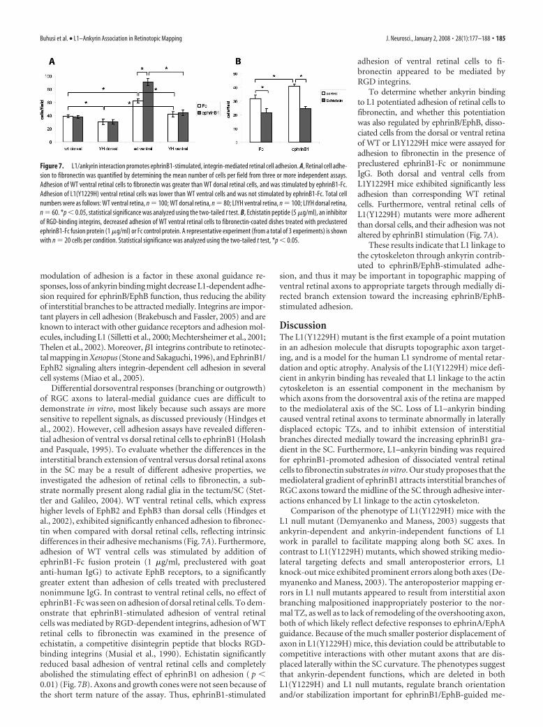
Misdirected interstitial branching ofretinal axons in developing L1(Y1229H)mutant miceTo investigate the mechanism responsiblefor the formation of ectopic terminationzones of RGC axons in the L1(Y1229H)SC, the development of the contralateralretinocollicular projection was analyzed inmutants and WT littermates at P2–P3.Embryonic RGC axons at E14 enter the SCat its anterior border and grow posteriorlyacross the SC until covering it completelyaround P0, thus overshooting their futureTZ location along the anteroposterior axis(McLaughlin and O’Leary, 2005). AroundP2, the overshooting RGC axons branchinterstitially with a mediolateral bias forthe topographically correct site of their fu-ture TZ. The direction of interstitial axonbranching is mediated by a balance of at-tractant and repellent forces including thehigh medial to low lateral gradients of eph-rinB1 and Wnt3 in the SC (Hindges et al.,2002; McLaughlin et al., 2003b; Schmitt etal., 2006). As the TZ matures, the over-shooting axon is then remodeled.
To investigate the hypothesis that theL1–ankyrin interaction influencesephrinB1-dependent interstitial branch-ing, RGC axons in the SC and their devel-oping interstitial branches at P2–P3 wereanalyzed after DiI injections into the VTretina at P1–P2. In WT mice, DiI labeling(Fig. 5A–D) showed labeled VT axons en-tering the SC spread along the mediolat-eral SC axis with a bias toward the positionof their future correct anteromedial TZ(Fig. 5A). Interstitial branches extendingfrom RGC axon shafts were preferentiallyoriented toward the future TZ. Branchesextending from axons navigating in thelateral SC were oriented medially (Fig.5C,D, arrowheads), whereas branches ex-tending from axons in the medial SC wereoriented laterally. In L1(Y1229H) mutant
mice (Fig. 5E–H), DiI labeling of VT RGC axons at P1–P2 re-vealed that most branches extending from the shafts of axonsnavigating in the lateral SC were directed laterally; i.e., away fromthe correct termination site (Fig. 5G,H, arrowheads). In somemutants, a developing ectopic TZ was observed between theaxons in the lateral SC (Fig. 5E).
Figure 4. Displacement of termination zones of RGC axons in L1(Y1229H) mutant mice. A, Percentage of mice with displacedTZs in the SC after temporal retinal injections at P10 –P12. B, Schematic representation of the location of TZs after temporal retinalinjections in L1(Y1229H) mutant mice at P10. The centers of TZs and eTZs from mutant mice (n � 10) are marked and connected.Range of location of TZs of WT mice in the anteromedial SC is depicted by the gray circle. C, Percentage of mutant mice withdisplaced TZs after ventrotemporal retinal injections at P10. D, Schematic representation of the aberrant eTZs after ventrotem-poral retinal injections in mutant mice (n � 6). L, Lateral; M, medial; A, anterior; P, posterior.
Figure 5. Abnormal directional branch extension in WT and L1(Y1229H) mutant mice at P2. A, B, Labeling of RGC axons at P2after focal injections of DiI in the ventrotemporal retina of WT mice show interstitial branches extending toward the nascent TZ inthe anteromedial SC. C, D, Most of the branches extending from axons that navigate lateral to the future TZ are oriented mediallyas shown in a higher magnification of the boxed area in B from confocal z-stacks (arrows). E, F, DiI labeling of VT axons in the SCof L1(Y1229H) mice at P2 reveal an accumulation at an inappropriate ectopic TZ (eTZ) as well as at a more appropriate future TZ.G, H, Many interstitial branches of mutant VT axons are abnormally oriented toward the lateral SC, as shown in a higher magni-fication of the boxed area in F from z-stacks (arrows). M, Medial; L, lateral; P, posterior.
Buhusi et al. • L1–Ankyrin Association in Retinotopic Mapping J. Neurosci., January 2, 2008 • 28(1):177–188 • 183

To quantitatively evaluate branch ori-entation of VT axons in L1(Y1229H) andWT mice at P2–P3, the SC was dividedinto three bins along the mediolateral axisrelative to the emerging TZ: lateral to TZ,within TZ, and medial to TZ (Fig. 6). Thetotal number of labeled axons andbranches was counted, and medial or lat-eral branch orientation was recorded foreach bin (Fig. 6A,B). The results were ex-pressed as a DC, which was calculated foreach subject in each of the bins, as the dif-ference in number of medial and lateralbranches, divided by the total number ofbranches, as defined in (Hindges et al.,2002). A positive DC indicated more me-dial than lateral branches, whereas a nega-tive DC indicated more lateral than medialbranches. Analysis of RGC axons in the SCat P2–P3 after DiI injection into the VTretina confirmed that in WT mice (n � 8)interstitial axon branches were preferen-tially oriented toward the future TZ alongthe mediolateral axis. The majority ofbranches in the lateral bin were orientedmedially toward the TZ, branches of axonsin the TZ bin were largely unbiased, andthe majority of branches in the medial binwere oriented laterally toward the TZ (Fig.6B). In contrast, VT axons of L1(Y1229H)mutants (n � 10) in the lateral bin wereoriented laterally with respect to the TZ,whereas branches of axons in the TZ andmedial bins were normally oriented.
The DCs were compared by a mixedANOVA for WT (n � 8 mice; 437branches) and L1(Y1229H) mutants (n �10 mice; 754 branches). The statisticalanalysis revealed a significant main effectof genotype (F(1,16) � 75.28; p � 0.00001),demonstrating that branch orientation inL1(Y1229H) mice was different from WT. Analysis of the inter-action between bin and genotype indicated a significant differ-ence in branching of WT and mutant axons in the lateral bin(F(2,32) � 29.99; p � 0.00001), but not the medial bin (t(16) �0.32; p � 0.75). Analyses also showed no significant differencewithin the TZ (t(16) � 1.55; p � 0.14), where the DC was notsignificantly different from zero for either genotype (WT, t(7) �1.18, p � 0.28; L1(1229H) mice, t(9) � 0.92, p � 0.38).
In summary, these results suggested that disruption of L1 as-sociation with ankyrin resulted in a decrease in the ability ofinterstitial branches of VT axons to be attracted medially towardan increasing ephrinB1 gradient, similar to that seen in micedeficient in EphB2/EphB3 receptors (Hindges et al., 2002),whereas repellent guidance of medial axons away from the eph-rinB1 gradient was preserved.
To evaluate whether disruption of the L1–ankyrin interactionresulted in different patterns of axonal entrance into the SC, thedistribution of DiI-labeled VT axons entering the SC was evalu-ated at P2-P3, after retinal injections at P1. For each mouse theentrance zone into the SC was divided into 10 equal bins: fourbins located medial to the TZ, and six bins lateral to the TZ. Thepercentage of axons entering the SC in each bin was scored for
each subject (Fig. 6C), and averaged over groups (Fig. 6D). Thedistribution of axons (expressed as percentage of total for eachbin) for WT (n � 6 mice; 291 axons) and L1(Y1229H) mice (n �9 mice; 442 axons) was then analyzed by a mixed ANOVA. Theanalysis revealed no significant differences between WT andL1(Y1229H) mice (F(9,117) � 0.66; p 0.75). As expected, therewas a significant effect of the entry position of VT axons in bothWT and mutant mice (F(9,117) � 10.54; p � 0.0001), such thatmore axons entered the SC in the vicinity of the TZ, decreasingmedially and laterally. Thus, laterally displaced ectopic TZs in theL1(Y1229H) mutant were probably not caused by impaired ax-onal entrance into the SC.
L1–ankyrin binding is required for ephrinB1-inducedmodulation of cell adhesionMice deficient in both EphB2 and EphB3 display topographicallyaberrant projections along the mediolateral SC axis resulting inlaterally displaced ectopic TZs and disrupted branch extension(Hindges et al., 2002). The strong similarity of mapping defects inL1(Y1229H) mice and EphB2/EphB3 knock-out mice suggestedthat the lateral displacement of temporal and VT RGC axons inL1(Y1229H) mice might be caused by altered responsiveness toephrinB1-mediated attraction to medial positions. If dynamic
Figure 6. Branching and axonal positioning patterns in the SC of WT and L1(Y1229H) mice at P2. A, Schematic representationof directed branch extension along the mediolateral axis. The SC was divided in three bins [lateral (L), TZ, medial (M)] separatedby dash lines, in relation to the forming TZ (gray circle). The orientation of each branch was recorded and graphed by bin. B,Distribution of branches in P2–P3 WT and L1(Y1229H) mutant mice after VT retinal injections. Arrows represent the direction ofthe branching preference in each region. The results were expressed as a branch directional coefficient as described (Hindges et al.,2002), calculated for each subject for each of the three bins of the SC as the difference in the number of medially oriented branchesminus the number of laterally oriented branches, divided by the total number of branches. C, Schematic representation of RGCaxon positioning in the SC in WT and mutant mice. The SC was divided into 10 segments along the mediolateral axis at the anteriorborder, and all labeled RGC axons within each bin were counted and represented relative to the position of the developing TZ foreach injection. D, Distribution of labeled axons along the mediolateral SC axis (expressed as percentage of total) in L1(Y1229H)mice compared to WT littermates at P2, after VT retinal injections. Error bars indicate SEM.
184 • J. Neurosci., January 2, 2008 • 28(1):177–188 Buhusi et al. • L1–Ankyrin Association in Retinotopic Mapping
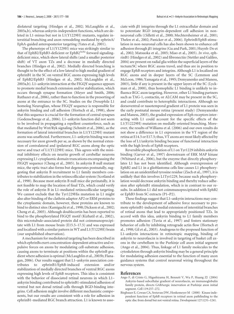
modulation of adhesion is a factor in these axonal guidance re-sponses, loss of ankyrin binding might decrease L1-dependent adhe-sion required for ephrinB/EphB function, thus reducing the abilityof interstitial branches to be attracted medially. Integrins are impor-tant players in cell adhesion (Brakebusch and Fassler, 2005) and areknown to interact with other guidance receptors and adhesion mol-ecules, including L1 (Silletti et al., 2000; Mechtersheimer et al., 2001;Thelen et al., 2002). Moreover, �1 integrins contribute to retinotec-tal mapping in Xenopus (Stone and Sakaguchi, 1996), and EphrinB1/EphB2 signaling alters integrin-dependent cell adhesion in severalcell systems (Miao et al., 2005).
Differential dorsoventral responses (branching or outgrowth)of RGC axons to lateral-medial guidance cues are difficult todemonstrate in vitro, most likely because such assays are moresensitive to repellent signals, as discussed previously (Hindges etal., 2002). However, cell adhesion assays have revealed differen-tial adhesion of ventral vs dorsal retinal cells to ephrinB1 (Holashand Pasquale, 1995). To evaluate whether the differences in theinterstitial branch extension of ventral versus dorsal retinal axonsin the SC may be a result of different adhesive properties, weinvestigated the adhesion of retinal cells to fibronectin, a sub-strate normally present along radial glia in the tectum/SC (Stet-tler and Galileo, 2004). WT ventral retinal cells, which expresshigher levels of EphB2 and EphB3 than dorsal cells (Hindges etal., 2002), exhibited significantly enhanced adhesion to fibronec-tin when compared with dorsal retinal cells, reflecting intrinsicdifferences in their adhesive mechanisms (Fig. 7A). Furthermore,adhesion of WT ventral cells was stimulated by addition ofephrinB1-Fc fusion protein (1 �g/ml, preclustered with goatanti-human IgG) to activate EphB receptors, to a significantlygreater extent than adhesion of cells treated with preclusterednonimmune IgG. In contrast to ventral retinal cells, no effect ofephrinB1-Fc was seen on adhesion of dorsal retinal cells. To dem-onstrate that ephrinB1-stimulated adhesion of ventral retinalcells was mediated by RGD-dependent integrins, adhesion of WTretinal cells to fibronectin was examined in the presence ofechistatin, a competitive disintegrin peptide that blocks RGD-binding integrins (Musial et al., 1990). Echistatin significantlyreduced basal adhesion of ventral retinal cells and completelyabolished the stimulating effect of ephrinB1 on adhesion ( p �0.01) (Fig. 7B). Axons and growth cones were not seen because ofthe short term nature of the assay. Thus, ephrinB1-stimulated
adhesion of ventral retinal cells to fi-bronectin appeared to be mediated byRGD integrins.
To determine whether ankyrin bindingto L1 potentiated adhesion of retinal cells tofibronectin, and whether this potentiationwas also regulated by ephrinB/EphB, disso-ciated cells from the dorsal or ventral retinaof WT or L1Y1229H mice were assayed foradhesion to fibronectin in the presence ofpreclustered ephrinB1-Fc or nonimmuneIgG. Both dorsal and ventral cells fromL1Y1229H mice exhibited significantly lessadhesion than corresponding WT retinalcells. Furthermore, ventral retinal cells ofL1(Y1229H) mutants were more adherentthan dorsal cells, and their adhesion was notaltered by ephrinB1 stimulation (Fig. 7A).
These results indicate that L1 linkage tothe cytoskeleton through ankyrin contrib-uted to ephrinB/EphB-stimulated adhe-
sion, and thus it may be important in topographic mapping ofventral retinal axons to appropriate targets through medially di-rected branch extension toward the increasing ephrinB/EphB-stimulated adhesion.
DiscussionThe L1(Y1229H) mutant is the first example of a point mutationin an adhesion molecule that disrupts topographic axon target-ing, and is a model for the human L1 syndrome of mental retar-dation and optic atrophy. Analysis of the L1(Y1229H) mice defi-cient in ankyrin binding has revealed that L1 linkage to the actincytoskeleton is an essential component in the mechanism bywhich axons from the dorsoventral axis of the retina are mappedto the mediolateral axis of the SC. Loss of L1–ankyrin bindingcaused ventral retinal axons to terminate abnormally in laterallydisplaced ectopic TZs, and to inhibit extension of interstitialbranches directed medially toward the increasing ephrinB1 gra-dient in the SC. Furthermore, L1–ankyrin binding was requiredfor ephrinB1-promoted adhesion of dissociated ventral retinalcells to fibronectin substrates in vitro. Our study proposes that themediolateral gradient of ephrinB1 attracts interstitial branches ofRGC axons toward the midline of the SC through adhesive inter-actions enhanced by L1 linkage to the actin cytoskeleton.
Comparison of the phenotype of L1(Y1229H) mice with theL1 null mutant (Demyanenko and Maness, 2003) suggests thatankyrin-dependent and ankyrin-independent functions of L1work in parallel to facilitate mapping along both SC axes. Incontrast to L1(Y1229H) mutants, which showed striking medio-lateral targeting defects and small anteroposterior errors, L1knock-out mice exhibited prominent errors along both axes (De-myanenko and Maness, 2003). The anteroposterior mapping er-rors in L1 null mutants appeared to result from interstitial axonbranching malpositioned inappropriately posterior to the nor-mal TZ, as well as to lack of remodeling of the overshooting axon,both of which likely reflect defective responses to ephrinA/EphAguidance. Because of the much smaller posterior displacement ofaxon in L1(Y1229H) mice, this deviation could be attributable tocompetitive interactions with other mutant axons that are dis-placed laterally within the SC curvature. The phenotypes suggestthat ankyrin-dependent functions, which are deleted in bothL1(Y1229H) and L1 null mutants, regulate branch orientationand/or stabilization important for ephrinB1/EphB-guided me-
Figure 7. L1/ankyrin interaction promotes ephrinB1-stimulated, integrin-mediated retinal cell adhesion. A, Retinal cell adhe-sion to fibronectin was quantified by determining the mean number of cells per field from three or more independent assays.Adhesion of WT ventral retinal cells to fibronectin was greater than WT dorsal retinal cells, and was stimulated by ephrinB1-Fc.Adhesion of L1(Y1229H) ventral retinal cells was lower than WT ventral cells and was not stimulated by ephrinB1-Fc. Total cellnumbers were as follows: WT ventral retina, n � 100; WT dorsal retina, n � 80; LIYH ventral retina, n � 100; LIYH dorsal retina,n � 60. *p � 0.05, statistical significance was analyzed using the two-tailed t test. B, Echistatin peptide (5 �g/ml), an inhibitorof RGD-binding integrins, decreased adhesion of WT ventral retinal cells to fibronectin-coated dishes treated with preclusteredephrinB1-Fc fusion protein (1 �g/ml) or Fc control protein. A representative experiment (from a total of 3 experiments) is shownwith n � 20 cells per condition. Statistical significance was analyzed using the two-tailed t test, *p � 0.05.
Buhusi et al. • L1–Ankyrin Association in Retinotopic Mapping J. Neurosci., January 2, 2008 • 28(1):177–188 • 185
diolateral targeting (Hindges et al., 2002; McLaughlin et al.,2003a,b), whereas ankyrin-independent functions, which are de-leted in L1-minus but not in L1(Y1229H) mutants, regulate in-terstitial branching and axon remodeling mediated by ephrinA/EphA-guided anteroposterior targeting (Yates et al., 2001).
The phenotype of L1(Y1229H) mice was strikingly similar tothat of EphB2/EphB3-deficient or EphB2 ki/ki forward signaling-deficient mice, which show lateral shifts (and a smaller posteriorshift) of VT axon TZs and a decrease in medially directedbranches (Hindges et al., 2002). Medially directed branching isthought to be the effect of a low lateral to high medial gradient ofephrinB1 in the SC on ventral RGC axons expressing high levelsof EphB2/EphB3 (Hindges et al., 2002; McLaughlin et al.,2003a,b). L1–ankyrin interaction at the FIGQY sequence appearsto promote medial branch extension and/or stabilization, whichoccurs through synapse formation (Meyer and Smith, 2006;Ruthazer et al., 2006), rather than to function in the presorting ofaxons at the entrance to the SC. Studies on the Drosophila L1homolog Neuroglian, whose FIGQY sequence is responsible forankyrin binding and cell adhesion (Hortsch et al., 1998), showthat this sequence is crucial for the formation of central synapses(Godenschwege et al., 2006). L1–ankyrin function did not seemto be involved in propagating a repellent counterforce, such asthat mediated by Wnt/Ryk signaling (Schmitt et al., 2006), as theformation of lateral interstitial branches in L1(Y1229H) mutantaxons was unaffected. Furthermore, L1–ankyrin binding was notnecessary for axon growth per se, as shown by the normal exten-sion of contralateral and ipsilateral RGC axons along the opticnerve and tract of L1(Y1229H) mice. This agrees with the mini-mal inhibitory effects on neurite growth of cerebellar neuronsexpressing L1 cytoplasmic domain truncations encompassing theFIGQY sequence (Cheng et al., 2005). In ankyrin B null mutantmice, the optic tract also forms but degenerates postnatally, sug-gesting that ankyrin B recruitment to L1 family members con-tributes to stabilization in the retinocollicular system (Scotland etal., 1998). Because most ankyrin B null mice die perinatally, it isnot feasible to map the location of final TZs, which could verifythe role of ankyrin B in L1-mediated retinocollicular targeting.We cannot exclude that the Tyr1229His mutation in L1 mightalso alter binding of the clathrin adaptor AP2 or ERM proteins tothe cytoplasmic domain, however, these proteins are known tobind to distinct sites (Kamiguchi et al., 1998; Dickson et al., 2002;Cheng et al., 2005). Although doublecortin has been reported tobind to the phosphorylated FIGQY motif (Kizhatil et al., 2002),this microtubule-associated protein did not coimmunoprecipi-tate with L1 from mouse brain (E15.5–17.5) and was expressedand localized with a similar pattern in WT and L1(Y1229H) brain(our unpublished observation).
A mechanism for mediolateral targeting has been described inwhich ephrinBs exert concentration-dependent attractive and re-pulsive forces on axons by modulating cell-substrate adhesion,causing axons to terminate at positions within the ephrinB gra-dient where adhesion is optimal (McLaughlin et al., 2003b; Flana-gan, 2006). Our results suggest that L1–ankyrin association con-tributes to ephrinB1/EphB-induced extension and/orstabilization of medially directed branches of ventral RGC axonsexpressing high levels of EphB receptors. This idea is consistentwith the behavior of dissociated retinal neurons in which L1–ankyrin binding contributed to ephrinB1-stimulated adhesion ofventral but not dorsal retinal cells through RGD-binding inte-grins. Cell adhesion might involve different mechanistic compo-nents, but our results are consistent with a role for adhesion inephrinB1-mediated RGC branch attraction. L1 is known to asso-
ciate with �1 integrins through the L1 extracellular domain andto potentiate RGD integrin-dependent cell adhesion in non-neuronal cells ((Silletti et al., 2000; Mechtersheimer et al., 2001;Thelen et al., 2002; Panicker et al., 2006). EphrinB/EphB stimu-lation in non-neuronal cells has also been shown to enhance celladhesion through �1 integrins (Gu and Park, 2001; Huynh-Do etal., 2002; Matsuoka et al., 2005; Miao et al., 2005). In vivo, eph-rinB1 (Hindges et al., 2002) and fibronectin (Stettler and Galileo,2004) are present on radial glia within the superficial layers of thetectum/SC where RGC axons travel, and thus are in position toengage EphB receptors and integrins. Although L1 is localized onRGC axons and in deeper layers of the SC (Lemmon andMcLoon, 1986; Yamagata et al., 1995; Demyanenko and Maness,2003), little if any is present in the upper layers of the SC (Lyck-man et al., 2000), thus homophilic L1 binding is unlikely to in-fluence RGC axon targeting. However, other L1 binding partnerssuch as TAG-1, contactin, or ALCAM may be present in the SC,and could contribute to heterophilic interactions. Although nodorsoventral or nasotemporal gradient of L1 protein was seen inthe early postnatal mouse retina (present study) (Demyanenkoand Maness, 2003), the graded expression of Eph receptors inter-acting with L1 could account for the specific effects of theL1(Y12229H) mutation on medially oriented branching. More-over, the results of Williams et al. (2006) and our own results donot show a difference in L1 expression in the VT region of theretina at E14.5 or E17.5; thus, VT axons are most likely affected bythe loss of L1/ankyrin binding because of functional interactionwith the high levels of EphB receptors.
Reversible phosphorylation of L1 on Tyr1229 inhibits ankyrinbinding (Garver et al., 1997) downstream of ERK MAP kinase(Whittard et al., 2006), but the enzyme that directly phosphory-lates L1 has not been identified. Although overexpression ofEphB2 and L1 in a glioblastoma cells line causes L1 phosphory-lation on an unidentified tyrosine residue (Zisch et al., 1997), it isunlikely that this involves L1Tyr1229, because such phosphory-lation would decrease ankyrin binding and thereby reduce adhe-sion after ephrinB1 stimulation, which is in contrast to our re-sults. In addition L1 did not coimmunoprecipitated with EphB2or EphB3 from SC extracts (P1–P2).
These findings suggest that L1–ankyrin interactions may con-tribute to the development of adhesive force necessary to pro-mote ephrinB1-induced medial extension of interstitial branchesof retinal axons that lead to appropriately positioned TZs. Inaccord with this idea, ankyrin binding to L1 family memberspromotes adhesion (Tuvia et al., 1997) and fosters stationarybehavior of cells by inhibiting retrograde actin flow (Hortsch etal., 1998; Gil et al., 2003). Analogous to the proposed function ofL1–ankyrin interactions in retinotopic mapping, binding ofankyrin to neurofascin is involved in targeting of basket cell ax-ons in the cerebellum to the Purkinje cell axon initial segment(Ango et al., 2004). Thus, linkage of L1 family molecules to thecytoskeleton through ankyrin binding may be a general principlefor modulating adhesion essential to the function of many axonguidance systems that control neuronal wiring throughout thenervous system.
ReferencesAngo F, di Cristo G, Higashiyama H, Bennett V, Wu P, Huang ZJ (2004)
Ankyrin-based subcellular gradient of neurofascin, an immunoglobulinfamily protein, directs GABAergic innervation at Purkinje axon initialsegment. Cell 119:257–272.
Birgbauer E, Cowan CA, Sretavan DW, Henkemeyer M (2000) Kinase inde-pendent function of EphB receptors in retinal axon pathfinding to theoptic disc from dorsal but not ventral retina. Development 127:1231–1241.
186 • J. Neurosci., January 2, 2008 • 28(1):177–188 Buhusi et al. • L1–Ankyrin Association in Retinotopic Mapping

Brakebusch C, Fassler R (2005) beta 1 integrin function in vivo: adhesion,migration and more. Cancer Metastasis Rev 24:403– 411.
Burgoon MP, Grumet M, Mauro V, Edelman GM, Cunningham BA (1991)Structure of the chicken neuron-glia cell adhesion molecule, Ng-CAM:origin of the polypeptides and relation to the Ig superfamily. J Cell Biol112:1017–1029.
Chan W, Kordeli E, Bennett V (1993) 440-kD ankyrinB: structure of themajor developmentally regulated domain and selective localization inunmyelinated axons. J Cell Biol 123:1463–1473.
Cheng L, Itoh K, Lemmon V (2005) L1-mediated branching is regulated bytwo ezrin-radixin-moesin (ERM)-binding sites, the RSLE region and anovel juxtamembrane ERM-binding region. J Neurosci 25:395– 403.
Davis JQ, Bennett V (1994) Ankyrin binding activity shared by the neuro-fascin/L1/NrCAM family of cell adhesion molecules. J Biol Chem269:27163–27166.
Demyanenko G, Tsai A, Maness PF (1999) Abnormalities in neuronal pro-cess extension, hippocampal development, and the ventricular system ofL1 knockout mice. J Neurosci 19:4907– 4920.
Demyanenko GP, Maness PF (2003) The L1 cell adhesion molecule is essen-tial for topographic mapping of retinal axons. J Neurosci 23:530 –538.
Dickson TC, Mintz CD, Benson DL, Salton SR (2002) Functional bindinginteraction identified between the axonal CAM L1 and members of theERM family. J Cell Biol 157:1105–1112.
Drescher U, Kremoser C, Handwerker C, Loschinger J, Noda M, BonhoefferF (1995) In vitro guidance of retinal ganglion cell axons by RAGS, a 25kDa tectal protein related to ligands for Eph receptor tyrosine kinases. Cell82:359 –370.
Feldheim DA, Kim YI, Bergemann AD, Frisen J, Barbacid M, Flanagan JG(2000) Genetic analysis of ephrin-A2 and ephrin-A5 shows their require-ment in multiple aspects of retinocollicular mapping [see comments].Neuron 25:563–574.
Feldheim DA, Nakamoto M, Osterfield M, Gale NW, DeChiara TM, RohatgiR, Yancopoulos GD, Flanagan JG (2004) Loss-of-function analysis ofEphA receptors in retinotectal mapping. J Neurosci 24:2542–2550.
Felding-Habermann B, Silletti S, Mei F, Siu CH, Yip PM, Brooks PC, ChereshDA, O’Toole TE, Ginsberg MH, Montgomery AM (1997) A singleimmunoglobulin-like domain of the human neural cell adhesion mole-cule L1 supports adhesion by multiple vascular and platelet integrins.J Cell Biol 139:1567–1581.
Flanagan JG (2006) Neural map specification by gradients. Curr Opin Neu-robiol 16:59 – 66.
Garver TD, Ren Q, Tuvia S, Bennett V (1997) Tyrosine phosphorylation at asite highly conserved in the L1 family of cell adhesion molecules abolishesankyrin binding and increases lateral mobility of neurofascin. J Cell Biol137:703–714.
Gil OD, Sakurai T, Bradley AE, Fink MY, Cassella MR, Kuo JA, Felsenfeld DP(2003) Ankyrin binding mediates L1CAM interactions with static com-ponents of the cytoskeleton and inhibits retrograde movement of L1CAMon the cell surface. J Cell Biol 162:719 –730.
Godenschwege TA, Kristiansen LV, Uthaman SB, Hortsch M, Murphey RK(2006) A conserved role for Drosophila Neuroglian and human L1-CAMin central-synapse formation. Curr Biol 16:12–23.
Gu C, Park S (2001) The EphA8 receptor regulates integrin activity throughp110gamma phosphatidylinositol-3 kinase in a tyrosine kinase activity-independent manner. Mol Cell Biol 21:4579 – 4597.
Hansen MJ, Dallal GE, Flanagan JG (2004) Retinal axon response toephrin-as shows a graded, concentration-dependent transition fromgrowth promotion to inhibition. Neuron 42:717–730.
Hindges R, McLaughlin T, Genoud N, Henkemeyer M, O’Leary DD (2002)EphB forward signaling controls directional branch extension and ar-borization required for dorsal-ventral retinotopic mapping. Neuron35:475– 487.
Holash JA, Pasquale EB (1995) Polarized expression of the receptor proteintyrosine kinase Cek5 in the developing avian visual system. Dev. Biol172:683– 693.
Hortsch M, Homer D, Malhotra JD, Chang S, Frankel J, Jefford G, DubreuilRR (1998) Structural requirements for outside-in and inside-out signal-ing by Drosophila neuroglian, a member of the L1 family of cell adhesionmolecules. J Cell Biol 142:251–261.
Huynh-Do U, Vindis C, Liu H, Cerretti DP, McGrew JT, Enriquez M, Chen J,Daniel TO (2002) Ephrin-B1 transduces signals to activate integrin-
mediated migration, attachment and angiogenesis. J Cell Sci115:3073–3081.
Jenkins SM, Kizhatil K, Kramarcy NR, Sen A, Sealock R, Bennett V (2001)FIGQY phosphorylation defines discrete populations of L1 cell adhesionmolecules at sites of cell-cell contact and in migrating neurons. J Cell Sci114:3823–3835.
Kalil K, Dent EW (2005) Touch and go: guidance cues signal to the growthcone cytoskeleton. Curr Opin Neurobiol 15:521–526.
Kamiguchi H, Long KE, Pendergast M, Schaefer AW, Rapoport I, Kirch-hausen T, Lemmon V (1998) The neural cell adhesion molecule L1 in-teracts with the AP-2 adaptor and is endocytosed via the clathrin-mediated pathway. J Neurosci 18:5311–5321.
Kenwrick S, Watkins A, Angelis ED (2000) Neural cell recognition moleculeL1: relating biological complexity to human disease mutations. Hum MolGenet 9:879 – 886.
Kizhatil K, Wu YX, Sen A, Bennett V (2002) A new activity of doublecortinin recognition of the phospho-FIGQY tyrosine in the cytoplasmic domainof neurofascin. J Neurosci 22:7948 –7958.
Kunimoto M, Otto E, Bennett V (1991) A new 440-kd isoform is the majorankyrin in neonatal brain. J Cell Biol 115:1319 –1331.
Lemke G, Reber M (2005) Retinotectal mapping: new insights from molec-ular genetics. Annu Rev Cell Dev Biol 21:551–580.
Lemmon V, McLoon SC (1986) The appearance of an L1-like molecule inthe chick primary visual pathway. J Neurosci 6:2987–2994.
Lyckman AW, Moya KL, Confaloni A, Jhaveri S (2000) Early postnatal ex-pression of L1 by retinal fibers in the optic tract and synaptic targets of theSyrian hamster. J Comp Neurol 423:40 –51.
Maness PF, Schachner M (2007) Neural recognition molecules of the im-munoglobulin superfamily: signaling transducers of axon guidance andneuronal migration. Nat Neurosci 10:19 –26.
Mann F, Harris WA, Holt CE (2004) New views on retinal axon develop-ment: a navigation guide. Int J Dev Biol 48:957–964.
Matsuoka H, Obama H, Kelly ML, Matsui T, Nakamoto M (2005) Biphasicfunctions of the kinase-defective Ephb6 receptor in cell adhesion andmigration. J Biol Chem 280:29355–29363.
McLaughlin T, O’Leary DD (2005) Molecular gradients and developmentof retinotopic maps. Annu Rev Neurosci 28:327–355.
McLaughlin T, Hindges R, O’Leary DD (2003a) Regulation of axial pattern-ing of the retina and its topographic mapping in the brain. Curr OpinNeurobiol 13:57– 69.
McLaughlin T, Hindges R, Yates PA, O’Leary DD (2003b) Bifunctional ac-tion of ephrin-B1 as a repellent and attractant to control bidirectionalbranch extension in dorsal-ventral retinotopic mapping. Development130:2407–2418.
Mechtersheimer S, Gutwein P, Agmon-Levin N, Stoeck A, Oleszewski M,Riedle S, Fogel M, Lemmon V, Altevogt P (2001) Ectodomain sheddingof L1 adhesion molecule promotes cell migration by autocrine binding tointegrins. J Cell Biol 155:661– 673.
Meyer MP, Smith SJ (2006) Evidence from in vivo imaging that synaptogen-esis guides the growth and branching of axonal arbors by two distinctmechanisms. J Neurosci 26:3604 –3614.
Miao H, Strebhardt K, Pasquale EB, Shen TL, Guan JL, Wang B (2005)Inhibition of integrin-mediated cell adhesion but not directional cell mi-gration requires catalytic activity of EphB3 receptor tyrosine kinase. Roleof Rho family small GTPases. J Biol Chem 280:923–932.
Musial J, Niewiarowski S, Rucinski B, Stewart GJ, Cook JJ, Williams JA,Edmunds Jr LH (1990) Inhibition of platelet adhesion to surfaces ofextracorporeal circuits by disintegrins. RGD-containing peptides fromviper venoms. Circulation 82:261–273.
Needham LK, Thelen K, Maness PF (2001) Cytoplasmic domain mutationsof the L1 cell adhesion molecule reduce L1- ankyrin interactions. J Neu-rosci 21:1490 –1500.
Otto E, Kunimoto M, McLaughlin T, Bennett V (1991) Isolation and char-acterization of cDNAs encoding human brain ankyrins reveal a family ofalternatively spliced genes. J Cell Biol 114:241–253.
Panicker AK, Buhusi M, Erickson A, Maness PF (2006) Endocytosis of beta1integrins is an early event in migration promoted by the cell adhesionmolecule L1. Exp Cell Res 312:299 –307.
Ruthazer ES, Li J, Cline HT (2006) Stabilization of axon branch dynamics bysynaptic maturation. J Neurosci 26:3594 –3603.
Schmidt JT (2004) Activity-driven sharpening of the retinotectal projec-
Buhusi et al. • L1–Ankyrin Association in Retinotopic Mapping J. Neurosci., January 2, 2008 • 28(1):177–188 • 187
tion: the search for retrograde synaptic signaling pathways. J Neurobiol59:114 –133.
Schmitt AM, Shi J, Wolf AM, Lu CC, King LA, Zou Y (2006) Wnt-Ryksignalling mediates medial-lateral retinotectal topographic mapping. Na-ture 439:31–37.
Scotland P, Zhou D, Benveniste H, Bennett V (1998) Nervous system de-fects of AnkyrinB (�/�) mice suggest functional overlap between the celladhesion molecule L1 and 440-kD AnkyrinB in premyelinated axons.J Cell Biol 143:1305–1315.
Silletti S, Mei F, Sheppard D, Montgomery AM (2000) Plasmin-sensitivedibasic sequences in the third fibronectin-like domain of L1-cell adhesionmolecule (CAM) facilitate homomultimerization and concomitant inte-grin recruitment. J Cell Biol 149:1485–1502.
Simon DK, O’Leary DD (1992) Influence of position along the medial-lateral axis of the superior colliculus on the topographic targeting andsurvival of retinal axons. Brain Res Dev Brain Res 69:167–172.
Stettler EM, Galileo DS (2004) Radial glia produce and align the ligand fi-bronectin during neuronal migration in the developing chick brain.J Comp Neurol 468:441– 451.
Stone KE, Sakaguchi DS (1996) Perturbation of the developing Xenopusretinotectal projection following injections of antibodies against beta1integrin receptors and N-cadherin. Dev Biol 180:297–310.
Thelen K, Kedar V, Panicker AK, Schmid RS, Midkiff BR, Maness PF (2002)
The neural cell adhesion molecule L1 potentiates integrin-dependent cellmigration to extracellular matrix proteins. J Neurosci 22:4918 – 4931.
Tuvia S, Garver TD, Bennett V (1997) The phosphorylation state of theFIGQY tyrosine of neurofascin determines ankyrin-binding activity andpatterns of cell segregation. Proc Natl Acad Sci USA 94:12957–12962.
Whittard JD, Sakurai T, Cassella MR, Gazdoiu M, Felsenfeld DP (2006)MAP kinase pathway-dependent phosphorylation of the L1-CAMankyrin binding site regulates neuronal growth. Mol Biol Cell17:2696 –2706.
Williams SE, Mann F, Erskine L, Sakurai T, Wei S, Rossi DJ, Gale NW, HoltCE, Mason CA, Henkemeyer M (2003) Ephrin-B2 and EphB1 mediateretinal axon divergence at the optic chiasm. Neuron 39:919 –935.
Williams SE, Grumet M, Colman DR, Henkemeyer M, Mason CA, Sakurai T(2006) A role for Nr-CAM in the patterning of binocular visual path-ways. Neuron 50:535–547.
Yamagata M, Herman JP, Sanes JR (1995) Lamina-specific expression ofadhesion molecules in developing chick optic tectum. J Neurosci15:4556 – 4571.
Yates PA, Roskies AL, McLaughlin T, O’Leary DD (2001) Topographic-specific axon branching controlled by ephrin-As is the critical event inretinotectal map development. J Neurosci 21:8548 – 8563.
Zisch AH, Stallcup WB, Chong LD, Dahlin-Huppe K, Voshol J, Schachner M,Pasquale EB (1997) Tyrosine phosphorylation of L1 family adhesion mol-ecules: implication of the Eph kinase Cek5. J Neurosci Res 47:655–665.
188 • J. Neurosci., January 2, 2008 • 28(1):177–188 Buhusi et al. • L1–Ankyrin Association in Retinotopic Mapping