manual: pbluescript® ii phagemid...
TRANSCRIPT

pBluescript® II Phagemid Vectors
INSTRUCTION MANUALCatalog #212205, #212206, #212207 and #212208
Revision #083001m
For In Vitro Use Only
*212205-12_083001m/*
LIMITED PRODUCT WARRANTY
This warranty limits our liability to replacement of this product. No other warranties of any kind,express or implied, including without limitation, implied warranties of merchantability or fitness fora particular purpose, are provided by Stratagene. Stratagene shall have no liability for any direct,indirect, consequential, or incidental damages arising out of the use, the results of use, or theinability to use this product.
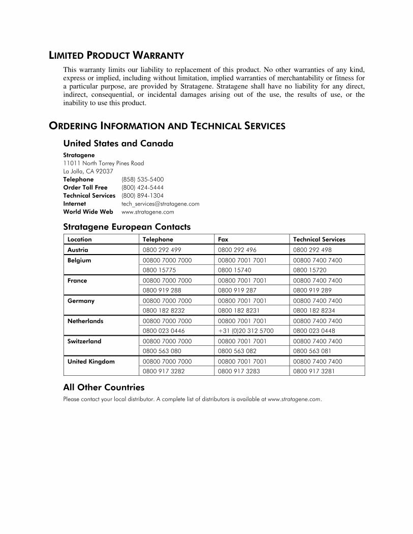
ORDERING INFORMATION AND TECHNICAL SERVICES
United States and CanadaStratagene11011 North Torrey Pines RoadLa Jolla, CA 92037Telephone (858) 535-5400Order Toll Free (800) 424-5444Technical Services (800) 894-1304Internet [email protected] Wide Web www.stratagene.com
Stratagene European ContactsLocation Telephone Fax Technical Services
Austria 0800 292 499 0800 292 496 0800 292 498
00800 7000 7000 00800 7001 7001 00800 7400 7400Belgium
0800 15775 0800 15740 0800 15720
00800 7000 7000 00800 7001 7001 00800 7400 7400France
0800 919 288 0800 919 287 0800 919 289
00800 7000 7000 00800 7001 7001 00800 7400 7400Germany
0800 182 8232 0800 182 8231 0800 182 8234
00800 7000 7000 00800 7001 7001 00800 7400 7400Netherlands
0800 023 0446 +31 (0)20 312 5700 0800 023 0448
00800 7000 7000 00800 7001 7001 00800 7400 7400Switzerland
0800 563 080 0800 563 082 0800 563 081
00800 7000 7000 00800 7001 7001 00800 7400 7400United Kingdom
0800 917 3282 0800 917 3283 0800 917 3281
All Other CountriesPlease contact your local distributor. A complete list of distributors is available at www.stratagene.com.
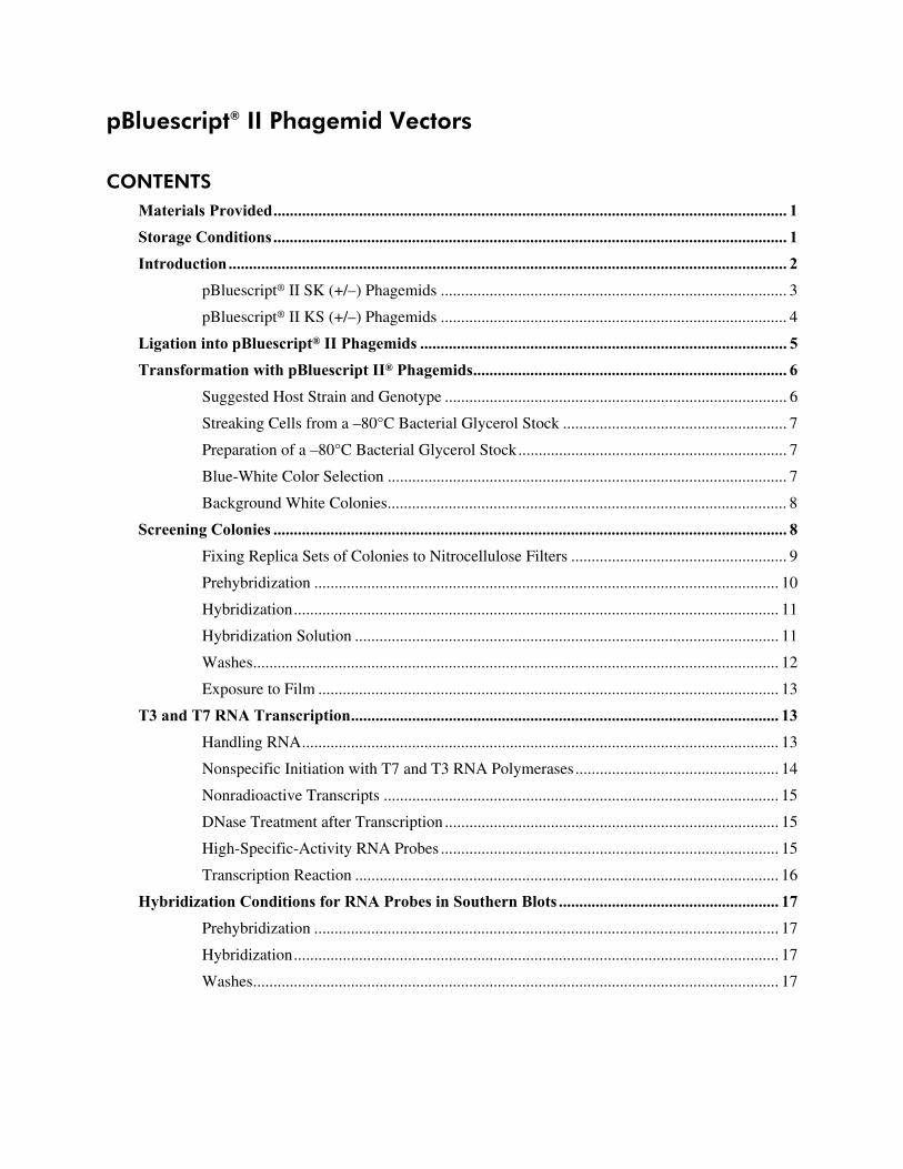
pBluescript® II Phagemid Vectors
CONTENTSMaterials Provided.............................................................................................................................. 1Storage Conditions.............................................................................................................................. 1Introduction......................................................................................................................................... 2
pBluescript® II SK (+/–) Phagemids ..................................................................................... 3
pBluescript® II KS (+/–) Phagemids ..................................................................................... 4
Ligation into pBluescript® II Phagemids .......................................................................................... 5Transformation with pBluescript II® Phagemids............................................................................. 6
Suggested Host Strain and Genotype .................................................................................... 6
Streaking Cells from a –80°C Bacterial Glycerol Stock ....................................................... 7
Preparation of a –80°C Bacterial Glycerol Stock.................................................................. 7
Blue-White Color Selection .................................................................................................. 7
Background White Colonies.................................................................................................. 8
Screening Colonies .............................................................................................................................. 8Fixing Replica Sets of Colonies to Nitrocellulose Filters ..................................................... 9
Prehybridization .................................................................................................................. 10
Hybridization....................................................................................................................... 11
Hybridization Solution ........................................................................................................ 11
Washes................................................................................................................................. 12
Exposure to Film ................................................................................................................. 13
T3 and T7 RNA Transcription......................................................................................................... 13Handling RNA..................................................................................................................... 13
Nonspecific Initiation with T7 and T3 RNA Polymerases.................................................. 14
Nonradioactive Transcripts ................................................................................................. 15
DNase Treatment after Transcription .................................................................................. 15
High-Specific-Activity RNA Probes ................................................................................... 15
Transcription Reaction ........................................................................................................ 16
Hybridization Conditions for RNA Probes in Southern Blots ...................................................... 17Prehybridization .................................................................................................................. 17
Hybridization....................................................................................................................... 17
Washes................................................................................................................................. 17

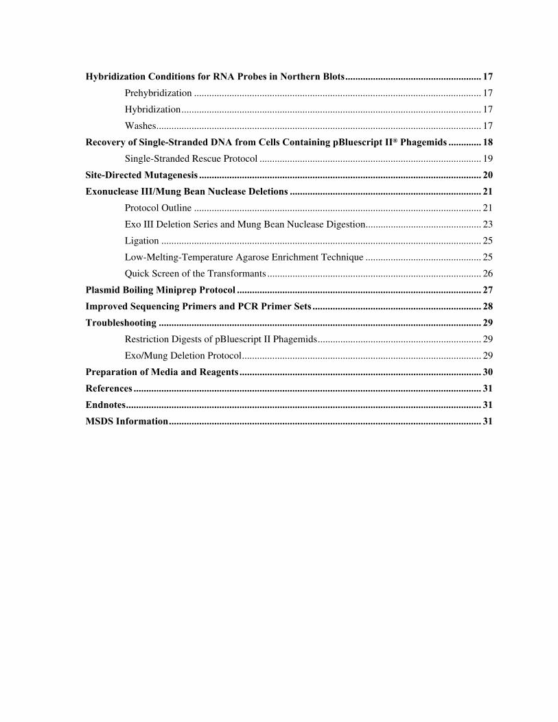
Hybridization Conditions for RNA Probes in Northern Blots...................................................... 17Prehybridization .................................................................................................................. 17
Hybridization....................................................................................................................... 17
Washes................................................................................................................................. 17
Recovery of Single-Stranded DNA from Cells Containing pBluescript II® Phagemids ............. 18Single-Stranded Rescue Protocol ........................................................................................ 19
Site-Directed Mutagenesis ................................................................................................................ 20Exonuclease III/Mung Bean Nuclease Deletions ............................................................................ 21
Protocol Outline .................................................................................................................. 21
Exo III Deletion Series and Mung Bean Nuclease Digestion.............................................. 23
Ligation ............................................................................................................................... 25
Low-Melting-Temperature Agarose Enrichment Technique .............................................. 25
Quick Screen of the Transformants ..................................................................................... 26
Plasmid Boiling Miniprep Protocol ................................................................................................. 27Improved Sequencing Primers and PCR Primer Sets ................................................................... 28Troubleshooting ................................................................................................................................ 29
Restriction Digests of pBluescript II Phagemids................................................................. 29
Exo/Mung Deletion Protocol............................................................................................... 29
Preparation of Media and Reagents ................................................................................................ 30References .......................................................................................................................................... 31Endnotes............................................................................................................................................. 31MSDS Information............................................................................................................................ 31

pBluescript® II Phagemid Vectors 1
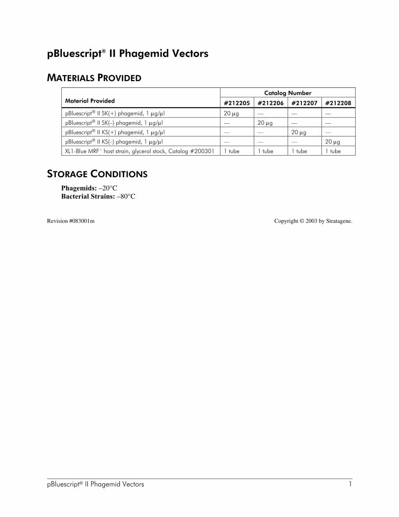
pBluescript® II Phagemid Vectors
MATERIALS PROVIDED
Catalog NumberMaterial Provided #212205 #212206 #212207 #212208
pBluescript® II SK(+) phagemid, 1 µg/µl 20 µg — — —
pBluescript® II SK(–) phagemid, 1 µg/µl — 20 µg — —
pBluescript® II KS(+) phagemid, 1 µg/µl — — 20 µg —
pBluescript® II KS(–) phagemid, 1 µg/µl — — — 20 µg
XL1-Blue MRF´ host strain, glycerol stock, Catalog #200301 1 tube 1 tube 1 tube 1 tube
STORAGE CONDITIONS
Phagemids: –20°CBacterial Strains: –80°C
Revision #083001m Copyright © 2003 by Stratagene.

2 pBluescript® II Phagemid Vectors
INTRODUCTION
The pBluescript® II phagemids (plasmids with a phage origin) are cloningvectors designed to simplify commonly used cloning and sequencingprocedures, including the construction of nested deletions for DNAsequencing, generation of RNA transcripts in vitro and site-specificmutagenesis and gene mapping. The pBluescript II phagemids have anextensive polylinker with 21 unique restriction enzyme recognition sites.Flanking the polylinker are T7 and T3 RNA polymerase promoters that canbe used to synthesize RNA in vitro.1,2 The choice of promoter used toinitiate transcription determines which strand of the insert cloned into thepolylinker will be transcribed.
Circular maps and lists of features for the pBluescript II phagemids areshown in figures 1 and 2. The polylinker and T7 and T3 RNA polymerasepromoter sequences are present in the N-terminal portion of a lacZ genefragment. A total of 131 amino acids of β-galactosidase coding sequence ispresent in the pBluescript II phagemid, but the coding sequence isinterrupted by the large polylinker. (There are 36 amino acids from theinitiator Met sequence to the EcoR I site.) pBluescript II phagemids havingno inserts in the polylinker will produce blue colonies in the appropriatestrains of bacteria (i.e., strains containing lacZ∆M15 on an F´ episome, suchas XL1-Blue MRF´, among others). pBluescript II phagemids that haveinserts will produce white colonies using the same strain, because the insertsdisrupt the coding region of the lacZ gene fragment.
pBluescript II (+) and (–) are available with two polylinker orientationsdesignated as either KS or SK using the following convention: (1) in the KSorientation, the Kpn I restriction site is nearest the lacZ promoter and theSac I restriction site is farthest from the lacZ promoter; and (2) in the SKorientation, the Sac I site is the closest restriction site to the lacZ promoterand the Kpn I site is the farthest.
Flanking the T3 and T7 promoters are BssH II sites. This rare six-base cutterwill allow the insert plus the T phage RNA promoters to be excised andused for gene mapping.
pBluescript II phagemids can be rescued as single-stranded (ss) DNA.pBluescript II phagemids contain a 454-bp filamentous f1 phage intergenicregion (M13 related), which includes the 307-bp origin of replication. The(+) and (–) orientations of the f1 intergenic region allow the rescue of senseor antisense ssDNA by a helper phage. This ssDNA can be used fordideoxynucleotide sequencing (Sanger method) or site-specific mutagenesis.
Note We have discovered that the use of excess amounts of EcoR I todigest pBluescript II results in EcoR I prime activity. This appearsas cleavage at a non-EcoR I site at the 3´ end of the f1 intergenicregion, causing confusion when interpreting results from anagarose gel. If a restriction pattern appears incorrect, checkwhether reducing the units of EcoR I restores a normal restrictionpattern.
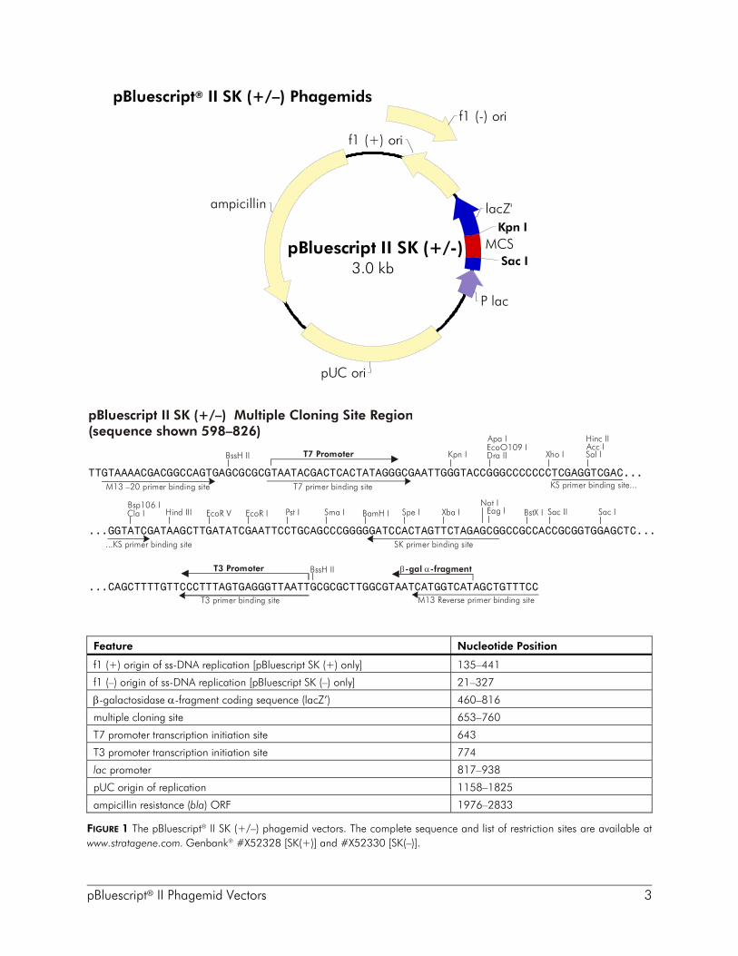
pBluescript® II Phagemid Vectors 3
pBluescript® II SK (+/–) Phagemids
Feature Nucleotide Position
f1 (+) origin of ss-DNA replication [pBluescript SK (+) only] 135–441
f1 (–) origin of ss-DNA replication [pBluescript SK (–) only] 21–327
β-galactosidase α-fragment coding sequence (lacZ’) 460–816
multiple cloning site 653–760
T7 promoter transcription initiation site 643
T3 promoter transcription initiation site 774
lac promoter 817–938
pUC origin of replication 1158–1825
ampicillin resistance (bla) ORF 1976–2833
FIGURE 1 The pBluescript® II SK (+/–) phagemid vectors. The complete sequence and list of restriction sites are available atwww.stratagene.com. Genbank® #X52328 [SK(+)] and #X52330 [SK(–)].
f1 (+) ori
f1 (-) ori
MCS
lacZ'
P lac
ampicillin
pUC ori
Kpn I
Sac IpBluescript II SK (+/-)
3.0 kb
pBluescript II SK (+/–) Multiple Cloning Site Region(sequence shown 598–826)
Sac IBstX I
...GGTATCGATAAGCTTGATATCGAATTCCTGCAGCCCGGGGGATCCACTAGTTCTAGAGCGGCCGCCACCGCGGTGGAGCTC...
EcoR IEcoR V Sac IISpe ISma I Xba IPst IHind IIIBsp106 ICla I BamH I
Not IEag I
TTGTAAAACGACGGCCAGTGAGCGCGCGTAATACGACTCACTATAGGGCGAATTGGGTACCGGGCCCCCCCTCGAGGTCGAC...
Acc IKpn I Xho I
Hinc II
BssH IIEcoO109 IDra II
Apa I
Sal IT7 Promoter
T7 primer binding site
...CAGCTTTTGTTCCCTTTAGTGAGGGTTAATTGCGCGCTTGGCGTAATCATGGTCATAGCTGTTTCC
M13 –20 primer binding site
BssH IIT3 Promoter
M13 Reverse primer binding siteT3 primer binding site
β α-gal -fragment
KS primer binding site...
SK primer binding site...KS primer binding site
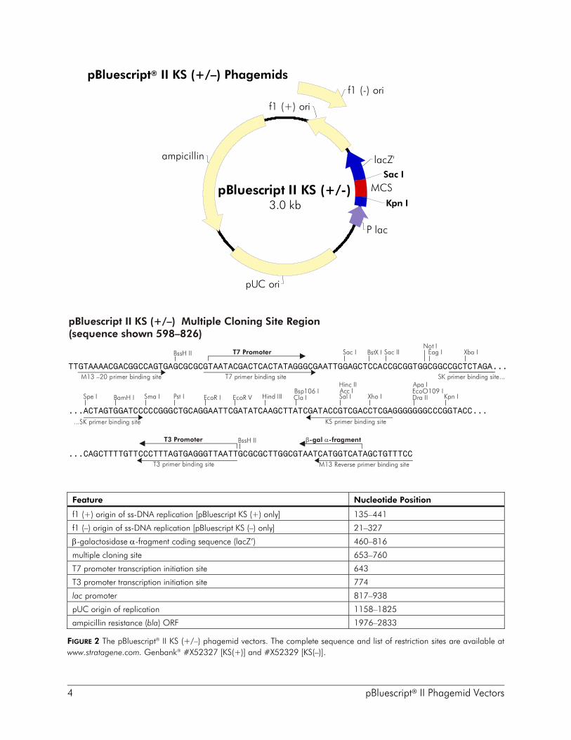
4 pBluescript® II Phagemid Vectors
pBluescript® II KS (+/–) Phagemids
Feature Nucleotide Position
f1 (+) origin of ss-DNA replication [pBluescript KS (+) only] 135–441
f1 (–) origin of ss-DNA replication [pBluescript KS (–) only] 21–327
β-galactosidase α-fragment coding sequence (lacZ’) 460–816
multiple cloning site 653–760
T7 promoter transcription initiation site 643
T3 promoter transcription initiation site 774
lac promoter 817–938
pUC origin of replication 1158–1825
ampicillin resistance (bla) ORF 1976–2833
FIGURE 2 The pBluescript® II KS (+/–) phagemid vectors. The complete sequence and list of restriction sites are available atwww.stratagene.com. Genbank® #X52327 [KS(+)] and #X52329 [KS(–)].
ampicillin
f1 (+) ori
MCS
lacZ'
P lac
pUC ori
Kpn I
Sac I
f1 (-) ori
pBluescript II KS (+/-)3.0 kb
Sac I BstX I
...ACTAGTGGATCCCCCGGGCTGCAGGAATTCGATATCAAGCTTATCGATACCGTCGACCTCGAGGGGGGGCCCGGTACC...
EcoR I EcoR V
Sac II
Spe I Sma I
Xba I
Pst I Hind IIIBsp106 ICla IBamH I
Not IEag I
TTGTAAAACGACGGCCAGTGAGCGCGCGTAATACGACTCACTATAGGGCGAAT ...
TGGAGCTCCACCGCGGTGGCGGCCGCTCTAGA
Kpn IXho I
BssH II
EcoO109 IDra II
Apa IAcc IHinc II
Sal I
T7 Promoter
...CAGCTTTTGTTCCCTTTAGTGAGGGTTAATTGCGCGCTTGGCGTAATCATGGTCATAGCTGTTTCC
BssH IIT3 Promoter
M13 –20 primer binding site T7 primer binding site
T3 primer binding site M13 Reverse primer binding site
β α-gal -fragment
...SK primer binding site KS primer binding site
SK primer binding site...
pBluescript II KS (+/–) Multiple Cloning Site Region(sequence shown 598–826)
pBluescript® II Phagemid Vectors 5
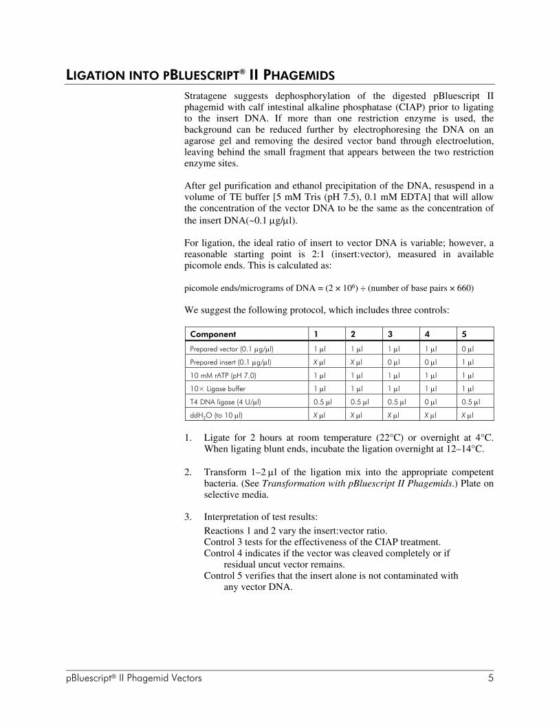
LIGATION INTO PBLUESCRIPT® II PHAGEMIDS
Stratagene suggests dephosphorylation of the digested pBluescript IIphagemid with calf intestinal alkaline phosphatase (CIAP) prior to ligatingto the insert DNA. If more than one restriction enzyme is used, thebackground can be reduced further by electrophoresing the DNA on anagarose gel and removing the desired vector band through electroelution,leaving behind the small fragment that appears between the two restrictionenzyme sites.
After gel purification and ethanol precipitation of the DNA, resuspend in avolume of TE buffer [5 mM Tris (pH 7.5), 0.1 mM EDTA] that will allowthe concentration of the vector DNA to be the same as the concentration ofthe insert DNA(~0.1 µg/µl).
For ligation, the ideal ratio of insert to vector DNA is variable; however, areasonable starting point is 2:1 (insert:vector), measured in availablepicomole ends. This is calculated as:
picomole ends/micrograms of DNA = (2 × 10⁶) ÷ (number of base pairs × 660)
We suggest the following protocol, which includes three controls:
Component 1 2 3 4 5
Prepared vector (0.1 µg/µl) 1 µl 1 µl 1 µl 1 µl 0 µl
Prepared insert (0.1 µg/µl) X µl X µl 0 µl 0 µl 1 µl
10 mM rATP (pH 7.0) 1 µl 1 µl 1 µl 1 µl 1 µl
10× Ligase buffer 1 µl 1 µl 1 µl 1 µl 1 µl
T4 DNA ligase (4 U/µl) 0.5 µl 0.5 µl 0.5 µl 0 µl 0.5 µl
ddH2O (to 10 µl) X µl X µl X µl X µl X µl
1. Ligate for 2 hours at room temperature (22°C) or overnight at 4°C.When ligating blunt ends, incubate the ligation overnight at 12–14°C.
2. Transform 1–2 µl of the ligation mix into the appropriate competentbacteria. (See Transformation with pBluescript II Phagemids.) Plate onselective media.
3. Interpretation of test results:Reactions 1 and 2 vary the insert:vector ratio.Control 3 tests for the effectiveness of the CIAP treatment.Control 4 indicates if the vector was cleaved completely or if
residual uncut vector remains.Control 5 verifies that the insert alone is not contaminated with
any vector DNA.
6 pBluescript® II Phagemid Vectors
4. Expected plating results:Plates 1 and 2 should have mostly white colonies, representing
recombinants.Plate 3 should have low numbers of blue colonies if the CIAP
treatment was effective.Plate 4 should have no colonies if the digest was complete.Plate 5 should have no colonies if the insert was pure.
TRANSFORMATION WITH PBLUESCRIPT II® PHAGEMIDS
Note pBluescript II phagemids will replicate autonomously as plasmids.Therefore, colonies—not plaques—are obtained followingtransformation.
Suggested Host Strain and GenotypeStratagene recommends the host strain XL1-Blue MRF´ for propagation ofpBluescript II phagemids and for transformation of recombinant phagemids.XL1-Blue MRF´ allows blue-white color selection and single-stranded DNArescue, and is restriction-deficient aiding in the construction of librariesmade from methylated DNA.3
XL1-Blue MRF´ Genotype: ∆(mcrA)183 ∆(mcrCB-hsdSMR-mrr)173endA1 supE44 thi-1 recA1 gyrA96 relA1 lac [F´ proAB lacIqZ∆M15 Tn10(Tetr)]
Note The XL1-Blue MRF´ is provided as a glycerol stock. Additionaltubes of glycerol stock can be purchased from Stratagene (Catalog#200301); alternatively, XL1-Blue MRF´ is available fromStratagene as high-efficiency frozen competent cells(>1 × 10⁹ colonies/µg of pUC 18, Catalog #200230).
For the appropriate media and plates for growth of XL1-Blue MRF´, pleaserefer to the following table:
Bacterial strainPlates for bacterialstreak
Media for glycerolstock
XL1-Blue MRF´ LB–tetracycline agara LB–tetracyclinea
a12.5 µg/ml.
pBluescript® II Phagemid Vectors 7
Streaking Cells from a –80°C Bacterial Glycerol Stock
Prepare the following from a frozen glycerol stock:
Note Do not allow the contents of the vial to thaw. The vials can bestored at –20° or –80°C, but most strains remain viable longer ifstored at –80°C.
1. Revive the stored cells by scraping off splinters of solid ice with asterile wire loop.
2. Streak the splinters onto an LB plate containing the appropriateantibiotic.
Restreak the cells fresh each week.
Preparation of a –80°C Bacterial Glycerol Stock
1. In a sterile 50-ml conical tube, inoculate 10 ml of the appropriate liquidmedia with one or two colonies from a plate of freshly-streaked cells.Grow the cells to late log phase.
2. Add 4.5 ml of a sterile glycerol–liquid media solution (prepared bycombining 5 ml of glycerol + 5 ml of liquid media) to the bacterialculture from step 1. Mix well.
3. Aliquot into sterile centrifuge tubes (1 ml/ tube). This preparation maybe stored at –20°C for 1–2 years or at –80°C for more than 2 years.
Blue-White Color SelectionThe XL1-Blue MRF´ strain allows blue–white color selection forpBluescript II phagemids because of lacZ∆M15 complementation on theF´ episome. The color selection may be seen when plating on LB platescontaining 100 µg/ml of ampicillin, 80 µg/ml of fresh X-gal, and 20 mMIPTG. Alternatively, plates for color selection can be prepared by spreading100 µl of 40 mM IPTG and 100 µl of 2% X-gal on LB–ampicillin plates30 minutes prior to plating your transformants. X-gal should be prepared indimethyl formamide and IPTG in sterile, distilled H2O (store stock solutionsat –20°C until use). Colonies containing phagemids without inserts will beblue after incubation for 12–18 hours at 37°C. Colonies with phagemidscontaining inserts will remain white. Further enhancement of the blue colormay be obtained by placing plates at 4°C for 2 hours following overnightgrowth at 37°C.
Occasionally, β-galactosidase fusion proteins are toxic to the host bacteria.If there is any suspicion that an insert might be toxic, the X-gal and IPTGmay be left out of the ampicillin plates. Under these conditions there will beno color selection, but recombinants will express lower levels of thepotentially toxic proteins.
8 pBluescript® II Phagemid Vectors
Background White ColoniesSince the ∆M15 lac gene carried on the F´ episome is needed for the blue–white color assay, host bacteria that have lost the F´ episome will remain aswhite colonies on an X-gal/IPTG agar plate even if the pBluescript IIphagemid does not contain an insert. XL1-Blue MRF´ is a lac– AG1derivative with Tn10, lacIq, and lacZ∆M15 on the F´. Selection for bacteriacontaining the F´ in this strain is accomplished by plating on 12.5 µg/mltetracycline instead of minimal media plates. XL1-Blue MRF´ transformantscontaining pBluescript II phagemids can be plated on tetracycline–ampicillin plates to select for colonies that contain both the F´ and thepBluescript II phagemid. This advantage further reduces the background offalse positives.
For bacteria containing an F´ without a Tn10 gene, growth on a minimalmedium plate supplemented with 1 mM thiamine-HCl will maintainselection for the F´; however, colonies will grow more slowly. If there isany doubt about whether a white colony represents a pBluescript IIrecombinant or a colony lacking the F´, streak it onto a minimal mediumplate.4 A cell lacking an F´ will not grow; an F+ will grow slowly since itcarries the proAB genes on the F´ episome.
SCREENING COLONIES
Colonies containing pBluescript II phagemids may be screened forrecombinants by double-stranded DNA, RNA, or oligonucleotidehybridization.5 Colonies may also be screened by restriction mapping or bysequencing miniprep plasmid DNA. Antibodies may be used to screencolonies6 since cDNA cloned into the appropriate reading frame of the lacZgene will be expressed as fusion proteins.
When screening with antibodies, the bacteria produce fusion proteinscontaining several amino acids from the amino-terminus of theβ-galactosidase protein (3.5 kDa to the EcoR I site). Some fusion proteinsare toxic to E. coli. Therefore, it is best to initially plate transformants onnitrocellulose filters on top of ampicillin plates lacking IPTG. After8–10 hours (when the colonies are 1 mm in diameter), transfer the filters toplates containing 5 mM IPTG for several hours. This will induce synthesisof the fusion proteins. When screening with antibodies, Stratagene'spicoBlue™ immunoscreening kit is recommended. To synthesize largeamounts of the fusion proteins in liquid culture, grow the cells to anOD600 = 0.7 in the absence of IPTG. Add IPTG to 5 mM and grow foranother 2–3 hours. The β-galactosidase portion of the fusion protein is~3.5 kDa from the Met amino acid to the EcoR I site in the polylinker.
Identification of recombinant clones within pBluescript II can be performedby colony hybridization. The following protocol minimizes problemsassociated with colony screening procedures. For the following protocol tobe effective, the screening should be performed on duplicate sets of filters.

pBluescript® II Phagemid Vectors 9
Fixing Replica Sets of Colonies to Nitrocellulose FiltersUse the following protocol to make multiple replica plates of transformants.Keep the original or master filter to pick colonies identified by the screeningof the replica filters.
1. Place 100–mm Duralon–UV™ or nitrocellulose filters on 150–mmLB–ampicillin plates.
2. Spread ~1.0 × 10⁶ cfu on the filters.
3. Incubate the plates at 37°C overnight or until colonies are 1.0 mm indiameter (~7–10 hours).
4. Make a replica of the library growing on the nitrocellulose filter:
a. Place a piece of sterile Whatman® 3MM paper on a glass surface.
b. Remove the filter from the agar and place it colony side up on theWhatman 3MM paper.
c. Align a fresh filter, prewetted on an LB plate, over the masterfilter and cover with another piece of Whatman 3MM paper. Pressin place with a glass plate.
d. Mark the filters with a small needle to aid in realignment afterhybridization.
e. Separate the master and replica filters and place face up on LBagar plates containing ampicillin.
f. Incubate both the master and replica filters for at least 4 hoursat 37°C.
g. Seal the master plate with Parafilm® and store at 4°C.
5. The replica filter is then prepared for hybridization:
a. Place the replica filter colony side up for 30 seconds on the surfaceof Whatman 3MM paper prewetted with 0.5 M NaOH.
b. Remove filter and place on another sheet of Whatman 3MM paperprewetted with 1 M Tris-HCl (pH 7.6) for 30 seconds.
c. Remove the filter and place on a third piece of Whatman 3MMpaper prewetted with 1 M Tris-HCl (pH 7.6) and 1.5 M NaCl for30 seconds.
d. Immerse the filter in 1 M Tris-HCl (pH 7.6) and 1.5 M NaCl andremove bacterial debris by rubbing the filter gently with a glovedhand.
10 pBluescript® II Phagemid Vectors
e. Rinse the filter in 1 M Tris-HCl (pH 7.6) and 1.5 M NaCl. Blotdry on paper towels.
f. Crosslink the DNA to the filters using the autocrosslink setting onthe Stratalinker® UV crosslinker (120,000 µJ of UV energy).Alternatively, oven bake at 80°C for ~1.5–2 hours.
PrehybridizationPrehybridization Solution for Oligonucleotide Probe
6× SSC20 mM NaH2PO4
0.4% sodium dodecyl sulfate* (SDS)5× Denhardt'sDenatured, sonicated salmon sperm DNA (500 µg/ml)
OR
Prehybridization Solution for Double-Stranded Probe2× Pipes buffer50% Deionized formamide0.5% SDS*Denatured, sonicated salmon sperm DNA (100 µg/ml)
The amount of prehybridization solution to make is dependent on thenumber of filters used (generally 2–3 ml/membrane).
1. Preheat the prehybridization solution to ~50°C without the salmonsperm DNA. Preboil the salmon sperm DNA for ~10 minutes and add itto the warm prehybridization solution.
2. Wet each filter (quickly) in the prehybridization buffer in a tray,placing each filter on top of the next, until each is wet through. Addmore prehybridization solution as necessary. (This helps wet the filterscompletely to allow more even hybridization later.)
3. Put the wet prehybridization filter "stack" in a heat-seal bag, add theremaining prehybridization buffer and heat seal.
4. Calculate the hybridization temperature (generally 42°C) andprehybridize for a minimum of 1 hour.
5. Prehybridize and hybridize a blank filter ("background") along with therest and wash it to determine when and at what temperature thebackground counts disappear.
* For Stratagene's Duralon-UV membranes, increase the SDS concentration to 1% (w/v).
pBluescript® II Phagemid Vectors 11
Hybridization
Labeling Oligonucleotide ProbesLabel oligonucleotides with fresh [γ-32P]ATP. High-specific-activityγ-label yields the best results.
a. Perform a polynucleotide kinase (PNK) labeling in 1× ligase buffer for30 minutes at 37°C.
b. Incubate for 15 minutes at 65°C to inactivate the kinase.
c. Run the solution over a G-50 column or a NucTrap® probe purificationcolumn to get rid of the unincorporated counts.
Labeling Double-Stranded ProbesWhen using double-stranded probes, nick translate with fresh [α-32P]dATP.
Stratagene offers the Prime-It® II random primer kit designed to producehigh-specific-activity DNA probes in 2 minutes.
It is best to use ~1 × 10⁶–5 × 10⁶ counts/ml of hybridization solution. Keepthe concentration of counts high and use ~1 × 10⁷ counts/filter.
Hybridization SolutionHybridization Solution for Oligonucleotide Probes
6× SSC20 mM NaH2PO40.4% SDS*Denatured, sonicated salmon sperm DNA (500 µg/ml)
1. Make the hybridization solution.
2. Boil the salmon sperm DNA and then add it to the prewarmedhybridization solution.
3. Pour out the prehybridization buffer from the filter bag. Add thehybridization solution and then the appropriate amount of labeledoligonucleotide.
* For Stratagene's Duralon-UV membranes, increase the SDS concentration to 1% (w/v).
12 pBluescript® II Phagemid Vectors
4. Heat seal and hybridize at 5–10°C below Tm. Calculate Tm using thefollowing formula:
Note The first method below overestimates the Tm of hybrids involvinglonger nucleotides.
OLIGONUCLEOTIDES SHORTER THAN 18 BASESTm = 2°C(A + T) + 4°C(G + C)
OLIGONUCLEOTIDES 14 BASES AND LONGER (UP TO 60–70 NUCLEOTIDES)Tm = 81.5 – 16.6 (log10[Na+]) + 0.41(%G + C) – (600/N), where N = chain length
Hybridization Solution for Double-Stranded Probes2× Pipes buffer50% Deionized formamide0.5% SDS*Denatured, sonicated salmon sperm DNA (100 µg/ml)
1. Prepare the hybridization solution.
2. Warm the solution, boil the appropriate amount of salmon sperm DNAwith the probe for 4 minutes and then add it to the hybridization buffer.
3. Decant the prehybridization buffer and replace it with the hybridizationsolution and probe. Hybridize overnight at 42°C.
WashesOligonucleotide Probes
Use 6× SSC buffer and 0.1% (w/v) SDS. Wash the filters three times for5 minutes each at room temperature. The final washing temperature dependson the GC ratio of the probe. It is best to stay several degrees below themelting temperature. A rough estimate of the melting temperature of anoligonucleotide probe can be determined by the following formula:
Tm = 4(G + C) + 2(T + A)
If the probe sequence is unknown, start with a room temperature wash andgradually increase the temperature until the background diminishes. DONOT allow the membranes to completely dry out or the probe may beirreversibly bound.
Double-Stranded Probes
Use 0.1× SSC buffer and 0.1% (w/v) SDS. Wash the filters at 50–65°C withagitation.
* For Stratagene's Duralon-UV membranes, increase the SDS concentration to 1% (w/v).
pBluescript® II Phagemid Vectors 13
Exposure to FilmAfter washing, remove the excess liquid by blotting on Whatman 3MMpaper and place the filters between two sheets of plastic wrap in cassetteswith intensifying screens. Leave overnight at –80°C. (By keeping the filtersslightly moist between plastic wrap, you can wash again if the backgroundis high.)
T3 AND T7 RNA TRANSCRIPTION
The RNA transcripts synthesized from inserts cloned into vectors containingeither T3 or T7 polymerase promoters can be used for many purposes.Transcripts can be used for both Southern and Northern hybridizationexperiments and for either S1 or RNase A analysis. In addition, RNAtranscripts can be used to produce protein by translation in vitro ortranslation in vivo after microinjection into Xenopus oocytes or tissueculture cells. Stratagene’s mCAP™ RNA capping kit may be used with bothT3 and T7 RNA polymerases to incorporate 5´-7MeGpppG-5´ cap analogs,increasing RNA stability by up to 95%.
The pBluescript II vectors have a BssH II site outside each RNA promoter.This feature allows the excising of the insert with the promoters andsubsequent mapping using phosphorylated T3 and/or T7 primers.
Handling RNA
Note Wear gloves at all times to prevent RNase contamination.
When working with RNA, caution must be used to eliminate RNasecontamination from any source. The following general principles will helpin the production of full-length transcripts:
1. Make all buffers, DTT, and rNTPs in highly pure water treated withdiethylpyrocarbonate (DEPC) as follows:
Add DEPC to water to a final concentration of 0.1%, heat to 37°C for8 hours and autoclave. If DEPC scent remains after autoclaving, placethe water in a 90°C water bath for at least 1 hour or until thescent is gone.
Note Do not treat Tris solutions with DEPC!! Instead, use water thathas been treated with DEPC to make up all Tris solutions.
Stratagene’s RNAMaxx™ high-yield transcription kit(Catalog #200339) may be used for transcription reactions performedwith T7 RNA polymerase.
14 pBluescript® II Phagemid Vectors
2. All tubes and pipet tips should be autoclaved and baked for severalhours at 80°C. A common source of RNase contamination on gelelectrophoresis equipment comes from DNA mini–preps which havebeen treated with RNase A. Thoroughly clean all gel tanks, gel combs,gel spacers and glassware, using soap and water. Followed with anethanol rinse. Next, soak the equipment in 3% hydrogen peroxide for10 minutes at room temperature and rinse with DEPC-treated water.Keep cleaned items covered and away from bare hands. Autoclave allglass plates and other appropriate materials on dry cycle prior to use.
3. Phagemid templates for transcription must be RNase-free. Cesiumchloride preps are advisable, but minipreps may be used if care is takento remove contaminating RNases. Generally the plasmid template islinearized with an enzyme that cleaves "downstream" of the RNApolymerase promoter and the insert in the multiple cloning site. It isstrongly advised to purify the post-restriction digest DNA by adding50 µg/ml proteinase K to the restriction buffer at 37°C for 30 minutes,followed by two phenol–chloroform [1:1 (v/v)] extractions and ethanolprecipitation prior to the transcription reaction. Resuspend digested,proteinase K treated DNA at 1 mg/ml in a 10 mM Tris (pH 7.4) and0.1 mM EDTA solution made with DEPC-treated water.
4. Working with RNA is simplified by using a ribonuclease inhibitor intranscription reactions. Stratagene's RNase Block RibonucleaseInhibitor has been tested and adjusted to work optimally withStratagene transcription kits.
Nonspecific Initiation with T7 and T3 RNA PolymerasesT7 and T3 RNA polymerases are highly specific for their respectivepromoters,1 however, nonspecific initiation of RNA transcripts may occur atthe ends of the DNA template. This is most prevalent with a 3´-protrudingterminus. Nonspecific initiation may be reduced by increasing the NaClconcentration in the transcription buffers to 100 mM, although this willresult in a decrease of the total transcription efficiency by ~50%. Whenpossible, use restriction enzymes that leave blunt or 5´-protruding ends.
When the T7 or T3 polymerase enzymes are used in molar excess of theDNA template, there is a risk of polymerization from the wrong promoter.T7 polymerase can synthesize RNA inefficiently from a plasmid containingonly a T3 promoter. Conversely, T3 polymerase can synthesize RNAinefficiently from a plasmid containing only a T7 promoter. Synthesis isextremely promoter specific when both promoters are present, provided thatthe enzyme is not in molar excess of the specific promoter. Do not useexcessive amounts of the polymerases if promoter specificity is important toyour experiment. Best results are obtained when the ratios stated in thismanual are followed.
pBluescript® II Phagemid Vectors 15
Nonradioactive TranscriptsNonradioactive transcripts can be used for nucleotide sequencing, in vitrotranslation and injection into cells for in vivo translation. Set up thetranscription reaction as described, but add 1 µl of 10 mM rUTP instead ofradioactive rUTP. For larger amounts of RNA, scale up the reactionappropriately. Each molecule of DNA template yields 10–20 nonradioactiveRNA molecules if the ribonucleotides are not a limiting factor.
DNase Treatment after TranscriptionThe DNA template will be present after the transcription reaction and can beremoved with RNase-free DNase. After the transcription reaction, add 10 Uof RNase-free DNase/µg of DNA template and incubate at 37°C for15 minutes. Extract with phenol–chloroform [1:1 (v/v)], add 1/10 volume of3 M sodium acetate at pH 5.2 and precipitate RNA with 2.5 volumes of100% (v/v) ethanol.
High-Specific-Activity RNA ProbesAny vector containing T3 and T7 RNA promoters can be used to synthesizehigh specific activity, strand-specific RNA probes. The choice between T3and T7 RNA polymerase will determine which strand will be used as thetemplate. This is important because probes used for Northern or S1 analysismust complement the RNA targeted for detection.
The initiation of RNA transcription requires rGTP; the reaction has a Km of~180 µM. The elongation reaction has a Km of 40 µM for eachribonucleotide. Therefore, radioactive rGTP should not be used to generatehigh specific-activity probes unless the concentration of rGTP exceeds180 µM. This usually means supplementing the radioactive rGTP with coldrGTP. Adding 50 µCi of 500 Ci/mmol [32P]rXTP to a 25-µl reaction onlyproduces a rXTP concentration of 4 µM. To generate high specific-activityprobes, we suggest using radioactive rATP, rCTP, or rUTP as the labelednucleotide. However, any triphosphate present at just 4 µM will not producemany transcripts per template molecule because the reaction simply runs outof radioactive rXTP. To make large amounts of long, radioactive transcripts,the reactions must be supplemented with cold rXTP. It is thereforenecessary to choose between full length, quantity and high-specific-activitywhen producing probes.
16 pBluescript® II Phagemid Vectors
Transcription Reaction
Note Stratagene’s RNAMaxx high-yield transcription kit (Catalog#200339) may be used for transcription reactions performed withT7 RNA polymerase.
1. In the order given, add5 µl of 5× transcription buffer§
1 µg of restricted, proteinase K-treated DNA template1 µl of 10 mM rATP1 µl of 10 mM rCTP1 µl of 10 mM rGTP
[1 µl of 1 mM rUTP is optional (see above)]1 µl of 0.75 M dithiothreitol (DTT)1 µl of RNase Block Ribonuclease Inhibitor (optional)5 µl of 400–800 Ci/mmol, 10 µCi/µl [α-32P]rUTP10 U of T3 or T7 RNA polymerase*DEPC-treated water to a final volume of 25 µl
2. Incubate at 37°C for 30 minutes.
3. RNA transcripts may be purified away from the unincorporatednucleotides using Stratagene's NucTrap probe purification columnwith a push-column beta-shield device.
Alternatively, an RNase-free G-50 column can be used. However, care mustbe taken that there are no ribonucleases present in the column that coulddegrade the probe.
Note Do not use large excesses of T3 polymerase (10 U ofpolymerase/pmol of promoter is sufficient). T3 RNA polymerasemay utilize the T7 promoter 1 in 20 times when the T3 enzymeconcentration exceeds the T3 promoter concentration by 10-fold.However, T3 polymerase in the recommended concentrations willnot make T7 transcripts in the presence of a T3 promoter. If anyT7 hybridization should result from a T3 transcription, decreasethe amount of T3 polymerase by a factor of 5 or 10.
§ See Preparation of Media and Reagents.* Use supplied RNA polymerase dilution buffer to dilute enzymes just before use.

pBluescript® II Phagemid Vectors 17
HYBRIDIZATION CONDITIONS FOR RNA PROBES IN SOUTHERN BLOTS
PrehybridizationPrehybridize the membrane with 0.1–0.5 ml/cm2 of the following solutionfor 2 hours at 42°C with constant agitation in a heat-sealable bag:
6× SSC5× Denhardt’s (see Preparation of Media and Reagents)20 mM NaH2PO4
500 µg/ml of denatured, sonicated salmon sperm DNA
HybridizationPour off the prehybridization solution and add the probe to the bag with theminimum volume of the following hybridization solution:
6× SSC20 mM NaH2PO4
0.4% SDS*500 µg/ml denatured sonicated salmon sperm DNA
Incubate overnight at 42°C with constant agitation.
WashesWash in 2× SSC buffer and 0.1% (w/v) SDS twice for 15 minutes each at55°C and twice in 0.1× SSC buffer and 0.1% (w/v) SDS for 15 minutes eachat 55°C.
HYBRIDIZATION CONDITIONS FOR RNA PROBES IN NORTHERN BLOTS
PrehybridizationPrehybridize the membrane with 0.1–0.5 ml/cm2 of the following solutionfor ~1 hour at 42°C with constant agitation in a heat-sealable bag:
50% deionized formamide10% dextran sulfate1% SDS*1 M NaCl100 µg/ml of denatured sonicated salmon sperm DNA
HybridizationHybridize overnight with the riboprobe at the same temperature and in theprehybridization solution.
WashesWash in 2× SSC buffer and 0.1% (w/v) SDS twice for 15 minutes each at42°C and twice in 0.1× SSC buffer and 0.1% (w/v) SDS for 15 minutes eachat 42°C. If a high background is observed, the temperature may be increasedor the NaCl concentration may be decreased for greater stringency.
* For Stratagene's Duralon-UV™ and Illuminator™ membranes, increase the SDSconcentration to 1% (w/v).
18 pBluescript® II Phagemid Vectors
RECOVERY OF SINGLE-STRANDED DNA FROM CELLS CONTAININGPBLUESCRIPT II® PHAGEMIDS
pBluescript II is a phagemid that can be secreted as single-stranded DNA inthe presence of M13 helper phage. These phagemids contain the intergenic(IG) region of a filamentous f1 phage. This region encodes all of thecis-acting functions of the phage required for packaging and replication. InE. coli with the F+ phenotype (containing an F´ episome), pBluescript IIphagemids will be secreted as single-stranded f1 "packaged" phage when thebacteria has been infected by a helper phage. Since these filamentous helperphages (M13, fI) will not infect E. coli without an F´ episome coding forpili, it is essential to use XL1-Blue MRF´ or a similar strain containingthe F´ episome.7,8
Stratagene offers helper phages that preferentially package pBluescript IIphagemids. Typically, 30–50 pBluescript II molecules are packaged/helperphage DNA molecule. pBluescript II phagemids are offered with the IGregion in either of two orientations: pBluescript II (+) is replicated such thatthe sense strand of the β-galactosidase gene is secreted within the phageparticles; pBluescript II (–) is replicated such that the antisense strand of theβ-galactosidase gene is secreted in the phage particles.
Yields of single-stranded (ss)DNA depend on the specific insert sequence.For most inserts, over 1 µg of ssDNA can be obtained from a 1.5-mlminiprep if grown in XL1-Blue MRF´. A faint single-strand helper phageband may appear on a gel at ~4 kb for R408 or at 6 kb for VCSM13. ThisDNA mixture can be sequenced with primers that are specific forpBluescript II and do not hybridize to the helper phage genome.
Site-specific mutagenesis is also possible using standard techniques. Theadvantages of using pBluescript II phagemids for either purpose are asfollows: (1) pBluescript II phagemids do not replicate via the M13 cycle,lessening the tendency to delete DNA inserts, therefore it is unlikely thateven 10-kb inserts will be deleted. (2) "Packaging" of pBluescript IIphagemids containing inserts is efficient since the pBluescript II vector issignificantly smaller than wild-type M13. (3) Oligonucleotide mutagenesisin pBluescript II vectors is advantageous because the mutagenized insert islocated between the T3 and T7 promoters. The resultant mutant transcriptscan be synthesized in vitro without further subcloning.
pBluescript® II Phagemid Vectors 19
VCSM13 and R408 helper phage produce the largest amount of single-strand pBluescript II. R408 (single-strand size ~4 kb) is more stable and canbe grown more easily. VCSM13 (single-strand size ~6 kb), is more efficientat single-stranded DNA rescue and yields more single-stranded phagemid;however it is more unstable and reverts to wild-type more frequently. Thisdifficulty can be addressed by periodically propagating VCSM13 in thepresence of kanamycin. VCSM13 (a derivative of M13KO7) has akanamycin gene inserted into the intergenic region, while R408 has adeletion in that region. We suggest R408 for excision of pBluescript II fromthe Lambda ZAP vector and VCSM13 for single-stranded rescue.
Single-Stranded Rescue Protocol
1. Inoculate a single colony into 5 ml of 2× YT containing 100 µg/mlampicillin and VCM13 or R408 helper phage at 107–108 pfu/ml(MOI ~10).
2. Grow the culture at 37°C with vigorous aeration for 16–24 hours, oruntil growth has reached saturation.
Note If using VCSM13, after 1–2 hours, add kanamycin to70 µg/ml to select for infected cells.
3. Centrifuge 1.5 ml of the cell culture for 5 minutes in a microcentrifuge.
4. Remove 1 ml of the supernatant to a fresh tube, then add 150 µl of asolution containing 20% PEG8000 and 2.5 M NaCl. Allow phageparticles to precipitate on ice for 15 minutes.
Note For increased yield, perform the PEG precipitation overnightat 4°C.
5. Centrifuge for 5 minutes in a microcentrifuge. (A pellet shouldbe obvious.)
6. Remove supernatant. Centrifuge the PEG pellets a few seconds more tocollect residual liquid, then remove and discard the residual liquid.
7. Resuspend the pellet in 400 µl of 0.3 M NaOAc (pH 6.0) and1 mM EDTA by vortexing vigorously.
8. Extract with 1 volume phenol–chloroform and centrifuge for1–2 minutes to separate phases.
9. Transfer the aqueous phase to a fresh tube and add 1 ml of ethanol.Centrifuge for 5 minutes.
10. Remove ethanol and dry the DNA pellet.
11. Dissolve the pellet in 25 µl of TE buffer.
12. Analyze 1–2 µl on an agarose gel.

20 pBluescript® II Phagemid Vectors
SITE-DIRECTED MUTAGENESIS
Isolated single-stranded DNA (see Recovery of Single–Stranded DNA fromCells Containing pBluescript II Phagemids) can be used for site-directedoligonucleotide mutagenesis.9 The following protocol is recommended:
1. Phosphorylation of the oligonucleotide with polynucleotide kinase:100 ng of oligonucleotide4 µl of 10× ligase buffer§
4 µl of 10 mM rATP2 µl of polynucleotide kinase (10 U)Water to 40 µl final volumeIncubate at 37°C for 30 minutes.
2. Synthesis of mutant DNA strand
a. Anneal Oligonucleotide
20 µl of oligonucleotide from the kinase reaction (50 ng)5 µl of salmon sperm DNA (1 µg template)Incubate at 65°C for 10 minutes, then at room temperature for
5 minutes.
b. Primer Extension ReactionAdd the following to the annealing reaction:
4.0 µl of 10× ligase buffer§
2.0 µl of 2.5 mM dNTPs (N = A, C, G and T in equalconcentration)
4.0 µl of 10 mM rATP1.0 µg of single-stranded DNA binding protein1.5 U of Klenow0.5 µl of T4 DNA ligase (2 U)Water to 40 µl final volume
Incubate at room temperature for 3–4 hours.
3. Transform XL1-Blue MRF´ E. coli with 10 µl of synthesis reaction andplate onto nitrocellulose filters across three plates.
4. Screen as described in Screening Colonies. One percent mutants shouldbe obtained.
§ See Preparation of Media and Reagents.
pBluescript® II Phagemid Vectors 21
EXONUCLEASE III/MUNG BEAN NUCLEASE DELETIONS
Stratagene's Exo III/Mung Bean Nuclease Deletion Kit (Catalog #200330)has been optimized to produce unidirectional deletions of predictable sizes.The technique takes advantage of the properties of exonuclease III andpBluescript II phagemids. Exonuclease III will not digest 3´-single-strandedoverhangs ≥4 bases, but will digest 3´ ends from blunt ends or5´ overhangs.4 The polylinker in the pBluescript II phagemids has uniquerestriction sites on the outside edges of the polylinker with 3´ overhangs andinternal sites with 5´-overhangs or blunt ended cleavage products. To createdeletions in the insert but not in the vector DNA, simply double-digest theclone with a 3´-overhang-producing restriction enzyme and 5´-overhang orblunt end-producing restriction enzyme, creating a substrate forunidirectional exonuclease digestion by exonuclease III. Afterward, mungbean nuclease is used to digest the single-stranded DNA ends to allow blunt-end ligation of the deletion products. Taking advantage of the convenientrestriction sites and the predictable progression of exonuclease III, nesteddeletion construction can be accomplished very quickly.
Stratagene's Exo III/Mung Bean Nuclease Deletion Kit provides thefollowing buffers:
2× Exo III buffer§
10× mung bean buffer§
1× mung bean dilution buffer§
The 2× Exo III buffer and 10× mung bean buffer are used for theexonuclease III and mung bean nuclease digestions, respectively. The 10×mung bean buffer is also used to terminate the exonuclease III digestion.The 1× mung bean dilution buffer is used to dilute the mung bean nucleaseto the appropriate concentration for the reaction. A fresh dilution of mungbean nuclease is necessary, because dilute concentrations of mung beannuclease are not stable.
It is necessary to start the digestions with highly supercoiled DNA (>85%).Exonuclease III can initiate digestion from nicks in the DNA, producinghigh background and making it more difficult to interpret results. Restrictionenzymes with any nicking activity will contribute to these problems.Therefore, use the highest quality restriction enzymes available.
Protocol Outline
1. Clone the inserts into internal restriction sites of pBluescript IIphagemid (EcoR I or Pst I are best).
2. Perform cesium chloride banding and purification of the dsDNA.
§ See Preparation of Media and Reagents.
22 pBluescript® II Phagemid Vectors
3. Double-digest the clones to COMPLETION at a unique restriction siteproducing 3´-overhangs and a unique restriction site producing5´ overhangs or blunt ends that lies between the insert and the3´-overhang site chosen. Check for completion of the first digest onan agarose gel. Make sure that the 5´-overhang or blunt end-producingrestriction site, where deletions will be initiated by Exo III, is betweenthe 3´-overhang-producing restriction site and the insert. Ensure thatthe 3´-overhang is ≥4 nucleotides in length; shorter 3´-overhangs aresusceptible to cleavage by Exo III. The 3´-overhang-producing digestcan be replaced with a 5´-overhang-producing digest if the overhang isfilled in with deoxythio-derivatives by Klenow fragment to block theend from Exo III digestion.10 When protecting withdeoxythioderivatives, Stratagene recommends the following protocol.If a 3’-overhang was produced in a double-digest, proceed directly tostep 4.
Thioderivative Fill-In (for Dual 5’-Overhangs)
a. Select two unique, 5’-overhang-producing restriction sites on thesame side of the insert within the polylinker. Digest ~20–30 µg ofDNA in a 500-µl reaction with the restriction enzyme whose site isto be protected (i.e. the site farthest from the insert). Do not digestthe DNA at the second site, to be used for unidirectional deletions,until step h.
b. Heat the restriction digest to 75°C for 15 minutes.
c. Add 2 µl of a 1 mM stock of thio-dNTP mix and 5 U ofKlenow fragment.
d. Incubate at room temperature for 10 minutes.
e. Extract with phenol–chloroform [1:1 (v/v)].
f. Ethanol precipitate the DNA.
g. Verify the success of the fill-in reaction by incubating 1 µg offilled-in DNA with 20 U of exonuclease III for 15 minutes at37°C. Run the products on an agarose gel to check for protectionagainst deletion.
h. Proceed with the second 5´-overhang-producing restrictiondigestion.
i. Extract with phenol–chloroform [1:1 (v/v)].
j. Ethanol precipitate the DNA.
4. Treat the double-digested DNA with exonuclease III (as described inExo III Deletion Series and Mung Bean Nuclease Digestion) so that aportion of the insert is made single stranded.
pBluescript® II Phagemid Vectors 23
5. Digest the ssDNA with mung bean nuclease to create blunt ends.
6. Ligate the ends to recircularize.
7. Transform the DNA into competent E. coli cells.
To obtain unidirectional deletions, it is important that the DNA iscompletely digested, phenol–chloroform extracted and ethanol precipitated(as described in Exo III Deletion Series and Mung Bean Nuclease Digestion,below). When selecting sites to use for digestion prior to exonucleasetreatment, select restriction sites as far apart as possible to increase thelikelihood of obtaining a complete double-digestion. Stratagene hasobserved that the overhang from Sac II digestion does not protect againstexonuclease III digestion.
Keep mung bean nuclease concentrated until just before use; store the mungbean nuclease on ice for only short periods of time. Check restrictionenzymes for nicking activity before use (see Troubleshooting).
Exo III Deletion Series and Mung Bean Nuclease DigestionThe length of DNA converted from double stranded to single stranded byexonuclease III can be controlled by the reaction temperature and time ofincubation:
At 37°C, ~400 bp are converted per minuteAt 34°C, ~375 bp are converted per minuteAt 30°C, ~230 bp are converted per minuteAt 23°C, ~125 bp are converted per minute
When using the exonuclease III/mung bean nuclease system, it is possible toproduce nested deletions of varying lengths simultaneously by setting up asingle reaction for exonuclease III and removing aliquots at varying timepoints. Each aliquot is then treated with mung bean nuclease and is ligatedseparately. The following protocol has been optimized to obtain multiplenested deletions:
1. Prepare a stop solution for each exonuclease III time point. Dilute 20 µlof 10× mung bean buffer into 155 µl of water in a microcentrifuge tubefor each time interval desired. Use this diluted mung bean buffer toterminate the exonuclease III deletions at the desired time points.
2. Start the reaction by adding 20 U of exonuclease III for each picomoleof susceptible 3´ ends of DNA. Incubate reaction at the desiredtemperature (see the guidelines for conversion at different temperaturesabove) and remove 25-µl aliquots from the reaction mixture at theappropriate time intervals. Add the 25-µl aliquot directly to the tubescontaining the 175-µl aliquots of diluted mung bean nuclease bufferprepared in step 1 above and place the tubes on dry ice.
24 pBluescript® II Phagemid Vectors
The exonuclease reactions for all time points are started in a single tube, andaliquots are removed at each time point. For each time point, the reactioncontains the following components:
5.0 µg of double-digested DNA (1 µg/µl)12.5 µl of 2× Exo III buffer 2.5 µl of fresh 100 mM β-mercaptoethanolX µl of exonuclease III (20 U/pmol end)Water to 25 µl (total reaction volume per time point)
Multiply each component by the total number of time points to be taken. Anexample for an exonuclease III/mung bean nuclease deletion with five timepoints is as follows:
5.0 µl × 5 = 25 µl of double-digested DNA (1 µg/µl) 12.5 µl × 5 = 62.5 µl of 2× Exo III buffer 2.5 µl × 5 = 12.5 µl of fresh 100 mM β-mercaptoethanol100.0 U × 5 = 500.0 U of exonuclease IIIWater to 125 µl (total reaction volume for 5 time points)
3. When all aliquots have been removed, heat the tubes at 68°C for15 minutes and then place the tubes on ice.
4. Dilute mung bean nuclease to 15 U/µl in 1× mung bean dilution buffer.Add 1 µl to each time point tube and incubate for 30 minutes at 30°C.
Optional Steps 5–11 below are optional and are performed tocompletely remove residual mung bean nuclease from the DNA.
5. Add the following components:10 µl of 1 M Tris-HCl (pH 9.5)20 µl of 8 M LiCl 4 µl of 20% (w/v) SDS250 µl of buffer-equilibrated phenol–chloroform
6. Vortex and then spin for 1 minute in a microcentrifuge. Transfer theupper aqueous layer to a fresh tube and extract the upper layerwith chloroform.
7. Add 25 µl of 3 M sodium acetate at pH 7.0 to the aqueous phase.Transfer RNA (tRNA) may be added to a final concentration of10 ng/µl as a carrier for the precipitation.
8. Add 650 µl of cold ethanol. Chill on dry ice for 10 minutes and spin ina microcentrifuge for 20 minutes.
9. Drain off the supernatants and wash the pellets with 80% (v/v) ethanol.
10. Dry the pellet.
11. Redissolve the DNA pellet in 15 µl of TE buffer.
pBluescript® II Phagemid Vectors 25
Ligation
12. Ligate the DNA deletions by adding the following:
1.0 µl (~3 µg) of exonuclease III/mung bean nuclease-treated DNA2.0 µl of 10× ligase buffer1.0 µl of 10 mM rATP (pH 7.0–7.5)0.5 µl of T4 DNA ligase (2 U)15.5 µl of water20.0 µl total volume
Incubate at room temperature for 4 hours or at 4°C overnight.
13. Use 7 of the remaining 14 µl (20 of 200 µl if steps 5–11 were omitted)of the exonuclease/mung bean nuclease-treated DNA for gelelectrophoresis analysis. The deletions can only be visualized aftertreatment with mung bean nuclease. Before treatment, there will beonly a slight difference in mobility between the exonuclease-digestedDNA and the full-length, linearized DNA.
14. Use 1 µl of the ligation reaction to transform 100 µl of E. colicompetent cells (such as Stratagene's XL1-Blue MRF´ competent cells)and plate the cells on LB–ampicillin plates (100 µg/ml ampicillin).
Low-Melting-Temperature Agarose Enrichment TechniqueTo minimize screening of the deletions, run a portion of the deletion in low-melting-temperature (LMT) agarose, excise the band of interest and proceedwith the ligation. Stratagene recommends keeping the agarose level below0.5% in the ligation reaction.
1. Perform steps 1–4 from Deletions.
2. Add 10 µl of 3 M sodium acetate at pH 5.2 and 0.5 ml of cold ethanol.Chill on ice for 10 minutes and spin in a microcentrifuge for20 minutes.
3. Dry the pellet.
4. Redissolve the DNA pellet in 15 µl of TE buffer.
5. Load 7 µl in a 1% low-melting-point agarose gel and separate by gelelectrophoresis.
6. Excise deletion band. Heat agarose to 68°C for 30 minutes, then use10 ng for ligation.
26 pBluescript® II Phagemid Vectors
7. Ligate DNA deletions using the following conditions:1.0 µl of Exo/Mung-treated DNA2.0 µl of 10× ligase buffer§
1.0 µl of 10 mM rATP (pH 7.0–7.5)0.5 µl of T4 DNA ligase (4 U/µl)15.5 µl of water(20.0 µl total reaction volume)Incubate at room temperature for 4 hours or 4°C overnight.
8. Use 1 µl of the ligation reaction to transform 100 µl of competentE. coli (e.g. Stratagene's XL1-Blue MRF´ competent cells) and plate onLB-ampicillin plates.
Quick Screen of the Transformants
1. Isolate three to four colonies from each time interval with steriletoothpicks and streak each as a single line onto LB–ampicillin plates(~12 streaks/plate).
2. Grow overnight at 37°C.
3. Scrape bacteria with sterile toothpick and resuspend in 40 µl of1× STE buffer.§
4. Add 40 µl of phenol–chloroform and vortex.
5. Microcentrifuge for 1 minute.
6. Transfer supernatant to a microcentrifuge tube and add 1 µl ofRNase A (1 mg/ml).
7. Incubate at room temperature for 2 minutes. Add loading buffer. Load20 µl onto a 1% agarose gel and separate by gel electrophoresis tocompare supercoiled Exo/Mung-deleted plasmids. *
§ See Preparation of Media and Reagents.* WARNING: Samples cannot be restriction digested with this technique. Care must
be taken when loading the gel since high sample viscosity may make it difficult to keepthe sample in the wells. Make sure the wells are deeply immersed in running bufferwhile loading. If restriction digestion is desired, see Plasmid Boiling Miniprep Protocol.
pBluescript® II Phagemid Vectors 27
PLASMID BOILING MINIPREP PROTOCOL
The following protocol yields high-quality dsDNA template simply andrapidly. (Caution: Escherichia coli strain HB101 and derivatives give lowyields using this protocol.) This DNA is suitable for restriction enzymedigestion or for enzyme sequencing.11
1. Grow a 3-ml culture overnight in LB broth plus ampicillin (100 µg/ml)from a single colony.
2. Pellet 1.5 ml of the culture in a microcentrifuge at 4°C for 2 minutes.Remove the supernatant by aspiration.
3. Resuspend the pellet in 110 µl of STETL buffer (see Preparation ofMedia and Reagents).
4. Place the tube in a boiling water bath for 30 seconds.
5. Immediately spin the tube in a microcentrifug for 15 minutes at roomtemperature.
6. Remove and discard the pellet with a sterile toothpick. Save thesupernatant. [RNase treatment (20 µg/ml) is optional at this stage.)]
7. Add 110 µl of isopropanol to the supernatant and immediately spin thetube in a microcentrifuge for 15 minutes.
8. Resuspend the pellet in 100 µl of TE buffer.
9. Extract twice with an equal volume of phenol–chloroform [1:1 (v/v)]and once with chloroform.
Note To purify the sample, StrataClean™ resin may be used inplace of the phenol–chloroform extraction.
10. Add an equal volume of 7.5 M ammonium acetate and precipitate with2.5 volumes of ethanol. Incubate on ice 15 minutes and spin at 4°C for20 minutes.
11. Rinse with 1 ml of 80% (v/v) ethanol and spin in a microcentrifuge for1 minute.
12. Vacuum dry the pellets.
13. Resuspend the pellets in 15 µl of TE buffer.
14. Use 5 µl of this DNA (about 2.0 µg) for sequencing.
28 pBluescript® II Phagemid Vectors
IMPROVED SEQUENCING PRIMERS AND PCR PRIMER SETS
The traditional primers designed for the pBluescript phagemid vector and itsderivatives were used primarily for primer extension reactions at 37°C orless. The advent of PCR and cycle sequencing requires that these primersbind efficiently at higher temperatures. Stratagene has redesigned theseprimers for exceptional performance in high-temperature primer extensionreactions. The new primers maintain nearly the same template positions, butnow have higher melting temperatures.
Improved Sequencing Primers
Primer Sequence Catalog #
T3 5´ AATTAACCCTCACTAAAGGG 3´ 300301
T7 5´ GTAATACGACTCACTATAGGGC 3´ 300302
M13 (–20) 5´ GTAAAACGACGGCCAGT 3´ 300303
M13 reverse 5´ GGAAACAGCTATGACCATG 3´ 300304
SK 5´ CGCTCTAGAACTAGTGGATC 3´ 300305
KS 5´ TCGAGGTCGACGGTATC 3´ 300306
Improved PCR Primer Sets
Primer set Catalog #
T3/T7 302001
M13 (–20)/Reverse 302003
SK/KS 302005
pBluescript® II Phagemid Vectors 29
TROUBLESHOOTING
Restriction Digests of pBluescript II PhagemidsObservation Suggestion
Digestion with EcoR I producesmultiple bands
Using excess amounts of EcoR I to digest pBluescript II vectors results in EcoR I primeactivity. This appears as cleavage at a non-EcoR I site at the 3´ end of the f1intergenic region, causing confusion when interpreting results from an agarose gel.Test whether reducing the units of EcoR I restores a normal restriction pattern
Nae I fails to cleave thepBluescript II vector
Stratagene has observed that the Nae I site in the pBluescript II phagemids presentsa more challenging substrate for digestion than the sites in pBR322. Use 16U Nae Ienzyme per µg DNA and increase the digestion period (overight digestion may benecessary). Even under these more stringent conditions, Nae I may not producecomplete cleavage.
Exo/Mung Deletion ProtocolObservation Suggestion
Gel electrophoresis analysisreveals the same size bandbefore and after deletion. Theband is the same molecularweight as the linearized startingplasmid vector. A large numberof colonies are obtained afterligation and transformation
Incomplete digestion with restriction endonuclease that leaves a 5´ overhang.Increase the units of the appropriate restriction enzyme.
Transformants are obtained onlyfor short fragments
Incomplete digestion with restriction endonuclease that leaves a 3´ overhang.Increase the units of the appropriate enzyme.
Deletions are observed on gelelectrophoresis, but they are lessextensive than expected. Fewtransformants are obtained afterligation of deleted DNA
Mung bean nuclease digestion did not go to completion. Increase the units of mungbean nuclease to remove all ssDNA.
Gel electrophoresis analysisreveals a smear on the gelinstead of discrete deletionbands
Exonuclease III can delete from a 5´ overhang, a blunt end, or any nick in theplasmid.a. The initial plasmid should be greater than 85% supercoiled.b. The restriction enzymes should be checked for nonspecific nicking activity
by incubating a supercoiled plasmid that does not contain the restriction sitewith the restriction enzyme and checking for change in mobility on a 1%agarose gel. Change in mobility indicates nicking activity.

30 pBluescript® II Phagemid Vectors
PREPARATION OF MEDIA AND REAGENTS
5× Transcription Buffer200 mM Tris, pH 8.0 40 mM MgCl2
10 mM spermidine250 mM NaCl
10× Ligase Buffer500 mM Tris-HCl (pH 7.5) 70 mM MgCl2
10 mM dithiothreitol (DTT)Note rATP is added separately in the ligation
reaction.
2× Exo III Buffer100 mM Tris-HCl (pH 8.0) 10 mM MgCl2
10× Mung Bean Buffer300 mM NaOAc (pH 5.0)500 mM NaCl 10 mM ZnCl2
50% (v/v) glycerol
1× Mung Bean Dilution Buffer 10 mM NaOAc (pH 5.0)0.1 mM ZnOAc 1 mM cysteine0.01% (v/v) Triton® X-100 50% (v/v) glycerol
20× SSC175.3 g of NaCl 88.2 g of sodium citrate800.0 ml of water10.0 N NaOHAdjust to pH 7.0 with a few drops of 10.0 N
NaOHAdjust volume to 1 liter with water
M9 Minimal Medium (per Liter)750 ml of sterile deionized water (cooled to
50°C)200 ml of 5× M9 saltsSterile deionized water to 1 liter20 ml of a 20% solution of the appropriate
carbon source (e.g., 20% glucose)
50× Denhardt's Reagent (per 500 ml)5 g of Ficoll5 g of polyvinylpyrrolidone5 g of BSA (Fraction V)Add deionized H2O to a final volume of 500 mlFilter through a disposable filterDispense into aliquots and store at –20°C
LB Broth (per Liter)10 g of NaCl10 g of tryptone 5 g of yeast extractAdd deionized H2O to a final volume of
1 literAdjust to pH 7.0 with 5 N NaOHAutoclave
1× STE Buffer100 mM NaCl 20 mM Tris-HCl (pH 7.5) 10 mM EDTA
STETL Buffer 8.0% sucrose 0.5% Triton X-10050.0 mM Tris (pH 8.0)50.0 mM EDTA 0.5 mg/ml lysozymeAll components except lysozyme can be
prepared and stored indefinitely at 4°C.The lysozyme is made as a 5 mg/ml stockand stored in small aliquots at –20°C.Do not reuse the lysozyme stock afterthawing.
pBluescript® II Phagemid Vectors 31
REFERENCES
1. Morris, C. E., Klement, J. F. and McAllister, W. T. (1986) Gene 41(2–3): 193–200. 2. Studier, F. W. and Moffatt, B. A. (1986) J Mol Biol 189(1): 113–30. 3. Jerpseth, B., Greener, A., Short, J. M., Viola, J. and Kretz, P. L. (1992) Strategies
5(3): 81–83. 4. Sambrook, J., Fritsch, E. F. and Maniatis, T. (1989). Molecular Cloning: A Laboratory
Manual. Cold Spring Harbor Laboratory Press, Cold Spring Harbor, NY. 5. Short, J. M., Fernandez, J. M., Sorge, J. A. and Huse, W. D. (1988) Nucleic Acids Res
16(15): 7583–600. 6. Helfman, D. M., Feramisco, J. R., Fiddes, J. C., Thomas, G. P. and Hughes, S. H.
(1983) Proc Natl Acad Sci U S A 80(1): 31–5. 7. Dente, L., Cesareni, G. and Cortese, R. (1983) Nucleic Acids Res 11(6): 1645–55. 8. Mead, D. A., Skorupa, E. S. and Kemper, B. (1985) Nucleic Acids Res 13(4): 1103–18. 9. Craik, C. S. (1985) Biotechniques 3: 12–19.10. Putney, S. D., Benkovic, S. J. and Schimmel, P. R. (1981) Proc Natl Acad Sci U S A
78(12): 7350–4.11. Holmes, D. S. and Quigley, M. (1981) Anal Biochem 114(1): 193–7.
ENDNOTES
pBluescript®, NucTrap®, Prime-It®, and Stratalinker® are registered trademarks of Stratagenein the United States.
picoBlue, mCAP, Duralon-UV, Illuminator, RNAMaxx, and StrataClean are trademarks ofStratagene.
GenBank® is a registered trademark of the U.S. Department of Health and Human Services.Parafilm® is a registered trademark of American Can Company.Whatman® is a registered trademark of Whatman Ltd.Triton® is a registered trademark of Rohm and Haas Co.
MSDS INFORMATION
The Material Safety Data Sheet (MSDS) information for Stratagene products is provided on Stratagene’swebsite at http://www.stratagene.com/MSDS/. Simply enter the catalog number to retrieve any associatedMSDS’s in a print-ready format. MSDS documents are not included with product shipments.