lab diagnosis---staphylococcus
TRANSCRIPT
Lab diagnosis---Staphylococcus Sample: Nasal swab, skin swab, urine sample
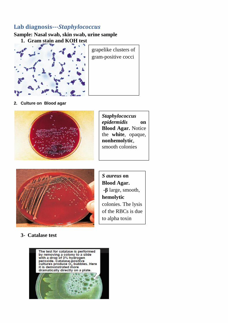
1. Gram stain and KOH test
2. Culture on Blood agar
3- Catalase test
grapelike clusters of
gram-positive cocci
Staphylococcus
epidermidis on
Blood Agar. Notice
the white, opaque,
nonhemolytic,
smooth colonies
S aureus on
Blood Agar.
large, smooth, β -
hemolytic
colonies. The lysis
of the RBCs is due
to alpha toxin
production.
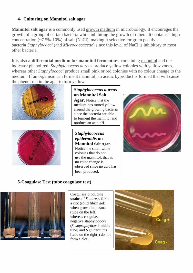
4- Culturing on Mannitol salt agar
Mannitol salt agar is a commonly used growth medium in microbiology. It encourages the
growth of a group of certain bacteria while inhibiting the growth of others. It contains a high
concentration (~7.5%-10%) of salt (NaCl), making it selective for gram positive
bacteria Staphylococci (and Micrococcaceae) since this level of NaCl is inhibitory to most
other bacteria.
It is also a differential medium for mannitol fermentors, containing mannitol and the
indicator phenol red. Staphylococcus aureus produce yellow colonies with yellow zones,
whereas other Staphylococci produce small pink or red colonies with no colour change to the
medium. If an organism can ferment mannitol, an acidic byproduct is formed that will cause
the phenol red in the agar to turn yellow.
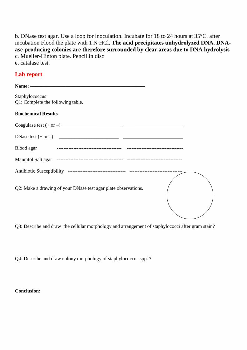
5-Coagulase Test (tube coagulase test)
Staphylococcus aureus
on Mannitol Salt
Agar. Notice that the
medium has turned yellow
around the growing bacteria
since the bacteria are able
to ferment the mannitol and
produce an acid pH.
Staphylococcus
epidermidis on
Mannitol Salt Agar.
Notice the small white
colonies that do not
use the mannitol; that is,
no color change is
observed since no acid has
been produced.
Coagulase producing
strains of S. aureus form
a clot (solid fibrin gel)
when grown in plasma
(tube on the left),
whereas coagulase
negative staphylococci
(S. saprophyticus [middle
tube] and S.epidermidis
[tube on the right]) do not
form a clot.

Principle of Tube coagulase test:
The free coagulase secreted by S. aureus reacts with coagulase reacting factor (CRF) present
in plasma to form a complex, thrombin. This converts fibrinogen to fibrin resulting in
clotting of plasma.
Procedure for Tube coagulase test:
1. Take three test tubes and label them as “test”, “negative control” and “positive
control”.
2. Fill each test tube with 1 ml of 1:6 dilution of rabbit or human plasma in normal
saline.
3. Add 0.1 ml of overnight broth culture to the tube labeled test. Also add 0.1 ml of
overnight broth culture of known S. aureus to the tube labeled positive control and 0.1
ml of sterile broth to the tube labeled negative control.
4. Incubate all the tubes at 37oC and observe up to four hours.
6-Novobiocin Susceptibility Test.
7- DNase test :
Pathogenic strains of staphylococci produce a nuclease enzyme called DNase. DNase
degrades host DNA and increases the pathogenicity of staphylococci that possess it. To
demonstrate the presence of DNase, agar containing dissolved DNA is spot inoculated with
staphylococci. A zone of clearing around the colony indicates a positive DNase test. Three
versions of the test are available, One formulation for DNase Agar consists of peptides,
sodium chloride, and DNA as the substrate. After incubation, 1N HCl is added. Intact DNA
will form a cloudy precipitate with the HCl, but not with nucleotides. Therefore, clearing
around the growth is an indication of DNase activity (DNA hydrolysis).A modification of
DNase test agar includes toluidineblue, which forms a blue complex with intact DNA, but
appears pinkish when complexed with nucleotides. DNase activity is indicated by a pink
(Left on plate) Novobiocin resistance
evidenced by lack of zone of
inhibition (or a zone less than 17
mm) surrounding a novobiocin disk.
Resistance is typical of
Staphylococcus saprophyticus. (Right
on plate) Novobiocin susceptibility
evidenced by a zone of inhibition
greater than 16 mm surrounding the
novobiocin disk. Sensitivity is typical
of Staphylococcus epidermidis and
other coagulase-negative
staphylococci, other than S.
saphrophyticus.

coloration around the growth. An alternate modification of DNase Test Agar contains methyl
green dye. Bacterial colonies that secrete DNase produce clearing around the growth of
DNase positive organisms.
Lab Work:
Materials required
1. Swabs for taking samples (nasal, skin), Normal saline (0.09% NaCl)
2. Syringe for taking blood plasma, heprinized tube
3. Preparation of blood agar, mannitol salt agar, DNase agar, Muller hinton agar
4. Gram stain (KOH), slides, Oil , HCL
5. 3% H2O2 6. Novobiocin antibiotic disc
First Period
1. Taking sample
Obtain a nose swab by rotating a moistened (0.85% saline) swab thoroughly around
the perimeter of both nares (nostrils). Avoid touching the outside skin area.
Obtain a skin culture by rolling a moistened swab up and down the arm. An alternative
method is to swab underneath the fingernails of one hand.
Obtain a urine sample (mid stream urine)
2. Inoculate Blood agar plate, Mannitol salt agar, Incubate for 24 hours at 37°C.
Second period
3. Choose one presumptive colony of S. aureus and S. epidermidis for further
characterization. If you did not obtain one of each from your body, use a plate from one of
the other students in the laboratory and inoculate the following:
a. Citrated rabbit plasma.
b. DNase test agar. Use a loop for inoculation. Incubate for 18 to 24 hours at 35°C. after
incubation Flood the plate with 1 N HCl. The acid precipitates unhydrolyzed DNA. DNA-
ase-producing colonies are therefore surrounded by clear areas due to DNA hydrolysis
c. Mueller-Hinton plate. Pencillin disc
e. catalase test.
Lab report
Name: ———————————————————————
Staphylococcus
Q1: Complete the following table.
Biochemical Results
Coagulase test (+ or –) ________________________ ________________________
DNase test (+ or –) ________________________ ________________________
Blood agar --------------------------------------- ----------------------------------
Mannitol Salt agar ---------------------------------------- ---------------------------------
Antibiotic Susceptibility ----------------------------------- --------------------------------
Q2: Make a drawing of your DNase test agar plate observations.
Q3: Describe and draw the cellular morphology and arrangement of staphylococci after gram stain?
Q4: Describe and draw colony morphology of staphylococcus spp. ?
Conclusion:
