lasers and its application in periodontics
TRANSCRIPT


Lasers and its applications in Periodontology
Presented By:Shilpa ShivanandII MDS

Contents
Introduction.Historical perspective.LASER properties.LASER physics.LASER parts and delivery systems.LASER classification.Biologic rationale: LASER –tissue interactions.LASERS-
Argon, Diode, Nd:YAG, Er:YAG, Co 2 laser.

Contents
Laser Safety.Applications in dentistry.Clinical Applications in Periodontics.Literature review.ConclusionReferences

introduction
L- Light A- Amplification
S- StimulatedE- EmissionR- Radiation

Historical perspective1917- Principle of stimulated emission by Albert
Einstein. “Zur Quantern Theorie der Strahlung”
1954: Townes and Gordon- MASER.
1957- Gordon Gould introduced the term LASER.
1960- Theodore Maiman- First LASER- ruby – active medium.
1961- Javan.- He-Ne laser.

Historical perspective
1964- Nd YAG- Geusic.
1965- Co2 Laser- Patel.
1989- Myers and Myers-FDA approval for use of laser in dentistry- Nd YAG laser.
1990- Opthalmic application- ruby laser.
1995- dental use started.
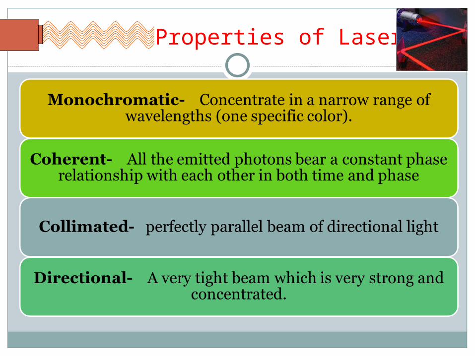
Properties of Laser

Monochromatic lightMono chromatic light has a very narrow range of
frequency and single wavelength (i.e. it is only made of light of one colour)

Coherence• All the emitted photons are in phase with each
other and have identical peaks and valleys.

Directionality

LASER physicsHow is it produced??

Laser%2C_quantum_principle.ogv.720p.webm
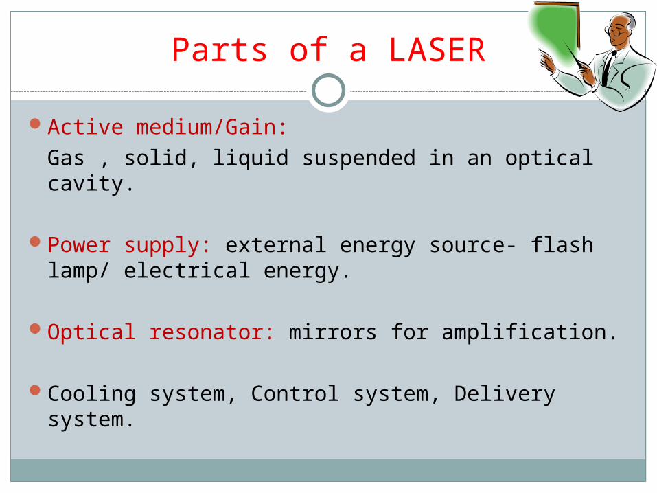
Parts of a LASERActive medium/Gain:
Gas , solid, liquid suspended in an optical cavity.
Power supply: external energy source- flash lamp/ electrical energy.
Optical resonator: mirrors for amplification.
Cooling system, Control system, Delivery system.


Active mediumPhase ExampleGas Argon, Co2Solid DiodeSolid Nd:YAGSolid Er:YAGLiquid Red dye
Usually defines laser type.

LASER DELIVERY SYSTEMS

ARTICULATED ARMS

Consist of a series of rigid hollow tubes with mirrors at each joint (called a knuckle)
Mirrors reflect the energy down the length of the tube.
The laser energy exits the tube through a handpiece
DISADVANTAGES1 . Awkward 3-D maneuverability of the arm.
2 . Mirrors at each knuckle must be aligned precisely.
A misalignment of the mirrors could cause a drop-off in the amount of energy transmitted to the handpiece.
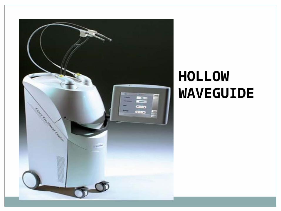
HOLLOW WAVEGUIDE

Semi rigid hollow tube with reflective interior mirror finish.
Laser energy is reflected along this tube and exits through a handpiece at the surgical end with the beam striking the tissue in a noncontact fashion.

This handpiece can be attached to an accessory tip of sapphire or hollow metal for contact with the surgical site.
lenses within the laser instrument focus the beam

OPTIC FIBER
Smaller in diameter with sizes ranging from 200 -1000 μm in diameter
Fits into a handpiece
Used in contact or noncontact mode
Focal point is at or near the tip, which has the greatest energy.
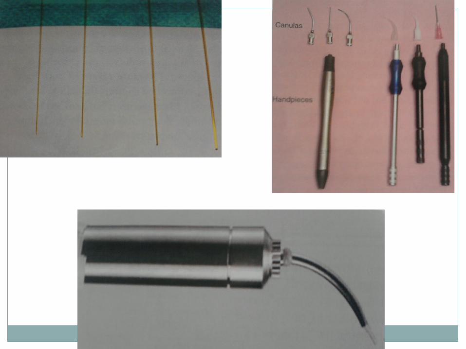

LASER Delivery systemsDelivery system typeArticulated arm Hollow tubes, 45 degree mirrorsHollow waveguide Semi-rigid tube with internal reflective
pathwayOptic fiber/ rigid tip Quartz-silica flexible fiber with quartz,
sapphire tipHand held unit Low power lasers.
Erbium family- fibers with low content of OH ion are used. (eg) Zirconium fluoride

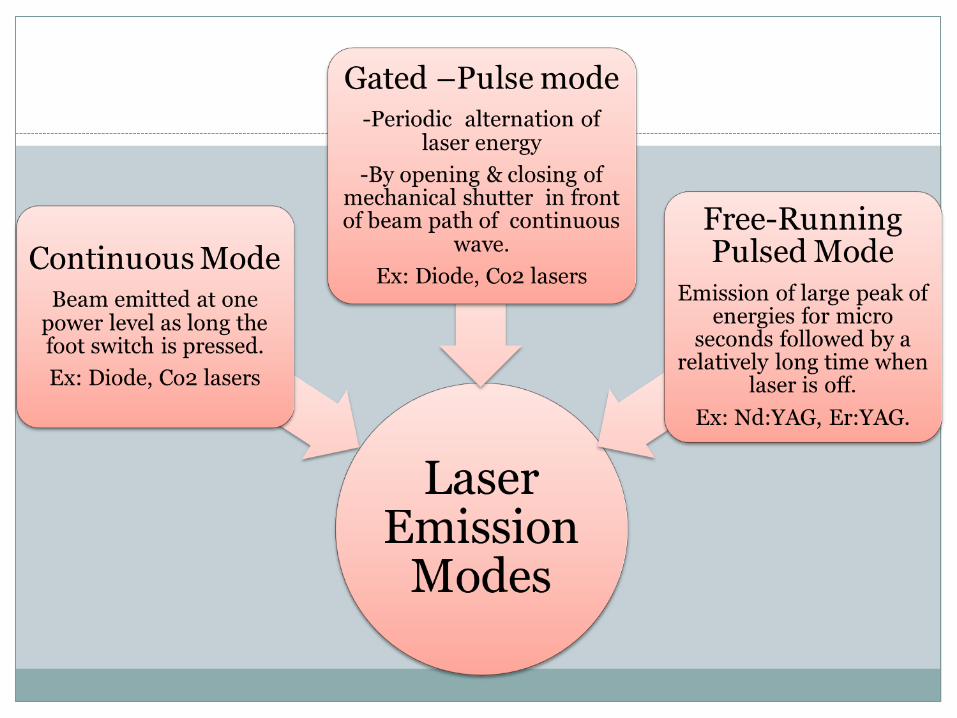
Modes of operation of LASER
Continuous wave
Gated pulsed mode (Physical gating of beam)
Free running pulsed mode (Property of the active medium)


Focused De-focused
Laser beam hits tissue at its focal point- narrowest diameter.
Cutting mode
Beam moved away from its focal point.
Wider area of tissue affected as beam diameter increases.
Ablative mode.LLLT.
Laser operation parameters

Contact Non- contact
Tip is in contact with tissue.
Concentrated delivery of laser energy.
Char tissue formation at tip.
Tactile feedback is available
Tip is kept 0.5 to 1 mm away from tissue.
Laser energy delivered at the surface is reduced.
Laser Operation parameters
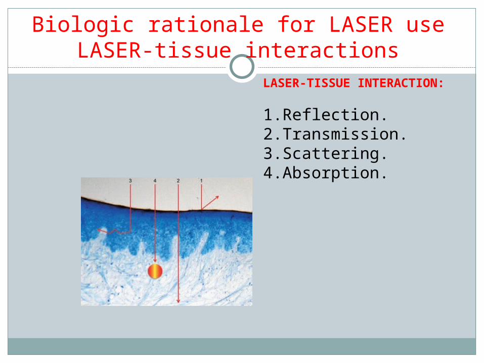
Biologic rationale for LASER use LASER-tissue interactions
LASER-TISSUE INTERACTION:
1. Reflection.2. Transmission.3. Scattering.4. Absorption.
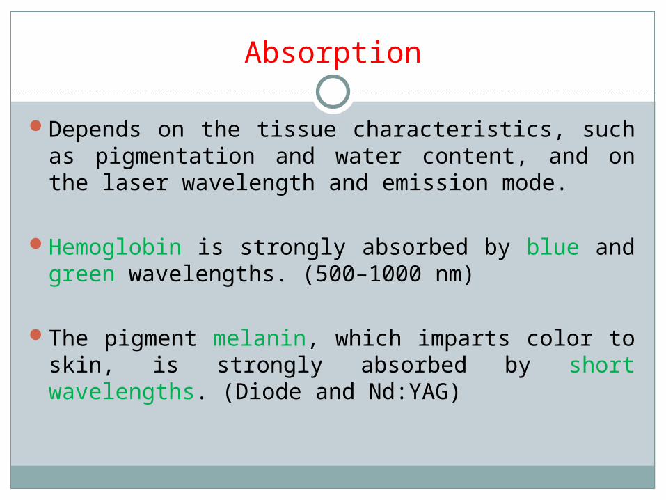
Absorption
Depends on the tissue characteristics, such as pigmentation and water content, and on the laser wavelength and emission mode.
Hemoglobin is strongly absorbed by blue and green wavelengths. (500–1000 nm)
The pigment melanin, which imparts color to skin, is strongly absorbed by short wavelengths. (Diode and Nd:YAG)

Transmission
Water, for example, is relatively transparent to the shorter wavelengths like argon, diode, and Nd:YAG, whereas tissue fluids readily absorb the erbium family and CO2 at the outer surface, so there is little energy transmitted to adjacent tissues.

Reflection
A caries-detecting laser device uses the reflected light to measure the degree of sound tooth structure.
This reflection can be dangerous because the energy is directed to an unintentional target, such as the eyes; this is a major safety concern for laser operation.

Scattering
Weakening the intended energy and possibly producing no useful biologic effect.
Cause heat transfer to the tissue adjacent to the surgical site, and unwanted damage could occur.
However a beam deflected in different directions is useful in facilitating the curing of composite resin or in covering a broad area.

THEORETICAL ZONES OF TISSUE CHANGE ASSOCIATED WITH SOFT TISSUE EXPOSURE TO LASER
LIGHT

BENEFITS OF LASER – TISSUE INTERACTION
Soft tissue: Cut, coagulate, ablate or vaporize target tissue
elements Sealing of small blood vessels Sealing of small lymphatic vessels Sterilizing of tissue- Eschar Decreased post-operative tissue shrinkage

THEORETICAL ZONE OF TISSUE CHANGE ASSOCIATED WITH HARD DENTAL TISSUE EXPOSURE TO LASER LIGHT

LASER effects are due to:
Photothermal.Photochemical.Photoacoustic.Biostimulation.Photodynamic.Photovaporolysis.Photoplasmolysis.

Photothermal effects
Tissue temperature (degree celsius)
Observed effect
37-50 Hyperthermia
> 60 Coagulation, protein denaturation
70-90 Welding
100-150 Vaporization
>200 Carbonization

Photoacoustic
The photoacoustic effect is a conversion between light and acoustic waves due to absorption and localized thermal excitation.
When rapid pulses of light are incident on a sample of matter, they can be absorbed and the resulting energy will then be radiated as heat.
This heat causes detectable sound waves due to pressure variation in the surrounding medium.

Photovaporolysis Photoplasmolysis
Ascendant heat levels- phase transfer from liquid to vapor.
Tissue removed by formation of electrically charged ions and particles in a semi-gaseous high energy state.
LASER effects

Photochemical Biostimulation
Absorption by chromophores-
Tissue response in terms of change of covalent structure.
Believed to work towards healing by stimulation of factors and processes involved in healing.
Below surgical threshold.Useful for pain relief,
increased collagen growth and anti-inflammatory activity
LASER effects


WHAT DOES THE OPERATOR CONTROL?

CLASSIFICATIONS
Lasers are named according to:
Active mediumWavelengthDelivery systemsEmission modes Tissue absorption Clinical Application

Classification

Classification of LASER (Periodontology 2000, 2009)

Classification of LASER- based on safety
Based on the potential of the primary laser beam or the reflected beam to cause biologic damage to the eye or skin.
Four basic classes: Class I. Class II: a,b Class III: a, b Class IV.

Classification of LasersClass I lasersDo not pose a health
hazard.Beam is completely
enclosed and does not exit the housing.
Max power output: 1/10 th of milliwatt
Eg: CD player.
Class II Lasers:Visible light with low
power output.No hazard- blinking and
aversion reaction.Max power output is 1
mW.Eg: bar code scanner,
laser pointerTwo subdivisions:
IIa: dangerous- >1000 sec.IIb: ¼ th of second.

Laser classificationClass IIIa:Any wavelength.Max Output power:
0.1 to 0.5 W.Danger > ¼ th of a
second.Caution label.
Class IIIb:Hazard to eye- direct
or reflected beam, irrespective of time of exposure.
Safe with matted surface and no fire hazard.
Max output power: 0.5 to 5W.

Classification of lasers
Class IV lasers:Hazardous for direct viewing and reflection.Max output power > 5 W.Fire and skin hazards.Use safety glasses
Dental lasers are Class IIIb or Class IV lasers.
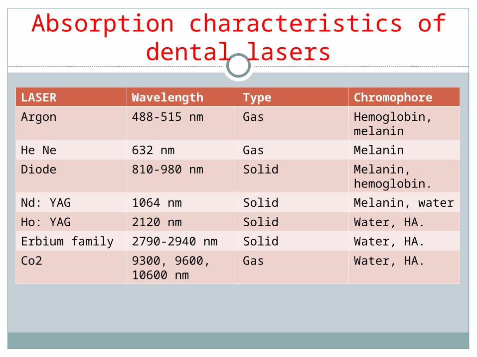
Absorption characteristics of dental lasers
LASER Wavelength Type ChromophoreArgon 488-515 nm Gas Hemoglobin,
melaninHe Ne 632 nm Gas MelaninDiode 810-980 nm Solid Melanin,
hemoglobin.Nd: YAG 1064 nm Solid Melanin, waterHo: YAG 2120 nm Solid Water, HA.Erbium family 2790-2940 nm Solid Water, HA.Co2 9300, 9600,
10600 nmGas Water, HA.
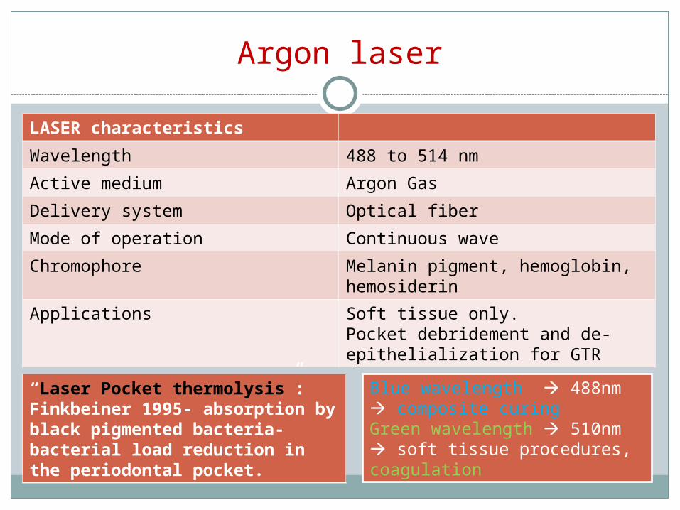
Argon laser
LASER characteristicsWavelength 488 to 514 nmActive medium Argon GasDelivery system Optical fiberMode of operation Continuous waveChromophore Melanin pigment, hemoglobin,
hemosiderinApplications Soft tissue only.
Pocket debridement and de-epithelialization for GTR
“Laser Pocket thermolysis”: Finkbeiner 1995- absorption by black pigmented bacteria- bacterial load reduction in the periodontal pocket.
Blue wavelength 488nm composite curingGreen wavelength 510nm soft tissue procedures, coagulation

Argon
Acute inflammatory periodontal disease and highly vascularized lesions, such as a hemangioma, are ideally suited for treatment.The poor absorption into enamel and dentin is advantageous when using this laser for cutting and sculpting gingival tissues because there is minimal interaction and thus no damage to the tooth surface during those procedures.

Diode laser
LASER characteristicsWavelength 810 to 980 nmActive medium Semi-conductor diodeDelivery system Optical fiber- quartz or silicaMode of operation Continuous wave, gated pulsed
mode. Used in focused and de-focused modes.
Chromophore Melanin, hemoglobin.Applications Primarily soft tissue applications-
all minor surgical procedures.The chief advantage of the diode lasers is one of a smaller size, portable instrument.HOT TIP EFFECT
heat accumulation at
tip thick coagulating layer
Less tissue penetration ,
Deeper coagulation
DIODENTVisible red diode
655nm1mW
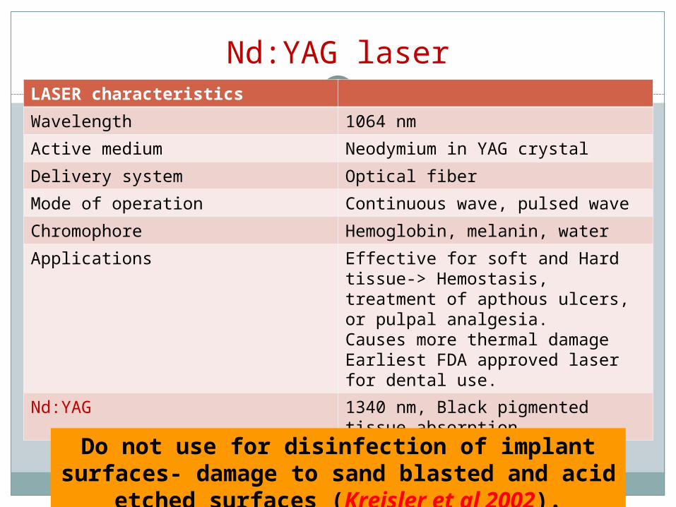
Nd:YAG laserLASER characteristicsWavelength 1064 nmActive medium Neodymium in YAG crystalDelivery system Optical fiberMode of operation Continuous wave, pulsed waveChromophore Hemoglobin, melanin, waterApplications Effective for soft and Hard tissue-
> Hemostasis, treatment of apthous ulcers, or pulpal analgesia.Causes more thermal damageEarliest FDA approved laser for dental use.
Nd:YAG 1340 nm, Black pigmented tissue absorption.
Do not use for disinfection of implant surfaces- damage to sand blasted and acid etched surfaces (Kreisler et al 2002).

Erbium family of lasers
Er YAG- 2940 nm: Zharikov et al 1975.Er Cr YSGG- 2780 nm: Zharikov et al 1984 and
Moulton et al 1988.1988: Phagdiwala: Er YAG laser: ability to ablate
the dentinal hard tissue.1989: Pulsed Erbium laser: Keller and Hibst-
enamel , dentin and bone.1995: Commercially available.1997: introduced for use in dentistry.
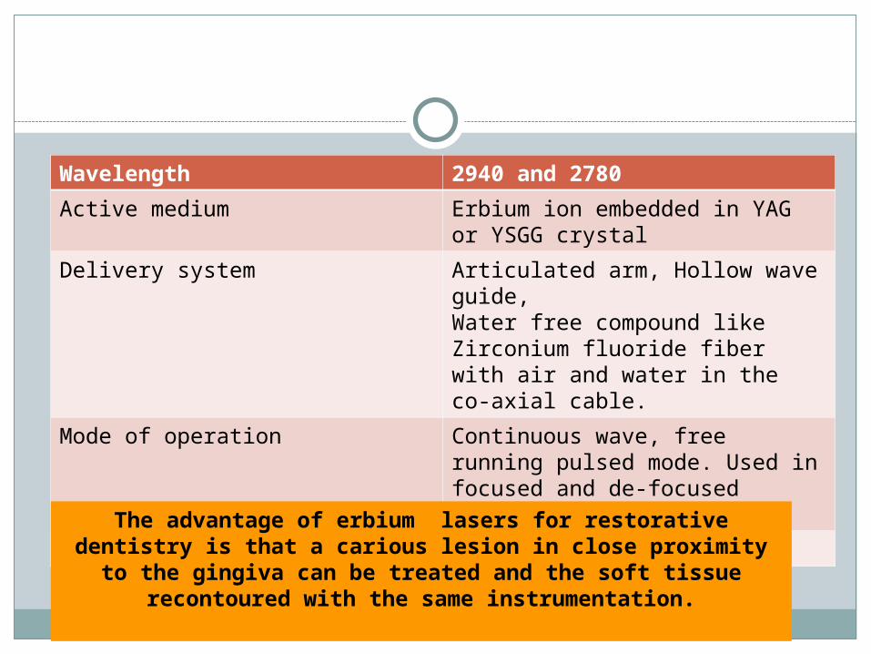
Wavelength 2940 and 2780Active medium Erbium ion embedded in YAG or
YSGG crystalDelivery system Articulated arm, Hollow wave
guide,Water free compound like Zirconium fluoride fiber with air and water in the co-axial cable.
Mode of operation Continuous wave, free running pulsed mode. Used in focused and de-focused modes.
Chromophore Water, HydroxyapatiteThe advantage of erbium lasers for restorative dentistry is that a carious lesion in close proximity to the gingiva can be treated and the soft tissue recontoured with the same
instrumentation.
MOA of Er laser photoablationLayers formed superficial significantly altered
intermediate deeper/ less affected
Superficial layer micro-cracking, disorganization, slight recrystallization of apatite, reduction of surrounding organic matrix
Intermediate layer micro-explosion due to energy accumulation
Deep no change

Co2 laser
Wavelength 9300, 9600, 10600 nmActive medium Carbon dioxide Gas
Delivery system Articulated armMode of operation Continuous wave, gated pulsed
mode. Used in focused and de-focused modes.
Chromophore Water, Hydroxyapatite
Limitation: High risk of carbonization (water absorption generates more heat carbonizes tissue)Advantage : carbonized / charred layer acts as biological dressing
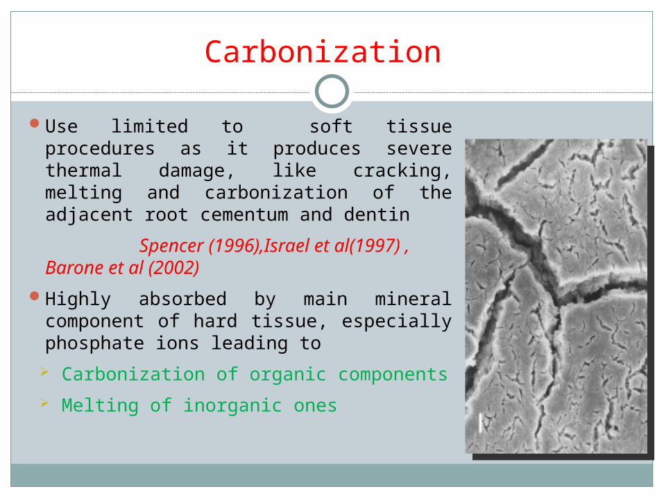
Carbonization
Use limited to soft tissue procedures as it produces severe thermal damage, like cracking, melting and carbonization of the adjacent root cementum and dentin
Spencer (1996),Israel et al(1997) , Barone et al (2002)
Highly absorbed by main mineral component of hard tissue, especially phosphate ions leading to Carbonization of organic components Melting of inorganic ones

Advantages Disadvantages
Hemostasis.Ablation.Detoxification.Bactericidal activity.Osseous tissue
removal and contouring easy with Er family
Hard tissue damage (bone)
High cost.Risk of pulpal
damage.No single
wavelength can treat all diseases
Lasers

LASER safetyRegulatory
organizations: CDRH center for
devices and radiologic health
ANSIAmerican National Standards Institute
OSHA occupational safety and health administration
Laser safety officer.
Environment: warning signs, restricted access, reflective surface minimized.
Laser use documentation.
Training.Eye and tissue
protection.

Eye damage
Part of eye damaged Laser typeCorneal damage Er Cr YSGG, Ho YAG, Er YAG, Co2Lens damage Diode, Nd YAG, Ho YAG, Er Cr YSGG,
Er YAGAqueous damage Ho YAG, Er Cr YSGG, Er YAGRetinal damage Argon, He Ne, Diode, Nd YAG

Laser Safety Officer (LSO)Knowledge of
operational characteristics.
Supervises staff education and training.
Laser maintenance and calibration.
Posts warning signs.Oversees personal
protection.Incident reporting.Knowledge about
regulations.Regulates working
area.

Applications in dentistryBiopsy.Apicoectomy.Teeth preparation.Epulis fissuratum.Residual ridge
modification.Bleaching.Impaction.
Pontic site preparation.Tori reduction.Soft tissue modification
around laminates.Impacted teeth
exposure- orthodontic movement.
Caries removal.Root canal disinfection.

Clinical Applications in PeriodonticsInitial non-surgical
pocket therapy.Frenectomy.Gingivectomy.Soft tissue grafting.De-pigmentation.Desensitization
Removal of granulation tissue.
Osseous recontouring.
Crown lengthening.Surgery- implants.Peri-implantitis.Operculectomy.

Conventional methods LASER
Bleeding- surgical field.Suturing.Local anesthesia.Post-operative discomfort.Healing time.Post-operative
complications.Infection.Periodontal dressings.
Effective hemostasis.No sutures. (concept of
tissue welding).Topical anesthetic- some
procedures.Faster healing.Minimal/no post operative
complications.Laser sterilization of wound
site.Laser bandage.
Why Lasers in Periodontics…

LASERS used in Periodontics

Gingival soft tissue procedures
Advantages of lasers over conventional: Hemostasis. Ablation. Little wound contraction/ minimal scarring. Faster healing. Less post-operative discomfort. Less risk of damage to underlying structures as
compared to cautery.

Gingival soft tissue proceduresIndications:
Gingivectomy. Gingivoplasty. Frenectomy/
frenotomy. Vestibuloplasty. Operculectomy. Depigmentation.
Lasers used; Diode. Nd YAG. Er YAG. Co2.Diode and Nd YAG: deep
penetration .Er YAG, Co2: superficial
action.

Gingival soft tissue proceduresDiode and Nd YAG:
Effective for cutting and reshaping of soft tissue.
Good hemostasisGreater thermal
effects.Thicker coagulated
layer.
Co2 laser:
Rapid ablation of soft tissue.
Good hemostasis.Effective even for
thick tissue.Risk of charring-
thermal damage.

Gingival soft tissue procedures.Er YAG :Fine cutting can be
done.Less hemostasis as
compared to other lasers.
Very less thermal damage: use with irrigation.
Width of thermally affected layer: 5-20 microns (Aoki et al 2005)
Er YAG:Safer even in thin
tissues.Useful to remove
melanin and metal tattoos.

Non Surgical therapy
Introduction:Primarily aimed at efficient removal of plaque
and calculus and reduction of bacterial load, inflammation.
Conventional therapy limitations: Incomplete removal of calculus. Incomplete elimination of inflamed pocket lining.
Lasers used: Diode, Nd YAG, Er YAG, Co2 lasers.

Subgingival calculus detection- Unique application for LASER
Conventional method- tactile feel.
Latest: Er YAG laser with fluorescent feedback system for calculus detection.
Rationale: Difference in the fluorescence emission
properties of calculus and dental hard tissue when subjected to irradiation with 655 nm diode laser.

Author and year
Study design Objective Findings
Folwaczny M et al 2002
In vitro- extracted teeth
Assess efficacy of fluorescence induced by 655 nmdiode laser to detect subgingival calculus
655 nm diode laser- effective for calculus detection
Krause F et al 2003
In vitro- histologic study ( in presence of saline/ blood)
Efficacy for calculus detection
The laser fluorescence values co-relate strongly with calculus presence.
Scharwz F et al 2003
In vivo and in vitro.Er YAG with Diode 655 nm combined
Compare the new system with SRP for calculus removal efficacy
Selective removal of sub-gingival calculus.
Sculean A et al 2004
Er YAG+ diode vs SRP
Improvement of clinical parameters
Similar results with both systems
Tung OH et al 2008
Detection through the gingiva- based on autofluorescence- Ti Sapphire laser
Studies- sub gingival calculus detection system
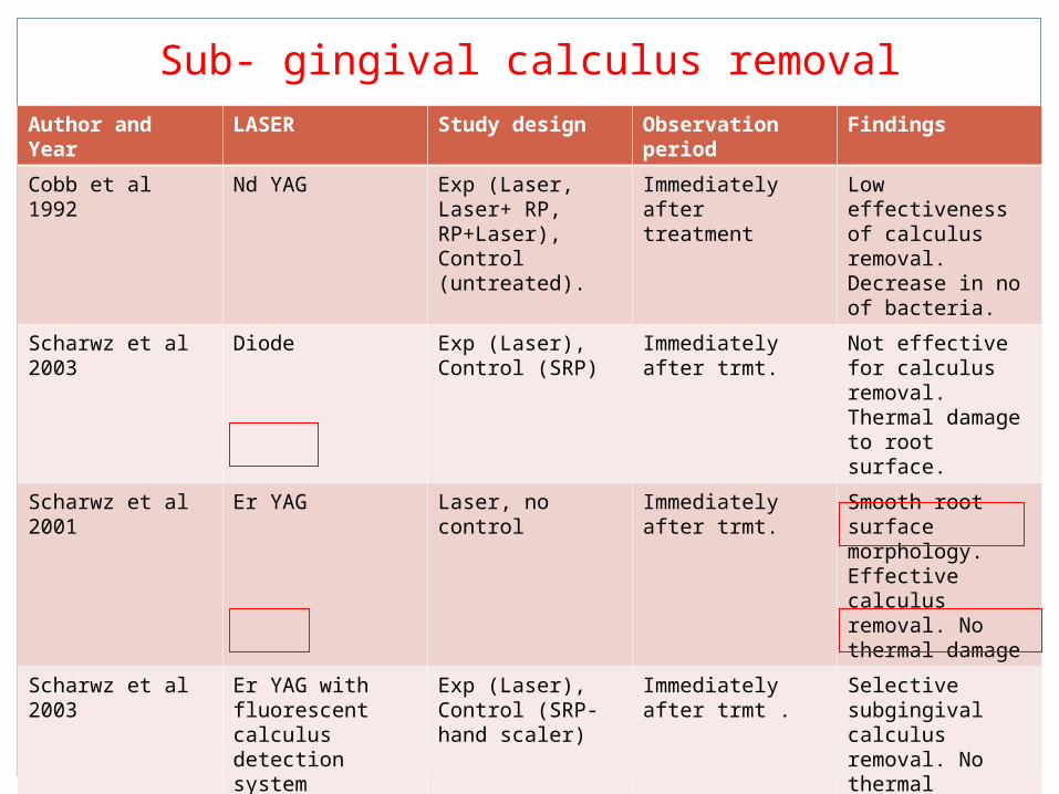
Sub- gingival calculus removalAuthor and Year
LASER Study design Observation period
Findings
Cobb et al 1992 Nd YAG Exp (Laser, Laser+ RP, RP+Laser), Control (untreated).
Immediately after treatment
Low effectiveness of calculus removal. Decrease in no of bacteria.
Scharwz et al 2003
Diode Exp (Laser), Control (SRP)
Immediately after trmt.
Not effective for calculus removal.Thermal damage to root surface.
Scharwz et al 2001
Er YAG Laser, no control Immediately after trmt.
Smooth root surface morphology. Effective calculus removal. No thermal damage
Scharwz et al 2003
Er YAG with fluorescent calculus detection system
Exp (Laser), Control (SRP- hand scaler)
Immediately after trmt .
Selective subgingival calculus removal. No thermal damage, less cementum removal.

Diode laser Nd YAG laser
Dry or saline moistened root surfaces- no detectable alterations.
Blood coated specimens- charring (Kreisler M et al 2002)
Morlock BJ et al 1992: surface pitting, craters, melting, carbonization of root surface.
Spencer et al 1992, 1996: decrease in protein/mineral ratio, production of protein by-products.
Trylovich DJ et al 1992: Nd YAG treated root surface – not favorable for fibroblast attachment.
Thomas D et al 1994: Laser followed by SRP- restores the biocompatibility of root surface
Root surface alterations

Co2 laser Erbium family
SpencerP, Cobb CM et al 1996: Carbonized layer on root
surface. Cyanamide , cyanate ions-
detected on the carbonized layer- FTIS method.
Gopin BW et al 1997: Char layer inhibits periodontal soft tissue attachment.
Co2 laser contraindicated for root surface treatment in focused mode.
• Aoki et al 2000: Er YAG with coolant:
micro-irregular surface. No thermal effects such
as cracking, fissuring.• Sasaki KM et al 2002: no
major chemical or compositional change- on root cementum or dentin.
• Biocompatability of root surface: micro-irregularity offers better attachment to fibroblasts (Scharwz F et al 2003).
Root surface alterations

Bacterial reduction
The only two soft tissue wavelengths that currently meet the criterion of having a delivery system able to deliver laser energy efficiently and effectively to the periodontal pockets for nonsurgical periodontal therapy are Nd:YAG and diode.
Well absorbed by melanin and hemoglobin and other chormophores present in periodontally diseased tissues. The laser energy is transmitted through water and poorly absorbed in hydroxyapitite.
Both of these wavelengths have been shown to be extremely effective against periodontal pathogens in vivo and in vitro ( Moritz 1998, Pinhero J 1997)
These investigators concluded that the diode laser revealed a bactericidal effect, helped reduce inflammation, and supported healing of the periodontal pockets through the elimination of bacteria.
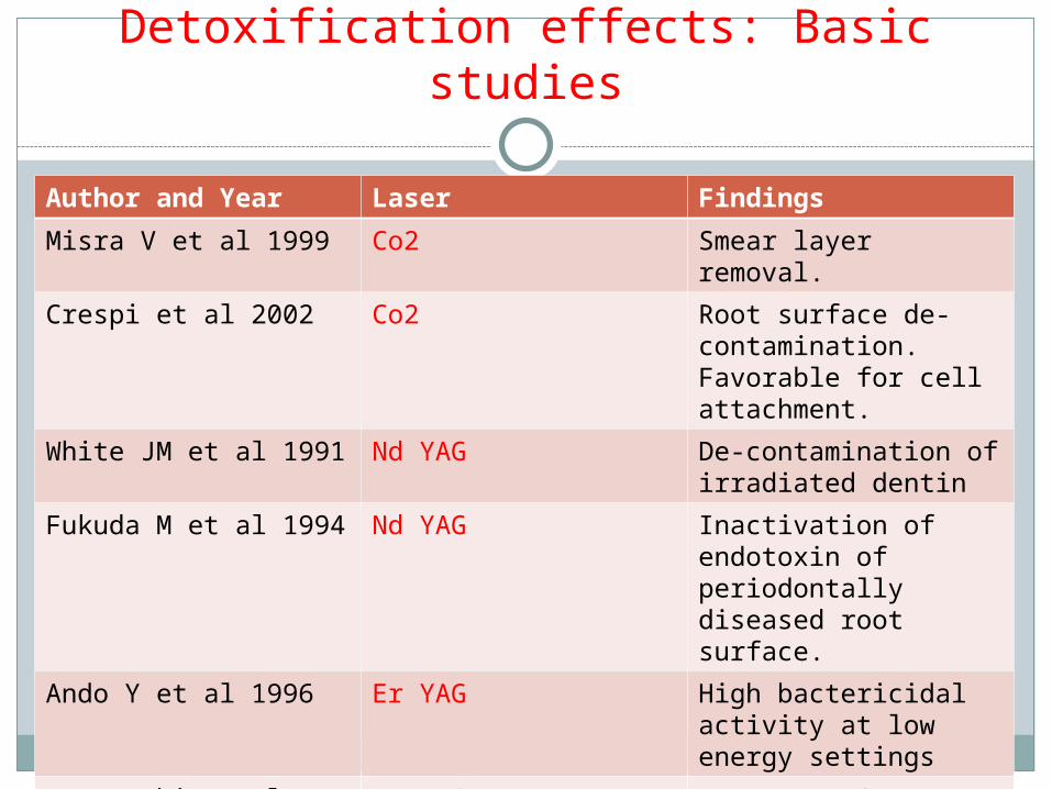
Detoxification effects: Basic studies
Author and Year Laser FindingsMisra V et al 1999 Co2 Smear layer removal.Crespi et al 2002 Co2 Root surface de-
contamination. Favorable for cell attachment.
White JM et al 1991 Nd YAG De-contamination of irradiated dentin
Fukuda M et al 1994 Nd YAG Inactivation of endotoxin of periodontally diseased root surface.
Ando Y et al 1996 Er YAG High bactericidal activity at low energy settings
Yamaguchi et al 1997 Er YAG Removes LPS diffused into root surface

Low Level Lasers

1903: N.R. FinesenN.R. Finesen, the father of modern , the father of modern phototherapy, first described low level ultraviolet in phototherapy, first described low level ultraviolet in the treatment of lupus vulgaris.the treatment of lupus vulgaris. The treatment was low level effect of non coherent The treatment was low level effect of non coherent light.light.
1960s: The use of LLLT first initiated in medicine The use of LLLT first initiated in medicine for in vitro experiments to determine the effects on for in vitro experiments to determine the effects on cell cultures and increased blood circulation within cell cultures and increased blood circulation within regenerating tissue.regenerating tissue.
Historical aspects

• Generally smaller, less expensive lasers that operate in the milliwatts range, 1-500 mw are used.
• The therapy performed with such lasers are often called “low level laser therapy” (LLLT) or also known as “therapeutic lasers”
• Generally operate in the visible and the infrared spectrum, 600 – 900 nm wavelengths.
• The energy used is indicated in joules. • Suitable therapeutic energies range from 1 – 10
joules per point.

All commercially available LLLT system are generally variants of Gallium, Aluminum, Arsenide (Ga, Al, As) which emit in the near infrared spectrum (700 – 940 nm).

Low Level LasersLow Level Lasers Helium – neon laserHelium – neon laser
Gallium – aluminum – arsenide diode laserGallium – aluminum – arsenide diode laser
Gallium – arsenide diode laserGallium – arsenide diode laser
Argon ion laser Argon ion laser
Defocused CoDefocused Co22 laser laser
Defocused Nd:YAG laserDefocused Nd:YAG laser

Biostimulation effects of low level laser
Reduction of discomfort / pain (Kreisler MB et al 2004). Promotion of wound healing (Qadri T et al 2005). Bone regeneration (Merli LA et al 2005). Suppression of inflammatory process. (Qadri T et al
2005). Activation of gingival and periodontal ligament fibroblast
(Kreisler M et al 2003), growth factor release (Saygun I et al 2007).
Alteration of gene expression of inflammatory cytokines (Safavi SM et al 2007).
Photobiostimulaation (Garcia et al 2012)

Laser Assisted New Attachment Procedure
LANAPGold and Villardi 1994, safe application of the Nd YAG laser
for removal of pocket epithelium lining without carbonization of the underlying connective tissue.
Approved by FDA for use.Yukna et al 2007- case series- histologic study- new
cementum with new connective tissue attachment on previously diseased root surface.
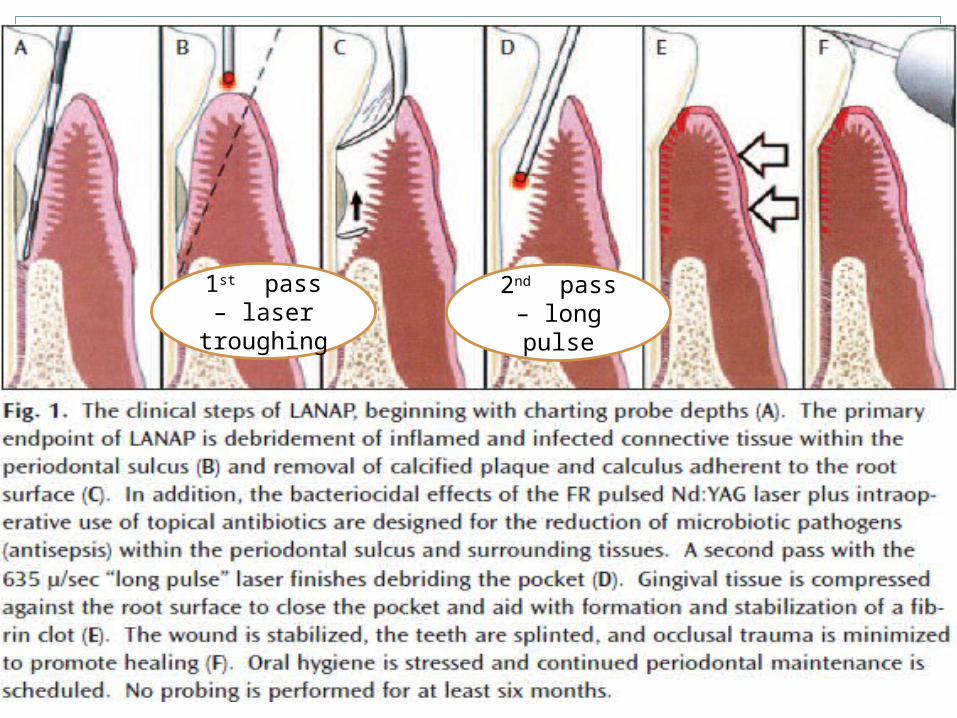
1st pass – laser
troughing2nd pass – long pulse
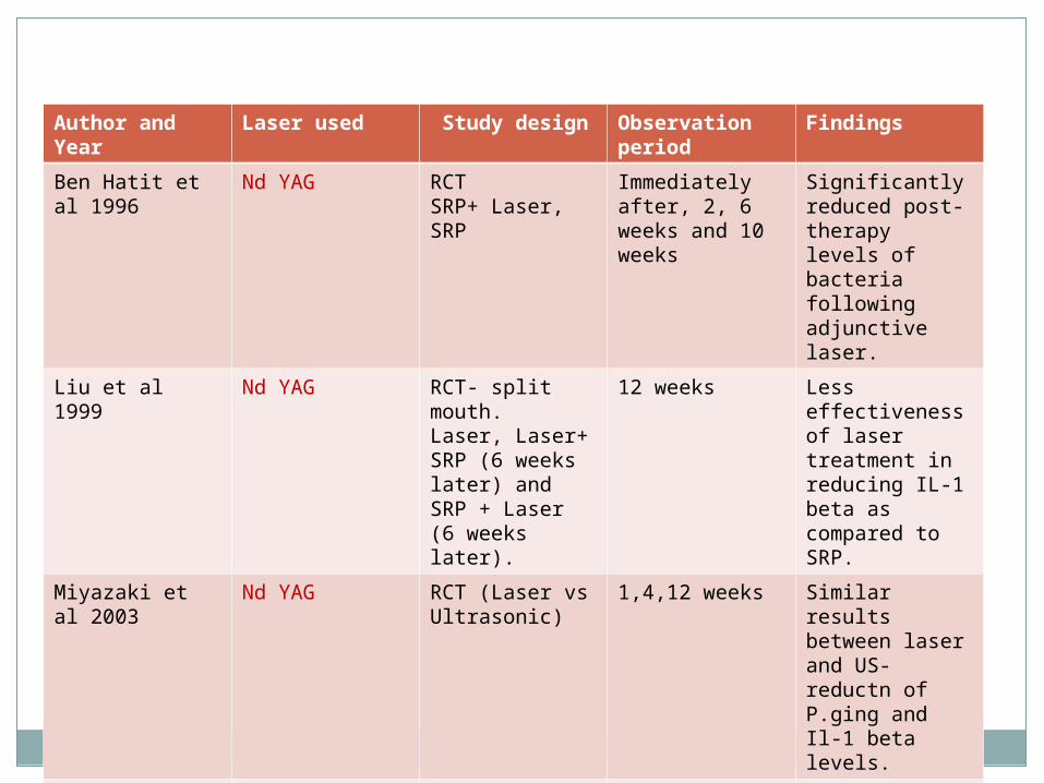
Author and Year
Laser used Study design Observation period
Findings
Ben Hatit et al 1996
Nd YAG RCTSRP+ Laser, SRP
Immediately after, 2, 6 weeks and 10 weeks
Significantly reduced post-therapy levels of bacteria following adjunctive laser.
Liu et al 1999 Nd YAG RCT- split mouth.Laser, Laser+ SRP (6 weeks later) and SRP + Laser (6 weeks later).
12 weeks Less effectiveness of laser treatment in reducing IL-1 beta as compared to SRP.
Miyazaki et al 2003
Nd YAG RCT (Laser vs Ultrasonic)
1,4,12 weeks Similar results between laser and US- reductn of P.ging and Il-1 beta levels.
Noguchi et al 2005
Nd YAG Laser, Laser+ local minocycline, Laser+ povidone iodine
1, 3 months Greater reduction of bacteria on laser+ minocycline treated sites

Author and Year
Laser used Study design
Observation periods
Findings
Moritz et al 1997
Diode SRP+ Laser, SRP
1,2 weeks High bacterial reduction in SRP+ laser as compared to SRP sites alone.
Moritz et al 1998
Diode SRP + Laser, SRP + H2 O2 rinse
6 months Higher reductn in bacterial, BOP, PD in SRP + laser sites.
Kresiler et al 2005
Diode RCT, split mouth design.SRP + Laser, SRP alone.
3 months Greater reduction of PD and attachment gain in Laser adjunct sites
Miyazaki et al 2003
Co2 RCT, Laser vs ultrasonic
1,4,12 weeks No decrease of P.ging and IL-1 in laser sites. But significant decrease in US sites.

Author and Year
Laser used Study design
Observation period
Findings
Watanabe et al 1996
Er YAG Case series (Laser only)
4 weeks Safe and effective calculus removal .
Schwarz et al 2001
Er YAG Split mouth design, RCT (Laser vs SRP)
6 months Clinical outcome similar to SRP.
Sculean et al 2004
Er YAG RCT, split mouth (Laser vs Ultrasonic)
6 months Clinical outcome similar to Ultrasonic scaling.
Tomasi et al 2006
Er YAG RCT, split mouth (Laser vs Ultrasonic)
6 months 1 month- following therapy, laser treated sites- better clinical outcomes, no difference in microbiological levels

Surgical pocket therapy- Lasers
Lasers used: Co2 and Erbium familyInvolves use of lasers for calculus removal, osseous surgery, de-toxification of the root surface and bone, granulation tissue removal
Advantage of Laser:Better access in furcation areas, hemostasis, less post-operative discomfort, faster healing.

Author and Year
Laser used Animal model Study Objective
Findings
Nelson et al 1989
Er YAG Rabbit Irradiated tissue characteristics
Found useful as a bone cutting tool
Lewandrowski et al 1996
Er YAG Rat Cutting efficiency and tissue characteristics
Comparable thermal damage as mechanical cut bone. Normal fracture healing.
Friesen et al 1999
Co2, Nd YAG Rat Tissue characteristics
Residual carbonized tissue and thermal necrosis.
Sasaki et al 2002
Co2 and Er YAG Rat Tissue characteristics
Er YAG- Tissue removal, no charring. Two distinct layers.Co2- charring and no tissue removal.
Stubinger et al 2007
Er YAG Clinical- human Depth control of laser
Laser could not offer precise depth control in preparing osetotomies.
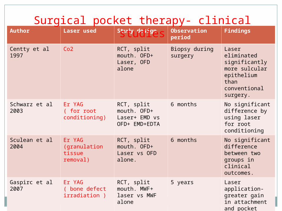
Author Laser used Study design Observation period
Findings
Centty et al 1997
Co2 RCT, split mouth. OFD+ Laser, OFD alone
Biopsy during surgery
Laser eliminated significantly more sulcular epithelium than conventional surgery.
Schwarz et al 2003
Er YAG( for root conditioning)
RCT, split mouth. OFD+ Laser+ EMD vs OFD+ EMD+EDTA
6 months No significant difference by using laser for root conditioning
Sculean et al 2004
Er YAG(granulation tissue removal)
RCT, split mouth. OFD+ Laser vs OFD alone.
6 months No significant difference between two groups in clinical outcomes.
Gaspirc et al 2007
Er YAG( bone defect irradiation )
RCT, split mouth. MWF+ laser vs MWF alone
5 years Laser application- greater gain in attachment and pocket depth reduction.
Surgical pocket therapy- clinical studies

Implant therapy- Management of Peri-implantitis
Peri-implantitis – Management options- Conventional- plastic curettes and antibiotics.New option- Laser Rationale:
Disinfection and de-contamination of implant surface. Granulation tissue removal.Lasers used: Diode, Co2, Erbium family.Lasers contra-indicated: Nd YAG (Implant damage).
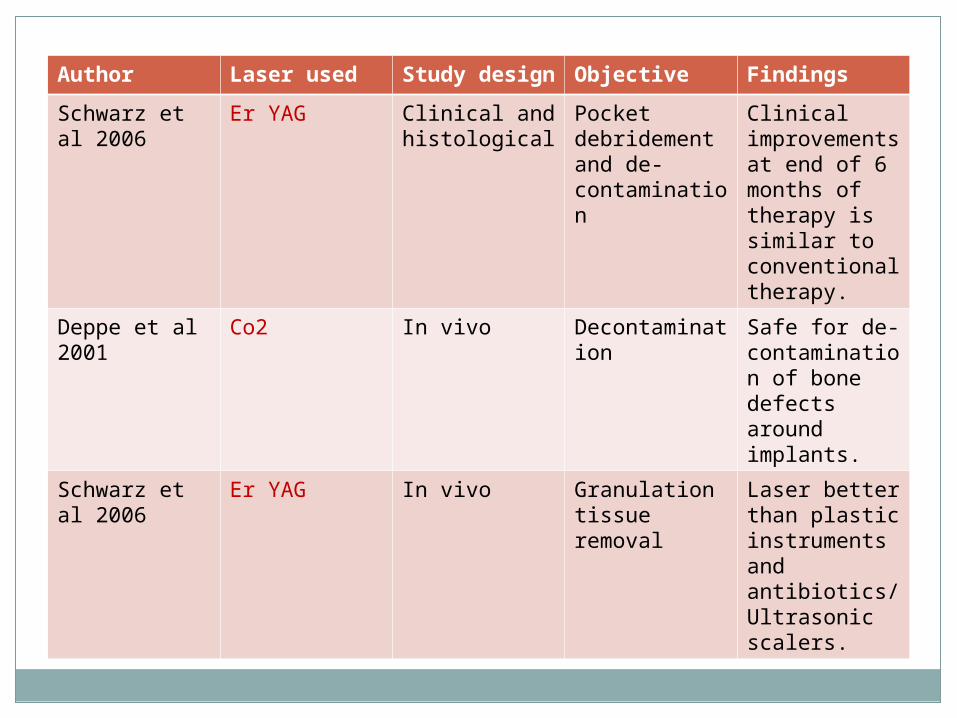
Author Laser used Study design
Objective Findings
Schwarz et al 2006
Er YAG Clinical and histological
Pocket debridement and de-contamination
Clinical improvements at end of 6 months of therapy is similar to conventional therapy.
Deppe et al 2001
Co2 In vivo Decontamination
Safe for de-contamination of bone defects around implants.
Schwarz et al 2006
Er YAG In vivo Granulation tissue removal
Laser better than plastic instruments and antibiotics/ Ultrasonic scalers.

Photodynamic therapy in laser
Main objective of periodontal therapy: eliminate the deposits of bacteria.
Conventional mechanical therapy: incomplete elimination due to
Anatomical complexity of root.Deep periodontal pockets.

PRINCIPLES BEHIND PDT
TARGET CELL
1O2
1O2
1O2
1O2
lightPS
Cell death

Destruction of periodontopathogenic bacteria
Polysaccharides in biofilm are highly sensitive to
singlet oxygen. During inflammation reduced O2
consumption change in pH growth of
anaerobes

PDT tissue blood flow and venous congestion
oxygenation of gingival tissues 21– 47 %
The activity of PDT ..been reported in vitro and in vivo .
Greater bacterial reduction of S.sanguis numbers compared with A. actinomycetamcomitans.
F.D.L. Mattiello et al.(2011)


Anti- microbial photosensitizing agents and the wavelengths used.
Healing after laser therapy
o Reports - laser created wounds heal more quickly and produce less scar tissue than conventional scalpel surgery.
o Contrary evidence from studies in pigs, rats and dogs indicate that the healing of laser wounds is delayed, that more initial tissue damage may result, and that wounds have less tensile strength during the early phase of healing.
(Pick et al 1990)

Abergel et al (1984) experiment with cultured human skin fibroblasts showed that collagen production and DNA synthesis were delayed when the fibroblasts were exposed to Nd: YAG laser radiation.
Iliria et al (2003) analyzed the biocompatibility of root surfaces treated by Er: YAG laser and concluded that laser irradiation promoted faster fibroblast adhesion and growth than surfaces treated with root planing.
Garcia et al (2012) LLLT enhanced healing biostimulation
RECENT ADVANCESWATERLASE
Device that uses laser energized water to cut and coaglate soft and hard tissue.
Er, Cr: YSGG laser 2,780nm - available as WATERLASE


USES
Full thickness flap
Partial thickness flap
Split thickness
Laser soft tissue curettage
Laser removal of diseasd, infected, inflamed, necrosed tissue within the periodontal pocket
Removal of inflamed tissue, osteoplasty, osseous recontouring……

PERIOWAVE
Photodynamic disinfection system utilizes nontoxic dye in combination with a low-intensity lasers enabling singlet oxygen molecules to destroy bacteria.

conclusion
Lasers in dentistry offer incredible precision, less pain, faster healing and many more advantages.
It is most important for the dental practitioner to become familiar with those principles and choose the proper laser for the intended clinical application.

References
Dental clinics of North America. “ Lasers in Clinical dentistry”. Oct 2004. Vol 48. Issue 4.
Application of antimicrobial photodynamic therapy in periodontal and peri-implant diseases. Periodontology 2000, Vol. 51, 2009, 109–140.
Application of lasers in periodontics: true innovation or myth? Periodontology 2000, Vol. 50, 2009, 90–126.
The impact of laser application on periodontal and peri-implant wound healing. Periodontology 2000, Vol. 51, 2009, 79–108
Laser applications in dentistry – Robert N Conviesar
The biologic rationale for the use of lasers in dentistry. Robert Convissar. DCNA 48(2004) 771-794.
Lasers in periodontics . J Periodontol 2002,73:1231-1239
