late-treated individuals with suboptimal cd4r t-cell ... · late-treated individuals with...
TRANSCRIPT

Impaired gut junctio
nal complexes featurelate-treated individuals with suboptimal CD4RT-cell recovery upon virologically suppressivecombination antiretroviral therapy
Camilla Tincatia,b,c, Esther Merlinia,b,c, Paola Braidottib,d,
Giuseppe Anconaa,b,c, Federica Savic,d, Delfina Tosib,c,d, Elisa Borghib,e,
Maria Luisa Callegarif, Benedetto Mangiavillanoc,g,
Alessandra Barassib,c,h, Gaetano Bulfamanteb,c,d,
Antonella d’Arminio Monfortea,b,c, Solange Romagnolib,c,d,i,
Nicolas Chomontj,k and Giulia Marchettia,b,c
aClinic of InfectiodPathology Unit, e
gGastroenterologyInstitute Florida, PMicrobiologie, Inf
Correspondence toSan Paolo Hospita
Tel: +3902818430Received: 31 July
DOI:10.1097/QAD
ISSN
Objective: HIV-infected individuals with incomplete CD4þ T-cell recovery uponcombination antiretroviral therapy (cART) display high levels of immune activationand microbial translocation. However, whether a link exists between gut damage andpoor immunological reconstitution remains unknown.
Design: Cross-sectional study of the gastrointestinal tract in late cART-treated HIV-infected individuals: 15 immunological nonresponders (CD4þ <350 cells/ml and/ordelta CD4þ change from baseline<30%); 15 full responders (CD4þ>350 cells/ml and/or delta CD4þ change from baseline >30%).
Methods: We assessed gut structure (junctional complex proteins in ileum and colon)and function (small intestine permeability/damage and microbial translocationparameters). The composition of the fecal microbiome and the size of the HIV reservoirin the gut and peripheral blood were investigated as possible mechanisms underlyingmucosal impairment.
Results: Markers of intestinal permeability, damage, systemic inflammation, andmicrobial translocation were comparable in all study individuals, yet the expressionof junctional complex proteins in gut biopsies was significantly lower in HIV-infectedpatients with incomplete CD4þ restoration and negatively correlated with markers ofCD4þ reconstitution. Electron microscopy revealed dilated intercellular spaces inindividuals lacking immunological response to cART, yet not in patients displayingCD4þ T-cell recovery. Analysis of the fecal microbiome revealed an overall outgrowthof Bacteroides–Prevotella spp. with no differences according to CD4þ T-cell recon-stitution. Interestingly, HIV reservoirs in peripheral CD4þ T cells and intestinal tissuenegatively correlated with immune recovery.
Copyright © 2016 Wolters Kluwer Health, Inc. All rights reserved.
us Diseases, bDepartment of Health Sciences, University of Milan, cSan Paolo University Hospital, Milan,Microbiology Laboratory, fBiotechnology Research Centre, Sacred Heart Catholic University, Cremona, Italy,Unit, hBiochemistry Laboratory, iRoche Innovation Center, Penzberg, Germany, jVaccine and Gene Therapyort St Lucie, Florida, USA, and kCentre de Recherche du CHUM et Universite de Montreal, Departement deectiologie et Immunologie, Montreal, Quebec, Canada.
Giulia Marchetti, MD, PhD, Clinic of Infectious Diseases, Department of Health Sciences, University of Milan,l, Milan, Italy, Via di Rudini’ 8, 20142, Milan, Italy.
46; fax: +390281843054; e-mail: [email protected]; revised: 7 December 2015; accepted: 28 December 2015.
.0000000000001015
0269-9370 Copyright Q 2016 Wolters Kluwer Health, Inc. All rights reserved. 991

992 AIDS 2016, Vol 30 No 7
Conclusion: These observations establish gut damage and the size of the HIV reservoiras features of deficient immunological response to cART and provide new elements forinterventional strategies in this setting.
Copyright � 2016 Wolters Kluwer Health, Inc. All rights reserved.
AIDS 2016, 30:991–1003
Keywords: gut junctional complex proteins, HIV, HIV reservoir, immunereconstitution, immunological nonresponders, microbial translocation
Introduction
The gastrointestinal tract is a major site of HIVpathogenesis [1–11]. The impairment of the gastroin-testinal epithelial barrier structure and function,occurring in the natural course of disease [12–19], leadsto the translocation of microbial bioproducts from the gutlumen to the systemic circulation [20,21], causingperipheral immune activation [20,21] and, in turn,driving CD4þ T-cell loss and disease progression[22–25].
Combination antiretroviral therapy (cART) suppressesviral replication and leads to CD4þ T-cell reconstitutionin peripheral blood, yet seems less successful in loweringHIV-RNA and HIV-DNA in the gastrointestinal mucosa[26–28], as well as reconstituting gastrointestinal CD4þ
T cells [4,6,8,11,26,29–32] and repairing the epithelialbarrier [33,34]. Further, up to 30% of treated patients failto recover peripheral CD4þ [immunological nonrespon-ders (INR)] [35], particularly those starting cART late inthe course of disease, with severe CD4þ depletion. Thisparticular outcome has been associated with a high risk ofclinical events and death [36–38] as well as poor responseto experimental treatments [39–44].
Studies addressing the pathogenesis of inefficient CD4þ
T-cell recovery in INR have shown increased levels ofcirculating lipopolysaccharide (LPS) [20,45,46], along-side high levels of immune activation [47,48]. Literature,however, has so far not disentangled the links betweengastrointestinal barrier dysfunction and poor immunereconstitution in the course of effective cART.
Given these premises, we hypothesized that impairmentof the gut epithelial barrier persists in late cART-treatedindividuals with suboptimal CD4þ T-cell reconstitutionand not in those with complete immune recovery.
We, thus, analyzed the structure and function of thegastrointestinal mucosa, focusing on both ileum andcolon districts in HIV-infected individuals with diverseresponse to treatment and evaluated the relationshipbetween CD4þ T-cell reconstitution, expression of gutjunctional complex proteins, size of the HIV reservoir,and composition of the fecal microbiome.
Copyright © 2016 Wolters Kluwer H
Materials and methods
Study patientsHIV-positive individuals were recruited at the Clinic ofInfectious Diseases and Tropical Medicine, San PaoloHospital (University of Milan, Italy) following theprovision of informed consent. Participants were requiredto be undergoing cART for at least 12 months, feature aCD4þ cell nadir less than 350 cells/ml and HIV-RNA lessthan 40 copies/ml. Patients with current CD4þ less than350 cells/ml and/or delta CD4þ change from baseline lessthan 30% were defined as INR (n¼ 15). Patients withcurrent CD4þ more than 350 cells/ml and/or deltaCD4þ change from baseline higher than 30% weredefined as full responders (n¼ 15).
The Institutional Review Board at the San PaoloHospital, Milan, Italy, approved the study. To minimizepossible bias, all laboratory analyses were performed in ablinded fashion.
Flow cytometrySurface phenotypes were evaluated using fresh peripheralblood (FACSCanto II; Becton Dickinson ItaliaSpa, Milan, Italy), CD4-PE-cy7, CD8-PE-cy5, CD38-FITC, CD45RO-PE, and CD45RA-FITC (BectonDickinson).
ColonoscopyIndividuals with a known gastrointestinal disease orclinical symptoms were excluded from the study; eight of15 INR and 13 of 15 full responders were willing toundergo routine screening colonoscopy [49] (bowelpreparation with Moviprep-Norgine, Marburg,Germany). Biopsies were performed in colon and distalileum. Three pinch biopsies were collected at each site.Fresh tissue (i.e. unfixed) was disposed in tissue paper andimmediately transported to the pathology lab forcryopreservation in liquid nitrogen with optimal cuttingtemperature compound and stored at �808C. Followingthis procedure, all colonic tracts, rectum and distal ileumwere biopsied (1 pinch biopsy per site). These sampleswere fixed through immersion in a solution of 10%buffered formalin.
ealth, Inc. All rights reserved.

Gut junctional complexes in treated HIV Tincati et al. 993
Small fragments of biopsies with larger specimen yieldswere fixed in glutaraldehyde for electron microscopyevaluation (see subsections below).
Immunohistochemical stainingBiopsies were selected to assess major structural proteinsof tight and adherens junctions (TJ and AJ) forming thegut junctional complex, T-lymphocyte sub-populations,proliferation index, and microbial translocation byimmunohistochemistry (IHC).
Biopsies were fixed in formalin and paraffin-embedded;3 mm sections were stained with hematoxylin and eosinand selected antibodies.
For junction protein analysis, cadherin 1 (Cdh1,1 : 15000; Abnova, Taipei, Taiwan), Zonula occludensprotein 1 (ZO-1, 1 : 200; Zymed, ThermoFisher Scien,Milan, Italy), claudin 1 (1 : 100; Invitrogen, Lifetch Italia,Monza, Italy) and claudin 7 (1 : 100, Zymed) were used.For the study of T-lymphocyte subpopulations, CD3(clone F7.2.38, 1 : 200; Dako Italia srl, Milan, Italy),CD4þ (clone 1F6, 1 : 50; Leica Microsystems Srl, Milan,Italy), and CD8þ (clone C8/144B, 1 : 50; Dako)were used.
Proliferation index was determined by Ki67 (cloneMIB1, 1 : 100; Dako) (clone Asp175, 1 : 500; CellSignaling Technologies Inc., Danvers, Massachusetts,USA) staining. The translocation of microbial productsthrough the colonic lamina propria was evaluated bymonoclonal antibody staining against the LPS-coreantigen (clone WN1 222–5, 1 : 100, Hycult Biotech,Uden, The Netherlands).
For antigen retrieval, slides were incubated at 97.58C in athermostatic bath for 35 min, with EDTA (pH 8) orcitrate buffer (pH 6). Endogenous peroxidase activity wasblocked by hydrogen peroxide (0.3%) for 10 min. Allslides were counterstained with hematoxylin. Negativecontrols were incubated in the absence of primaryantibodies. IHC was performed using a Biogenex i6000Automated Staining System (BioGenex srl, Bergamo,Italy).
Reactions were detected by Novolink Max polymerdetection system (Novocastra Laboratories L.T.D., LeicaMicrosystems), using diaminobenzidine as chromogen.
A semiquantitative score was used for the study ofintestinal junction proteins, by evaluating: the reactionintensity of stained epithelial cells of colon and ileum(0¼ no staining, 1¼weak staining, 2¼ intermediatestaining, 3¼ intense staining); the percentage of positivecells, by counting stained cells on 100 superficialconsecutive intestinal cells at high magnification (40�);and subcellular localization (membranous as complete,lateral, basal, basolateral; cytoplasmic; paranuclear).
Copyright © 2016 Wolters Kluwe
Lymphocyte subpopulations were determined by count-ing positive cells in three consecutive high-power field(40�) in ileum and colon ‘lamina propria’. CD4þ
expression was also evaluated in terms of distribution(subepithelial or diffuse).
Proliferation index (Ki67) was determined by countingpositive cells on 100 intestinal cells at high magnification(40�) and reported as percentage.
Electron microscopyIleum and colon samples from biopsies were fixed in 2.5%glutaraldehyde in phosphate buffer and routinelyprocessed for electron microscopy examination (trans-mission electron microscope JEOL JEM 1010, Tokyo,Japan).
Microbial translocation and systemicinflammationPlasma sCD14 and endotoxin core antibodies weremeasured by ELISA (R&D Systems, Minneapolis,Minnesota, USA), in accordance with the manufacturer’sinstructions. Samples were diluted 1000 times.
Circulating LPS was assessed using the lymulus amebo-cyte lysate test (Lonza Group Ltd, Basel, Switzerland), asper the manufacturer’s instructions. Samples were diluted1 : 150 and preheated at 958C for 10 min.
IL-6 and D-dimer levels were measured by ELISA (R&DSystems) and immunoturbidimetric determinationassay (Instrumentation Laboratory SPA, Milan, Italy)respectively.
Urinary lactulose–mannitol fractional excretionratio and intestinal fatty-acid-binding proteinParticipants were asked to fast the night before and tocollect morning urine before drinking a sugar probesolution containing 5 g lactulose and 1 g mannitol. Urinewas collected for 5 h following administration of thedouble sugar solution and participants did not eat or drink(with the exception of water) until the end of the 5-hcollection. The total volume of urine was recorded and a30 ml aliquot of chlorhexidine-preserved (0.236 mg/mlof urine; Sigma Chemical, St Louis, Missouri, USA) wasfrozen and stored for HPLC analysis of lactulose andmannitol (Dionex MA-1 ion exchange column withpulsed amperometric detection on a Dionex IonChromatograph 3000; Thermo Scientific, Sunnyvale,California, USA). Intestinal fatty-acid-binding protein(I-FABP) was assessed by ELISA (Hycult Biotech).
Fecal microbial population analysisTotal bacterial DNA was extracted from 100 mg of feces,using the PSP Spin Stool DNA Plus kit (StratecMolecular, Berlin, Germany).
r Health, Inc. All rights reserved.

994 AIDS 2016, Vol 30 No 7
The V2–V3 region of the 16S ribosomal DNA (rDNA)gene was amplified with primers HDA1-GC/HDA2 [50].Denaturing gradient gel electrophoresis (DGGE) wasperformed with the use of a PhorU-2 system (Ingeny Int,Goes, The Netherlands) [51]. Banding patterns of DGGEprofiles were analyzed with Fingerprinting II software(Bio-Rad Laboratories, Hercules, California, USA).
To confirm the identification of the bacteria examined byDGGE, individual bands were carved out of the gel andthe DNA was reamplified and sequenced with the sameprimers (BMR Genomics, Padua, Italy). We subsequentlyconducted a search of sequences deposited in theGenBank DNA database by the Basic Local AlignmentSearch Tool algorithm (http://www.ncbi.nlm.nih.gov/).
Given that Bacteroides appeared to be the mostrepresentative group, it was analyzed using a nestedapproach [51], followed by DGGE and real-time PCR toquantify Bacteroides–Prevotella.
Virological studiesCD4þ T cells were isolated by negative selection(StemCell Technologies, Vancouver, Canada) fromperipheral blood mononuclear cells obtained by Ficollprocedure (Biospa, Milan, Italy). Cryopreserved ileumand colon biopsies were thawed and optimal cuttingtemperature compound was removed following washeswith PBS.
A pellet of 1� 106 peripheral CD4þT cells and entire gutbiopsies were digested with lysis buffer overnight at 558C
Copyright © 2016 Wolters Kluwer H
Colonoscopywith ileum and colon tissue biop
INR, n = 8; FR, n = 13
Study cohortINR, n = 15; FR,
n = 15
Peripheral bloodINR, n = 15; FR, n = 15
UrineINR, n = 12; FR, n = 11
StoolINR, n = 13; FR, n = 12
Fig. 1. Cohort diagram illustrating the assignment of intestinal banalytical method. Fifteen immunological nonresponders (INRs)different analytical methods for the study of gastrointestinal barrierunderwent colonscopy. In 30 patients (15 INRs and 15 FRs) blood ssamples were collected. In 25 patients (13 INRs, 12 FRs) stool sam
and the lysate was used directly in a nested PCR toquantify both HIV and CD3 gene copy numbers, aspreviously described [52].
Statistical analysisDescriptive and statistical analyses were performed withthe use of GraphPad Prism 6.0 (GraphPad Inc., La Jolla,California, USA). Categorical variables are presented asthe number of cases and percentages and analyzed by x2 ortwo-sided Fisher’s exact test were appropriate. Continu-ous variables are presented as median values andinterquartile range and analyzed by a two-tailedMann–Whitney test. Correlations were assessed by anonparametric, two-tailed Spearman’s rank coefficient.A P-value< 0.05 was considered statistically significant.
Results
Patient populationFifteen INR and 15 full responders were enrolled andassigned to different analytical methods for the study ofgastrointestinal barrier structure and function (Fig. 1).
No differences were observed between groups in termsof demographics, HIV duration, and length of cART(Table 1).
Compared to full responders patients, INR featuredsignificantly lower CD4þ as per inclusion criteria, lowerchange in CD4þ T cells since cART introduction and
ealth, Inc. All rights reserved.
sies
Lactulose-mannitolfractional excretion ratio
(LAC/MAN)
Denaturing gradient gel electrophoresis(DGGE)
Immunohistochemistry
Electron microscopy
HIV reservoirs
Microbial translocation/intestinal damagein plasma
Surface phenotype on T cells
HIV reservoirs (matched samples of subjectswith intestinal biopsies)INR, n = 8; FR, n = 13
iopsies, peripheral blood, urine, and stool samples to eachand 15 full responders (FRs) were enrolled and assigned tostructure and function. Twenty-one patients (8 INRs, 13 FRs)amples were collected. In 23 patients (12 INRs, 11 FRs) urineples were collected.

Gut junctional complexes in treated HIV Tincati et al. 995
Table 1. Clinical characteristics of study patients.
Immunological nonresponder (n¼15) Full responder (n¼15) P value
Age (years) (IQR) 53 (50–70) 56 (43–71) 0.99Sex, no females (%) 2 (13) 1 (7) 1.00Ongoing antibiotic prophylaxis, n (%) 4 (26) 0 (0) 0.10Risk factors for HIV infection 0.06
Heterosexual, n (%) 10 (67) 4 (27)MSM, n (%) 3 (20) 9 (60)Intravenous drug use, n (%) 2 (13) 2 (13)
HCV-Ab positivity, n (%) 2 (13) 0 (0) 0.48Duration of HIV infection (months) (IQR) 59 (32–71) 61 (54–91) 0.55Duration of cART (months) (IQR) 54 (30–67) 57 (50–60) 0.79AIDS diagnosis, n (%) 8 (53) 5 (33) 0.87Nadir CD4þ T cells
Absolute counts (cells/ml) (IQR) 68 (36–120) 196 (50–237) 0.11Current CD4þ T cells
Absolute counts (cells/ml) (IQR) 265 (195–327) 516 (418–639) <0.0001Percentage (IQR) 20 (14–25) 25 (21–33) 0.01
Change in CD4þ T cells since cARTAbsolute counts (cells/ml) (IQR) 159 (106–249) 351 (294–515) 0.001
Current CD4þ/CD8þ T-cell ratio 0.4 (0.3–0.6) 0.6 (0.5–0.9) 0.01Current CD4þCD45RAþ (%) 6.0 (4.0–9.0) 6.0 (3.0–9.0) 0.84Current CD8þCD38þ (%) 4.0 (3.0–7.0) 2.0 (1.0–4.0) 0.01Current CD8þCD38þCD45ROþ (%) 1.0 (1.0–3.0) 1.0 (0.0–1.0) 0.01Current CD8þCD45RAþ (%) 14.0 (13.0–27.0) 14.0 (11.0–19.0) 0.18Current CD8þCD45ROþ (%) 14.0 (10.0–18.0) 10.0 (7.0–18.0) 0.59Zenith HIV-RNA load, log10 copies/ml (IQR) 5 (5–6) 5 (5–6) 0.50LPS (pg/ml) 209 (75–431) 342 (125–515) 0.46EndocAb (MMU/ml) 50.4 (13.4–65.6) 34.0 (17.2–50.2) 0.78sCD14 (mg/ml) 4.8 (3.1–7.9) 7.9 (3.8–10.3) 0.23IL-6 (pg/ml) 1.07 (0.78–1.47) 0.90 (0.70–1.60) 0.62D-dimer (ng/ml) 80 (62–436) 122 (61–253.5) 0.77Ongoing cART regimen, no patients (%) 0.56
NNRTI based 4 (27) 5 (33)PI based 8 (53) 9 (60)Other 3 (20) 1 (7)
Antibiotic prophylaxis includes trimetroprim/sulfametoxazole, atovaquone. cART, combination antiretroviral therapy; EndocAb, endotoxin coreantibodies; HCV, hepatitis C virus; IL-6, interleukin-6; IQR, interquartile range; LPS, lipopolysaccharide; NNRTI, nonnucleoside transcriptaseinhibitor; PI, protease inhibitor; sCD14, soluble CD14. Data were analyzed by Fisher’s exact, x2 and Mann–Whitney test where appropriate.
lower CD4þ/CD8þ ratio (Table 1). INR also displayedsignificantly higher activated CD8þCD38þ and mem-ory-activated CD8þCD38þCD45ROþ cells (Table 1).
No difference was observed between study groups interms of endoscopic and histological evaluation inileum and colon biopsies in INR and full responders(Supplemental Digital Content, SDC, 1, http://links.lww.com/QAD/A866).
Immunological nonresponders and fullresponders show comparable CD4R T-lymphocyte reconstitution in the ileum andcolon, yet immunological nonresponders displayhigher intestinal proliferationCD3, CD4þ, and CD8þ expression were measured byIHC. Participants showed comparable CD4þ expressionand distribution in both the ileum (high-power field,respectively, 105, IQR 84–207 vs. 98, 67–152; P¼ 0.61)and colon (respectively, 107, IQR 100–144 vs. 128, IQR79–186; P¼ 0.7). The expression of CD3 and CD8þ atboth mucosal sites were also similar in INR and fullresponders (not shown).
Copyright © 2016 Wolters Kluwe
We further investigated gut cellular proliferationthrough Ki67 IHC measurement. No differencesbetween study groups were detected in Ki67 expressionin ileum (INR 27%, 16–35; full responders 22%,19–33%; P¼ 0.09; SDC 2A, http://links.lww.com/QAD/A866). Interestingly, however, a significantlyhigher Ki67 expression was detected in colon biopsiesof INR (35%, 31–44 vs. 25%, 15–30; P¼ 0.004; SDC2B, http://links.lww.com/QAD/A866). Colon Ki67levels negatively correlated with colon CD4þ cell counts(r¼�0.6, P¼ 0.04) (SDC 2C, http://links.lww.com/QAD/A866).
Overall, these results show similar CD4þ T-cell levels inthe gut of INR and full responders, despite poor CD4þ
recovery in peripheral blood and heightened activation/proliferation in both peripheral blood and gut mucosain the former.
Damage of gut junctional complexes featuresimmunological nonresponder patientsWe analyzed colon and ileum junctional complex bystaining for claudin 1, cadherin (Cdh1), and Zonulaoccludens protein 1 (ZO-1).
r Health, Inc. All rights reserved.
996 AIDS 2016, Vol 30 No 7
0
FRIN
R%
CD
h1 p
ositi
ve c
ells
ICH
sta
inin
g in
teni
sty
20
40
60
80
100
*
0
FRIN
R FRIN
R
% z
o-1
posi
tive
cells
IHC
sta
inin
g in
teni
sty
20
40
60
80
100
*
Zonula occludens 1 Zonula occludens 13
2
1
0
Cadherin
HIV-negative
(a) (b)
(d) (e)(c) FR INR
HIV-negative (i)
(f) (g)
(j)(h) FR INR
Cadherin
3
* *2
1
0
FRIN
R
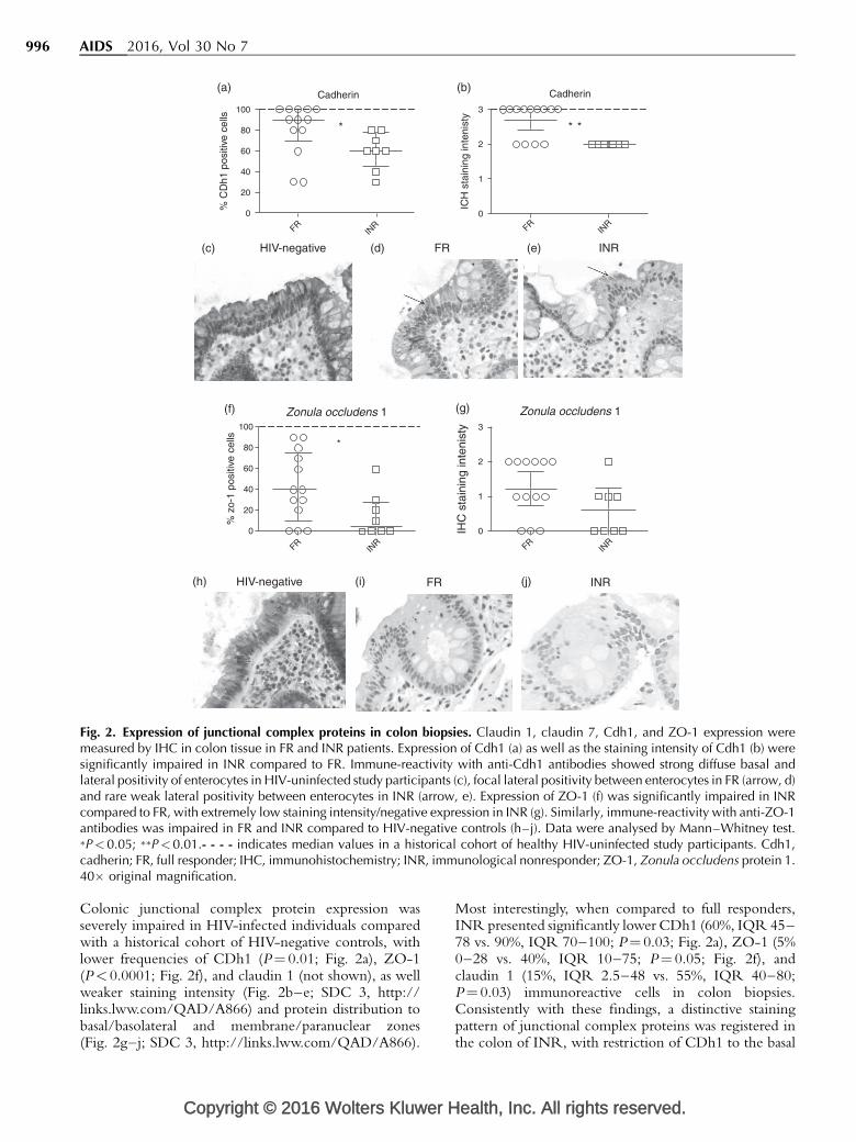
Fig. 2. Expression of junctional complex proteins in colon biopsies. Claudin 1, claudin 7, Cdh1, and ZO-1 expression weremeasured by IHC in colon tissue in FR and INR patients. Expression of Cdh1 (a) as well as the staining intensity of Cdh1 (b) weresignificantly impaired in INR compared to FR. Immune-reactivity with anti-Cdh1 antibodies showed strong diffuse basal andlateral positivity of enterocytes in HIV-uninfected study participants (c), focal lateral positivity between enterocytes in FR (arrow, d)and rare weak lateral positivity between enterocytes in INR (arrow, e). Expression of ZO-1 (f) was significantly impaired in INRcompared to FR, with extremely low staining intensity/negative expression in INR (g). Similarly, immune-reactivity with anti-ZO-1antibodies was impaired in FR and INR compared to HIV-negative controls (h–j). Data were analysed by Mann–Whitney test.�P< 0.05; ��P<0.01.- - - - indicates median values in a historical cohort of healthy HIV-uninfected study participants. Cdh1,cadherin; FR, full responder; IHC, immunohistochemistry; INR, immunological nonresponder; ZO-1, Zonula occludens protein 1.40� original magnification.
Colonic junctional complex protein expression wasseverely impaired in HIV-infected individuals comparedwith a historical cohort of HIV-negative controls, withlower frequencies of CDh1 (P¼ 0.01; Fig. 2a), ZO-1(P< 0.0001; Fig. 2f), and claudin 1 (not shown), as wellweaker staining intensity (Fig. 2b–e; SDC 3, http://links.lww.com/QAD/A866) and protein distribution tobasal/basolateral and membrane/paranuclear zones(Fig. 2g–j; SDC 3, http://links.lww.com/QAD/A866).
Copyright © 2016 Wolters Kluwer H
Most interestingly, when compared to full responders,INR presented significantly lower CDh1 (60%, IQR 45–78 vs. 90%, IQR 70–100; P¼ 0.03; Fig. 2a), ZO-1 (5%0–28 vs. 40%, IQR 10–75; P¼ 0.05; Fig. 2f), andclaudin 1 (15%, IQR 2.5–48 vs. 55%, IQR 40–80;P¼ 0.03) immunoreactive cells in colon biopsies.Consistently with these findings, a distinctive stainingpattern of junctional complex proteins was registered inthe colon of INR, with restriction of CDh1 to the basal
ealth, Inc. All rights reserved.

Gut junctional complexes in treated HIV Tincati et al. 997
(a) (b)
Electron microscopy of gut biopsies and evidenceof microbial translocation
(c)
(d) (e)
Fig. 3. Electron microscopy study of colon biopsies and evidence of microbial translocation in the lamina propria. Colon biopsyfrom a representative INR patient. (a) Arrows point to dilated intercellular space. Colon biopsy from a representative full responder(FR) patient. (b) Arrows point to normal intercellular space. Intestinal (ileocolic junction) biopsy from an INR patient. (c) Arrowpoints to an extracellular bacterium among microvilla, red arrow points to intracellular bacterium, arrow heads point to partlydigested intracytoplasmic bacteria. IHC staining for LPS-core antigen in the colonic lamina propria of a representative FR (d) andINR (e). A and B, scale bar¼500 nm; C, scale bar¼1 mm; D and E, scale bar¼ 50 mm. INR, immunological nonresponder.
surface (Fig. 2e; SDC 3, http://links.lww.com/QAD/A866) and negative expression of ZO-1 (Fig. 2j; SDC 3,http://links.lww.com/QAD/A866). We also found apositive correlation between colonic ZO-1 expressionand current CD4þ T cells (r¼ 0.5, P¼ 0.04), as well as atrend with the change in CD4þ cell count since cARTstart (r¼ 0.4, P¼ 0.07), confirming the IHC findings oflower TJ protein expression in the colon of INR.
Junctional complex protein expression and intensity werealso impaired in ileal tissue of HIV-infected patientscompared with HIV-negative controls (CDh1: P¼ 0.03;ZO-1: P< 0.0001; SDC 4A–J, http://links.lww.com/QAD/A866). Despite no significant differences in thenumber of Cdh1 and ZO-1 immune-reactive cellsbetween INR and full responders (SDC 4A, 4F, http://links.lww.com/QAD/A866), defective distribution, and
Copyright © 2016 Wolters Kluwe
staining intensity of junctional complex proteins wasregistered in the former (SDC 3, SDC 4D, E; 4I, J, http://links.lww.com/QAD/A866), as well as a positive corre-lation between ileal ZO-1 expression and measures ofperipheral CD4þ T-cell reconstitution (current CD4þ
T cells: r¼ 0.6, P¼ 0.01; change in CD4þ cell count sincecART start: r¼ 0.5, P¼ 0.02).
In accordance with the role played by immune activationin hampering CD4þ T-cell recovery on cART, we alsofound a negative correlation between peripheralCD8þCD38þ T-cell frequencies and ZO-1 expressionin both colon (r¼�0.6, P¼ 0.01) and ileum (r¼�0.5,P¼ 0.02).
To confirm intestinal epithelial barrier dysfunction inINR, electron microscopy evaluation of ileum and colon
r Health, Inc. All rights reserved.

998 AIDS 2016, Vol 30 No 7
HIV reservoirs and correlation with measures of peripheral CD4+ T cell reconstitution
10000
1000
100
10
1
0.1
(e) (f)
Tota
l HIV
DNA
(log
cp/1
0^6
cells
)Peripheral CD4+ T cells
Inte
grat
ed H
IV D
NA2 L
TR
r = -0.6P <0.01
r = -0.5P = 0.05
r = -0.5P = 0.05
r = -0.6P = 0.02
0 20 40 60
Current CD4+ T cell count (%)
Current CD4+ T cell count (%)
0.0 0.5 1.0 1.5
CD4+/CD8+ ratio
10000
1000
100
10
1
0.1
Per
iphe
ral 2
LTR
(cp^
6/C
D4+
T c
ells
)
Per
iphe
ral 2
LTR
(cp^
6/C
D4+
T c
ells
) 10000
1000
100
10
1
0.1
10000
1000
100
10
1
0.1
Tota
l HIV
DNA
(log
cp/1
0^6
cells
)
lleum tissue
(b) (c)
Inte
grat
ed H
IV D
NA2 L
TR
(d)
(a)
10000r = -0.5
P = 0.04
1000
100
100 20 40 60
Current CD4+ T cell count (%)
Per
iphe
ral t
otal
HIV
DN
A(c
p/10
^6 C
D4+
T c
ells
)
(i)
10000 r = -0.5P = 0.04
100
100.0 0.5 1.0 1.5
CD4+/CD8+ ratio
Col
on to
tal H
IV D
NA
(cp/
1o^6
cel
ls)
(h)
10000
100
1000
1
10
0 20 40 60
Col
on to
tal H
IV D
NA
(cp/
1o^6
cel
ls)
Current CD4+ T cell count (%)
(g)
10000
100
1000
1
10
0 500 1000 1500
Col
on to
tal H
IV D
NA
(cp/
1o^6
cel
ls)
10000
1000
100
10
1
0.1
Tota
l HIV
DNA
(log
cp/1
0^6
cells
)
Colon tissue
Inte
grat
ed H
IV D
NA2 L
TR
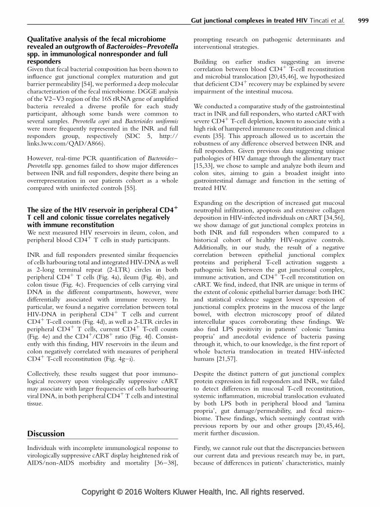
Fig. 4. HIV reservoirs and correlation with measures of peripheral CD4R T-cell reconstitution. Total HIV DNA, integrated HIVDNA, and 2-LTR circles were measured in peripheral CD4þ T cells (a) ileum (b) and colon (c) of INR and FR study participants. Nodifferences in the size of the reservoir were registered between the study groups, nor differences noted between anatomicalcompartments. Peripheral CD4þ T-cell counts negatively correlated with HIV reservoirs in circulating CD4þ T cells (d, e, for totalHIV DNA and 2-LTR circles, respectively). CD4þ/CD8þ ratio in the periphery was also negatively associated with the frequency ofblood CD4þ T cells harboring 2 LTR (f). Similarly, peripheral CD4þ T-cell counts (g, h) and CD4þ/CD8þ ratio (i) negativelycorrelated with total HIV-DNA in the ileum. Data were analyzed by Mann–Whitney test. Spearman’s rank coefficient was used forcorrelations. INR, immunological nonresponder; FR, full responder.
junctional complex was performed. Striking differencesbetween INR and full responders were observed in colontissue: the intercellular space between adjacent cellsdetermined by TJ and AJ was focally wider in INR(Fig. 3a and c). Of note, intracellular bacteria were seen in1 INR (Fig. 3c); furthermore, IHC staining for LPS waspositive in the gut ‘lamina propria’ of both full respondersand INR (Fig. 3d and e), in all indicating microbialtranslocation through a damaged mucosa.
Comparable small intestine permeability/damage, microbial translocation, and systemicinflammation in immunological nonresponderand full respondersHaving shown impairment of gut junctional complexin INR, we assessed intestinal permeability (LAC/
Copyright © 2016 Wolters Kluwer H
MAN) and function (I-FABP). No differencesin urinary LAC/MAN ratio were found betweenstudy groups (INR: 0.03, IQR 0.02–0.07; fullresponders: 0.020, IQR 0.02–0.14; P¼ 0.6), pointingto a similar degree of gastrointestinal permeabilityin INR and full responders with increased paracellularabsorption of high molecular weight molecules [53].Consistently with this finding and with the resultof similar LPS staining in colon tissue, studyparticipants also showed comparable I-FABP (721 pg/ml, IQR 612–994 vs. 858 pg/ml, IQR 660–1157;P¼ 0.4), LPS, endotoxin core antibodies, sCD14, IL-6,and D-dimer levels in plasma (Table 1), suggestinganalogous gut epithelial damage, microbial transloca-tion, and systemic inflammation in INR and fullresponders.
ealth, Inc. All rights reserved.
Gut junctional complexes in treated HIV Tincati et al. 999
Qualitative analysis of the fecal microbiomerevealed an outgrowth of Bacteroides–Prevotellaspp. in immunological nonresponder and fullrespondersGiven that fecal bacterial composition has been shown toinfluence gut junctional complex maturation and gutbarrier permeability [54], we performed a deep molecularcharacterization of the fecal microbiome. DGGE analysisof the V2–V3 region of the 16S rRNA gene of amplifiedbacteria revealed a diverse profile for each studyparticipant, although some bands were common toseveral samples. Prevotella copri and Bacterioides uniformiswere more frequently represented in the INR and fullresponders group, respectively (SDC 5, http://links.lww.com/QAD/A866).
However, real-time PCR quantification of Bacteroides–Prevotella spp. genomes failed to show major differencesbetween INR and full responders, despite there being anoverrepresentation in our patients cohort as a wholecompared with uninfected controls [55].
The size of the HIV reservoir in peripheral CD4R
T cell and colonic tissue correlates negativelywith immune reconstitutionWe next measured HIV reservoirs in ileum, colon, andperipheral blood CD4þ T cells in study participants.
INR and full responders presented similar frequenciesof cells harbouring total and integrated HIV-DNA as wellas 2-long terminal repeat (2-LTR) circles in bothperipheral CD4þ T cells (Fig. 4a), ileum (Fig. 4b), andcolon tissue (Fig. 4c). Frequencies of cells carrying viralDNA in the different compartments, however, weredifferentially associated with immune recovery. Inparticular, we found a negative correlation between totalHIV-DNA in peripheral CD4þ T cells and currentCD4þ T-cell counts (Fig. 4d), as well as 2-LTR circles inperipheral CD4þ T cells, current CD4þ T-cell counts(Fig. 4e) and the CD4þ/CD8þ ratio (Fig. 4f). Consist-ently with this finding, HIV reservoirs in the ileum andcolon negatively correlated with measures of peripheralCD4þ T-cell reconstitution (Fig. 4g–i).
Collectively, these results suggest that poor immuno-logical recovery upon virologically suppressive cARTmay associate with larger frequencies of cells harbouringviral DNA, in both peripheral CD4þT cells and intestinaltissue.
Discussion
Individuals with incomplete immunological response tovirologically suppressive cART display heightened risk ofAIDS/non-AIDS morbidity and mortality [36–38],
Copyright © 2016 Wolters Kluwe
prompting research on pathogenic determinants andinterventional strategies.
Building on earlier studies suggesting an inversecorrelation between blood CD4þ T-cell reconstitutionand microbial translocation [20,45,46], we hypothesizedthat deficient CD4þ recovery may be explained by severeimpairment of the intestinal mucosa.
We conducted a comparative study of the gastrointestinaltract in INR and full responders, who started cARTwithsevere CD4þ T-cell depletion, known to associate with ahigh risk of hampered immune reconstitution and clinicalevents [35]. This approach allowed us to ascertain therobustness of any difference observed between INR andfull responders. Given previous data suggesting uniquepathologies of HIV damage through the alimentary tract[15,33], we chose to sample and analyze both ileum andcolon sites, aiming to gain a broadest insight intogastrointestinal damage and function in the setting oftreated HIV.
Expanding on the description of increased gut mucosalneutrophil infiltration, apoptosis and extensive collagendeposition in HIV-infected individuals on cART [34,56],we show damage of gut junctional complex proteins inboth INR and full responders when compared to ahistorical cohort of healthy HIV-negative controls.Additionally, in our study, the result of a negativecorrelation between epithelial junctional complexproteins and peripheral T-cell activation suggests apathogenic link between the gut junctional complex,immune activation, and CD4þ T-cell reconstitution oncART. We find, indeed, that INR are unique in terms ofthe extent of colonic epithelial barrier damage: both IHCand statistical evidence suggest lowest expression ofjunctional complex proteins in the mucosa of the largebowel, with electron microscopy proof of dilatedintercellular spaces corroborating these findings. Wealso find LPS positivity in patients’ colonic ‘laminapropria’ and anecdotal evidence of bacteria passingthrough it, which, to our knowledge, is the first report ofwhole bacteria translocation in treated HIV-infectedhumans [21,57].
Despite the distinct pattern of gut junctional complexprotein expression in full responders and INR, we failedto detect differences in mucosal T-cell reconstitution,systemic inflammation, microbial translocation evaluatedby both LPS both in peripheral blood and ‘laminapropria’, gut damage/permeability, and fecal micro-biome. These findings, which seemingly contrast withprevious reports by our and other groups [20,45,46],merit further discussion.
Firstly, we cannot rule out that the discrepancies betweenour current data and previous research may be, in part,because of differences in patients’ characteristics, mainly
r Health, Inc. All rights reserved.

1000 AIDS 2016, Vol 30 No 7
CD4þ nadir, which appears to be lower in literaturestudies [34,45] compared to the present report.Additionally, a proportion of INR in our study werereceiving antibiotics, which may have contributed tocontaining microbial translocation in these patients.
Although similar CD4þ levels are reported in the gut ofINR and full responders, we find higher colonic Ki67 inthe former; this negatively correlates with CD4þ cellcounts in the same district. Based on animal datademonstrating ongoing damage and subsequent repair inincreased gut epithelial proliferation, measured by Ki67[21], our findings are suggestive of compensatoryregenerative activity in the colon of INR, resulting inequal CD4þ cell counts in the two study groups. Thisfinding is in contrast with what described by Somsouket al. [34], who show reduced Ki67 in the colon of acohort of INR. Although we cannot exclude technicaldifferences in Ki67 staining and interpretation, as well asdifferent Ki67 expression according to the gut anatomicalsite, we do believe that a likely interpretation of suchdiscrepant findings might reside in differences in thepatients populations studied: in Somsouk’s paper INRfeature very low CD4þ nadir, of 21 (7–56) cells/ml,inferior than our values of 68 (36–120) CD4þ cells/ml.Given that CD4þ nadir has been consistently proven tocapture the pretherapy HIV-mediated damage, and tostrongly dictate immune reconstitution [58], it is possiblethat Somsouk et al. probed a very drained and exhaustedpopulation with no further potential for compensatoryepithelial proliferation. At the other end, Somsoukdescribe quite high median colonic Ki67 stain in their fullresponders as compared with ours (about 50 vs. 25%),again to possibly reflect patients with a better preservedmucosal immunity.
Differently to data by Somsouk et al. [34] we also showcomparable I-FABP and urinary LAC/MAN in INR andfull responders, despite marked differences in gutjunctional complex protein expression between the studygroups. In this respect, INR display severe damage ofboth TJ (claudin-1, ZO-1) and AJ (Cdh1) in the colonand impairment of only Cdh1 in the ileum. Thesefindings explain the reportedly similar gut damage/permeability markers in INR and full responders, giventhat they mirror small bowel function. Furthermore, theyexpand a recent observation of the reduced proximal-to-distal expression of colonic TJ, yet not AJ, in virallysuppressed HIV-infected individuals [33], by showing thatdifferential defects of the mucosal barrier may exist atdifferent gut sites according to the degree of immunerecovery on cART. In this respect, while not designed tospecifically dissect the reasons behind anatomical differ-ences in gut junctional complex expression, our data doconfirm that a unique HIV-driven pathology throughoutthe alimentary tract persist on cART [15,33]. Indeed,concordant with data in untreated HIV, we hereby showless severe junctional complex damage in patients’ ileum,
Copyright © 2016 Wolters Kluwer H
that is limited to AJ (as opposed to both TJ and AJ proteinsdownregulation in the colon), entirely in line with themodel of epithelial apoptosis as predominant mechanismof small bowel damage as opposed to paracellularpermeability in the large intestine [33].
We also analyzed fecal microbiome, which, on the onehand, promotes TJ protein expression [54] and on theother, is shaped by the gut barrier structure [59–61].Despite an overall outgrowth of Bacteroides–Prevotella[55], INR and full responders displayed similar fecalmicrobial composition, suggesting analogous impairmentof the gut microflora [62–65].
Finally, in the face of our starting hypothesis, our findingsmay be also consistent with the alternative possibility thatdespite reduced junctional complex protein expression,INR are able to preserve mucosal integrity/functionsimilar to that observed in full responders. Investigation ofgenes and markers regulating gut permeability andimmunity is thus needed to shed light on the mechanismsby which the junctional complex affects peripheral CD4þ
T-cell reconstitution. Indeed, by showing similar levels ofbacterial translocation in both peripheral blood and colon‘lamina propria’ of INR and full responders despitestriking differences in gut junctional complex protein,our data seem to propose a model whereby cART startedin a context of severe CD4þ T-cell depletion, while notrestoring gut epithelium adhesion molecules, maynonetheless recuperate gut barrier function, thereforecontaining systemic microbial translocation. As corollary,our data leads to the supposition that bacterial transloca-tion in treated HIV may have a limited impact on clinicaloutcome, as also indicated in recent reports from largerclinical cohorts, which failed to capture an independentassociation between markers of microbial translocationand clinical events in treated infection [47,66,67].Whether or not HIV-driven gut barrier anatomy andfunction might be reversible upon antiviral treatment andhow such reversibility might associate with starting CD4þ
strata should be tested in ad hoc-designed studies.
Based on earlier research showing higher gut versusperipheral blood HIV-DNA in cART-treated patients[26–28], we asked whether greater HIV reservoirsassociate with gastrointestinal damage and poor immunerecovery. No differences in gut and circulating HIVreservoirs were observed between INR and fullresponders, which may be explained by the reportedlow CD4þ nadir [68]. However, HIV reservoirs in bothperipheral blood and gut negatively correlate withmarkers of CD4þ T-cell reconstitution in periphery,suggesting that poor immune recovery on cART may beassociated with greater HIV reservoirs. Our result of aninverse correlation between HIV reservoirs and CD4þ/CD8þ ratio, which has been shown to associate withimmune activation/senescence and disease progression[69], further highlights the possible link between HIV
ealth, Inc. All rights reserved.

Gut junctional complexes in treated HIV Tincati et al. 1001
reservoir, persistent immunological alterations, andclinical risk in INR [48].
Our study rests on a few caveats, including our inability tomeasure HIV reservoirs in sorted gut cells and to correctfor CD4þ frequency in gastrointestinal tissue, thusimpeding the comparison of HIV reservoirs amongcompartments.
Another substantial abnormality described in thesecondary lymphoid organs and gastrointestinal mucosaof HIV-infected patients are profibrotic changes, that arenot fully reversed by cART, mainly if started late in theinfection [56,70]. Interestingly, tissue fibrosis has alsobeen implicated in failed immune reconstitution follow-ing effective cART [71]. Although our study was notspecifically designed to investigate fibrosis in the gut ofINR and full responders, it is highly likely that ourpatients display wide tissue fibrosis, in turn furthercontributing to gastrointestinal damage.
The findings outlined above shed light, nonetheless, onthe possible reasons behind the ineffectiveness oftreatments tested to sustain CD4þ T-cell recovery inINR. A key implication of our study is, indeed, thatunless the gastrointestinal tract is specifically targetedvis-a-vis HIV burden and mucosal repair, interventions areunlikely to be successful. cART intensification[35,39,40,42,43] may result in the inefficient concen-tration of antiretrovirals in the gut [72]; IL-7 immune-adjuvant therapy may contribute to replenishment of theHIV reservoir rather than its reduction [41,73]; further,rifaximin may not result in the effective control ofmicrobial translocation because of the lasting damage ofthe gastrointestinal tract [44].
In conclusion, we show that INR feature severe gutjunctional complex damage and increased HIV reservoirin both circulating CD4þ and gastrointestinal tissue, thusproviding new directions for therapeutic interventions inthis setting.
Acknowledgements
The authors would like to acknowledge Giulia Moraceand Francesca Ghilardi for their help and advice. Theauthors would like to thank Carlotta Tincati fordiscussion, all patients who took part in the study, staffmembers of the Clinic of Infectious Diseases, Pathologyand Endoscopy Units of the San Paolo UniversityHospital in Milan, Italy.
The study was supported by the Italian Ministry ofHealth, Regione Lombardia, grant ‘Giovani Ricercatori’(number GR-2009-1592029).
Copyright © 2016 Wolters Kluwe
Author contributions: C.T. and E.M. conducted thecellular biology experiments; P.B, F.S., D.T., G.B., andS.R. performed the pathology experiments; E.B. andM.L.C conducted the microbiological analyses and A.B.was responsible for HPLC data. E.M. and N.C.performed the studies on HIV reservoirs. G.A andA.d’M. recruited study participants and B.M. performedthe endoscopy examinations. C.T. and G.M. designed thestudy and conducted the data analyses. All authorscontributed to the writing of the manuscript. G.M.supervised the project.
Conflicts of interestThere are no conflicts of interest.
Presented in part at CROI 2014, 3–6 March 2014, Boston(Massachusetts), US – abstract # 231.
References
1. Veazey RS, DeMaria M, Chalifoux LV, Shvetz DE, Pauley DR,Knight HL, et al. Gastrointestinal tract as a major site of CD4RT cell depletion and viral replication in SIV infection. Science1998; 280:427–431.
2. Fackler OT, Schafer M, Schmidt W, Zippel T, Heise W, Schnei-der T, et al. HIV-1 p24 but not proviral load is increased in theintestinal mucosa compared with the peripheral blood in HIV-infected patients. AIDS 1998; 12:139–146.
3. Mattapallil JJ, Douek DC, Hill B, Nishimura Y, Martin M,Roederer M. Massive infection and loss of memory CD4R Tcells in multiple tissues during acute SIV infection. Nature2005; 434:1093–1097.
4. Mehandru S, Poles MA, Tenner-Racz K, Horowitz A, Hurley A,Hogan C, et al. Primary HIV-1 infection is associated withpreferential depletion of CD4R T lymphocytes from effectorsites in the gastrointestinal tract. J Exp Med 2004; 200:761–770.
5. Mehandru S, Poles MA, Tenner-Racz K, Jean-Pierre P, ManuelliV, Lopez P, et al. Lack of mucosal immune reconstitution duringprolonged treatment of acute and early HIV-1 infection. PLoSMed 2006; 3:e484.
6. Mehandru S, Poles MA, Tenner-Racz K, Manuelli V, Jean-PierreP, Lopez P, et al. Mechanisms of gastrointestinal CD4R T-celldepletion during acute and early human immunodeficiencyvirus type 1 infection. J Virol 2007; 81:599–612.
7. Ciccone EJ, Read SW, Mannon PJ, Yao MD, Hodge JN, Dewar R,et al. Cycling of gut mucosal CD4R T cells decreases afterprolonged antiretroviral therapy and is associated with plasmaLPS levels. Mucosal Immunol 2010; 3:172–181.
8. Gordon SN, Cervasi B, Odorizzi P, Silverman R, Aberra F,Ginsberg G, et al. Disruption of intestinal CD4R T cell home-ostasis is a key marker of systemic CD4R T cell activation inHIV-infected individuals. J Immunol 2010; 185:5169–5179.
9. Schneider T, Jahn HU, Schmidt W, Riecken EO, Zeitz M, UllrichR. Loss of CD4 T lymphocytes in patients infected with humanimmunodeficiency virus type 1 is more pronounced in theduodenal mucosa than in the peripheral blood. Berlin Diar-rhea/Wasting Syndrome Study Group. Gut 1995; 37:524–529.
10. Brenchley JM, Schacker TW, Ruff LE, Price DA, Taylor JH,Beilman GJ, et al. CD4R T cell depletion during all stages ofHIV disease occurs predominantly in the gastrointestinal tract.J Exp Med 2004; 200:749–759.
11. Guadalupe M, Reay E, Sankaran S, Prindiville T, Flamm J,McNeil A, et al. Severe CD4R T-cell depletion in gut lymphoidtissue during primary human immunodeficiency virus type1 infection and substantial delay in restoration followinghighly active antiretroviral therapy. J Virol 2003; 77:11708–11717.
r Health, Inc. All rights reserved.

1002 AIDS 2016, Vol 30 No 7
12. Kotler DP, Gaetz HP, Lange M, Klein EB, Holt PR. Enteropathyassociated with the acquired immunodeficiency syndrome.Ann Intern Med 1984; 101:421–428.
13. Lim SG, Menzies IS, Lee CA, Johnson MA, Pounder RE.Intestinal permeability and function in patients infected withhuman immunodeficiency virus. A comparison with coeliacdisease. Scand J Gastroenterol 1993; 28:573–580.
14. Keating J, Bjarnason I, Somasundaram S, Macpherson A, FrancisN, Price AB, et al. Intestinal absorptive capacity, intestinalpermeability and jejunal histology in HIV and their relation todiarrhoea. Gut 1995; 37:623–629.
15. Epple HJ, Schneider T, Troeger H, Kunkel D, Allers K, Moos V,et al. Impairment of the intestinal barrier is evident in untreatedbut absent in suppressively treated HIV-infected patients. Gut2009; 58:220–227.
16. Nazli A, Chan O, Dobson-Belaire WN, Ouellet M, TremblayMJ, Gray-Owen SD, et al. Exposure to HIV-1 directly impairsmucosal epithelial barrier integrity allowing microbial trans-location. PLoS Pathog 2010; 6:e1000852.
17. Bjarnason I, Sharpstone DR, Francis N, Marker A, Taylor C,Barrett M, et al. Intestinal inflammation, ileal structure andfunction in HIV. AIDS 1996; 10:1385–1391.
18. Stockmann M, Fromm M, Schmitz H, Schmidt W, Riecken EO,Schulzke JD. Duodenal biopsies of HIV-infected patients withdiarrhoea exhibit epithelial barrier defects but no active secre-tion. AIDS 1998; 12:43–51.
19. Stockmann M, Schmitz H, Fromm M, Schmidt W, Pauli G,Scholz P, et al. Mechanisms of epithelial barrier impairment inHIV infection. Ann N Y Acad Sci 2000; 915:293–303.
20. Brenchley JM, Price DA, Schacker TW, Asher TE, Silvestri G,Rao S, et al. Microbial translocation is a cause of systemicimmune activation in chronic HIV infection. Nat Med 2006;12:1365–1371.
21. Estes JD, Harris LD, Klatt NR, Tabb B, Pittaluga S, Paiardini M,et al. Damaged intestinal epithelial integrity linked to microbialtranslocation in pathogenic simian immunodeficiency virusinfections. PLoS Pathog 2010; 6:e1001052.
22. Liu Z, Hultin LE, Cumberland WG, Hultin P, Schmid I, Matud JL,et al. Elevated relative fluorescence intensity of CD38 antigenexpression on CD8R T cells is a marker of poor prognosis inHIV infection: results of 6 years of follow-up. Cytometry 1996;26:1–7.
23. Giorgi JV, Hultin LE, McKeating JA, Johnson TD, Owens B,Jacobson LP, et al. Shorter survival in advanced humanimmunodeficiency virus type 1 infection is more closely asso-ciated with T lymphocyte activation than with plasma virusburden or virus chemokine coreceptor usage. J Infect Dis 1999;179:859–870.
24. Deeks SG, Kitchen CM, Liu L, Guo H, Gascon R, Narvaez AB,et al. Immune activation set point during early HIV infectionpredicts subsequent CD4R T-cell changes independent of viralload. Blood 2004; 104:942–947.
25. Hazenberg MD, Otto SA, van Benthem BH, Roos MT, CoutinhoRA, Lange JM, et al. Persistent immune activation in HIV-1infection is associated with progression to AIDS. AIDS 2003;17:1881–1888.
26. Chun TW, Nickle DC, Justement JS, Meyers JH, Roby G,Hallahan CW, et al. Persistence of HIV in gut-associatedlymphoid tissue despite long-term antiretroviral therapy. JInfect Dis 2008; 197:714–720.
27. Yukl SA, Gianella S, Sinclair E, Epling L, Li Q, Duan L, et al.Differences in HIV burden and immune activation within thegut of HIV-positive patients receiving suppressive antiretrovir-al therapy. J Infect Dis 2010; 202:1553–1561.
28. Yukl SA, Shergill AK, Ho T, Killian M, Girling V, Epling L, et al.The distribution of HIV DNA and RNA in cell subsetsdiffers in gut and blood of HIV-positive patients on ART:implications for viral persistence. J Infect Dis 2013;208:1212–1220.
29. Macal M, Sankaran S, Chun TW, Reay E, Flamm J, Prindiville TJ,et al. Effective CD4R T-cell restoration in gut-associated lym-phoid tissue of HIV-infected patients is associated with en-hanced Th17 cells and polyfunctional HIV-specific T-cellresponses. Mucosal Immunol 2008; 1:475–488.
30. Kotler DP, Shimada T, Snow G, Winson G, Chen W, Zhao M,et al. Effect of combination antiretroviral therapy upon rectalmucosal HIV RNA burden and mononuclear cell apoptosis.AIDS 1998; 12:597–604.
Copyright © 2016 Wolters Kluwer H
31. Sheth PM, Chege D, Shin LY, Huibner S, Yue FY, Loutfy M, et al.Immune reconstitution in the sigmoid colon after long-termHIV therapy. Mucosal Immunol 2008; 1:382–388.
32. Chege D, Kovacs C, la Porte C, Ostrowski M, Raboud J, Su D,et al. Effect of raltegravir intensification on HIV proviral DNAin the blood and gut mucosa of men on long-term therapy: arandomized controlled trial. AIDS 2012; 26:167–174.
33. Chung CY, Alden SL, Funderburg NT, Fu P, Levine AD. Pro-gressive proximal-to-distal reduction in expression of the tightjunction complex in colonic epithelium of virally-suppressedHIVR individuals. PLoS Pathog 2014; 10:e1004198.
34. Somsouk M, Estes JD, Deleage C, Dunham RM, Albright R,Inadomi JM, et al. Gut epithelial barrier and systemicinflammation during chronic HIV infection. AIDS 2015;29:43–51.
35. Gazzola L, Tincati C, Bellistrı GM, Monforte A, Marchetti G.The absence of CD4R T cell count recovery despite receipt ofvirologically suppressive highly active antiretroviral therapy:clinical risk, immunological gaps, and therapeutic options. ClinInfect Dis 2009; 48:328–337.
36. Lapadula G, Cozzi-Lepri A, Marchetti G, Antinori A, ChioderaA, Nicastri E, et al. Risk of clinical progression among patientswith immunological nonresponse despite virological suppres-sion after combination antiretroviral treatment. AIDS 2013;27:769–779.
37. Engsig FN, Zangerle R, Katsarou O, Dabis F, Reiss P, Gill J, et al.Long-term mortality in HIV-positive individuals virally sup-pressed for >3 years with incomplete CD4 recovery. Clin InfectDis 2014; 58:1312–1321.
38. Zoufaly A, Cozzi-Lepri A, Reekie J, Kirk O, Lundgren J, Reiss P,et al. Immuno-virological discordance and the risk of non-AIDSand AIDS events in a large observational cohort of HIV-pa-tients in Europe. PLoS One 2014; 9:e87160.
39. Rusconi S, Vitiello P, Adorni F, Colella E, Foca E, Capetti A, et al.Maraviroc as intensification strategy in HIV-1 positive patientswith deficient immunological response: an Italian randomizedclinical trial. PLoS One 2013; 8:e80157.
40. Hunt PW, Shulman NS, Hayes TL, Dahl V, Somsouk M, Funder-burg NT, et al. The immunologic effects of maraviroc intensi-fication in treated HIV-infected individuals with incompleteCD4R T-cell recovery: a randomized trial. Blood 2013; ;121:4635–4646.
41. Levy Y, Sereti I, Tambussi G, Routy JP, Lelievre JD, Delfraissy JF,et al. Effects of recombinant human interleukin 7 on T-cellrecovery and thymic output in HIV-infected patients receivingantiretroviral therapy: results of a phase I/IIa randomized,placebo-controlled, multicenter study. Clin Infect Dis 2012;55:291–300.
42. Massanella M, Negredo E, Puig J, Puertas MC, Buzon MJ, Perez-Alvarez N, et al. Raltegravir intensification shows differingeffects on CD8 and CD4 T cells in HIV-infected HAART-suppressed individuals with poor CD4 T-cell recovery. AIDS2012; 26:2285–2293.
43. Negredo E, Massanella M, Puertas MC, Buzon MJ, Puig J, Perez-Alvarez N, et al. Early but limited effects of raltegravir intensi-fication on CD4 T cell reconstitution in HIV-infected patientswith an immunodiscordant response to antiretroviral therapy. JAntimicrob Chemother 2013; 68:2358–2362.
44. Tenorio AR, Chan ES, Bosch RJ, Macatangay BJ, Read SW,Yesmin S, et al. Rifaximin Has a Marginal Impact on MicrobialTranslocation, T-cell Activation and Inflammation in HIV-Positive Immune Nonresponders to Antiretroviral Therapy -ACTG A5286. J Infect Dis 2015; 211:780–790.
45. Marchetti G, Bellistrı GM, Borghi E, Tincati C, Ferramosca S, LaFrancesca M, et al. Microbial translocation is associated withsustained failure in CD4R T-cell reconstitution in HIV-infectedpatients on long-term highly active antiretroviral therapy.AIDS 2008; 22:2035–2038.
46. Jiang W, Lederman MM, Hunt P, Sieg SF, Haley K, Rodriguez B,et al. Plasma levels of bacterial DNA correlate with immuneactivation and the magnitude of immune restoration in personswith antiretroviral-treated HIV infection. J Infect Dis 2009;199:1177–1185.
47. Hunt PW, Martin JN, Sinclair E, Bredt B, Hagos E, Lampiris H,et al. T cell activation is associated with lower CD4R T cellgains in human immunodeficiency virus-infected patients withsustained viral suppression during antiretroviral therapy.J Infect Dis 2003; 187:1534–1543.
ealth, Inc. All rights reserved.

Gut junctional complexes in treated HIV Tincati et al. 1003
48. Marchetti G, Gori A, Casabianca A, Magnani M, Franzetti F,Clerici M, et al. Comparative analysis of T-cell turnover andhomeostatic parameters in HIV-infected patients with discor-dant immune-virological responses to HAART. AIDS 2006;20:1727–1736.
49. Kasapovic A, Boesecke C, Schwarze-Zander C, Anadol E, VogelM, Hippe V, et al. Screening colonoscopy in HIV-infectedpatients: high rates of mucosal abnormalities in a GermanHIV-infected cohort. HIV Med 2014; 15:175–181.
50. Walter J, Tannock GW, Tilsala-Timisjarvi A, Rodtong S, LoachDM, Munro K, et al. Detection and identification of gastro-intestinal Lactobacillus species by using denaturing gradientgel electrophoresis and species-specific PCR primers. ApplEnviron Microbiol 2000; 66:297–303.
51. Patrone V, Ferrari S, Lizier M, Lucchini F, Minuti A, Tondelli B,et al. Short-term modifications in the distal gut microbiota ofweaning mice induced by a high-fat diet. Microbiology 2012;158 (Pt 4):983–992.
52. Vandergeeten C, Fromentin R, Merlini E, Lawani MB, DaFon-seca S, Bakeman W, et al. Cross-clade ultrasensitive PCR-basedassays to measure HIV persistence in large-cohort studies.J Virol 2014; 88:12385–12396.
53. Cobden I, Dickinson RJ, Rothwell J, Axon AT. Intestinal perme-ability assessed by excretion ratios of two molecules: results incoeliac disease. BMJ 1978; 2:1060.
54. Patel RM, Myers LS, Kurundkar AR, Maheshwari A, Nusrat A,Lin PW. Probiotic bacteria induce maturation of intestinalclaudin 3 expression and barrier function. Am J Pathol 2012;180:626–635.
55. Rinttila T, Kassinen A, Malinen E, Krogius L, Palva A. Devel-opment of an extensive set of 16S rDNA-targeted primers forquantification of pathogenic and indigenous bacteria in faecalsamples by real-time PCR. J Appl Microbiol 2004; 97:1166–1177.
56. Estes JD, Haase AT, Schacker TW. The role of collagen deposi-tion in depleting CD4R T cells and limiting reconstitution inHIV-1 and SIV infections through damage to the secondarylymphoid organ niche. Semin Immunol 2008; 20:181–186.
57. Smith AJ, Schacker TW, Reilly CS, Haase AT. A role forsyndecan-1 and claudin-2 in microbial translocation duringHIV-1 infection. J Acquir Immune Defic Syndr 2010; 55:306–315.
58. Moore RD, Keruly JC. CD4R cell count 6 years after com-mencement of highly active antiretroviral therapy in personswith sustained virologic suppression. Clin Infect Dis 2007;44:441–446.
59. George MD, Sankaran S, Reay E, Gelli AC, Dandekar S. High-throughput gene expression profiling indicates dysregulationof intestinal cell cycle mediators and growth factors duringprimary simian immunodeficiency virus infection. Virology2003; 312:84–94.
60. Sankaran S, George MD, Reay E, Guadalupe M, Flamm J,Prindiville T, et al. Rapid onset of intestinal epithelial barrierdysfunction in primary human immunodeficiency virus infec-tion is driven by an imbalance between immune response andmucosal repair and regeneration. J Virol 2008; 82:538–545.
Copyright © 2016 Wolters Kluwe
61. Sheng YH, Lourie R, Linden SK, Jeffery PL, Roche D, Tran TV,et al. The MUC13 cell-surface mucin protects against intestinalinflammation by inhibiting epithelial cell apoptosis. Gut 2011;60:1661–1670.
62. Mutlu EA, Keshavarzian A, Losurdo J, Swanson G, Siewe B,Forsyth C, et al. A compositional look at the human gastro-intestinal microbiome and immune activation parameters inHIV infected subjects. PLoS Pathog 2014; 10:e1003829.
63. Gori A, Tincati C, Rizzardini G, Torti C, Quirino T, Haarman M,et al. Early impairment of gut function and gut flora supportinga role for alteration of gastrointestinal mucosa in humanimmunodeficiency virus pathogenesis. J Clin Microbiol 2008;46:757–758.
64. Perez-Santiago J, Gianella S, Massanella M, Spina CA, KarrisMY, Var SR, et al. Gut Lactobacillales are associated withhigher CD4 and less microbial translocation during HIV in-fection. AIDS 2013; 27:1921–1931.
65. Vujkovic-Cvijin I, Dunham RM, Iwai S, Maher MC, Albright RG,Broadhurst MJ, et al. Dysbiosis of the gut microbiota is asso-ciated with HIV disease progression and tryptophan catabo-lism. Sci Transl Med 2013; 5:193ra91.
66. Hunt PW, Sinclair E, Rodriguez B, Shive C, Clagett B, Funder-burg N, et al. Gut epithelial barrier dysfunction and innateimmune activation predict mortality in treated HIV infection.J Infect Dis 2014; 210:1228–1238.
67. Tenorio AR, Zheng Y, Bosch RJ, Krishnan S, Rodriguez B, HuntPW, et al. Soluble markers of inflammation and coagulation butnot T-cell activation predict non-AIDS-defining morbid eventsduring suppressive antiretroviral treatment. J Infect Dis 2014;210:1248–1259.
68. Boulassel MR, Chomont N, Pai NP, Gilmore N, Sekaly RP,Routy JP. CD4 T cell nadir independently predicts the magni-tude of the HIV reservoir after prolonged suppressive anti-retroviral therapy. J Clin Virol 2012; 53:29–32.
69. Serrano-Villar S, Sainz T, Lee SA, Hunt PW, Sinclair E, ShacklettBL, et al. HIV-infected individuals with low CD4/CD8 ratiodespite effective antiretroviral therapy exhibit altered T cellsubsets, heightened CD8R T cell activation, and increased riskof non-AIDS morbidity and mortality. PLoS Pathog 2014;10:e1004078.
70. Zeng M, Southern PJ, Reilly CS, Beilman GJ, Chipman JG,Schacker TW, et al. Lymphoid tissue damage in HIV-1 infectiondepletes naıve T cells and limits T cell reconstitution afterantiretroviral therapy. PLoS Pathog 2012; 8:e1002437.
71. Asmuth DM, Pinchuk IV, Wu J, Vargas G, Chen X, Mann S, et al.Role of intestinal myofibroblasts in HIV-associated intestinalcollagen deposition and immune reconstitution following com-bination antiretroviral therapy. AIDS 2015; 29:877–888.
72. Fletcher CV, Staskus K, Wietgrefe SW, Rothenberger M, ReillyC, Chipman JG, et al. Persistent HIV-1 replication is associatedwith lower antiretroviral drug concentrations in lymphatictissues. Proc Natl Acad Sci U S A 2014; 111:2307–2312.
73. Vandergeeten C, Fromentin R, DaFonseca S, Lawani MB, SeretiI, Lederman MM, et al. Interleukin-7 promotes HIV persistenceduring antiretroviral therapy. Blood 2013; 121:4321–4329.
r Health, Inc. All rights reserved.