lec32 f08 handout
TRANSCRIPT

8/3/2019 Lec32 F08 Handout
http://slidepdf.com/reader/full/lec32-f08-handout 1/11
Bioc 460 - Dr. Miesfeld Fall 2008
1 of 11 pages
Figure 1.
Lecture 32 - Carbohydrate Metabolism
Key Concepts- Pentose Phosphate Pathway
Enzymatic reactions in the oxidative phase
Enzymatic reactions in the nonoxidative phaseGlucose-6P dehydrogenase deficiency in humans
- GluconeogenesisThree steps in glycolysis are bypassed by gluconeogenesisReciprocal control of glycolysis and gluconeogenesisThe Cori Cycle provides glucose to muscle cells during exercise
Key Questions about the Pentose Phosphate Pathway and Gluconeogenesis:What is the biochemical basis for favism and how is it related to malarial resistance? How does fructose-2,6-bisphosphate control flux through gluconeogenesis and glycolysis?
Biochemical Applications:Monitoring blood glucose levels throughout the day is critical to diabetics whoneed insulin injections. Glucose monitoring devices are based on an assay usingthe enzyme glucose oxidase which produces gluconate and hydrogen peroxide(H2O2) from glucose. The level of H2O2 in the sample is detected by an indicator dye that is oxidized in a reaction catalyzed by peroxidase.
In lectures 22-32, we examined the regulation of energy conversion pathways in cells and sawhow the absorption of sunlight by photosynthetic organisms was used as an energy source to drivecarbohydrate synthesis in the form of starch and sucrose. We now begin the second half of our
journey through the metabolic forest by exploring biochemical pathways in the cell that control the
synthesis and degradation of a variety of biomolecules. The energy required for biomolecular synthesis comes from phosphoryl transfer energy available from ATP, and from a redox energyprovided by the phosphorylated form of NADH called NADPH. Degradation of biomolecules is animportant process in cells because it not only scavengesbuilding blocks for biosynthesis, but it is a form of metabolicregulation by controlling the steady-state level of activebiomolecules.
The three primary pathways in anabolic carbohydratemetabolism in non-photosynthetic organisms are thepentose phosphate pathway, gluconeogenesis and
glycogen metabolism. We describe the pentosephosphate pathway and gluconeogenesis here in lecture 33,and then discuss glycogen metabolism in lectures 34(enzyme reactions) and 35 (regulation of glycogenmetabolism). The major sources of carbon ingluconeogenesis are amino acids and glycerol in animals,and glyceraldehyde-3-phosphate (GAP) in plants.Metabolism of ribose sugars in the pentose phosphatepathway is used to generate NADPH and to provide thecarbohydrate component of nucleotides (figure 1).

8/3/2019 Lec32 F08 Handout
http://slidepdf.com/reader/full/lec32-f08-handout 2/11
Bioc 460 - Dr. Miesfeld Fall 2008
2 of 11 pages
Figure 2.
Pentose Phosphate Pathway The pentose phosphate pathway takes place entirely within the cytoplasm and is also known asthe hexose monophosphate shunt or phosphogluconate pathway. The most important function of the pentose phosphate pathway is to reduce two molecules of NADP+ to NADPH for each glucose-6-phosphate (glucose-6P) that is oxidatively decarboxylated to ribulose-5-phosphate (ribulose-5P).The pentose phosphate pathway is also responsible for producing ribose-5-phosphate (ribose-
5P) from glucose-6P. NADPH (nicotinamide adenine dinucleotide phosphate) is structurallyidentical to NADH with the exception of a phosphate group on the C-2 carbon of the sugar moietyof the adenine nucleotide. Another difference between the conjugate redox pairs of NAD+/NADHand NADP+/NADPH is that NAD+ functions as the primary oxidant in the cell (accepts electrons),whereas, NADPH is the primary reductant in the cell (donates electrons). The distinct roles of these two related coenzymes can be seen in the very different steady-state levels of the conjugateredox pairs. In liver cells, the [NAD+]/[NADH] ratio is close to 1,000, whereas, the[NADP+]/[NADPH] ratio is 0.01, i.e., NAD+ and NADPH are the most abundant redox species.
The pentose phosphate pathway can be divided into two phases, the oxidative phase which generates NADPH, and the nonoxidative phase, which interconverts C3, C4, C5, C6 and C7 sugar phosphates using many of the same "carbon shuffle" reactions we saw in the Calvin cycle.
Figure 2 provides an overview of the pentosephosphate pathway illustrating the function of theoxidative and nonoxidative phases in producingNADPH and ribose-5P, respectively. Flux throughthe oxidative and nonoxidative phases of thepathway is tightly regulated in response to energyneeds of the cell, the NADP+/NADPH ratio, andrequirements for nucleotide and coenzymebiosynthesis (see #1, #2 and #3 in figure 2). For example, when NADPH is needed, ribulose-5P isconverted back into glucose-6P to maintain flux
through the pathway (#1), however, if ATP andNADPH are needed (which would be the case for most anabolic pathways), then some of theribulose-5P is used to synthesis hexosephosphates for glycolysis (#2). Finally, if the cellneeds to increase the rate of nucleotide andcoenzyme biosynthesis, then most of the ribulose-5P is shunted toward ribose-5P synthesis (#3).
1. What does the pentose phosphate pathway accomplish for the cell?• The oxidative phase generates NADPH which is required for many biosynthetic pathways and for
detoxification of reactive oxygen species.• The nonoxidative phase interconverts C3, C4, C5, C6 and C7 monosaccharides to produce ribose-5P for nucleotide synthesis, and also to regenerate glucose-6P to maintain NADPHproduction by the oxidative phase.
2. What is the overall net reaction of the pentose phosphate pathway when it is utilized togenerate the maximum amount of NADPH?
6 Glucose-6P + 12 NADP+ + 12 H2O --> 5 Glucose-6P + 12 NADPH + 12 H+ + 6 CO2
8/3/2019 Lec32 F08 Handout
http://slidepdf.com/reader/full/lec32-f08-handout 3/11
Bioc 460 - Dr. Miesfeld Fall 2008
3 of 11 pages
Figure 3.
Figure 4.
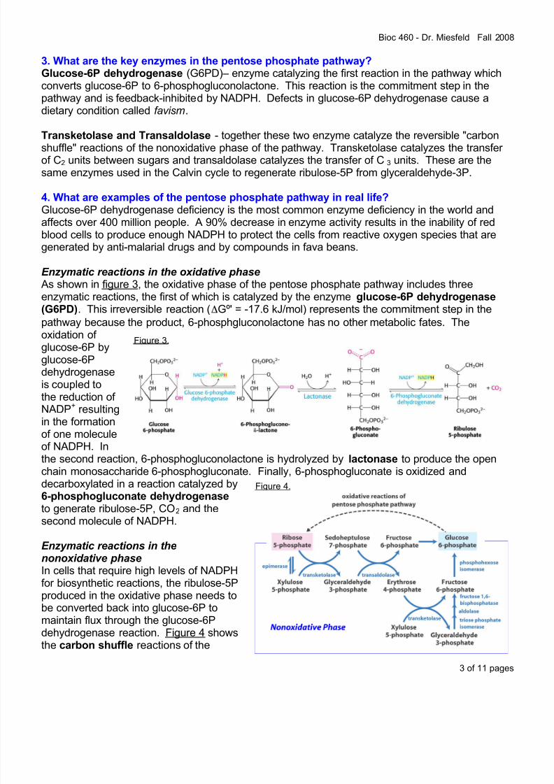
3. What are the key enzymes in the pentose phosphate pathway? Glucose-6P dehydrogenase (G6PD)– enzyme catalyzing the first reaction in the pathway whichconverts glucose-6P to 6-phosphogluconolactone. This reaction is the commitment step in thepathway and is feedback-inhibited by NADPH. Defects in glucose-6P dehydrogenase cause adietary condition called favism.
Transketolase and Transaldolase - together these two enzyme catalyze the reversible "carbonshuffle" reactions of the nonoxidative phase of the pathway. Transketolase catalyzes the transfer of C2 units between sugars and transaldolase catalyzes the transfer of C3 units. These are thesame enzymes used in the Calvin cycle to regenerate ribulose-5P from glyceraldehyde-3P.
4. What are examples of the pentose phosphate pathway in real life? Glucose-6P dehydrogenase deficiency is the most common enzyme deficiency in the world andaffects over 400 million people. A 90% decrease in enzyme activity results in the inability of redblood cells to produce enough NADPH to protect the cells from reactive oxygen species that aregenerated by anti-malarial drugs and by compounds in fava beans.
Enzymatic reactions in the oxidative phaseAs shown in figure 3, the oxidative phase of the pentose phosphate pathway includes threeenzymatic reactions, the first of which is catalyzed by the enzyme glucose-6P dehydrogenase(G6PD). This irreversible reaction (ΔGº' = -17.6 kJ/mol) represents the commitment step in the
pathway because the product, 6-phosphgluconolactone has no other metabolic fates. Theoxidation of glucose-6P byglucose-6Pdehydrogenaseis coupled tothe reduction of
NADP+
resultingin the formationof one moleculeof NADPH. Inthe second reaction, 6-phosphogluconolactone is hydrolyzed by lactonase to produce the openchain monosaccharide 6-phosphogluconate. Finally, 6-phosphogluconate is oxidized anddecarboxylated in a reaction catalyzed by6-phosphogluconate dehydrogenase to generate ribulose-5P, CO2 and thesecond molecule of NADPH.
Enzymatic reactions in thenonoxidative phaseIn cells that require high levels of NADPHfor biosynthetic reactions, the ribulose-5Pproduced in the oxidative phase needs tobe converted back into glucose-6P tomaintain flux through the glucose-6Pdehydrogenase reaction. Figure 4 showsthe carbon shuffle reactions of the

8/3/2019 Lec32 F08 Handout
http://slidepdf.com/reader/full/lec32-f08-handout 4/11
Bioc 460 - Dr. Miesfeld Fall 2008
4 of 11 pages
Figure 5.
Figure 6.
nonoxidative phase which ultimately are used to regenerate glucose-6P using the sametransketolase and transaldolase enzyme reactions that we saw in the Calvin cycle (lecture 32).Since enzymes in the pentose phosphate pathway are localized to the cytosol, as are the enzymesfor glycolysis and gluconeogenesis, fructose-6P is readily converted to glucose-6P by the enzymephosphoglucose isomerase. Similarly, glyceraldehyde-3P is converted to fructose-6P by aseries of three reactions involving the enzymes
triosephosphate isomerase, aldolase andfructose-1,6-bisphosphatase. With theexception of fructose-1,6-bisphosphatasewhich catalyzes a highly exergonic reaction ingluconeogenesis (ΔG'º=-16.3 kJ/mol), the other
three enzymes function at or near equilibrium inboth the glycolytic and gluconeogenic pathways.Figure 5 illustrates how six C5 molecules(ribose-5P and xylulose-5P) are used toresynthesize five C6 molecules (glucose-6P) inthe nonoxidative phase.
Since glucose-6P is a substrate for boththe glycolytic pathway and the pentosephosphate pathway, what controls flux throughthese two pathways? As shown in figure 6,when the rates of NADPH-dependent biosynthetic reactions are high in the cytosol, then the[NADP+]/[NADPH] ratio increases, leading to allosteric activation of glucose-6P dehydrogenaseactivity by NADP+. This in turn, increases flux through the pentose phosphate pathway to producemore NADPH by stimulating oxidative decarboxylation of glucose-6P by enzymes in the oxidativephase of the pathway. When the level of NADPH rises in the cell, it competes with NADP+ for binding to glucose-6P dehydrogenase, therebyreducing the activity of the enzyme. This results in
decreased flux through the pentose phosphatepathway and the available glucose-6P is thenmetabolized by the glycolytic pathway as a source of energy for the production of ATP. This makes sensebecause biosynthetic pathways require ATP, and whenATP levels drop (low energy charge in the cell), thedemand for NADPH will also diminish causing glucose-6P to be shunted away from the pentose phosphatepathway and toward glycolysis.
Glucose-6P dehydrogenase deficiency in humans
In addition to its role in generating NADPH for biosynthetic pathways in the liver (primarily fattyacid and cholesterol biosynthesis), the pentose phosphate pathway is also responsible for maintaining high levels of NADPH in red blood cells (erythrocytes) for use as a reductant in theglutathione reductase reaction shown in figure 7. Glutathione is a tripeptide (γ-
glutamylcysteinylglycine) that has a free sulfhydryl group which functions as an electron donor in avariety of coupled redox reactions in the cell. In erythrocytes, electrons from glutathione are usedto keep cysteine residues in hemoglobin in the reduced state, and for reducing harmful reactiveoxygen species and hydroxyl free radicals that damage proteins and lipids through oxidation-induced cleavage reactions. Glutathione reductase is a flavoprotein that contains the coenzyme

8/3/2019 Lec32 F08 Handout
http://slidepdf.com/reader/full/lec32-f08-handout 5/11
Bioc 460 - Dr. Miesfeld Fall 2008
5 of 11 pages
Figure 7.
Figure 8.
Figure 9.
FAD and is related to ferredoxin-NADP+ reductase.As shown in figure 8, glutathione reductase uses thetwo electrons available from NADPH to maintainglutathione in the reduced state (GSSG ---> 2 GSH).
High levels of GSH, and therefore high levelsof NADPH, are needed in erythrocytes to reduce
hydrogen peroxide (H2O2) levels through a GSH-dependent redox reaction catalyzed by the enzymeglutathione peroxidase. When erythrocytes areexposed to chemicals that generate high levels of superoxide radicals, GSH is required to reduce thesedamaging compounds. An active pentose phosphatepathway in erythrocytes normally provides sufficientlevels of NADPH to maintain the GSH:GSSG ratio atabout 500:1. Glucose-6P dehydrogenase (G6PD)deficiency is the most common enzyme deficiency inthe world, effecting over 400 million people.
The discovery of G6PD deficiency inthe mid 1950s came as a result of observations made 30 years earlier when itwas noticed that the anti-malarial drugprimaquine induced acute hemolyticanemia (red blood cell lysis) in a smallpercentage of people who had been givenprimaquine prophylatically. Thebiochemical basis for this drug-inducedillness was found to be lower than normal levels of NADPH in erythrocytes due to a G6PDdeficiency. People with G6PD deficiency cannot tolerate primaquine because their erythrocytes do
not contain enough GSH to detoxify the reactive oxygen species produced by the compound. Infact, the reason primaquine works as an anti-malarial drug is because productive infection of themosquito-borne microorganism Plasmodium is inhibited in erythrocytes under conditions in whichNADPH levels are reduced due to increased oxidative stress. Figure 9 shows the chemicalstructure of anti-malarial drug primaquine, aswell as, vicine, a compound found at high levelsin fava beans. Interestingly, it has been foundthat people who inherit the G6PD mutationactually have lower incidence of malarial infection. The explanation for this is thatreduced levels of NADPH, and the associated
increase in oxidative stress in erythrocytes(coming from normal biochemical processes inthe cell), creates a hostile environment for themalarial pathogen. This would be analogous tohow the hemoglobin S gene defect (HbS), whichcauses sickle cell anemia, affords protectionagainst malaria because of the reduced ability of the pathogen to infect HbS-containing cells.
The finding that people with G6PD deficiency are for the most part asymptomatic (no signsof illness), but can get gravely sick when given primaquine, led to the realization that another

8/3/2019 Lec32 F08 Handout
http://slidepdf.com/reader/full/lec32-f08-handout 6/11
Bioc 460 - Dr. Miesfeld Fall 2008
6 of 11 pages
Figure 10.
mysterious illness called favism was also caused by the same enzyme defect. As far back as the6th century B.C. in the times of Pythagoras, it was observed that if certain people ate foodscontaining fava beans, a main ingredient in the Mediterranean dish falafel, they would becomevery sick. It is now known that the same acute hemolytic anemia seen in individuals with G6PDwho are treated with primaquine (or any other drug that induces oxidative stress in erythrocytes),also explains the symptoms of favism. One of the active compounds in fava beans is called
vicine, a toxic glycoside that induces oxidative stress in erythrocytes.
GluconeogenesisGlucose is the primary chemical energy source for most non-photosynthetic organisms (and for plants at night), andtherefore must be readily available at all times. When dietarysources of glucose are insufficient, and glucose stores havebeen depleted (starch in plants and glycogen in animals),glucose is synthesized from non-carbohydrate compounds by aseries of cytosolic reactions called the gluconeogenicpathway as shown in figure 10. Gluconeogenesis converts
pyruvate to glucose using a set of reactions that require energyinput in the form of ATP and GTP (gluconeogenesis costs 4ATP and 2 GTP to synthesize one glucose from two pyruvate).Importantly, gluconeogenesis is not simply the reversal of glycolysis, and is in fact, a highly regulated pathway (as isglycolysis) to prevent futile cycling between glucosedegradation by glycolysis and glucose synthesis bygluconeogenesis.
1. What does gluconeogenesis accomplish for the organism?• The liver and kidney generate glucose from noncarbohydrate sources (lactate, amino acids,
glycerol) for export to other tissues that depend on glucose for energy, primarily the brainand erythrocytes.• Plants use the gluconeogenic pathway to convert GAP, the product of the Calvin cycle, into
glucose which is used to make sucrose and starch.
2. What is the overall net reaction of gluconeogenesis?2 pyruvate + 2NADH + 4ATP + 2GTP + 6H2O
Glucose + 2NAD+ + 2H+ + 4ADP + 2GDP + 6Pi
3. What are the key enzymes in gluconeogenesis?Pyruvate carboxylase – is a mitochondrial enzyme that catalyzes a carboxylation reaction
converting pyruvate to oxaloacetate using a reaction mechanism involving a biotinyl "swinging arm"and ATP hydrolysis. Pyruvate carboxylase is dependent on allosteric activation by acetyl CoA.
Phosphoenolpyruvate carboxykinase (PEPCK) – is localized to either the mitochondrial matrixor the cytosol (or both in the case of human liver cells) and catalyzes a phosphoryl transfer reaction that converts oxaloacetate to phosphoenolpyruvate (PEP) using the energy released bydecarboxylation and GTP hydrolysis. Transcription of the PEPCK gene is regulated by hormones.

8/3/2019 Lec32 F08 Handout
http://slidepdf.com/reader/full/lec32-f08-handout 7/11
Bioc 460 - Dr. Miesfeld Fall 2008
7 of 11 pages
Figure 11.
Figure 12.
Fructose-1,6-bisphosphatase-1 (FBPase-1) - catalyzes the dephosphorylation of fructose-1,6BPto form fructose-6P; this is the bypass reaction for PFK-1 in glycolysis. FBPase-1 is inhibited bythe allosteric regulators F2,6BP and AMPwhich are also allosteric activators of PFK-1.Glucose-6-phosphatase - is an enzyme inliver and kidney cells (not present in muscle
cells) that catalyzes the dephosphorylation of glucose-6P to form glucose which can beexported out of the cell. Glucose-6-phosphatase is located in the lumen of theendoplasmic reticulum.
4. What are examples of gluconeogenesisin real life?Athletes that exercise intensely for shortperiods of time, such as in a sprint race, buildup large amounts of lactate in their muscles as
a result of anaerobic glycolysis. The "warmingdown" period of continual movement under aerobic conditions performed by athletes for ~15 minutes after a race increases circulationand removes lactate from the muscle. Thelactate is transported to the liver where it isconverted to glucose by the gluconeogenicpathway and shipped back to the muscle toreplenish glycogen. This process is called theCori cycle.
Three steps in glycolysis are bypassed by gluconeogenesis As shown in figure 11, glycolysis andgluconeogenesis are opposing pathways thatserve the critical function of degrading or synthesizing glucose in response to energy demands inthe cell (glycolysis to generate ATP), and in the whole animal (gluconeogenesis to export glucose).These two pathways share seven of the same enzymes,with additional pathway-specific enzymes required at thethree key regulatory steps shown in figure 11. Two of thebypass enzymes in gluconeogenesis, fructose-1,6-bisphosphatase-1 (FBPase-1) and glucose-6-
phosphatase, simply reverse the reaction catalyzed by thecorresponding glycolytic enzymes phosphofructokinase-1(PFK-1) and hexokinase, respectively. However, as shownin figure 12, two gluconeogenic enzymes, pyruvatecarboxylase and phosphoenolypyruvate carboxykinase(PEPCK), are required to catalyze the bypass reaction thatconverts pyruvate to PEP. Pyruvate carboxylase is amitochondrial enzyme that requires the cofactor biotin tofunction as a carboxyl group carrier in a two step enzymereaction. Pyruvate carboxylase is activated by acetyl CoA

8/3/2019 Lec32 F08 Handout
http://slidepdf.com/reader/full/lec32-f08-handout 8/11
Bioc 460 - Dr. Miesfeld Fall 2008
8 of 11 pages
Figure 13.
Figure 14.
and has an important role in supplying OAA to the citrate cycle when acetyl CoA levels are highand the energy charge in the cell is low. The cellular location of PEPCK differs depending on thespecies. Humans actually contain two distinct PEPCK genes that encode mitochondrial andcytosolic PEPCK enzymes. As shown in figure 13, when NADH equivalents need to be movedfrom the mitochondrial matrixto the cytosol to maintain flux
through the glyceraldehyde-3P dehydrogenase reactionin gluconeogenesis (e.g.,when amino acids are usedas a source of pyruvate), theoxaloacetate generated bypyruvate carboxylase in themitochondria is converted tomalate which is shuttled tothe cytosol. Oxidation of thismalate by cytosolic malate
dehydrogenase results in thegeneration of NADH in thecytosol, thereby effectivelytransporting NADHequivalents from themitochondrial matrix to thecytosol. The cytosolicoxaloacetate is thenconverted to PEP bycytosolic PEPCK. Figure 13also shows an alternate
pathway from pyruvate to PEP that is utilized in humans when lactate builds up due to anaerobicmetabolism in muscle cells (lactate is transported to the liver as part of the Cori cycle). In thiscase, oxidation of lactate by lactate dehydrogenase in the cytosol generates pyruvate and thenecessary NADH for the glyceraldehyde-3P dehydrogenase reaction without requiring the malateshuttle. The oxaloacetate produced in the mitochondria by pyruvate carboxylase is converted toPEP by mitochondrial PEPCK. The PEP produced in the mitochondria is shuttled by a specifictransport system to the cytosol where it enters gluconeogenesis.
Reciprocal control of glycolysis and gluconeogenesisAs shown in figure 14, the activities of PFK-1 and FBPase-1 are regulated by the allostericeffectors AMP, citrate and fructose-2,6-bisphosphate (F-2,6-BP), but in a reciprocal manner.
Reciprocal regulation refers to the fact that the sameregulatory molecule has opposite effects on two enzymesthat control a shared step in two reaction pathways. For example, when energy charge in the cell is low, AMP levelsare high leading to activation of PFK-1 (increased fluxthrough glycolysis) and inhibition of FBPase-1 (decreasedflux through gluconeogenesis). This makes sense becausethe pyruvate generated by glycolysis can then be used inthe energy conversion pathways to replenish ATP, while atthe same time, glucose synthesis is shutdown resulting in a

8/3/2019 Lec32 F08 Handout
http://slidepdf.com/reader/full/lec32-f08-handout 9/11
Bioc 460 - Dr. Miesfeld Fall 2008
9 of 11 pages
Figure 15.
Figure 16.
Figure 17.
build-up of pyruvate. In contrast, when citrate levels are high in the cytosol it means that the citratecycle is backed up and glycolysis needs to be inhibited while at the same time converting the left-over pyruvate to glucose by activating gluconeogenesis. The allosteric regulator F-2,6-BP is aneven more potent regulator of these two enzymes than either AMP or citrate. Note that while F-2,6-BP is structurally related to fructose-6P and fructose-1,6BP (the only difference is the positionof the phosphate groups), F-2,6-BP not a metabolic intermediate in either the glycolytic or
gluconeogenic pathways, instead it is an allosteric regulator that activates PFK-1 and inhibitsFBPase-1. As shown in figure 15, in thepresence of F-2,6-BP, the affinity of PFK-1 for its substrate fructose-6P is 25 timeshigher than it is in the absence of F2,6BP. Looking at the activity curves for FBPase-1 in the presence and absenceof F-2,6-BP it can be seen that the affinityof FBPase-1 for its substrate fructose-1,6BP is 15 times lower in the presenceof F-2,6-BP. As with other allosteric
regulators, F-2,6-BP binds to PFK-1 andFBPase-1 at sites outside of the active site resulting in protein conformational changes that effectsubstrate binding affinities.
The amount of F-2,6-BP in the cell is regulated by hormone signaling through glucagonand insulin which control the activity of a dual function enzyme containing two catalytic activities,1) a kinase activity called phosphofructokinase-2 (PFK-2) thatphosphorylates fructose-6P to form F-2,6-BP, and 2) aphosphatase activity called fructose-2,6-bisphosphatase(FBPase-2) that dephosphorylates F-2,6-BP to form fructose-6P (figure 16). When the PFK-2/FBPase-2 dual functionenzyme is unphosphorylated, then the PFK-2 activity in the
enzyme is stimulated and the FBPase-2 activity is inhibited ,resulting in the net phosphorylation of fructose-6P to producemore F-2,6-BP which stimulates glycolytic flux. Incontrast, when PFK-2/FBPase-2 is phosphorylated, theactivity of PFK-2 is inhibited and the activity of FBPase-2 is stimulated . Under these conditions,the PFK-2/FBPase-2 enzyme dephosphorylates F-2,6-BP resulting in lower levels of F-2,6-BPwhichstimulatesgluconeogenicflux by "de-repressing"
FBPase-1activity.
Figure 17shows thatactivation of theglucagonreceptor in liver cells results instimulation of

8/3/2019 Lec32 F08 Handout
http://slidepdf.com/reader/full/lec32-f08-handout 10/11
Bioc 460 - Dr. Miesfeld Fall 2008
10 of 11 pages
Figure 18.
protein kinase A signaling which leads to phosphorylation of the PFK-2/FBPase-2 enzyme,thereby leading to decreased levels of F-2,6-BP and increased activity of the gluconeogenicenzyme FBPase-1. In contrast, insulin signaling stimulates protein phosphatase-1 activityresulting in the dephosphorylation of the PFK-2/FBPase-2 enzyme leading to higher levels of F-2,6-BP and activation of the glycolytic enzyme PFK-1. Taken together, glucagon signalingstimulates gluconeogenesis to elevate blood glucose levels, while insulin stimulates glycolysis
which serves to lower blood glucose levels by increasing the rate of glucose degradation inside thecell.
The Cori Cycle provides glucose to muscle cells during exerciseRegulating metabolic flux through glycolysis and gluconeogenesis in the same cell requires thatthe opposing enzymes in both pathways be reciprocally regulated to avoid futile cycling (ATPhydrolysis in the absence of net chemical work).However, there is one situation in which havingboth pathways active at the same time, but indifferent cells, can be quite advantageous. Asshown in figure 18, the Cori cycle, which was
first described by Carl and Gerty Cori in 1929,provides a mechanism to convert lactateproduced by anaerobic glycolysis in musclecells to glucose using the gluconeogenicpathway in liver cells. Although it costs four high energy phosphate bonds to run the Coricycle (the difference between 2 ATP producedby anaerobic glycolysis and 4 ATP and 2 GTPconsumed by gluconeogenesis), the benefit to the organism is that glycogen stores in the musclecan be quickly replenished following prolonged exercise. Studies on athletes have shown thatwithin 30 minutes of completing a vigorous workout, the majority of lactate produced during
anaerobic glycolysis in the muscle has been converted to glucose in the liver and used to replenishmuscle glycogen stores. In fact, the reason you should "warm down" after exercise (samemovement but under aerobic conditions) is to enhance circulation so that lactate will be clearedfrom the muscle and be used in the liver for glucose synthesis via the Cori cycle.
ANSWER TO K EY Q UESTI ons About the Pentose Phosphate Pathway and Gluconeogenesis
What is the biochemical basis for favism and how is it related to malarial resistance?
Favism is a deficiency in the enzyme glucose-6-phosphate dehydrogenase (G6PD) which is
required to maintain flux through the pentose phosphate pathway and provide erythrocytes withNADPH. NADPH is a coenzyme in the glutathione reductase reaction which generates reducedglutathione for cellular detoxification of superoxide radicals by the enzyme glutathione peroxidase.Fava beans are rich in a compound called vicine which is a toxic glycoside that induces oxidativestress in erythrocytes. Individuals with a G6PD deficiency are not able to produce enough NADPHthrough the pentose phosphate pathway to detoxify vicine, and as a result, suffer from symptomsof diet-induced acute hemolytic anemia, a condition known as favism. It turns out that a productiveinfection of the mosquito born malarial pathogen Plasmodium requires that erythrocytes have highlevels of NADPH to control oxidative stress which would otherwise inhibit the Plasmodium lifecycle. Individuals with a G6PD deficiency have naturally reduced levels of NADPH in their

8/3/2019 Lec32 F08 Handout
http://slidepdf.com/reader/full/lec32-f08-handout 11/11
Bioc 460 - Dr. Miesfeld Fall 2008
11 of 11 pages
erythrocytes and are found to be more resistant to malarial infection than normal individuals. Thisis consistent with this observation that the anti-malarial drug primaquine, which induces oxidativestress in erythrocytes, causes severe hemolytic anemia in individuals with G6PD deficiency.
How does fructose-2,6-bisphosphate control flux through gluconeogenesis and glycolysis?
Fructose-2,6-bisphosphate (F-2,6-BP) controls flux through gluconeogenesis and glycolysis by reciprocal allosteric regulation of the glycolytic enzyme phosphofructokinase-1 (PFK-1) and thegluconeogenic enzyme fructose-1,6-bisphophatase (FBPase-1). F-2,6-BP is structurally related tofructose-6P and fructose-1,6BP, but is not a metabolic intermediate in either the glycolytic or gluconeogenic pathways. F-2,6-BP is an allosteric activator of PFK-1 activity and an allostericinhibitor of FBPase-1, therefore in the presence of elevated levels of F-2,6-BP, metabolic fluxthrough glycolysis is much greater than through gluconeogenesis. The amount of F-2,6-BP in thecell is determined by a dual function enzyme that contains a kinase activity calledphosphofructokinase-2 (PFK-2) which phosphorylates fructose-6P to form F-2,6-BP, and aphosphatase activity called fructose-2,6-bisphosphatase (FBPase-2) that dephosphorylates F-2,6-
BP to form fructose-6P. The activity of the PFK-2/FBPase-2 dual function enzyme is controlled byhormone-dependent phosphorylation of a serine residue in the protein. Glucagon signalingthrough protein kinase A (PKA) results in phosphorylation of the enzyme which inhibits PFK-2activity and stimulates FBPase-2 activity, thereby leading to decreased levels of F-2,6-BP andincreased flux through gluconeogenesis. In contrast, insulin signaling in leads to stimulation of protein phosphatase-1 (PP-1) activity and subsequent dephosphorylation of the PFK-2/FBPase-2enzyme. This leads to an increase in F-2,6-BP levels which stimulates PFK-1 activity and inhibitsFBPase-1 activity, thereby, increasing flux through glycolysis.