lecture presentations for campbell biology, ninth...
TRANSCRIPT
LECTURE PRESENTATIONS
For CAMPBELL BIOLOGY, NINTH EDITION Jane B. Reece, Lisa A. Urry, Michael L. Cain, Steven A. Wasserman, Peter V. Minorsky, Robert B. Jackson
© 2011 Pearson Education, Inc.
Lectures by
Erin Barley
Kathleen Fitzpatrick
Viruses
Chapter 19

Overview: A Borrowed Life
• Viruses called bacteriophages can infect and set
in motion a genetic takeover of bacteria, such as
Escherichia coli
• Viruses lead “a kind of borrowed life” between life-
forms and chemicals
• The origins of molecular biology lie in early studies
of viruses that infect bacteria
© 2011 Pearson Education, Inc.
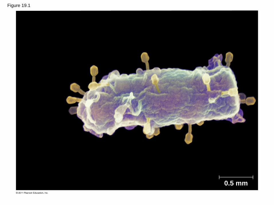
Figure 19.1
0.5 mm

Concept 19.1: A virus consists of a nucleic
acid surrounded by a protein coat
• Viruses were detected indirectly long before they
were actually seen
© 2011 Pearson Education, Inc.
The Discovery of Viruses: Scientific Inquiry
• Tobacco mosaic disease stunts growth of tobacco
plants and gives their leaves a mosaic coloration
• In the late 1800s, researchers hypothesized that a
particle smaller than bacteria caused the disease
• In 1935, Wendell Stanley confirmed this
hypothesis by crystallizing the infectious particle,
now known as tobacco mosaic virus (TMV)
© 2011 Pearson Education, Inc.

Figure 19.2
Extracted sap from tobacco plant with tobacco mosaic disease
RESULTS
Passed sap through a porcelain filter known to trap bacteria
Healthy plants became infected
Rubbed filtered sap on healthy tobacco plants
1 2 3
4

Structure of Viruses
• Viruses are not cells
• A virus is a very small infectious particle
consisting of nucleic acid enclosed in a protein
coat and, in some cases, a membranous envelope
© 2011 Pearson Education, Inc.

Viral Genomes
• Viral genomes may consist of either
– Double- or single-stranded DNA, or
– Double- or single-stranded RNA
• Depending on its type of nucleic acid, a virus is
called a DNA virus or an RNA virus
© 2011 Pearson Education, Inc.

Capsids and Envelopes
• A capsid is the protein shell that encloses the viral
genome
• Capsids are built from protein subunits called
capsomeres
• A capsid can have various structures
© 2011 Pearson Education, Inc.

Figure 19.3
Capsomere of capsid
RNA Capsomere
DNA
Glycoprotein Glycoproteins
Membranous envelope RNA
Capsid
Head
DNA
Tail sheath
Tail fiber
18 250 nm 80 225 nm 70–90 nm (diameter) 80–200 nm (diameter)
20 nm 50 nm 50 nm 50 nm (a) Tobacco
mosaic virus (b) Adenoviruses (c) Influenza viruses (d) Bacteriophage T4
• Some viruses have membranous envelopes that
help them infect hosts
• These viral envelopes surround the capsids of
influenza viruses and many other viruses found in
animals
• Viral envelopes, which are derived from the host
cell’s membrane, contain a combination of viral
and host cell molecules
© 2011 Pearson Education, Inc.
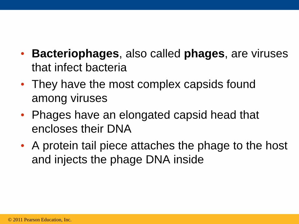
• Bacteriophages, also called phages, are viruses
that infect bacteria
• They have the most complex capsids found
among viruses
• Phages have an elongated capsid head that
encloses their DNA
• A protein tail piece attaches the phage to the host
and injects the phage DNA inside
© 2011 Pearson Education, Inc.

Concept 19.2: Viruses replicate only in
host cells
• Viruses are obligate intracellular parasites, which
means they can replicate only within a host cell
• Each virus has a host range, a limited number of
host cells that it can infect
© 2011 Pearson Education, Inc.

General Features of Viral Replicative
Cycles
• Once a viral genome has entered a cell, the cell
begins to manufacture viral proteins
• The virus makes use of host enzymes, ribosomes,
tRNAs, amino acids, ATP, and other molecules
• Viral nucleic acid molecules and capsomeres
spontaneously self-assemble into new viruses
© 2011 Pearson Education, Inc.
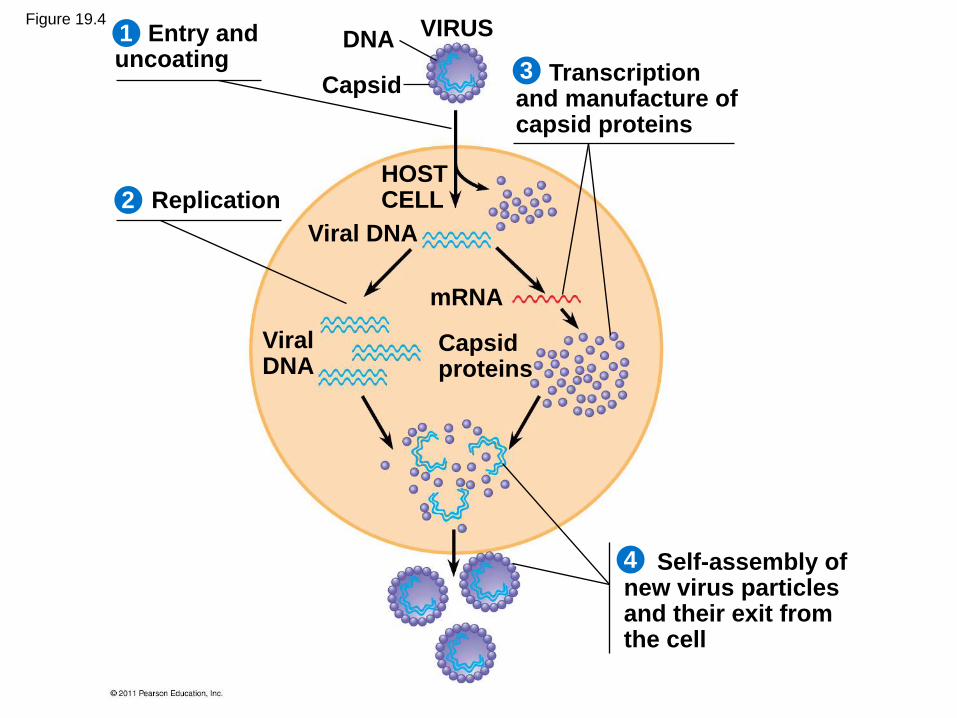
VIRUS
2
1
3
4
Entry and uncoating
Replication
Transcription and manufacture of capsid proteins
Self-assembly of new virus particles and their exit from the cell
DNA
Capsid
HOST CELL
Viral DNA
Viral DNA
mRNA
Capsid proteins
Figure 19.4
Replicative Cycles of Phages
• Phages are the best understood of all viruses
• Phages have two reproductive mechanisms: the
lytic cycle and the lysogenic cycle
© 2011 Pearson Education, Inc.

The Lytic Cycle
• The lytic cycle is a phage replicative cycle that
culminates in the death of the host cell
• The lytic cycle produces new phages and lyses
(breaks open) the host’s cell wall, releasing the
progeny viruses
• A phage that reproduces only by the lytic cycle is
called a virulent phage
• Bacteria have defenses against phages, including
restriction enzymes that recognize and cut up
certain phage DNA
© 2011 Pearson Education, Inc.

Figure 19.5-5
Attachment
2
1
5
4 3
Entry of phage DNA and degradation of host DNA
Release
Synthesis of viral genomes and proteins
Assembly
Phage assembly
Head Tail Tail fibers

The Lysogenic Cycle
• The lysogenic cycle replicates the phage
genome without destroying the host
• The viral DNA molecule is incorporated into the
host cell’s chromosome
• This integrated viral DNA is known as a prophage
• Every time the host divides, it copies the phage
DNA and passes the copies to daughter cells
© 2011 Pearson Education, Inc.

• An environmental signal can trigger the virus
genome to exit the bacterial chromosome and
switch to the lytic mode
• Phages that use both the lytic and lysogenic
cycles are called temperate phages
© 2011 Pearson Education, Inc.
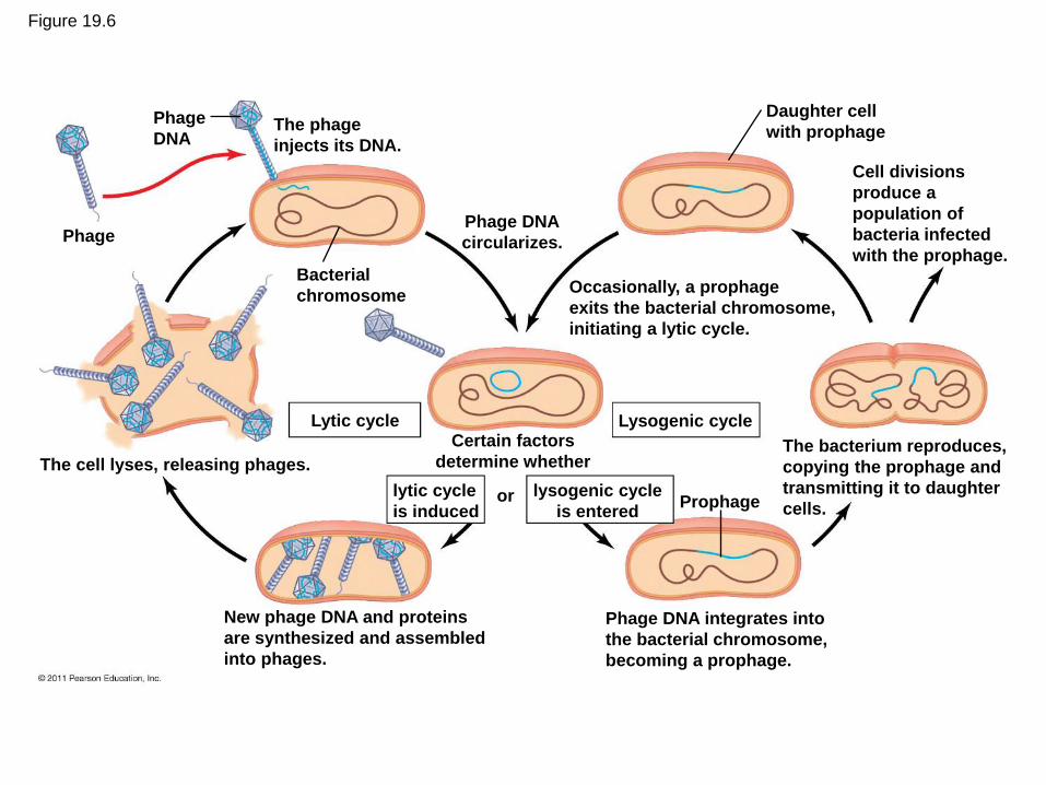
Figure 19.6
New phage DNA and proteins
are synthesized and assembled
into phages.
The cell lyses, releasing phages.
Phage
Phage
DNA The phage
injects its DNA.
Bacterial
chromosome
Lytic cycle
lytic cycle
is induced or
Phage DNA
circularizes.
Certain factors
determine whether
lysogenic cycle
is entered
Lysogenic cycle
Prophage
Daughter cell
with prophage
Occasionally, a prophage
exits the bacterial chromosome,
initiating a lytic cycle.
Cell divisions
produce a
population of
bacteria infected
with the prophage.
The bacterium reproduces,
copying the prophage and
transmitting it to daughter
cells.
Phage DNA integrates into
the bacterial chromosome,
becoming a prophage.

Replicative Cycles of Animal Viruses
• There are two key variables used to classify
viruses that infect animals
– DNA or RNA?
– Single-stranded or double-stranded?
© 2011 Pearson Education, Inc.

Table 19.1
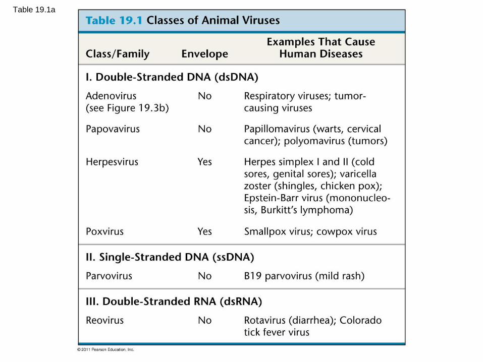
Table 19.1a
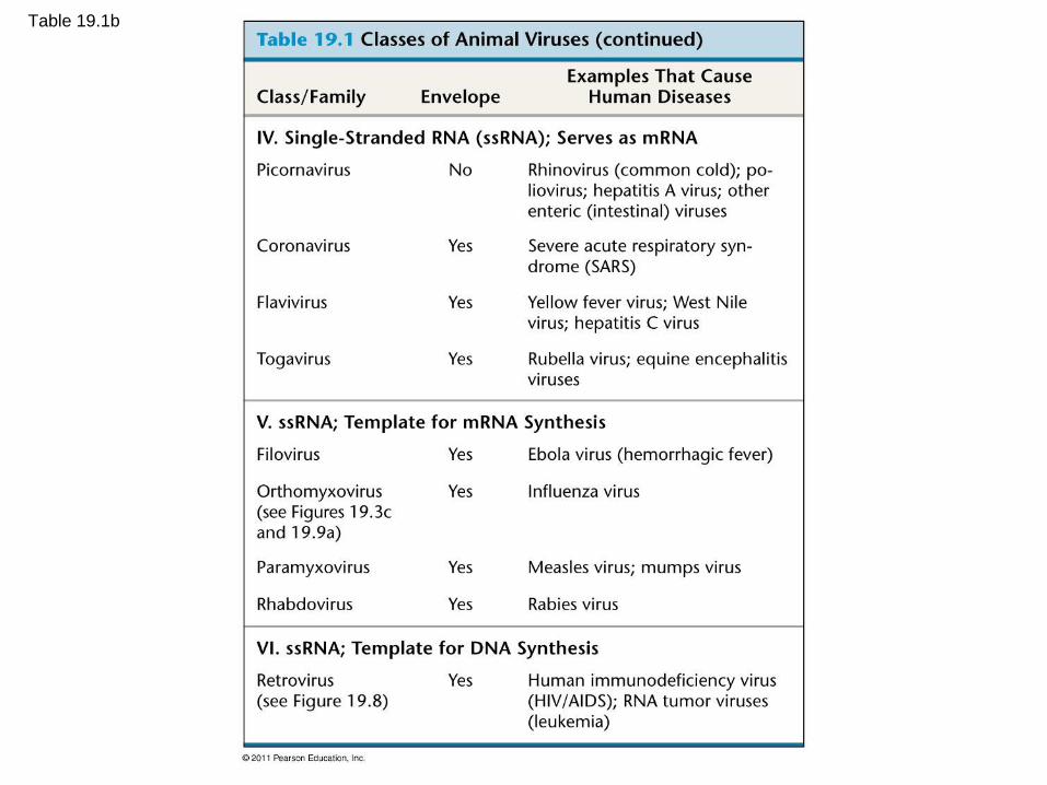
Table 19.1b
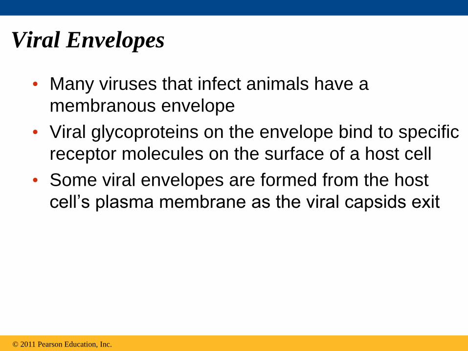
Viral Envelopes
• Many viruses that infect animals have a
membranous envelope
• Viral glycoproteins on the envelope bind to specific
receptor molecules on the surface of a host cell
• Some viral envelopes are formed from the host
cell’s plasma membrane as the viral capsids exit
© 2011 Pearson Education, Inc.

• Other viral membranes form from the host’s
nuclear envelope and are then replaced by an
envelope made from Golgi apparatus membrane
© 2011 Pearson Education, Inc.
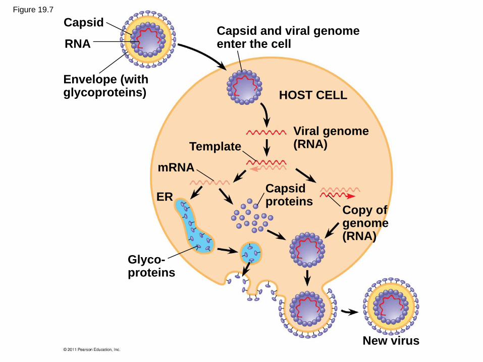
Figure 19.7
Capsid
RNA
Envelope (with glycoproteins)
Capsid and viral genome enter the cell
HOST CELL
Viral genome (RNA) Template
mRNA
ER Capsid proteins
Copy of genome (RNA)
New virus
Glyco- proteins

RNA as Viral Genetic Material
• The broadest variety of RNA genomes is found in
viruses that infect animals
• Retroviruses use reverse transcriptase to copy
their RNA genome into DNA
• HIV (human immunodeficiency virus) is the
retrovirus that causes AIDS (acquired
immunodeficiency syndrome)
© 2011 Pearson Education, Inc.

Glycoprotein
Reverse
transcriptase HIV
Viral envelope
Capsid
RNA (two
identical
strands) HOST
CELL
Viral RNA
Reverse
transcriptase
RNA-DNA
hybrid
DNA
NUCLEUS
Provirus Chromosomal
DNA
RNA genome
for the
next viral
generation mRNA
New virus
HIV
Membrane
of white
blood cell
0.25 m
HIV entering a cell
New HIV leaving a cell
Figure 19.8

• The viral DNA that is integrated into the host genome is called a provirus
• Unlike a prophage, a provirus remains a permanent resident of the host cell
• The host’s RNA polymerase transcribes the proviral DNA into RNA molecules
• The RNA molecules function both as mRNA for synthesis of viral proteins and as genomes for new Avirus particles released from the cell
© 2011 Pearson Education, Inc.
Evolution of Viruses
• Viruses do not fit our definition of living organisms
• Since viruses can replicate only within cells, they probably evolved as bits of cellular nucleic acid
• Candidates for the source of viral genomes are plasmids, circular DNA in bacteria and yeasts, and transposons, small mobile DNA segments
• Plasmids, transposons, and viruses are all mobile genetic elements
© 2011 Pearson Education, Inc.
• Mimivirus, a double-stranded DNA virus, the largest virus yet discovered, is the size of a small bacterium
• There is controversy about whether this virus evolved before or after cells
© 2011 Pearson Education, Inc.
Concept 19.3: Viruses, viroids, and prions
are formidable pathogens in animals and
plants
• Diseases caused by viral infections affect humans,
agricultural crops, and livestock worldwide
• Smaller, less complex entities called viroids and
prions also cause disease in plants and animals,
respectively
© 2011 Pearson Education, Inc.
Viral Diseases in Animals
• Viruses may damage or kill cells by causing the
release of hydrolytic enzymes from lysosomes
• Some viruses cause infected cells to produce
toxins that lead to disease symptoms
• Others have molecular components such as
envelope proteins that are toxic
© 2011 Pearson Education, Inc.

• Vaccines are harmless derivatives of pathogenic
microbes that stimulate the immune system to
mount defenses against the harmful pathogen
• Vaccines can prevent certain viral illnesses
• Viral infections cannot be treated by antibiotics
• Antiviral drugs can help to treat, though not cure,
viral infections
© 2011 Pearson Education, Inc.

Emerging Viruses
• Emerging viruses are those that suddenly become
apparent
• Recently, a general outbreak (epidemic) of a flu-
like illness appeared in Mexico and the United
States, caused by an influenza virus named H1N1
• Flu epidemics are caused by new strains of
influenza virus to which people have little immunity
© 2011 Pearson Education, Inc.
• Viral diseases in a small isolated population can emerge and become global
• New viral diseases can emerge when viruses spread from animals to humans
• Viral strains that jump species can exchange genetic information with other viruses to which humans have no immunity
© 2011 Pearson Education, Inc.

• These strains can cause pandemics, global
epidemics
• The 2009 flu pandemic was likely passed to
humans from pigs; for this reason it was originally
called the “swine flu”
© 2011 Pearson Education, Inc.

Figure 19.9
(c) 1918 flu pandemic
2009 pandemic
screening
(b) 2009 pandemic H1N1
influenza A virus
(a)
1 m

Viral Diseases in Plants
• More than 2,000 types of viral diseases of plants
are known and cause spots on leaves and fruits,
stunted growth, and damaged flowers or roots
• Most plant viruses have an RNA genome
© 2011 Pearson Education, Inc.
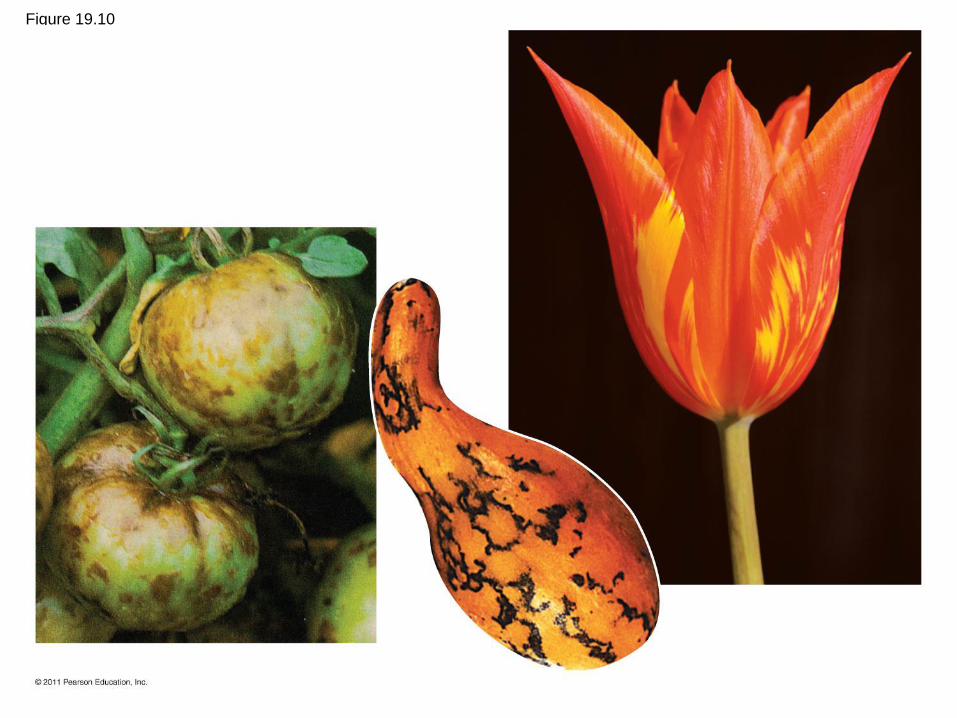
Figure 19.10

• Plant viruses spread disease in two major modes
– Horizontal transmission, entering through
damaged cell walls
– Vertical transmission, inheriting the virus from a
parent
© 2011 Pearson Education, Inc.

Viroids and Prions: The Simplest Infectious
Agents
• Viroids are small circular RNA molecules that infect plants and disrupt their growth
• Prions are slow-acting, virtually indestructible infectious proteins that cause brain diseases in mammals
• Prions propagate by converting normal proteins into the prion version
• Scrapie in sheep, mad cow disease, and Creutzfeldt-Jakob disease in humans are all caused by prions
© 2011 Pearson Education, Inc.

Figure 19.11
Prion
Normal protein
Original prion
New prion
Aggregates of prions

Figure 19.UN01
Phage
DNA
The phage attaches to a
host cell and injects its DNA.
Bacterial
chromosome Prophage
Lytic cycle Lysogenic cycle
• Temperate phage only • Virulent or temperate phage
• Genome integrates into bacterial
chromosome as prophage, which
(1) is replicated and passed on to
daughter cells and
(2) can be induced to leave the chromo-
some and initiate a lytic cycle
• Lysis of host cell causes release
of progeny phages
• Destruction of host DNA • Production of new phages
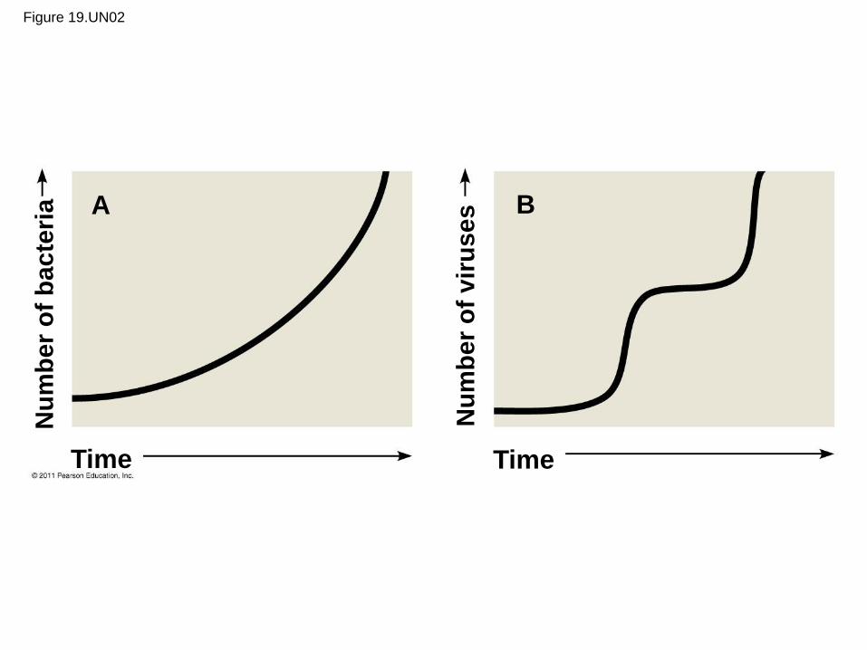
Figure 19.UN02
Time Time
A B
Nu
mb
er
of
ba
cte
ria
Nu
mb
er
of
vir
uses

Figure 19.UN03
• Prokaryotes have considerable genetic variation
• Three factors contribute to this genetic diversity:
– Rapid reproduction
– Mutation
– Genetic recombination
BACTERIA 27.2: Rapid reproduction,
mutation, and genetic recombination
promote genetic diversity in prokaryotes
© 2011 Pearson Education, Inc.

Rapid Reproduction and Mutation
• Prokaryotes reproduce by binary fission, and
offspring cells are generally identical
• Mutation rates during binary fission are low, but
because of rapid reproduction, mutations can
accumulate rapidly in a population
• High diversity from mutations allows for rapid
evolution
© 2011 Pearson Education, Inc.
Genetic Recombination
• Genetic recombination, the combining of DNA
from two sources, contributes to diversity
• Prokaryotic DNA from different individuals can
be brought together by transformation,
transduction, and conjugation
• Movement of genes among individuals from
different species is called horizontal gene
transfer
© 2011 Pearson Education, Inc.

Transformation and Transduction
• A prokaryotic cell can take up and incorporate
foreign DNA from the surrounding environment in
a process called transformation
• Transduction is the movement of genes
between bacteria by bacteriophages (viruses that
infect bacteria)
© 2011 Pearson Education, Inc.
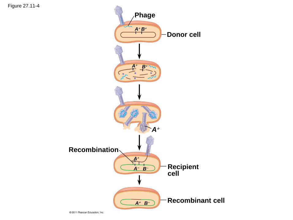
Figure 27.11-4
Recombinant cell
Recipient cell
Recombination
A
A
A B
B A
Donor cell
A B
B A
Phage

Conjugation and Plasmids
• Conjugation is the process where genetic
material is transferred between prokaryotic cells
• In bacteria, the DNA transfer is one way
• A donor cell attaches to a recipient by a pilus,
pulls it closer, and transfers DNA
• A piece of DNA called the F factor is required
for the production of pili
© 2011 Pearson Education, Inc.
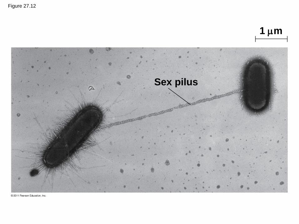
Figure 27.12
Sex pilus
1 m

The F Factor as a Plasmid
• Cells containing the F plasmid function as DNA
donors during conjugation
• Cells without the F factor function as DNA
recipients during conjugation
• The F factor is transferable during conjugation
© 2011 Pearson Education, Inc.
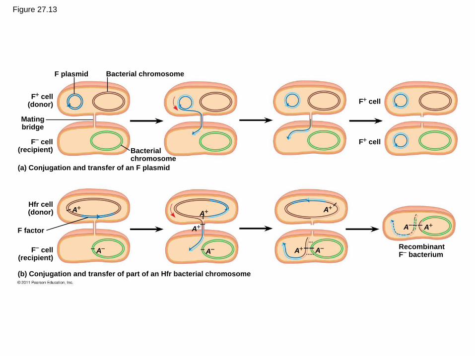
Figure 27.13
F plasmid Bacterial chromosome
F cell (donor)
F cell (recipient)
Mating bridge
Bacterial chromosome
(a) Conjugation and transfer of an F plasmid
Hfr cell (donor)
F cell (recipient)
(b) Conjugation and transfer of part of an Hfr bacterial chromosome
F factor
A
A
A
A
A
A A
F cell
F cell
A A
Recombinant F bacterium
A
The F Factor in the Chromosome
• A cell with the F factor built into its chromosomes
functions as a donor during conjugation
• The recipient becomes a recombinant bacterium,
with DNA from two different cells
© 2011 Pearson Education, Inc.

Figure 27.13b-3
Hfr cell (donor)
F cell (recipient)
(b) Conjugation and transfer of part of an Hfr bacterial chromosome
F factor A A
Recombinant F bacterium
A
A A A
A
A
A
A

R Plasmids and Antibiotic Resistance
• R plasmids carry genes for antibiotic resistance
• Antibiotics kill sensitive bacteria, but not bacteria
with specific R plasmids
• Through natural selection, the fraction of
bacteria with genes for resistance increases in a
population exposed to antibiotics
• Antibiotic-resistant strains of bacteria are
becoming more common
© 2011 Pearson Education, Inc.