leftatrialand pulmonary capillary venous pressures …
TRANSCRIPT

LEFT ATRIAL AND PULMONARY CAPILLARY VENOUS PRESSURESIN MITRAL STENOSIS
BY
R. G. EPPS AND RICHARD H. ADLER*From the Department of Cardiology, Brompton Hospital
Received February 10, 1953
A method of measuring pulmonary " capillary " pressure by wedging a catheter into a terminalbranch of a pulmonary artery was introduced by Hellems et al. in 1948. To differentiate this frompressures recorded in a blocked pulmonary vein (e.g. through an atrial septal defect) this pulmonary" capillary" pressure will be referred to as pulmonary capillary venous pressure (P.C.V.P.), follow-ing the lead of Lagerl6f and Werk6 (1949). Subsequent investigations have produced conffictingevidence concerning the interpretation and accuracy of such pressure measurements, and this reportpresents the results of a new method used to investigate the relationship between P.C.V.P. and leftatrial pressure.
Dow and Gorlin (1950) found a close correlation between P.C.V.P. and left atrial pressure indogs; the two pressures also varied directly under abnormal conditions. Several investigatorshave found the P.C.V.P. to be higher than the pressure in the left atrium, considerably so in someinstances (Hellems et al., 1948, 1949; Lagerlofand Werko, 1949; Calazel etal., 1951; and Ankeney,1952). Ankeney has recently questioned whether phasic variations in P.C.V.P. records reflectsimilar phasic pressure changes in the left atrium, and suggests that the true P.C.V.P. is essentiallydevoid of phasic variations. Opinion among investigators also varies with regard to the significanceand interpretation of the waves seen in P.C.V.P. tracings.
So far all published attempts to record pressures from a blocked pulmonary artery and from theleft atrium simultaneously have been in dogs under general anmsthesia or in human subjects withatrial septal defect. In this investigation the problem has been tackled in the conscious human sub-ject in the absence of a septal defect. Certain patients with mitral valve disease were selected for thisstudy, in view of the practical importance of measuring the P.C.V.P. in that condition.
It is common knowledge that the tracheal bifurcation and left atrium are in close proximity, andthat an enlarged left atrium in mitral valve disease may widen the carina and compress the left mainbronchus. Post-mortem examinations confirmed the fact, first demonstrated by Allison (1952) inthis country, that a needle could be introduced through the left main bronchus into the left atriumwithout endangering any nearby vital structure.
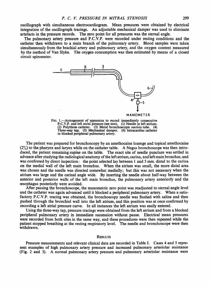
Method. After studying relevant anatomy in the cadaver, a No. 22 gauge needle, 5 cm. long,was selected for the left atrial puncture. The needle was attached to the distal end of a metalbronchoscopic suction tube. A polythene catheter inside the metal tube connected the needle tothe recording machine. Fig. 1 illustrates the position of a three-way tap that provided the essentialmeans of recording immediately consecutive pressures from the left atrium and from a blockedperipheral pulmonary artery.
Cardiac catheterization was performed by the standard method of Cournand on the consciouspatient 30 minutes after oral administration of 3 grains of sodium amytal and 0-5 grains of codeinephosphate. Heparin, 5000 units (50 mg.), was injected through the catheter. Pressures wererecorded in mm. Hg. by means of a Sanborn electromanometer and a direct writing multichannel* Work undertaken during the tenure of a fellowship from the Dazian Foundation for Medical Research, U.S.A.
298
P. C. V. PRESSURE IN MITRAL STENOSIS
oscillograph with simultaneous electrocardiogram. Mean pressures were obtained by electricalintegration of the oscillograph tracings. An adjustable mechanical damper was used to eliminateartefacts in the pressure records. The zero point for all pressures was the sternal angle.The pulmonary artery pressure and P.C.V.P. were recorded under resting conditions and the
catheter then withdrawn to a main branch of the pulmonary artery. Blood samples were takensimultaneously from the brachial artery and pulmonary artery, and the oxygen content measuredby the method of Van Slyke. The oxygen consumption was then estimated by means of a closedcircuit spirometer.
MANOMETERFIG. 1.-Arrangement of apparatus to record immediately consecutive
P.C.V.P. and left atrial pressure (see text). (1) Needle in left atrium.(2) Polythene catheter. (3) Metal bronchoscopic suction tube. (4)Three-way tap. (5) Mechanical damper. (6) Intracardiac catheterto blocked peripheral pulmonary artery.
The patient was prepared for bronchoscopy by an amethocaine lozenge and topical amethocaine(2%) to the pharynx and larynx while on the catheter table. A Negus bronchoscope was then intro-duced, the patient remaining supine on the table. The exact site of needle puncture was settled inadvance after studying the radiological anatomy ofthe left atrium, carina, and left main bronchus, andwas confirmed by direct inspection: the point selected lay between 1 and 5 mm. distal to the carinaon the medial wall of the left main bronchus. When the atrium was small, the more distal areawas chosen and the needle was directed somewhat medially; but this was not necessary when theatrium was large and the carinal angle wide. By inserting the needle about half-way between theanterior and posterior walls of the left main bronchus, the pulmonary artery anteriorly and thecesophagus posteriorly were avoided.
After passing the bronchoscope, the manometric zero point was readjusted to sternal angle leveland the catheter was again advanced until it blocked a peripheral pulmonary artery. When a satis-factory P.C.V.P. tracing was obtained, the bronchoscopy needle was flushed with saline and thenpushed through the bronchial wall into the left atrium, and this position was at once confirmed byrecording a left atrial pressure curve. In all instances the left atrium was easily entered.
Using the three-way tap, pressure tracings were obtained from the left atrium and from a blockedperipheral pulmonary artery in immediate succession without pause. Electrical mean pressureswere recorded from both sites in the same way, and these procedures were then repeated while thepatient stopped breathing at the resting respiratory level. The needle and bronchoscope were thenwithdrawn.
RESULTS
Pressure measurements and relevant clinical data are recorded in Table I. Cases 4 and 5 repre-sent examples of high pulmonary artery pressure and increased pulmonary arteriolar resistance(Fig. 2 and 3). A normal pulmonary artery pressure and pulmonary arteriolar resistance were
299
30EPPSAND ADLER
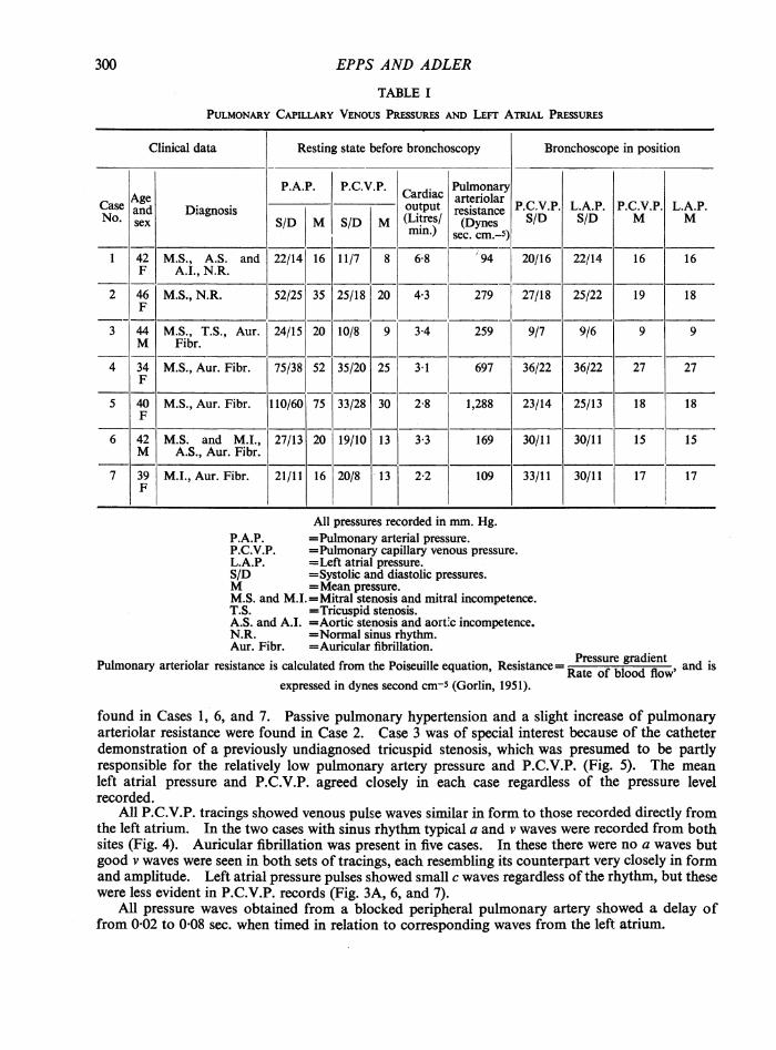
TABLE I
PULMONARY CAPILLARY VENOUS PRESSURES AND LEFT A uRAL PRESSURES
Clinical data Resting state before bronchoscopy Bronchoscope in position
P.A.P. P.C.V.P. Cardiac PulmonaryCase ____and t output arteriar P.C.V.P. L.A.P. P.C.V.P. L.A.P.Case and Diagnosis Lte/resistance SD SDN.sex S/D M S/D M (irs (Dynes S/ SD M M
min.) sec. cm.-5)
1 42 M.S., A.S. and 22/14 16 11/7 8 6-8 94 20/16 22/14 16 16F AlI., N.R.
2 46 M.S., N.R. 52/25 35 25/18 20 4 3 279 27/18 25/22 19 18F
3 44 M.S., T.S., Aur. 24/15 20 10/8 9 3-4 259 9/7 9/6 9 9M Fibr.
4 34 M.S., Aur. Fibr. 75/38 52 35/20 25 3 1 697 36/22 36/22 27 27F
5 40 M.S., Aur. Fibr. 110/60 75 33/28 30 2 8 1,288 23/14 25/13 18 18F
6 42 M. S. and M.I., 27/13 20 19/10 13 3 3 169 30/11 30/11 15 15M A.S., Aur. Fibr.
7 39 M.I., Aur. Fibr. 21/11 16 20/8 13 2-2 109 33/11 30/11 17 17F
All pressures recorded in mm. Hg.P.A.P. =Pulmonary arterial pressure.P.C.V.P. =Pulmonary capillary venous pressure.L.A.P. =Left atrial pressure.S/D =Systolic and diastolic pressures.M =Mean pressure.M.S. and M.I.=Mitral stenosis and mitral incompetence.T.S. =Tricuspid stenosis.A.S. and A.I. =Aortic stenosis and aort-c incompetence.N.R. =Normal sinus rhythm.Aur. Fibr. =Auricular fibrillation.
Pulmonary arteriolar resistance is calculated from the Poiseuille equation, Resistance= Rtessoufreblgoad iflnt and isexpressed in dynes second cm-5 (Gorlin, 1951).
found in Cases 1, 6, and 7. Passive pulmonary hypertension and a slight increase of pulmonaryarteriolar resistance were found in Case 2. Case 3 was of special interest because of the catheterdemonstration of a previously undiagnosed tricuspid stenosis, which was presumed to be partlyresponsible for the relatively low pulmonary artery pressure and P.C.V.P. (Fig. 5). The meanleft atrial pressure and P.C.V.P. agreed closely in each case regardless of the pressure levelrecorded.
All P.C.V.P. tracings showed venous pulse waves similar in form to those recorded directly fromthe left atrium. In the two cases with sinus rhythm typical a and v waves were recorded from bothsites (Fig. 4). Auricular fibrillation was present in five cases. In these there were no a waves butgood v waves were seen in both sets of tracings, each resembling its counterpart very closely in formand amplitude. Left atrial pressure pulses showed small c waves regardless of the rhythm, but thesewere less evident in P.C.V.P. records (Fig. 3A, 6, and 7).
All pressure waves obtained from a blocked peripheral pulmonary artery showed a delay offrom 002 to 0-08 sec. when timed in relation to corresponding waves from the left atrium.
300

S/D BREATHIlNQ
CI.. jLEFTATRIUM P.CC.V.P.PEF 4TIULEFTATRIU
0-
MEA)NO BREATHINQrPCv.P.LEFT ATRIUM'4PCVP P.-.-vP. LEFT ATRIUM
50--
1.5.3.~ ~ ~ /D NOT BREATHIN&CILEFTATW,PV'LATRTRIUM.C.V.cv.L 4LEFTRATRIU
O40 o
2%t1qb¶/htAtN/%/0td¶/h%A0A-0 ~Fx.3-A eodo meitl osctv rsuetrcnsi uiuairlain0hwn iiaiyo
vEnou wave0- patr setx) ntn ftpsic smre yarw ae pe 5m.ascnd Cs)(B)Immediatelyconsecutive pressure records in a case with auricular fibrillation showing equal systolic,diastolic,~..........and mea prsue ilce prheapumnRyE ArTer an left aru.Isato awthiakdbarrow.aper sEed 10m.a eod(CsD)

302 EPPS AND ADLER
SP BRERATHItLEFT ATRIUM P. C.v. P.
2 30~~~~~~~~~~~~~~~~~.
ECIl_;1_:w<:.;, -...-
....................... 7
FRG. 4. -Pressure tracings from blocked peripheral pulmonary artery and left atrium with sinus rhythm to showsimilarity of venous pulse wave (see text). The pressures are not immediately consecutive. Paper speed 25 mm.a second (Case 2).
NOT BREATHINGMEAN 5/DLEFT ATRIUM | p c v P LEFT ATRIVM P C V Pr7T| t1 ei !! i. I4s-w-.4
10~
to~~~~~~~~~~~~~~~~~~~~~~oA,_~~w *__
FIG. 5.-Immediately consecutive pressure tracings from blocked peripheralpulmonary artery and left atrium in a-case with auricular fibrillation showing identical pressures. This is the only case with a P.C.V.P. venous pulse
wave that was not shown clearly (see text). Instant of tap switch is marked byacuow. Paper speed 10 mm. asecond (Case 3).
P C.V. P. LEFT ATRIUM P
Ii 2~~~~~~~~~~~~
EI- - - i _ . t-*.
I .~~~~~~~~~~~~~~~~~~~~~~~~~~~~~~~~~~~~~~~~~~~~~~~~~~~~~~~~~~~~~~~~~~~~~~~~~~~~~~~~~~~~~~~--.<.
FIG. 6.-Immediately consecutive pressure record from left atrium and blocked peripheral pulmonary artery in mitralstenosis and regurgitation with auricular fibrillation to show similarity of pressure and wave pattern. Instantof tap switch is marked by arrow. Paper speed 25 mm. a second (Case 6).

P. C. V. PRESSURE IN MITRAL STENOSIS 303
S/D NOT BREATHINTP.C.V.P. LLEFT ATRIUM
40.
.... ...
0-
E C . =
FIG. 7.-Immediately consecutive pressure tracing from left atrium and blocked peripheral-pulmonary artery in mitralincompetence with auricular fibrillation showing the type of venous pattern recorded. The mean pressures wereidentical (see text). Instant of tap switch is marked by arrow. Paper speed 25 mm. a second (Case 7).
DISCUSSIONFor the purpose of this study, cases were specially selected to include examples of sinus rhythm,
auricular fibrillation, low and high left atrial pressures, and low and high pulmonary arteriolarresistances. One case of pure mitral incompetence was included.
We have been greatly impressed by the marked lability of the left atrial pressure and P.C.V.P.in relation to the phases of respiration and to minor variations in the resting physiological state ofthe patient. Misleading figures could easily be obtained in comparing these two pressure recordsunless simultaneous or immediately consecutive tracings were taken. In general there was a con-siderable rise in P.C.V.P. during insertion of the bronchoscope, followed by a slow return to theresting level.
Proper damping is essential in obtaining accurate pressure curves. Although some difficultyin this respect was encountered, since accurate tracings had to be recorded from two different sitesand through two different systems in rapid succession, critical damping was achieved in mostinstances (Fig. 3 and 6). Mean pressures were not subject to damping errors and gave convincingproof that P.C.V. and left atrial pressures were identical in all the cases studied.
From previous catheter studies in mitral stenosis carried out in this department, it has long beenmaintained that a proper P.C.V.P. tracing should be venous in form (Wood, 1952), and the evidencenow presented shows clearly that these waves are derived from the left atrium (Fig. 4). In thepresence of auricular fibrillation there can be no a wave but only Mackenzie's ventricular form ofthe venous pulse, the v wave (Fig. 3 and 6).
Despite proper blocking of a peripheral pulmonary artery by the catheter the typical venous wavepattern may not be present. A low or normal pressure in the left atrium may be associated with aand v waves of such low amplitude that they cannot be detected in the P.C.V.P. tracing, presumablybecause of the damping effect of the intervening capillaries. Extreme pulmonary vaso-constrictionin certain cases of mitral stenosis might also be expected to damp out the venous waves from theP.C.V.P. tracing. However, in Cases 4 and 5 the venous wave pattern was transmitted in fullstrength under just these conditions.
No immediate or delayed complications from the left atrial puncture were encountered, but twocases showed a considerable rise in P.C.V.P. as a result of inserting the bronchoscope. Thisemphasizes the potential danger of the procedure, which might well precipitate pulmonarycedema under certain conditions; fortunately this did not occur during the present study.

SUMMARYA method of recording immediately consecutive pressures in a blocked peripheral pulmonary
artery (P.C.V.P.) and the left atrium in conscious patients with mitral valve disease is presented.Bronchoscopy was performed during cardiac catheterization and a needle introduced into the leftatrium through the medial wall of the left main bronchus. The necessity for recording simultaneousor immediately consecutive pressures from the two sites is emphasized, because of their labilityduring bronchoscopy.
Seven patients suffering from mitral valve disease were selected to include examples of sinusrhythm, auricular fibrillation, low and high left atrial pressures, and low and high pulmonaryarteriolar resistances. Mitral stenosis was present in five cases, mitral stenosis and incompetencein one, and pure mitral incompetence in the seventh.
In every instance the mean pressures in the left atrium and blocked peripheral pulmonary arterywere found to be equal, and this confirms the view that the P.C.V.P. measured by cardiac catheteri-zation is an accurate measurement of the left atrial pressure in mitral valve disease.
The P.C.V.P. tracing showed venous pulse waves similar in form to those recorded directly fromthe left atrium in every case, confirming the view that the true P.C.V.P. tracing should be venous inform and that the waves recorded are derived from the left atrium.
The authors wish to give grateful acknowledgrnent to Dr. Paul Wood for his encouragement during the investi-gation, and advice in the preparation of this paper. They also wish to express their appreciation for the technicalassistance of Miss P. Scarlett, Miss D. Anderson, and Mr. J. Manders.
REFERENCESAllison, P. R. (1952). Personal communication.Ankeney, J. L. (1952). Amer. J. Physiol., 169, 40.Calazel, R. R., Gerard, R., Daley, R., Daley, A., Foster, J., and Bing, R. (1952. Bull. Johns Hopkins Hosp., 88, 20.Dexter, L., Dow, J. W., Haynes, F. W., Whittenberger, J. L., Ferris, B. G., Goodale, W. T., Hellems, H. K. (1950).
J. Clin. Invest., 29, 602.Dow, J. W., and Gorlin, R. (1950). Fed. Proc., 9, 33.Gorlin, R., Haynes, F. W., Goodale, W. T., Sawyer, C. G., Dow, J. W., Dexter, L. (1951). Amer. Heart J., 41, 30.
Lewis, B. M., Haynes, F. W., Dexter, L. (1952). Amer. Heart J., 43, 357.-,- , -, Spiegl, R. J., and Dexter, L. (1951). Amer. Heart J., 41, 834.
Hellems, H. K., Haynes, F. W., and Dexter, L. (1949). J. App. Physiol., 2, 24.- -, , - , and Kinney, T. D. (1948). Amer. J. Physiol., 155, 98.Lagerlof, H., and Werko, L. (1949). Scand. J. Clin. Lab. Invest., 1, 147.Lewis, B. M., Gorlin, R., Houssay, H. J., Haynes, F. W., Dexter, L. (1952). Amer. Heart J., 43, 2.Mackenzie, J. (1902). The Study of the Pulse. Young J., Pentland, Edinburgh.Wood, P. (1952). European Congress of Cardiology, Abstracts of Scientific Communications, p. 25.
304 EPPS AND ADLER