liberato berrino - clinical cancer...
TRANSCRIPT

1
Primary and acquired resistance of colorectal cancer to anti-EGFR monoclonal antibody can be overcome by combined treatment of regorafenib with cetuximab.
Stefania Napolitano1, Giulia Martini1, Barbara Rinaldi2, Erika Martinelli1, Maria Donniacuo2,
Liberato Berrino2, Donata Vitagliano1, Floriana Morgillo1, Giusy Barra3, Raffaele De Palma3,
Francesco Merolla4, Fortunato Ciardiello1 and Teresa Troiani1.
1Oncologia Medica and 3Immunologia Clinica,Dipartimento Medico-Chirurgico di Internistica
Clinica e Sperimentale “F. Magrassi e A. Lanzara”, Seconda Università degli Studi di Napoli, Via
S. Pansini 5, 80131 Naples, Italy.
2Sezione di Farmacologia, Dipartimento di Medicina Sperimentale “L.Donatelli”, Seconda
Università degli Studi di Napoli, Via L. De Crecchio 7, 80138 Naples, Italy.
3Dipartimento di Scienze Biomediche Avanzate, Universita Federico II , Napoli , via S.
Pansini,80131 Naples, Italy.
Dr. Napolitano and all coauthors have no conflicts of interest to declare for the following manuscript.
Corresponding Author:Teresa Troiani, Dipartimento Medico-Chirurgico di Internistica Clinica e
Sperimentale “F. Magrassi e A. Lanzara”, Seconda Università degli Studi di Napoli, Italy; Via S.
Pansini 5, 80131 Naples, Italy.
Running title: Regorafenib and cetuximab overcome resistance to cetuximab
Keywords: Regorafenib, cetuximab, CRC,primary and acquired resistance.
Financial Support:This research has been supported by a grant from AssociazioneItaliana per la
RicercasulCancro (AIRC) and a grant from Ministero dell’ Istruzione, Università e Ricerca
(MIUR)-PRIN 2010-2011.
Word Count: 4477
Total number of figures and tables: 4 figures, 2 Tables and 4supplementary figures
Research. on February 15, 2019. © 2015 American Association for Cancerclincancerres.aacrjournals.org Downloaded from
Author manuscripts have been peer reviewed and accepted for publication but have not yet been edited. Author Manuscript Published OnlineFirst on April 2, 2015; DOI: 10.1158/1078-0432.CCR-15-0020

2
Translational Relevance
The introduction in clinical practice of MAbs against the EGFR, such as cetuximab or
panitumumab, in combination with chemotherapy has demonstrated therapeutic efficacy in
metastatic CRC patients with all RAS wild-type tumors. However, efficacy of these MAbs is limited
by development of resistance mechanisms in cancer cells. Activation of alternative signaling
pathways, that bypass the EGFR, has been implicated in the resistance to anti-EGFR therapies.
Therefore, the blockade of multiple growth factor and receptor pathways could be necessary to
increase the efficacy of anti-EGFR monoclonal antibodies. In this study, we have demonstrated that,
in human CRC cells with either primary or acquired resistance to cetuximab, the combined
treatment with cetuximab and regorafenib induced synergistic anti-proliferative and apoptotic
effects and causes significant tumor growth inhibition. This study provides a rationale for
evaluating combined treatment with cetuximab and regorafenib as a therapeutic strategy for
preventing and/or overcoming cetuximab resistance in mCRC patients.
Research. on February 15, 2019. © 2015 American Association for Cancerclincancerres.aacrjournals.org Downloaded from
Author manuscripts have been peer reviewed and accepted for publication but have not yet been edited. Author Manuscript Published OnlineFirst on April 2, 2015; DOI: 10.1158/1078-0432.CCR-15-0020

3
Abstract
Purpose: In colorectal cancer (CRC) the activation of the intracellular RAS/RAF and
PIK3CA/AKT pathwayshas been implicated in the resistance to anti-epidermal growth factor
receptor (EGFR) monoclonal antibodies (MAbs).Wehave investigated the role of regorafenib,an
oral multikinase inhibitor, in combination withcetuximab, an anti-EGFR MAb, to overcome anti-
EGFR resistance.
Experimental Design: We have tested, in vitro and in vivo,the effects of regorafenib in a panel of
human CRC cell lines with a KRAS mutation (SW480, SW620, HCT116, LOVO, HCT15) or with a
BRAF mutation (HT29), as models of intrinsic resistance to cetuximab treatment, and in two human
CRC cell lines (GEO and SW48),that are cetuximab-sensitive, as well as in their derived cells with
acquired resistance to cetuximab (GEO-CR and SW48-CR).
Results: Treatment with regorafenib determined a dose-dependent growth inhibition in all CRC cell
lines.The combined treatment with cetuximab and regorafenib induced synergistic anti-proliferative
and apoptotic effects in cetuximab-resistant cell lines by blocking MAPK and AKT pathways. Nude
mice were injected subcutaneously with HCT116, HCT15, GEO-CRand SW48-CR cells. The
combined treatment caused significant tumor growth inhibition. Synergistic antitumor activity of
regorafenib plus cetuximab was also observed in an orthotopic CRC model of HCT116 cells. In
particular, the combined treatment induceda significant tumor growth inhibition in the primary
tumor site (cecum) and completely preventedmetastasis formation.
Conclusion:The combined treatment with cetuximab and regorafenib could be a strategy to
overcome resistance to anti-EGFR therapies in metastatic CRC patients.
Research. on February 15, 2019. © 2015 American Association for Cancerclincancerres.aacrjournals.org Downloaded from
Author manuscripts have been peer reviewed and accepted for publication but have not yet been edited. Author Manuscript Published OnlineFirst on April 2, 2015; DOI: 10.1158/1078-0432.CCR-15-0020

4
Introduction
CRC is one of the leading causes of cancer-related mortality worldwide, with over 1,2
million new cases and 608,700 deaths estimated in 2008 (1).Despite improvements made in
screening strategies, a significant number of patients are still diagnosed at late stages of the disease.
In the last decade, the introduction of targeted therapies in clinical practice, in particular of
agents targeting the vascular endothelial growth factor (VEGF)- related pathway (bevacizumab and
aflibercept) and the EGFR (cetuximab and panitumumab) has changed the therapeutic approach to
metastatic CRC patients, with a significant improvement in progression free survival (PFS) and
overall survival (OS) (2).Cetuximab and panitumumab are monoclonal antibodies (Mabs) that block
the activation of the EGFR and of its downstream intracellular signals, the RAS-RAF-MEK-MAPK
and the PTEN-PIK3CA-AKT pathways (3-6).These two drugs are currently approved for the
treatment of metastatic CRCpatients with all-RAS wild-type tumors. Nevertheless, prognosis
remains poor for most of these patients. In fact, the use of these monoclonal antibodiesis limited by
the presence of pre-existing intrinsic resistance mechanisms or by the ability of cancer cells to
acquire resistance. Possible mechanisms for primaryand acquired resistance to cetuximab include
mutations in the KRAS, BRAF and NRAS genes,secondary mutation (S492R) in the extracellular
domain of EGFR,HER2gene amplification and/or increased HER2 signaling and overexpression of
the METpathway(7-10).
Recently, it has been elucidated thatin the resistance to anti-EGFR therapiesdifferent growth
factors and receptors could be activated in the cancer cell to drive alternative signaling pathways
that bypass the EGFR(11,12). Molecular heterogeneity also plays an important role in the context of
resistance, by limiting the success of therapies against a single target. Colorectal cancer patients can
harbor different gene mutations in distinct tumor lesions, or even within different regions of the
same lesion (13).All these alterations could converge on activation of the RAS–MEK–ERK
pathway (9,10,14,15). Understanding the biology of such complex gene heterogeneity in tumors is
Research. on February 15, 2019. © 2015 American Association for Cancerclincancerres.aacrjournals.org Downloaded from
Author manuscripts have been peer reviewed and accepted for publication but have not yet been edited. Author Manuscript Published OnlineFirst on April 2, 2015; DOI: 10.1158/1078-0432.CCR-15-0020
5
necessaryfor developing rational combination therapies. In fact, blockade of multiple growth factor
and growth factor receptor pathways could be needed to increase the efficacy of anti-EGFR targeted
therapies (16).
Regorafenibis an oral multikinase inhibitor, that could target three key oncogenic pathways,
such as a) cell growth by inhibition of KIT, RET, RAF-1 and BRAF; b)tumor-induced
angiogenesisby targeting VEGFR1, 2 and-3, and the tyrosine kinase with immunoglobulin and
epidermal growth factor homology domain 2 (TIE2); and c) tumor microenvironment by
blockingplatelet-derived growth factor receptor-β (PDGR-β) and fibroblast growth factor receptor
(FGFR)(17-19). In preclinical studies, regorafenib exhibited antitumor activity in different tumor
xenografts (17).Recently, a phase III study showed that regorafenib treatment significantly
improved OS and PFS in patients with metastatic CRC who failed all available therapies (20).Thus,
both the Food and Drug Administration (FDA) and the European Medicines Agency (EMA) have
approved regorafenib for the treatment of such metastatic CRC patients.
In the present study, we have evaluated the efficacy of regorafenib in combination with
cetuximab to overcoming resistance to anti-EGFR MAbsby using different human CRC cell
models. We have selected five CRC cell lines with KRAS mutations (SW480, SW620, HCT116,
LOVO, HCT15)one with BRAF mutation (HT29) and two cell lines with acquired resistance to
cetuximab, that were originally obtained in our laboratory (10,14,21) (Supplementary Table 1). We
have found that combined treatment with cetuximab and regorafenib induced synergistic anti-
proliferative and pro-apoptotic effects by blocking MAPK and AKT pathways in these CRC cell
lines. Moreover, a similar synergistic antitumor activity has beenconfirmed byin vivosubcutaneous
and orthotopic CRC xenograftmodels.
Research. on February 15, 2019. © 2015 American Association for Cancerclincancerres.aacrjournals.org Downloaded from
Author manuscripts have been peer reviewed and accepted for publication but have not yet been edited. Author Manuscript Published OnlineFirst on April 2, 2015; DOI: 10.1158/1078-0432.CCR-15-0020

6
Material and Methods
Drugs. Cetuximab, an anti-EGFR human-mouse chimeric monoclonal antibody,was kindly
provided by Merck Serono Italy (Rome, Italy) and it was ready to use. Regorafenib was kindly
provided by Bayer Pharma Italy (Milan, Italy). For in vitro applications, regorafenibwasdissolved in
sterile dimethylsulfoxide (DMSO) and the 10 mMstock solution wasstored in aliquots at -20°C.
Working concentrations were diluted in culture medium just before each experiment.For in vivo
applications, regorafenibwas solubilized in 0.5% Tween-80 in sterile Phosphate Buffered Saline
(PBS).
Cell Lines. The human HT29, SW620, LOVO, HCT15CRC cell lines were obtained from the
American Type Culture Collection (ATTC) (Manassas, VA) and have been authenticated by IRCCS
“AziendaOspedalieraUniversitaria San Martino-IST IstitutoNazionale per la RicercasulCancro,
Genova” Italy. The humanSW48 (catalogue number: HTL99020), SW480 (catalogue number:
HTL95025) and HCT116 (catalogue number: HTL99017)CRC cell lines were obtained from
IRCCS “AziendaOspedalieraUniversitaria San Martino-IST IstitutoNazionale per la
RicercasulCancro, Genova” Italy. The human GEO colon cancer cell linewas kindly provided by
Dr. N. Normanno (National Cancer Institute, Naples, Italy). GEO-CR and SW48-CR cells were
established as previously described (10,14,21). GEO and GEO-CR cell lines were grown in DMEM
(Lonza, Cologne, Germany),supplemented with 10% fetal bovine serum (FBS) (Lonza), 1%
penicillin/streptomycin (Lonza). SW48, SW480, HCT116, LOVO, HCT15, SW48-CR, cells were
grown in RPMI-1640 (Lonza) supplemented with 10% FBS, 1% penicillin/streptomycin. SW620
and HT29 cancer cells were grown in McCoy medium (Lonza) supplemented with 20% FBS
(Lonza), 1% penicillin/streptomycin (Lonza).All cell lines were grown in a humidified incubator
with 5% of carbon dioxide (CO2) and 95% air at 37°C. All cell lines were routinely screened for the
presence of mycoplasma (Mycoplasma Detection Kit, Roche Diagnostics, Monza, Italy).
Research. on February 15, 2019. © 2015 American Association for Cancerclincancerres.aacrjournals.org Downloaded from
Author manuscripts have been peer reviewed and accepted for publication but have not yet been edited. Author Manuscript Published OnlineFirst on April 2, 2015; DOI: 10.1158/1078-0432.CCR-15-0020
7
Proliferation Assay. Cancercell lines were seeded in 24-well plates and were treated with different
concentrations ofcetuximab (range, 0.001 to 20 µg/ml) alone or in combination with
regorafenib(range, 0.001 to 5 µM)for 96 hours. Cell proliferation was measured with the 3-(4,5-
dimethylthiazol-2-yl)-2,5-diphenyltetrazolium bromide (MTT). The IC50 was determined by
interpolation from the dose-response curves. Results represent the median of three separate
experiments, each performed in quadruplicate. Results of the combination treatment were analyzed
according to the method of Chou and Talalay by using the CalcuSyn software programme (Biosoft,
Cambridge, UK).
Apoptosis assay. HT29, SW480, SW620, HCT116,LOVO, HCT15, GEO-CR and SW48-CRcells
were seeded in six-well plates, treated with cetuximab, regorafenib or their combination at different
concentrations 72 hours and stained with Annexin V-fluorescein isothiocynate (FITC) (Invitrogen,
CA, USA). Apoptotic cell death was assessed by counting the numbers of cells that stained positive
for Annexin V-FITC using an Apoptosis Annexin V-FITC Kit (Invitrogen, CA, USA), coupled with
fluorescence-activated cell sorting (FACS) analysis, by following manufacturer’s protocol.
Immunoblotting.SW480, SW620, HCT116, LOVO, HCT15, GEO-CR and SW48-CR cells were
seeded into 100 mm3 dishes and treated with vehicle, cetuximab, regorafenib, or their combination
for 24 hours at different concentration as following indicated. Fifty mg of protein lysates, estimated
by a modified Bradford assay (Bio-Rad, Munich, Germany), were subjected to Western blot, as
previously described(22), by using the following antibodies:AKT polyclonal antibody (#9272),
pAKT monoclonal antibody (#4060), phospho-S6 ribosomial protein (#4856), p44/42
MAPKpolyclonal antibody (#9102), phospho-p44/42MAPK monoclonal antibody (#9106)were
from Cell Signaling (Beverly, MA, USA).Monoclonal anti-α-tubulin antibody (T8203) was from
Sigma Chemical Co. (St. Louis, MO, USA).Goat anti-rabbit IgG and rabbit anti-mouse
IgGsecondary antibodies were from Bio-rad (Hercules, CA, USA). Immunoreactive proteins were
Research. on February 15, 2019. © 2015 American Association for Cancerclincancerres.aacrjournals.org Downloaded from
Author manuscripts have been peer reviewed and accepted for publication but have not yet been edited. Author Manuscript Published OnlineFirst on April 2, 2015; DOI: 10.1158/1078-0432.CCR-15-0020

8
visualized by enhanced chemiluminescence. (ECL plus, ThermoFisherScientific, Rockford, IL,
USA). Each experiment was done intriplicate.
Tumor xenografts in nudemice. Four- to six-week old female balb/c athymic (nu+/nu+) mice
were purchased fromCharles River Laboratories (Milan, Italy). The research protocol was approved
and mice were maintained in accordance with the institutional guidelines of the Second University
of Naples Animal Care and Use Committee. Animal care was in compliance with Italian (Decree
116/92) and European Community (E.C. L358/1 18/12/86) guidelines on the use and protection of
laboratory animals. Mice were acclimatized at the Second University of Naples Medical School
Animal Facility for 1 week prior to being injected with cancer cells and then caged in groups of five
under controlled conditions (12–12 h light-dark cycle; room temperature 20±22°C; humidity 55–
60%). A total number of 3.5 x 10⁶ GEO-CR, SW48-CRcells and 2 x 10⁶ HCT116, HCT15cells in in
200 µl of matrigel (BD Biosciences, Milan, IT):PBS (1:1) were subcutaneously injected to the
dorsal flank of mice. When the mean values of tumors were between 200-300 mm3, mice were
randomly assigned to one of the following groups (10 mice per group).Group 1: vehicles
administrated orally and intraperitoneally (i.p.). Group 2:cetuximab injected twice a week i.p. at the
dose of 1 mg for 3 weeks. Group 3:regorafenib administered by daily oral gavage at the dose of 10
mg/kg for 3 weeks.Group 4:combination of regorafenib and cetuximab.Monitoring of tumor growth
was performed until tumors reached approximately2.000 mm3, when mice were euthanized. Tumor
size was evaluated twice a week by calliper measurements using the following formula: π/6 x larger
diameter x (smaller diameter)2. Student's t test was used to evaluate the statistical significance of the
results.
Orthotopic colorectal cancer model.Four- to six-week old female balb/c athymic (nu+/nu+) mice
purchased from Charles River Laboratories (Milan, Italy)were used. The orthotopic implantation
was performed as described by Hoffman and colleagues (23).In brief, subcutaneous tumors derived
from HCT116 cells were obtained.When tumors reached a mean volume of 500 mm3, animals were
Research. on February 15, 2019. © 2015 American Association for Cancerclincancerres.aacrjournals.org Downloaded from
Author manuscripts have been peer reviewed and accepted for publication but have not yet been edited. Author Manuscript Published OnlineFirst on April 2, 2015; DOI: 10.1158/1078-0432.CCR-15-0020
9
euthanized, the tumors were removed using sterile techniques, divided into 2-3 mm-sized pieces,
and harvested in PBS on ice. Mice were treated with antibiotics, ticarcillin (50 mg kg−1i.v.), two
hours before and after tumor implantation.Animals were anesthetized with 2,2,2-tribomoethanol
97% TBE, Avertin (Sigma-Aldrich, St. Louis, MO, USA). TBE solution was prepared fresh daily
by mixing 0.625 g of 97% crystalline TBE powder with 25 ml sterile 0.9% saline and then injected
intraperitoneally at 0.01 ml/g body mass (250 mg/kg).The abdomen was prepped with betadine
solution and the surgical site was isolated in a sterile fashion.A laparotomy of 0.5 cm was
conducted; the cecum was exteriorized and isolated using pre-cut, sterile gauze. A warm saline
solution was used to keep the cecum wet. Subsequently, the cecum wall waslightly damaged and a
single tumor fragment from HCT-116 subcutaneous tumors was sutured to the mesenteric border of
the cecum wall using 6.0 nylon surgical sutures. Upon completion, the cecum was placed into the
abdominal cavity and the abdominal wound was sutured using a 6.0 Ethicon absorbable stitches.
(Ethicon Inc., Somerville, NJ).Fourteen days after the injection, mice were randomly assigned to
four groups (7 mice for each group) to receive one of the following treatments.Group 1:daily
administration of PBS/0.5% Tween 80 by oral gavage for 5 days a week and i.p. injection of PBS
twice a week (control group). Group 2: daily administration of diluent for 5 days a week and i.p.
injection of cetuximab 1 mg twice a week. Group 3:daily administration of regorafenib 10 mg/kg by
oral gavage for 5 days a week and i.p. injection of PBS twice a week. Group 4: combination of oral
regorafenib and i.p. cetuximab. Treatment was continued for 3 weeks, and the mice were euthanized
1 week later. The body weights were monitored daily. Primary tumors in the cecum were excised
andweighed. The final tumor was measured with a caliper and the volume was calculated by
thefollowing formula: π/6 x larger diameter x (smaller diameter)2. The presence of metastasis was
evaluated in the peritoneum, liver, intestines, lungs, rectum, and spleen and confirmed by histologic
review.The tumor excised from each mouse was divided into 3 parts. One piece was formalin-fixed;
the other two pieces were frozen at -80 C in RNAlater. Hematoxylin and eosin staining confirmed
the presence of tumors in each sample.
Research. on February 15, 2019. © 2015 American Association for Cancerclincancerres.aacrjournals.org Downloaded from
Author manuscripts have been peer reviewed and accepted for publication but have not yet been edited. Author Manuscript Published OnlineFirst on April 2, 2015; DOI: 10.1158/1078-0432.CCR-15-0020

10
Results
Sensitivity to cetuximab and regorafenib treatment in a panel of human CRC cell lines.
We first tested in vitrothe activity of cetuximab and regorafenib, as single agents, in a panel
ofhuman CRC cell lines to characterize their spectrum of activity. We selected eight human CRC
(GEO, SW48, HT29, SW480, SW620, HCT116, LOVO, HCT15) cell lines, having different
mutation profiles in KRAS, NRAS, BRAF, and PIK3CA genes (Supplementary Table1). Cancer cells
were treated with cetuximab at concentrations ranging from 0.01 to 20 µg/ml and with regorafenib
at concentrations ranging from 0.05 to 5 µg/ml for 96 hours. The drug concentrations required to
inhibit cell growth by 50% (IC50) were determined by interpolation from the dose-response curves.
Two CRC cell lines were sensitive to cetuximab: SW48, a cell line “quadruple wild type” for KRAS,
BRAF, NRAS and PIK3CA genes, and GEO cells with a KRAS codon 12 mutation, with IC50 of 0.5
and 0.1µg/ml, respectively. Despite GEO cells harbor a KRAS gene mutation, previous studies from
different laboratories, including our own, demonstrated that this CRC cell line is one of the most
sensitive to the in vitro and in vivo antitumor activity of cetuximab treatment (14,21,24-26).HT29,
SW480, SW620, HCT116, LOVO and HCT15 were primarily resistant to cetuximab, as shown in
Figure 1. These cells have an activating KRAS gene mutation in either codon 12 or 13 within exon
2, except HT29 cells that have a BRAF mutation (V600E). Cetuximab was also not effective in
GEO-CR and SW48-CR cells, two models of cetuximab-acquired resistance,previously obtained in
our laboratory (10,14,21). As shown in Figure 1, regorafenib show a different proliferation
inhibitory effect in these human CRC cell lines, with IC50 values ranging between 0.5 μM
(HCT116, HT29, LOVO),1μM (GEO), 2 μM (SW480, HCT15) and >2 μM (SW48, SW620, SW48-
CR, GEO-CR).No significantly differences in regorafenib efficacy were observed among CRC cell
lines harboring KRAS, NRAS, BRAF, PIK3CA mutations, indicating that its anti-tumor activity
seems to be independent of the molecular profile of CRC cell lines tested.
Research. on February 15, 2019. © 2015 American Association for Cancerclincancerres.aacrjournals.org Downloaded from
Author manuscripts have been peer reviewed and accepted for publication but have not yet been edited. Author Manuscript Published OnlineFirst on April 2, 2015; DOI: 10.1158/1078-0432.CCR-15-0020
11
Effects of cetuximab in combination with regorafenib in a panel of human CRC cell lines with
primary and acquired resistance to anti-EGFR drugsin vitro.
We evaluated the anti-proliferative activity of cetuximab and regorafenib in combination in the
panel of humanCRC cell lines. (Supplementary Figure 1 and 2). Combination Index (CI) values
were calculated according to the Chou and Talalay mathematical model for drug interactions using
the Calcusyn software, as previously described (10,14,24,27).A synergistic growth inhibitory effect
was observed in human CRC cell lines with both primary and acquired resistance to cetuximab. In
fact, the CI values for the combined treatments were significantly <1.0 for all the drug doses tested
(CI values ranging between 0.0001 and 0.7). (Supplementary Figure 1 and 2). In contrast,an
antagonistic effect of the combined treatment was observed in sensitive CRC cell lines (GEO and
SW48) with CI values significantly >1.0 (data not shown).
Effects of cetuximab in combination with regorafenib on intracellular signaling pathways in a
panel of human CRC cell lines with primary and acquired resistance to anti-EGFR drugs.
To examine the mechanism by which the combined treatment contributes to inhibition of
proliferation in CRC cell lines with primary or acquired resistance to anti-EGFR inhibitor, the
activation of EGFR downstream signaling molecules was evaluated. SW480, SW620, HCT116,
LOVO, HCT15, SW48-CR, and GEO-CR cells were treated with cetuximab, regorafenib
and/ortheir combination. The activation of PIK3CA/AKT and RAS/MAPK pathways wasanalyzed
by Western blotting. The combined treatment with cetuximab and regorafenib substantially
inhibited phosphorylation of both AKT and MAPK after 24 hours of treatment compared to
singleagent treatments (Figure 2). Astrong reduction of phosphorylated S6 ribosomal protein (pS6)
levels, the major downstream effector of AKT/m-TOR signaling, was observed in the combination
treatment (Figure 2). These findings suggested that cetuximab in combination with regorafenib
could overcome resistance to anti-EGFR treatment by inhibiting PIK3CA/AKT and MAPK
pathways.
Research. on February 15, 2019. © 2015 American Association for Cancerclincancerres.aacrjournals.org Downloaded from
Author manuscripts have been peer reviewed and accepted for publication but have not yet been edited. Author Manuscript Published OnlineFirst on April 2, 2015; DOI: 10.1158/1078-0432.CCR-15-0020
12
Pro-apoptotic effect of cetuximab in combination with regorafenib in CRC cell lines with
primary and acquired resistance to anti-EGFR drugs.
Wemeasured the ability of cetuximab and regorafenib as single agents or in combination, to induce
apoptosis in CRC cell lines by the Annexin V-FITC assay (Table 1 and Supplementary Figure 3).
Compared tosingle agent, the combined treatment induced significantly early and late apoptosis in
the whole panel of human CRC cell lines with primary or acquired resistance to cetuximab.
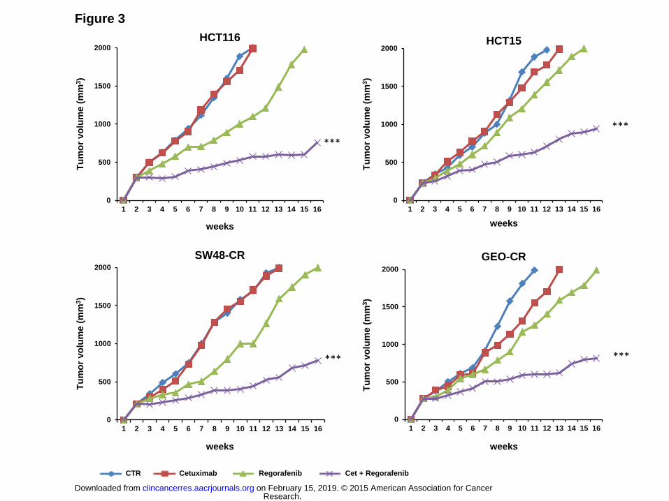
Cetuximabplus regorafenib combination exhibits antitumor activity in subcutaneous CRC
xenografts models.
We evaluated the in vivo activity of cetuximab alone or in combination with regorafenib in nude
mice subcutaneously injected with cetuximab-resistant HCT15, HCT116, GEO-CR or SW48-CR
cell lines. Mice were randomly assigned to receive vehicle, cetuximab, regorafenib or their
combination and were treated for three weeks. As shown in Figure 3, treatment with cetuximab had
little or no effect on tumor growth in all tumor xenografts. Similar results were obtained in the
groups treated with regorafenib alone. On the contrary, the combined treatment significantly
inhibited tumor growth compared both to control group and to single agent treatments in all tumor
xenografts (Figure 3). Single agent and combination treatment protocols were well tolerated by
mice and were not accompanied by any major side effect or treatment-related weight loss. No
cellular abnormalities were observed in the examined organs, including heart, lung, liver, kidney
and spleen derived from all xenograft mouse models (data not shown).
Cetuximab plus regorafenib combination inhibits tumor growth in an orthotopic human CRC
xenograft.
An orthotopicCRC model with HCT 116 CRC cells was established, as describedin Material and
Methods. Both cetuximab and regorafenib were well tolerated, and no significant loss of animal
weight was observed in the group of combined treatment, whereas a significant weight lossoccurred
Research. on February 15, 2019. © 2015 American Association for Cancerclincancerres.aacrjournals.org Downloaded from
Author manuscripts have been peer reviewed and accepted for publication but have not yet been edited. Author Manuscript Published OnlineFirst on April 2, 2015; DOI: 10.1158/1078-0432.CCR-15-0020
13
in the single agenttreatment groups, compared with the mice weight before treatment. The observed
weight loss in these groups was probably caused by the presence of growing tumors and peritoneal
metastases (Supplementary Figure 4).Of interest combined treatment showed a significant
antitumor effectcompared tovehicle, cetuximab or regorafenib single agent groups (Figure 4 and
Table 2).Mice treated with vehicle had large tumors in the cecum and peritoneum with 100%
incidence of regional (mesenteric) lymph node metastases. Mice receiving cetuximab orregorafenib
alone had large tumors with 80% and 70% respectively incidence of lymph node metastases.The
combined treatment strongly inhibitedthe tumor growth in the cecum and no peritoneum metastases
were observed (Figure 4 and Table 2). The combined treatmentwas also evident ontumor
vascularization. In fact, tumors in mice treated with vehicle, cetuximab or regorafenib were large
and highly vascularized, whereascetuximab plus regorafenib treated mice developed small tumors
without evidence of neovascularization. (Figure 4 and Table 2). No liver or lung metastases were
detected macroscopically in all groups (data not shown).
Research. on February 15, 2019. © 2015 American Association for Cancerclincancerres.aacrjournals.org Downloaded from
Author manuscripts have been peer reviewed and accepted for publication but have not yet been edited. Author Manuscript Published OnlineFirst on April 2, 2015; DOI: 10.1158/1078-0432.CCR-15-0020

14
Discussion
The development of targeted therapies has provided new options for the personalized
management of patients with advanced solid tumors.MAbsdirected against the EGFR, such as
cetuximab and panitumumab,have emerged as important therapeutic agents in the treatment of
metastatic CRC patients. However, their use is substantially limited by intrinsic and acquired cancer
cell resistance.Several hypothesis have been developed to explain why resistant cancer cell arises
and how it is possible to overcome it. One possibility is cancer intrinsic genetic heterogeneity,
which could be more prominent in the metastatic setting (28,29).Heterogeneous genetic alterations
in genes involved in the EGFR pathways have been hypothesized to play a role in resistance to anti-
EGFR drugs in CRC, including activating mutations in KRAS, NRAS, B-RAFand PIK3CA, and loss
of expression of PTEN(13). The overall scenario is complicated by presence of additional genetic
mechanismsable to activate the RAS pathway in the absence of molecular alterations affecting RAS
or itsimmediate downstream effectors (30-37).One strategy to overcome the limitations of targeting
an individual growth factorreceptor such as the EGFR is to combine different drugs that target
different growth controlling pathways.In fact, the use of MAbs blocking an individual pathway has
been largely limited by the presence of a compensatory feedback loop inother pathways. In our
study,in order to circumvent this compensatory feedback, we have tested cetuximab in combination
with regorafenib in human CRC cell lines with primary or with acquired resistance to the anti-
EGFR MAbcetuximab. The combined treatment with cetuximab plus regorafenib shows a
synergistic antitumor effect both in vitro and in vivo, providing the rational for the clinical
development of this combination. These results are consistent with previous reports, which showed
thatcombined inhibition of different growth controllingpathways might potentially exhibit a better
therapeutic efficacy compared with inhibition of a single pathway (38-40).In this respect,
regorafenib inhibits multiple cell membrane tyrosine kinase receptors that are involved in key
processes of cancer development and progression, including angiogenesis (17).Furterhmore,
Research. on February 15, 2019. © 2015 American Association for Cancerclincancerres.aacrjournals.org Downloaded from
Author manuscripts have been peer reviewed and accepted for publication but have not yet been edited. Author Manuscript Published OnlineFirst on April 2, 2015; DOI: 10.1158/1078-0432.CCR-15-0020

15
regorafenib antitumor activity could be also due in part by its ability to inhibit RAF serine/threonine
kinase (41-43).
We have previously shown that a mechanism of acquired resistance to EGFR inhibitors could be
the increased secretion of VEGF, suggestinga key role fortumor-induced angiogenesis in the
development of anti-EGFR resistance(21).Moreover, treatment withvandetanib, a dual inhibitor of
EGFR and VEGFRs, ofhuman EGFR inhibitor-sensitive CRC cellscould delay the onset of cancer
cell resistance(21).Bianco et al. have shown that human EGFR-inhibitor resistant cancer cells,
secrete VEGF and placental growth factor and express VEGFR-1. Treatment with vandetanib
significantly inhibits VEGFR-1 activation, cell proliferation and migration in these EGFR inhibitor
resistant human cancer cell lines(44).Martinelli et al have investigated the role of combined
treatment withselective anti-EGFR drugs, such as erlotinib or cetuximab, and sorafenib,
anothermultitargeted inhibitor of C-RAF and B-RAF and of all three VEGFRs (3,24). Also in this
study the combined treatment determined significant anti-proliferative and anti-migratory effects in
vitro and antitumor activity in vivo in xenografts models of human cancer cell lines (24).
In the clinical setting, several studies have explored the possibility of combining anti-EGFR
drugs such as cetuximab, panitumumab or erlotinib, with different anti-angiogenic drugs, including
bevacizumab or sorafenib. The results in unselected non small cell lung cancer (NSCLC) or CRC
patients have been contradictory (45-49).However, the results of a randomized phase II study in 154
advanced NSCLC patients that were selected for the presence of activating EGFR gene mutations
have recently demonstrated a statistically and clinically relevant increase in the efficacy of the
combined treatment with erlotinib plus bevacizumab compared to single agent standard therapy
with erlotinib. Median PFS was significantly longer in the combination arm (16 months) compared
to single agent erlotinibarm (9,7 months) (50).
A difficult question to answer is whether combining anti-VEGF and anti-EGFR
mAbsantibodies, at least in combination with cytotoxic drug, has definitively proven to be
Research. on February 15, 2019. © 2015 American Association for Cancerclincancerres.aacrjournals.org Downloaded from
Author manuscripts have been peer reviewed and accepted for publication but have not yet been edited. Author Manuscript Published OnlineFirst on April 2, 2015; DOI: 10.1158/1078-0432.CCR-15-0020

16
detrimental, or at least not effective in the first line treatment of metastic CRC. Two large
randomize phase III studies have evaluated the efficacy of adding an anti-EGFR monoclonal
antibody such as cetuximab (CAIRO-2) or panitumumab (PACCE), to an oxaliplatin-containing
chemotherapy doublet plus bevacizumab (48,49). Both studies have shown that the addition of the
anti-EGFR mAbs does not improve efficacy. The possibility of a negative interaction between
bevacizumab and anti-EGFR antibodies or of a negative interaction when the two antibodies and
chemotherapy are combined cannot be ruled out, although no mechanisms behind such potential
interactions is known.Although these studies have demonstrated a detrimental effect of the combine
treatment of cetuximab with bevacizumab in addition to chemotherapy in metastic CRC, in our
study we have explored the antitumor activity of cetuximab in combination with a different
antiangiogenic drug such as regorafenib. While bevacizumab is a monoclonal antibody directed
against VEGFA, regorafenib has a broader spectrum of activity blocking different tyrosine kinase
receptors , that are potentially involved in the mechanisms of resistance to cetuximab. This may
explain the synergistic effect that we have found in this study.
In summary, the present study provides experimental evidence that the combined treatment
with anti-EGFR drugs, such as cetuximab, and with a multiple signaling pathway inhibitor, such as
regorafenib, could be a potential therapeutic strategy to investigate in a clinical setting for
overcoming intrinsic or acquired resistance to EGFR inhibitors in CRC patients.
Research. on February 15, 2019. © 2015 American Association for Cancerclincancerres.aacrjournals.org Downloaded from
Author manuscripts have been peer reviewed and accepted for publication but have not yet been edited. Author Manuscript Published OnlineFirst on April 2, 2015; DOI: 10.1158/1078-0432.CCR-15-0020
17
Acknowledgments
This research has been supported by a grant from AssociazioneItaliana per la RicercasulCancro
(AIRC) and a grant from Ministero dell’ Istruzione, Università e Ricerca (MIUR)-PRIN 2010-2011.
Research. on February 15, 2019. © 2015 American Association for Cancerclincancerres.aacrjournals.org Downloaded from
Author manuscripts have been peer reviewed and accepted for publication but have not yet been edited. Author Manuscript Published OnlineFirst on April 2, 2015; DOI: 10.1158/1078-0432.CCR-15-0020
18
References
1. Jemal A, Bray F, Center MM, Ferlay J, Ward E, Forman D, et al. Global cancer statistics. CA
Cancer J Clin 2011; 61:69–90.
2. Cercek A, Saltz L. Evolving treatment of advanced colorectal cancer. CurrOncol Rep 2010;
12:153-9.
3. Ciardiello F, Tortora G.EGFR antagonists in cancer treatment. N Engl J Med 2008; 358:1160-
74.
4. Mendelsohn J, Baselga J. The EGF receptor family as targets for cancer therapy. Oncogene
2000; 19:6550-65.
5. Galizia G, Lieto E, De Vita F, Orditura M, Castellano P, Troiani T, et al. Cetuximab, a
chimeric human mouse anti-epidermal growth factor receptor monoclonal antibody, in the
treatment of human colorectal cancer. Oncogene 2007; 26:3654-60.
6. Martinelli E, De Palma R, Orditura M, De Vita F, Ciardiello F. Anti-
epidermalgrowthfactorreceptormonoclonalantibodies in cancertherapy.ClinExpImmunol
2009;158:1-9.
7. Bardelli A, Janne PA. The road to resistance: EGFR mutation and cetuximab. Nat Med 2012;
18:199-200.
8. Montagut C, Dalmases A, Bellosillo B, Crespo M, Pairet S, Iglesias M, et al. Identification of
a mutation in the extracellular domain of the Epidermal Growth Factor Receptor conferring
cetuximab resistance in colorectal cancer. Nat Med 2012; 18:221-23.
9. Bardelli A, Corso S, Bertotti A, Hobor S, Valtorta E, Siravegna G, et al. Amplification of the
MET receptor drives resistance to anti-EGFR therapies in colorectal cancer. Cancer Discov
2013; 3:658–73.
10. Troiani T, Martinelli E, Napolitano S, Vitagliano D, Ciuffreda LP, Costantino S, et al.
Increased TGF-α as a Mechanism of Acquired Resistance to the Anti-EGFR Inhibitor
Research. on February 15, 2019. © 2015 American Association for Cancerclincancerres.aacrjournals.org Downloaded from
Author manuscripts have been peer reviewed and accepted for publication but have not yet been edited. Author Manuscript Published OnlineFirst on April 2, 2015; DOI: 10.1158/1078-0432.CCR-15-0020

19
Cetuximab through EGFR-MET Interaction and Activation of MET Signaling in Colon
Cancer Cells.Clin Cancer Res 2013; (Epub ahead of print).
11. Wheeler DL, Huang S, Kruser TJ, Nechrebecki MM, Armstrong EA, Benavente S, et al.
Mechanisms of acquired resistance to cetuximab: role of HER (ErbB) family members.
Oncogene 2008; 27:3944-56.
12. Vlacich G, Coffey RJ. Resistance to EGFR-targeted therapy: a family affair. Cancer Cell
2011; 20:423-25.
13. Ciardiello F, Normanno N, Maiello E, Martinelli E, Troiani T, Pisconti S, et al. Clinical
activity of FOLFIRI plus cetuximab according to extended gene mutation status by next-
generation sequencing: findings from the CAPRI-GOIM trial. Ann Oncol 2014; 25:1756-56.
14. Troiani T, NapolitanoS, Vitagliano D, Morgillo F, Capasso A, Sforza V, et al. Primary and
acquired resistance of colorectal cancer cells to anti-EGFR antibodies converge on MEK/ERK
pathway activation and can be overcome by combined MEK/EGFR inhibition.Clin Cancer
Res 2014; 20:3775-86.
15. Russo M, Di Nicolantonio F and Bardelli A. Climbing RAS, the Everest of Oncogenes.
Cancer Discovery 2014; 4:19-21.
16. Yancopoulos GD, Davis S, Gale NW, Rudge JS, Wiegand SJ, Holash J. Vascular-specific
growth factors and blood vessel formation. Nature 2000; 407:242-48.
17. Wilhelm SM, Dumas J, Adnane L, Lynch M, Carter CA, Schutz G, et al. Regorafenib (BAY
73-4506): a new oral multikinase inhibitor of angiogenic, stromal and oncogenic receptor
tyrosine kinases with potent preclinical antitumor activity. Int J Cancer 2011; 129:245–55.
18. Tsai JH, Lee WM. Tie2 in tumor endothelial signaling and survival: implications for
antiangiogenic therapy. Mol Cancer Res 2009; 7: 300–10.
19. Schmieder R, Hoffmann J, Becker M, Bhargava A, Muller T, Kahmann N et al. Regorafenib
(BAY 73-4506): Antitumor and antimetastatic activities in preclinical models of colorectal
cancer. Int J Cancer 2014; 135:1487-96.
Research. on February 15, 2019. © 2015 American Association for Cancerclincancerres.aacrjournals.org Downloaded from
Author manuscripts have been peer reviewed and accepted for publication but have not yet been edited. Author Manuscript Published OnlineFirst on April 2, 2015; DOI: 10.1158/1078-0432.CCR-15-0020

20
20. Grothey A, Van Cutsem E, Sobrero A, Siena S, Falcone A, Ychou M et al. Regorafenib
monotherapy for previously treated metastatic colorectal cancer: an international, multicentre,
prospective, randomised, placebo- controlled phase 3 trial (CORRECT). Lancet 2013;
381:303–12.
21. Ciardiello F, Bianco R, Caputo R, Caputo R, Damiano V, Troiani T, et al. Antitumor activity
of ZD6474, a vascular endothelial growth factor receptor tyrosine kinase inhibitor, in human
cancer cells with acquired resistance to antiepidermal growth factor receptor therapy.Clin
Cancer Res2004; 10:784-93.
22. Martinelli E, Troiani T, D'Aiuto E, Morgillo F, Vitagliano D, Capasso A, et al. Antitumor
activity of pimasertib, a selective MEK 1/2 inhibitor, in combination with PI3K/mTOR
inhibitors or with multi-targeted kinase inhibitors in pimasertib-resistant human lung and
colorectal cancer cells. Int J Cancer 2013; 133:2089-101.
23. Hoffman RM. Orthotopic metastatic mouse models for anticancer drug discovery and
evaluation: a bridge to the clinic. Invest New Drugs 1999; 17:343–59.
24. Martinelli E, Troiani T, Morgillo F, Rodolico G, Vitagliano D, Morelli MP, et al. Synergistic
antitumor activity of sorafenib in combination with epidermal growth factor receptor
inhibitors in colorectal and lung cancercells.Clin Cancer Res2010; 16:4990-01.
25. Jhawer M, Goe S, Wilson AJ, Montagna C, Ling YH, Byun DS, et al. PI3KCA
mutation/PTEN expression status predicts response of colon cancer cells to the epidermal
growth factor receptor inhibitor cetuximab. Cancer Res 2008; 68:1953-61.
26. Liska D, Chen CT, Bachleitner-Hofmann, Christensen JG, Weiser M. HGF rescuses
colorectal cancer cells from EGFR inhibition via MET activation. Clin Cancer Res 2011;
17:472-482.
27. Troiani T, Vecchione L, Martinelli E, Capasso A, Costantino S, Ciuffreda LP, et al. Intrinsic
resistance to selumetinib, a selective inhibitor of MEK1/2, by cAMP-dependent protein
Research. on February 15, 2019. © 2015 American Association for Cancerclincancerres.aacrjournals.org Downloaded from
Author manuscripts have been peer reviewed and accepted for publication but have not yet been edited. Author Manuscript Published OnlineFirst on April 2, 2015; DOI: 10.1158/1078-0432.CCR-15-0020

21
kinase A activation in human lung and colorectal cancer cells. Br J Cancer 2012;106:1648–
59.
28. Vermaat JS, Nijman IJ, Koudijs MJ, Gerritse FL, Scherer SJ, Mokry M, et al. Primary
colorectal cancers and their subsequent hepatic metas- tases are genetically different:
implications for selection of patients for targeted treatment. Clin Cancer Res 2012; 18:688–
99.
29. Lee SY, Haq F, Kim D, Jun C, Jo HJ, Ahn SM, et al. Comparative genomic analysis of
primary and synchronous metastatic colorectal cancers. PLoS ONE 2014; 9:e90459.
30. MisaleS, Di Nicolantonio F, Sartore-BianchiA, SienaSetBardelli A Resistance to Anti-EGFR
Therapy in Colorectal Cancer: From Heterogeneity to Convergent Evolution. Cancer Discov
2014; 4: 1-12.
31. De Roock W, Claes B, Bernasconi D, De Schutter J, Biesmans B, Fountzilas G, et al. Effects
of KRAS, BRAF, NRAS, and PIK3CA mutations on the efficacy of cetuximab plus
chemotherapy in chemotherapy-refractory metastatic colorectal cancer: a retrospective
consortium analysis. Lancet Oncol 2010; 11:753–62.
32. Iwamoto S, Hazama S, Kato T, Miyake Y, Fukunaga M, Matsuda C, et al. Multicenter phase
II study of second-line cetuximab plus foli- nic acid/5-fluorouracil/irinotecan (FOLFIRI) in
KRAS wild-type meta- static colorectal cancer: the FLIER study. Anticancer Res 2014;
34:1967–73.
33. Karapetis CS, Jonker D, Daneshmand M, Hanson JE, O’Callaghan CJ, Marginean C, et al.
PIK3CA, BRAF, and PTEN status and benefit from cetuximab in the treatment of advanced
colorectal cancer–results from NCIC CTG/AGITG CO.17. Clin Cancer Res 2014; 20:744–53.
34. Perrone F, Lampis A, Orsenigo M, Di Bartolomeo M, Gevorgyan A, Losa M, et al.
PI3KCA/PTEN deregulation contributes to impaired responses to cetuximab in metastatic
colorectal cancer patients. Ann Oncol 2009; 20:84–90.
35. Prenen H, De Schutter J, Jacobs B, De Roock W, Biesmans B, Claes B, et al. PIK3CA
Research. on February 15, 2019. © 2015 American Association for Cancerclincancerres.aacrjournals.org Downloaded from
Author manuscripts have been peer reviewed and accepted for publication but have not yet been edited. Author Manuscript Published OnlineFirst on April 2, 2015; DOI: 10.1158/1078-0432.CCR-15-0020
22
mutations are not a major determinant of resistance to the epidermal growth factor receptor
inhibitor cetuximab in meta- static colorectal cancer. Clin Cancer Res 2009; 15:3184–88.
36. Smith CG, Fisher D, Claes B, Maughan TS, Idziaszczyk S, Peuteman G, et al. Somatic
profiling of the epidermal growth factor receptor pathway in tumors from patients with
advanced colorectal cancer treated with chemotherapy /−cetuximab. Clin Cancer Res
2013; 19:4104–13.
37. Pentheroudakis G, Kotoula V, De Roock W, Kouvatseas G, Papakostas P, Makatsoris T, et al.
Biomarkers of benefit from cetuximab-based therapy in metastatic colorectal cancer:
interaction of EGFR ligand expression with RAS/RAF, PIK3CA genotypes. BMC Cancer
2013; 13:49.
38. Shimizu T, Tolcher AW, Papadopoulos KP, Beeram M, Rasco DW, Smith LS, et al. The
clinical effect of the dual-targeting strategy involving PI3K/AKT/mTOR and
RAS/MEK/ERK pathways in patients with advanced cancer. Clin Cancer Res. 2012;
15:2316–25.
39. Britten CD. PI3K and MEK inhibitor combinations: examining the evidence in selected tumor
types. Cancer ChemotherPharmacol 2013; 71:1395–09.
40. E J, Xing J, Gong H, He J, Zhang W. Combine MEK inhibition with PI3K/mTOR inhibition
exert inhibitory tumor growth effect on KRAS and PIK3CA mutation CRC xenografts due to
reduced expression of VEGF and matrix metallopeptidase-9. Tumour Biol. 2014. [Epub ahead
of print].
41. Strumberg D, Scheulen ME, Schultheis B, Richly H, Frost A, Buchert M, et al. Regorafenib
(BAY 73-4506) in advanced colorectal cancer: a phase I study. Br J Cancer 2012; 106:1722–
27.
42. Abou-ElkacemL, ArnsS, BrixG, Gremse F, ZopfD, Fabian KiesslingF, et al. Regorafenib
Inhibits Growth, Angiogenesis, and Metastasis in a Highly Aggressive, Orthotopic Colon
Cancer Model. Mol Cancer Ther 2013; 12:1322–31.
Research. on February 15, 2019. © 2015 American Association for Cancerclincancerres.aacrjournals.org Downloaded from
Author manuscripts have been peer reviewed and accepted for publication but have not yet been edited. Author Manuscript Published OnlineFirst on April 2, 2015; DOI: 10.1158/1078-0432.CCR-15-0020

23
43. Fang JY, Richardson BC. The MAPK signalling pathways and colorectal cancer. Lancet
Oncol 2005; 6:322–27.
44. Bianco, R, Rosa R, Damiano V, Daniele G, Gelardi T, Garofalo S et al. Vascular endothelial
growth factor receptor-1 contributes to resistance to anti-epidermal growth factor receptor
drugs in human cancer cells. Clin Cancer Res 2008; 14:5069-80.
45. Saltz LB, Lenz HJ, Kindler HL, Hochster HS, Wadler S, Hoff PM, et al. Randomized Phase II
Trial of Cetuximab, Bevacizumab, and Irinotecan Compared With Cetuximab and
Bevacizumab Alone in Irinotecan-Refractory Colorectal Cancer: The BOND-2 Study. J
ClinOncol 2007; 5:4557-61.
46. GroupeCooperateurMultidisciplinaire en Oncologie (GERCOR). Optimized Chemotherapy
Followed by Maintenance With Bevacizumab With or Without Erlotinib in Treating Patients
With Metastatic Colorectal Cancer That Cannot be Removed by Surgery (DREAM).
ClinicalTrials.gov registration number:NCT00265824.
47. Gridelli C, Morgillo F, Favaretto A, de Marinis F, Chella A, Cerea G, et al. Sorafenib in
combination with erlotinib or gemcitabine in elderly patients with advanced non-small-cell
lung cancer: a randomized phase II study. Ann Oncol 2011; 22:1528-34.
48. Hecht JR, Mitchell E, Chidiac T, Scroggin C, Hagenstad C, Spigel D, et al. A randomized
Phase IIIB Trial of Chemotherapy, bevacizumab and panitumumab compared with
chemotherapy and bevacizumab alone for metastatic colorectal cancer. J ClinOncol 2009;
27:655-58.
49. Punt CJ, Tol J, Rodenburg CJ, Cats A, Creemers G, Schrama JG et al.Randomizedphase III
study of capecitabine, oxaliplatin, and bevacizumab with or without cetuximab in
advancedcolorectalcancer: The CAIRO2 study of the DutchColorectalCancer Group. J
ClinOncol 26:180s, 2008 (suppl; abstr LBA 4011)
50. Seto T, Kato T, Nishio M, Goto K, Atagi S, Hosomi Y, et al. Erlotinib alone or with
bevacizumab as first-line therapy in patients with advanced non-squamous non-small-cell
Research. on February 15, 2019. © 2015 American Association for Cancerclincancerres.aacrjournals.org Downloaded from
Author manuscripts have been peer reviewed and accepted for publication but have not yet been edited. Author Manuscript Published OnlineFirst on April 2, 2015; DOI: 10.1158/1078-0432.CCR-15-0020

24
lung cancer harbouring EGFR mutations (JO25567): an open-label, randomized, multicenter,
phase 2 study. Lancet 2014; 11:1236-44.
Research. on February 15, 2019. © 2015 American Association for Cancerclincancerres.aacrjournals.org Downloaded from
Author manuscripts have been peer reviewed and accepted for publication but have not yet been edited. Author Manuscript Published OnlineFirst on April 2, 2015; DOI: 10.1158/1078-0432.CCR-15-0020

25
Table 1. Pro-apoptotic effects of cetuximab in combination with regorafenib in CRC cell lines
with primary and acquired resistance to anti-EGFR inhibitor.
* The rate of apoptosis was expressed as a percentage of the total cells counted.
Research. on February 15, 2019. © 2015 American Association for Cancerclincancerres.aacrjournals.org Downloaded from
Author manuscripts have been peer reviewed and accepted for publication but have not yet been edited. Author Manuscript Published OnlineFirst on April 2, 2015; DOI: 10.1158/1078-0432.CCR-15-0020

26
Table 2. Cetuximab plus regorafenib combination inhibits growth of orthotopic HCT116
CRC xenografts.
Research. on February 15, 2019. © 2015 American Association for Cancerclincancerres.aacrjournals.org Downloaded from
Author manuscripts have been peer reviewed and accepted for publication but have not yet been edited. Author Manuscript Published OnlineFirst on April 2, 2015; DOI: 10.1158/1078-0432.CCR-15-0020

27
Figure Legends
Figure 1. Effects of cetuximab and regorafenib treatment on cell proliferation in a panel in a
panel of human CRC cell lines.
Cells were treated with different concentrations of cetuximab (range, 0.01 to 20 g/ml) and
regorafenib (range, 0.05 to 5 g/ml) for 96 hours and evaluated for proliferation by MTT staining,
as described in Materials and Methods. The IC50 was determined by interpolation from the dose-
response curves. Results represent the median of three separate experiments, each performed in
quadruplicate.
Figure 2. Effects of cetuximab in combination with regorafenib on intracellular signaling
pathways in a panel of CRC cell lines with primary and acquired resistance to anti-EGFR
inhibitor.
Cells were treated with cetuximab at dose of 1 mg/mL, with regorafenibat dose of 1μM) or with
their combinationfor 24 hrs. Total cell protein extracts (50μg) were subjected to immunoblotting
with the indicated antibodies, as described in Materials and Methods. Anti-tubulin antibody was
used for normalization of protein extract content. Experiments were repeated three times.
Figure 3. Effects of cetuximab in combination with regorafenib on HCT15, HCT116, GEO-
CR and SW48-CR tumor xenografts.
Mice bearing xenografts of the human CRC cell line HCT15, the human CRC cell line HCT116, the
human CRC cell line GEO-CR, or the human CRC cell line SW48-CR were treated with cetuximab
(1 mg/dose twice a week intraperitoneally) and/or regorafenib (10 mg/kg/daily oral gavage) for 3
weeks. Animals were sacrified when tumors achieved 2.000 mm3 in size. Each group consisted of
10 mice. *** = P<0.0005 (combination versus control).
Research. on February 15, 2019. © 2015 American Association for Cancerclincancerres.aacrjournals.org Downloaded from
Author manuscripts have been peer reviewed and accepted for publication but have not yet been edited. Author Manuscript Published OnlineFirst on April 2, 2015; DOI: 10.1158/1078-0432.CCR-15-0020

28
Figure 4. Cetuximab plus regorafenib combination inhibits growth of orthotopic HCT116
CRC xenografts.
HCT116 cells were injected into the cecal wall of nude mice. Two weeks later, the mice were
randomly assigned (7 mice each group) to receive: daily administration of PBS/0.5% Tween 80 by
oral gavage for 5 days a week and i.p. injection of PBS twice a week (control); daily administration
of diluent for 5 days a week and i.p. injection of cetuximab 1 mg twice a week; daily administration
of regorafenib 10 mg/kg by oral gavage for 5 days a week and i.p. injection of PBS twice a week;
combination of oral regorafenib and i.p. cetuximab. The treatment continued for 3 weeks, and 1
week later mice were killed and necropsied.
Research. on February 15, 2019. © 2015 American Association for Cancerclincancerres.aacrjournals.org Downloaded from
Author manuscripts have been peer reviewed and accepted for publication but have not yet been edited. Author Manuscript Published OnlineFirst on April 2, 2015; DOI: 10.1158/1078-0432.CCR-15-0020

Figure 1
GEO-CR
LOVO
HCT116
SW620
SW480
HT29
SW48
GEO
HCT150
SW48-CR
0
10
20
30
80
90
100
110
40
50
60
70
% o
f c
ell p
rolife
rati
on
0 5
Regorafenib (mM)
GEO-CR
LOVO
HCT116
SW620
SW480
HT29
SW48
GEO
HCT150
SW48-CR
0
10
20
30
80
90
100
110
40
50
60
70
% o
f c
ell p
rolife
rati
on
20 0 0,01 1 10 0,5 0,1 5 2,5
Cetuximab (mg/ml)
0,005 0,01 0,05 0,1 0,5 2
Research. on February 15, 2019. © 2015 American Association for Cancerclincancerres.aacrjournals.org Downloaded from
Author manuscripts have been peer reviewed and accepted for publication but have not yet been edited. Author Manuscript Published OnlineFirst on April 2, 2015; DOI: 10.1158/1078-0432.CCR-15-0020

Figure 2
Research. on February 15, 2019. © 2015 American Association for Cancerclincancerres.aacrjournals.org Downloaded from
Author manuscripts have been peer reviewed and accepted for publication but have not yet been edited. Author Manuscript Published OnlineFirst on April 2, 2015; DOI: 10.1158/1078-0432.CCR-15-0020
0
500
1000
1500
2000
1 2 3 4 5 6 7 8 9 10 11 12 13 14 15 16
HCT116
0
500
1000
1500
2000
1 2 3 4 5 6 7 8 9 10 11 12 13 14 15 16
HCT15
0
500
1000
1500
2000
1 2 3 4 5 6 7 8 9 10 11 12 13 14 15 16
SW48-CR
0
500
1000
1500
2000
1 2 3 4 5 6 7 8 9 10 11 12 13 14 15 16
GEO-CR
Tu
mo
r vo
lum
e (
mm
3)
Tu
mo
r vo
lum
e (
mm
3)
Tu
mo
r vo
lum
e (
mm
3)
Tu
mo
r vo
lum
e (
mm
3)
weeks weeks
weeks weeks
Figure 3
Cet + Regorafenib CTR Cetuximab Regorafenib
***
***
***
***
Research. on February 15, 2019. © 2015 American Association for Cancerclincancerres.aacrjournals.org Downloaded from
Author manuscripts have been peer reviewed and accepted for publication but have not yet been edited. Author Manuscript Published OnlineFirst on April 2, 2015; DOI: 10.1158/1078-0432.CCR-15-0020

Figure 4
CTR Cetuximab
Regorafenib Combination
Research. on February 15, 2019. © 2015 American Association for Cancerclincancerres.aacrjournals.org Downloaded from
Author manuscripts have been peer reviewed and accepted for publication but have not yet been edited. Author Manuscript Published OnlineFirst on April 2, 2015; DOI: 10.1158/1078-0432.CCR-15-0020

Published OnlineFirst April 2, 2015.Clin Cancer Res Stefania Napolitano, giulia martini, barbara rinaldi, et al. treatment of regorafenib with cetuximabanti-EGFR monoclonal antibody can be overcome by combined Primary and acquired resistance of colorectal cancer to
Updated version
10.1158/1078-0432.CCR-15-0020doi:
Access the most recent version of this article at:
Material
Supplementary
http://clincancerres.aacrjournals.org/content/suppl/2015/04/04/1078-0432.CCR-15-0020.DC1
Access the most recent supplemental material at:
Manuscript
Authoredited. Author manuscripts have been peer reviewed and accepted for publication but have not yet been
E-mail alerts related to this article or journal.Sign up to receive free email-alerts
Subscriptions
Reprints and
To order reprints of this article or to subscribe to the journal, contact the AACR Publications
Permissions
Rightslink site. Click on "Request Permissions" which will take you to the Copyright Clearance Center's (CCC)
.http://clincancerres.aacrjournals.org/content/early/2015/04/02/1078-0432.CCR-15-0020To request permission to re-use all or part of this article, use this link
Research. on February 15, 2019. © 2015 American Association for Cancerclincancerres.aacrjournals.org Downloaded from
Author manuscripts have been peer reviewed and accepted for publication but have not yet been edited. Author Manuscript Published OnlineFirst on April 2, 2015; DOI: 10.1158/1078-0432.CCR-15-0020