limited and unlimited growth of sv40- …jcs.biologists.org/content/joces/63/1/77.full.pdflimited...
TRANSCRIPT

J. Cell Sd. 63, 77-99 (1983) 77Printed in Great Britain © The Company of Biologists Limited 1983
LIMITED AND UNLIMITED GROWTH OF SV40-
TRANSFORMED CELLS FROM HUMAN DIPLOID
MRC-5 FIBROBLASTS
L. I. HUSCHTSCHA AND R. HOLLIDAYGenetics Division, National Institute for Medical Research, The Ridgeway, Mill Hill,London NW7 1AA, U.K.
SUMMARY
Human foetal lung strain, MRC-5, was treated with simian virus 40 and cultures were obtainedthat had many of the properties of transformed populations. In 10 experiments, only two producedpermanent lines, designated MRC-5V1 and MRC-5V2, which have grown to passage 750 and 650,respectively. In all cases, the SV40-treated cultures acquired many of the features of transformation,including production of T-antigen, loss of contact-inhibition, and ability to grow in low concentra-tions of serum. The presence or absence of other transformed characteristics, such as alteredmorphology, abnormal karyotype or ability to grow in soft agar, can be used to distinguish betweenindividual newly infected cultures. However, the cells invariably entered a period of slow growth,or crisis, and in eight experiments the cultures subsequently died without the emergence of apermanent line. The report that late-passage diploid cultures are more easily transformed to per-manent lines than young cultures has not been confirmed. MRC-5V1 initially had a sub-diploidchromosome number, but during serial passaging this gradually increased. MRC-5V2, which hasa more extreme transformed phenotype than MRC-5V1, had a hyper-diploid chromosome number,which also increased during long-term growth. MRC-5V1 became polymorphic for glucose-6-phosphate dehydrogenase, as judged by the heat-lability and electrophoretic mobility of the enzyme.Fusions between MRC-5VI and Lesch-Nyhan fibroblasts yielded hybrids with a limited lifespan,and certain sub-lines of MRC-5V1 also slowed down, exhibited characteristic signs of senescenceand ceased to grow.
INTRODUCTION
Human diploid fibroblasts have a finite lifespan when cultured in vitro and do notproduce permanent lines spontaneously. These can, however, be obtained followinginfection with simian virus 40 (SV40) (for a review, see Ponten, 1971). SV40-transformed permanent lines acquire new phenotypic features, including alteredmorphology, ability to grow in low concentrations of serum and soft agar, productionof T-antigen and an unstable karyotype (for a review, see Sack, 1980). Many proper-ties of transformation can be expressed in newly SV40-infected cultures of humanfibroblasts before they enter a period of cellular deterioration, generally known ascrisis (Sack & Obie, 1981; Oshima, Pellett, Robb & Schneider, 1977; Moyer, Wallace& Cox, 1964), during which growth eventually ceases. At this time the cells resemblesenescent diploid cells, since they are characteristically large, granular, irregular inshape and, in addition, they are continually shed into the medium (Girardi, Jensen& Koprowski, 1965). The crisis period has been shown to coincide roughly with thegeneral senescence of normal cultures (Jensen, Koprowski & Ponten, 1963), but a

78 L. I. Huschtscha and R. Holliday
direct relationship between crisis and senescence still remains to be established.Transformed permanent lines can emerge from crisis, forming foci of rapidly dividingcells. A general confusion exists concerning human transformation experiments, sinceauthors often make no distinction between transformation before crisis and the emer-gence of permanently transformed lines after crisis (Koprowski et al. 1962; Nishida,1970; Greiner, Evans & Di Paolo, 1981). For this reason, newly infected pre-crisiscultures in this study will be referred to as pre-transformed.
The ease with which permanent lines could be obtained after SV40 infection dif-fered greatly between laboratories. Girardi et al. (1965) and Todaro, Wolman &Green (1963) isolated permanent lines with high frequency, whilst others couldproduce none at all (Shein, Enders, Palmer &Grogan, 1964; Ruben & Rafferty, 1978;Rafferty, Ruben & Young, 1978; Gotoh, Gleb & Schlessinger, 1979; Sack & Obie,1981). We have confirmed the results of Moyer et al. (1964), who showed thatpermanent transformation is a rare event. In one study, susceptibility to transforma-tion was reported to increase with passage level of the diploid culture (Jensen et al.1963), but we have been unable to substantiate this observation.
MRC-5 has been widely used in biochemical and cellular studies of in vitro ageing(e.g. see Holliday & Tarrant, 1972; Thompson & Holliday, 1973; Petes, Farber,Tarrant & Holliday, 1974; Linn, Kairis & Holliday, 1976; Shakespeare & Buchanan,1978; Fulder, 1979; Murray & Holliday, 1981). In addition, experiments have beencarried out specifically to test the commitment theory of ageing, which provides apossible basis for the difference in growth potential between diploid populations andtransformed permanent lines (Kirkwood & Holliday, 1975; Holliday, Huschtscha,Tarrant & Kirkwood, 1977). In this study we have isolated and characterized twopermanent lines from MRC-5, which will be used for comparative studies betweentransformed and diploid cell populations.
MATERIALS AND METHODS
CellsThe male foetal lung fibroblast strain, MRC-5, was obtained by courtesy of J. P. Jacobs, National
Institute for Biological Standards and Control, Hampstead, London, where the strain was originallyisolated and characterized (Jacobs, Jones & Baillie, 1970).
Lesch-Nyhan skin fibroblasts from a 9-year-old male patient (A.C.) were provided by DrElizabeth Spellacy from the Clinical Research Centre, Northwick Park Hospital. The monkeykidney cell line (CV1) was obtained from Dr W. C. Russell, Virology Division, this Institute.
Media and cell growthIn the first experiments (Table 1, expt 1) human diploid cells were routinely subcultured in ISO ml
bow bottles in Eagle's BME medium (Gibco-Biocult Ltd, U.K.) containing 10% foetal calf serum(FCS), 10% tryptose-phosphate broth. This and all other media used contained 100units ofpenicillin and 100 /ig streptomycin per ml. The cells were incubated at 37 °C, with their bottle topsscrewed on tightly. For the remaining experiments in Table 1, the cells were subcultured in 25 cm2
flasks (Nunc Ltd, U.K.) and incubated, with loose bottle tops, in a humidified atmosphere of 5 %CO2. A richer medium was used in later experiments for routine cell growth (Table 1, expts 6-10).This was F-15 Eagle's MEM (Gibco-Biocult) supplemented with 10% FCS, 2mM-glutamine. Cellswere subcultured when the culture had attained confluence. They were first washed in

SV40-transformed human fibroblasts 79
phosphate-buffered saline (Dulbecco-PBS 'A') before trypsinizing with 0-5 ml of trypsin-versene(0'125% trypsin, 0-01 % EDTA). The cell suspension was diluted by the addition of 9-5mlmedium before subculture at 1:4 or 1:8 split ratios for younger cultures, or 1:2 split for slowergrowing cultures. The growth medium of non-confluent cultures was changed every 3—4 days. Thecumulative number of population doublings (p.d.) was calculated by counting the number of cellsharvested from each confluent bottle with a Coulter Counter (model ZB1, Coulter Electronics Ltd,Harpenden, U.K.). (To retain equivalence of p.d. with passages, we score a 1:4 split as 2 passagesand a 1: 8 split as 3 passages.) All cells were routinely screened for mycoplasmal contamination bythe method of Chen (1977). They were all found to be negative. Stocks of cells were maintained bystoring them in growth medium containing 10% glycerol in liquid nitrogen.
Pre-transformed cells were cultured in a manner similar to that for normal cells, but the growthmedium was supplemented with horse anti-SV40 serum (Flow Laboratories, U.K.) at TCD50 forthe first two experiments in Table 1. Anti-SV40 was not used in subsequent experiments, since itspresence did not affect the growth of pre-transformed cultures. The transformed lines were grownin MEM supplemented with 2 % FCS, 1 mg/ml glucose and 2mM-glutamine. The medium wasreplaced every 2-3 days.
Methionine-free medium consisted of F-ll (Gibco-Biocult) supplemented with 10% dialysedFCS, O'lmM-folic acid, 1-5/iM-hydroxy-cobalamine, 0-1 mM-homocysteine thiolactone (all fromSigma Chemical Co., U.K.). For methionine-supplemented medium 0-l mM-L-methioninereplaced homocysteine. The selective medium for the fusion experiment was methionine-free withthe addition of 10~4M-hypoxanthine, 4 X 10~7M-aminopterin, 1-6 X 10~5M-thymidine (HATmedium).
The doubling time of transformed cells was estimated by seeding 104 and 2 X 10⁴ cells, respective-ly, into each well of two Linbro trays (24 wells of 1-75 cm2 from Linbro Flow Laboratories). Sixhours after seeding and every 24 h thereafter, two wells from each dilution were trypsinized andcounted. Cells grown on coverslips were examined for morphological changes after fixation withmethanol/acetone in the ratio of 3:2 and then stained with Giemsa stain. The cells were alsoexamined by phase-contrast microscopy.
For growth in soft agar, 10s and 2 X 10⁵ cells were suspended separately in growth medium contain-ing 0-3 % Bacto-agar (Difco Laboratories, Detroit, Michigan, U.S.A.) and layered onto a base of0-45% Bacto-agar (Macpherson & Montagnier, 1964). Colonies were scored 10—14 days later.
SV40 virus and infectionThree strains of virus were used to infect cultures of MRC-5. These were DM, 45-54 and WT80,
kindly supplied by Dr D. Metz and Dr G. Stark, both from Virology Division of this Institute, andDr K. Rafferty of the Department of Anatomy, University of Illinois, Chicago, respectively.
Log-phase cultures of MRC-5 at various population doubling levels were infected with one of thestrains of SV40 at the specified level of plaque-forming units (p.f .u.)/ml (see Table 1). The virus wasleft to adsorb for 2 h at 37 °C with gentle shaking at frequent intervals. These cultures were then washedthree times in warm PBS to wash off any unadsorbed virus before adding fresh growth medium.
Isolation of transformed linesTransformed cell lines were isolated according to the following procedures. (1) In mass culture
(MC), the newly infected cells were subcultured when they reached confluence and maintained asa normal cell culture in its pre-transformed state until they entered crisis. (2) In the focal isolationmethod (FI), the cells were subcultured only once after SV40 infection and then left until fociappeared. The nutrient medium was replaced every week. A selected focus was then individuallyisolated by washing the cells with warm PBS, followed by the addition of 1 ml of trypsin. Before thecells detached, a long Pasteur pippette with a bent end was used to suck up the cells and redistributethem into a 5-5cm2 Leighton tube. When this was confluent, all the cells were transferred to a25 cm2 flask and grown as a pre-transformed culture. A cloning ring was not used, since cross-contamination did not occur between foci, as each pre-transformed culture isolated in this mannerhad its own transformed phenotype (see Results).
T-antigen test
Cells were examined for the presence of SV40 T-antigen by the indirect immunofluorescence test

80 L. I. Huschtscha and R. Holliday
(Pope& Rowe, 1964), using hamster anti-SV40 T serum (kindly supplied by Dr D. Lane, ImperialCollege, London) and fluorescein-conjugated goat anti-hamster immunoglobulin G (IgG) (FlowLaboratories, U.K.).
Determination of free virusSamples of 0' 1 ml of undiluted, and 1: 2 and 1:10 dilutions of nutrient medium taken from a flask
of growing transformed cells, were inoculated in pairs into Linbro trays (Linbro, FlowLaboratories) previously seeded with CV1 (monkey kidney) cells in log-phase. After incubating forl i h at 37°C with CO2, the medium was removed and 0-5 ml of overlay (20% 5 X MEM, 0-75%agarose, 0-04% glutamine, 2% FCS, 0-21 % sodium bicarbonate) at 37°C was added. Five dayslater another 0'5 ml of fresh overlay was overlaid. When plaques appeared, the wells were stainedwith 0-01 % neutral red (Gurr Ltd) in saline and scored.
Chromosome countsMetaphases were examined routinely in pre-transformed and transformed cultures. Cells were
arrested in division by addition of 0-08 ^g/ml colchicine for 3h, harvested and swollen for 20minat 37 °C in 0-003 M-potassium chloride and 0-01 M-sodium citrate. The cells were fixed in methanol/acetic acid (3:2), spread on slides and the chromosomes were then stained in 2 % Giemsa solution(Gurr Ltd) in phosphate buffer (pH 6-8) for 1 h. At least 100 metaphases were counted from eachpopulation. Karyotype analysis, without banding, was done on selected samples of metaphases oftransformed lines.
Glucose-6-phosphate dehydrogenase (Glc-6-Y* dehydrogenase)
Cell-free extracts were prepared from cultures before, during and after crisis, and the proportionof heat-labile Glc-6-P dehydrogenase was measured according to the methods described by Holliday& Tarrant (1972), except that 1 mM-mercaptoethanol was excluded from the extraction buffer.
Cell hybridizationEqual numbers of transformed MRC-5V1, passage 675, were fused to normal Lesch-Nyhan skin
fibroblasts at passage 21, according to the method described by Bunn & Tarrant (1980). The hybridswere selected in medium appropriate for the HAT selection regime and lacking methionine;7-5 X lO^nd 104 fused cells were inoculated into 1-75 cm2 wells (Linbro, Flow Laboratories, U.K.)and scored for hybrid colony growth. These were transferred into larger vessels only in those casesin which one clone appeared per well. The chromosomes were counted in each isolated hybrid cloneand these were scored for morphological appearance, and for finite and infinite gTOwth.
RESULTS
SV40 infection and pre-transformed cultures
Cultures of MRC-5 at various population doubling levels (p.d.l.) were infectedwith SV40 and the results of 10 such experiments are summarized in Table 1. In allcases cells that had some of the properties of transformed cells appeared after infectionand we refer to these as pre-transformed cells. These properties include changes inmorphology, presence of T-antigen, mitotic activity in confluent cultures (indicatingloss of contact-inhibition) and growth in soft agar, although in some cases the lattertest was not done. During the growth of pre-transformed cultures large numbers ofcells were continually being shed into the medium, indicating that the final lifespanis an underestimate.
Although these pre-transformed cultures were all positive for T-antigen, they werenot identical in other properties. For instance, in expt 8 the pre-transformed culture

Tab
le 1
. SV
40 t
rans
form
atio
n of
MR
C-5
Dea
th o
f T
ime
of
Tim
e of
S
trai
n of
un
trea
ted
entr
y in
to
Len
gth
of
Exp
t in
fect
ion
viru
s M
etho
d of
N
o. o
f fo
ci
cont
rol
cris
is
cris
is
Tra
nsfo
rmed
no
. (p
.d.1
.) (p
.f.u
./cel
l)
sele
ctio
n pe
r lo
6 ce
lls
(p.d
.1.)
(p.d
.1.)
(w
eeks
) li
ne
to
?
Q
1 29
D
M (1
000)
M
CC
-
52
54
8 M
RC
-5V
1 -
-
9
2 17
D
M (
1000
) M
C
42
52
h
-14
(2 8
FC
S)
2 3
48
DM
(10
00)
MC
-
54
58
35
-
4 23
45
-54
(660
) M C
45
48
3
5
-
f F
it (9
) 25
46
-68
2-12
M
RC
-5V
2 5
5 1
45-5
4 (7
50)
FI
(5)
8 N
Df
8 -
-
3-
6 25
W
830
(500
) M
C
- 46
53
N
D
-
L:
FI
(0)
0 7
52
W83
0 (5
00)
FI
(20)
11
N
D
4 -
-
$ 3
8 2
1 45
-54
(200
) M
C
-
43
47
32
-
h
FI
(0)
0 -
- -
9 5 3
45
-54
(130
) M
C
-
54
58
34
-
3- F
I (1
4)
10
8 -
-
10
43
45-5
4 (8
0)
FI
(11)
14
N
D
66-8
1 -1
-12
-
F z
+M
ass c
ultu
re m
etho
d.
t Foc
al i
sola
tion
met
hod,
and
num
ber
of f
oci
isol
ated
. f
ND
, no
t de
term
ined
. 8 A
ll fo
ci d
ied
befo
re g
row
ing
into
larg
e po
pula
tion
s.

82 L. I. Huschtscha and R. Holliday
exhibited fewer properties of transformation; such as, normal fibroblast morphologyand little mitotic activity at confluence. In expt 2, the cells exhibited an intermediatemorphology and high mitotic activity at confluence, which indicated a faster growthrate than the uninfected control. All pre-transformed cultures entered crisis later thantheir respective controls became senescent. During the crisis period, many more cellswere shed into the medium and those remaining attached were very granular inappearance, irregular in shape, often large and multinucleate. Cultures in crisis werediscarded when almost all cells had detached, which varied from 2 to 14 weeks. Inmany cases, clones of freshly dividing cells appeared against a background of non-growing cells during crisis, but these eventually died before the culture becameconfluent. Only one permanent line emerged during crisis (Table 1), and this wasdesignated MRC-5VI.
In the focal isolation (FI) experiments (Table 1, expts 4-10), the differencesbetween the individual focal isolates were more obvious. For example, in expt 10, oneof the newly isolated pre-transformed cultures had an epithelial morphology, highmitotic activity and increased growth rate, while another from the same experimentretained all the properties of a normal uninfected cell line, except high mitotic activ-ity. All these cultures entered crisis, which varied in length from 2 to 12 weeks. In thisseries of experiments only one culture survived crisis to form a permanent line, whichwas designated MRC-5V2.
There seems to be no particular feature of pre-transformed cells that makes itpossible to determine whether or not a cell line would eventually develop into apermanent line. For instance, the age at infection, the multiplicity of infection, thenumber of foci appearing shortly after SV40 infection, the method of selection orincreasing the size of the population of cells entering the crisis period, did not increasethe probability of cells surviving crisis. Growth in reduced serum (Table 1, expt 2)did not predispose the cells to survive crisis. We have no evidence that the viral strainis important in determining immortalization. We have also tried, with no success, toincrease the probability of transformation by treating pre-transformed cultures justprior to or during crisis with 1 /ig/ml of 4-nitroquinoline-oxide, which is a potentcarcinogen (Kakunaga, 1978), and bromodeoxyuridine, which is known to alter geneexpression in many systems (for a review, see Rutter, Pictet & Morris, 1973).
Emergence of permanent transformed linesIn this section we describe in more detail the origins of the two permanent lines,
MRC-5V1 and MRC-5V2.MRC-5V1. In expt 1 (Table 1) nine foci appeared 13 p.d. after mass culture infec-
tion. These foci were dispersed by treatment with trypsin. From this time onwardsthe pre-transformed culture exhibited increased mitotic activity, was positive forT-antigen, but still maintained a normal fibroblast-like morphology. The growth rateof this pre-transformed culture was similar to that of the control, uninfected culture(Fig. 1A). However, growth rapidly decreased as the culture entered crisis at p.d.l.S3. At the beginning of crisis nearly 2 X 10⁷ cells were accumulated and these werealways maintained at a reasonably high density by pooling the cells into smaller

SV40-transformed human fibroblasts 83
culture vessels approximately every 2—3 weeks. After 60 days, dividing cells could beseen again and a permanent line emerged (MRC-5V1).
MRC-5V2. In expt 4 (Table 1), the first foci appeared approximately 21 days afterSV40 infection. Nine of these were selected for further study using the focal isolationmethod. All the pre-transformed lines that could be sub-cultured were positive forT-antigen. Five of these could not be sub-cultured further, while three eventuallydied in crisis between p.d.l. 46 and 55. The properties of these three pre-transformedcultures differed from each other: one had a distinct epithelial morphology, thesecond a fibroblast-like morphology, while the third was epithelial-like in appearance.The latter two lines did not produce colonies of growth in soft agar, while the formerwas lost due to fungal contamination. These differences made it unlikely that therewas any cross-contamination between foci.
The ninth focal isolate exhibited many of the properties of fully transformed cells.The growth rate was greatly increased, the cells exhibited high mitotic activity, grewin reduced serum and soft agar (8—9% plating efficiency), and were distinctlyepithelial in morphology. When the ninth focal isolate attained full population size,the culture was divided into four sub-lines: two were grown in small flasks (25 cm2)in 2 % FCS and 10 % FCS, while the other two were grown in larger flasks (81 cm2)in 2% FCS and 10% FCS. The two sub-lines in the smaller vessels grew morerapidly than their counterparts in the larger vessels (Fig. 1B) , yet they failed to survivecrisis. They died at p.d.l. 67 and 68, respectively. The large population in 10 % FCSwas the first to enter crisis at p.d.l. 62. At this time many cells were being shed intothe medium, but crisis itself was not as severe as for MRC-5V1 (i.e. there were lessabnormal cells). Two weeks later many dividing clones appeared (170 were countedin one flask) and the dispersal of these with trypsin resulted in a line, which was grownfor 10 p.d. and then frozen. The other large population growing in reduced serumentered crisis later, at p.d.l. 68. A similar pattern of behaviour in crisis was alsoobserved for this line but its time in crisis was longer, approximately 3 weeks. Thispermanent line, designated MRC-5V2, was chosen for further study.
Transformed properties ofMRCSVl and MRC-5V2To demonstrate that MRC-5V1 and MRC-5V2 are derived from MRC-5 and were
not contaminants, the electrophoretic properties of certain enzymes were checked, incollaboration with Dr S. Povey, Galton Laboratory. MRC-5 is heterozygous forphosphoglucomutase and 6-phosphogluconate dehydrogenase loci (Jacobs, Garrett& Merton, 1979). MRC-5V1 and MRC-5V2 were shown to have electrophoreticprofiles similar to MRC-5 for these enzymes. Both MRC-5 VI and MRC-5V2 have theB form of Glc-6-/5 dehydrogenase, indicating that they could not be HeLacontaminants, since this line has the A form of the enzyme. The Y chromosome inMRC-5 was still present in both transformed lines, as detected by bright fluorescence,using the technique of Q banding, which shows it is of human origin (Miller et al.1971).
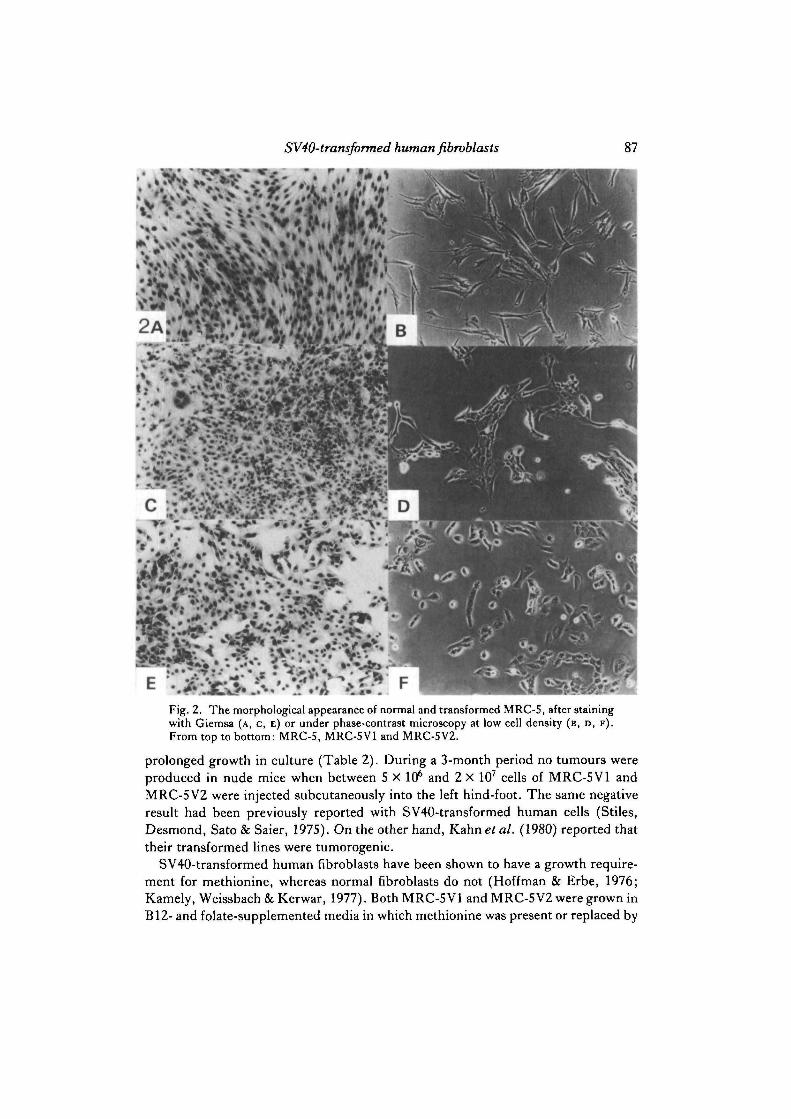
Table 2 summarizes the transformed properties of MRC-5V1 and MRC-5V2.MRC-5V2 always exhibited a distinct epithelial morphology (Fig. 2E), even in its

84 L. I. Huschtscha and R. Holliday
100 r~
75
CD
c23
oC
o 503Q.OQ_
25
—4
100 200Time (days)
300 400
Fig. 1. The growth of transformed MRC-5 following infection with SV40. A. MRC-5V1:infected by the mass culture method ( • • ) , and the uninfected control (O O),which died out at the same time. B. MRC-5V2: infected by the focal isolation method. Sub-lines grown in small Falcon flasks in 10% FCS (A A) and 2 % FCS (A A) andthen in large Falcon flasks in 10% FCS ( • • ) and 2% FCS (G O) (MRC-5V2). The arrow in B indicates that the culture was frozen in liquid nitrogen. Crossesindicate death of culture.

SV40-transformed human fibroblasts 85
pre-transformed state. On the other hand, MRC-5VI maintained a morphology be-tween epithelial and fibroblast-like when the culture was sparse (Fig. 2D), but ap-peared more epithelial as the culture became confluent (Fig. 2c). There was lessuniformity in cell size and shape for this line compared to MRC-5V2. For instance,large cells with up to four nuclei were present (Fig. 2c). Both MRC-5 VI and MRC-5V2 grew equally in reduced serum but MRC-5V1 needed twice the normal con-centration of glucose (2mg/ml) for maximum growth, whereas MRC-5V2 did nothave this requirement. Both transformed lines had lost contact-inhibition, since theycontinued to grow even when the cultures were confluent. However, this was only apartial effect with MRC-5 VI, since its growth rate was reduced at confluence (resultsnot shown). In growth experiments over 7 days, the doubling times for MRC-5 VI and
85
65
c33
oC
o
3Q.O
0-
45
2 5 * .100
Time (days)200
Fig. 1B

86 L. /. Huschtscha and R. Holliday
Table 2. Transformed characteristics ofMRCSVl and MRC-5VZ
Transformed phenotype
Morphology
Serum requirementGlucose requirementDoubling timeGrowth in soft agarT-antigenFree virus production
Tumorigenicity
Growth in mediumlacking methionine
ChromosomesGrowth potential
•P, passage level tested.
MRC-5V1
Intermediate(fibroblast-epithelial)
2%2 mg/ml36-40hPositivePositive
P83»20 p.f.u/mlP313 0 p.f.u/ml
Negative(0/7 mice)Negative
Modal no. 38-42750 + passages
MRC-5V2
Epithelial
2%1 mg/ml35-37hPositivePositive
P137 100 p.f.u/mlP365 0 p.f.u/ml
Negative(0/7 mice)
Positive
Modal no. 60-80650 + passages
MRC-5V2 were always slower than MRC-5; they did not significantly change withp.d.l., even when the cumulative growth rate increased (Fig. 3). For example, thedoubling time of MRC-5V1 at passage 84 was 37 h and at passage 416 it was 38 h. Thedoubling time for MRC-5V2 remained in the range 42-56, compared with 18-21 hfor early-passage MRC-5 cells (Holliday & Kirkwood, 1981). Since the doubling timeof the transformed strains was lower than MRC-5, it might be thought that anyspontaneously occurring transformed cells would be selected against in young or mid-term cultures and this might explain the failure to observe transformed foci in MRC-5 cultures. However, we have shown, by mixing a small proportion of MRC-5 VI cells(0-1-10%) with MRC-5, that the transformed cells invariably take over the cultureduring approximately 10—20 p.d. We assume that this is due to loss of contact-inhibition, which enables the transformed cell to grow even when the culture isconfluent.
Both MRC-5 VI and MRC-5V2 produced colonies in soft agar; however, no growthwas seen below a certain cell density. For MRC-5 VI this threshold was about 10s cellsper 55 mm plate and for MRC-5 V2 about 104 cells per plate. After about 200 p.d. thesethresholds were reduced 10-fold. Estimates of the proportion of cells that formcolonies at or above these thresholds were in the range 8-10 % for MRC-5 V2, but only0-6% for MRC-5V1 at passage 200, rising to 7% at passage 386. Both transformedlines have an extremely low plating efficiency in normal medium (<0*01 %), whereas,in our hands, the parental strain, MRC-5, has a plating efficiency of 2—10%.
Both MRC-5 VI and MRC-5 V2 had T-antigen in the nuclei of all their cells, but thenuclei exhibited a variable intensity of fluorescence, which did not seem to be relatedto the size of the nucleus. Free virus was still found in the medium shortly after crisis,from both MRC-5VI and MRC-5V2, but this ability to shed virus was lost with
SV40-transformed human fibroblasts 87
Fig. 2. The morphological appearance of normal and transformed MRC-S, after stainingwith Giemsa (A, C, E) or under phase-contrast microscopy at low cell density (B, D, F ) .From top to bottom: MRC-5, MRC-5V1 and MRC-5V2.
prolonged growth in culture (Table 2). During a 3-month period no tumours wereproduced in nude mice when between 5 X 10⁶ and 2 x 10⁷ cells of MRC-5V1 andMRC-5V2 were injected subcutaneously into the left hind-foot. The same negativeresult had been previously reported with SV40-transformed human cells (Stiles,Desmond, Sato & Saier, 1975). On the other hand, KahneJ al. (1980) reported thattheir transformed lines were tumorogenic.
SV40-transformed human fibroblasts have been shown to have a growth require-ment for methionine, whereas normal fibroblasts do not (Hoffman & Erbe, 1976;Kamely, Weissbach & Kerwar, 1977). Both MRC-5 VI and MRC-5V2 were grown inB12- and folate-supplemented media in which methionine was present or replaced by

88 L. I. Huschtscha and R. Holliday
homocysteine. MRC-5V1 divided two to three times in methionine-free medium andthen growth ceased, whereas MRC-5V2 grew equally well with or withoutmethionine.
The summary of results in Table 2 shows that MRC-5 VI and MRC-5 V2 have theirown individual characteristics but, in general, MRC-5 V2 was judged to possess a moreextreme transformed phenotype.
Heteroploidy of permanent lines
Metaphase preparations were examined at intervals throughout the growth ofMRC-5 VI and MRC-5 V2. Chromosome counts were made but no detailed karyotypeanalysis was done. These results are shown in Fig. 4. MRC-5 VI has a modal numberbetween 38 and 42, with only a small proportion of the metaphases in each populationhaving a hyper-diploid number of chromosomes - in the range of 60-70. Few othertransformed human lines have previously been found to be sub-diploid (Weinstein &Moorhead, 1965). We do not know if the predominantly low chromosome number inmost MRC-5V1 cells is compensated by rearrangements that might form larger
750
600
CO
a>^450n3o
T3co<o3 300aoa.
150
56
500 1000 1500Time (days)
2000
Fig. 3. Computer plot of the cumulative growth of MRC-5V1. The arrows show thepassage levels at which Glc-6-Pdehydrogenase from MRC-5V1 was examined. X indicatescessation of growth (see Fig. 5) and C that cultures were lost by contamination. I-IV werethe times when cultures were re-established after thawing frozen ampoules (see text).

SV40-transformed human fibroblasts 89
chromosomes. Fig. 4 also shows that there is a gradual increase in the proportion ofhyper-diploid for MRC-5V1 with time in culture. Only 15 % of the metaphases werehyper-diploid shortly after crisis, but the proportion had increased to 45 %approximately 600 p.d. later. This suggests that the MRC-5V1 population is continu-ally changing; either there is selection for cells with higher chromosome numbers, orhyper-diploid cells are continually being derived from sub-diploid ones. On the otherhand, MRC-5V2 is more stable, with no noticeable change in chromosome numberduring growth. Most of the metaphases are hyper-diploid, with a modal numberbetween 60 and 80 chromosomes.
In both strains abnormal chromosomes were common, including breaks, trans-locations, dicentrics and occasionally ring chromosomes. In some cases, all theseabnormalities could be seen in one metaphase. However, no particular marker
Passagenumber
49*98
193286330454557
MRC-5V1
<46 >46
MRC-5V2
100 80 60 40 20 20 40 60 80 100
Fig. 4. The percentages of hypo- and hyper-diploid metaphases in MRC-5V1 and MRC-5V2 at increasing passage levels. The asterisk indicates that the analysis was done beforecrisis.
CEL63
90 L. I. Huschtscha and R. Holliday
chromosome could be seen consistently in MRC-5V1, but in MRC-5V2 a long-armedacrocentric chromosome was detected in all metaphase karyotypes at passage 101, andagain at passage 286. It is interesting to note from Fig. 4 that the extent of heteroploidyin the pre-transformed state is probably related to the transformed state, since MRC-5V2 was hyper-diploid both before and after crisis.
Growth potential
MRC-5V2 seems to possess infinite growth potential, since it has shown no signsof slowing down during 5 years of continuous sub-culture (approx. 650p.d.). Thegrowth rate in terms of cumulative p.d., has increased slightly in later passages, butthere has been no discernible change in the doubling time, morphology orchromosome number.
On the other hand, MRC-5V1 did not grow as a continuous cell line (Fig. 3). Theprimary line was lost due to bacterial contamination at p.d.l. 300, and then severalampoules of frozen stocks had to be defrosted sequentially in order to recover this cellline. The first ampoule (I) was defrosted at p.d.l. 274 and as soon as cells wereavailable they were re-frozen (ampoule II). The remaining cells were lost almostimmediately, probably due to an unknown environmental cause. Ampoule II wasthen defrosted and the cells achieved 30 p.d. before the culture slowed down and diedout (Fig. 5). Some of these cells had been frozen at p.d.l. 287 (ampoule III) and, ondefrosting, six sub-lines were established. All these slowed down and died out after20—30 p.d. The cells became more granular, larger and more fibroblast-like, lookingvery much like senescent fibroblasts - a conclusion that was confirmed by two in-dependent observers. Finally, the cells were again removed from a frozen ampoule(IV) at p.d.l. 294. Two sub-lines died out, but two survived to maintain the cell line.During the course of 6 years of continuous sub-culture (~7S0 p.d.) its growth rate hasgradually increased (Fig. 3). However, the doubling time in 6-day growth experi-ments has been fairly stable throughout, so the faster cumulative growth could beattributed to a greater release from contact-inhibition. Further evidence for this isshown by the increased efficiency of growth in soft agar and the appearance of a moreepithelial-like morphology in the latter passages.
Effects of bromodeoxyuridine (BrdUrd) and temperature
It has been shown that the growth potential of MRC-5 is severely reduced by0-3 ̂ g/ml of BrdUrd, but there is little effect at lower concentrations (unpublishedresults). It was then found that MRC-5VI is much more sensitive to BrdUrd than theparent diploid strain. BrdUrd at 0-1 jug/ml prevented more than 1-2 p.d., but growthof MRC-5V2 continued at a concentration of 1-0/ig/ml.
Transformed lines also showed differences from MRC-5 in their tolerance of tem-peratures higher or lower than 37 °C. It was previously shown that MRC-5 can growat 40 °C and achieves roughly half the lifespan of control cultures at 37 °C (Thompson& Holliday, 1973). MRC-5V1 and MRC-5V2 had a very limited growth at 40°C. Inseveral experiments with MRC-5VI, the cells divided very slowly and achieved only1-5 p.d. However, the effect did not appear to be analogous to the premature

SV40-transformed human fibroblasts 913201 -
310
CD
c33oT3CoJ5Daoa.
300
290
50 100 150Time (days)
Fig. 5. The terminal growth of a subline of MRC-SV1, from defrosted ampoule II (seeFig. 3).
senescence of MRC-5 at 40 °C. At 39 °C MRC-5V1 died within 3-10 p.d., but MRC-5V2 continued to grow. MRC-5VI was also unable to grow continuously at 33 °C or34 °C, whereas MRC-5 achieved a considerable number of population doublings atthese temperatures. The results show the transformed lines are heat-sensitive andMRC-5V1 is cold-sensitive.
Glucose-6-phosphate dehydrogenase
It has been shown that senescent cultures of MRC-5 and skin fibroblasts producea significant fraction of heat-labile Glc-6-P dehydrogenase (Holliday & Tarrant, 1972;Holliday & Thompson, 1982). If the crisis of pre-transformed strains is the same as

92 L. I. Huschtscha and R. Holliday
senescence, it should be possible to detect altered enzyme during or before this period.However, the results obtained were not consistent. MRC-5V1 contained 20% heat-labile dehydrogenase during crisis, and when the permanent line emerged the propor-tion of heat-labile enzyme fell to control levels (0-10%). In the case of MRC-5V2,no significant proportion of heat-labile dehydrogenase was detected to 10 p.d. beforecrisis, and the results were variable when the enzyme was examined from otherpopulations during crisis.
In other experiments on Glc-6-P dehydrogenase during the growth of MRC-5V1,it was noticed that the heat-lability of the enzyme had altered dramatically after manyp.d.s. In cell-free extracts at least 50% of the enzyme was extremely sensitive toheat inactivation at 60 °C (Fig. 6, curve B). This inactivation curve is completelydifferent from those obtained with extracts from senescent cells (Fig. 6, curve A). Itled us to examine the enzyme by starch-gel electrophoresis. A new band was seen thatmigrates more quickly towards the cathode than either the parental B form of theenzyme of the A form. This band disappeared entirely when the gel was incubated at60 °C for 1—2 min prior to staining, whereas the normal band (approximately 50 % of
100
Time (min)0 5 10 15 20 25 30
Fig. 6. The heat inactivation of Glc-6-P dehydrogenase from senescent MRC-5 andMRC-5V1. Curve A, MRC-5, passage 56 (60°C);B, MRC-5V1, passage 270 (59°C); C,MRC-5V1, passage 509 (59-7°C).

SV40-transformed human fibroblasts 93
activity) was not inactivated (Fig. 7). Further experiments were carried out on MRC-5V1 at various passage levels (see Fig. 3) and it became evident that the populationhad become polymorphic for the enzyme at approximately passage 100 and thispersisted for about 200 further passages. Subsequently, the heat-labile form of theenzyme was no longer detectable on gels, although the heat-inactivation profileshowed that the enzyme was still quite unstable (Fig. 6, curve C).
Hybridization
To study the expression of the transformed phenotype in hybrids between normaland transformed cell lines, a new method of selection was devised. This entailed aHAT regime to select against the normal Lesch-Nyhan cells and medium lackingmethionine to inhibit growth of MRC-5V1. Fifteen hybrid clones appeared 2 weeksafter fusion, which represents a frequency of hybrid formation of 0-05 %. These weretransferred into two wells (3-7 cm2) and then into two Leighton tubes (11 cm2) whenthere were sufficient cells. Their morphology was essentially epithelial, although cellsin sparser areas exhibited a more fibroblast-like form. All the clones eventually died
A2
B2
i^^p^^ ^pi^^^^
Fig. 7. Horizontal starch gel electrophoresis of Glc-6-/* dehydrogenase Lanes 1, MRC-5,passage 25; 2, MRC-5V1, passage 96; 3, MRC-SV1, passage 306. A. Extracts at roomtemperature; B, extracts heated for 1 min at 60°C prior to electrophoresis.

94 L. I. Huschtscha and R. Holliday
between 34 and 40 p.d. A normal population of Lesch-Nyhan died after 37 p.d.Chromosomes were counted in five hybrid clones. Their modal number was 50-60chromosomes, which is more than that of either parent, but less than would beexpected from the sum of the two parental cells. The variation in chromosome num-ber was much less than in the MRC-5V1 parent. These observations are similar tothose of Bunn & Tarrant (1980) on hybrids between Lesch-Nyhan and HeLa cells.
DISCUSSION
In the experiments reported here we have reinvestigated the sequence of eventsinvolved in the transformation of cultured human fibroblasts by SV40, and charac-terized two permanent lines that were obtained. One of the aims of the investigationwas to find out whether the crisis period that terminates the first stage in transforma-tion is equivalent to the senescence of normal diploid fibroblasts, and another was totry to gain some insight into the basic difference between diploid and heteroploidtransformed cells, with regard to their limited and unlimited growth potential.
We found that many properties of transformation were acquired soon after viralinfection, such as high mitotic activity at confluence, presence of T-antigen, an-chorage independence and growth in low concentrations of serum. We refer to thesecultures as pre-transformed, because it was invariably observed that growth eventu-ally ceased and the cells entered a degenerative phase, commonly referred to as 'crisis'.The phenotypes of pre-transformed cultures were often quite distinct, but no par-ticular phenotype predisposed the culture to survive crisis and form a permanent line.In a similar study, Sack & Obie (1981) noted individual phenotypes in their clonedpre-transformed cultures. Oshima et al. (1977) were also unable to correlate any pre-transformed feature, such as virion production or T- and V-antigen expression, withthe ability to become fully transformed.
Our results demonstrate that the emergence of permanent lines is a very uncommonevent, since in 10 experiments with MRC-5 only two yielded transformed lines,indicating that the frequency of transformation at the cellular level is probably < 10~7,and there was also no indication that late-passage diploid cultures were more suscept-ible to transformation. These observations confirm those of several other investigators,who either had similar difficulties in obtaining established cell lines after SV40 infec-tion of human cells (Moyeretal. 1964), or did not obtain any at all (Shemetal. 1964;Ruben & Rafferty, 1978; Raffertyef a/. 1978; Gotohetal. 1979; Sack&Obie, 1981).They do not support the results of Jensen et al. (1963), Todaro et al. (1963) andGirardief al. (1965), who reported a high rate of success in producing permanent celllines, especially with senescent populations. The variable length of the crisis period(i.e. from 2—14 weeks) suggests that the rare events involved in immortalization occurduring rather than before crisis, as selection of an immortal clone would not take morethan about 4 weeks (assuming a doubling time of approximately 35 h). Moreover,within the crisis period foci may appear that have limited capacity for growth. In otherexperiments with SV40-treated human skin fibroblasts, the crisis period sometimeslasted up to 200 days before permanent lines emerged (unpublished results).

SV40-transformed human fibroblasts 95
The individual nature of the pre-transformed cultures was further exemplified bycomparing the histories of our two transformed lines, MRC-5 VI and MRC-5V2. Pre-transformed MRC-5V1 had fairly minor changes in growth properties, while thephenotype of pre-transformed MRC-5V2 was more extreme. Both permanent linesretained similar features of their pre-transformed state, including morphological ap-pearance and chromosome number. The major phenotypic differences betweenMRC-5VI and MRC-5V2 are their morphological appearance, modal chromosomenumber, glucose and methionine requirements, sensitivity to BrdUrd and elevatedincubation temperature. Long-term growth has shown that both lines have lost theirtendency to shed free virus into the medium, and rates of cumulative populationdoublings have increased in later passages, without a detectable change in growth ratein short-term experiments. The gradual increase in anchorage independence probablyindicates that there is also a greater release from contact-inhibition. In both lines, thechromosome number gradually increased during sub-culture, but this change wasmore marked in MRC-5VI. Karyotype instability has also been reported for otherSV40-transformed lines (Begovich & Francke, 1979).
MRC-5V1 showed two further interesting examples of instability. The growth ofsome sub-lines slowed over a period of many population doublings and the culturedied out, in a manner that was very reminiscent of the senescence and death of diploidfibroblasts. In several cases, cells recovered from liquid nitrogen also gave rise topopulations with finite growth. However, it has been possible to keep the line growingand it has now reached p.d.l. 750. The question arises as to whether this line containsan immortal sub-population that constitutes only a small minority of the cells. If thissub-population is lost (perhaps by drift or stochastic fluctuation in cell numbers), thenthe remaining cells may continue to divide for a while before eventually dying out. Wepreviously proposed that very early passage diploid fibroblasts contain a sub-population of immortal cells that are inevitably lost, because during growth theychange with a given frequency to cells with finite growth potential (Kirkwood &Holliday, 1975; Holliday et al. 1977). In transformed cultures, a similar sub-population would normally be retained, but it might occasionally be lost, especiallyafter a reduction in population size during freezing and thawing. Unfortunately, thedifficulty of growing individual clones from MRC-5 VI has not made it possible to testthis hypothesis directly. It is, however, known that many clones isolated from HeLacell populations have limited growth potential (Martinez, Norwood, Prothero &Martin, 1978; K. A. Rafferty, personal communication).
The instability of MRC-5V1 was also shown at the biochemical level. The propor-tion of heat-labile Glc-6-P dehydrogenase was studied at intervals to see whether thepopulations maintained the low levels observed in young cultures of diploid humanfibroblasts. Whereas in MRC-5V2 there was no significant change in the enzyme over600 p.d., MRC-5V1 acquired a new form of the enzyme, which was extremely sen-sitive to heat and was also distinguishable from the normal enzyme by its fastermigration in starch gels. However, this new form of Glc-6-P dehydrogenase disap-peared after a further 200 p.d. The heat-inactivation experiments also suggested thatother allelic forms of the dehydrogenase had been produced during the growth of

96 L. I. Huschtscha and R. Holliday
MRC-5V1. There have been two other reports of newly acquired isoenzymes of Glc-6-P dehydrogenase in mouse and human tumours (Hilf, Rector, Abraham & Lyon,1973; Khan, Shin & Steinberg, 1978). In the former, a faster migrating band of theenzyme in polyacrylamide gels was detected in pre-neoplastic and neoplastic mam-mary adenocarcinomas from Balb/c mice, while in the latter, a new modified form ofthe enzyme was observed by isoelectric focusing in various human leukaemic cells.Extracts of leukaemic cells, when incubated with normal Glc-6-P dehydrogenase, ledto a modification of this enzyme, identical to that seen in the leukaemic cells. We werenot able to show this to be the case when our normal and transformed cell-free extractswere mixed.
We do not know whether the new forms of Glc-6-P dehydrogenase are due to post-synthetic modifications, or to changes in the structural gene. It may be that the well-known karyotypic instability of transformed cells is also accompanied by instabilityat the gene level. For instance, the amplification of genes in response to anti-metabolite inhibitors has been demonstrated, so far as we are aware, only in per-manent lines (e.g. see Schimke, 1982). In any event, the widely held view that theelectrophoretic profile of any enzyme is an unchanging diagnostic characteristic of anyparticular cell strain or line, may need reassessment. In the case of MRC-5V1, itseemed that an initial homogeneous population, possibly a clone, had itself becomepolymorphic for Glc-6-P dehydrogenase. We presume that the changes seen in theproperties of the enzyme are at least in part due to continuous selection of fast-growingsub-populations of cells, and their subsequent replacement by other populations.
One of the aims of this study was to try to determine whether the crisis of pre-transformed cells is the same as senescence (phase III) of diploid populations. Cellsin crisis are invariably large, often multinucleate, granular and with variable morphol-ogy. Large numbers of cells detach and form debris in the medium. In these respectsthey resemble phase III diploid cells. We tried to exploit the observation that sene-scent MRC-5 cultures have elevated levels of heat-labile Glc-6-P dehydrogenase(Holliday & Tarrant, 1972). The results obtained were ambiguous, in that somecultures near to or within crisis contained 15-25% heat-labile enzyme, which ischaracteristic of senescent MRC-5, whereas others had a heat-labile fractionequivalent to young populations. (Note that any changes in a minor sub-fraction ofenzyme molecules should be clearly distinguished from the major alterations in theenzyme that we observed during the long-term growth of MRC-5VI.) It is possiblethat in some cases there was a loss of unstable enzyme molecules in the slowly dividingor non-growing cells, before the heat stability of Glc-6-P dehydrogenase wasexamined. Better biochemical methods are needed to compare pre-transformed cellsin crisis with senescent diploid fibroblasts.
The events that allow occasional cells to survive crisis and form a permanent lineremain obscure. These cells are, in a sense, rejuvenated. For instance, it was recentlyshown that they contain a low level of autofluorescence (AF), which is a specificcharacteristic of early-passage MRC-5 cells (Rattan, Keeler, Buchanan & Holliday,1982). It is probable that the escape from senescence is due to the loss of one or morecellular functions, since hybrids between diploid and transformed cells usually have

SV40-transformed human fibroblasts 97
a finite lifespan (Bunn & Tarrant, 1980; Muggleton-Harris & DeSimone, 1980;Periera-Smith & Smith, 1981). We exploited the methionine requirement of MRC-5V1 to select hybrids between this line and Lesch-Nyhan fibroblasts and also foundthat these eventually died out.
The early events in transformation clearly involve the frequent integration of SV40and the expression of the T-antigens, but the later event or events during the crisisperiod, which give rise to permanent lines, are unknown. Since immortalization isvery rare in human cells, it is reasonable to suppose that it depends on mutation,rearrangement or transposition of the SV40 genome in individual cells, althoughepigenetic switches in gene activity are also possible (Holliday, 1979; Harris, 1982).In rodent cells, immortalization occurs spontaneously and at high frequency afterSV40 infection (Ponten, 1971). A comparative study of in vitro transformation indifferent species may throw light on the mechanisms whereby cells escape fromsenescence.
We thank Dr S. J. Fulder for his initial support, Drs P. Gallimore, K. Rafferty and G. H. Sack,Jr, for their interest and helpful comments, and T. B. L. Kirkwood for computing the long-termgrowth curves. We also thank Dr M. Bobrow for the Q band staining, Drs D. Lane and J. ArTandfor supplying batches of hamster anti-SV40 T-antigen, S. Povey for her help in characterizingenzymes, and D. M. Field for her patience and accuracy in the typing of this script.
REFERENCES
BEGOVICH, A. & FRANCKE, U. (1979). Karyotype evolution of the simian virus 40-transformedhuman cell line LNSV. Cytogenet. Cell Genet. 23, 3-11.
BUNN, C. L. & TARRANT, G. M. (1980). Limited lifespan in somatic cell hybrids and cybrids. ExplCell Res. 127, 385-3%.
CHEN, T. R. (1977). In situ detection of mycoplasma contamination in cell cultures by fluorescentHoescht 33258 stain. Expl Cell Res. 104 255-262.
FULDER, S. J. (1979). Evidence for an increase in presumed somatic mutation during the ageing ofhuman cells in culture. Mech. Ageing Dev. 10, 101-115.
GIRARDI, A. J., JENSEN, F. C. & KOPROWSKI, H. (1965). SV40-induced transformation of humandiploid cells: crisis and recovery. J . cell. comp. Physiol. 65, 69-84.
GOTOH, S., GLEB, L. & SCHLESSINGER, D. (1979). SV40-transformed human diploid cells thatremain transformed throughout their limited lifespan. J. gen. Viml. 42, 409-414.
GREINER, J. W., EVANS, C. H. & Di PAOLO, J. A. (1981). Carcinogen-induced anchorage-independent gTowth and in vivo lethality of human MRC-5 cells. Carcinogenesis 2, 359-362.
HARRIS, M. (1982). Induction of thymidine kinase in enzyme deficient Chinese hamster cells. Cell29, 483-493.
HILF, R., RECTOR, W., ABRAHAM, S. & LYON, B. (1973). A glucose-6-phosphate dehydrogenaseisoenzyme characteristic of preneoplastic and neoplastic mouse mammary tissue. J. natn. CancerInst. 50, 1395-1398.
HOFFMAN, R. M. & ERBE, R. W. (1976). High in vivo rates of methionine biosynthesis in transfor-med human and malignant rat cells auxotrophic for methionine. Proc. natn.Acad. Sci. U.SA. 73,1523-1527.
HOLLIDAY, R. (1979). A new theory of carcinogenesis. Br.J. Cancer 40, 513-521.HOLLIDAY, R., HUSCHTSCHA, L. I., TAKRANT, G. M. & KIRKWOOD, T. B. L. (1977). Testing
the commitment theory of cellular ageing. Science, N.Y. 198, 366—372.HOLLIDAY, R. & KIRKWOOD, T. B. L. (1981). Predictions of the somatic mutation and mortiliza-
tion theories of cellular ageing are contrary to experimental observations. J. theor. Biol. 93,627-642.

98 L. /. Huschtscha and R. Holliday
HOLLIDAY, R. & TARRANT, G. M. (1972). Altered enzymes in ageing human fibroblasts. Nature,Land. 238, 26-30.
HOLLIDAY, R. & THOMPSON, K. V. A. (1982). Genetic effects on the longevity of cultured humanfibroblasts. III . Correlations with altered glucose-6-phosphate dehydrogenase. Gerontology (inpress).
JACOBS, J. P., GARRETT, A. J. & MERTON, R. (1979). Characteristics of a serially propagatedhuman diploid cell line designated MRC5-9. Bio. Standard. 7, 113-122.
JACOBS, J. P., JONES, C. M. & BAILLIE, J. P. (1970). Characteristics of a human diploid celldesignated MRC-5. Nature, Land. 227, 168-170.
JENSEN, F. C , KOPROWSKI, H. &PONTEN, J. A. (1963). Rapid transformation of human fibroblastcultures by simian virus 40. Proc. natn. Acad. Sci. U.SA. 50, 343-348.
KAHN, A., BOIVIN, P., RUBINSON, H., COTTREAU, D., MARIE, J. & DREYFUS, J. (1978). Modifi-cations of purified glucose-6-phosphate dehydrogenase and other enzymes by a factor of lowmolecular weight abundant in some leukaemic cells. Proc. natn. Acad. Set. U.SA. 73, 77-81.
KAHN, P., SHIN, S. & STEINBERG, B. (1980). Tumourigenicity of SV40-transformed primate cellsin immunodeficient mice. Eur.J. Cell Biol. 22, 527.
KAKUNAGA, T. (1978). Neoplastic transformation of human diploid fibroblast cells by chemicalcarcinogens. Proc. natn. Acad. Sci. U.SA. 75, 1334-1338.
KAMELY, D., WEISSBACH, H. & KERWAR, S. S. (1977). Methionine biosynthesis in normal andtransformed fibroblasts. Archs Biochem. Biophys. 179, 43-45.
KIRKWOOD, T. B. L. & HOLLIDAY, R. (1975). Commitment to senescence: a model for the finiteand infinite gTOwth of diploid and transformed human fibroblasts in culture. ,7. theor. Biol. 53,481-496.
KOPROWSKI, H., PONTEN, J. A., JENSEN, F. C , RAVDIN, R. G., MOORHEAD, P. & SAKSELA, E.
(1962). Transformation of cultures of human tissue infected with simian virus SV40. J. cell.comp. Physiol. 59, 281-292.
LINN, S., KAIRIS, M. & HOLLIDAY, R. (1976). Decreased fidelity of DNA polymerase activityisolated from ageing human fibroblasts. Proc. natn. Acad. Sci. U.SA. 73, 2818-2822.
MACPHERSON, I. & MONTAGNIER, L. (1964). Agar suspension culture for the selective assay of cellstransformed by polyoma virus. Virology 23, 291-294.
MARTINEZ, A. O., NORWOOD, T. H., PROTHERO, J. W. & MARTIN, G. M. (1978). Evidence for
clonal attenuation of growth potential in HeLa cells. In Vitro 14, 996-1002.MILLER, O. J., MILLER, D. A., ALDERDICE, P. W., DEV, E. G. & GREWAL, M. S. (1971).
Quinacrine fluorescent karyotypes of human diploid and heteroploid cell lines. Cytogenetics 10,338-346.
MOYER, A. W., WALLACE, R. &COX, H. R. (1964). Limited growth period of human lung cell linestransformed by simian virus 40. J. natn. Cancer Inst. 33, 227-232.
MUGGLETON-HARRIS, A. & DESIMONE, D. (1980). Replicative potentials of various fusionproducts between WI-38 and SV40 transformed WI-38 cells and their components. Somat. CellGenet. 6, 689-698.
MURRAY, V. & HOLLIDAY, R. (1981). Increased error frequency of DNA polymerases from sene-scent human fibroblasts. J. molec. Biol. 146, 55-76.
NISHIDA, S. (1970). Studies on the transformation of human fetal cell cultures by simian virus 40.Acta med., Okayama 24, 417-443.
OSHIMA, R. G., PELLETT, O. L., ROBB, J. A. & SCHNEIDER, J. A. (1977). Transformation of
human cystinotic fibroblasts by SV40: Characteristics of transformed cells with limited andunlimited growth potential. J . cell. Physiol. 93, 129-136.
PERIERA-SMITH, O. M. & SMITH, J. R. (1981). Expression of SV40 T-antigen in finite lifespanhybrids of normal and SV40 transformed fibroblasts. Somat. Cell Genet. 7, 411-421.
PETES, T. D., FARBER, R. A., TARRANT, G. M. & HOLLIDAY, R. (1974). Altered rate of DNAreplication in ageing human fibroblast cultures. Nature, Land. 251, 434—436.
POPE, J. H. & ROWE, W. P. (1964). Detection of specific antigen in SV40 transformed cells byimmunofluorescence. J. exp. Med. 120, 121-128.
PONTEN, J. (1971). Spontaneous and virus inducted transformation in cell culture. Virol. Monogr.8, 59-75.
RAFFERTY, K., RUBEN, R. L. & YOUNG, S. K. (1978). A virus-producing cell line produced bytransformation of human parapharyngeal cells with SV40. In Vitro 14, 227-235.

SV40-transformed human fibroblasts 99
RATTAN, S. I. S., KEELER, K. D., BUCHANAN, J. H. & HOLLIDAY, R. (1982). Autofluorescence
as an index of ageing in human fibroblasts in culture. BioScience Rep. 2, 561-567.RUBEN, R. L. & RAFFERTY, K. A. (1978). Limited in vitro replicative lifespan of cultured human
epithelial cells. Survey of recent literature. Growth 42, 357-368.RUTTER, W. J., PICTET, R. L. & MORRIS, P. W. (1973). Toward molecular mechanisms of develop-
mental processes. A. Rev. Biochem. 42, 601-646.SACK, G. H. JR (1980). Human cell transformation by simian virus SV40- a review. In Vitro 17,
1-19.SACK, G. H. J R & O B I E , C. (1981). Human cell transformation by simian virus 40; biologic features
of cloned lines. Expl Cell Res. 134, 425-432.SCHIMKE, R. T. (1982). Gene amplification. New York: Cold Spring Harbor Laboratory.SHAKESPEARE, V. A. & BUCHANAN, J. H. (1978). Studies of phosphoglucose isomerase from
altered human fibroblasts: absence of detectable age effect on the enzyme. .7. cell. Physiol. 94,105-116.
SHEIN, H. M., ENDERS, J. F., PALMER, L. & GROGAN, E. (1964). Further studies on SV40-induced transformation in human renal cell cultures. I. Eventual failure of subcultivation despitea continuing high rate of cell division. Proc. Soc. exp. Biol. Med. 115, 618—621.
STILES, C. D., DESMOND, W. JR, SATO, G. & SAIER, M. H. JR (1975). Failure of human cellstransformed by SV40 to form tumours in athymic nude mice. Proc. natn. Acad. Set. U.SA. 72,4971-4975.
THOMPSON, K. V. A. & HOLLIDAY, R. (1973). Effect of temperature on the longevity of humanfibroblasts in culture. Expl Cell Res. 80, 354-360.
TODARO, G. J., WOLMAN, S. R. & GREEN, H. (1963). Rapid transformation of human fibroblastswith low growth potential into established cell lines by SV40. J. cell. Physiol. 62, 257-265.
WEINSTEIN, D. & MOORHEAD, P. S. (1965). Karyology of permanent human cell line W-18VA2,originated by SV40 transformation. J . cell. comp. Physiol. 65, 85-92.
(Received 3 December 1982 -Accepted 28 March 1983)
