long bone and vertebral microanatomy and osteo-histology...
TRANSCRIPT
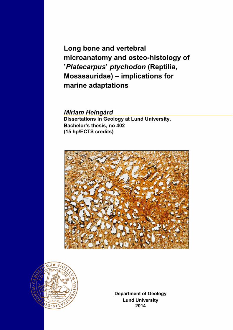
Long bone and vertebral
microanatomy and osteo-histology of
’Platecarpus’ ptychodon (Reptilia,
Mosasauridae) – implications for
marine adaptations
Miriam Heingård Dissertations in Geology at Lund University,
Bachelor’s thesis, no 402
(15 hp/ECTS credits)
Department of Geology
Lund University
2014

Long bone and vertebral microanatomy and osteo-histology of
’Platecarpus’ ptychodon (Reptilia, Mosasauridae) - implications for
marine adaptations
Bachelor’s thesis Miriam Heingård
Department of Geology Lund University
2014
Contents
1 Introduction ........................................................................................................................................................ 7
1.1 Bone development and aquatic adaptations in tetrapods 7
1.2 Mosasaur evolution and adaptation 8
1.3 Aim of the study 9
2 Material and methods ........................................................................................................................................ 9
3 Results ................................................................................................................................................................. 9
3.1 Humerus 9
3.2 Vertebra 10
4 Discussion.......................................................................................................................................................... 11
4.1 Humerus 11
4.2 Vertebra 12
4.3 Adaptive and ecological implications 12
5 Conclusions ....................................................................................................................................................... 13
6 Acknowledgements........................................................................................................................................... 13
7 References ......................................................................................................................................................... 13
Cover Picture: Humeral microanatomy of ’Platecarpus’ ptychodon.
Long bone and vertebral microanatomy and osteo-histology of
’Platecarpus’ ptychodon (Reptilia, Mosasauridae) - implications
for marine adaptations
MIRIAM HEINGÅRD
Heingård, M., 2014: Long bone and vertebral microanatomy and osteo-histology of ’Platecarpus’ ptychodon
(Reptilia, Mosasauridae) - implications for marine adaptations. Dissertations in Geology at Lund University, No.
402, 15 pp. 15 hp (15 ECTS credits).
Keywords: bone histology, bone microanatomy, mosasaurs, aquatic adaptations, growth rate
Supervisor(s): Johan Lindgren
Subject: Bedrock Geology
Miriam Heingård, Department of Geology, Lund University, Sölvegatan 12, SE-223 62 Lund, Sweden. E-mail:
Abstract: The inner bone architecture and histology provide information about life history traits of extant as well
as extinct animals. Mosasaurs (family Mosasauridae) are a group of secondarily adapted marine squamates that
radiated in the Late Cretaceous, resulting in the evolution of a body plan adapted for pelagic habitats. Specialisa-
tions in this group can be observed at different levels of skeletal anatomy, including bone microstructures and osteo
-histology. This study describes the histological features and microanatomy observed in bone sections of a vertebra
and a long bone (humerus) from derived mosasaur ’Platecarpus’ ptychodon. Bone sections consist mainly of can-
cellous bone with a gradual transition to a rather thin outer layer of cortical bone. The bone histology in both sec-
tions is characterised by poorly organised tissue; fibro-lamellar bone is abundant throughout the sections, display-
ing randomly organised lacunae and lamellar bone in osteons, whereas parallel-fibered bone (lamellar-zonal bone)
is observed only in one area of the vertebra. This implies a rapid growth rate possibly similar to some ichthyosaurs
and plesiosaurs. The present contribution demonstrates that ’P’. ptychodon exhibit microstructures consistent with
life in pelagic environments and show evidence for rapid growth rate and an active marine lifestyle. Furthermore, a
structure found in the vertebra is probably related to avascular necrosis, a disease caused by decompression sick-
ness, which adds information about the ecology of this species.
Osteohistologi och mikrostrukturer i ett överarmsben och en
ryggkota från ’Platecarpus’ ptychodon - implikationer för marina
anpassningar
MIRIAM HEINGÅRD
Heingård, M., 2014: Long bone and vertebral microanatomy and osteo-histology of ’Platecarpus’ ptychodon
(Reptilia, Mosasauridae) - implications for marine adaptations. Examensarbeten i geologi vid Lunds universitet, Nr.
402, 15 sid. 15 hp.
Nyckelord: benhistologi, mikroanatomi, mosasaurier, akvatisk anpassning, tillväxthastighet
Handledare: Johan Lindgren
Ämnesinriktning: Berggrundsgeologi
Miriam Heingård, Geologiska institutionen, Lunds universitet, Sölvegatan 12, 223 62 Lund, Sverige. E-post:
Sammanfattning: Den inre strukturen och vävnaden hos ben ger information om livsstil hos nutida och utdöda
djur. Mosasaurier (familj Mosasauridae) är en grupp sekundärt anpassade marina reptiler som utvecklades och
spred sig under sen krita, vilket resulterade i en kropp väl anpassad för marina miljöer. Marina anpassningar hos
den här gruppen kan observeras på olika nivåer i skelettanatomin, exempelvis mikrostrukturer och benvävnad. Den
här studien beskriver histologiska och mikroanatomiska kännetecken i ett överarmsben och en ryggkota hos den väl
anpassade mosasaurien ’Platecarpus’ ptychodon. Tunnslip visar att benen till stor del består av trabekulärt ben med
en gradvis övergång till det mer kompakta yttre benlagret. Detta är jämförbart med många moderna valar. Benhisto-
login karaktäriseras av en avsaknad av organiserad vävnad. Fibro-laminärt ben med slumpmässigt orienterade laku-
ner finns rikligt i båda benen och laminärt ben finns i osteoner. Parallellfibröst ben observeras endast i en del av
ryggkotan. Detta antyder en snabb tillväxthastighet, eventuellt liknande den hos vissa ichthyosaurier och plesiosau-
rier. Den här studien visar att ’P’ ptychodon har mikrostrukturer som stämmer överens med en aktiv pelagisk livs-
stil. Den uppvisar även tecken på en snabb tillväxthastighet. En struktur i ryggkotan är troligtvis relaterad till osteo-
nekros, en sjukdom som orsakas av tryckfallssjuka, vilket bidrar till förståelsen av den här artens ekologi.
7
1 Introduction Marine vertebrates that originated on land show a
range of aquatic adaptations, and to understand the
evolution of these groups the study of their fossilised
rema ins i s e s sen t ia l . I n extan t spec ie s the
determination of lifestyle and life history traits is
possible through direct observation, whereas in extinct
vertebrates skeletal remains provide some of that
information. The lifestyle of an animal is often
reflected at different levels of their skeletal anatomy,
ranging from gross anatomy to bone microstructures.
S tud ies have shown a re la t ionship be tween
microanatomy and lifestyle, e.g. in amniotes (Germain
& Laurin 2005; Kriloff et al. 2008; Canoville & Laurin
2010). The inner bone organisation of tissue is studied
in the field of osteo-histology. Analysis of the bone
tissue is a useful tool for providing additional
ecological data and work as a complement to
anatomical data from extant as well as extinct taxa.
The study of histological and microanatomical features
in fossil taxa can thus be used as a source of
information on life history traits. Additionally, the
lifestyle(s) of taxa whose habitats are uncertain can be
inferred to some extent.
1.1 Bone development and aquatic adaptations in tetrapods
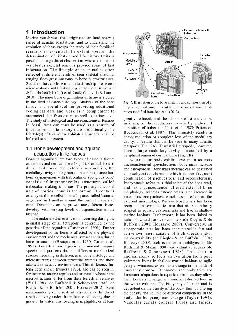
Bone is organised into two types of osseous tissue;
cancellous and cortical bone (Fig. 1). Cortical bone is
dense and forms the exterior surrounding the
medullary cavity in long bones. In contrast, cancellous
bone (synonymous with trabecular or spongious bone)
consists of interconnecting structures cal led
trabeculae, making it porous. The primary functional
unit of cortical bone is the osteon. It contains
osteocytes (bone cells) in small spaces called lacunae,
organised in lamellae around the central Haversian
canal. Depending on the growth rate different tissues
develop with varying levels of organisation of the
lacunae.
The endochondral ossification occurring during the
neonatal stage of all tetrapods is controlled by the
genetics of the organism (Carter et al. 1991). Further
development of the bone is affected by the physical
environment and the mechanical stresses acting during
bone maturation (Beaupre et al. 1990; Carter et al.
1991). Terrestrial and aquatic environments require
special adaptations due to different mechanical
stresses, resulting in differences in bone histology and
microstructures between terrestrial animals and those
adapted to aquatic environments. This difference has
long been known (Nopcsa 1923), and can be seen in,
for instance, marine reptiles and mammals whose bone
microstructures differ from their terrestrial relatives
(Wall 1983; de Buffrénil & Schoevaert 1988; de
Ricqlès & de Buffrénil 2001; Houssaye 2012). Bone
microanatomy of terrestrial tetrapods is the direct
result of living under the influence of loading due to
gravity. In water, this loading is negligible, or at least
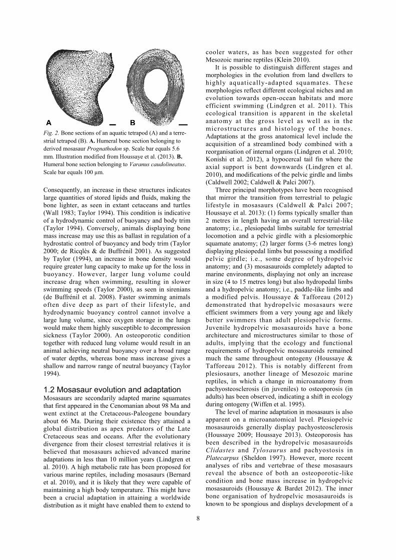
greatly reduced, and the absence of stress causes
infilling of the medullary cavity by endosteal
deposition of trabeculae (Pitts et al. 1983; Patterson-
Buckendahl et al. 1987). This ultimately results in
heavy reduction or complete loss of the medullary
cavity, a feature that can be seen in many aquatic
tetrapods (Fig. 2A). Terrestrial tetrapods, however,
have a large medullary cavity surrounded by a
peripheral region of cortical bone (Fig. 2B).
Aquatic tetrapods exhibit two main osseous
microanatomical specialisations: bone mass increase
and osteoporosis. Bone mass increase can be described
as pachyosteosclerosis which is the frequent
combination of pachyostosis and osteosclerosis.
Pachyostosis refers to a thickening of the bone wall,
and, as a consequence, altered external bone
morphology, whereas osteosclerosis is an increase in
inner bone compactness which has no effect on the
external morphology. Pachyosteosclerosis has been
recorded in semiaquatic taxa that are secondarily
adapted to aquatic environments and live in shallow
marine habitats. Furthermore, it has been linked to
rather slow and passive swimmers (de Ricqlès & de
Buffrénil 2001; Houssaye 2009). Conversely, an
osteoporotic state has been encountered in fast and
active swimmers capable of high speeds and/or
manoeuvrability (de Ricqlès & de Buffrénil 2001;
Houssaye 2009), such as the extinct ichthyosaurs (de
Buffrénil & Mazin 1990) and extant cetaceans (de
Buffrénil & Schoevaert 1988) . This shift in
microanatomy reflects an evolution from poor
swimmers living in shallow marine habitats to agile
pelagic swimmers, as well as a change in the mode of
buoyancy control. Buoyancy and body trim are
important adaptations in aquatic animals as they allow
them to stay submerged and remain at desired level in
the water column. The buoyancy of an animal is
dependent on the density of the body, thus, by altering
the density and volume of different components in the
body, the buoyancy can change (Taylor 1994).
Vascu la r cana l s co n ta i n f l u id s and l ip id s .
Fig. 1. Illustration of the bone anatomy and composition of a
long bone, displaying different types of osseous tissue. Illust-
ration modified from Bao et al. (2013).
8
Consequently, an increase in these structures indicates
large quantities of stored lipids and fluids, making the
bone lighter, as seen in extant cetaceans and turtles
(Wall 1983; Taylor 1994). This condition is indicative
of a hydrodynamic control of buoyancy and body trim
(Taylor 1994). Conversely, animals displaying bone
mass increase may use this as ballast in regulation of a
hydrostatic control of buoyancy and body trim (Taylor
2000; de Ricqlès & de Buffrénil 2001). As suggested
by Taylor (1994), an increase in bone density would
require greater lung capacity to make up for the loss in
buoyancy. However, larger lung volume could
increase drag when swimming, resulting in slower
swimming speeds (Taylor 2000), as seen in sirenians
(de Buffrénil et al. 2008). Faster swimming animals
often dive deep as part of their lifestyle, and
hydrodynamic buoyancy control cannot involve a
large lung volume, since oxygen storage in the lungs
would make them highly susceptible to decompression
sickness (Taylor 2000). An osteoporotic condition
together with reduced lung volume would result in an
animal achieving neutral buoyancy over a broad range
of water depths, whereas bone mass increase gives a
shallow and narrow range of neutral buoyancy (Taylor
1994).
1.2 Mosasaur evolution and adaptation Mosasaurs are secondarily adapted marine squamates
that first appeared in the Cenomanian about 98 Ma and
went extinct at the Cretaceous-Paleogene boundary
about 66 Ma. During their existence they attained a
global distribution as apex predators of the Late
Cretaceous seas and oceans. After the evolutionary
divergence from their closest terrestrial relatives it is
believed that mosasaurs achieved advanced marine
adaptations in less than 10 million years (Lindgren et
al. 2010). A high metabolic rate has been proposed for
various marine reptiles, including mosasaurs (Bernard
et al. 2010), and it is likely that they were capable of
maintaining a high body temperature. This might have
been a crucial adaptation in attaining a worldwide
distribution as it might have enabled them to extend to
cooler waters, as has been suggested for other
Mesozoic marine reptiles (Klein 2010).
It is possible to distinguish different stages and
morphologies in the evolution from land dwellers to
highly aquatically-adapted squamates. These
morphologies reflect different ecological niches and an
evolution towards open-ocean habitats and more
efficient swimming (Lindgren et al. 2011). This
ecological transition is apparent in the skeletal
anatomy at the gross level as well as in the
micros tructures and histology of the bones.
Adaptations at the gross anatomical level include the
acquisition of a streamlined body combined with a
reorganisation of internal organs (Lindgren et al. 2010;
Konishi et al. 2012), a hypocercal tail fin where the
axial support is bent downwards (Lindgren et al.
2010), and modifications of the pelvic girdle and limbs
(Caldwell 2002; Caldwell & Palci 2007).
Three principal morphotypes have been recognised
that mirror the transition from terrestrial to pelagic
lifestyle in mosasaurs (Caldwell & Palci 2007;
Houssaye et al. 2013): (1) forms typically smaller than
2 metres in length having an overall terrestrial-like
anatomy; i.e., plesiopedal limbs suitable for terrestrial
locomotion and a pelvic girdle with a plesiomorphic
squamate anatomy; (2) larger forms (3-6 metres long)
displaying plesiopedal limbs but possessing a modified
pelvic girdle; i.e., some degree of hydropelvic
anatomy; and (3) mosasauroids completely adapted to
marine environments, displaying not only an increase
in size (4 to 15 metres long) but also hydropedal limbs
and a hydropelvic anatomy; i.e., paddle-like limbs and
a modified pelvis. Houssaye & Tafforeau (2012)
demonstrated that hydropelvic mosasaurs were
efficient swimmers from a very young age and likely
better swimmers than adult plesiopelvic forms.
Juvenile hydropelvic mosasauroids have a bone
architecture and microstructures similar to those of
adults, implying that the ecology and functional
requirements of hydropelvic mosasauroids remained
much the same throughout ontogeny (Houssaye &
Tafforeau 2012). This is notably different from
plesiosaurs, another lineage of Mesozoic marine
reptiles, in which a change in microanatomy from
pachyosteosclerosis (in juveniles) to osteoporosis (in
adults) has been observed, indicating a shift in ecology
during ontogeny (Wiffen et al. 1995).
The level of marine adaptation in mosasaurs is also
apparent on a microanatomical level. Plesiopelvic
mosasauroids generally display pachyosteosclerosis
(Houssaye 2009; Houssaye 2013). Osteoporosis has
been described in the hydropelvic mosasauroids
Clidastes and Tylosaurus and pachyostosis in
Platecarpus (Sheldon 1997). However, more recent
analyses of ribs and vertebrae of these mosasaurs
reveal the absence of both an osteoporotic -like
condition and bone mass increase in hydropelvic
mosasauroids (Houssaye & Bardet 2012). The inner
bone organisation of hydropelvic mosasauroids is
known to be spongious and displays development of a
Fig. 2. Bone sections of an aquatic tetrapod (A) and a terre-
strial tetrapod (B). A. Humeral bone section belonging to
derived mosasaur Prognathodon sp. Scale bar equals 5.6
mm. Illustration modified from Houssaye et al. (2013). B.
Humeral bone section belonging to Varanus caudolineatus.
Scale bar equals 100 µm.
9
true trabecular organisation, while the peripheral layer
of the cortical bone is generally rather thin (Sheldon
1997; Houssaye 2012; Houssaye & Bardet 2012;
Houssaye et al. 2013).
1.3 Aim of the study The present study describes histological features and
microanatomical structures observed in a humerus and
a vertebra of ‘Platecarpus’ ptychodon, a hydropelvic
and hydropedal mosasaur hi ther to descr ibed
exclusively from isolated teeth (Arambourg 1952).
Sections of the bones were examined in order to
describe and analyse the bone histology and
microanatomy, and to relate these structures to marine
adaptations in this derived mosasaur.
2 Material and methods Mosasaur bone samples (a humerus and a vertebra)
belonging to different individuals of ’Platecarpus’
ptychodon were collected from marine sediments of
Maastrichtian age near Bentiaba, Angola (Mateus et al.
2012). The vertebra belongs to a semi-complete
skeleton. The humerus was identified from a
characteristic crest in the distal lateral part of the bone.
Prior to histological sectioning the bones were
photographed and casted. Casts were made using clay
moulds and silicone mixed with a hardening agent.
The bones were cut longitudinally into three pieces, of
which the middle piece of each bone was selected for
analysis. This was encased in a two component epoxy
(Araldite) to create support and to prevent the bones
from shattering during cutting and polishing. A
Buehler Isomet low speed diamond saw was used to
cut two sections of approximately 1 mm thickness
from each of the encased bones. The sections were
attached to glass slides using epoxy and polished to
desired thickness. Bone sections were examined under
an optical microscope, using magnifications ranging
from 4x to 50x.
The histological terminology is based on Houssaye
et al. (2013).
3 Results
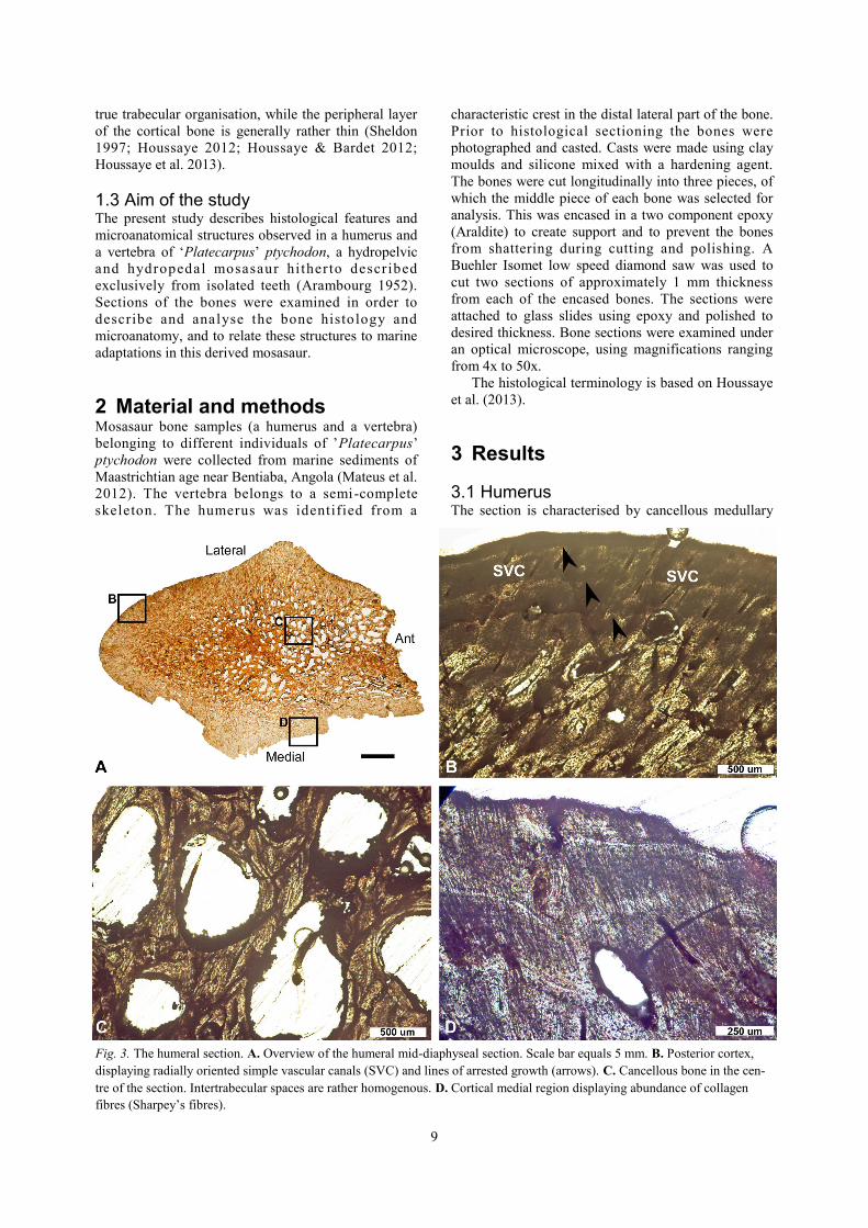
3.1 Humerus The section is characterised by cancellous medullary
Fig. 3. The humeral section. A. Overview of the humeral mid-diaphyseal section. Scale bar equals 5 mm. B. Posterior cortex,
displaying radially oriented simple vascular canals (SVC) and lines of arrested growth (arrows). C. Cancellous bone in the cen-
tre of the section. Intertrabecular spaces are rather homogenous. D. Cortical medial region displaying abundance of collagen
fibres (Sharpey’s fibres).
10
bone surrounded by a peripheral layer of cortical bone;
however, there is no clear transition from the
cancellous bone to the peripheral layer but rather a
gradual change in density (Fig. 3A). The central
spongiosa consists of a rather homogenous trabecular
network (Fig. 3C), al though the size of the
intertrabecular spaces varies over the section. The
shape of the intertrabecular spaces is consistent
throughout most of the section but variations do occur,
specifically towards the anterior end where they are
elongated (Fig. 3A). Some central trabeculae are
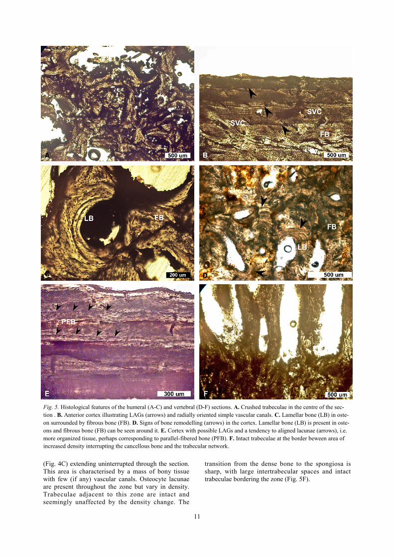
crushed (Fig. 5A).
The cortex is penetrated by numerous radially
oriented, simple vascular canals (Fig. 3B, 5B). Primary
and secondary osteons are present, the latter indicating
some degree of bone remodelling. At least three,
possibly up to five, lines of arrested growth (LAGs)
are visible in the cortex (Fig. 3B, 5B). Bundles of
collagenous fibres occur frequently in the peripheral
region (Fig. 3D).
Different types of bone tissue are present: fibrous
bone with randomly oriented lacunae is abundant
throughout most of the section (Fig. 3D, 5B-C)
whereas lamellar bone with aligned lacunae is present
in osteons (Fig. 5C). Notably, the bone lacks true
parallel-fibered bone.
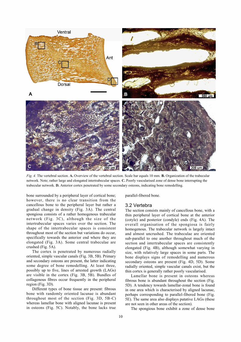
3.2 Vertebra The section consists mainly of cancellous bone, with a
thin peripheral layer of cortical bone at the anterior
(cotyle) and posterior (condyle) ends (Fig. 4A). The
overall organisation of the spongiosa is fairly
homogenous. The trabecular network is largely intact
and almost uncrushed. The trabeculae are oriented
sub-parallel to one another throughout much of the
section and intertrabecular spaces are consistently
elongated (Fig. 4B), although somewhat varying in
size, with relatively large spaces in some parts. The
bone displays signs of remodelling and numerous
secondary osteons are present (Fig. 4D, 5D). Some
radially oriented, simple vascular canals exist, but the
thin cortex is generally rather poorly vascularised.
Lamellar bone is present in osteons whereas
fibrous bone is abundant throughout the section (Fig.
5D). A tendency towards lamellar-zonal bone is found
in one area which is characterised by aligned lacunae,
perhaps corresponding to parallel-fibered bone (Fig.
5E). The same area also displays putative LAGs (these
are not seen in other areas of the section).
The spongious bone exhibit a zone of dense bone
Fig. 4. The vertebral section. A. Overview of the vertebral section. Scale bar equals 10 mm. B. Organization of the trabecular
network. Note; rather large and elongated intertrabecular spaces. C. Poorly vascularised zone of dense bone interrupting the
trabecular network. D. Anterior cortex penetrated by some secondary osteons, indicating bone remodelling.
11
(Fig. 4C) extending uninterrupted through the section.
This area is characterised by a mass of bony tissue
with few (if any) vascular canals. Osteocyte lacunae
are present throughout the zone but vary in density.
Trabeculae adjacent to this zone are intact and
seemingly unaffected by the density change. The
transition from the dense bone to the spongiosa is
sharp, with large intertrabecular spaces and intact
trabeculae bordering the zone (Fig. 5F).
Fig. 5. Histological features of the humeral (A-C) and vertebral (D-F) sections. A. Crushed trabeculae in the centre of the sec-
tion . B. Anterior cortex illustrating LAGs (arrows) and radially oriented simple vascular canals. C. Lamellar bone (LB) in oste-
on surrounded by fibrous bone (FB). D. Signs of bone remodelling (arrows) in the cortex. Lamellar bone (LB) is present in oste-
ons and fibrous bone (FB) can be seen around it. E. Cortex with possible LAGs and a tendency to aligned lacunae (arrows), i.e.
more organized tissue, perhaps corresponding to parallel-fibered bone (PFB). F. Intact trabeculae at the border beween area of
increased density interrupting the cancellous bone and the trabecular network.
12
4 Discussion
4.1 Humerus The humeral section displays cancellous bone
occupying the medullary cavity, and this tissue is
surrounded by dense but vascularised bone. The
medullary cavity is completely filled with trabeculae.
This is consistent with the condition in other derived
mosasauroids, as well as derived aquatic tetrapods in
general. The spongiosa is rather homogenous
compared to some hydropelvic mosasauroids
(Houssaye et al. 2013) and lacks distinct zones. It
could be argued that the few elongated intertrabecular
spaces present make up a zone, and since the anterior
end is broken other microanatomical structures might
be missing.
The remodelling of the bone reveals that this
individual is not a juvenile, which is corroborated by
the presence of 3–5 LAGs. Lines of arrested growth,
also known as Harris lines, are lines marking periods
of temporary slower bone growth rate generally linked
to a seasonal change in resources.
4.2 Vertebra The majority of the vertebral section consists of
cancellous bone with occasionally very large
intertrabecular spaces. The cortex at the condyle is
very thin and somewhat thicker at the cotyle. Parallel-
fibered bone has a slower growth rate than fibrous
bone and indicates varying growth rates within the
bone. Although the section displays putative LAGs, it
is primarily the relatively extensive remodelling of the
bone tissue that implies that this individual is not a
juvenile animal.
The peculiar band of dense bone diagonally across
the section (Fig. 4A, 4C) is probably related to
avascular necrosis, a disease caused by interrupted
blood supply to certain skeletal components (Resnick
et al. 1981). When the blood supply ceases the bone
tissue dies, resulting in the loss of structure and a
possible collapse, leaving a permanent zone of dead
bone. In this case, a change has occurred in a distinct
zone without affecting any of the surrounding
trabeculae. The density change is localised to a distinct
band and the overall intactness of the trabecular
network indicates little, if any, crushing during burial.
This implies that this structure is not taphonomic in
origin. A collapse of the trabecular network due to
taphonomic processes would probably yield a different
pattern than that observed. The structure seen in the
material at hand is superficially very similar to what
Rothschild & Mar tin (1987) documented in
Platecarpus vertebrae, with the difference that the
structure related to avascular necrosis therein was
described as a band of decreased density. The increase
in density seen herein could perhaps be explained by
secondary infilling of some cavities. However,
acellular areas would be expected if post-collapse
infilling of the cavities had occurred, but such areas
are not observed.
4.3 Adaptive and ecological implications Convergent evolution has created analogous characters
in mosasaurs, cetaceans, ichthyosaurs, and other
marine vertebrates, including similar skeletal
microanatomy, acquisition of a streamlined body
shape and a hypocercal tail fin in some marine reptiles
(Lindgren et al. 2010; Konishi et al. 2012). The
evolution seen in mosasaurs shows a clear transition
from life on land to active pelagic swimming, and
suggests similar ecological adaptations to those seen in
other groups secondarily adapted to an aquatic
existence. The change in microanatomy and thus
change from hydrostatic to hydrodynamic buoyancy
control and body trim is comparable to what has been
observed in early Cetacea (Gray et al. 2007; Uhen
2010).
’Platecarpus’ ptychodon is considered a highly
derived mosasaur. The features observed in the present
study corroborate this notion. Both the humeral and
the vertebral section display features typical of pelagic
animals. The reduced medullary cavity is consistent
with life in water where the force of gravity is less
apparent. Both sections are largely dominated by
cancellous bone, the vertebral section in particular,
indicating that considerable quantities of lipids and
other fluids were located in the voids, hence making
the skeleton lighter. This suggests that the animal
relied on a hydrodynamic buoyancy control, which
wo u ld i mp l y t h a t i t i n h ab i t ed o p e n -o c ea n
environments. The lightened skeleton probably also
has implications for speed and manoeuvrability, as
these aspects are known to correlate with a light
skeleton in marine tetrapods (de Ricqlès & de
Buffrénil 2001; Houssaye 2009). ’P’. ptychodon was
thus likely a fast and active animal capable of high
speed swimming.
In a comparative study, Houssaye (2012)
concluded that collagenous weave and vascular
network in different groups of aquatic reptiles seem to
inform mainly of growth rate and basal metabolic
rates. Generally, poorly organised bone tissue is
indicative of elevated growth rates (de Margerie et al.
2002). Growth rate increases with the density of the
vascularisation; conversely, it decreases with increased
organisation of the collagenous fibres. Bone growth
rate has indirectly been linked to basal metabolic rate
(Montes et al. 2007). It is thus possible to use bone
tissue to make inferences about basal metabolic rate
and the thermal physiology of an animal (Montes et al.
2007).
Primary periosteal bone can be divided into two
main types: lamellar-zonal bone (lamellar or parallel-
fibered bone) and fibro-lamellar bone. Fibro-lamellar
bone is typically highly vascularised with frequent
osteons, whereas lamellar-zonal bone is generally
poorly vascularised with few primary osteons. Fibro-
lamellar bone has a faster rate of bone deposition and
is present in all extant endotherms and has been linked
13
to a high body temperature (de Ricqlès et al. 2003;
Chinsamy et al. 2009). However fibro-lamellar bone is
also present in animals considered ectothermic, which
implies that the formation of fibro-lamellar bone does
not necessarily require a high metabolic rate
(Chinsamy-Turan 2005). Parallel-fibered bone has
been recorded in vertebrae (Houssaye & Bardet 2012;
Houssaye & Tafforeau 2012) and long bones
(Houssaye et al. 2013) of hydropelvic mosasauroids.
However, the sections examined herein largely lack
apparent parallel-fibered bone and instead they display
a bony tissue more similar to fibro-lamellar bone. This
reflects a faster growth rate than typical parallel-
fibered bone. The dominance of fibro-lamellar bone in
ichthyosaurs and plesiosaurs (Houssaye 2012;
Houssaye et al. 2014) indicates rather high growth and
basal metabolic rates in these groups compared to
hydropelvic mosasaurs (Motani 2002). However, the
sections studied in the present study indicate that the
growth and basal metabolic rate of ’Platecarpus’
ptychodon might have been comparable to that of
some ichthyosaurs and plesiosaurs.
Avascular necrosis has been described in different
genera of mosasaurs, and is interpreted as an effect of
decompression sickness (Rothschild & Martin 1987).
The phenomenon has also been identified in extant as
well as extinct marine turtles and in Jurassic
ichthyosaurs (Rothschild et al. 2012), of which at least
the latter are believed to have engaged in deep diving
(Motani et al. 1999). Some whales are known to
frequently dive deep to feed. Consequently,
decompression sickness would be expected in these
animals but is nonetheless strikingly absent in all
modern taxa (Beatty & Rothschild 2008). Since
avascular necrosis could inflict damage to bone it
would be expected that animals frequently engaging in
deep diving would evolve protective mechanisms to
counteract decompression sickness. Indeed, the
absence of avascular necrosis in modern cetaceans has
long been attributed to anatomical modifications, such
as improved circulatory physiology (Beatty &
Rothschild 2008). The frequent occurrence of
avascular necrosis in mosasaurs suggests they lacked
physiological adaptations similar to those seen in
cetaceans. Mosasaurs existed for a geologically brief
period of time (~32 million years) compared to
whales, ichthyosaurs and plesiosaurs. It is possible that
it was not enough time to develop protective measures
against decompression syndrome (Rothschild &
Martin 1987). Although ichthyosaurs existed for a
significantly longer time than did mosasaurs, the
presence of avascular necrosis has been attributed to a
possible change in lifestyle and prey (Rothschild et al.
2012). Avascular necrosis would imply tha t
’Platecarpus’ ptychodon had a pelagic lifestyle that
included occasional deep diving habits as a means of
feeding and/or escaping from predators.
5 Conclusions Mosasaurs radiated in the Late Cretaceous, resulting in
different adaptations and the evolution of a body plan
adapted for pelagic environments. The present study
shows that the humeral and vertebral microanatomy of
'Platecarpus' ptychodon is consistent with highly
derived mosasauroids. The vertebral section consists
mainly of cancellous bone and a very thin cortex,
whereas the humeral section displays medullary
cancellous bone and a gradual transition to a thin
peripheral layer of cortical bone. This is largely
consistent with an active aquatic lifestyle and more
specifically tetrapods in open-ocean habitats relying
on a hydrodynamic buoyancy and body trim control.
Histological features show a general tendency for
rapid growth rates. The dominant type of bone in both
sections is fibro-lamellar; i.e., fibrous bone with
lamellar bone in osteons, which is indicative of
elevated growth rates and a high metabolic rate
compared to that of extant reptiles. A high growth rate
is also indicated by the prevalence of radially oriented,
simple vascular canals that occur in both sections, but
are more frequent in the long bone.
The possible presence of avascular necrosis adds
information on the ecology of ’P’. ptychodon.
6 Acknowledgements I am very grateful for all the assistance and
encouragement I have received from my supervisor
Johan Lindgren, who made this project possible by
providing me with material and reading and
commenting on my manuscript. I am also thankful to
Johan Gren for useful instructions on microscopy and
photography.
7 References Arambourg, C., 1952: Les vertébrés fossiles des
gisements de phosphates (Maroc-Algérie-Tunisie).
Service Géologique du Maroc, Notes et Mémoires
92, 396 pp.
Bao, C. L. M., Teo, E. Y., Chong, M. S. K., Liu, Y.,
Choolani, M. & Chan, J. K. Y., 2013: Advances in
Bone Tissue Engineering. In J. A. Andrades (ed.):
Regenerative Medicine and Tissue Engineering,
866 pp. Intech.
Beatty, B. L. & Rothschild, B. M., 2008:
Decompression syndrome and the evolution of
deep diving physiology in the Cetacea.
Naturwissenschaften 95, 793-801.
Beaupre, G. S., Orr, T. E. & Carter, D. R., 1990: An
Approach for Time-Dependent Bone Modeling and
Remodeling -Theoretical Development. Journal of
Orthopaedic Research 8, 651-661.
Bernard, A., Lecuyer, C., Vincent, P., Amiot, R.,
Bardet, N., Buffetaut, E., Cuny, G., Fourel, F.,
Martineau, F., Mazin, J. M. & Prieur, A., 2010:
Regulation of body temperature by some Mesozoic
14
marine reptiles. Science 328, 1379-1382.
Caldwell, M. W., 2002: From fins to limbs to fins:
limb evolution in fossil marine reptiles. Am J Med
Genet 112, 236-249.
Caldwell, M. W. & Palci, A., 2007: A new basal
mosasauroid from the Cenomanian (U. Cretaceous)
of Slovenia with a review of mosasauroid
phylogeny and evolution. Journal of Vertebrate
Paleontology 27, 863-880.
Canoville, A. & Laurin, M., 2010: Evolution of
humeral microanatomy and lifestyle in amniotes,
and some comments on palaeobiological
inferences. Biological Journal of the Linnean
Society 100, 384-406.
Carter, D. R., Wong, M. & Orr, T. E., 1991:
Musculoskeletal ontogeny, phylogeny, and
functional adaptation. Journal of Biomechanics 24,
3-16.
Chinsamy-Turan, A., 2005: The microstructure of
dinosaur bone: deciphering biology with fine scale
techniques. Johns Hopkins University Press. i-xii,
1-195 pp.
Chinsamy, A., Codorniu, L. & Chiappe, L., 2009:
Palaeobiological Implications of the Bone
Histology of Pterodaustro guinazui. Anatomical
Record-Advances in Integrative Anatomy and
Evolutionary Biology 292, 1462-1477.
De Buffrénil, V., Astibia, H., Suberbiola, X. P.,
Berreteaga, A. & Bardet, N., 2008: Variation in
bone histology of middle Eocene sirenians from
western Europe. Geodiversitas 30, 425-432.
De Buffrénil, V. & Mazin, J. M., 1990: Bone
Histology of the Ichtyosaurs: Comparative Data
and Functional Interpretation. Paleobiology 16,
435-447.
De Buffrénil, V. & Schoevaert, D., 1988: On how the
periosteal bone of the delphinid humerus becomes
cancellous: ontogeny of a histological
specialization. Journal of Morphology 198, 149-
164.
De Margerie, E., Cubo, J. & Castanet, J., 2002: Bone
typology and growth rate testing and quantifying
‘Amprino’s rule’ in the mallard (Anas
platyrhynchos). Comptes Rendus - Biologies 325,
221-230.
De Ricqlès, A. & De Buffrénil, V. 2001: Bone
histology, heterochronies and the return of
tetrapods to life in water: where are we? In J.-M.
Mazin & V. de Buffrénil (eds.): Secondary
adaptation of tetrapods to life in water:
proceedings of the international meeting, Poitiers,
1996., 289-310. Dr Friedrich Pfeil.
De Ricqlès, A. J., Padian, K. & Horner, J. R., 2003:
On the bone histology of some Triassic
pseudosuchian archosaurs and related taxa.
Annales de Paleontologie 89, 67-101.
Germain, D. & Laurin, M., 2005: Microanatomy of the
radius and lifestyle in amniotes (Vertebrata,
Tetrapoda). Zoologica Scripta 34, 335-350.
Gray, N. M., Kainec, K., Madar, S., Tomko, L. &
Wolfe, S., 2007: Sink or swim? Bone density as a
mechanism for buoyancy control in early
cetaceans. Anat Rec (Hoboken) 290, 638-653.
Houssaye, A., 2009: "Pachyostosis" in aquatic
amniotes: a review. Integr Zool 4, 325-340.
Houssaye, A., 2012: Bone histology of aquatic
reptiles: what does it tell us about secondary
adaptation to an aquatic life? Biological Journal of
the Linnean Society 108, 3-21.
Houssaye, A., 2013: Palaeoecological and
morphofunctional interpretation of bone mass
increase: an example in Late Cretaceous shallow
marine squamates. Biol Rev Camb Philos Soc 88,
117-139.
Houssaye, A. & Bardet, N., 2012: Rib and vertebral
micro-anatomical characteristics of hydropelvic
mosasauroids. Lethaia 45, 200-209.
Houssaye, A., Lindgren, J., Pellegrini, R., Lee, A. H.,
Germain, D. & Polcyn, M. J., 2013:
Microanatomical and histological features in the
long bones of mosasaurine mosasaurs (Reptilia,
Squamata)--implications for aquatic adaptation and
growth rates. PLoS One 8, e76741.
Houssaye, A., Scheyer, T. M., Kolb, C., Fischer, V. &
Sander, P. M., 2014: A new look at ichthyosaur
long bone microanatomy and histology:
implications for their adaptation to an aquatic life.
PLoS One 9, e95637.
Houssaye, A. & Tafforeau, P., 2012: What vertebral
microanatomy reveals about the ecology of
juvenile mosasaurs (Reptilia, Squamata). Journal
of Vertebrate Paleontology 32, 1042-1048.
Klein, N., 2010: Long Bone Histology of
Sauropterygia from the Lower Muschelkalk of the
Germanic Basin Provides Unexpected Implications
for Phylogeny. PLoS One 5, e11613.
Konishi, T., Lindgren, J., Caldwell, M. W. & Chiappe,
L., 2012: Platecarpus tympaniticus(Squamata,
Mosasauridae): osteology of an exceptionally
preserved specimen and its insights into the
acquisition of a streamlined body shape in
mosasaurs. Journal of Vertebrate Paleontology 32,
1313-1327.
Kriloff, A., Germain, D., Canoville, A., Vincent, P.,
Sache, M. & Laurin, M., 2008: Evolution of bone
microanatomy of the tetrapod tibia and its use in
palaeobiological inference. J Evol Biol 21, 807-
826.
Lindgren, J., Caldwell, M. W., Konishi, T. & Chiappe,
L. M., 2010: Convergent Evolution in Aquatic
Tetrapods: Insights from an Exceptional Fossil
Mosasaur. PLoS One 5, e11998.
Lindgren, J., Polcyn, M. J. & Young, B. A., 2011:
Landlubbers to leviathans: evolution of swimming
in mosasaurine mosasaurs. Paleobiology 37, 445-
469.
Mateus, O., Polcyn, M., Jacobs, L., Araújo, R., Schulp,
A., Marinheiro, J., Pereira, B. & Vineyard, D.,
2012: Cretaceous amniotes from Angola:
dinosaurs, pterosaurs, mosasaurs, plesiosaurs, and
15
turtles. V Jornadas Internacionales sobre
Paleontología de Dinosaurios y su Entorno, 71-
105.
Montes, L., Le Roy, N., Perret, M., De Buffrénil, V.,
Castanet, J. & Cubo, J., 2007: Relationships
between bone growth rate, body mass and resting
metabolic rate in growing amniotes: a phylogenetic
approach. Biological Journal of the Linnean
Society 92, 63-76.
Motani, R., 2002: Swimming speed estimation of
extinct marine reptiles: energetic approach
revisited. Paleobiology 28, 251-262.
Motani, R., Rothschild, B. M. & Wahl, W., 1999:
Large eyeballs in diving ichthyosaurs - The huge
eyes of these extinct reptiles may have been useful
deep in the ocean. Nature 402, 747-747.
Nopcsa, F., 1923: Vorläufige Notiz über die
Pachyostose und Osteosklerose einiger mariner
Wirbeltiere. Anatomischer Anzeiger 56, pp. 353-
359.
Patterson-Buckendahl, P., Arnaud, S. B., Mechanic, G.
L., Martin, R. B., Grindeland, R. E. & Cann, C. E.,
1987: Fragility and composition of growing rat
bone after one week in spaceflight. American
Journal of Physiology 252, R240-R246.
Pitts, G. C., Ushakov, A. S., Pace, N., Smith, A. H.,
Rahlmann, D. F. & Smirnova, T. A., 1983: Effects
of wightlessness on body composition in the rat.
American Journal of Physiology 244, R332-R337.
Resnick, D., Niwayama, G., Guerra, J., Vint, V. &
Usselman, J., 1981: Spinal vacuum phenomena -
anatomical study and review. Radiology 139, 341-
348.
Rothschild, B. & Martin, L. D., 1987: Avascular
Necrosis: Occurrence in Diving Cretaceous
Mosasaurs. Science 236, 75-77.
Rothschild, B. M., Xiaoting, Z. & Martin, L. D., 2012:
Adaptations for marine habitat and the effect of
Triassic and Jurassic predator pressure on
development of decompression syndrome in
ichthyosaurs. Naturwissenschaften 99, 443-448.
Sheldon, A. 1997: Ecological Implications of
Mosasaur Bone Microstructure. In J. M. Callaway
& E. L. Nivholls (eds.): Ancient Marine Reptiles,
293-332. Academic Press.
Taylor, M. A. 1994: Stone, bone or blubber?
Buoyancy control strategies in aquatic tetrapods. In
L. Maddock, Q. Bone & J. M. V. Rayner (eds.):
Mechanics and Physiology of Animal Swimming,
151-161. Cambridge Univ Press.
Taylor, M. A., 2000: Functional significance of bone
ballastin in the evolution of buoyancy control
strategies by aquatic tetrapods. Historical Biology
14, 15-31.
Uhen, M. D., 2010: The Origin(s) of Whales. Annual
Review of Earth and Planetary Sciences 38, 189-
219.
Wall, W. P., 1983: The Correlation between High
Limb-Bone Density and Aquatic Habits in Recent
Mammals. Journal of Paleontology 57, 197-207.
Wiffen, J., De Buffrénil, V., De Ricqlès, A. & Mazin,
J.-M., 1995: Ontogenetic evolution of bone
structure in Late Cretaceous Plesiosauria from New
Zealand. GEOBIOS 28, 625-640.

16
Tidigare skrifter i serien
”Examensarbeten i Geologi vid Lunds
universitet”:
355. Månsson, Anna, 2013: Hydrogeologisk
kartering av Hultan, Sjöbo kommun. (15
hp)
356. Larsson, Emilie, 2013: Identifying the
Cretaceous–Paleogene boundary in North
Dakota, USA, using portable XRF. (15 hp)
357. Anagnostakis, Stavros, 2013: Upper Creta-
ceous coprolites from the Münster Basin
(northwestern Germany) – a glimpse into
the diet of extinct animals. (45 hp)
358. Olsson, Andreas, 2013: Monazite in meta-
sediments from Stensjöstrand: A pilot stu-
dy. (15 hp)
359. Westman, Malin, 2013: Betydelsen av
raka borrhål för större geoenergisystem.
(15 hp)
360. Åkesson, Christine, 2013: Pollen analyti-
cal and landscape reconstruction study at
Lake Storsjön, southern Sweden, over the
last 2000 years. (45 hp)
361. Andolfsson, Thomas, 2013: Analyses of
thermal conductivity from mineral compo-
sition and analyses by use of Thermal
Conductivity Scanner: A study of thermal
properties in Scanian rock types. (45 hp)
362. Engström, Simon, 2013: Vad kan inneslut-
ningar i zirkon berätta om Varbergschar-
nockiten, SV Sverige. (15 hp)
363. Jönsson, Ellen, 2013: Bevarat maginnehåll
hos mosasaurier. (15 hp)
364. Cederberg, Julia, 2013: U-Pb baddeleyite
dating of the Pará de Minas dyke swarm in
the São Francisco craton (Brazil) – three
generations in a single swarm. (45 hp)
365. Björk, Andreas, 2013: Mineralogisk och
malmpetrografisk studie av disseminerade
sulfider i rika och fattiga prover från Kle-
va. (15 hp)
366. Karlsson, Michelle, 2013: En MIFO fas 1-
inventering av förorenade områden: Kvar-
nar med kvicksilverbetning Jönköpings
län. (15 hp)
367. Michalchuk, Stephen P., 2013: The Säm
fold structure: characterization of folding
and metamorphism in a part of the eclogite
-granulite region, Sveconorwegian orogen.
(45 hp)
368. Praszkier, Aron, 2013: First evidence of
Late Cretaceous decapod crustaceans from
Åsen, southern Sweden. (15 hp)
369. Alexson, Johanna, 2013: Artificial ground-
water recharge – is it possible in Mozambi-
que? (15 hp)
370. Ehlorsson, Ludvig, 2013: Hydrogeologisk
kartering av grundvattenmagasinet Åsums-
fältet, Sjöbo. (15 hp)
371. Santsalo, Liina, 2013: The Jurassic extinc-
tion events and its relation to CO2 levels in
the atmosphere: a case study on Early Ju-
rassic fossil leaves. (15 hp)
372. Svantesson, Fredrik, 2013: Alunskiffern i
Östergötland – utbredning, mäktigheter,
stratigrafi och egenskaper. (15 hp)
373. Iqbal, Faisal Javed, 2013: Paleoecology
and sedimentology of the Upper Creta-
ceous (Campanian), marine strata at Åsen,
Kristianstad Basin, Southern Sweden,
Scania. (45 hp)
374. Kristinsdóttir, Bára Dröfn, 2013: U-Pb, O
and Lu-Hf isotope ratios of detrital zircon
from Ghana, West-African Craton – For-
mation of juvenile, Palaeoproterozoic
crust. (45 hp)
375. Grenholm, Mikael, 2014: The Birimian
event in the Baoulé Mossi domain (West
African Craton) — regional and global
context. (45 hp)
376. Hafnadóttir, Marín Ósk, 2014: Under-
standing igneous processes through zircon
trace element systematics: prospects and
pitfalls. (45 hp)
377. Jönsson, Cecilia A. M., 2014: Geophysical
ground surveys of the Matchless Amphibo-
lite Belt in Namibia. (45 hp)
378. Åkesson, Sofia, 2014: Skjutbanors påver-
kan på mark och miljö. (15 hp)
379. Härling, Jesper, 2014: Food partitioning
and dietary habits of mosasaurs (Reptilia,
Mosasauridae) from the Campanian
(Upper Cretaceous) of the Kristianstad
Basin, southern Sweden. (45 hp)
380. Kristensson, Johan, 2014: Ordovicium i
Fågelsångskärnan-2, Skåne – stratigrafi
och faciesvariationer. (15 hp)
381. Höglund, Ida, 2014: Hiatus - Sveriges
första sällskapsspel i sedimentologi. (15
hp)
382. Malmer, Edit, 2014: Vulkanism - en fara
för vår hälsa? (15 hp)
383. Stamsnijder, Joaen, 2014: Bestämning av
kvartshalt i sandprov - medtodutveckling
med OSL-, SEM– och EDS-analys. (15
hp)
384. Helmfrid, Annelie, 2014: Konceptuell
modell över spridningsvägar för glas-
bruksföroreningar i Rejmyre samhälle. (15
hp)
385. Adolfsson, Max, 2014: Visualizing the
volcanic history of the Kaapvaal Craton
using ArcGIS. (15 hp)
386. Hajny, Casandra, 2014: Ett mystiskt
ryggradsdjursfossil från Åsen och dess
koppling till den skånska, krittida
ryggradsdjursfaunan. (15 hp)
387. Ekström, Elin, 2014: Geologins betydelse
för geotekniker i Skåne. (15 hp)
388. Thuresson, Emma, 2014: Systematisk
sammanställning av större geoener-
gianläggningar i Sverige. (15 hp)
389. Redmo, Malin, 2014: Paleontologiska och
impaktrelaterade studier av ett anomalt
lerlager i Schweiz. (15 hp)
390. Artursson, Christopher, 2014: Comparison
of radionuclide-based solar reconstruc-
tions and sunspot observations the last
2000 years. (15 hp)
391. Svahn, Fredrika, 2014: Traces of impact in
crystalline rock – A summary of processes
and products of shock metamorphism in
crystalline rock with focus on planar de-
formation features in feldspar. (15 hp)
392. Järvin, Sara, 2014: Studie av faktorer som
påverkar skredutbredningen vid
Norsälven, Värmland. (15 hp)
393. Åberg, Gisela, 2014: Stratigrafin i
Hanöbukten under senaste glaciationen: en
studie av borrkärnor från IODP’s expedi-
tion nr 347. (15 hp)
394. Westlund, Kristian, 2014: 5 Geomor-
phological evidence for an ongoing trans-
gression on northwestern Svalbard. (15 hp)
395. Rooth, Richard, 2014: Uppföljning av ut-
lastningsgrad vid Dannemora gruva; april
2012- april 2014. (15 hp)
396. Persson, Daniel, 2014: Miljögeologisk
undersökning av deponin vid Getabjär,
Sölvesborg. (15 hp)
397. Jennerheim, Jessica, 2014: Undersökning
av långsiktiga effekter på mark och grund-
vatten vid infiltration av lakvatten – fältun-
dersökning och utvärdering av förhållan-
den vid Kejsarkullens avfallsanläggning,
Hultsfred. (15 hp)
398. Särman, Kim, 2014: Utvärdering av be-
fintliga vattenskyddsområden i Sverige.
(15 hp)
399. Tuvesson, Henrik, 2014: Från hav till land
– en beskrivning av geologin i Skrylle. (15
hp)
400. Nilsson Brunlid, Anette, 2014: Paleoeko-
logisk och kemisk-fysikalisk undersökning
av ett avvikande sedimentlager i Barse-
bäcks mosse, sydvästra Skåne, bildat för
ca 13 000 år sedan. (15 hp)
401. Falkenhaug, Jorunn, 2014: Vattnets kret-
slopp i området vid Lilla Klåveröd: ett
kunskapsprojekt med vatten i focus. (15
hp)
402. Heingård, Miriam, 2014: Long bone and
vertebral microanatomy and osteo-
histology of ’Platecarpus’ ptychodon
(Reptilia, Mosasauridae) – implications for
marine adaptations. (15 hp)
Geologiska institutionen
Lunds universitet
Sölvegatan 12, 223 62 Lund
