lots of help from cristian botez, ashfia huq, yuan-hon kiang, silvina pagola, many other people who...
TRANSCRIPT

Lots of help from Cristian Botez, Ashfia Huq, Yuan-Hon Kiang, Silvina Pagola, many other people who don’t care to be mentioned.
http://[email protected]
Use of Synchrotron Radiation to Study Polymorphs of
Pharmaceuticals

Why powders?
Why synchrotron radiation?
Examples:•Real problem - distinguishing polymorphs with similar x-ray patterns
•Real problem – determining small concentrations of a protected polymorph
•Crystal structure solutions•Proxy for a real problem – determining solid form of a small amount of API in a finished tablet
How synchrotron radiation?
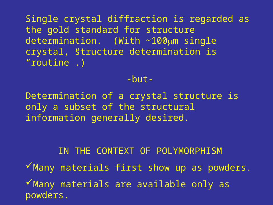
Single crystal diffraction is regarded as the gold standard for structure determination. (With ~100m single crystal, structure determination is “routine”.)
-but-
Determination of a crystal structure is only a subset of the structural information generally desired.
IN THE CONTEXT OF POLYMORPHISM
Many materials first show up as powders.
Many materials are available only as powders.
Measurements of solid mixtures are important, even independent of the crystal structure.
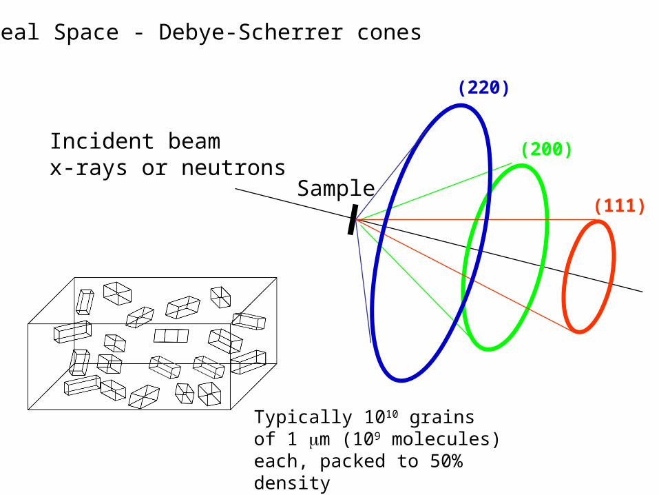
Incident beamx-rays or neutrons
Sample(111)
(200)
(220)
Real Space - Debye-Scherrer cones
Typically 1010 grains of 1 m (109 molecules) each, packed to 50% density

Where does a powder diffraction pattern come from?
Instrument – Is your instrument aligned correctly?
(Can you index simple patterns such as lactose hydrate)Are relative intensities measured accurately?
(Preferred orientation, illuminated volume, …)Strong pitch for use of synchrotron radiation
(Not giving an unbiased comparison of available instruments)
Three levels of understanding of a powder diffraction pattern1. Collection of peaks (fingerprint)2. PROFILE FIT. Use known lattice (or determine lattice) to
constrain all peak positions. Instead of ~20 peak positions, you have 1 to 6 lattice parameters. Intensitites are matched to the data.
3. RIETVELD FIT. Use known structure (or solve structure). Intensitites determined by crystal structure.

5 7 9 11 13 15 17 19 21 232 the ta (degrees)
0
10000
20000
30000
40000
50000
No
rma
lze
d X
-ra
y In
ten
sity
(co
un
ts /
se
c)
"unknow n" sam ple,0.6997 Å , cap illa ry
lact_raw .grf
What’s in your pill? (fake)
Data taken with very good (~0.007º FWHM) resolution at NSLS
Example 1

A little work turns up this entry in the Powder Diffraction File

5 7 9 11 13 15 17 19 21 232 the ta (degrees)
0
10000
20000
30000
40000
50000
No
rma
lze
d X
-ra
y In
ten
sity
(co
un
ts /
se
c)Lactose M onohydrate0.6997 Å , cap illa ry
lact_pdf.grf
Pow der D iffraction F Ile #27-1947Lactose H ydrate (1975)
What are these weak peaks? The active ingredient?
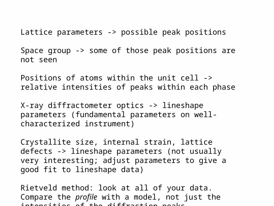
Lattice parameters -> possible peak positions
Space group -> some of those peak positions are not seen
Positions of atoms within the unit cell -> relative intensities of peaks within each phase
X-ray diffractometer optics -> lineshape parameters (fundamental parameters on well-characterized instrument)
Crystallite size, internal strain, lattice defects -> lineshape parameters (not usually very interesting; adjust parameters to give a good fit to lineshape data)
Rietveld method: look at all of your data. Compare the profile with a model, not just the intensities of the diffraction peaks.
4 6 8 10 12 14 16 18 20 22 242 the ta (degrees)
0
10000
20000
30000
40000
50000N
orm
alz
ed
X-r
ay
Inte
nsi
ty (
cou
nts
/ s
ec)
-5000
0
5000
Diff
ere
nce
R ietveld
Lactose M onohydrate0.6997 Å , cap illaryR ietve ld re finem ent
lac tgsas.grf
Pow der D iffraction F ile
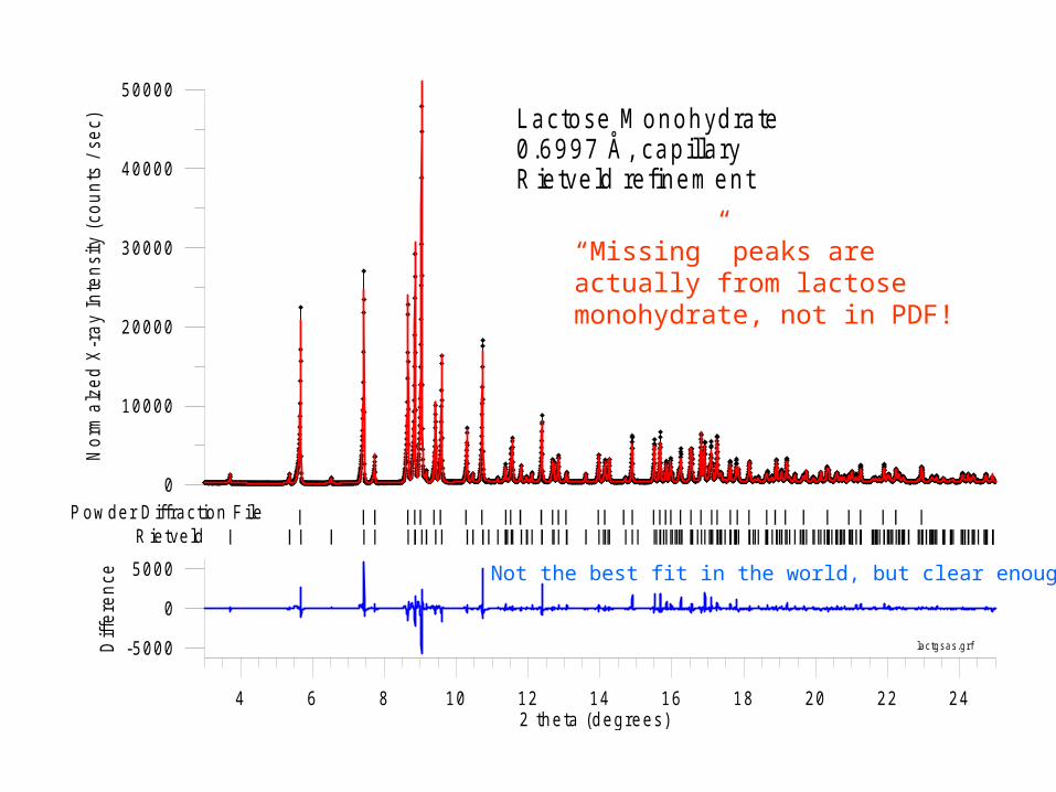
Not the best fit in the world, but clear enough
“Missing” peaks are actually from lactose monohydrate, not in PDF!
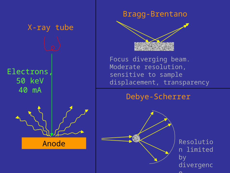
Anode
Electrons,50 keV40 mA
X-ray tube
Bragg-Brentano
Focus diverging beam. Moderate resolution, sensitive to sample displacement, transparency
Debye-Scherrer
Resolution limited by divergence, parallax

Magnetic Field
Electrons,2.8 GeV = 5500
mc2
300 mA99.9999983% of c
1/ ~ 0.01
Synchrotron Radiation
X-ray Energy [keV ]
Inte
nsity
[pho
tons
/ se
c / m
rad2 /
0.1%
ban
dwid
th]
APS
N S LS
C H E SS
ESR F
PF
SR S
SSR L
sources.g rf
1 0 8
1 0 6
1 0 1 0
1 0 1 2
1 0 1 4
1 0 4
2 1 05 2 0 5 0 1 0 0
C u t u b e
X-r
ay b
rig
htn
ess
Ph
oto
ns/
tim
e/s
olid
an
gle
/ban
dw
idth
105
(This is NOT an x-ray laser)
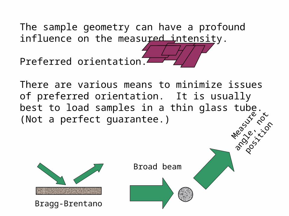
The sample geometry can have a profound influence on the measured intensity.
Preferred orientation.
There are various means to minimize issues of preferred orientation. It is usually best to load samples in a thin glass tube.(Not a perfect guarantee.)
Bragg-Brentano
Broad beam
Mea
sure
angl
e, n
ot
positio
n
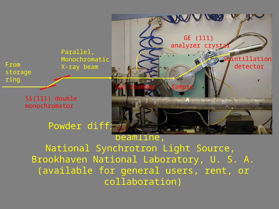
Powder diffraction station at X3B1 beamline, National Synchrotron Light Source,
Brookhaven National Laboratory, U. S. A.(available for general users, rent, or
collaboration)
Ion chamber sample
GE (111) analyzer crystal
Scintillation detector
Parallel,MonochromaticX-ray beam
Si(111) double monochromator
From storagering
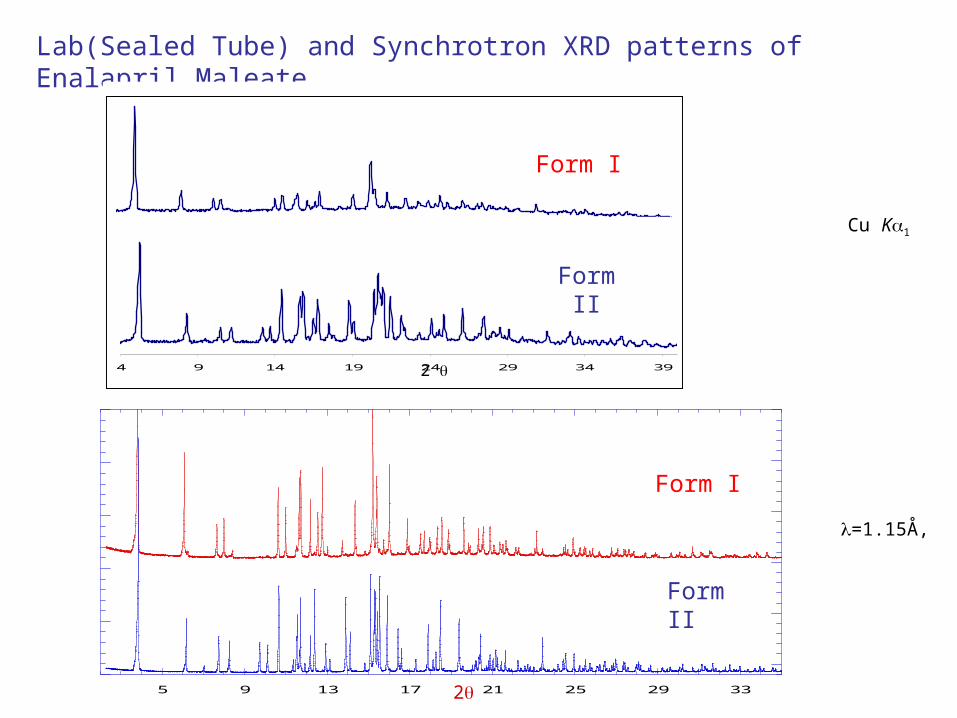
5 9 13 17 21 25 29 33
Form I
Form II
Lab(Sealed Tube) and Synchrotron XRD patterns of Enalapril Maleate
2
=1.15Å,
4 9 14 19 24 29 34 39
2
Cu K1
Form I
Form II
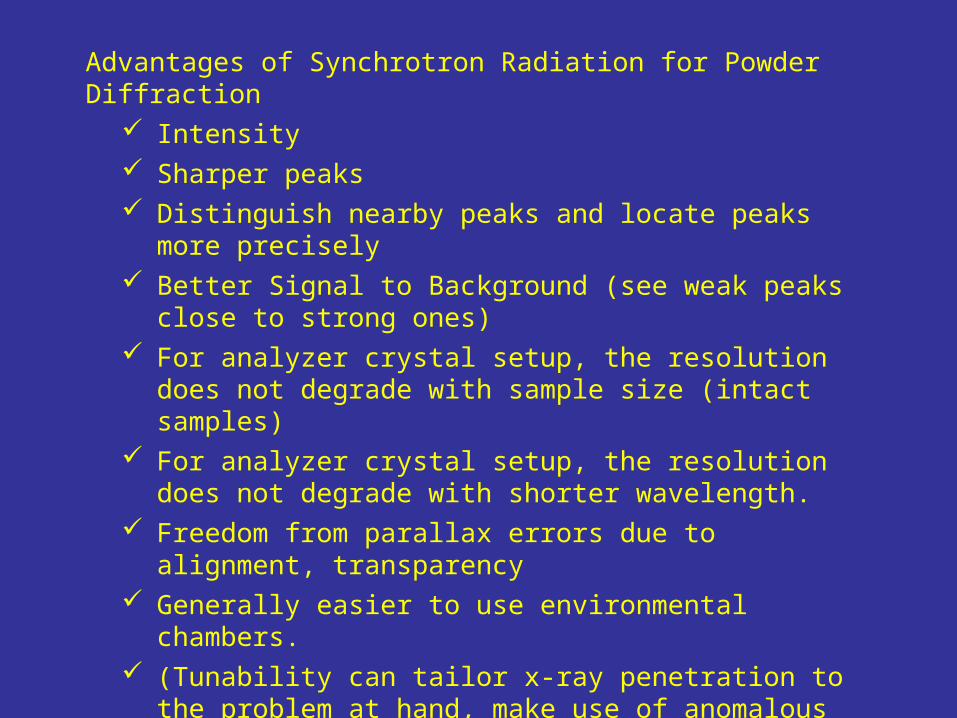
Advantages of Synchrotron Radiation for Powder Diffraction
Intensity Sharper peaks Distinguish nearby peaks and locate peaks more
precisely Better Signal to Background (see weak peaks close
to strong ones) For analyzer crystal setup, the resolution does not
degrade with sample size (intact samples) For analyzer crystal setup, the resolution does not
degrade with shorter wavelength. Freedom from parallax errors due to alignment,
transparency Generally easier to use environmental chambers. (Tunability can tailor x-ray penetration to the
problem at hand, make use of anomalous dispersion.)

Examples of identification of polymorphs with synchrotron radiation data

Form 1 -- Lab
Form 2 -- Lab
Form 1 -- S ynchrotron
Form 2 -- S ynchrotron
This drug product has two polymorphs that can’t be quantified except by Rietveld. Compare lab vs. synchrotron data sets.

8 10 12 14 16 18 20 22 24 26 28 30
Diffraction angle 2
0
10000
X-r
ay I
nten
sity
(ar
b.
unit
s)
0
1000
0
P ure Form 1
1 .0% Form 2 in Form 1
P ure Form 2
Det ect ion sensit ivit y depends on s ignal int ensit y shar p peaks low backgr oundA ll ar e enhanced by use of synchr ot r on r adiat ion
(m ax 12K )
(m ax 176K )
Two polymorphs of Zantac®,ranitidine hydrochloride
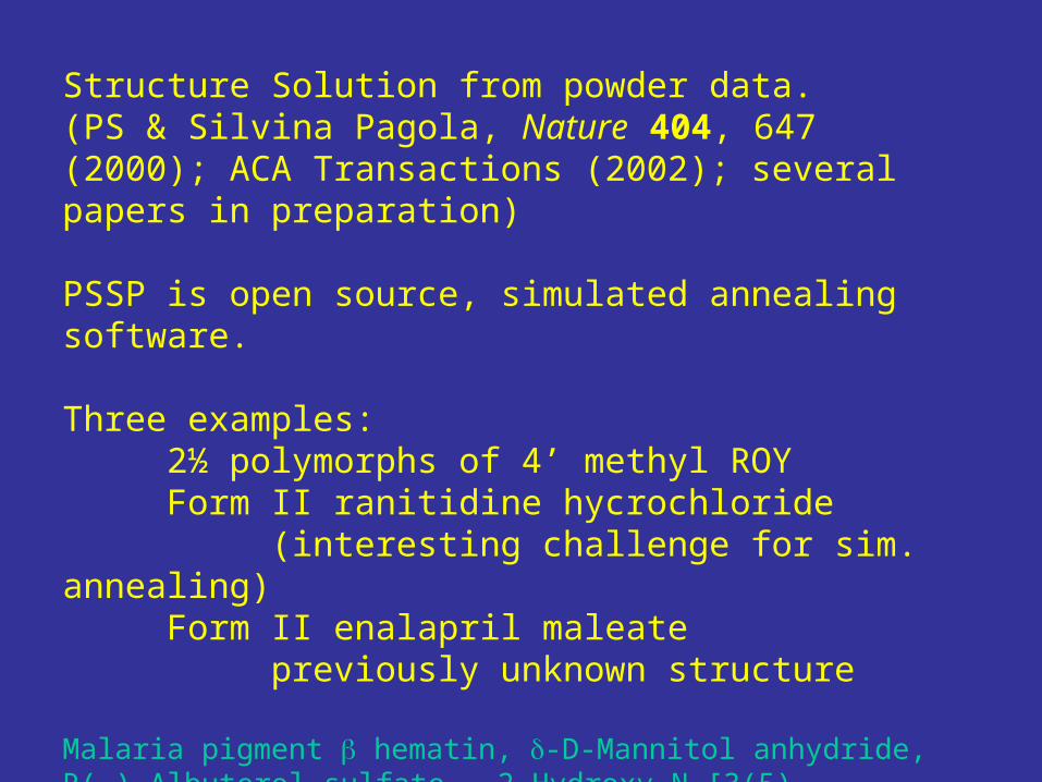
Structure Solution from powder data. (PS & Silvina Pagola, Nature 404, 647 (2000); ACA Transactions (2002); several papers in preparation)
PSSP is open source, simulated annealing software.
Three examples:2½ polymorphs of 4’ methyl ROYForm II ranitidine hycrochloride
(interesting challenge for sim. annealing)
Form II enalapril maleatepreviously unknown structure
Malaria pigment hematin, -D-Mannitol anhydride, R(-)-Albuterol sulfate, 2-Hydroxy-N-[3(5)-Pyrazolyl]-1,4-napthoquinone-4-imine, N-(p-tolyl)-dodecylsulfonamide, …
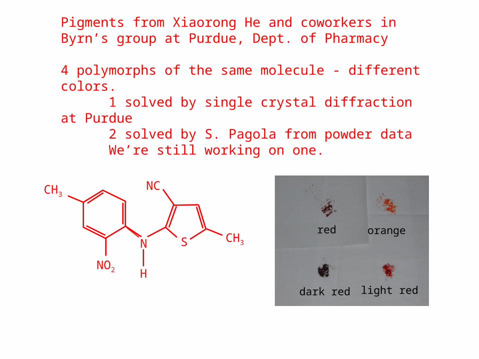
Pigments from Xiaorong He and coworkers in Byrn’s group at Purdue, Dept. of Pharmacy
4 polymorphs of the same molecule - different colors.1 solved by single crystal diffraction at Purdue2 solved by S. Pagola from powder dataWe’re still working on one.
CH3
N
NO2
NC
CH3
H
Sorangered
dark red light red

light red
orange
dark red
Comparison of powder vs. single crystal solution of DR

Ranitidine hydrochloride form IIUndertake a project like this with very good data

Space Group : P 21/n
6 Spatial coordinates : position 3 Eulerian angles : orientation 11 Torsions.20 parameters
Ranitidine HCl (Zantac®) is a very widely used drug for ulcers,
excess production of stomach acid. There is an interesting subtlety in its crystal structure.
Monoclinic, Z=4a=18.808Å, b=12.981Å, c=7.211Å
=95.057°,

Two candidate solutions from PSSP
Two others
All four, superimposed.
Disorder, or inability of
powder data to distinguish
a few of the atoms?

Atomic structure of our best Rietveld refinement of a single molecule.Essentially independent of which solution we start from.
Rwp = 11.12%, 2 = 10.56
Rietveld plot of Ranitidine Hydrochloride single configuration (from pssp)
(a)
(b)
(c)
5.00 15.00 25.00 35.00 45.00
Two Theta (degrees)
-6000-3000
030006000
Dif
fere
nce
20000
40000
60000
No
rma
lize
d X
-ra
y co
un
ts
x5
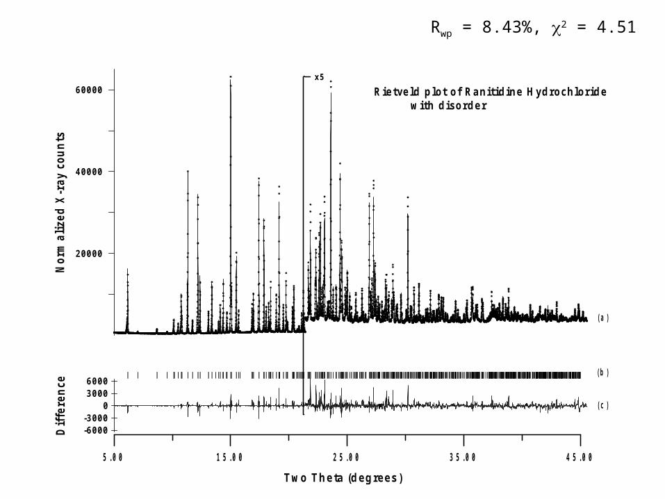
Rwp = 8.43%, 2 = 4.51
Rietveld plot of Ranitidine H ydrochlor ide with disorder
(a)
(b)
(c)
5.00 15.00 25.00 35.00 45.00
Two Theta (degrees)
-6000-3000
030006000
Dif
fere
nce
20000
40000
60000
No
rma
lize
d X
-ra
y co
un
tsx5

Refinement incorporating disorder. 50% occupancy of each of two sites for N14, C16, C18, O20, and O21.
Rwp =8.39%, 2 = 4.51
All thermal parameters independently refined! Gentle restraints on bond lengths.
This is clearly the correct solution, which includes molecular disorder.

The answer, including disorder, was already known from single crystal experiment. T. Ishida, Y. In, M. Inoue (1990)

The crystallographic problem:
1. Single crystals, small molecules: # of observations >> # of atoms Demand reasonable atomic distances, angles, etc.
2. Proteins (single crystals, data with resolution ~ 2–3 Å ): Use sequence data, strong constraints on amino acid structure.
3. Structures determined from powders by direct methods, etc.:
Demand reasonable atomic distances, angles, etc.
Structures from powders using direct space: models of known molecular structure Caution: many bond distances and angles are built in, so there is less redundancy.
No rigorous argument that any solution we find is correct. We look for heuristic consistency checks, generally based on getting a “reasonable” solution, and having redundant data.
2
exp using }{ from }{ Determine j
jhkljhklhklj RiGfIIR

Conclusions for Ranitidine HCl
•In this case, the crystal structure contains subtleties not expected in the starting model.
•Careful monitoring of the progress of the refinement shows trouble if you ignore the disorder that is suggesting itself through nonsense thermal parameters.
•Careful monitoring of the simulated annealing steps even show that the correct answer is knocking at the door, even though it was not originally invited.
Enalapril Maleate is a potent angiotensin converting enzyme (ACE) inhibitor with two known polymorphs, Form I and Form II. The single crystal structure of Form I has been known for almost twenty years. On the other hand, the crystal structure of Form II has never been reported before because of the difficulty to obtain single crystals of this polymorph, which is made by water slurry of Form I.
The crystal structure of Form II is of interest for several reasons:
1. Form II is the more stable of the two polymorphs.
2. The two forms are structurally similar based on X-ray, IR, and solid-state NMR.
3. The conformation of ACE inhibitors is important to their biological activity.

LeBail RefinementCell indexed using 25 peaks in ITO
Index measured pattern and extract intensities
Orthorhombic P212121
a=33.987 b=6.642 c=11.210

a=17.838
b=6.640c=11.64
9form I
a=33.987 b=6.642 c=11.210
form II
No Solution from simulated annealing methods• Powder Solve Crashed• PSSP : ran for time without success• DASH – we haven’t tried it yet.
23 parameters: 11 enalapril torsions (+2 maleate) + 12 orientation + position
Enalapril Maleate
Monoclinic P21/c
Orthorhombic P212121
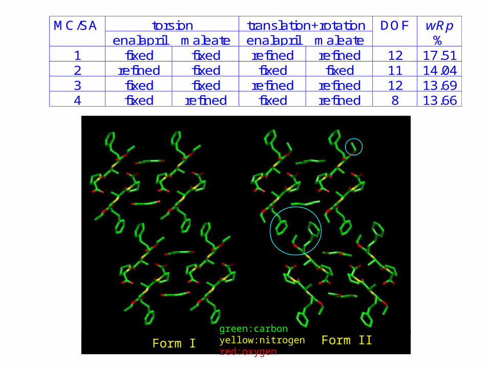
Form I Form IIgreen:carbonyellow:nitrogenred:oxygen
MC/SA torsion translation+rotation DOF wRpenalapril maleate enalapril maleate %
1 fixed fixed refined refined 12 17.512 refined fixed fixed fixed 11 14.043 fixed fixed refined refined 12 13.694 fixed refined fixed refined 8 13.66


Real business problem:_____ has a patented polymorph of _____ , and suspects that _____ is selling material that infringes. It is desired to examine the commercial tablets and determine the polymorph of the API for potential litigation.
Proxy:Examine commercial tablet of Endocet 500/7.5Gross tablet 607 mgAcetaminophen 500 mg – known lattice & structureOxycodone (as HCl) 7.5 mg – pattern in PDF but lattice unknown,*
* In general, I’d like to get better info into the PDF database. Please get in touch if you can help.

Endocet (in tact tab le t)500 m g Acetam inophen7.5 m g. oxycodone H C l
2 4 6 8 10 12 14 16 18 20
2theta (degrees)
0
20000
40000
60000
80000X
-ra
y In
ten
sity
(co
un
ts p
er
10
^6 m
on
itor)
Acetam inophen
data: endocet.631
M easured
Profile fitx 50

Powder patterns of oxycodone hydrochloride from ICDD Powder Diffraction File. Strucutures and lattices are not known.
0 4 8 12 16 202 (degrees) a t = 0 .70Å
0
50
100
0
50
100
Pe
ak
Inte
nsi
ty
PD F 38-1799
PD F 06-0014

Endocet (in tact tab le t)500 m g Acetam inophen7.5 m g. oxycodone H C l
3.0 4.0 5.0 6.0 7.0 8.0 9.0 10.0 11.0 12.0 13.0 14.0 15.0 16.0
2theta (degrees)
0
1000
2000
3000
4000
X-r
ay
Inte
nsi
ty
(co
un
ts p
er
10
^6 m
on
itor)
0
100
Oxy
cod
on
e H
Cl p
ea
ksfr
om
PD
F
A cetam inophen
data: endocet.631M easured P rofile fit
?
?

There are five synchrotron x-ray sources in the US suitable for experiments like these. ~40 worldwide.
In my humble opinion, the most usable is NSLS at Brookhaven Lab. 2500 users per year, of whom ~30% are new.
All of these facilities have active programs to serve the interests of people who want to use them.
Modes of access:Write a proposal, wait several months, no charge for x-rays, publish results.Pay ~$300 - $5000/hr for time used, you own the data, get access within hours to weeks.
http://nslsweb.nsls.bnl.gov

What is the real difference between working at a synchrotron radiation source vs. with a laboratory x-ray diffractometer?
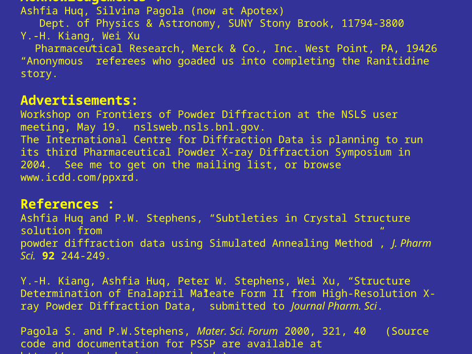
Acknowledgements :Ashfia Huq, Silvina Pagola (now at Apotex) Dept. of Physics & Astronomy, SUNY Stony Brook, 11794-3800Y.-H. Kiang, Wei Xu Pharmaceutical Research, Merck & Co., Inc. West Point, PA, 19426“Anonymous” referees who goaded us into completing the Ranitidine story.
Advertisements:Workshop on Frontiers of Powder Diffraction at the NSLS user meeting, May 19. nslsweb.nsls.bnl.gov.The International Centre for Diffraction Data is planning to run its third Pharmaceutical Powder X-ray Diffraction Symposium in 2004. See me to get on the mailing list, or browse www.icdd.com/ppxrd.
References :Ashfia Huq and P.W. Stephens, “Subtleties in Crystal Structure solution frompowder diffraction data using Simulated Annealing Method”, J. Pharm Sci. 92 244-249.
Y.-H. Kiang, Ashfia Huq, Peter W. Stephens, Wei Xu, “Structure Determination of Enalapril Maleate Form II from High-Resolution X-ray Powder Diffraction Data,” submitted to Journal Pharm. Sci.
Pagola S. and P.W.Stephens, Mater. Sci. Forum 2000, 321, 40 (Source code and documentation for PSSP are available at http://powder.physics.sunysb.edu)
Conclusions:
Think about x-ray diffraction as giving information about the fundamental structure of your material, not just a list of peaks.
This is a data-driven enterprise. High quality data is very important.
I do not want to leave the impression that synchrotron radiation is prerequisite to good data. Nor that SR is guaranteed to provide an important breakthrough. It certainly helps.
Research carried out in part at the National Synchrotron Light Source at Brookhaven National Laboratory, which is supported by the US Department of Energy, Division of Materials Sciences and Division of Chemical Sciences. The SUNY X3 beamline at NSLS is supported by the Division of Basic Energy Sciences of the US Department of Energy under Grant No. DE-FG02-86ER45231.