low-power laser irradiation promotes cell proliferation by activating pi3k/akt pathway
TRANSCRIPT
ORIGINAL ARTICLE 553J o u r n a l o fJ o u r n a l o f
CellularPhysiologyCellularPhysiology
Low-Power Laser IrradiationPromotes Cell Proliferation byActivating PI3K/Akt Pathway
LINGLING ZHANG, DA XING,* XUEJUAN GAO, AND SHENGNAN WUMOE Key Laboratory of Laser Life Science & Institute of Laser Life Science, South China Normal University, Guangzhou, China
Low-power laser irradiation (LPLI) can stimulate cell proliferation through a wide network of signals. Akt is an important protein kinase inmodulating cell proliferation. In this study, using real-time single-cell analysis, we investigated the activity of Akt and its effects on cellproliferation induced by LPLI in African green monkey SV40-transformed kidney fibroblast cells (COS-7). We utilized a recombinantfluorescence resonance energy transfer (FRET) Akt probe (BKAR) to dynamically detect the activation of Akt after LPLI treatment. Ourresults show that LPLI induced a gradual and continuous activation of Akt. Moreover, the activation of Akt can be completely abolished bywortmannin, a specific inhibitor of PI3K, suggesting that the activation of Akt caused by LPLI is a PI3K-dependent event. Src family isinvolved in Akt activation as demonstrated by the part inhibition of Akt activity in samples treated with PP1 (an inhibitor of Src family). Incontrast, loading Go 6983, a PKC inhibitor, did not affect this response. Further experiments performed using GFP-Akt fluorescenceimaging and Western blot analysis demonstrate that, the activation of Akt is a multi-step process in response to LPLI, involving membranerecruitment, phosphorylation, and membrane detachment. LPLI promotes cell proliferation through PI3K/Akt activation since the cellviability was significantly inhibited by PI3K inhibitor. All these studies create a concernful conclusion that PI3K/Akt signaling pathway is wellinvolved in LPLI triggered cell proliferation that acts as a time- and dose-dependent manner.
J. Cell. Physiol. 219: 553–562, 2009. � 2009 Wiley-Liss, Inc.
Abbreviations: BKAR, B kinase activity reporter; CCK-8, CellCounting Kit-8; CFP and YFP, cyan and yellow fluorescent protein;GFP, green fluorescent protein; EGF, epidermal growth factor;FRET, fluorescence resonance energy transfer; LPLI, low-powerlaser irradiation; PI3K, phosphoinosotide 3-kinase; ROS, reactiveoxygen species; RTK, receptor tyrosine kinase.
Contract grant sponsor: National Natural Science Foundation ofChina;Contract grant numbers: 30627003, 30870676.Contract grant sponsor: Natural Science Foundation of GuangdongProvince;Contract grant number: 7117865.
*Correspondence to: Da Xing, MOE Key Laboratory of Laser LifeScience & Institute of Laser Life Science, South China NormalUniversity, Guangzhou 510631, China. E-mail: [email protected]
Received 8 September 2008; Accepted 10 December 2008
Published online in Wiley InterScience(www.interscience.wiley.com.), 13 January 2009.DOI: 10.1002/jcp.21697
Low-power laser irradiation (LPLI) is a non-thermal irradiationwithin the visible to near infrared range of light spectrum whichhas been used clinically to accelerate wound healing and reducepain and inflammation in a variety of pathologies (Schindl et al.,1999). In the latest decades, a large body of evidence hasshowed that He–Ne laser light can stimulate a number ofbiological processes, including cell growth, proliferation (Yuet al., 1996) and differentiation (Bibikova and Oron, 1993;Conlan et al., 1996). In vitro, the effects on cell proliferation byLPLI have been studied in various cell types including fibroblasts,endothelial cells, skeletal cells, keratinocytes, myoblasts, andother types (Yu et al., 1996; Stadler et al., 2000; Shefer et al.,2002; Jia and Guo, 2004; Stein et al., 2005). However, themechanism associated with the stimulatory effects of LPLI hasnot been fully clarified. One classic mechanism involved is that,the laser energy is absorbed by intracellular chromophores andconverted to metabolic energy, since cellular ATP levelsincrease almost twofold after He–Ne laser irradiation (Karuet al., 1995). To further understand the mechanism, it isnecessary to identify the signal transduction pathways of cellproliferation stimulated by LPLI. Shefer et al. show that, LPLIspecifically activates MAPK/ERK pathway and consequentlyinduces satellite cell proliferation (Ben-Dov et al., 1999; Sheferet al., 2001). Our previous studies have demonstrated that LPLIspecifically activates RTK/PKCs signaling pathway to promotecell proliferation (Gao et al., 2006). We have also shown thatLPLI triggers a significant activation of ROS/Src pathway (Zhanget al., 2008). Furthermore, Akt can be activated by either Src orPKCs protein kinase (Kassenbrock et al., 2002; Kawakami et al.,2004; Partovian and Simons, 2004; Bentley et al., 2007).Therefore, it is likely that Akt is involved in LPLI-induced cellproliferation. On the other hand, LPLI treatment can increasethe level of intracellular ROS generation (Karu, 1999;Alexandratou et al., 2002; Jou et al., 2002; Zhang et al., 2008).The increased intracellular oxidants can mediate the activationof Akt (Ushio-Fukai et al., 1999; Wang et al., 2000). Thesereports suggest the existence of ROS/Akt signaling pathwayduring LPLI-induced proliferation. Therefore, the workinghypothesis is to explore the functions of Akt in modulating cellproliferation under LPLI treatment.
Serine/threonine protein kinase, Akt, also known as proteinkinase B, regulates a variety of cellular processes, such as cell
� 2 0 0 9 W I L E Y - L I S S , I N C .
growth, proliferation, and survival (Brazil et al., 2004; Bellacosaet al., 2005). Its activation can be initiated by variousextracellular signals that turn on PI3K (Engelman et al., 2006).After stimulus affecting on its respective receptor (Kumar et al.,2007), PI3K is activated and then phosphorylates inositol lipids(Bevan, 2001). This results in the production of PI3,4,5P3 andPI3,4P2 at the plasma membrane (Cantrell, 2001). Akt isrecruited to the membrane from the cytosol through specificbinding of the N-terminal PH domain to PI3,4,5P3 and/orPI3,4P2 (Andjelkovic et al., 1997; Frech et al., 1997), where it isthought to undergo a conformational change and becomeactivated by phosphorylation of two residues: Thr308 andSer473 (Alessi et al., 1996; Bayascas and Alessi, 2005; Song et al.,2005). Once activated, Akt translocates to various sites withinthe cell and phosphorylates a number of ‘‘effector’’ substratesto exert its biological effects (Catalucci and Condorelli, 2006).Akt promotes proliferation through regulating certain cell cycleproteins to drive quiescent cells into cell cycle and to speed upthe progression of the cell cycle. This process is accomplishedby phosphorylating p21, down-regulating transcription of p27

Fig. 1. A cartoon depicting the working of genetic reporter BKAR.BKAR, an Akt kinase activity reporter, consists of mCFP, the FHA2domain of Rad53p, a consensus Akt phosphorylation sequence andmYFP. In the unphosphorylated state, mCFP and mYFP are in aproximity and orientation resulting in FRET. Once phosphorylated byAkt at the threonine within the substrate sequence, the FHA2 domainbinds the phosphorylated sequence resulting in a conformationalchange that decreases the FRET ratio. Since it does not contain thefull Akt sequence, but an Akt substrate, it monitors the activation ofendogenous Akt in live cells.
554 Z H A N G E T A L .
and promoting stabilization of cyclin D1 protein via inactivationof GSK3 (Diehl et al., 1998; Sun et al., 1999; Alt et al., 2000;Gesbert et al., 2000; Graff et al., 2000). Akt is thendephosphorylated and inactivated by protein phosphatasessuch as protein phosphatase 2A (PP2A) (Andjelkovic et al.,1996; Gao et al., 2005).
Fluorescence resonance energy transfer (FRET) is a non-radiative transfer of energy from an excited donor molecule toa suitable acceptor molecule in close proximity. It has become apowerful tool for investigating of molecular events in living cells(Uchiyama et al., 1996; Zhang et al., 2002). To examine Aktsignaling in living cells, we used a B kinase activity reporter(BKAR) that generally consists of two different fluorescentproteins flanking a phosphoamino acid-binding domain and akinase substrate sequence (Kunkel et al., 2005).Phosphorylation of the substrate sequence causesintramolecular complexation by the phosphoamino acid-binding domain, thus, altering FRET between the fluorescentproteins. This provides a means of powerful visualization ofkinase signaling in situ in living cells. This cannot be fullyelucidated by traditional biophysical or biochemical approachesthat can only measure average behavior of a cell population andstatic spatial information from fixed cells.
Although a large number of recent studies havedemonstrated that growth factors can induce cell proliferationthrough activating PI3K/Akt signaling pathway, few studies havebeen performed during LPLI-induced cell proliferation. In thepresent study, using fluorescent imaging and Western blotanalysis, great efforts have been focused on the investigation ofAkt activity in COS-7 cells under LPLI treatment, and theassociated mechanism was discussed. Our findings will extendthe knowledge about the cellular signaling mechanismsmediating LPLI-induced proliferation.
Materials and MethodsMaterials
Dulbecco’s modified Eagle’s medium (DMEM) was purchased fromGIBCO (Grand Island, NY). Epidermal growth factor (EGF)(diluted in DMSO) was purchased from PeproTech (Rocky Hill,NJ). Wortmannin was purchased from BIOMOL ResearchLaboratories, Inc. (Plymouth, PA). 4-Amino-5-(4-methylphenyl)-7-(t-butyl)pyrazolo[3,4-d]-pyrimidine (PP1) was purchased fromInvitrogen (Carlsbad, CA). Go 6983 was purchased from Merck(Darmstadt, Germany). Anti-phospho-Akt (Thr308) antibody andanti-Akt antibody were purchased from Cell Signaling (Beverly,MA). Cell Counting Kit-8 (CCK-8) was purchased from DojindoLaboratories (Kumamoto, Japan). LipofectamineTM Reagent waspurchased from Invitrogen. DNA Extraction kit was purchasedfrom Qiagen (Valencia, CA). Other chemicals were mainly fromSigma (St. Louis, MO).
Cell culture and transfection
African green monkey SV40-transformed kidney fibroblast cell line(COS-7) was obtained from Department of Medicine, ZhongshanUniversity. The cells were cultured in DMEM mediumsupplemented with 15% fetal calf serum, penicillin (100 units/ml),and streptomycin (100 mg/ml) in 5% CO2 at 378C in a humidifiedincubator. Transient transfections were performed with 1 mg ofexpression vectors using the LipofectamineTM 2000 reagentaccording to the manufacturer’s instructions in serum-freemedium. The serum-free medium was replaced with fresh culturemedium after 5 h and incubated for an additional 24 h forexpression. The cells were then starved for 24 h in serum-freeDMEM before treatment and were examined, according to theprotocol, for the following 24 h.
Plasmid DNA of BKAR (a kind gift from Dr. Newton, Universityof California, San Diego) was used to monitor Akt activity. It
JOURNAL OF CELLULAR PHYSIOLOGY
consists of mCFP (cyan fluorescent protein), the FHA2 domain ofRad53p, a consensus Akt phosphorylation sequence and mYFP(yellow fluorescent protein) (Kunkel et al., 2005). In theunphosphorylated state, mCFP and mYFP are in a proximity andorientation resulting in FRET. Once phosphorylated by Akt at thethreonine within the substrate sequence, the FHA2 domain bindsthe phosphorylated sequence resulting in a conformational changethat decreases the FRET ratio (Fig. 1). Thus the ratio of YFP/CFPincreases with the inactivation of Akt (unphosphorylated state),while the ratio of YFP/CFP decreases with the activation of Akt(phosphorylated state). pDp85 (a kind gift from Dr. Cooper,Harvard Medical School, Boston) (Pap and Cooper, 1998) is adominant-negative construct of the p85a regulatory subunit ofPI3K, making it incapable to binds to the p110 catalytic subunit. Soover-expression of Dp85 in cells indicates that it is unable to elicitthe protein PI3K activation in vivo. pEGFP-Akt was kindly suppliedby Dr. Badger (He et al., 2006).
LPLI treatment and cell viability assays
COS-7 cells were cultured in DMEM supplemented with 15%serum at a density of 4� 103 cells/well in 96-well microplates. After24 h serum starvation in DMEM, the cells were divided into fourgroups and each group was irradiated with He–Ne laser (632.8 nm,10 mW, 12.74 mW/cm2, HN-1000, Guangzhou, China) at dose of0, 0.2, 0.4, 0.8, and 1.2 J/cm2 or treated with EGF (50 ng/ml). Theinterval wells were filled with black ink in order to minimize thelight scattering. After irradiation, the cells were maintained inserum-free DMEM and the 96-well microplates were returned tothe incubator for a further culture at 378C, 5% CO2. The irradiationwas performed on monolayer cells. In all cases, control (non-irradiated) cells were kept in the same conditions as the treatedcells.
Cell viability was assessed with CCK-8 at 1, 2, and 3 days afterthe laser irradiation, respectively. At the indicated time, CCK-8was added to the cells and incubated for 1.5 h. OD450, theabsorbance value at 450 nm, was read with a 96-well plate reader(DG5032, Hua dong, Nanjing, China). The value is directlyproportional to the number of viable cells in a culture medium andthe cell proliferation.

Fig. 2. Dose- and time-dependent proliferation of COS-7 cellsinduced by LPLI. A: LPLI induces dose-dependent proliferation. COS-7 cells were seeded on 96-well microplates for 24 h in 15% serumcontaining medium and maintained in serum-free medium for 24 h.Cells were then irradiated with He–Ne laser at dose of 0, 0.2, 0.4, 0.8,and 1.2 J/cm2, respectively or treated with 50 ng/ml EGF. Afterirradiation or EGF treatment, cells were maintained in serum-freemedium for 2 days and cell viability was assessed by the CCK-8 assay.Error bars are s.e.m. from four independent experiments. MP < 0.05;MMP < 0.01. B: LPLI induces time-dependent proliferation. Cell viabilitywas assessed by the CCK-8 assay at 0, 1, 2, 3 days after 1.2 J/cm2 LPLI.Error bars are s.e.m. from four independent experiments. MP < 0.01.
L P L I P R O M O T E S A C T I V A T I O N O F P I 3 K / A k t P A T H W A Y 555
Laser scanning microscopy (LSM) and FRET analysis
FRET was performed on a commercial Laser Scanning Microscope(LSM510/ConfoCor2) combination system (Zeiss, Jena, Germany).For excitation, the 458 nm line of an Ar-Ion Laser was attenuatedwith an acousto-optical tunable filter (AOTF), reflected by adichroic mirror (main beam splitter HFT 458 nm), and focusedthrough a Zeiss C-Apochromat 40�, NA 1.3 objective onto thesample. The emission fluorescence was split by a second dichroicmirror (secondary beam splitter NFT 515 nm) into two separatechannels: a 470–500 nm bandpass (CFP channel) and a 530 nmlongpass (YFP channel), respectively. For intracellularmeasurement, a single cell was chosen in the LSM image. Toquantify the results, images of CFP and YFP emission intensitieswere processed with Zeiss Rel3.2 image processing software(Zeiss). After background subtraction, the average fluorescenceintensity per pixel was calculated.
GFP fluorescence was excited at 488 nm with an argon ion laserand emission was recorded through a 500–550 nm band pass filter.With control experiments, it was confirmed that the bleaching ofthe probe was negligible for all protocols.
Spectrofluorometric analysis
COS-7 cells transfected with BKAR reporter were grown inDMEM for 24 h. Then, the cells were treated with LPLI (1.2 J/cm2).After irradiation, the cells were immediately transferred into aquartz cuvette. The quartz cuvette was then placed inside thesample chamber of a luminescence spectrometer (LS55,PerkinElmer, Wellesley, MA). The fluorescence emission spectrafrom 0 to 30 min after the LPLI treatment were then acquired. Theexcitation wavelength was 434 centered. The excitation andemission slits were set for 10 and 15 nm, respectively. The scanningspeed was 200 nm/sec. The fluorescence emission spectra of cellsfrom same generation were recorded in the similar ways, 0–30 minafter EGF treatment. The corresponding background spectra ofcell-free culture medium were subtracted.
GFP-Akt translocation assay
COS-7 cells were transfected with pGFP-Akt and with/withoutDp85 and then treated with LPLI in the presence or absence ofwortmannin. Using the Zeiss LSM 510 confocal microscope, weimaged the distribution pattern of GFP-Akt during LPLI-inducedproliferation.
Western blot analysis
After different treatments, cells were scraped from the dish, thenwashed twice with ice-cold phosphate-buffered saline (PBS, pH7.4), and lysed with ice-cold lysis buffer (50 mmol/L Tris–HCl pH8.0, 150 mmol/L NaCl, 1% Triton X-100, 100 mg/ml PMSF) for30 min on ice. The lysates were centrifuged at 12,000 rpm for 5 minat 48C, and the protein concentration was determined. Equivalentsamples were subjected to SDS–PAGE on 12% gel. The proteinswere then transferred onto nitrocellulose membranes, and probedwith primary antibody: anti-phospho-Akt (Thr308) and anti-Akt ata dilution of 1:1,000, followed by secondary antibodies, goat anti-mouse conjugated to Alexa Fluor 680 for phospho-Akt and goatanti-rabbit conjugated to IRDyeTM800 for Akt. Detection wasperformed using the LI-COR Odyssey Infrared Imaging System(LI-COR, Inc., Lincoln, NE).
Statistics analysis
All assays were repeated independently for a minimum of threetimes. Data are represented as mean� SEM. Statistical analysis wasperformed with Student’s paired t-test. Differences wereconsidered statistically significant at P< 0.05.
JOURNAL OF CELLULAR PHYSIOLOGY
ResultsLPLI promotes cell proliferation in dose- andtime-dependent manner
To establish a proper laser irradiation dose to induceproliferation, we used CCK-8 to observe cell viability after cellswere treated with different doses of laser irradiation. Serum-starved COS-7 cells were treated with LPLI at the dose of 0, 0.2,0.4, 0.8, 1.2 J/cm2, respectively, and the cell viabilities wereobserved 2 days following the irradiation. As shown inFigure 2A, the cell viability significantly increased in LPLI-treatedgroups (�0.4 J/cm2) compared with that of non-treated cells.The data show that, in the range 0.4–1.2 J/cm2, laser irradiationhad a significant promotive effect on cell proliferation and theeffect of LPLI on proliferation of COS-7 cells was dose-dependent. Therefore, in our following experiments, weselected 1.2 J/cm2 as the irradiation dose.
We next analyzed the cell proliferation kinetics induced byLPLI. After 24 h serum starvation, COS-7 cells were irradiatedwith LPLI (1.2 J/cm2) and then maintained in serum-freemedium. The cell viability was analyzed by CCK-8 at 1, 2, and

556 Z H A N G E T A L .
3 days, respectively. Compared with non-irradiated cells,LPLI-treated cells showed an increase in cell viability, and apositive correlation was observed with post-treatment period(Fig. 2B), indicating that the effect of LPLI on cell proliferation ofCOS-7 cells is time-dependent.
Real-time monitoring of Akt activation induced by LPLIin single living cell
To explore the mechanism of LPLI promoting proliferation, theeffects of LPLI on Akt activation in single living cell weremonitored by FRET technique with the plasmid BKAR. After 24h starvation, COS-7 cells transfected with BKAR were treatedwith LPLI, and then the real-time CFP, YFP and YFP/CFPfluorescence images were collected with LSM microscopy for30 min (Fig. 3A). The fluorescence intensities of the CFP andYFP images and their ratio (YFP/CFP) are shown inFigure 3B. The results show that, as the time lapsed, the CFPfluorescence increased, while the YFP fluorescence and theYFP/CFP ratio decreased, during the 30 min observation periodpost-irradiation. This indicates that, Akt is activated after LPLItreatment and this activation is gradual and continuous in thefirst 30 min. Similar result was obtained in cells treated withEGF, except for the YFP/CFP ratio decreased more rapidly(Fig. 3C,D), suggesting a higher degree of Akt activationstimulated by EGF. The results also suggest that BKAR is aneffectively reporter for reflecting the activation of Akt. Tofurther validate our results, in the control group (No LPLI orEGF), the fluorescence intensities of CFP and YFP, and theirratio (YFP/CFP) remained stable for more than 30 min(Fig. 3C,D), which well excluded the possibility of thespontaneously FRET change induced by scar laser and otherfactors in our experiment.
Spectrofluorometric analysis, a technique for monitoring theoverall profile of FRET fluorescence emission from a group ofcells, was recommended to further confirm that the activationof Akt was a common phenomenon in our experimental mode.After 24 h serum deprivation, COS-7 cells transfected withBKAR were treated with LPLI. The emission peak of CFP(476 nm) increased gradually, while that of YFP (527 nm)decreased during the first 30 min post-irradiation (Fig. 3E).Similar results were also obtained in cells treated with EGF(Fig. 3F). These results further confirm that BKAR is a specificreporter for Akt activation, meanwhile, support the resultsshown in Figure 3A–D that Akt is activated in response to LPLIstimulation.
Since Akt activation indicated by FRET effect of Akt reporter(BKAR) is only determined by the balance between Akt kinasesand protein tyrosine phosphatases (PTPs), we next exploredwhether Akt activation induced by LPLI depends upon anincreased level of Akt phosphorylation. Western blot analysisshow that the level of Akt phosphorylation at Thr308 waselevated as early as 10 min after LPLI treatment and graduallyincreased during the next 20 min. Similar results were obtainedin EGF-treated cells, with the level of Akt phosphorylationincreasing more significantly and rapidly (Fig. 3G). However,Akt phosphorylation decreased at 1.5 h and remained at a lowlevel up to 72 h after LPLI stimulation (Fig. 3H). Taken together,these results suggest that, LPLI effectively promotes Aktactivation due to its phosphorylation on Thr308.
Signal propagation of Akt from plasma membraneto nucleus
It is well known that the translocation of Akt from cytosol toplasma membrane is crucial for Akt activation (Andjelkovicet al., 1997; Frech et al., 1997). We therefore examined thesubcellular location of Akt in response to LPLI. A GFP-Aktplasmid was transfected into COS-7 cells and the intracellularlocalization of GFP-Akt was monitored by confocal microscopy.
JOURNAL OF CELLULAR PHYSIOLOGY
To exclude the potential effects due to over-expression of GFP-Akt under our experimental conditions, we examineddistribution of GFP-Akt in cells without any treatment. GFP-Akt remained uniformly distributed throughout the cell forover 30 min (Fig. 4A, upper). Upon LPLI stimulation, as shown inFigure 4A (lower), a significant portion of GFP-Akt translocatedfrom cytosol to plasma membrane at 5 min and this migrationlasted for more than 30 min. Interestingly, at about 10 min post-irradiation, we found that GFP-Akt started to return intocytosol and nucleus, and a great deal of GFP-Akt reachednucleus at 30 min after LPLI. Line-scan plots of these GFP-Aktimages emphasized the subcellular location changes of Akt(Fig. 4B). These results strongly indicate that the activation ofAkt occurs on the plasma membrane and this activation lasts forat least 30 min. The activated Akt disengages from themembrane and diffuses through the cytosol and nucleus tofunction throughout the cell after LPLI treatment.
LPLI induces Akt activation through aPI3K-dependent pathway
Akt is a downstream target of PI3K during growth factorsstimulation (Engelman et al., 2006). Therefore, we sought todetermine whether Akt activation was dependent on PI3Kactivity in respond to LPLI stimulation. After 24 h serumstarvation, COS-7 cells transfected with BKAR were treatedwith LPLI. Wortmannin was added 10 min later. Arepresentative temporal sequence of the pseudocolor imagesfor the ratio of YFP/CFP fluorescence are shown inFigure 5A. The quantitative analysis for the ratio of YFP/CFPfluorescence intensities is shown in Figure 5B. The ratio of YFP/CFP remained stable without treatment, and started todecrease immediately once the LPLI treatment was initiated.The decreased trend of the ratio was reversed in response towortmannin exposure. The ratio resumed to at least its pre-LPLI treatment level. In control cells, the ratio of YFP/CFPfluorescence intensities remained unchanged over 30 min(Fig. 5B). These results indicate that wortmannin couldcompletely abolish the activation of Akt induced by LPLI. Thissuggests that, LPLI-induced Akt activation is a PI3K-dependentevent.
Subsequently, we tested the effects of Dp85 (a dominant-negative mutant of PI3K) on the activation of Akt under LPLItreatment. As shown in Figure 5C, from cells co-transfectedwith pDp85 and BKAR, we found that there was a slight increaseof the YFP/CFP ratio, indicating that over-expression of Dp85completely blocks the activation of Akt induced by LPLI. Thisalso confirms the finding shown in Figure 5A,B that LPLI inducesAkt activation through a PI3K-dependent pathway.
To investigate the relationship between Akt membranetranslocation and PI3K activity under LPLI treatment, COS-7cells were either transfected with pGFP-Akt and subjected towortmannin treatment or co-transfected with pGFP-Akt andpDp85 before LPLI treatment. Both protocols resulted incomplete inhibition of GFP-Akt membrane location as thefluorescence remained uniformly distributed throughout thecells during the whole observation period (Fig. 5D), indicatingthat Akt membrane translocation is dependent on PI3K activityin response to LPLI stimulation.
Akt activation induced by LPLI is partially inhibited byPP1, but not by Go 6983
Our recent work prove that PKCs and Src can be activated byLPLI (Gao et al., 2006; Zhang et al., 2008) and several PKCisoforms and Src family have been shown to lie upstream of Akt(Aeder et al., 2004; Kawakami et al., 2004; Partovian andSimons, 2004; Thamilselvan et al., 2007). To investigate whetherPKCs and Src contribute to Akt activation, we treated COS-7cells with PKCs inhibitor (Go 6983) or Src inhibitor (PP1) prior

Fig. 3. Real-timemonitoringofAktactivation inducedbyLPLI.COS-7cellswere starved for24handthentreatedwith1.2 J/cm2 LPLI or50ng/mlEGF. A–D: Single-cell imaging analysis of COS-7 cells transfected with BKAR in different conditions. The fluorescence images of CFP and YFPchannels excited by Ar-Ion laser (458 nm) and YFP/CFP ratio were recorded with LSM microscope. The decreased ratio of YFP/CFP indicates theactivation ofAkt. A:Representative fluorescence images ofCFP, YFP and YFP/CFP ratio ofcells treatedwithLPLI. B: Quantitativeanalysisof CFPandYFP intensitiesandYFP/CFP ratio corresponding tothe images inA.The CFPandYFPintensitiesat the first time pointarenormalized to100,and the YFP/CFP ratio is normalized to 1. C: Representative fluorescence images of CFP, YFP and YFP/CFP ratio of EGF-treated cells and controlcells.D: Comparisonof YFP/CFP ratios ofcontrol cellsor cells treated withLPLI or EGF.The YFP/CFP ratios at the first time point arenormalizedto 1. Results represent 1 of 4 replicates. Scale bar: 10mm. E,F: Spectrofluorometric analysis of Akt activation induced by LPLI or EGF in living cellsexpressing BKAR. The cells were excited at the excitation wavelength of CFP (434 W 5 nm), resulting in a CFP emission peak (476 nm) and YFPemission peak (528 nm) caused by FRET from CFP. And the fluorescence emission spectra were obtained by luminescence spectrometer. E: TheemissionspectraofBKARafterLPLItreatment.F:TheemissionspectraofBKARafterEGFtreatment.G,H:RepresentativeWesternblotanalysisforAktphosphorylation(G)with30minaftereitherLPLIorEGFtreatmentand(H)upto72hafterLPLI.COS-7cellswerecollectedattheindicatedtimeafterEGFandLPLI treatment,andwereanalyzed forAktphosphorylationbyWesternblotting.Aktandb-actinwasusedasa loadingcontrol.Results represent one of three replicates. [Color figure can be viewed in the online issue, which is available at www.interscience.wiley.com.]
JOURNAL OF CELLULAR PHYSIOLOGY
L P L I P R O M O T E S A C T I V A T I O N O F P I 3 K / A k t P A T H W A Y 557

Fig. 4. Dynamics of GFP-Akt distribution after LPLI stimulation.After 24 h serum starvation, COS-7 cells transfected with GFP-Aktwere stimulated with 1.2 J/cm2 LPLI and time-lapse imaged. A:Confocal imaging temporal sequences of GFP-Akt distribution incontrol and LPLI-treated cells. Scale bar: 10 mm. B: Along the whitelines in A, GFP fluorescence intensity is plotted. The width of cellalong the white line is normalized to the width of cell at 0 min. [Colorfigure can be viewed in the online issue, which is available atwww.interscience.wiley.com.]
Fig. 5. LPLI induces the activation of Akt through a PI3K-dependentpathway. A,B: Effects of wortmannin on the activation of Akt inducedby LPLI. COS-7 cells transfected with BKAR were serum starved for24 h and then treated with 1.2 J/cm2 LPLI, 10 min after stimulation,cells were subjected to wortmannin. A: Representative pseudocolorimages of YFP/CFP ratio in COS-7 cells at various time during thecause of LPLI followed by wortmannin treatment. Scale bar: 10mm. B:The quantitative time course of YFP/CFP ratio corresponding to theimages in A. C: The quantitative time course of YFP/CFP ratio inCOS-7 cells co-transfected with pDp85 (a dominant-negative mutantof PI3K) and BKAR after 1.2 J/cm2 LPLI treatment. The inset showspseudocolor images of YFP/CFP ratio in COS-7 cells before and afterLPLI treatment. D: Real-time monitoring of GFP-Akt distribution incells co-transfected with pGFP-Akt and pDp85 (upper) or transfectedwith pGFP-Akt in the presence of wortmannin (10�6 M) (bottom)after 1.2 J/cm2 LPLI treatment. Results represent one of threereplicates. [Color figure can be viewed in the online issue, which isavailable at www.interscience.wiley.com.]
JOURNAL OF CELLULAR PHYSIOLOGY
558 Z H A N G E T A L .

L P L I P R O M O T E S A C T I V A T I O N O F P I 3 K / A k t P A T H W A Y 559
to LPLI stimulation. As established in the current work thatwortmannin could completely inhibit Akt activation in cellstreated with LPLI (Fig. 5A,B), we selected wortmannin as apositive control for the subsequent experiment. Arepresentative temporal sequence of the pseudocolor imagesof YFP/CFP ratio are shown in Figure 6A. Figure 6B is thequantitative analysis of the corresponding YFP/CFP ratio. Asshown in these figures, YFP/CFP ratio was significantly reducedby the LPLI treatment, while adding Go 6983 had minimal effecton the ratio. With PP1, the ratio was also reduced, but only to aless extent. These results indicate that PKCs have no effect onthe activation of Akt induced by LPLI. Src is involved in LPLI-induced Akt activation, but not necessarily as a prerequisite.This is because PP1 could not completely block Akt activationlike what wortmannin did. These results were also confirmed
Fig. 6. Role of PI3K, Src and PKC in LPLI-induced Akt activation. A,B: EffeFRET of BKAR. A: Representative pseudocolor imaging series of YFP/CFabsence of wortmannin, PP1 and Go 6983, respectively. B: The time courseoneofthreereplicates.Scalebar:10mm.C,D:Effectsofwortmannin,PP1ancollected at 15 min after 1.2 J/cm2 LPLI treatment in the presence or abseblotting for Akt phosphorylation. Akt andb-actin were used as loading contby densitometry (with an LAS-1000 image analyzer). Data are from three[Color figure can be viewed in the online issue, which is available at www
JOURNAL OF CELLULAR PHYSIOLOGY
by the Western blot analysis. As shown in Figure 6C,D, Go 6983had minimal effect on the level of Akt phosphorylation in cellstreated with LPLI, while PP1 showed a moderate inhibitiveeffect. All these results strongly indicate that Src, but not PKCs,is involved in the activation of Akt induced by LPLI.
Blockade of PI3K/Akt signaling pathway inhibits cellproliferation induced by LPLI
The results in the present study show that, Akt activity wasenhanced during LPLI-induced cell proliferation. The followingexperiments were performed to examine the cell viability whenAkt activation was inhibited by wortmannin. COS-7 cells after24 h starvation were treated with wortmannin, and the cellviability was analyzed using CCK-8 at various time points after
cts of wortmannin, Go 6983 and PP1 on Akt activation indicated by theP ratio at the indicated times after 1.2 J/cm2 LPLI in the presence ors of YFP/CFP ratio corresponding to the images in A. Results representdGo6983onAktphosphorylationinducedbyLPLI.C:COS-7cellswerence of wortmannin, PP1 and Go 6983, and were analyzed by Westernrols. D: The levels of endogenous Akt phosphorylation were quantifiedindependent experiments and presented as average W SE. MP < 0.01.
.interscience.wiley.com.]

Fig. 7. Effects of wortmannin on LPLI-induced cell proliferation.Cell viability was assessed by the CCK-8 assay at 0, 6, 12, and 24 h after1.2 J/cm2 LPLI in the presence or absence of wortmannin. Error barsare s.e.m. from four independent experiments. MP < 0.01.
560 Z H A N G E T A L .
the irradiation. As shown in Figure 7, compared to that of thenon-treated cells, significant decrease of cell viability wasobserved in cells treated with wortmannin. The viability of cellstreated with LPLI in conjunction with wortmannin was alsodecreased compared to that of cells with/without LPLItreatment. These results suggest that PI3K/Akt signal pathwayplays a crucial role in cell proliferation induced by LPLI.
Discussion
In the present study, a novel link between LPLI stimulation andcell proliferation is identified. Our results contribute to thegeneral idea that LPLI promotes cell proliferation through PI3K/Akt signaling pathway.
Akt serine/threonine protein kinases are critical for theregulation of fundamental cellular processes including cellproliferation, differentiation, cell shape, adhesion, migration,and survival (Brazil et al., 2004; Bellacosa et al., 2005).Therefore, we explored the activity of Akt involved in LPLI-induced cell proliferation. In the present study, significantactivation of Akt was observed in cells treated with LPLI. Wehave demonstrated, via four distinctive evidences, that LPLIpromotes cell proliferation by activating PI3K/Akt signalingpathway and conducted preliminary work to explore its cross-talk with ROS/Src or RTKs/PKCs pathway: (1) LPLI couldinduce Akt activation and the activation occurs on plasmamembrane (Figs. 3 and 4). (2) The activation of Akt triggered byLPLI is in a PI3K-dependent manner (Fig. 5). (3) Src familypartially participates in the activation of Akt caused by LPLI,while PKCs have no contribution (Fig. 6). (4) Blockade of Aktactivation by wortmannin negatively affects cell proliferationunder LPLI treatment (Figs. 2 and 7).
In order to investigate the activity of Akt under LPLItreatment, we used three different physical–chemical methods.Firstly, we used single-cell FRET analysis, which is a powerfultechnique that can provide insight into the spatial and temporaldynamics of protein kinase activity using reporter plasmid invivo. Using this method, we show that LPLI induced gradual andcontinuous activation of Akt compared to the intense and rapidactivation caused by EGF (Fig. 3A–D). The second approach,spectrofluorometric analysis further confirmed the aboveresult on multi-cell and statistical level (Fig. 3E,F). Finally, weused Western blot analysis, a traditional method, to detect the
JOURNAL OF CELLULAR PHYSIOLOGY
phosphorylation of Akt after LPLI treatment. The level ofThr308 phosphorylation was observed to increase in cellstreated with LPLI compared to that of control cells (Fig. 3G).These results strongly suggest that Akt can be effectivelyactivated by phosphorylation in response to LPLI stimulation.
Recent studies indicate that Akt activation induced bygrowth factors occurs on plasma membrane (Watton andDownward, 1999). Once activated, Akt migrates to subcellularorganelles, including nuclei, mitochondria, and other cytosoliclocations, and then phosphorylates a number of ‘‘effector’’substrates throughout the cell. Therefore, the regulation ofspecific cellular functions is exerted by Akt at the level of theplasma membrane, nucleus, mitochondria, and cytosol in multi-protein complexes (Catalucci and Condorelli, 2006). Akt isthen dephosphorylated and inactivated by proteinphosphatases (Andjelkovic et al., 1996; Gao et al., 2005). Toinvestigate this intracellular process, we employed a live-cell insitu fluorescent imaging to examine the cellular distribution ofAkt after LPLI treatment. As shown in Figure 4, after LPLItreatment, GFP-Akt translocated from cytosol to plasma at5 min, and then returned to cytosol and nucleus at 10 min post-irradiation. Interestingly, the migration from cytosol to plasmamembrane lasted for at least 30 min, indicating the continuousaccumulation of activated Akt during this time period after LPLItreatment. Based on our results, it is reasonable to suggest theevents involved in LPLI-induced Akt activation as following: Aktis activated on the plasma membrane and maintains its activityduring the migration from plasma into cytosol and nucleus,presumably by maintaining its phosphorylation status (Fig. 3G).Akt-mediated phosphorylation sustained in the cytosol andnucleus for 10–30 min after LPLI stimulation, which might bedue to a large amount of activated Akt continuouslytranslocating from plasma membrane to cytosol and nucleus,although protein phosphatase simultaneously hydrolyzed somephosphorylated Akt in cells.
How does LPLI activate Akt? One possible mechanism is thatLPLI induces ligand-free dimerization and transactivation ofRTKs which are in the ‘‘right energetic state’’ to accept the laserenergy, leading to their auto-phosphorylation and activation(Karu, 1999; Shefer et al., 2001). Activated RTKs can activatePI3K (Araki et al., 1994; Bruning et al., 1997), resulting in anincrease in PI (3, 4, 5) P3 at the plasma membrane (Cantrell,2001). The binding of PI (3, 4, 5) P3 to the PH domain anchorsAkt on the plasma membrane and allows its phosphorylationand activation by PDK1 and other kinases (Alessi et al., 1996,1997; Andjelkovic et al., 1997; Alessi, 2001; Mora et al., 2004;Dong and Liu, 2005; Sarbassov et al., 2005). In our experiments,wortmannin and Dp85 completely inhibited Aktphosphorylation and activation induced by LPLI (Fig. 5),suggesting this activation of Akt is dependent on PI3K activity.Therefore, we prefer that RTKs/PI3K/Akt pathway mediatesAkt activation under LPLI treatment.
There are other potential mechanisms may contribute to theactivation of Akt induced by LPLI. It is possible that LPLIactivates Akt through ROS. LPLI has been demonstrated toincrease the level of intracellular ROS generation (Matsui et al.,2007; Zhang et al., 2008). With LPLI treatment, light is absorbedby endogenous photosensitizers (porphyrins or cytochromes)that dominantly locate at plasma membrane, mitochondria orlysomes. The photosensitizers activation results in ROS (1O2,O�
2 , and H2O2) production (Lavi et al., 2003). Intracellularoxidants can mediate the activation of Akt (Ushio-Fukai et al.,1999; Wang et al., 2000). Our previous work prove that ROSproduction induced by LPLI can promote the activation of Src(Zhang et al., 2008) and there are several evidences indicate thatSrc family is an upstream activator of Akt (Kassenbrock et al.,2002; Bentley et al., 2007). Jiang and Qiu (2003) provide that Srcdirectly regulates Akt activity by phosphorylating Tyr315 andTyr326 in the activation loop of Akt. In recent studies, there are
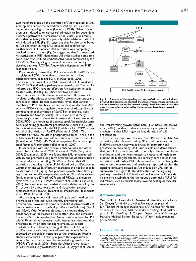
Fig. 8. A model of the signaling pathways of Akt activation inducedby LPLI. Broken lines mean that the downstream changes producedby the upstream do not be proved clearly. Real lines mean that thedownstream effects induced by the upstream have been provedalready.
L P L I P R O M O T E S A C T I V A T I O N O F P I 3 K / A k t P A T H W A Y 561
two major opinions on the activation of Akt mediated by Src.One opinion is that the activation of Akt by Src is a PI3K-dependent signaling pathway (Liu et al., 1998). Others showpressure-induced colon cancer cell adhesion via Src-dependentPI3K/Akt pathways (Thamilselvan et al., 2007). Our resultsshow that Src family inhibitor partially inhibited the activation ofAkt induced by LPLI (Fig. 6), suggesting that Src also contributesto Akt activation during LPLI-induced cell proliferation.Furthermore, LPLI-induced Akt activation was completelyblocked by wortmannin (Fig. 5), suggesting that Src regulatedAkt activation is PI3K dependent. All these studies come to aconclusion that LPLI-induced Akt activation is dominated by theRTKs/PI3K/Akt signaling pathway. There is a secondarysignaling pathway, ROS/Src/Akt, which is dependent on PI3K inresponse to LPLI.
Previous study shows that LPLI can activate RTKs/PKCs in adiacylglycerol (DG)-dependent manner in human lungadenocarcinoma cells (ASTC-a-1) (Gao et al., 2006).Therefore, the possibility of PKCs involved in LPLI mediatedRTKs/PI3K/Akt signaling pathway was investigated. The resultsindicate that PKCs have no effect on Akt activation in cellstreated with LPLI (Fig. 6). There are two possibleinterpretations for this phenomenon: either PKCs are notinvolved, or the effects of various PKC isoforms counteract andcancel each other. Recent researches reveal that variousmembers of PKC family can either increase or decrease Aktactivity. PKCa can up-regulate the activity of Akt by directlystimulating phosphorylation of Ser473 in endothelial cells(Partovian and Simons, 2004). PKCbII can also directlyphosphorylate and activate Akt in mast cells (Kawakami et al.,2004). PKCh can modulate the activation of Akt in glioblastomacells, supporting glioblastoma proliferation (Aeder et al., 2004).By contrast, treating cell with PKCb inhibitor LY 379196 causesAkt phosphorylation at Ser473 (Wen et al., 2003). Theactivation of PKCz results in phosphorylation of Thr34 in thePH domain of Akt and limits its activation in response to insulin(Powell et al., 2003). c-PKCs down-regulate Akt activity uponsteel factor (SF) stimulation (Edling et al., 2007).
In accordance with our previous observations and otherresearches (Shefer et al., 2001; Gao et al., 2006; Tuby et al.,2007; Zhang et al., 2008), we found that LPLI enhanced cellviability and promoted long-term proliferation of cells culturedon serum-free medium (Fig. 2). We also found that Aktactivation plays a key role in LPLI-induced cell proliferation aswortmannin andDp85 effectively decreased the viability of cellstreated with LPLI (Fig. 7). Akt promotes proliferation throughregulating some cell cycle proteins, such as p21 and the relatedfamily members p27/Kip1 (p27) and p57/Kip2, to inhibit cellcycle arrest (Sun et al., 1999; Gesbert et al., 2000; Graff et al.,2000). Akt can promote translation and stabilization of cyclinD1 protein by phosphorylation and inactivation glycogensynthase kinase 3 (GSK3) (Diehl et al., 1998; Muise-Helmerickset al., 1998; Alt et al., 2000).
Akt drives quiescent cells into cell cycle and speed up theprogression of the cell cycle, thereby promoting cellproliferation. However, the time period of Akt activation (0.5 h)is not consistent with that of cell proliferation (2–3 days) (Figs. 2and 3G,H). With Western blotting, it is showed that, Aktphosphorylation decreased at 1.5 h after LPLI, and remainedlow up to 72 h. It is possible that, Akt activation induced by LPLIwithin 30 min drives quiescent cells into at least one round ofproliferation which lasts for approximately 24 h post-irradiation. The relatively prolonged effect of LPLI on theproliferation of cells may be attributed to growth factorssecreted by the cells in response to the irradiation. Recentstudies show that LPLI can up-regulate the expression ofgrowth factors, such as vascular endothelial growth factor(VEGF) (Tuby et al., 2006), basic fibroblast growth factor(bFGF), insulin like growth factor-1 (IGF-1) (Saygun et al., 2008)
JOURNAL OF CELLULAR PHYSIOLOGY
and transforming growth factor-beta (TGF-beta), etc. (Safaviet al., 2008). Further studies are necessary to pinpoint themechanisms that LPLI triggered long duration of cellproliferation.
For the first time, we conclude that LPLI can stimulate Aktactivation, which is mediated by PI3K, and the activation ofPI3K/Akt signaling pathway is crucial in promoting cellproliferation induced by LPLI. Our results also demonstratethat, with LPLI stimulation, Akt is initially activated on plasmamembrane, and then translocates to cytosol and nucleus tofunction its biological effects. Src partially participates in theactivation of Akt, while PKCs show no effect. By combining theresults on the presented and previously reported studies, thesignaling pathways related to Akt induced by LPLI can besummarized in Figure 8. The delineation of the signalingpathways involved in LPLI-induced proliferation will provideinsight into establishing the therapeutic potential of LPLI forindications such as muscle injury, wound healing or vascularregeneration.
Acknowledgments
We thank Dr. Alexandra C. Newton (University of California,San Diego) for kindly providing Akt reporter plasmid,Dr. Thomas M. Badger (University of Arkansas for MedicalSciences, Little Rock, AR) for kindly providing pEGFP-Akt2plasmid, Dr. Geoffrey M. Cooper (Department of Pathology,Harvard Medical School, Boston, MA) for kindly providingpD85 plasmid.
Literature Cited
Aeder SE, Martin PM, Soh JW, Hussaini IM. 2004. PKC-eta mediates glioblastoma cellproliferation through the Akt and mTOR signaling pathways. Oncogene 23:9062–9069.
Alessi DR. 2001. Discovery of PDK1, one of the missing links in insulin signal transduction.Colworth Medal Lecture. Biochem Soc Trans 29:1–14.
Alessi DR, Andjelkovic M, Caudwell B, Cron P, Morrice N, Cohen P, Hemmings BA. 1996.Mechanism of activation of protein kinase B by insulin and IGF-1. EMBO J 15:6541–6551.

562 Z H A N G E T A L .
Alessi DR, James SR, Downes CP, Holmes AB, Gaffney PR, Reese CB, Cohen P. 1997.Characterization of a 3-phosphoinositide-dependent protein kinase which phosphorylatesand activates protein kinase Balpha. Curr Biol 7:261–269.
Alexandratou E, Yova D, Handris P, Kletsas D, Loukas S. 2002. Human fibroblast alterationsinduced by low power laser irradiation at the single cell level using confocal microscopy.Photochem Photobiol Sci 1:547–552.
Alt JR, Cleveland JL, Hannink M, Diehl JA. 2000. Phosphorylation-dependent regulation ofcyclin D1 nuclear export and cyclin D1-dependent cellular transformation. Genes Dev14:3102–3114.
Andjelkovic M, Jakubowicz T, Cron P, Ming XF, Han JW, Hemmings BA. 1996. Activation andphosphorylation of a pleckstrin homology domain containing protein kinase (RAC-PK/PKB) promoted by serum and protein phosphatase inhibitors. Proc Natl Acad Sci USA93:5699–5704.
Andjelkovic M, Alessi DR, Meier R, Fernandez A, Lamb NJ, Frech M, Cron P, Cohen P, LucocqJM, Hemmings BA. 1997. Role of translocation in the activation and function of proteinkinase B. J Biol Chem 272:31515–31524.
Araki E, Lipes MA, Patti ME, Bruning JC, Haag B III, Johnson RS, Kahn CR. 1994. Alternativepathway of insulin signalling in mice with targeted disruption of the IRS-1 gene. Nature372:186–190.
Bayascas JR, Alessi DR. 2005. Regulation of Akt/PKB Ser473 phosphorylation. Mol Cell18:143–145.
Bellacosa A, Kumar CC, Di Cristofano A, Testa JR. 2005. Activation of AKT kinases in cancer:Implications for therapeutic targeting. Adv Cancer Res 94:29–86.
Ben-Dov N, Shefer G, Irintchev A, Wernig A, Oron U, Halevy O. 1999. Low-energy laserirradiation affects satellite cell proliferation and differentiation in vitro. Biochim BiophysActa 1448:372–380.
Bentley JK, Newcomb DC, Goldsmith AM, Jia Y, Sajjan US, Hershenson MB. 2007. Rhinovirusactivates interleukin-8 expression via a Src/p110beta phosphatidylinositol 3-kinase/Aktpathway in human airway epithelial cells. J Virol 81:1186–1194.
Bevan P. 2001. Insulin signaling. J Cell Sci 114:1429–1430.Bibikova A, Oron U. 1993. Promotion of muscle regeneration in the toad (Bufo viridis)
gastrocnemius muscle by low-energy laser irradiation. Anat Rec 235:374–380.Brazil DP, Yang ZZ, Hemmings BA. 2004. Advances in protein kinase B signalling: AKTion on
multiple fronts. Trends Biochem Sci 29:233–242.Bruning JC, Winnay J, Cheatham B, Kahn CR. 1997. Differential signaling by insulin receptor
substrate 1 (IRS-1) and IRS-2 in IRS-1-deficient cells. Mol Cell Biol 17:1513–1521.Cantrell DA. 2001. Phosphoinositide 3-kinase signalling pathways. J Cell Sci 114:1439–1445.Catalucci D, Condorelli G. 2006. Effects of Akt on cardiac myocytes: Location counts. Circ
Res 99:339–341.Conlan MJ, Rapley JW, Cobb CM. 1996. Biostimulation of wound healing by low-energy laser
irradiation. A review. J Clin Periodontol 23:492–496.Diehl JA, Cheng M, Roussel MF, Sherr CJ. 1998. Glycogen synthase kinase-3beta regulates
cyclin D1 proteolysis and subcellular localization. Genes Dev 12:3499–3511.Dong LQ, Liu F. 2005. PDK2: The missing piece in the receptor tyrosine kinase signaling
pathway puzzle. Am J Physiol Endocrinol Metab 289:E187–E196.Edling CE, Pedersen M, Carlsson L, Ronnstrand L, Palmer RH, Hallberg B. 2007.
Haematopoietic progenitor cells utilise conventional PKC to suppress PKB/Akt activity inresponse to c-Kit stimulation. Br J Haematol 136:260–268.
Engelman JA, Luo J, Cantley LC. 2006. The evolution of phosphatidylinositol 3-kinases asregulators of growth and metabolism. Nat Rev Genet 7:606–619.
Frech M, Andjelkovic M, Ingley E, Reddy KK, Falck JR, Hemmings BA. 1997. High affinitybinding of inositol phosphates and phosphoinositides to the pleckstrin homology domain ofRAC/protein kinase B and their influence on kinase activity. J Biol Chem 272:8474–8481.
Gao T, Furnari F, Newton AC. 2005. PHLPP: A phosphatase that directly dephosphorylatesAkt, promotes apoptosis, and suppresses tumor growth. Mol Cell 18:13–24.
Gao X, Chen T, Xing D, Wang F, Pei Y, Wei X. 2006. Single cell analysis of PKCactivation during proliferation and apoptosis induced by laser irradiation. J Cell Physiol206:441–448.
Gesbert F, Sellers WR, Signoretti S, Loda M, Griffin JD. 2000. BCR/ABL regulates expressionof the cyclin-dependent kinase inhibitor p27Kip1 through the phosphatidylinositol 3-Kinase/AKT pathway. J Biol Chem 275:39223–39230.
Graff JR, Konicek BW, McNulty AM, Wang Z, Houck K, Allen S, Paul JD, Hbaiu A, Goode RG,Sandusky GE, Vessella RL, Neubauer BL. 2000. Increased AKT activity contributes toprostate cancer progression by dramatically accelerating prostate tumor growth anddiminishing p27Kip1 expression. J Biol Chem 275:24500–24505.
He L, Simmen FA, Mehendale HM, Ronis MJ, Badger TM. 2006. Chronic ethanol intake impairsinsulin signaling in rats by disrupting Akt association with the cell membrane. Role of TRB3in inhibition of Akt/protein kinase B activation. J Biol Chem 281:11126–11134.
Jia YL, Guo ZY. 2004. Effect of low-power He-Ne laser irradiation on rabbit articularchondrocytes in vitro. Lasers Surg Med 34:323–328.
Jiang T, Qiu Y. 2003. Interaction between Src and a C-terminal proline-rich motif of Akt isrequired for Akt activation. J Biol Chem 278:15789–15793.
Jou MJ, Jou SB, Chen HM, Lin CH, Peng TI. 2002. Critical role of mitochondrial reactiveoxygen species formation in visible laser irradiation-induced apoptosis in rat brainastrocytes (RBA-1). J Biomed Sci 9:507–516.
Karu T. 1999. Primary and secondary mechanisms of action of visible to near-IR radiation oncells. J Photochem Photobiol B 49:1–17.
Karu T, Pyatibrat L, Kalendo G. 1995. Irradiation with He-Ne laser increases ATP level in cellscultivated in vitro. J Photochem Photobiol B 27:219–223.
Kassenbrock CK, Hunter S, Garl P, Johnson GL, Anderson SM. 2002. Inhibition of Src familykinases blocks epidermal growth factor (EGF)-induced activation of Akt, phosphorylationof c-Cbl, and ubiquitination of the EGF receptor. J Biol Chem 277:24967–24975.
Kawakami Y, Nishimoto H, Kitaura J, Maeda-Yamamoto M, Kato RM, Littman DR,Leitges M, Rawlings DJ, Kawakami T. 2004. Protein kinase C betaII regulates Aktphosphorylation on Ser-473 in a cell type- and stimulus-specific fashion. J Biol Chem279:47720–47725.
JOURNAL OF CELLULAR PHYSIOLOGY
Kumar N, Afeyan R, Sheppard S, Harms B, Lauffenburger DA. 2007. Quantitative analysis ofAkt phosphorylation and activity in response to EGF and insulin treatment. BiochemBiophys Res Commun 354:14–20.
Kunkel MT, Ni Q, Tsien RY, Zhang J, Newton AC. 2005. Spatio-temporal dynamics of proteinkinase B/Akt signaling revealed by a genetically encoded fluorescent reporter. J Biol Chem280:5581–5587.
Lavi R, Shainberg A, Friedmann H, Shneyvays V, Rickover O, Eichler M, Kaplan D, Lubart R.2003. Low energy visible light induces reactive oxygen species generation and stimulates anincrease of intracellular calcium concentration in cardiac cells. J Biol Chem 278:40917–40922.
Liu AX, Testa JR, Hamilton TC, Jove R, Nicosia SV, Cheng JQ. 1998. AKT2, a member of theprotein kinase B family, is activated by growth factors, v-Ha-ras, and v-src throughphosphatidylinositol 3-kinase in human ovarian epithelial cancer cells. Cancer Res58:2973–2977.
Matsui S, Tsujimoto Y, Matsushima K. 2007. Stimulatory effects of hydroxyl radical generationby Ga-Al-As laser irradiation on mineralization ability of human dental pulp cells. BiolPharm Bull 30:27–31.
Mora A, Komander D, van Aalten DM, Alessi DR. 2004. PDK1, the master regulator of AGCkinase signal transduction. Semin Cell Dev Biol 15:161–170.
Muise-Helmericks RC, Grimes HL, Bellacosa A, Malstrom SE, Tsichlis PN, Rosen N. 1998.Cyclin D expression is controlled post-transcriptionally via a phosphatidylinositol 3-kinase/Akt-dependent pathway. J Biol Chem 273:29864–29872.
Pap M, Cooper GM. 1998. Role of glycogen synthase kinase-3 in the phosphatidylinositol3-Kinase/Akt cell survival pathway. J Biol Chem 273:19929–19932.
Partovian C, Simons M. 2004. Regulation of protein kinase B/Akt activity and Ser473phosphorylation by protein kinase Calpha in endothelial cells. Cell Signal 16:951–957.
Powell DJ, Hajduch E, Kular G, Hundal HS. 2003. Ceramide disables 3-phosphoinositidebinding to the pleckstrin homology domain of protein kinase B (PKB)/Akt by a PKCzeta-dependent mechanism. Mol Cell Biol 23:7794–7808.
Safavi SM, Kazemi B, Esmaeili M, Fallah A, Modarresi A, Mir M. 2008. Effects of low-level He-Ne laser irradiation on the gene expression of IL-1beta, TNF-alpha, IFN-gamma, TGF-beta,bFGF, and PDGF in rat’s gingiva. Lasers Med Sci 23:331–335.
Sarbassov DD, Guertin DA, Ali SM, Sabatini DM. 2005. Phosphorylation and regulation ofAkt/PKB by the rictor-mTOR complex. Science 307:1098–1101.
Saygun I, Karacay S, Serdar M, Ural AU, Sencimen M, Kurtis B. 2008. Effects of laser irradiationon the release of basic fibroblast growth factor (bFGF), insulin like growth factor-1 (IGF-1),and receptor of IGF-1 (IGFBP3) from gingival fibroblasts. Lasers Med Sci 23:211–215.
Schindl A, Schindl M, Pernerstorfer-Schon H, Kerschan K, Knobler R, Schindl L. 1999.Diabetic neuropathic foot ulcer: Successful treatment by low-intensity laser therapy.Dermatology 198:314–316.
Shefer G, Oron U, Irintchev A, Wernig A, Halevy O. 2001. Skeletal muscle cell activation bylow-energy laser irradiation: A role for the MAPK/ERK pathway. J Cell Physiol 187:73–80.
Shefer G, Partridge TA, Heslop L, Gross JG, Oron U, Halevy O. 2002. Low-energy laserirradiation promotes the survival and cell cycle entry of skeletal muscle satellite cells. J CellSci 115:1461–1469.
Song G, Ouyang G, Bao S. 2005. The activation of Akt/PKB signaling pathway and cell survival.J Cell Mol Med 9:59–71.
Stadler I, Evans R, Kolb B, Naim JO, Narayan V, Buehner N, Lanzafame RJ. 2000. In vitro effectsof low-level laser irradiation at 660 nm on peripheral blood lymphocytes. Lasers Surg Med27:255–261.
Stein A, Benayahu D, Maltz L, Oron U. 2005. Low-level laser irradiation promotesproliferation and differentiation of human osteoblasts in vitro. Photomed Laser Surg23:161–166.
Sun H, Lesche R, Li DM, Liliental J, Zhang H, Gao J, Gavrilova N, Mueller B, Liu X, Wu H. 1999.PTEN modulates cell cycle progression and cell survival by regulating phosphatidylinositol3,4,5,-trisphosphate and Akt/protein kinase B signaling pathway. Proc Natl Acad Sci USA96:6199–6204.
Thamilselvan V, Craig DH, Basson MD. 2007. FAK association with multiple signal proteinsmediates pressure-induced colon cancer cell adhesion via a Src-dependent PI3K/Aktpathway. FASEB J 21:1730–1741.
Tuby H, Maltz L, Oron U. 2006. Modulations of VEGF and iNOS in the rat heart by low levellaser therapy are associated with cardioprotection and enhanced angiogenesis. Lasers SurgMed 38:682–688.
Tuby H, Maltz L, Oron U. 2007. Low-level laser irradiation (LLLI) promotes proliferation ofmesenchymal and cardiac stem cells in culture. Lasers Surg Med 39:373–378.
Uchiyama H, Hirano K, Kashiwasake-Jibu M, Taira K. 1996. Detection of undegradedoligonucleotides in vivo by fluorescence resonance energy transfer. Nuclease activities inliving sea urchin eggs. J Biol Chem 271:380–384.
Ushio-Fukai M, Alexander RW, Akers M, Yin Q, Fujio Y, Walsh K, Griendling KK. 1999.Reactive oxygen species mediate the activation of Akt/protein kinase B by angiotensin II invascular smooth muscle cells. J Biol Chem 274:22699–22704.
Wang X, McCullough KD, Franke TF, Holbrook NJ. 2000. Epidermal growth factor receptor-dependent Akt activation by oxidative stress enhances cell survival. J Biol Chem275:14624–14631.
Watton SJ, Downward J. 1999. Akt/PKB localisation and 3’ phosphoinositide generation atsites of epithelial cell-matrix and cell-cell interaction. Curr Biol 9:433–436.
Wen HC, Huang WC, Ali A, Woodgett JR, Lin WW. 2003. Negative regulation ofphosphatidylinositol 3-kinase and Akt signalling pathway by PKC. Curr Biol 15:37–45.
Yu HS, Chang KL, Yu CL, Chen JW, Chen GS. 1996. Low-energy helium-neon laser irradiationstimulates interleukin-1 alpha and interleukin-8 release from cultured humankeratinocytes. J Invest Dermatol 107:593–596.
Zhang J, Campbell RE, Ting AY, Tsien RY. 2002. Creating new fluorescent probes for cellbiology. Nat Rev Mol Cell Biol 3:906–918.
Zhang J, Xing D, Gao X. 2008. Low-power laser irradiation activates Src tyrosine kinasethrough reactive oxygen species-mediated signaling pathway. J Cell Physiol217:518–528.