lumbar – transforaminal lumbar interbody fusion (tlif) · pdf file introduction a...
TRANSCRIPT
www.peakorthopedics.com/content/welcome-peak-orthopedics-spine
Lumbar – Transforaminal Lumbar Interbody Fusion(TLIF)
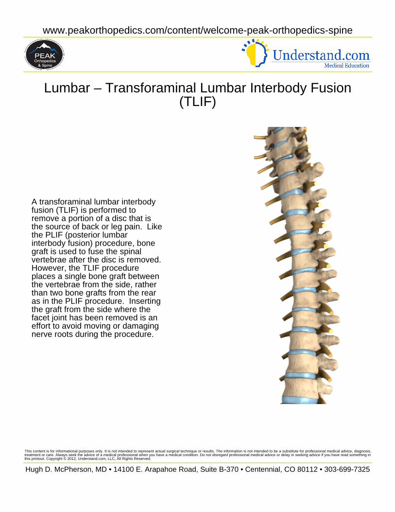
A transforaminal lumbar interbodyfusion (TLIF) is performed toremove a portion of a disc that isthe source of back or leg pain. Likethe PLIF (posterior lumbarinterbody fusion) procedure, bonegraft is used to fuse the spinalvertebrae after the disc is removed.However, the TLIF procedureplaces a single bone graft betweenthe vertebrae from the side, ratherthan two bone grafts from the rearas in the PLIF procedure. Insertingthe graft from the side where thefacet joint has been removed is aneffort to avoid moving or damagingnerve roots during the procedure.
This content is for informational purposes only. It is not intended to represent actual surgical technique or results. The information is not intended to be a substitute for professional medical advice, diagnosis,treatment or care. Always seek the advice of a medical professional when you have a medical condition. Do not disregard professional medical advice or delay in seeking advice if you have read something inthis printout. Copyright © 2012, Understand.com, LLC, All Rights Reserved.
Hugh D. McPherson, MD • 14100 E. Arapahoe Road, Suite B-370 • Centennial, CO 80112 • 303-699-7325
www.peakorthopedics.com/content/welcome-peak-orthopedics-spine
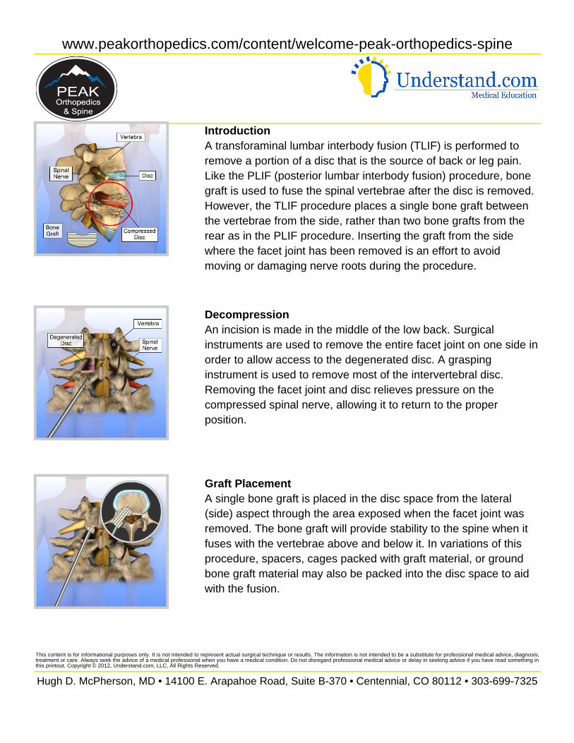
IntroductionA transforaminal lumbar interbody fusion (TLIF) is performed toremove a portion of a disc that is the source of back or leg pain.Like the PLIF (posterior lumbar interbody fusion) procedure, bonegraft is used to fuse the spinal vertebrae after the disc is removed.However, the TLIF procedure places a single bone graft betweenthe vertebrae from the side, rather than two bone grafts from therear as in the PLIF procedure. Inserting the graft from the sidewhere the facet joint has been removed is an effort to avoidmoving or damaging nerve roots during the procedure.
DecompressionAn incision is made in the middle of the low back. Surgicalinstruments are used to remove the entire facet joint on one side inorder to allow access to the degenerated disc. A graspinginstrument is used to remove most of the intervertebral disc.Removing the facet joint and disc relieves pressure on thecompressed spinal nerve, allowing it to return to the properposition.
Graft PlacementA single bone graft is placed in the disc space from the lateral(side) aspect through the area exposed when the facet joint wasremoved. The bone graft will provide stability to the spine when itfuses with the vertebrae above and below it. In variations of thisprocedure, spacers, cages packed with graft material, or groundbone graft material may also be packed into the disc space to aidwith the fusion.
This content is for informational purposes only. It is not intended to represent actual surgical technique or results. The information is not intended to be a substitute for professional medical advice, diagnosis,treatment or care. Always seek the advice of a medical professional when you have a medical condition. Do not disregard professional medical advice or delay in seeking advice if you have read something inthis printout. Copyright © 2012, Understand.com, LLC, All Rights Reserved.
Hugh D. McPherson, MD • 14100 E. Arapahoe Road, Suite B-370 • Centennial, CO 80112 • 303-699-7325
www.peakorthopedics.com/content/welcome-peak-orthopedics-spine
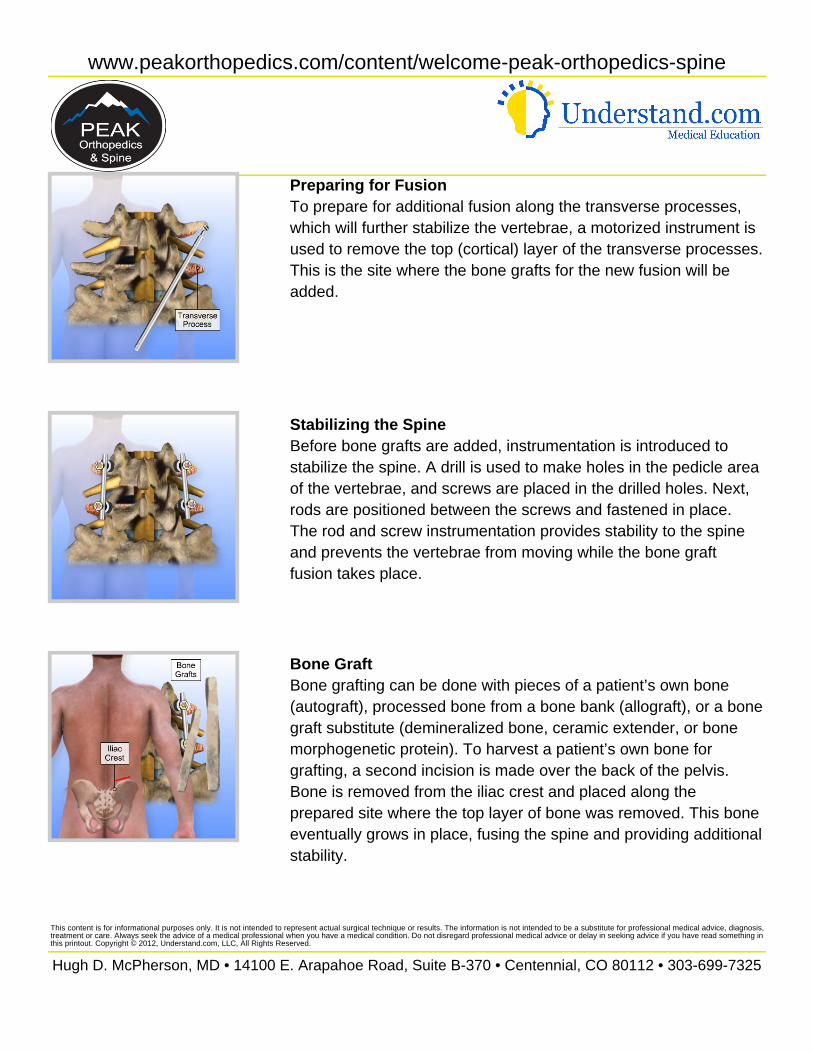
Preparing for FusionTo prepare for additional fusion along the transverse processes,which will further stabilize the vertebrae, a motorized instrument isused to remove the top (cortical) layer of the transverse processes.This is the site where the bone grafts for the new fusion will beadded.
Stabilizing the SpineBefore bone grafts are added, instrumentation is introduced tostabilize the spine. A drill is used to make holes in the pedicle areaof the vertebrae, and screws are placed in the drilled holes. Next,rods are positioned between the screws and fastened in place.The rod and screw instrumentation provides stability to the spineand prevents the vertebrae from moving while the bone graftfusion takes place.
Bone GraftBone grafting can be done with pieces of a patient’s own bone(autograft), processed bone from a bone bank (allograft), or a bonegraft substitute (demineralized bone, ceramic extender, or bonemorphogenetic protein). To harvest a patient’s own bone forgrafting, a second incision is made over the back of the pelvis.Bone is removed from the iliac crest and placed along theprepared site where the top layer of bone was removed. This boneeventually grows in place, fusing the spine and providing additionalstability.
This content is for informational purposes only. It is not intended to represent actual surgical technique or results. The information is not intended to be a substitute for professional medical advice, diagnosis,treatment or care. Always seek the advice of a medical professional when you have a medical condition. Do not disregard professional medical advice or delay in seeking advice if you have read something inthis printout. Copyright © 2012, Understand.com, LLC, All Rights Reserved.
Hugh D. McPherson, MD • 14100 E. Arapahoe Road, Suite B-370 • Centennial, CO 80112 • 303-699-7325
www.peakorthopedics.com/content/welcome-peak-orthopedics-spine
SummaryThe incisions are closed and dressed to complete the procedure.Adding the instrumentation with bone graft fusion increases thestrength of the spine directly after surgery, and may decrease theneed for a post-operative brace. Patients often remain in thehospital for two to four days following the procedure and shouldavoid heavy lifting, bending, twisting, and turning for six to twelveweeks.
This content is for informational purposes only. It is not intended to represent actual surgical technique or results. The information is not intended to be a substitute for professional medical advice, diagnosis,treatment or care. Always seek the advice of a medical professional when you have a medical condition. Do not disregard professional medical advice or delay in seeking advice if you have read something inthis printout. Copyright © 2012, Understand.com, LLC, All Rights Reserved.
Hugh D. McPherson, MD • 14100 E. Arapahoe Road, Suite B-370 • Centennial, CO 80112 • 303-699-7325