lymphoid tissue objectives: by the end of the lecture, the student should describe the microscopic...
TRANSCRIPT

LYMPHOID TISSUE
Objectives:By the end of the lecture, the student should describe
the microscopic structure of the following organs in correlation with their functions:
1- Lymph nodes.
2- Spleen.
3- Tonsils.
4- Thymus.
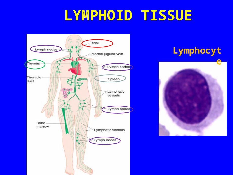
LYMPHOID TISSUE
Lymphocyte
LYMPHOID TISSUE
A) Diffuse lymphoid tissue B) Encapsulated lymphoid organs:
1 -Lymph nodes. 2 -Spleen.
3 -Tonsils (are incompletely encapsulated)
4 -Thymus. N.B. Both red bone marrow & Thymus are
considered 1ry. Lymphoid organs.

LYMPH NODES (L.N.)
(A) Stroma: 1- Capsule 2- Trabeculae (septa) 3- Reticular C.T.(B) Parenchyma: (lymphoid tissue + lymph sinuses) 1- Cortex 2- Paracortex 3- Medulla

1 .CORTEX OF L.N.
1- Lymphatic nodules (follicles):
a- 1ry: without germinal center
b- 2ry: with germinal center: Lighter
2- Cortical lymph sinuses.
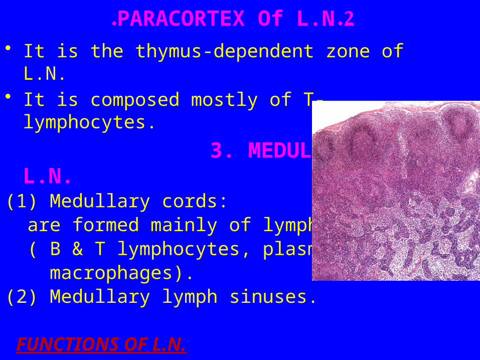
2 .PARACORTEX Of L.N.• It is the thymus-dependent zone of L.N.• It is composed mostly of T-lymphocytes.
3. MEDULLA OF L.N.(1) Medullary cords: are formed mainly of lymphoid cells ( B & T lymphocytes, plasma cells, macrophages).(2) Medullary lymph sinuses. FUNCTIONS OF L.N.• 1- Production of immunocompetent cells.• 2- Filtration of lymph.

SPLEEN
A. Stroma:
1- Capsule.
2- Trabeculae.
3- Reticular C.T.
B. PARENCHYMA:• (A) White pulp.• (B) RED PULP.
N.B. No cortex, no medulla.
FUNCTIONS OF SPLEEN
1- Filtration of blood.
2- Phagocytosis of old RBCs &
old blood platelets & invading
microorganisms.
3- Production & proliferation of
immunocompetent B & T lymphocytes.
4- Production of antibodies.

Tonsils
(1) Palatine Tonsils.
(2) Pharyngeal Tonsil.
(3) Lingual Tonsils.
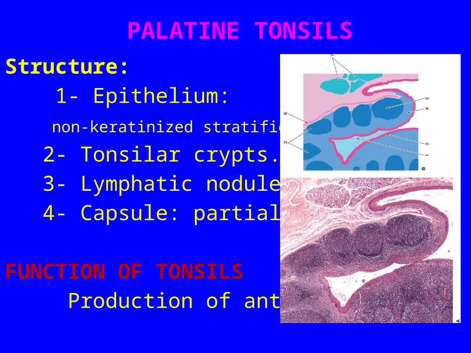
PALATINE TONSILS
Structure:
1- Epithelium:
non-keratinized stratified squamous.
2- Tonsilar crypts.
3- Lymphatic nodules.
4- Capsule: partial.
FUNCTION OF TONSILS
Production of antibodies.

THYMUS
A) Stroma:
1-Capsule
2-Interlobular trabeculae: incomplete
B) Thymic lobule:
1-Cortex
2-Medulla

CORTEX OF THYMIC LOBULEA) It contains developing (immature) T- lymphocytes
(thymocytes).
98% of thymocytes die?
B) Epithelial reticular cells
C) Macrophages.
N.B. No lymphatic nodules
No plasma cells
No B-lymphocytes
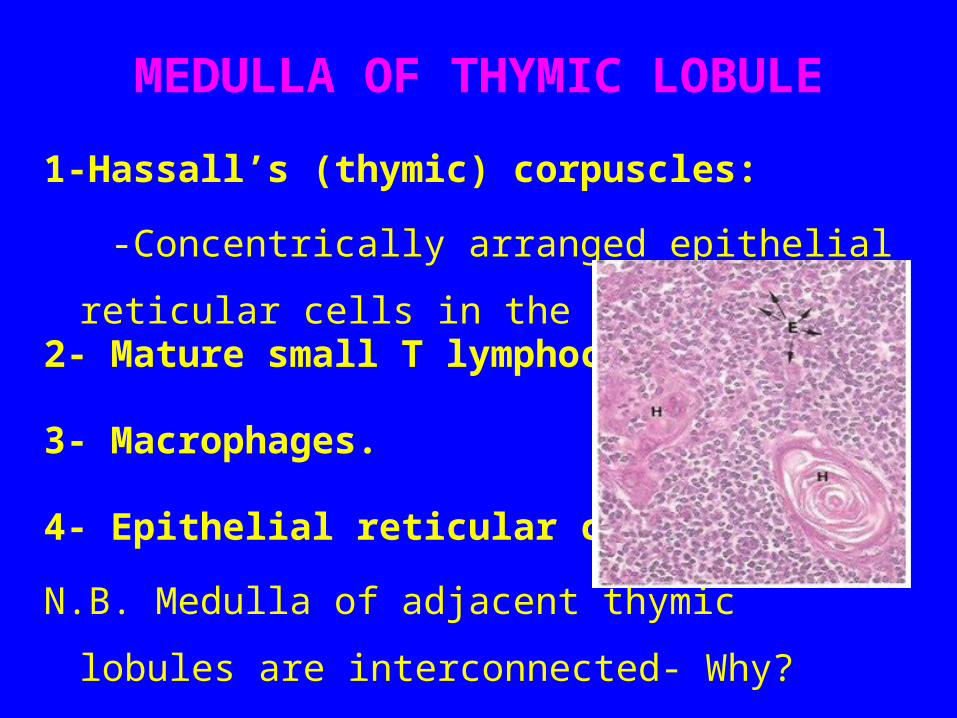
MEDULLA OF THYMIC LOBULE
1-Hassall’s (thymic) corpuscles:
-Concentrically arranged epithelial reticular cells in the
medulla.2- Mature small T lymphocytes
3- Macrophages.
4- Epithelial reticular cells.
N.B. Medulla of adjacent thymic lobules are
interconnected- Why? Incomplete trabeculae

FUNCTION OF THYMUS
Maturation of T lymphocytes.
(Immunoincompetent T cells →Immunocompetent T cells).
General notes about thymus
• No lymphoid nodules
• No reticular fibers
• No sinuses or sinusoids

Clinical ApplicationsAutoimmune disease
Def: mal-function of the immune system that results in the loss of immunological tolerance.
Example:
Grave’s Disease: in which the receptors for thyroid stimulating hormone (TSH) on follicular cells of thyroid gland are perceived to be antigens leading to the formation of antibodies against these TSH receptors.
These antibodies will bind to TSH receptors, so the cells will be stimulated to release excess amount of thyroid hormones leading to Hyperthyroidism

Clinical ApplicationAcquired Immunodeficiency Syndrome
(AIDS)• Causative organism: Human
immunodeficiency virus (HIV)• Pathogenesis: HIV binds to CD4 molecules
of TH resulting in incapacitating TH cells, leading to Spread of the virus, so other TH cells will be infected leading to incapability of immune response against bacterial or viral infections.

Clinical ApplicationsDiGeorge’s syndrome
• Def: Congenital failure of the thymus to
develop.
• Result: Failure to produce T cells leading
to non-functional cell-mediated immune
response which leads to early death due
to infection.
Clinical ApplicationsPalpable lymph node
• The presence of antigen or bacteria leads to
rapid proliferation of lymphocytes of the
lymph nodes (L.N), leading to increase of L.N.
to several times of its normal size, so the L.N.
becomes hard and palpable to the touch.
Clinical ApplicationsRupture of Spleen
• Spleen is a fragile or friable organ so major
trauma to the upper left abdominal quadrant
usually leads to rupture spleen, leading to
surgical removal of that ruptured spleen.

Thank You