maeder - thibodeau-beganny et al 2010
TRANSCRIPT

8/8/2019 Maeder - Thibodeau-Beganny Et Al 2010
http://slidepdf.com/reader/full/maeder-thibodeau-beganny-et-al-2010 1/61
Oligomerized Pool ENgineering (OPEN): An “Open-Source”
Protocol for Making Customized Zinc Finger Arrays
Morgan L. Maeder 1,2 , Stacey Thibodeau-Beganny 1, Jeffry D. Sander 3, Daniel F. Voytas 3,5 ,and J. Keith Joung 1,2,41Molecular Pathology Unit, Center for Cancer Research, and Center for Computational andIntegrative Biology, Massachusetts General Hospital, Charlestown, MA 02129, USA2Biological and Biomedical Sciences Program, Harvard Medical School, Boston, MA 02115, USA3Department of Genetics, Development & Cell Biology, 1043 Roy J. Carver Co-Laboratory, IowaState University, Ames, IA 50011, USA4Department of Pathology, Harvard Medical School, Boston, MA 02115, USA5Department of Genetics, Cell Biology & Development and Center for Genome Engineering, 321Church Street SE, University of Minnesota, Minneapolis, MN 55455, USA
AbstractEngineered zinc finger nucleases (ZFNs) form the basis of a broadly applicable method fortargeted, efficient modification of eukaryotic genomes. In recent work, we described OPEN(Oligomerized Pool ENgineering), an “open-source,” combinatorial selection-based method forengineering zinc finger arrays that function well as ZFNs. We have also shown in directcomparisons that the OPEN method has a higher success rate than previously described “modularassembly” methods for engineering ZFNs. OPEN selections are performed in E. coli using abacterial two-hybrid system and do not require specialized equipment. Here we provide a detailedprotocol for performing OPEN to engineer zinc finger arrays that have a high probability of functioning as ZFNs. Using OPEN, researchers can generate multiple customized ZFNs inapproximately 8 weeks.
IntroductionEngineered zinc finger nucleases (ZFNs) can be used as an important and broadly applicabletool for inducing highly efficient, targeted genome modification. 1-6 ZFNs consist of acustom-made DNA-binding zinc-finger array fused to a non-specific nuclease domain7 , 8(Fig. 1a). These artificial nucleases bind to DNA as dimers, with ZFN monomers binding to9 bp half-sites separated by a spacer sequence of variable length into which a double-stranded DNA break (DSB) is introduced (Fig. 1b).9 , 10 Repair of a ZFN-induced DSB bynon-homologous end-joining can lead to the introduction of mutagenic insertions or
deletions (indels) with high frequency. 11 -22 In addition, DSBs created by ZFNs alsostimulate homologous recombination-mediated repair;12 , 22, 23 therefore, ZFNs can be usedto induce high frequency gene targeting by introducing a homologous “donor DNAtemplate” harboring investigator-specified mutations or insertions into cells. 11 , 16, 24, 25
ZFN-induced modifications of endogenous genes have been reported to be as high as 50%and to work in a variety of cell types including Drosophila ,12, 13, 26, 27 somatic C. elegans ,28 zebrafish, 18-20 plants, 11 , 29, 30 and mammalian cells. 11 , 14-17, 24, 25
Correspondence to J. Keith Joung, [email protected].
NIH Public AccessAuthor Manuscript
Nat Protoc . Author manuscript; available in PMC 2010 April 22.
Published in final edited form as:Nat Protoc . 2009 ; 4(10): 1471–1501. doi:10.1038/nprot.2009.98.N
I H -P A A u
t h or Manus c r i pt
N I H -P A A ut h or Manus c r i pt
N I H -P A A ut h or M
anus c r i pt

8/8/2019 Maeder - Thibodeau-Beganny Et Al 2010
http://slidepdf.com/reader/full/maeder-thibodeau-beganny-et-al-2010 2/61
Implementing ZFN technology requires the capability to generate custom zinc finger arraysneeded to direct DSBs to specific genomic targets. We recently described a highly effectiveand “open-source” method for engineering zinc finger arrays which we termed OPEN (forOligomerized Pool ENgineering). 11 OPEN utilizes an archive of zinc finger pools, eachconsisting of a small number (95 or fewer) of different fingers designed to bind to aparticular 3 bp “subsite” (Fig. 2a). To perform an OPEN selection, a combinatorial library of multi-finger arrays from these pools is generated for a target 9 bp site of interest (Fig. 2a).
Members of this library that bind efficiently to the target site are then isolated using abacterial two-hybrid ( B2H ) selection method (Fig. 2c), which has been shown to identifymulti-finger arrays that possess high affinities and high specificities 31 and that functionefficiently as ZFNs in cells. 11 , 20, 32, 33 Thus, OPEN identifies combinations of fingers thatwork well toget her, thereby accounting for the context-depend ent DNA-binding activities of zinc fingers in an a rray. Because ZFNs function as dim ers, OPEN selections must beperformed for two 9 bp target sites in order to generate a ZFN pair.
To date, we have used OPEN to engineer, sequence, and characterize ∼ 500 zinc-fingerarrays targeted to 42 different nine bp target sequences. 11 , 20, 30, 34 The sequences,activities, and cognate target sites of all these multi-finger arrays have been deposited intothe freely available web-based Zinc Finger Database 34, 35 (ZiFDB; available athttp://www.zincfingers.org/software-tools.htm). Using a subset of these arrays, we have
successfully constructed and validated three-finger ZFN pairs for 17 different full targetsites from an integrated EGFP reporter gene (four sites) in human cells, three endogenoushuman genes (six sites), five endogenous zebrafish genes (five sites), and one endogenousplant gene (two sites). 11 , 20 , 30 At present, zinc finger pools have been described for allGNN subsites and for a subset of TNN subsites.
Other zinc finger engineering methods have also been used to create customized ZFNs.Various groups have made ZFNs using “modular assembly”, 12, 13, 26-28, 36-38 an approachfor engineering multi-finger arrays which treats individual fingers as independent units. 39-45
The success rate of modular assembly for making three-finger arrays has been reported to below 46 and in direct comparisons we have demonstrated that OPEN is more robust andefficient than modular assembly for constructing three-finger ZFNs. 11 The low success rateof modular assembly may result from its failure to account for the context-dependent
activity of zinc finger domains in an array. 47-51 The company Sangamo BioSciences, Inc.has also made four-finger ZFNs using their proprietary zinc finger engineering method.16-18, 25 ZFNs made using this approach are now commercially available through Sigma-Aldrich under the brand-name CompoZr™.18 , 52 We have performed an indirectcomparison of ZFN pairs made by OPEN with one ZFN pair made by the proprietarySangamo BioSciences approach (designed to different target sites) and found that theactivities and toxicities of these ZFNs made by the two approaches were comparable. 11
Despite the fact that we have successfully used OPEN to identify zinc finger arrays for alarge number of ZFN target half-sites, 11 , 20 the efficacy of the method is certainly not 100%.Our overall success rate to date is ∼ 70-80% for obtaining zinc finger arrays that can activatetranscription in the B2H system. However, we have also only focused on target half-sitesthat have one or more GNN “subsite.” Thus, we do not know how well the method will
work for sequences that do not contain any GNN subsites. We have deposited the results of both successful and failed selections in the publicly available Zinc Finger ConsortiumDatabase 34 at http://bindr.gdcb.iastate.edu/ZiFDB/ (or through the Zinc Finger Consortiumwebsite at: http://www.zincfingers.org/software-tools.htm) and we encourage all future usersof OPEN to do the same. Nonetheless, the less-than-perfect success rate of OPEN suggeststhat users should target more than one full ZFN site for their gene or locus of interest to
Maeder et al. Page 2
Nat Protoc . Author manuscript; available in PMC 2010 April 22.
N I H -P A A
ut h or Manus c r i pt
N I H -P A A ut h or Manus c r i pt
N I H -P A A ut h or
Manus c r i pt

8/8/2019 Maeder - Thibodeau-Beganny Et Al 2010
http://slidepdf.com/reader/full/maeder-thibodeau-beganny-et-al-2010 3/61
improve the chances of successfully obtaining functional zinc finger arrays for pairs of ZFNhalf-sites.
Although the emphasis of this protocol is on using OPEN zinc finger arrays to constructZFNs, we note that engineered zinc finger arrays have also been fused to other functionaldomains to create custom targeted transcription factors and recombinases. Both the modularassembly and proprietary Sangamo BioSciences approaches have been used successfully to
generate three, four-, five-, and six-finger arrays for these various fusion proteins.53-70
Todate, zinc finger arrays made by OPEN have only been used to make ZFNs, although inprinciple the method could also be used to construct zinc finger transcription factors andrecombinases. One potential limitation of OPEN for these other applications is that it hasonly been used successfully to create three-finger arrays. It remains unknown whether it,like the modular assembly and proprietary Sangamo approaches, can be successfullyadapted and used to create four-, five-, or six-finger arrays.
Here we describe a detailed protocol for using publicly available software and reagents toplan and perform OPEN selections. All experimental steps are performed using E. coli anddo not require specialized equipment. The OPEN platform was developed and validated bythe Zinc Finger Consortium, a group of academic scientists committed to continuedresearch, use, and application of engineered zinc finger technology (see
http://www.zincfingers.org). Software programs used in the OPEN protocol are freelyavailable on the web and do not require registration(http://www.zincfingers.org/software-tools.htm). Protocol-specific reagents required topractice OPEN are available to academic researchers through the non-profit plasmiddistribution service Addgene (http://www.addgene.org/zfc) and by request from the Jounglab. All other required materials and reagents are available through standard commercialvendors.
Overview of the ProcedureThe process of engineering zinc finger arrays using OPEN can be divided into five parts: (1)identifying potential full ZFN target sites using web-based software, (2) constructing B2Hselection strains harboring ZFN target half-sites, (3) creating combinatorial zinc finger array
libraries for the ZFN target half-sites, (4) selecting zinc finger arrays using OPEN, and (5)quantifying zinc finger array binding activity in B2H reporter strains harboring the ZFNtarget half-sites.
Identifying potential target sites using we b-based software
In thi s initial step, genomic sequence of the gene of interest is entered into the ZiFiTprogram11 , 35 which will identify potential full ZFN sites. This step is required because, asnote d in the Introduction, at pre sent OPEN can not target all possible sequences due to theavailability of “pools” for only a subset of all possible three base pair “subsites” at eachfinger position (currently 66 of the potential 192 pools are available). ZiFiT will alsoexclude sites that will be methylated by Dam and Dcm methylases in E. coli . Note that eachfull ZFN site consists of two 9 bp target “half-sites” separated by a user-defined spacer of 5,6, or 7 bps and that an OPEN selection must be performed for each of these half-sites. ZFNswith appropriate linkers (between the zinc finger array and the nuclease domain) canrecognize full ZFN sites with variable length spacers. 71, 72
Constructing B2H selection strains harboring target sites
B2H selection strains harbor a single copy F ′ episome with a ZFN target “half-site”positioned upstream of a B2H promoter which drives expression of the HIS3 and aadA
Maeder et al. Page 3
Nat Protoc . Author manuscript; available in PMC 2010 April 22.
N I H -P A A
ut h or Manus c r i pt
N I H -P A A ut h or Manus c r i pt
N I H -P A A ut h or
Manus c r i pt

8/8/2019 Maeder - Thibodeau-Beganny Et Al 2010
http://slidepdf.com/reader/full/maeder-thibodeau-beganny-et-al-2010 4/61
selectable marker genes 11 , 31, 73 (Fig. 2c). The ZFN target half-site used actually consists of the 9 bp target sequence identified by ZiFiT plus an additional upstream and downstreambase from the genomic sequence (i.e.—an 11 bp sequence); we use these additional basesbecause previous studies have shown that the identities of these external bases can influencebinding of a zinc finger array. 48 To construct a B2H selection strain, a target 11 bp site isfirst cloned into a reporter plasmid. The selection reporter plasmid contains lacI q and lacZ sequences which flank the binding site-promoter- HIS3 -aadA cassette and which can
recombine with sequences present on an F ′ episome present in the CSH100 strain74, 75
(Fig.3a). As shown in Fig. 3b, a double cross-over event between these two homologous regionsleads to the introduction of the binding site-promoter- HIS3 -aadA cassette onto the F ′(although this event will be more infrequent than a single cross-over event or norecombination). Mating of CSH100 cells harboring these various recombinant and non-recombinant F ′ episomes with the F - strain KJ1C will enable transfer of the F ′ episomesfrom the former cells to the latter. As shown in Fig. 3b, KJ1C cells harboring the desireddouble-recombinant F ′ can be identified by their growth on plates containing tetracycline,kanamycin, and sucrose (LB/TKS plates). Construction of a B2H selection strain iscompleted by transforming KJ1C cells bearing a recombinant F ′ reporter plasmid with anadditional low-copy plasmid which expresses the hybrid “alpha-Gal4” protein consisting of the amino-terminal domain and inter-domain linker of the E. coli RNA polymerase α-subunit fused to a fragment of the yeast Gal4 protein.
Constructing combinatorial zinc finger array libraries
Zinc finger array libraries are built by using PCR to stitch together random combinations of the three OPEN finger pools corresponding to the three subsites in a given target (Fig. 2a).The DNA encoding these randomly recombined finger arrays is then cloned into a low-copyexpression phagemid which expresses them as fusions to a fragment of the yeast Gal11Pprotein (Fig. 2a). Expression of these Gal11P-zinc finger array hybrid proteins is controlledby the lac repressor and is therefore inducible by the addition of IPTG to the medium.
To facilitate highly efficient transduction of a B2H selection strain by various Gal11P-zincfinger array hybrids, the library of phagemids is converted into infectious phage particles(Fig. 2b). We have found that efficient selection on histidine-deficient NM medium worksonly with B2H selection cells that have not been grown in rich medium (J.K. Joung,unpublished observations). Use of phage-based transduction enables us to achieve hightransformation efficiencies with the B2H selection strain without the need to useelectroporation, a method that requires cells to recover in rich SOC medium. In addition, thephage-based approach helps to ensure that only one zinc finger array is introduced into mostcells; this can be simply accomplished by using an excess of selection strain cells relative tophage. Finally, the use of phage enables rescue of the zinc finger array-encoding phagemidfrom selection strain cells. This capability is important for the two-stage selection procedure(see below).
Selecting zinc finger arrays using OPEN
OPEN selections are performed in two stages: an initial lower stringency selection enrichesfor zinc finger arrays that bind to the ZFN target half-site; a second higher stringency
selection identifies the final candidates. In the first selection stage, IPTG is added to inducehigher levels of Gal11P-zinc finger array and alpha-Gal4 hybrid protein expression. In thesecond selection step, IPTG is omitted so that the two hybrid proteins are expressed at lowerlevels. Both selection steps are performed by introducing a combinatorial zinc finger arrayphagemid library into a B2H selection strain and by plating on media which selects for cellsthat exhibit increased expression of the HIS3 and aadA marker genes. If a Gal11P-zincfinger array hybrid protein can bind to the 11 bp ZFN target half-site on the reporter, then
Maeder et al. Page 4
Nat Protoc . Author manuscript; available in PMC 2010 April 22.
N I H -P A A
ut h or Manus c r i pt
N I H -P A A ut h or Manus c r i pt
N I H -P A A ut h or
Manus c r i pt
8/8/2019 Maeder - Thibodeau-Beganny Et Al 2010
http://slidepdf.com/reader/full/maeder-thibodeau-beganny-et-al-2010 5/61
transcription of the selectable marker genes is activated because the DNA-bound Gal11P-zinc finger hybrid protein recruits RNA polymerase complexes harboring the alpha-Gal4protein to the promoter via a protein-protein interaction between the Gal11P and Gal4protein fragments (Fig. 2c).
The ability to rescue the zinc finger-encoding phagemids from selection strain cells is acritical capability utilized during the two-stage selection. Following the initial stage of
selection, zinc finger-encoding phagemids are rescued from these cells by infection withM13K07 helper phage and subsequent packaging of single-stranded phagemids in infectiousphage particles. This “enriched phagemid library” is then harvested and used to transducefresh B2H selection strain cells for the second round of selection.
Quantifying zinc finger array DNA-binding activity using B2H reporter strains
To confirm that zinc finger arrays identified in the selection process bind to the ZFN targethalf-site, the phagemids encoding these candidates are isolated from colonies on theselection plates and then introduced by transformation into a B2H reporter strain. 76 TheB2H reporter strain is similar to the B2H selection strain in that it harbors: (1) a single-copyplasmid (in this case a mini-BAC plasmid instead of a full F ′ episome) with the ZFN targethalf-site positioned upstream of a weak promoter which, in turn, controls expression of alacZ reporter gene; (2) a low-copy number plasmid expressing the alpha-Gal4 hybridprotein. If a Gal11P-zinc finger array hybrid that can bind to the ZFN target half-site isexpressed in the B2H reporter strain, then transcription of lacZ will be activated. Becausethe lacZ gene encodes β-galactosidase, its expression level can be measured by a simplequantitative assay. 75, 76 By comparing β-galact osidase expression in the presence andabsence of a given zinc finger array, a “fold-activation” value can be calculated which canguide the choice of which arrays to carry forward for testing as ZFNs.
Experimental DesignInitial trial selections
Before attempting to perform selections for new target sites of interest, we recommend thatusers first complete the entire OPEN protocol start-to-finish with at least one target site that
has worked successfully in previous experiments. We define a “successful” selection as onethat has previously yielded one or more zinc finger arrays that activate transcription morethan three-fold in the B2H system. The choice of this positive control target site may beinfluenced by the pools that the investigator has requested and therefore has on hand. TheZinc Finger Database (ZiFDB; available at http://www.zincfingers.org/software-tools.htm)contains information on target DNA sites for which OPEN selections have been successfulas well as the sequences and B2H activities of finger arrays obtained from those experimentswith which investigators can compare their own results.
For any initial selection, we strongly recommend that investigators follow the protocoloutlined here precisely. We have noted throughout the protocol certain steps that areparticularly critical to success. Although some of these suggestions may not appear to beimportant or significant to the first-time user, we have learned that these parameters can be
critical to success of the protocol. Examples of such recommendations include the use of aspecific thermostable polymerase for amplification of the finger pools, the use of glucosefrom a specific vendor for the NM medium, and the method for inoculating colonies intoNM medium.
Maeder et al. Page 5
Nat Protoc . Author manuscript; available in PMC 2010 April 22.
N I H -P A A
ut h or Manus c r i pt
N I H -P A A ut h or Manus c r i pt
N I H -P A A ut h or
Manus c r i pt

8/8/2019 Maeder - Thibodeau-Beganny Et Al 2010
http://slidepdf.com/reader/full/maeder-thibodeau-beganny-et-al-2010 6/61
Time required to complete the protocol
Although an experienced lab can complete the entire OPEN protocol in eight weeks or less,investigators should anticipate that the protocol will require significantly more time the firstfew times they perform it due to the inevitable need to repeat certain steps. In addition,completing OPEN in the optimal timeframe requires significant planning and coordination.We anticipate which plasmids, PCR products, restriction and modification enzymes, plates,medium, and cells will be required at least several days in advance to ensure that these
reagents do not become a rate limiting step. An optimal timeline for performing each part of the protocol is given below in the TIMING section. Once the protocol has been mastered, inour experience it is possible for a single individual to perform 12 or more selections inparallel in eight weeks time or less.
MaterialsReagents
• OPEN zinc finger pools (available by request from the Joung lab)
• Plasmids and expression vectors (see REAGENT SET UP)
• Bacterial strain CSH100 (F ′ lacproA+,B+(lacIq lacPL8)/araD(gpt-lac)5 ; availableby request from the Joung lab); this strain is used to construct B2H selectionstrains.
• Bacterial strain KJ1C (F - ΔhisB463 Δ(gpt-proAB-arg-lac)XIII zaj ∷ Tn10 ); availableby request from the Joung lab); this strain is required to construct B2H selectionstrains.
• Bacterial strain XL-1 Blue ( recA1 endA1 gyrA96 thi-1 hsdR17 supE44 relA1 lac [F ′ proAB lacIq lacZDM15 Tn10 (Tet R)]; Stratagene cat. no. 200249); this strain isused for routine subcloning of plasmids and for building the OPEN zinc fingerlibraries.
• Bacterial strain Transformax EPI300 (F − mcrA Δ (mrr-hsdRMS-mcrBC )Φ80d lacZDM15 ΔlacX74 recA1 endA1 araD139 Δ(ara, leu )7697 galU galK λ −
rpsL nupG trfA dhfr ; Epicentre, cat. no. C300C105); this strain is used to performsubcloning steps with the pBAC-lacZ-derived B2H reporter plasmids.
• Bacterial strain KJBAC1 (F - lacIq ΔhisB463 Δ(gpt-proAB-arg-lac ) XIII zaj ∷ Tn10 ;strain available through Addgene; http://www.addgene.org/zfc); this strain isrequired for propagation and subcloning of pBAC-lacZ-derived B2H reporterplasmids.
• M13K07 helper phage (New England Biolabs, Cat #N0315S; store indefinitely at-20°C)
<!>CAUTION Care should be taken to avoid contaminating laboratory equipmentand benches with bacteriophage
• Restriction enzymes (all from New England Biolabs): BamH I (cat. no. R0136S), Bbs I (cat. no R0539L), Bsa I (cat. no. R0533L), EcoR I (cat. no. R0101L), Hind III(cat. no. R0104L), Pst I (cat. no. R0140S), Sap I (cat. no. R0569L), Xba I (cat. no.R0145L)
• 10× restriction enzyme buffers (New England Biolabs, included with enzymes)
• T4 DNA Ligase and associated standard T4 DNA Ligase reaction buffer (NewEngland Biolabs, cat. no. M0202S)
Maeder et al. Page 6
Nat Protoc . Author manuscript; available in PMC 2010 April 22.
N I H -P A A
ut h or Manus c r i pt
N I H -P A A ut h or Manus c r i pt
N I H -P A A ut h or
Manus c r i pt

8/8/2019 Maeder - Thibodeau-Beganny Et Al 2010
http://slidepdf.com/reader/full/maeder-thibodeau-beganny-et-al-2010 7/61
• Quick Ligation Kit (New England Biolabs, cat. no. M2200S)
• T4 Polynucleotide Kinase (New England Biolabs, cat. no. M0201S)
• Cloned Pfu polymerase and associated 10× reaction buffer (Stratagene, cat. no.600159-81)
• Expand High-Fidelity thermostable polymerase and associated 10× Expand bufferwith MgCl2 (Roche, cat. no. 11732641001)
<!>CRITICAL We have found the use of Expand enzyme to be critical forsuccessful amplification of the zinc finger pools
• AccuGel 29:1 acrylamide:bis-acrylamide solution (National Diagnostics, cat. no.EC-852)
<!>CAUTION Acrylamide is a neurotoxin and should be handled with gloves.
• 10% (w/v) ammonium persulfate in ddH 2O (Fisher, cat. no. 7727-54-0; storeindefinitely at -20°C)
• TEMED (Fisher, cat. no. BP150-100; store indefinitely at 4°C)
• 100% ethanol (Pharmco, cat. no. 111ACS200)
• 70% (v/v) ethanol in ddH 2O• QIAprep Spin Miniprep kit (Qiagen, cat. no. 27106)
• QIAquick PCR Purification kit (Qiagen, cat. no. 28106)
• MinElute PCR Purification kit (Qiagen, cat. no. 28006)
• LB medium (Difco, cat. No. 244620)
• LB agar medium (Difco, cat. No 244520)
• 2xYT medium (Difco, cat. No. 244020)
• SOB medium (Difco, cat. No. 244310)
• SOC medium (SOB medium with 0.4% (v/v) glucose)
• Bacto-Agar (Difco, cat. no. 214010)
• Bacto-Tryptone (Difco, cat. no. 211705)
• Sterile 10% (v/v) glyercol and sterile 50% (v/v) glycerol both in ddH2O (100%glycerol – Fisher, cat. no. BP229-1)
• 10× M9 salts (Difco, cat. no. 248510)
• 20% (w/v) Glucose in ddH2O (Mallinckrodt Baker, cat. No. 4912-06)
<!>CRITICAL We have found the use of glucose from this specific vendor to becritical for high density growth of our B2H selection strains in NM medium
• 20 mM Adenine HCl in ddH 2O (Sigma, cat. no A8751; store indefinitely at room
temperature (20-25°C)
• Amino Acid Mixture (see REAGENT SET UP)
• Individual amino acid powders (all available through Sigma): Phenylalanine(#P5482), Lysine (#L5626), Arginine (#A6969), Glycine, Valine (#V0500),Alanine (#A7469), Tryptophan (#T8941), Threonine (#T8625), Serine (#S4311),Proline (#P0380), Asparagine(#A4159), Aspartic Acid (#A4534), Glutamine
Maeder et al. Page 7
Nat Protoc . Author manuscript; available in PMC 2010 April 22.
N I H -P A A
ut h or Manus c r i pt
N I H -P A A ut h or Manus c r i pt
N I H -P A A ut h or
Manus c r i pt

8/8/2019 Maeder - Thibodeau-Beganny Et Al 2010
http://slidepdf.com/reader/full/maeder-thibodeau-beganny-et-al-2010 8/61
(#G3126), Isoleucine (#I2752), Leucine (#L8000), L-Glutamic acid Potassium saltmonohydrate (#G1501), and Tyrosine (#T3754).
• 1 M MgSO 4 in ddH 2O (Fisher, cat. no. BP213-1)
• Thiamine (10 mg ml -1 stock solution in ddH 2O; filter sterilize and store indefinitelyat -20°C)
• 10 mM ZnSO 4 in ddH 2O
• 100 mM CaCl 2 in ddH 2O
• Carbenicillin (Sigma, cat. no. T4625; 50 mg ml -1 stock solution in ddH 2O; filtersterilize and store indefinitely as 1 ml aliquots at -20°C)
• Chloramphenicol (Sigma, cat. no. C0378; 30 mg ml -1 stock solution in 100%ethanol; store indefinitely at -20°C)
• Kanamycin (Sigma, cat. no. K4000-5G; 30 mg ml -1 stock solution in ddH 2O; filtersterilize and store indefinitely at 4°C)
• Tetracycline (Sigma, cat. no. T3383; 12.5 mg ml -1 stock solution in 80% (v/v)ethanol; filter sterilize and store indefinitely wrapped in foil at -20°C)
• Streptomycin (Sigma, cat. no. S6501; 100 mg ml -1 stock solution in ddH2O; filter
sterilize and store indefinitely at 4°C)
• 3-aminotriazole (3-AT; US Biochemical, cat. no. 11245; 1 M stock solution inddH 2O; filter sterilize and store indefinitely at -20°C)
• Sucrose (Fisher, cat. no. 8360; 50% (w/v) stock solution in ddH 2O; storeindefinitely at room temperature)
• Glycogen (Sigma, cat. no. G1767; 10 mg ml -1 stock solution; store indefinitely at-20°C)
• IPTG (isopropyl-beta-D-thiogalactopranoside; Sigma, cat. no. 16758; 1 M stock solution in ddH 2O; filter sterilize and store indefinitely as 1 ml aliquots at -20°C)
• Sterile 100 mM ZnSO 4 in ddH 2O (Fisher, cat. no. Z68-500)
• Ice-cold Solution A for preparing competent cells (10 mM MnCl 2 (Fisher, cat. no.BP541-100), 50 mM CaCl 2 (Fisher, cat. no. BP510-250), 10 mM MES, pH 6.3(Fisher, cat. no. BP300-11) in ddH 2O; filter sterilize and store wrapped in foil at 4°C; solution is usable until it acquires brown discoloration; Note: to make SolutionA, use a 100 mM MES stock solution prepared in ddH 2O which has been broughtto a pH of 6.3 using KOH)
• Ice-cold Solution A with 15% (v/v) glycerol for preparing competent cells(Solution A with 15% (v/v) glycerol; filter sterilize and store wrapped in foil at4°C; solution is usable until it acquires brown discoloration)
• ONPG (Sigma, cat. no. N1127; o-nitrophenyl-beta-D-galactopryanoside, 4 mg ml -1
in ddH 2O; solution can be stored indefinitely as 10 ml aliquots wrapped in foil at
-20°C.
• Z-buffer (1 liter: 16.1 g of Na 2HPO 4-7H 2O, 5.5 g of NaH 2PO 4-H2O, 0.75 g of KCl,0.246 g of MgSO 4-7H 2O dissolved in ddH 2O; filter sterilize and store at roomtemperature)
• Z-buffer with β-mercaptoethanol (prepare fresh by adding 2.7 μl of β-mercaptoethanol to every 1 ml of Z-buffer)
Maeder et al. Page 8
Nat Protoc . Author manuscript; available in PMC 2010 April 22.
N I H -P A A
ut h or Manus c r i pt
N I H -P A A ut h or Manus c r i pt
N I H -P A A ut h or
Manus c r i pt

8/8/2019 Maeder - Thibodeau-Beganny Et Al 2010
http://slidepdf.com/reader/full/maeder-thibodeau-beganny-et-al-2010 9/61
• Popculture reagent (Novagen, cat. no. 71092)
• R-Lysozyme (30,000 units μl-1) and associated dilution buffer (Novagen, cat. no.71110)
<!>CRITICAL Once diluted in dilution buffer, R-Lysozyme can not be re-frozen.R-Lysozyme can be stored indefinitely at -20°C when diluted to 400 units μl-1 indilution buffer containing 50% glycerol.
• Lysis Master Mix (10:1 mixture of Popculture reagent to diluted R-Lysozyme [4units μl-1])
• 7.5 M NH 4OAc in ddH 2O (Fisher, cat. no. A637-500; store indefinitely at roomtemperature)
<!>CRITICAL Due to the volatility of NH 4OAc, we seal storage containers bycapping tightly and wrapping with Parafilm.
• 10% SDS (Fisher, cat. no. BP2436-200)
• 1 M MgOAc in ddH 2O (Fisher, cat. no. M13-500)
• 0.5 M EDTA, pH 8.0 (Fisher, cat. no. S311-500)
• 1M Tris, pH 8 in ddH2O (Fisher, cat. no. BP152-1)
• 1M MgCl 2 in ddH 2O (Sigma, cat. no. M2393)
• 5M NaCl in ddH 2O (Fisher, cat. no. BP358-212)
• Ammonium acetate elution buffer (see REAGENT SET UP)
• 5× PEG/NaCl solution (see REAGENT SET UP)
• dNTP nucleotide set (Roche, cat. no. 11969064001; use this to make a 10 mMstock solution of dCTP, a 10 mM stock solution of dTTP and a 10 mM stock solution of dNTP mixture; store indefinitely in 200 μl aliquots at -20°C)
• 10× BSA (bovine serum albumin; 1 mg ml -1 solution; New England Biolabs, cat.no. B9001S; store indefinitely at -20°C)
• PEG8000 (Fisher, cat. no. BP233-1)
• 1 M Arabinose in ddH 2O(Sigma, cat. no. A-3256)
• 10× Annealing Buffer (see REAGENT SET UP)
• Sequencing and PCR primers (see Table 1)
Equipment
• 96-well thermocycler
• Wooden sticks (Fisher, cat. no. 23-400-102; sterilize by autoclaving)
• 200 μl filtered pipet tips (CLP, cat. no. BT200)
• 25 mm glass culture tubes (Fisher, cat. no. 14-961-34)• 18 mm glass culture tubes (Fisher, cat. no. 14-961-32)
• Glass beads, 3mm (Fisher, cat. no. 11-312A)
• Rotating wheel drum (for bacterial cultures)
• LabQuake shaker/rotisserie (Barnstead Thermolyne, cat. no. 415110)
Maeder et al. Page 9
Nat Protoc . Author manuscript; available in PMC 2010 April 22.
N I H -P A A
ut h or Manus c r i pt
N I H -P A A ut h or Manus c r i pt
N I H -P A A ut h or
Manus c r i pt
8/8/2019 Maeder - Thibodeau-Beganny Et Al 2010
http://slidepdf.com/reader/full/maeder-thibodeau-beganny-et-al-2010 10/61
• 1 mm gap electroporation cuvettes (BTX, cat. no. 45-0124, model no. 610)
• Electroporation device with adjustable settings
• Programmable, multi-channel (8 or 12) pipet
• 250 ml, 1 liter, and 2 liter glass flasks
• Orbital platform shaker with adjustable speed
• 2 ml cryogenic storage vials (Corning, cat. no. 430659)
• 0.22 μm PES (polyethersulfone) filter units (Millipore, cat. no. SLGP033RS)
• 245 mm square plates (Corning, cat. no. 431301)
• 100 × 15 mm round Petri plates (Fisher, cat. no. 08-757-13)
• 96-well flat-bottom, microtiter plates (Corning-Costar, cat. no. 3596)
• Deep-well 96-well blocks (optional; Corning, cat. no. 3960)
• Microtiter plate reader with temperature control option
• Sterile 250-ml centrifuge bottles
• Sterile 1 liter centrifuge bottles
• Sterile 50 ml conical tubes (Corning, cat. no. 430290)
• Autoclave
• Polyacrylamide gel running apparatus with gel casting system (Thermo Scientific,cat. no. P9DS-1)
• Biorad Model 680 Microplate Reader (or other microtiter plate reader withtemperature control)
• Microtitertron orbital shaker for 96-well blocks (Appropriate Technical Resources;optional)
Reagent Set Up
• Plasmids and expression vectors: The following are available by request throughthe Joung lab: pKJ1712 reporter plasmid: KAN R, p15A origin of replication, fullsequence and plasmid features described in Supplementary Fig. 1; pBR-UV5-GP-FD2 zinc finger B2H expression plasmid: AMP R, ColEI origin of replication, fullsequence and plasmid features described in Supplementary Fig. 2; pAC-alphaGal4expression plasmid: CAM R, p15A origin of replication, full sequence and plasmidfeatures described in Supplementary Fig. 3. The following plasmids (together withtheir full sequences and maps) are available through Addgene(http://www.addgene.org/zfc): pBAC-lacZ reporter plasmid: CAM R, primary F ′and secondary oriV origins of replication; pAC-KAN-alphaGal4 expressionplasmid: KAN R, p15A origin of replication.
<!>CRITICAL Plasmids pBR-UV5-GP-FD2, pAC-alphaGal4, and pAC-KAN-alphaGal4 must be propagated in a lacI q strain (e.g.--XL-1 Blue) in order to avoidtoxicity due to unregulated expression of fusion proteins encoded on theseplasmids.
<!>CRITICAL pBAC-lacZ is a single-copy plasmid that gives low yields whenpropagated in standard E. coli strains. However, it also harbors a second, highercopy origin ( oriV ) that requires for activity a protein encoded by the trfA gene. For
Maeder et al. Page 10
Nat Protoc . Author manuscript; available in PMC 2010 April 22.
N I H -P A A
ut h or Manus c r i pt
N I H -P A A ut h or Manus c r i pt
N I H -P A A ut h or
Manus c r i pt

8/8/2019 Maeder - Thibodeau-Beganny Et Al 2010
http://slidepdf.com/reader/full/maeder-thibodeau-beganny-et-al-2010 11/61
routine sub-cloning, pBAC-lacZ and its derivatives should therefore be propagatedin Transformax EPI300 cells. These cells express trfA from a promoter that can beinduced with arabinose (S.T.B. and J.K.J., unpublished data).
• Ammonium acetate elution buffer: To prepare 25 ml, add the following to 22.8 mlddH 2O: 1.65 ml of 7.5 M NH 4OAc, 250 μl of 10% (w/v) SDS, 250 μl of 1 MMgOAc, and 50 μ1 of 0.5 M EDTA. Store at room temperature for no more thanone month.
<!>CRITICAL To avoid irreversible precipitation of SDS, do not store elutionbuffer at temperatures below room temperature. Also, seal storage container tightlywith Parafilm to avoid loss of ammonium acetate through volatilization.
• 10× Annealing Buffer: To prepare 1 ml, combine the following: 400 μl 1 M Tris(pH 8), 200 μl 1 M MgCl 2, 100 μl 5 M NaCl, 20 μl 0.5 M EDTA (pH 8), and 280μl ddH 2O. Buffer can be stored indefinitely at -20°C.
• M9 minimal medium agar plates: To prepare 500 ml, autoclave 439 ml H 2O with7.5 g Bacto-agar and a stir bar. When agar has cooled to approximately 65°C, add50 ml 10× M9 salts, 1 ml 1 M MgSO 4, 10 ml 20% (w/v) glucose and 0.5 ml100mM CaCl 2 and then pour plates. Plates can be stored indefinitely at 4°C insealed plastic bags.
• LB/CK plates (LB agar supplemented with 30 μg ml -1 chloramphenicol and 30 μgml -1 kanamycin). Plates can be stored for up to two months at 4°C in sealed plasticbags.
• LB/CCK plates (LB agar supplemented with 100 μg ml -1 carbenicillin, 30 μg ml -1
chloramphenicol and 30 μg ml -1 kanamycin). Plates can be stored for up to twomonths at 4°C in sealed plastic bags.
• LB/TK plates (LB agar supplemented with 12.5 μg ml -1 tetracycline and 30 μg ml -1
kanamycin). Plates can be stored for up to two months at 4°C in sealed plastic bagswrapped in aluminum foil to protect the tetracycline from light.
• LB/TKS plates (LB agar supplemented with 12.5 μg ml -1 tetracycline, 30 μg ml -1
kanamycin, and 5% (w/v) sucrose). Plates can be stored for up to two months at4°C in sealed plastic bags wrapped in aluminum foil to protect the tetracycline fromlight.
• LB/TC plates (LB agar supplemented with 12.5 μg ml -1 tetracycline and 100 μgml -1 carbenicillin). Plates can be stored for up to two months at 4°C in sealedplastic bags wrapped in aluminum foil to protect the tetracycline from light.
• LB/KCarb plates (LB agar supplemented with 100 μg ml -1 carbenicillin and 70 μgml -1 kanamycin). Plates can be stored for up to two months at 4°C in sealed plasticbags.
• NM medium: To prepare 500 ml, combine the following components in the orderlisted: 418 ml ddH 2O, 50 ml 10× M9 salts, 10 ml 20% (w/v) glucose, 5 ml 20 mMAdenine HCl, 15 ml Amino Acid Mixture, 500 μl 1M MgSO 4, 500 μl thiamine (10
mg/ml), 500 μl 10 mM ZnSO 4 and 500 μl 100 mM CaCl 2. Filter sterilize and storeat 4°C. Wrap container in alumnimum foil to protect from light.
• NM plates: To prepare 500 ml, autoclave 7.5g Bacto-agar, 418 ml H 2O and a stirbar in a 1 or 2 liter Erlenmyer flask. In a separate sterile container, mix together thefollowing components in the order listed: 50 ml 10× M9 salts, 10 ml 20% (w/v)glucose, 5 ml 20mM Adenine HCl, 15 ml Amino Acid Mixture, 500 μl 1MMgSO 4, 500 μl thiamine (10mg/ml), 500 μl 10 mM ZnSO 4, 500 μl 100 mM CaCl 2
Maeder et al. Page 11
Nat Protoc . Author manuscript; available in PMC 2010 April 22.
N I H -P A A
ut h or Manus c r i pt
N I H -P A A ut h or Manus c r i pt
N I H -P A A ut h or
Manus c r i pt

8/8/2019 Maeder - Thibodeau-Beganny Et Al 2010
http://slidepdf.com/reader/full/maeder-thibodeau-beganny-et-al-2010 12/61
and carbenicillin, chloramphenicol, kanamycin, IPTG, 3-AT, and streptomycin asneeded. When agar has cooled to ∼ 65-70°C, add the separate mixture, mix well andpour plates. Plates can be stored for up to two months at 4°C in sealed plastic bags.
• NM/CCK plates (NM agar supplemented with 100 μg ml -1 carbenicillin, 30 μg ml -1
chloramphenicol and 30 μg ml -1 kanamycin). Plates can be stored for up to twomonths at 4°C in sealed plastic bags.
• NM/CCKI plates (NM agar supplemented with 100 μg ml-1
carbenicillin, 30 μgml -1 chloramphenicol, 30 μg ml -1 kanamycin, and 50 μM IPTG). Plates can bestored for up to two months at 4°C in sealed plastic bags.
• Z-buffer: To prepare 1 liter, dissolve 16.1 g of Na 2HPO 4-7H 2O, 5.5 g of NaH 2PO 4-H2O, 0.75 g of KCl and 0.246 g of MgSO 4-7H 2O in ddH 2O; filter sterilize. Buffercan be stored indefinitely at room temperature.
• Z-buffer with β-mercaptoethanol: Prepare fresh on day of use by adding 2.7 μl of β-mercaptoethanol to every 1 ml of Z-buffer.
• H Top Agar: To prepare 100 ml, autoclave 0.8 g Bacto-agar, 0.8 g NaCl, and 1 gBacto-Tryptone in 100 ml ddH 2O. Media can be stored indefinitely in a sealedcontainer at room temperature.
• 5× PEG/NaCl Solution: To prepare 500 ml, dissolve 87.5 g PEG 8000 and 62.5 gNaCl in ddH 2O to a final volume of 500 ml. Filter sterilize. Solution can be storedindefinitely at room temperature.
<!>CRITICAL Dissolving the PEG and NaCl into solution may require stirringfor many hours. We typically leave the mixture stirring overnight.
• Custom synthesized oligonucleotides required to create Selection ReporterPlasmids bearing ZFN half-site target sequences
<!>CRITICAL Two oligonucleotides must be synthesized for each ZFN half-sitetarget sequence. Details regarding design of these oligonucleotides are provided inBox 1.
• Amino Acid Mixture: Prepare six separate amino acid solutions by dissolving thecomponents listed below in ddH 2O to a final volume of 100 ml each. Afterpreparing all six solutions, combine them together and filter sterilize. Storewrapped in foil at 4°C. Solution is good for at least 30 days.
– Solution 1: 0.99 g Phenylalanine, 1.1 g Lysine, 2.5 g Arginine
– Solution 2: 0.2 g Glycine, 0.7 g Valine, 0.84 g Alanine, 0.41 g Tryptophan
– Solution 3: 0.71 g Threonine, 8.4 g Serine, 4.6 g Proline, 0.96 gAsparagine
– Solution 4: 9.1 ml HCl, 1.04 g Aspartic Acid, 14.6 g Glutamine
– Solution 5: 18.7 g L-Glutamic acid Potassium salt monohydrate, 0.36 gTyrosine, 4 g NaOH
– Solution 6: 0.79 g Isoleucine, 0.79 g Leucine
<!>CRITICAL The six solutions must first be made up individually and thencombined together. For each of the six solutions, add each amino acid componentin the order listed and make sure that each component is completely dissolvedbefore adding the next.
Maeder et al. Page 12
Nat Protoc . Author manuscript; available in PMC 2010 April 22.
N I H -P A A
ut h or Manus c r i pt
N I H -P A A ut h or Manus c r i pt
N I H -P A A ut h or
Manus c r i pt
8/8/2019 Maeder - Thibodeau-Beganny Et Al 2010
http://slidepdf.com/reader/full/maeder-thibodeau-beganny-et-al-2010 13/61
ProcedureIdentifying potential OPEN ZFN sites using the web-based Zinc Finger Targeter (ZiFiT)program
1| Open a web browser to the ZiFiT v3.0 program webpage (address:http://binder.gdcb.iastate.edu/ZiFiT; a link to the website is also permanently available at:http://www.zincfingers.org/software-tools.html).
2| Click on the ZiFiT option on the left hand menu and then click on the “Design ZincFinger Nucleases” option under the “OPEN (Oligomerized Pool Engineering)” menu. Notethat on the sequence entry page, the DNA triplets for which OPEN pools are currentlypublicly available are already automatically checked. Additional pools for other triplets canalso be selected by the user.
3| In the “Sequence” box, type or paste in the DNA sequence that will be searched forpotential OPEN sites. Note that any spaces or numbers are ignored and that the DNAsequence to be analyzed can be pasted in as raw sequence or in FASTA format.
4| Select the length of the target site spacer sequence desired from the Spacer drop downbox. Various ZFN expression vectors encoding different length linkers between the zinc
finger arrays and the Fok I nuclease domain have been described that enable cleavage of ZFN target sites harboring spacer sequences of five, six, or seven base pairs. 71, 72 The ZFNlinker encoded in expression vectors available from the Zinc Finger Consortium 76 worksbest on target sites with spacers of five or six base pairs. 71
5| Click the “Advanced” button to set upper limits on the number of GNN, ANN, CNN, orTNN subsites that will be allowed for a target ZFN site. This setting is useful especially if alarge number of potential target sites are returned. The number of GNN subsites selected canbe a useful parameter to alter because to date all target ZFN half-sites for which OPEN hasworked have harbored one or more GNN subsites.
6| Click the “Submit” button to retrieve a list of ZFN target sites. For each potential targetsite, ZiFiT returns the DNA sequence of the full site with the 3 bp subsites of each half-site
highlighted in different colors and a number indicating the nucleotide position of the sitewithin the submitted sequence. The subsites are highlighted on the so-called “primarystrand” which is the one that is predominantly contacted by amino acid residues in the zincfinger recognition helix. Because the nucleotide just 3 ′ to a given triplet subsite caninfluence binding of a zinc finger, some F1 subsites have multiple pools that were eachgenerated in the context of a different 3 ′ nucleotide. If for a given F1 target subsite, a pool isavailable that was generated in the context of the specific 3 ′ nucleotide adjacent to the targetsite, then only that pool is returned in the output. However, if no pool was specificallygenerated in the context of the 3 ′ nucleotide present adjacent to the target site, then all of theavailable pools for that F1 target subsite (selected in the context of various other 3 ′nucleotides) are returned. Note that each pool is assigned a unique “Reference Number”which can be used when requesting pools from the Joung lab.
? TROUBLESHOOTING
7| Use the nucleotide position information in the ZiFiT output to locate the target withinyour region of interest. Determine if the target location is compatible with your application.Ideally, for homologous recombination, ZFN cleavage sites should fall very close to thealteration or insertion to be introduced. Targets for gene knockout via NHEJ shouldpreferably be located in the beginning or in a critical region of the coding sequence.
Maeder et al. Page 13
Nat Protoc . Author manuscript; available in PMC 2010 April 22.
N I H -P A A
ut h or Manus c r i pt
N I H -P A A ut h or Manus c r i pt
N I H -P A A ut h or
Manus c r i pt

8/8/2019 Maeder - Thibodeau-Beganny Et Al 2010
http://slidepdf.com/reader/full/maeder-thibodeau-beganny-et-al-2010 14/61
8| Determine whether highly similar off-target sites exist in your cell type of interest. To dothis, use the BLAST button and organism list that is present in each target's ZiFiT outputwindow. The BLAST button queries the NCBI BLAST servers for highly similar ZFN targetsequences within the host genome. ZiFiT substitutes N's for the nucleotides within thespacer. This prevents matches to sequence within the spacer from positively influencing theBLAST results. ZiFiT implements the following the BLAST parameters to locate similarZFN target matches: Database=NCBI Genomes, Expect=100000, word size=7, Match/
Mismatch=1/-1. Filtering, masking, and automatic parameter adjustment by the BLASTprogram are disabled. The query may take up to a minute and results are returned to theZiFiT window.
9| Check whether individual three-finger zinc finger arrays have already been successfully(or unsuccessfully) identified for your ZFN half-sites. To do this, use the Zinc FingerDatabase (ZiFDB) at http://bindr.gdcb.iastate.edu/ZiFDB/ (or through the Zinc FingerConsortium website at: http://www.zincfingers.org/software-tools.htm), a repository whichdescribes sequences and activities of engineered zinc finger arrays previously described inthe literature. 34 The color-coded arrays in the ZiFiT output are hyperlinks whichautomatically query ZiFDB for zinc finger arrays generated for exact and similar sites. Inaddition, the color coded DNA triplets in the table are set to query ZiFDB for all knownrecognition helices that are believed to bind specifically to the given triplet.
10| To determine whether a potential ZFN target site occurs within a repeat sequence (e.g. atransposon) or a low complexity region, use the RepeatMasker Web Server athttp://www.repeatmasker.org/. Follow the RepeatMasking link from the services section andpaste the entire sequence for the region of interest. (Note: Be sure to use at least severalhundred base pairs on both sides of the target because pasting just the ZFN site alone is notsufficient for the program to recognize longer repeats.) Select your target organism from theDNA source drop down list and click the submit button. RepeatMasker will return asummary page identifying known repeat and low complexity regions. Select one of theannotation files under the Results section to verify your target is not in one of these regions.
Construction of B2H Selection Strains
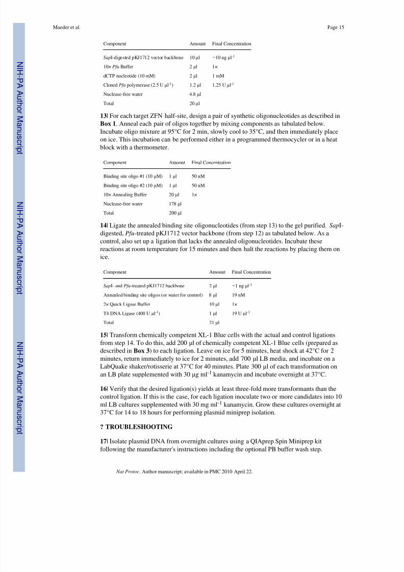
11 | To construct a Selection Reporter Plasmid bearing a ZFN half-site target sequence,digest plasmid pKJ1712 with Sap I (as tabulated below) at 37°C for 2 hours and purify thedigested vector backbone on a 5% non-denaturing polyacrylamide gel. The full sequence of pKJ1712 is given in Supplementary Fig. 1. Elute and ethanol-precipitate the digested vectorDNA as described in Box 2 .
Component Amount Final Concentration
Plasmid pKJ1712 1 μl (1 μg) 20 ng μl-1
10× Buffer (NEBuffer 4) 5 μl 1×
Sap I (2 U μl-1) 5 μl 0.04 U μl-1
Nuclease-free water 39 μl
Total 50 μl
<!>CAUTION Acrylamide is a neurotoxin and therefore polyacrylamide g els should beprepared wearing gloves.
12 | Create extended overhangs by treating the Sap I-digested pKJ1712 vector backbone of step 11 with Pfu polymerase in the presence of dCTP nucleotide (as tabulated below).Incubate this reaction for 15 minutes at 72°C and then place immediately at 4°C.
Maeder et al. Page 14
Nat Protoc . Author manuscript; available in PMC 2010 April 22.
N I H -P A A
ut h or Manus c r i pt
N I H -P A A ut h or Manus c r i pt
N I H -P A A ut h or
Manus c r i pt
8/8/2019 Maeder - Thibodeau-Beganny Et Al 2010
http://slidepdf.com/reader/full/maeder-thibodeau-beganny-et-al-2010 15/61
Component Amount Final Concentration
Sap I-digested pKJ1712 vector backbone 10 μl ∼ 10 ng μl-1
10× Pfu Buffer 2 μl 1×
dCTP nucleotide (10 mM) 2 μl 1 mM
Cloned Pfu polymerase (2.5 U μl-1) 1.2 μl 1.25 U μl-1
Nuclease-free water 4.8 μl
Total 20 μl
13| For each target ZFN half-site, design a pair of synthetic oligonucleotides as described inBox 1 . Anneal each pair of oligos together by mixing components as tabulated below.Incubate oligo mixture at 95°C for 2 min, slowly cool to 35°C, and then immediately placeon ice. This incubation can be performed either in a programmed thermocycler or in a heatblock with a thermometer.
Component Amount Final Concentration
Binding site oligo #1 (10 μM) 1 μl 50 nM
Binding site oligo #2 (10 μM) 1 μl 50 nM
10× Annealing Buffer 20 μl 1×
Nuclease-free water 178 μl
Total 200 μl
14| Ligate the annealed binding site oligonucleotides (from step 13) to the gel purified, Sap I-digested, Pfu -treated pKJ1712 vector backbone (from step 12) as tabulated below. As acontrol, also set up a ligation that lacks the annealed oligonucleotides. Incubate thesereactions at room temperature for 15 minutes and then halt the reactions by placing them onice.
Component Amount Final Concentration
Sap I- and Pfu -treated pKJ1712 backbone 2 μl ∼ 1 ng μl-1
Annealed binding site oligos (or water for control) 8 μl 19 nM
2× Quick Ligase Buffer 10 μl 1×
T4 DNA Ligase (400 U μl-1) 1 μl 19 U μl-1
Total 21 μl
15| Transform chemically competent XL-1 Blue cells with the actual and control ligationsfrom step 14. To do this, add 200 μl of chemically competent XL-1 Blue cells (prepared asdescribed in Box 3 ) to each ligation. Leave on ice for 5 minutes, heat shock at 42°C for 2minutes, return immediately to ice for 2 minutes, add 700 μl LB media, and incubate on aLabQuake shaker/rotisserie at 37°C for 40 minutes. Plate 300 μl of each transformation onan LB plate supplemented with 30 μg ml -1 kanamycin and incubate overnight at 37°C.
16 | Verify that the desired ligation(s) yields at least three-fold more transformants than thecontrol ligation. If this is the case, for each ligation inoculate two or more candidates into 10ml LB cultures supplemented with 30 mg ml -1 kanamycin. Grow these cultures overnight at37°C for 14 to 18 hours for performing plasmid miniprep isolation.
? TROUBLESHOOTING
17| Isolate plasmid DNA from overnight cultures using a QIAprep Spin Miniprep kitfollowing the manufacturer's instructions including the optional PB buffer wash step.
Maeder et al. Page 15
Nat Protoc . Author manuscript; available in PMC 2010 April 22.
N I H -P A A
ut h or Manus c r i pt
N I H -P A A ut h or Manus c r i pt
N I H -P A A ut h or
Manus c r i pt

8/8/2019 Maeder - Thibodeau-Beganny Et Al 2010
http://slidepdf.com/reader/full/maeder-thibodeau-beganny-et-al-2010 16/61
18| Confirm the reporter plasmid candidates by digesting them with EcoR I and Hind III for 1hour at 37°C under the conditions described below. Digestion products can be visualized ona 5% non-denaturing polyacrylamide gel (a typical gel is shown in Supplementary Fig. 4).Reporter plasmids that possess the binding sites should yield fragments of sizes 6108, 1006,963, 431 , and 190 bp, compared with the control parental pKJ1712 plasmid, which willyield fragments of sizes 6108, 1006, 963, 456 , and 190 bp.
Component Amount Final Concentration
Plasmid DNA 5 μl (∼ 0.75 μg) ∼ 25 ng / μl-1
10× EcoR I Buffer 3 μl 1×
EcoR I (20 U μl-1) 1 μl 0.66 U μl-1
Hind III (20 U μl-1) 1 μl 0.66 U μl-1
Nuclease-free water 20 μl
Total 30 μl
<!>CAUTION Acrylamide is a neurotoxin and therefore polyacrylamide gels should beprepared wearing gloves.
19 | Confirm the sequence of selection reporter plasmids between the EcoR I and Sal I sites
that flank the target ZFN half-site using sequencing primer OK181 (this primer is an anti-sense primer that anneals ∼ 270 bp downstream of and points back toward the binding site).Candidate reporter plasmids can be aligned with the sequence in Supplementary Fig. 5(“ EcoR I-Sal I binding site reporter”). Note that the target 11 bp ZFN half-site is shown as aseries of Xs in this file.
Recombination-based Transfer of Selection Reporter Plasmid Sequences to a Single-CopyEpisome
20| Transform sequence-confirmed selection reporter plasmids (one for each ZFN targethalf-site) into bacterial strain CSH100. Add 1 μl of mini prep plasmid DNA ( ∼ 0.1 μg) to 50μl of ice-cold chemically competent CSH100 cells (prepared as described in Box 4),incubate at 42°C for 2 minutes, return immediately to ice for 2 minutes, add 250 μl of LBmedium, incubate at 37°C for 40 minutes on a LabQuake shaker/rotisserie, plate entiretransformation on a LB plate containing 30 μg ml -1 kanamycin, and incubate overnight at37°C.
21 | On the same day that step 20 is performed, inoculate a fresh colony of bacterial strainKJ1C into 10 ml of LB medium supplemented with 12.5 μg ml -1 tetracycline and grow withagitation on a roller drum ( ∼ 60 rpm) overnight at 37°C.
22 | Examine plates with transformants of CSH100 (from step 20 above) and confirm thepresence of thousands of confluent colonies. Harvest these transformants by scraping all of the colonies from a single plate using a sterile wooden stick and resuspend these cells into10 ml of LB medium using gentle vortexing in a sterile 25 mm glass tube.
CRITICAL STEP: Only very gentle vortexing should be used to resuspend the CSH100transformants to minimize damage to the F pili expressed on the surface of these cells.
23 | Subculture ∼ 200 μl of the resuspended CSH100 transformants (prepared in step 22) intoa sterile 25 mm glass tube containing 5 ml of LB (without antibiotics). Also subculture ∼ 200μl of the overnight culture of strain KJ1C (from step 21 above) into a sterile 25 mm glasstube containing 5 ml of LB (without antibiotics). The density of cells in each of thesesubcultures should initially resemble a prelog phase culture by visual inspection (i.e.—an
Maeder et al. Page 16
Nat Protoc . Author manuscript; available in PMC 2010 April 22.
N I H -P A A
ut h or Manus c r i pt
N I H -P A A ut h or Manus c r i pt
N I H -P A A ut h or
Manus c r i pt

8/8/2019 Maeder - Thibodeau-Beganny Et Al 2010
http://slidepdf.com/reader/full/maeder-thibodeau-beganny-et-al-2010 17/61
OD 600 of ∼ 0.1). As a control, also add 10 ml of LB to a sterile 25 mm glass tube. Incubateall of these tubes for 2 hours at 37°C without agitation.
24 | Perform matings by setting up the following mixtures of subcultures prepared in step 23above in sterile 18 mm glass tubes:
Actual Mating: 1 ml of CSH100 transformants + 1 ml of KJ1C
Controls: 1 ml of CSH100 transformants + 1 ml of LB
1 ml of KJ1C + 1 ml of LB
2 ml of LB
Incubate matings at 37°C for 1 hr without agitation and then transfer tubes to a roller drum(∼ 60 rpm) at 37°C for 90 minutes.
25 | To identify desired double recombinant F's that have been successfully transferred tostrain KJ1C (Fig. 3), plate 300 μl of the actual desired mating from step 24 above on a LB/ TKS plate (as a control, also plate 300 μl of the actual mating on a LB/TK plate). Spot 5 μlof each of the control matings from step 24 on LB/TKS and LB/TK plates. Incubate allplates overnight at 37°C.
26 | Inspect all plates from step 25 for bacterial colony growth. KJ1C cells that havesuccessfully received the desired double-recombinant F ′ should be able to grow on LB/TKSplates (see Fig. 3). For the actual mating, we will typically see hundreds of colonies on theLB/TK control plate and about a ∼ 10-fold decrease in colony number observed on thematched LB/TKS plate. This reduction indicates that the sacB gene on the selection reporterplasmid is expressed, a critical requirement for successful identification of doublerecombinant F's (see Fig. 3). All plates on which control matings were spotted should be freeof colonies or bacterial growth (although occasionally, we will observe a few colonies onspots from the controls on LB/TK plates).
? TROUBLESHOOTING
27| For each mating, pick two independent colonies (designated “A” and “B”) to carryforward for confirmation. To purify clonal isolates, serially re-streak each colony to a LB/ TKS plate (one plate can be divided in two and a candidate streaked on each side) and growfor 12-18 hours at 37°C. Colonies for each candidate are then re-streaked again to LB/TKSplates and grown at 37°C for 12-18 hours.
28 | As an additional check for successful transfer of the F ′ to strain KJ1C, test the re-streaked colonies of candidates “A” and “B” from step 27 for their abilities to grow in theabsence of proline by resuspending a colony for each candidate in 100 μl of 1× M9 salts(this is conveniently done in the wells of a sterile 96-well plate) and spotting on a M9minimal medium plate. Incubate this plate overnight at 37°C. (Strain KJ1C lacks the proABgenes for proline biosynthesis and the F ′ from CSH100 contains the proAB genes. Thus,KJ1C cells that have successfully acquired the F ′ from CSH100 cells should be prolineprototrophs (i.e.—they should be able to grow in the absence of exogenously suppliedproline) and therefore should be able to grow on M9 minimal medium plates.)
29 | On the same day that candidates are spotted on M9 plates (as described in step 28), use20 μl of each cell resuspension (from step 28) to inoculate 4 ml of LB medium containing 30μg ml -1 kanamycin and grow these cultures overnight at 37°C on a roller drum ( ∼ 60 rpm).
30 | After overnight incubation, verify the growth of cells spotted on the M9 minimalmedium plate (from step 28) and discard any candidates that fail to grow.
Maeder et al. Page 17
Nat Protoc . Author manuscript; available in PMC 2010 April 22.
N I H -P A A
ut h or Manus c r i pt
N I H -P A A ut h or Manus c r i pt
N I H -P A A ut h or
Manus c r i pt
8/8/2019 Maeder - Thibodeau-Beganny Et Al 2010
http://slidepdf.com/reader/full/maeder-thibodeau-beganny-et-al-2010 18/61
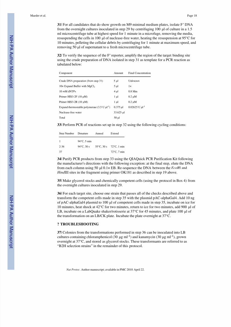
31| For all candidates that do show growth on M9 minimal medium plates, isolate F ′ DNAfrom the overnight cultures inoculated in step 29 by centrifuging 100 μl of culture in a 1.5ml microcentrifuge tube at highest speed for 1 minute in a microfuge, removing the media,resuspending the cells in 100 μl of nuclease-free water, heating the resuspension at 95°C for10 minutes, pelleting the cellular debris by centrifuging for 1 minute at maximum speed, andremoving 50 μl of supernatant to a fresh microcentrifuge tube.
32 | To verify the sequence of the F ′ reporter, amplify the region of the target binding siteusing the crude preparation of DNA isolated in step 31 as template for a PCR reaction astabulated below:
Component Amount Final Concentration
Crude DNA preparation (from step 31) 5 μl Unknown
10× Expand Buffer with MgCl 2 5 μl 1×
10 mM dNTPs 4 μl 0.8 Mm
Primer HIS3-2F (10 μM) 1 μl 0.2 μM
Primer HIS3-2R (10 μM) 1 μl 0.2 μM
Expand thermostable polymerase (3.5 U μl-1) 0.375 μl 0.02625 U μl-1
Nuclease-free water 33.625 μl
Total 50 μl
33| Perform PCR of reactions set up in step 32 using the following cycling conditions:
Step Number Denature Anneal Extend
1 94°C, 5 min
2-36 94°C, 30 s 55°C, 30 s 72°C, 1 min
37 72°C, 7 min
34| Purify PCR products from step 33 using the QIAQuick PCR Purification Kit followingthe manufacturer's directions with the following exception: at the final step, elute the DNAfrom each column using 50 μl 0.1× EB. Re-sequence the DNA between the EcoR I and
Hind III sites in the fragment using primer OK181 as described in step 19 above.
35 | Make glycerol stocks and chemically competent cells (using the protocol in Box 4) fromthe overnight cultures inoculated in step 29.
36 | For each target site, choose one strain that passes all of the checks described above andtransform the competent cells made in step 35 with the plasmid pAC-alphaGal4. Add 10 ngof pAC-alphaGal4 plasmid to 100 μl of competent cells made in step 35, incubate on ice for10 minutes, heat shock at 42°C for two minutes, return to ice for two minutes, add 900 μl of LB, incubate on a LabQuake shaker/rotisserie at 37°C for 45 minutes, and plate 100 μl of the transformation on an LB/CK plate. Incubate the plate overnight at 37°C.
? TROUBLESHOOTING
37| Colonies from the transformations performed in step 36 can be inoculated into LBcultures containing chloramphenicol (30 μg ml -1) and kanamycin (30 μg ml -1), grownovernight at 37°C, and stored as glycerol stocks. These transformants are referred to as“B2H selection strains” in the remainder of this protocol.
Maeder et al. Page 18
Nat Protoc . Author manuscript; available in PMC 2010 April 22.
N I H -P A A
ut h or Manus c r i pt
N I H -P A A ut h or Manus c r i pt
N I H -P A A ut h or
Manus c r i pt
8/8/2019 Maeder - Thibodeau-Beganny Et Al 2010
http://slidepdf.com/reader/full/maeder-thibodeau-beganny-et-al-2010 19/61
Construction of Combinatorial Zinc Finger Libraries
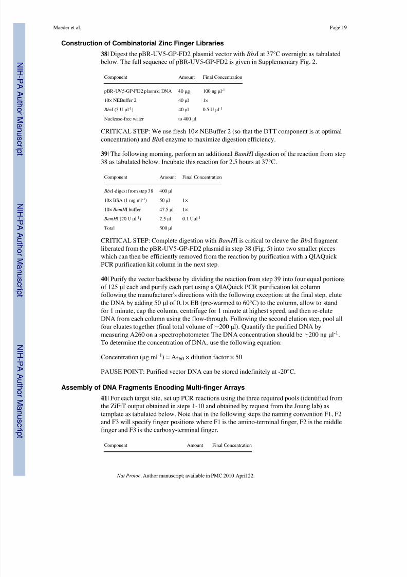
38| Digest the pBR-UV5-GP-FD2 plasmid vector with Bbs I at 37°C overnight as tabulatedbelow. The full sequence of pBR-UV5-GP-FD2 is given in Supplementary Fig. 2.
Component Amount Final Concentration
pBR-UV5-GP-FD2 plasmid DNA 40 μg 100 ng μl-1
10× NEBuffer 2 40 μl 1× Bbs I (5 U μl-1) 40 μl 0.5 U μl-1
Nuclease-free water to 400 μl
CRITICAL STEP: We use fresh 10× NEBuffer 2 (so that the DTT component is at optimalconcentration) and Bbs I enzyme to maximize digestion efficiency.
39 | The following morning, perform an additional BamH I digestion of the reaction from step38 as tabulated below. Incubate this reaction for 2.5 hours at 37°C.
Component Amount Final Concentration
Bbs I-digest from step 38 400 μl
10× BSA (1 mg ml-1
) 50 μl 1×10× BamH I buffer 47.5 μl 1×
BamH I (20 U μl-1) 2.5 μl 0.1 U μl-1
Total 500 μl
CRITICAL STEP: Complete digestion with BamH I is critical to cleave the Bbs I fragmentliberated from the pBR-UV5-GP-FD2 plasmid in step 38 (Fig. 5) into two smaller pieceswhich can then be efficiently removed from the reaction by purification with a QIAQuick PCR purification kit column in the next step.
40 | Purify the vector backbone by dividing the reaction from step 39 into four equal portionsof 125 μl each and purify each part using a QIAQuick PCR purification kit columnfollowing the manufacturer's directions with the following exception: at the final step, elute
the DNA by adding 50 μl of 0.1× EB (pre-warmed to 60°C) to the column, allow to standfor 1 minute, cap the column, centrifuge for 1 minute at highest speed, and then re-eluteDNA from each column using the flow-through. Following the second elution step, pool allfour eluates together (final total volume of ∼ 200 μl). Quantify the purified DNA bymeasuring A260 on a spectrophotometer. The DNA concentration should be ∼ 200 ng μl-1.To determine the concentration of DNA, use the following equation:
Concentration ( μg ml -1) = A 260 × dilution factor × 50
PAUSE POINT: Purified vector DNA can be stored indefinitely at -20°C.
Assembly of DNA Fragments Encoding Multi-finger Arrays
41| For each target site, set up PCR reactions using the three required pools (identified fromthe ZiFiT output obtained in steps 1-10 and obtained by request from the Joung lab) astemplate as tabulated below. Note that in the following steps the naming convention F1, F2and F3 will specify finger positions where F1 is the amino-terminal finger, F2 is the middlefinger and F3 is the carboxy-terminal finger.
Component Amount Final Concentration
Maeder et al. Page 19
Nat Protoc . Author manuscript; available in PMC 2010 April 22.
N I H -P A A
ut h or Manus c r i pt
N I H -P A A ut h or Manus c r i pt
N I H -P A A ut h or
Manus c r i pt

8/8/2019 Maeder - Thibodeau-Beganny Et Al 2010
http://slidepdf.com/reader/full/maeder-thibodeau-beganny-et-al-2010 20/61
Zinc Finger Pool DNA ( ∼ 0.15-0.2 μg) 1 μl 3-4 ng μl-1
10× Expand Buffer with MgCl 2 5 μl 1×
10mM dNTPs 4 μl 0.8 mM
Forward primer (10 μM) 3 μl 0.6 μM
for F1 use OK1424
for F2 use OK1426
for F3 use OK1428Reverse primer (10 μM) 3 μl 0.6 μM
for F1 use OK1425
for F2 use OK1427
for F3 use OK1429
Nuclease-free H2O 33.625 μl
Expand Enzyme (3.5 U μl-1) 0.375 μl 0.02625 U μl-1
Total 50 μl
42| Perform PCR using the finger position-specific cycling conditions listed in Tables 2, 3and 4.
PAUSE POINT: PCR products can be stored indefinitely at -20°C.43 | Purify PCR products from step 42 by electrophoresis on a 10% non-denaturingpolyacrylamide gel run at ∼ 100 volts. Stain the gel with ethidium bromide and use long-wave UV light to visualize the desired ∼ 100 bp products (note that these products willtypically run as a smeared, rather than a distinct, band and that a significant “primer dimer”product is always seen with the F1 primers). Excise the PCR products in a gel slice, crushthe gel piece, and elute DNA overnight at 37°C in 700 μl of ammonium acetate elutionbuffer.
<!>CAUTION Acrylamide is a neurotoxin and therefore polyacrylamide gels should beprepared wearing gloves.
? TROUBLESHOOTING
44| Purify the DNA fragments from the overnight elution of step 43 by spinning at highestspeed in a benchtop microfuge for 2 minutes and removing as much of the supernatant aspossible (typically ∼ 600 μl) being very careful to avoid the acrylamide pellet and transfer toa fresh microfuge tube.
45 | Spin the recovered supernatant of step 44 at highest speed in the microfuge for 2minutes, remove 475 μl of supernatant (again taking care to avoid the residual acrylamidepelleted at the bottom of the tube) and transfer to a fresh microfuge tube.
46 | Add 2 μl of 10 mg ml -1 glycogen to the recovered supernatant of step 45, mix well, add 1ml of 100% ethanol, mix well, place on dry ice for >15 minutes, and spin the tube at highestspeed in a microfuge for 15 minutes at 4°C. Remove and discard the supernatant (carefullyavoiding the pellet), add 500 μl of 70% (v/v) ethanol, spin at highest speed in a microfugefor 5 minutes, remove all residual liquid (again avoiding the pellet), air dry the pellet for5-10 minutes, and resuspend the pellet in 40 μl of nuclease-free water.
PAUSE POINT: Purified DNA can be stored indefinitely at -20°C.
Maeder et al. Page 20
Nat Protoc . Author manuscript; available in PMC 2010 April 22.
N I H -P A A
ut h or Manus c r i pt
N I H -P A A ut h or Manus c r i pt
N I H -P A A ut h or
Manus c r i pt

8/8/2019 Maeder - Thibodeau-Beganny Et Al 2010
http://slidepdf.com/reader/full/maeder-thibodeau-beganny-et-al-2010 21/61
47| Fuse together the purified F1, F2 and F3 PCR fragments purified in step 46 by setting upa PCR reaction as tabulated below.
Component Amount Final Concentration
Purified F1 PCR product (from step 46) 3 μl unknown
Purified F2 PCR product (from step 46) 3 μl unknown
Purified F3 PCR product (from step 46) 3 μl unknown10× Expand Buffer with MgCl 2 5 μl 1×
10mM dNTPs 4 μl 0.8 mM
Nuclease-free water 31.625 μl
Expand Enzyme (3.5 U μl-1) 0.375 μl 0.02625 U μl-1
Total 50 μl
48| Perform PCR of reaction set up in step 47 using the following cycling conditions:
Step number Denature Anneal Extend
1 94°C, 5 min
2-11 94°C, 30 s 50°C, 30 s 72°C, 1 min
12 72°C, 7 min
PAUSE POINT: PCR product can be stored indefinitely at -20°C.
49 | Purify the DNA from the PCR reaction of step 48 using a QIAQuick PCR purificationkit column following the manufacturer's directions with the following exception: at the finalstep, elute the DNA from each column using 50 μl 0.1× EB.
PAUSE POINT: Purified DNA can be stored indefinitely at -20°C.
50 | Amplify the fused PCR product encoding various combinations of three-finger arrays bysetting up the PCR reaction tabulated below.
Component Amount Final Concentration
Purified PCR product of step 49 24 μl unknown
Forward primer OK1430 (10 μM) 3 μl 0.6 μM
Reverse primer OK 1432 (10 μM) 3 μl 0.6 μM
10× Expand Buffer with MgCl 2 5 μl 1×
10mM dNTPs 4 μl 0.8 mM
Nuclease-free water 10.625 μl
Expand Enzyme (3.5 U μl-1) 0.375 μl 0.02625 U μl-1
Total 50 μl
51| Perform PCR of reaction set up in step 50 using the following cycling conditions:
Step number Denature Anneal Extend
1 94°C, 5 min
2-11 94°C, 30 s 56°C, 30 s 72°C, 1 min
12-31 94°C, 30 s 64°C, 30 s 72°C, 1 min
32 72°C, 7 min
Maeder et al. Page 21
Nat Protoc . Author manuscript; available in PMC 2010 April 22.
N I H -P A A
ut h or Manus c r i pt
N I H -P A A ut h or Manus c r i pt
N I H -P A A ut h or
Manus c r i pt
8/8/2019 Maeder - Thibodeau-Beganny Et Al 2010
http://slidepdf.com/reader/full/maeder-thibodeau-beganny-et-al-2010 22/61
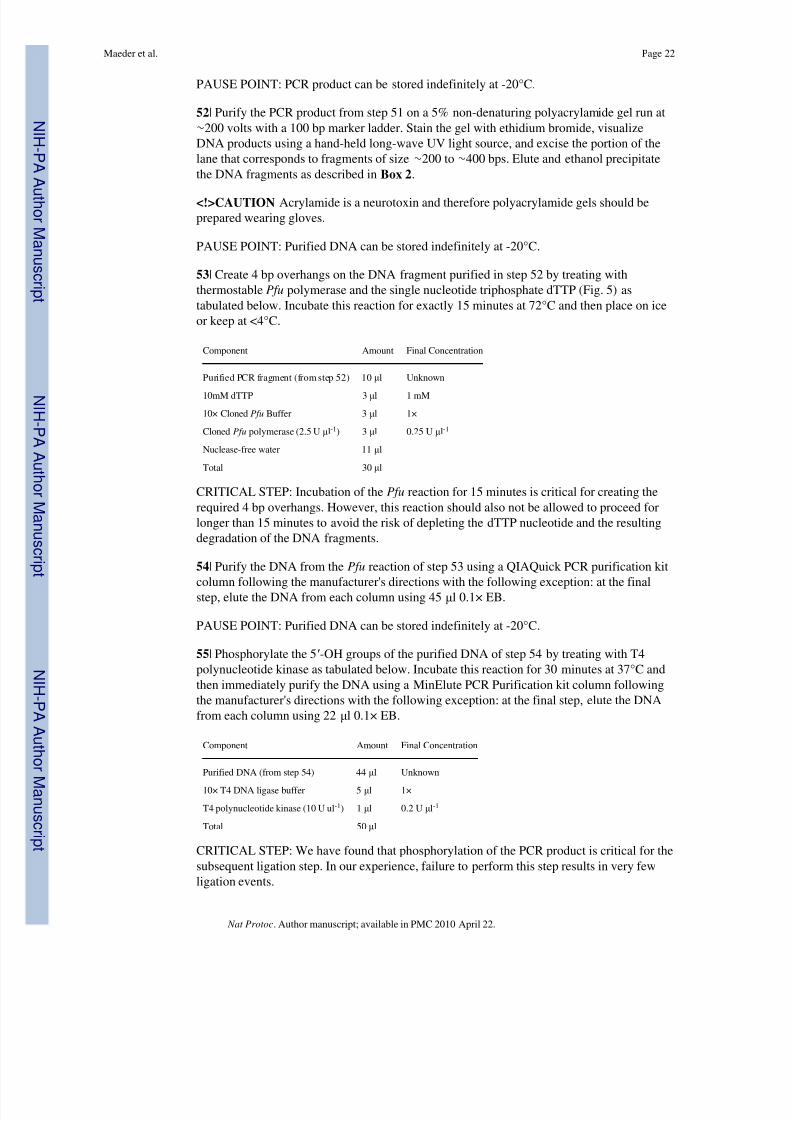
PAUSE POINT: PCR product can be stored indefinitely at -20°C.
52 | Purify the PCR product from step 51 on a 5% non-denaturing polyacrylamide gel run at∼ 200 volts with a 100 bp marker ladder. Stain the gel with ethidium bromide, visualizeDNA products using a hand-held long-wave UV light source, and excise the portion of thelane that corresponds to fragments of size ∼ 200 to ∼ 400 bps. Elute and ethanol precipitatethe DNA fragments as described in Box 2 .
<!>CAUTION Acrylamide is a neurotoxin and therefore polyacrylamide gels should beprepared wearing gloves.
PAUSE POINT: Purified DNA can be stored indefinitely at -20°C.
53 | Create 4 bp overhangs on the DNA fragment purified in step 52 by treating withthermostable Pfu polymerase and the single nucleotide triphosphate dTTP (Fig. 5) astabulated below. Incubate this reaction for exactly 15 minutes at 72°C and then place on iceor keep at <4°C.
Component Amount Final Concentration
Purified PCR fragment (from step 52) 10 μl Unknown
10mM dTTP 3 μl 1 mM
10× Cloned Pfu Buffer 3 μl 1×
Cloned Pfu polymerase (2.5 U μl-1) 3 μl 0.25 U μl-1
Nuclease-free water 11 μl
Total 30 μl
CRITICAL STEP: Incubation of the Pfu reaction for 15 minutes is critical for creating therequired 4 bp overhangs. However, this reaction should also not be allowed to proceed forlonger than 15 minutes to avoid the risk of depleting the dTTP nucleotide and the resultingdegradation of the DNA fragments.
54 | Purify the DNA from the Pfu reaction of step 53 using a QIAQuick PCR purification kitcolumn following the manufacturer's directions with the following exception: at the finalstep, elute the DNA from each column using 45 μl 0.1× EB.
PAUSE POINT: Purified DNA can be stored indefinitely at -20°C.
55 | Phosphorylate the 5 ′-OH groups of the purified DNA of step 54 by treating with T4polynucleotide kinase as tabulated below. Incubate this reaction for 30 minutes at 37°C andthen immediately purify the DNA using a MinElute PCR Purification kit column followingthe manufacturer's directions with the following exception: at the final step, elute the DNAfrom each column using 22 μl 0.1× EB.
Component Amount Final Concentration
Purified DNA (from step 54) 44 μl Unknown
10× T4 DNA ligase buffer 5 μl 1×
T4 polynucleotide kinase (10 U ul -1) 1 μl 0.2 U μl-1
Total 50 μl
CRITICAL STEP: We have found that phosphorylation of the PCR product is critical for thesubsequent ligation step. In our experience, failure to perform this step results in very fewligation events.
Maeder et al. Page 22
Nat Protoc . Author manuscript; available in PMC 2010 April 22.
N I H -P A A
ut h or Manus c r i pt
N I H -P A A ut h or Manus c r i pt
N I H -P A A ut h or
Manus c r i pt

8/8/2019 Maeder - Thibodeau-Beganny Et Al 2010
http://slidepdf.com/reader/full/maeder-thibodeau-beganny-et-al-2010 23/61
PAUSE POINT: Purified DNA can be stored indefinitely at -20°C.
Ligation of Digested Vector to DNA Fragments Encoding Multi-finger Arrays
56| Ligate the purified Bbs I-digested pBR-UV5-GP-FD2 vector (from step 40) to thepurified, Pfu -treated, phosphorylated PCR fragment encoding various combinations of threefingers (from step 55) as tabulated below. Incubate ligations overnight at 16°C. In parallel,also set up a control ligation reaction where 10 μl of nuclease-free water is substituted for
the PCR fragment encoding the three-finger arrays.
Component Amount Final Concentration
pBR-UV5-GP-FD2 backbone 1 μg 20 ng μl-1
PCR fragment encoding three-finger arrays 10 μl Unknown
10× standard T4 DNA Ligase buffer (NEB) 5 μl 1×
T4 DNA ligase (400U μl-1) 2 μl 16 U μl-1
Nuclease-free water to 50 μl
PAUSE POINT: Ligations can be stored indefinitely at -20°C.
57 | Check the efficiency of the ligation from step 56 by transforming the actual and control
ligations into chemically competent E. coli XL-1 Blue cells. Add 1 μl of actual ligation or 10μl of control each to 200 μl of chemically competent XL-1 Blue cells (prepared as describedin Box 3 ) in a sterile microfuge tube. Incubate transformations on ice for 10 minutes, heatshock by placing in a 42°C water bath for 2 minutes, return immediately to ice for 2minutes, add 900 μl of LB, and incubate on a roller drum ( ∼ 60 rpm) at 37°C for 40 minutes.Make 10 -1 and 10 -2 dilutions of the transformations by serial dilution as described in Box 5 .Spot 5 μl of the undiluted and diluted transformations in triplicate (i.e.—15 μl total) on LB/ TC plates and incubate overnight at 37°C.
58 | The next morning, count colonies from the highest dilution spots for which distinctcountable colonies are visible. Calculate the number of transformants μl-1 of ligationreaction by using the equations below.
Equation for transformants μl-1 of the actual ligation:
(# of colonies in 3 spots × 1100)/(15 × dilution factor)
Equation for transformants μl-1 of the control ligation:
(# of colonies in 3 spots × 1100)/(15 × dilution factor × 10)
CRITICAL STEP: The number of transformants μl-1 from the actual ligation should be >10-fold the number of transformants from the control ligation. Note that Steps 57 and 58 onlyneed to be performed once for each preparation of purified, digested vector backbone.
? TROUBLESHOOTING
Introduction of Combinatorial Zinc Finger Library into E. coli Cells
59| Purify 25 μl of the actual ligation from step 56 using a QIAgen MinElute PCRPurification Kit column following the manufacturer's instructions but eluting at the final stepinto 10 μl of nuclease-free water.
PAUSE POINT: Purified ligation DNA can be stored indefinitely at -20°C.
Maeder et al. Page 23
Nat Protoc . Author manuscript; available in PMC 2010 April 22.
N I H -P A A
ut h or Manus c r i pt
N I H -P A A ut h or Manus c r i pt
N I H -P A A ut h or
Manus c r i pt
8/8/2019 Maeder - Thibodeau-Beganny Et Al 2010
http://slidepdf.com/reader/full/maeder-thibodeau-beganny-et-al-2010 24/61
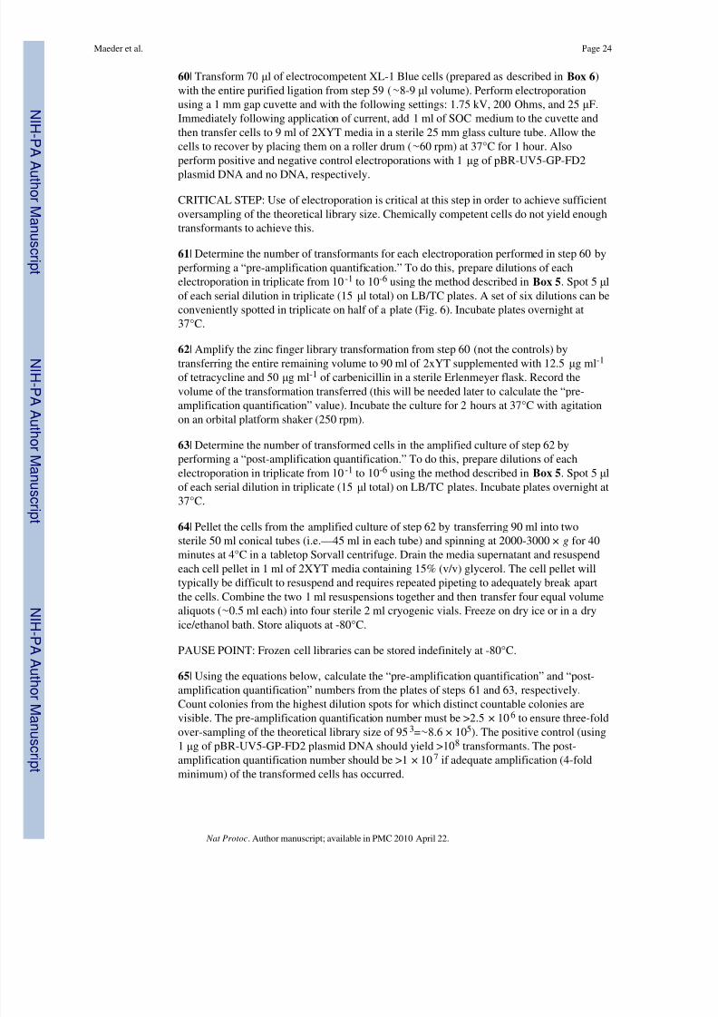
60| Transform 70 μl of electrocompetent XL-1 Blue cells (prepared as described in Box 6 )with the entire purified ligation from step 59 ( ∼ 8-9 μl volume). Perform electroporationusing a 1 mm gap cuvette and with the following settings: 1.75 kV, 200 Ohms, and 25 μF.Immediately following application of current, add 1 ml of SOC medium to the cuvette andthen transfer cells to 9 ml of 2XYT media in a sterile 25 mm glass culture tube. Allow thecells to recover by placing them on a roller drum ( ∼ 60 rpm) at 37°C for 1 hour. Alsoperform positive and negative control electroporations with 1 μg of pBR-UV5-GP-FD2
plasmid DNA and no DNA, respectively.
CRITICAL STEP: Use of electroporation is critical at this step in order to achieve sufficientoversampling of the theoretical library size. Chemically competent cells do not yield enoughtransformants to achieve this.
61 | Determine the number of transformants for each electroporation performed in step 60 byperforming a “pre-amplification quantification.” To do this, prepare dilutions of eachelectroporation in triplicate from 10 -1 to 10 -6 using the method described in Box 5 . Spot 5 μlof each serial dilution in triplicate (15 μl total) on LB/TC plates. A set of six dilutions can beconveniently spotted in triplicate on half of a plate (Fig. 6). Incubate plates overnight at37°C.
62 | Amplify the zinc finger library transformation from step 60 (not the controls) bytransferring the entire remaining volume to 90 ml of 2xYT supplemented with 12.5 μg ml -1
of tetracycline and 50 μg ml -1 of carbenicillin in a sterile Erlenmeyer flask. Record thevolume of the transformation transferred (this will be needed later to calculate the “pre-amplification quantification” value). Incubate the culture for 2 hours at 37°C with agitationon an orbital platform shaker (250 rpm).
63 | Determine the number of transformed cells in the amplified culture of step 62 byperforming a “post-amplification quantification.” To do this, prepare dilutions of eachelectroporation in triplicate from 10 -1 to 10 -6 using the method described in Box 5 . Spot 5 μlof each serial dilution in triplicate (15 μl total) on LB/TC plates. Incubate plates overnight at37°C.
64 | Pellet the cells from the amplified culture of step 62 by transferring 90 ml into twosterile 50 ml conical tubes (i.e.—45 ml in each tube) and spinning at 2000-3000 × g for 40minutes at 4°C in a tabletop Sorvall centrifuge. Drain the media supernatant and resuspendeach cell pellet in 1 ml of 2XYT media containing 15% (v/v) glycerol. The cell pellet willtypically be difficult to resuspend and requires repeated pipeting to adequately break apartthe cells. Combine the two 1 ml resuspensions together and then transfer four equal volumealiquots ( ∼ 0.5 ml each) into four sterile 2 ml cryogenic vials. Freeze on dry ice or in a dryice/ethanol bath. Store aliquots at -80°C.
PAUSE POINT: Frozen cell libraries can be stored indefinitely at -80°C.
65 | Using the equations below, calculate the “pre-amplification quantification” and “post-amplification quantification” numbers from the plates of steps 61 and 63, respectively.Count colonies from the highest dilution spots for which distinct countable colonies arevisible. The pre-amplification quantification number must be >2.5 × 10 6 to ensure three-foldover-sampling of the theoretical library size of 95 3=∼ 8.6 × 10 5). The positive control (using1 μg of pBR-UV5-GP-FD2 plasmid DNA should yield >10 8 transformants. The post-amplification quantification number should be >1 × 10 7 if adequate amplification (4-foldminimum) of the transformed cells has occurred.
Maeder et al. Page 24
Nat Protoc . Author manuscript; available in PMC 2010 April 22.
N I H -P A A
ut h or Manus c r i pt
N I H -P A A ut h or Manus c r i pt
N I H -P A A ut h or
Manus c r i pt

8/8/2019 Maeder - Thibodeau-Beganny Et Al 2010
http://slidepdf.com/reader/full/maeder-thibodeau-beganny-et-al-2010 25/61
PRE-AMPLIFICATION QUANTIFICATION = (Total # of colonies from 45 μl of transformation dilution for which distinct countable colonies are visible × volume in μltransferred in step 62)/(45 × dilution factor)
POST-AMPLIFICATION QUANTIFICATION = (Total # of colonies from 45 μl of transformation dilution for which distinct countable colonies are visible × 90,000)/(45 ×dilution factor)
? TROUBLESHOOTING
Conversion of Combinatorial Zinc Finger Library into Phage
66| Based on the post-amplification quantification number obtained in step 65, thaw enoughvials (typically 1 or 2) of the frozen cell library of step 64 to inoculate >2.5 × 10 6
transformed cells into 9 ml of 2XYT medium containing 12.5 μg ml -1 of tetracycline and 50μg ml -1 of carbenicillin in a sterile 25 mm glass tube. Incubate on a roller drum ( ∼ 60 rpm) at37°C for 1.5 hours.
67 | Determine the number of transformed cells in the subculture of step 66 by performing a“pre-infection titer.” To do this, prepare dilutions in triplicate from a small amount of thesubculture from 10 -1 to 10 -6 using the method described in Box 5 . Spot 5 μl of each serial
dilution in triplicate (15 μl total) on LB/TC and on LB/KCarb plates. Cells transformed withzinc finger plasmid will grow on LB/TC plates but should fail to grow on LB/KCarb plates(the latter is a control to check that cells are not already infected with M13K07 helper phagewhich confers resistance to kanamycin). Incubate plates overnight at 37°C.
68 | Infect the subculture of step 66 with helper phage by adding 140 μl of high titer M13K07stock (10 9 KTU μl-1) and mixing well to ensure even distribution of the phage throughoutthe culture medium. The high-titer M13K07 stock is prepared as described in Box 7 . Allowthe infected culture to sit at room temperature for 15 minutes without agitation and thenincubate on a roller drum ( ∼ 60 rpm) for 1.5 hours at 37°C.
69 | Determine the number of helper phage-infected cells in the culture of step 68 byperforming a “post-infection titer.” To do this, prepare dilutions in triplicate from a small
amount of the subculture from 10-1
to 10-6
using the method described in Box 5 . Spot 5 μl of each serial dilution in triplicate (15 μl total) on LB/TC and on LB/KCarb plates. Cellstransformed with zinc finger plasmid will grow on LB/TC plates and helper phage-infectedcells will also grow on LB/KCarb plates. Incubate plates overnight at 37°C.
70 | Add the remainder of the culture from step 68 to a sterile flask containing 90 ml of 2XYT medium containing 50 μg ml -1 carbenicillin and 70 μg ml -1 kanamycin. Record thevolume of culture transferred to this flask (it will be important for calculations performedlater). Shake at 250 rpm, 37°C on an orbital platform shaker overnight.
71 | The next day, verify that the pre-infection titer plates of step 67 show that none of thecells were initially infected with helper phage (i.e.—that there are no colonies on the LB/ KCarb plates). In addition, confirm that the library cells have been successfully super-
infected by helper phage by verifying that the post-infection titer plates of step 69 show that>50% of the cells were infected by helper phage (i.e.—that the titers from the LB/KCarbplates are >50% those from the LB/TC plates). If these titers look appropriate, harvest thephage from the culture of step 70. To do this, transfer ∼ 30 ml of the culture to a sterile 50 mlconical tube, pellet the cells by centrifuging at ∼ 1500 × g in a tabletop Sorvall centrifuge for30 minutes, and filter the supernatant through a 0.22 μm PES filter.
Maeder et al. Page 25
Nat Protoc . Author manuscript; available in PMC 2010 April 22.
N I H -P A A
ut h or Manus c r i pt
N I H -P A A ut h or Manus c r i pt
N I H -P A A ut h or
Manus c r i pt

8/8/2019 Maeder - Thibodeau-Beganny Et Al 2010
http://slidepdf.com/reader/full/maeder-thibodeau-beganny-et-al-2010 26/61
PAUSE POINT: Phage libraries can be stored at 4°C for up to a month. For long-termstorage, freeze the phage libraries at -80°C.
? TROUBLESHOOTING
Performing OPEN Selections
72| To begin the first stage of OPEN selection, streak the selection strain harboring the target
DNA site from the glycerol stock made in step 37 onto a LB/CK plate and incubate at 37°Covernight.
73 | Inoculate two or three colonies from the plate streaked in step 72 into a sterile 125 mlErlenmeyer flask containing 20 ml NM medium supplemented with 30 μg ml -1 kanamycin,30 μg ml -1 chloramphenicol, 50 μM IPTG. Grow the culture overnight for 16-24 hours at37°C on an orbital platform shaker at 125 rpm.
CRITICAL STEP: Do not shake cells at speeds higher than 125 rpm. For reasons that we donot as yet understand, the cells will grow suboptimally if shaken at higher speeds in NMmedium.
CRITICAL STEP: When inoculating colonies into NM medium, it is important to make sure
that the colony is well dispersed into the medium. Failure to adequately disperse the colonywill result in failure of the cells to grow. A reliable way to disperse a colony is to take up theentire colony into the base of a CLP 200 μl filtered pipet tip and then to force air through thetip several times as it is pressed against the bottom of the flask. To do this, push airforcefully through while the tip is pressed against the bottom of the flask (bubbles should begenerated if this is done correctly), lift the tip out of the medium, draw air back into the tipgently by slowly releasing the pipet plunger, and then place the tip against the bottom of theflask and repeat several times.
74 | Measure the density of the selection strain culture inoculated in step 73 using aspectrophotometer. This culture should have an OD 600 ∼ 1.0.
? TROUBLESHOOTING
75| In a sterile 125 ml Erlenmeyer flask, infect 5 ml of the selection strain culture inoculatedin step 73 with 50 μl of the phage library from step 71 and mix well. Let infection proceed atroom temperature for 30 minutes without agitation.
76 | Add 20 ml of pre-warmed NM medium containing 30 μg ml -1 chloramphenicol, 30 μgml -1 kanamycin, and 50 μM IPTG to the infected cells of step 75 and slowly shake at 125rpm, 37°C, for 1.5 hours.
77 | Harvest infected cells by transferring the culture of step 76 to a sterile 50 ml conicaltube, centrifuging at 1300 × g in a tabletop Sorvall centrifuge for 25 minutes at roomtemperature, draining the supernatant media, and resuspending the cell pellet in 2.5 ml of pre-warmed NM media containing 30 μg ml -1 chloramphenicol, 30 μg ml -1 kanamycin, and50 μM IPTG.
78 | To determine the number of total and infected cells in the resuspended cells of step 77,prepare dilutions of these cells in triplicate ranging from 10 -1 to 10 -6 using the methoddescribed in Box 5 . Spot 5 μl of each serial dilution in triplicate (15 μl total) on LB/CK, LB/ CCK/ and NM/CCKI plates. Incubate the LB/CK and LB/CCK plates for 14-18 hours at37°C and the NM/CCKI plates for 24 hours at 37°C.
Maeder et al. Page 26
Nat Protoc . Author manuscript; available in PMC 2010 April 22.
N I H -P A A
ut h or Manus c r i pt
N I H -P A A ut h or Manus c r i pt
N I H -P A A ut h or
Manus c r i pt

8/8/2019 Maeder - Thibodeau-Beganny Et Al 2010
http://slidepdf.com/reader/full/maeder-thibodeau-beganny-et-al-2010 27/61
79| Plate 250 μl of the resuspended cells of step 77 onto two different NM/CCKI plates: onesupplemented with 10 mM 3-AT/20 μg ml -1 streptomycin and the other with 25 mM 3-AT/ 40 μg ml -1 streptomycin. Spread resuspended cells on these plates using sterile glass beads(∼ 10-12 per plate), allow plates to dry, cover, invert, and tap the beads down onto the lid.Incubate plates (do not remove the beads) for 40-48 hours at 37°C.
80 | After the plates from step 78 have completed their incubation, perform calculations using
the equations below by counting from the highest dilution spots for which distinct countablecolonies are visible. The total number of infected cells in 250 μl of resuspended cells fromstep 77 should be >2.5 × 10 6 (to ensure three-fold oversampling of the theoretical zinc fingerlibrary size of 95 3=∼ 8.6 × 10 5). The total number of cells in 250 μl of resuspended cellsfrom step 77 should be at least three-fold more than the total number of infected cells toensure that the vast majority of infected cells have been infected by only a single phageparticle).
Total number of cells in 250 μ l of resuspended cells from step 77 = (Total # of coloniesfrom 45 μl of dilution for which distinct countable colonies are visible on LB/CK plates ×250)/(45 × dilution factor)
Total number of infected cells in 250 μ l of resuspended cells from step 77 = (Total # of colonies from 45 μl of dilution for which distinct countable colonies are visible on LB/CCKplates × 250)/(45 × dilution factor)
Total number of infected cells capable of growing on NM medium in 250 μ l of resuspended cells from step 77 = (Total # of colonies from 45 μl of dilution for whichdistinct countable colonies are visible on NM/CCKI plates × 250)/(45 × dilution factor)
? TROUBLESHOOTING
81| After the plates from step 79 have completed their incubation, harvest the higheststringency plate that has >1000 colonies. To do this, invert the plate, tap the glass beadsback onto the agar, add 1.5 ml of pre-warmed NM media to the plate, gently swirl toresuspend the colonies in the medium, and transfer the cell suspension to a sterile glass tube.
82 | Rescue the phagemids encoding zinc finger arrays from the cell suspension harvested instep 81 by infecting with helper phage and inducing the production of infectious phageharboring these phagemids. To do this, add enough cell suspension from step 81 (typically25 to 150 μl) to a sterile 25 mm glass tube containing 9 ml of 2xYT media supplementedwith 50 μg ml -1 carbencillin and 30 μg ml -1 kanamycin to generate a subculture with anOD 600 of ∼ 0.1. Place tube on a roller drum ( ∼ 60 rpm) at 37°C for 1 hour. Add 10 11 KTU of M13K07 helper phage (100 μl of a 10 9 KTU μl-1 M13K07 stock), mix well, let sit withoutagitation at room temperature for 30 minutes, and then place on the roller drum ( ∼ 60 rpm) at37°C for 6 hours.
83 | While the infected culture of cells from step 82 are incubating, add glycerol to a final (v/ v) concentration of 15% to the remaining cell suspension of step 81 and freeze at -80°C.These cells can be used to repeat step 82 if necessary.
84 | Harvest the phage supernatant from the infected culture of step 82. To do this, transferthe culture to a sterile 50 ml conical tube, pellet the cells by centrifuging at 1300 × g in atabletop Sorvall centrifuge for 25 minutes, and pass the supernatant through a 0.22 μm PESfilter. This filtered supernatant is the enriched zinc finger phage library.
Maeder et al. Page 27
Nat Protoc . Author manuscript; available in PMC 2010 April 22.
N I H -P A A
ut h or Manus c r i pt
N I H -P A A ut h or Manus c r i pt
N I H -P A A ut h or
Manus c r i pt

8/8/2019 Maeder - Thibodeau-Beganny Et Al 2010
http://slidepdf.com/reader/full/maeder-thibodeau-beganny-et-al-2010 28/61
PAUSE POINT: The enriched zinc finger phage library stock can be stored at 4°C for up to1 month. After this, phage can be stored indefinitely at -80°C.
85 | To perform the second stage of OPEN selection, determine the titer of the enriched zincfinger phage library stock of step 84. To prepare cells for doing this, inoculate two or threecolonies of the selection strain from a freshly streaked plate (<1 week old) into a sterile 125ml Erlenmeyer flask containing 20 ml NM medium supplemented with 30 μg ml -1
kanamycin and 30 μg ml-1
chloramphenicol. Grow the culture overnight for 16-24 hours at37°C on an orbital platform shaker at 125 rpm.
86 | Measure the density of the selection strain culture inoculated in step 85 using aspectrophotometer. This culture should have an OD 600 >1.0.
87 | In a sterile 18 mm glass tube, infect 500 μl of the selection strain culture inoculated instep 85 with 200 μl of the enriched zinc finger phage library from step 84 and mix well bygently swirling. Let infection proceed at room temperature for 30 minutes without agitation.
88 | Add 2 ml of pre-warmed NM medium containing 30 μg ml -1 chloramphenicol and 30 μgml -1 kanamycin to the infected cells of step 87 and incubate on a roller drum ( ∼ 60 rpm) for1.5 hours at 37°C.
89 | To determine the number of total and infected cells in the culture of step 88, preparedilutions of these cells ranging from 10 -1 to 10 -6 using the method described in Box 5 . Spot5 μl of each serial dilution in triplicate (15 μl total) on LB/CK, LB/CCK and NM/CCKplates. Incubate the LB/CK and LB/CCK plates for 14-18 hours at 37°C and the NM/CCKplates for 24 hours at 37°C.
90 | After the plates from step 89 have completed their incubation, perform calculations usingthe equations below by counting from the highest dilution spots for which distinct countablecolonies are visible.
Total number of cells in the culture of step 88 = (Total # of colonies from 45 μl of dilution for which distinct countable colonies are visible on LB/CK plates × 2700)/(45 ×dilution factor)
Total number of infected cells in the culture of step 88 = (Total # of colonies from 45 μlof dilution for which distinct countable colonies are visible on LB/CCK plates × 2700)/(45 ×dilution factor)
Titer of the enriched zinc finger library phage stock (in carbenicillin-transducing units[CTU]/ μl) = (Total # of colonies from 45 μl of dilution for which distinct countable coloniesare visible on NM/CCK plates × 2700)/(45 × dilution factor × 200)
The total number of cells in the culture of step 88 should be at least three-fold the number of infected cells to ensure that the vast majority of infected cells harbor only a single zincfinger-encoding phagemid and therefore that the titer is accurate. The titer of the enrichedzinc finger library phage stock must be >1×10 4 CTU μl-1 in order to successfully perform
the next step. (Note that phage particles are measured in CTUs because the zinc finger array-encoding phagemid encodes resistance to carbenicillin.) If phage titer is not sufficientlyhigh, it may be necessary to concentrate the phage library by PEG precipitation ( Box 8 ).
? TROUBLESHOOTING
91| Perform the second stage of OPEN selection by infecting selection strain cells with theenriched zinc finger phage library. To do this, inoculate a colony of the selection strain from
Maeder et al. Page 28
Nat Protoc . Author manuscript; available in PMC 2010 April 22.
N I H -P A A
ut h or Manus c r i pt
N I H -P A A ut h or Manus c r i pt
N I H -P A A ut h or
Manus c r i pt
8/8/2019 Maeder - Thibodeau-Beganny Et Al 2010
http://slidepdf.com/reader/full/maeder-thibodeau-beganny-et-al-2010 29/61
a freshly streaked plate (<1 week old) into a sterile 125 ml Erlenmeyer flask containing 20ml NM medium supplemented with 30 μg ml -1 kanamycin and 30 μg ml -1 chloramphenicol.Grow the culture overnight for 16-24 hours at 37°C on an orbital platform shaker at 125rpm.
92 | Measure the density of the selection strain culture inoculated in step 91 using aspectrophotometer. This culture should have an OD 600 >1.0.
93 | In a sterile 18 mm glass tube, infect 500 μl of the selection strain culture inoculated instep 91 with 2.5 × 10 6 CTU of the enriched zinc finger phage library from step 84 and mixwell by gently swirling. Let infection proceed at room temperature for 30 minutes withoutagitation.
94 | Add 2 ml of pre-warmed NM medium containing 30 μg ml -1 chloramphenicol and 30 μgml -1 kanamycin to the infected cells of step 93 and incubate on the roller drum ( ∼ 60 rpm)for 1.5 hours at 37°C.
95 | To determine the number of total and infected cells in the culture of step 94, preparedilutions of these cells ranging from 10 -1 to 10 -6 using the method described in Box 5 . Spot5 μl of each serial dilution in triplicate (15 μl total) on LB/CK, LB/CCK and NM/CCKplates. Incubate the LB/CK and LB/CCK plates for 14-18 hours at 37°C and the NM/CCKplates for 24 hours at 37°C.
96 | Plate the remainder of the culture of step 94 on a 245 mm square NM/CCK platecontaining gradients of 3-AT (0 to 80 mM) and streptomycin (0 to 100 μg ml -1). Gradientplates are poured as described in Box 9 . Record the volume of culture plated (this will berequired for calculations performed in step 97). Incubate the gradient plate for 4 days at37°C.
97 | After the titer plates from step 95 have completed their incubation, perform calculationsusing the equations below by counting from the highest dilution spots for which distinctcountable colonies are visible.
Total number of cells on the gradient plate = (Total # of colonies from 45 μl of dilutionfor which distinct countable colonies are visible on LB/CK plates × volume in μl of cultureplated in step 96)/(45 × dilution factor)
Total number of infected cells on the gradient plate = (Total # of colonies from 45 μl of dilution for which distinct countable colonies are visible on LB/CCK plates × volume in μlof culture plated in step 96)/(45 × dilution factor)
Total number of infected cells on the gradient plate that are able to grow on NM plates= (Total # of colonies from 45 μl of dilution for which distinct countable colonies are visibleon NM/CCK plates × volume in μl of culture plated in step 96)/(45 × dilution factor)
The total number of cells in the culture of step 94 should be at least three-fold the number of infected cells to ensure that the vast majority of infected cells harbor only a single zinc
finger-encoding phagemid. The total number of infected cells plated on the gradient plate(and calculated based on the titers from the NM/CCK plate) should be >2.5 × 10 6.
? TROUBLESHOOTING
98| After the gradient plate of step 96 has completed its 4 day incubation, pick 6-12 coloniesfrom the most selective/stringent region of the plate (Fig. 8) and inoculate into 4 ml LBmedium containing 50 μg ml -1 carbenicillin. Grow cultures overnight and then isolate
Maeder et al. Page 29
Nat Protoc . Author manuscript; available in PMC 2010 April 22.
N I H -P A A
ut h or Manus c r i pt
N I H -P A A ut h or Manus c r i pt
N I H -P A A ut h or
Manus c r i pt

8/8/2019 Maeder - Thibodeau-Beganny Et Al 2010
http://slidepdf.com/reader/full/maeder-thibodeau-beganny-et-al-2010 30/61
plasmids with the QIAprep Spin Miniprep Kit using the manufacturer's instructions but withthe following modifications: perform two serial washes with PB buffer instead of one andelute with 0.1× EB that is pre-warmed to 65°C.
PAUSE POINT: Plasmid DNA can be stored indefinitely at -20°C.
99 | Sequence the plasmids isolated in step 98 using primer OK61 to determine the aminoacid residues selected in the recognition helices of the finger arrays. OK61 anneals justupstream of and fires toward the sequence encoding the zinc finger array. The resultingsequences can be aligned with the sequence in Supplementary Fig. 6 entitled “ Xba I- BamH Ithree-finger array” – note that the triplet codons encoding randomized positions of therecognition helices are shown as a series of Ns in this file. The sequences of the recognitionhelices in the selected clones will typically resemble one another closely and one or moreconsensus sequences can be observed at each position.
Constructing B2H reporter strains (steps 100-111 below are reproduced with minorchanges from steps 31-41 of Wright et al., 200676)
100 | Digest the single-copy vector pBAC-lacZ with Bsa I restriction enzyme using thefollowing conditions:
Component Amount Final Concentration
pBAC-lacZ plasmid DNA 1 μg 50 ng μl-1
10× NEBuffer 3 2 μl 1×
Bsa I (10 U μl-1) 0.5 μl 0.5 U μl-1
Nuclease-free water to 20 μl
101 | Purify the ∼ 11,100 bp vector backbone by electrophoresis through a 5%polyacrylamide gel. Elute and ethanol precipitate this purified vector as described in Box 2 .
CRITICAL STEP: Bsa I digests should be incubated at 50° C.
PAUSE POINT: Purified DNA can be stored indefinitely at -20° C.
102 | Incubate purified, Bsa I-digested pBAC-lacZ vector backbone with Pfu polymerase inthe presence of the dCTP only (i.e.--omit dATP, dGTP, and dTTP) at 72° C for 10 minutesusing the conditions listed below. The combined 5 ′ to 3 ′ polymerase activity and 3 ′ to 5 ′exonuclease activity of Pfu will generate the extended overhangs as previously described. 76
Component Amount Final Concentration
Bsa I-digested pBAC-lacZ vector backbone 10 μl ∼ 25 ng μl-1
10× Pfu Buffer 2 μl 1×
dCTP nucleotide (10 mM) 2 μl 1 mM
Cloned Pfu polymerase (2.5 U μl-1) 1.2 μl 1.25 U μl-1
Nuclease-free water 4.8 μl
Total 20 μl
103 | For each target binding site, ligate the pBAC-lacZ vector backbone from step 102 toannealed oligonucleotides from step 13. As a control, perform a ligation with the pBAC-lacZ vector backbone only. Detailed conditions are provided below. Ligations are incubatedfor 15 minutes at room temperature.
Maeder et al. Page 30
Nat Protoc . Author manuscript; available in PMC 2010 April 22.
N I H -P A A
ut h or Manus c r i pt
N I H -P A A ut h or Manus c r i pt
N I H -P A A ut h or
Manus c r i pt
8/8/2019 Maeder - Thibodeau-Beganny Et Al 2010
http://slidepdf.com/reader/full/maeder-thibodeau-beganny-et-al-2010 31/61
Component Amount Final Concentration
Bsa I- and Pfu -treated pBAC-lacZ vector 2 μl ∼ 2.5 ng μl-1
Annealed binding site oligos (or water for control) 8 μl 19 nM
2× Quick Ligase Buffer 10 μl 1×
T4 DNA Ligase (400 U μl-1) 1 μl 19 U μl-1
Total 21 μl
104 | Transform ligations into E. coli strain Transformax EPI300 using standard chemicaltransformation. To do this, add 200 μl of chemically competent cells (prepared as describedin Box 3 ) to each ligation. Leave on ice for 5 minutes, heat shock at 42°C for 2 minutes,return immediately to ice for 2 minutes, add 700 μl LB media, and incubate on a LabQuakeshaker/rotisserie at 37°C for 40 minutes. Plate transformations on LB plates supplementedwith 12.5 μg ml -1 chloramphenicol and incubate overnight (14-18 hours) at 37° C.
105 | Inspect and count colonies on the transformation plates from step 104. If thetransformation of a ligation yields at least three-fold more colonies than that of the controlligation, we consider that ligation to be successful. From each actual successful ligation/ transformation plate, inoculate two 3 ml cultures of LB containing 12.5 μg ml -1
chloramphenicol and grow overnight (14-18 hours) at 37° C for isolating plasmid DNA.
106 | Inoculate 1 ml of each overnight culture into 9 ml of LB containing 14 μg ml -1
chloramphenicol and 1.1 mM arabinose. Grow cultures for 6 hours at 37° C and then harvestcells and isolate plasmid DNA using a QIAprep Spin Miniprep Kit.
CRITICAL STEP: Subculturing the cells containing the plasmids in arabinose is critical toinduce expression of the trfA gene product, which in turn increases the copy number of thepBAC-lacZ plasmid.
PAUSE POINT: Plasmids can be stored indefinitely at -20° C.
107 | Digest the candidate reporter plasmids (and the original pBAC-lacZ plasmid as acontrol) with EcoR I and Hind III using the conditions detailed below and visualize thedigestions on a 5% polyacrylamide gel. Candidates that have taken up the annealedoligonucleotides should yield fragments of sizes 159 and 10,970 base pairs whereas theoriginal pBAC-lacZ plasmid should yield fragments of sizes 182 and 10,973 base pairs.
Component Amount Final Concentration
Plasmid DNA 5 μl (∼ 0.75 μg) ∼ 25 ng / μl-1
10× EcoR I Buffer 3 μl 1×
EcoR I (20 U μl-1) 1 μl 0.66 U μl-1
Hind III (20 U μl-1) 1 μl 0.66 U μl-1
Nuclease-free water 20 μl
Total 30 μl
108 | Confirm the sequence of the target DNA binding site in the candidate reporter plasmidby sequencing with primer OK163 (this primer is an anti-sense primer that anneals ∼ 150 bpdownstream of the binding site). We will refer to the sequence-confirmed reporter plasmidas the pBAC-ZFBS-lacZ plasmid (ZFBS = Zinc Finger Binding Site) in subsequent steps of the protocol.
Maeder et al. Page 31
Nat Protoc . Author manuscript; available in PMC 2010 April 22.
N I H -P A A
ut h or Manus c r i pt
N I H -P A A ut h or Manus c r i pt
N I H -P A A ut h or
Manus c r i pt
8/8/2019 Maeder - Thibodeau-Beganny Et Al 2010
http://slidepdf.com/reader/full/maeder-thibodeau-beganny-et-al-2010 32/61
109 | Transform strain KJBAC1 (which constitutively expresses high levels of lac repressor)with each pBAC-ZFBS-lacZ reporter plasmid. Plate transformations on LB plates containing12.5 μg ml -1 chloramphenicol and incubate overnight at 37° C.
110 | From each successful transformation of step 109, inoculate a single colony into a 3 mlLB culture supplemented with 12.5 μg ml -1 chloramphenicol. Grow cultures overnight(12-16 hours) with agitation at 37° C.
111 | Make chemically competent cells from each of the overnight cultures of step 110 asdescribed in Box 4 . Also prepare glycerol stocks of each of the B2H reporter strains forlong-term storage at -80° C.
PAUSE POINT: Competent B2H reporter strain cells can be stored and frozen indefinitelyat -80° C.
Quantitative B2H assay of OPEN-selected zinc finger arrays
112 | Prepare the zinc finger-encoding plasmid minipreps isolated in step 98 for use intransformation of their cognate B2H reporter strain by digestion with Pst I overnight at 37°Cas tabulated below. This digestion removes the pAC-alphaGal4 plasmid that is isolatedtogether with the desired zinc finger-encoding plasmid.
Component Amount Final Concentration
Miniprep DNA (of step 98) 3 μl ∼ 15 ng μl-1
10× NEBuffer 3 3 μl 1×
10× BSA (1 mg ml -1) 3 μl 1×
Nuclease-free water 19 μl
Pst I (20 U μl-1) 2 μl 1.3 U μl-1
Total 30 μl
113 | Inactivate the Pst I enzyme in the reactions of step 112 by heating at 80°C for 20minutes.
PAUSE POINT: Pst I-digested DNA can be stored indefinitely at -20°C.
114 | Perform a double plasmid transformation of each B2H reporter strain prepared in step111. To do this, add 5 μl (∼ 75 ng) of the Pst I-digested DNA of step 101 and 150 ng of thepAC-KAN-alphaGal4 plasmid to 50 μl of chemically competent B2H reporter strainprepared in step 111, keep on ice for 10 minutes, heat shock at 42°C for 2 minutes, returnimmediately to ice for 2 minutes, add 175 μl of LB medium and shake or agitate at 37°C for75 minutes. To obtain well-isolated colonies, make serial 10 -1 and 10 -2 dilutions and spot 5μl of the undiluted and 10 -1 and 10 -2 diluted transformations in triplicate on a LB agar platesupplemented with 100 μg ml-1 carbenicillin, 12.5 μg ml -1 chloramphenicol and 30 μg ml -1
kanamycin. Also perform an additional transformation of each B2H reporter strain inparallel using 10 ng of the pBR-UV5-GP-FD2 plasmid and 150 ng of the pAC-KAN-alphaGal4 plasmid (this serves as a control lacking expression of a zinc finger array).Incubate these plates overnight at 37°C.
115 | Check the transformation plates of step 114 for growth of colonies.
PAUSE POINT: These plates may be stored for up to a week at 4°C.
? TROUBLESHOOTING
Maeder et al. Page 32
Nat Protoc . Author manuscript; available in PMC 2010 April 22.
N I H -P A A
ut h or Manus c r i pt
N I H -P A A ut h or Manus c r i pt
N I H -P A A ut h or
Manus c r i pt

8/8/2019 Maeder - Thibodeau-Beganny Et Al 2010
http://slidepdf.com/reader/full/maeder-thibodeau-beganny-et-al-2010 33/61
116 | Using the colonies obtained from the plates of step 115, perform β-galactosidase assaysas described in Box 10. Zinc finger arrays that mediate three-fold activation of lacZ expression relative to control cells that do not express a zinc finger array have a highprobability of functioning as ZFNs in human and zebrafish cells. 11 , 20, 32, 33 Occasionally,we will observe zinc finger arrays that cause toxicity in E. coli cells. Typically, this willmanifest itself as the inability to sub-culture cells for β-galactosidase assay.
? TROUBLESHOOTING117 | Clone candidate zinc finger arrays selected by OPEN into ZFN expression vectors.DNA fragments encoding zinc finger arrays can be conveniently excised by digestion with
Xba I and BamH I and cloned into various Zinc Finger Consortium ZFN expression vectorsfor human, zebrafish, and plant cells (Table 5).
● TIMING
Steps 1-10, Identification of ZFN target sites using ZiFiT software: <1 d
Steps 11-37, Construction of B2H Selection Strains: ∼ 12-14 d
Steps 38-71, Construction of Combinatorial Zinc Finger Libraries: ∼ 6-8 d
Note: Steps 11-37 and 38-71 can be performed in parallel
Steps 72-99, Performing OPEN Selections: ∼ 11-13 d
Steps 100-111, Construction of B2H Reporter Strains: ∼ 7-9 d
Note: Steps 72-99 and 100-111 can be performed in parallel
Steps 112-117, Quantitative B2H assay of OPEN-selected zinc finger arrays, 3 d
? TROUBLESHOOTING
Troubleshooting advice can be found in Table 6.
Boxes
Box 1
Designing oligonucleotides to create the target DNA-binding site for the B2H reportervector (reproduced from Box 2 of Wright et al., 200676); TIMING: <1 d
1. In the double-stranded DNA fragment shown in Fig. 4, replace the nine bpXXXXXXXXX sequence with the nine bp target site of the three-finger proteinto be tested. Replace the single bases (x) that flank the nine bp XXXXXXXXXsequence with the bases that flank the target site in the genomic sequence.
CRITICAL STEP: Note that the XXXXXXXXX site is positioned so that the F3triplet is on the “left” and the F1 triplet is on the “right” in the double-strandedfragment shown in Fig. 4. Replacement of the flanking “x” bases with the basespresent in the genomic target sequence is important because the identities of these adjacent bases can influence binding of the multi-finger domain to thetarget site.
Maeder et al. Page 33
Nat Protoc . Author manuscript; available in PMC 2010 April 22.
N I H -P A A
ut h or Manus c r i pt
N I H -P A A ut h or Manus c r i pt
N I H -P A A ut h or
Manus c r i pt

8/8/2019 Maeder - Thibodeau-Beganny Et Al 2010
http://slidepdf.com/reader/full/maeder-thibodeau-beganny-et-al-2010 34/61
2. Place an order for oligonucleotides corresponding to the first 29 bases of the topstrand of the fragment (starting from the 5 ′ end) and to the first 26 bases of thebottom strand (starting from the 5 ′ end) (Fig. 4).
Box 2
Purification of DNA from a polyacrylamide gel slice; TIMING: 1 d
1. Crush the excised gel slice in a microfuge tube using a thin metal spatula.
2. Add 700 μl of ammonium acetate elution buffer, seal the tube shut by wrappingParafilm around the top of the tube, and elute the DNA overnight at 37°C withagitation on a LabQuake shaker/rotisserie.
3. The next morning, spin the microfuge tube at 16,000 × g in a microcentrifugefor 2 minutes to pellet the acrylamide fragments. Transfer the supernatant ( ∼ 600μl) to a fresh microfuge tube being careful to avoid the acrylamide pellet.
4. Spin the supernatant at 16,000 × g in a microcentrifuge for 2 minutes. Transfer475 μl of the supernatant to a fresh microfuge tube. Add 950 μl of 100% ethanoland mix well. Place on dry ice for at least 15 minutes.
5. Spin the frozen sample at 16,000 × g in a microcentrifuge for 15 minutes atroom temperature. Immediately after the spin ends, remove the supernatantbeing careful not to dislodge or disturb the pellet.
6. Add 500 μl of 70% (v/v) ethanol to the tube. Invert gently several times. Spin at16,000 × g in a microcentrifuge for 5 minutes. Remove most of the supernatantwith a larger 1000 μl tip being careful to avoid the pellet. Re-spin the tubebriefly and then use a 200 μl pipet tip to remove all remaining supernatant.
7. Air dry the pellet at room temperature for 10 minutes. Resuspend the pellet in20 μl of nuclease-free water by gently pipeting up and down until the pellet iscompletely dissolved.
PAUSE POINT: Purified DNA fragments may be stored indefinitely at -20°C.
Box 3
Preparation of highly competent bacterial cells for chemical transformation; TIMING: 3d
1. Streak out cells (from frozen glycerol stock) on a LB plate with appropriateantibiotic and grow overnight at 37°C.
2. Autoclave 1 liter of LB medium in a 2 liter baffled Erlenmeyer flask.
3. The next day, pick a single, well-isolated colony from the plate of step 1 andstart a 10ml overnight culture in LB medium containing any appropriateantibiotics or chemical inducers. Incubate the culture overnight at 37°C on aroller drum ( ∼ 60 rpm).
4. Add 15 ml of 1M MgCl 2 to the LB prepared in step 2 and shake at 37°C, 250rpm overnight to check for any undesired contamination.
5. The next day, add any appropriate antibiotics or inducers to the LB mediumprepared in step 4 (if the medium appears to be free of any bacterial
Maeder et al. Page 34
Nat Protoc . Author manuscript; available in PMC 2010 April 22.
N I H -P A A
ut h or Manus c r i pt
N I H -P A A ut h or Manus c r i pt
N I H -P A A ut h or
Manus c r i pt
8/8/2019 Maeder - Thibodeau-Beganny Et Al 2010
http://slidepdf.com/reader/full/maeder-thibodeau-beganny-et-al-2010 35/61
contamination). Inoculate 1 ml of the overnight culture of step 3 into the LBmedium and incubate at 37°C with agitation on an orbital shaker (250 rpm).
6. Monitor the density of the culture of step 5 by taking OD 600 readings on aspectrophotometer. The first reading should be taken at approximately 2 hoursand subsequent readings should be taken every 15-45 minutes depending on thegrowth rate of the culture.
7. After the culture has reached an OD 600 between 0.4-0.6, pour the culture into asterile 1 liter centrifuge bottle and pellet the cells by centrifuging at 4°C, 2700 ×g for 30 minutes.
8. Decant the media and resuspend the cell pellet in 300 ml of ice-cold Solution A.Perform this resuspension step on ice and then incubate the resuspended cells onice for 20 minutes.
9. Centrifuge resuspended cells at 4°C, 2700 × g for 30 minutes and then decantthe supernatant.
10. Resuspend the remaining cell pellet in 60 ml of ice-cold Solution A with 15%(v/v) glycerol.
11. Make 500 μl and/or 1 ml aliquots of the cell suspension in sterile 1.5 ml
microfuge tubes and freeze on dry ice.12. Store competent cells at -80°C.
PAUSE POINT: Competent cells may be stored indefinitely at -80° C.
Box 4
Preparing competent bacterial cells for chemical transformation (reproduced with minoralterations from Box 3 of Wright et al., 200676); TIMING: 2 d
1. Inoculate a single colony of the bacterial strain to be made competent into a 2 mlculture of LB supplemented with any appropriate antibiotics. Grow overnight at37°C with agitation.
2. The next morning, subculture 1 ml of the saturated overnight culture from step 1into 50 ml LB supplemented with any appropriate antibiotics and 15 mM MgCl 2in a 250 ml flask. Grow for 1 hour with agitation at 37° C or until cells reach logphase growth (an OD 600 of ∼ 0.4-0.6).
3. Pellet cells in a sterile 50 ml conical tube by centrifuging at 3000-6000 × g in atabletop centrifuge.
4. Pour off media and resuspend cell pellet in 3 ml of ice-cold Solution A with15% glycerol.
5. Leave sitting on ice for 20 minutes and then aliquot into sterile microcentrifugetubes.
6. Snap freeze the tubes on dry ice
PAUSE POINT: Competent cells may be stored indefinitely at -80° C.
Box 5
Serial dilution of bacterial cultures; TIMING: <1 d
Maeder et al. Page 35
Nat Protoc . Author manuscript; available in PMC 2010 April 22.
N I H -P A A
ut h or Manus c r i pt
N I H -P A A ut h or Manus c r i pt
N I H -P A A ut h or
Manus c r i pt
8/8/2019 Maeder - Thibodeau-Beganny Et Al 2010
http://slidepdf.com/reader/full/maeder-thibodeau-beganny-et-al-2010 36/61

8/8/2019 Maeder - Thibodeau-Beganny Et Al 2010
http://slidepdf.com/reader/full/maeder-thibodeau-beganny-et-al-2010 37/61
11. Decant supernatant immediately after centrifugation and resuspend the cellpellet in 1 ml of sterile, ice-cold 10% (v/v) glycerol.
12. Make small aliquots ( ∼ 150 μl each) of the resuspension in pre-chilled, sterilemicrofuge tubes and freeze on dry ice. Store competent cells at -80°C.
13. Test the competency of cells by electroporating 1 μg of pBR-UV5-GP-FD2plasmid DNA into 70 μl of electrocompetent cells. Also perform a negative
control electroporation by using 70 μl of cells and no DNA. For theelectroporations, use a 1 mm gap cuvette and with the following settings: 1.75kV, 200 Ohms, and 25 μF. Pre-chill all cuvettes on ice and perform allelectroporation steps in the cold room. Immediately following application of current, add 1 ml of SOC medium to the cuvette and then transfer cells to 9 mlof 2xYT media in a sterile 25 mm glass culture tube. Allow the cells to recoverby incubating them on a roller drum ( ∼ 60 rpm) at 37°C for 1 hour.
14. Determine the number of transformants by preparing dilutions of eachelectroporation in triplicate from 10 -1 to 10 -6 using the method described in Box5. Spot 5 μl of each serial dilution in triplicate (15 μl total) on LB/TC plates. Aset of six dilutions can be conveniently spotted in triplicate on half of a plate(Fig. 6). Incubate plates overnight at 37°C.
15. Calculate the total number of transformants from the plates of step 14. Totalnumber of transformants in the 10 ml culture of step 14 should be in the range of 5 × 10 8 to 5 × 10 9.
Box 7
Protocol for preparing high-titer stock of M13K07 helper phage; TIMING: ∼ 4-5 d
<!>CAUTION Care should be taken to avoid contaminating laboratory equipment andbenches with bacteriophage. Use filtered pipet tips and pipets, dedicated tube racks, andchange bench paper frequently.
1. From a glycerol stock, streak out XL-1 Blue cells on a LB plate supplemented
with 12.5 mg ml-1
tetracycline. Incubate plate overnight at 37°C.2. The next day, inoculate a well-isolated colony of XL-1 Blue into a sterile 25
mm glass tube containing LB medium supplemented with 12.5 mg ml-1tetracycline. Incubate this inoculated culture on a roller drum ( ∼ 60 rpm)overnight at 37°C.
3. The next morning, melt a bottle of H Top Agar in the microwave and equilibratethe temperature of this molten agar to ∼ 60-65°C.
4. Dip the tip of a sterile loop or a sterile wooden stick into a commerciallyavailable stock of M13K07 and serially streak this tip with a side-to-side motionand down the surface of one LB plate supplemented with 12.5 mg ml -1
tetracycline and then another second plate. This process creates a progressive
dilution of the phage across the surfaces of these plates.5. Thoroughly mix together 7 ml of molten H Top Agar ( ∼ 60°C) with 0.5 ml of the
XL-1 Blue culture of step 2 and plate 3.75 ml onto each of the phage-streakedplates of step 4. When depositing the molten agar-bacteria mix, place it on theend of the plate that should contain the more dilute streaks of phage and then tiltthe plate slightly to allow the molten agar to cover the entire plate. After the top
Maeder et al. Page 37
Nat Protoc . Author manuscript; available in PMC 2010 April 22.
N I H -P A A
ut h or Manus c r i pt
N I H -P A A ut h or Manus c r i pt
N I H -P A A ut h or
Manus c r i pt

8/8/2019 Maeder - Thibodeau-Beganny Et Al 2010
http://slidepdf.com/reader/full/maeder-thibodeau-beganny-et-al-2010 38/61
agar has hardened (typically ∼ 2 minutes), incubate the plates face-up at 37°Covernight.
6. In the morning, use a sterile spatula to scoop off a region of the top agar ( ∼ 5 to25 cm 2) which contains dozens of well-isolated phage plaques and inoculate thisinto a baffled 2 liter flask containing 500 ml of 2xYT media supplemented with12.5 mg ml -1 tetracycline. Shake on an orbital shaker at 250 rpm, 37°C for 2hours.
7. Add 1.17 ml of 30 mg ml -1 kanamycin stock to the culture of step 6 to achieve afinal concentration of 70 μg ml -1. Continue to shake the culture at 250 rpm,37°C for an additional 22 hours.
8. Harvest the supernatant of the culture of step 7 by transferring the culture to asterile 1 liter centrifuge bottle and spinning the cells at ∼ 2700 × g. Removeresidual cells by filtering the culture supernatant through a 0.2 μm PES filterunit.
PAUSE POINT: Filtered phage stock can be stored for up to a month at 4°C.
9. Concentrate the phage by PEG precipitation. Divide the 500 ml of phage stock from step 8 into four sterile 250 ml centrifuge bottles (i.e.--125 ml of phage ineach bottle). Add 31.25 ml of 5× PEG/NaCl solution to each bottle, seal the captightly, and mix thoroughly by inverting each bottle several times. Incubate onice for at least 2.5 hours. Spin the bottles at ∼ 8600 × g in a Sorvall centrifuge,pour off and discard the supernatant. Resuspend phage pellets from the fourbottles in a total of 5 ml of 2xYT with 15% (v/v) glycerol. Make 500 μl aliquotsin 2 ml cryovials and store at -80°C.
10. Titer the phage stock of step 9 by serially diluting the stock in triplicate from10 -1 to 10 -8 in 2xYT and using 10 μl of each dilution to infect 50 μl of XL-1Blue cells from a fresh overnight culture. Allow the infection to proceed at roomtemperature for 20 minutes without shaking. Add 190 μl of 2xYT to eachinfection and incubate without shaking at 37°C for 90 minutes. Spot 5 μl of eachinfection in triplicate on 2xYT plates supplemented with 70 μg ml -1 kanamycin.Incubate plates overnight at 37°C.
11. Calculate the titer of the phage stock by counting colonies from the most dilutephage infections that yield distinct colonies.
Box 8
PEG precipitation of phage library stocks; TIMING: ∼ 1-2 d
1. In a sterile 2 ml microfuge tube, combine 1.44 ml of phage library with 360 μlof 5× PEG/NaCl solution and mix well. Set up four such tubes for each phagelibrary.
2. Incubate the tubes on ice for a minimum of 2.5 hours.
3. Spin the tubes at 4°C in a microcentrifuge at 16,000 × g for 30 minutes. Mark tubes so that you know where the pellet should be because the phage precipitatewill not be visible.
4. Immediately following completion of the spin, carefully remove the supernatantfrom each tube using a 1000 μl tip and resuspend the 4 pellets for each phagelibrary in a total of 500 μl of 2xYT media containing 15% (v/v) glycerol.
Maeder et al. Page 38
Nat Protoc . Author manuscript; available in PMC 2010 April 22.
N I H -P A A
ut h or Manus c r i pt
N I H -P A A ut h or Manus c r i pt
N I H -P A A ut h or
Manus c r i pt

8/8/2019 Maeder - Thibodeau-Beganny Et Al 2010
http://slidepdf.com/reader/full/maeder-thibodeau-beganny-et-al-2010 39/61
5. Titer the concentrated phage library as described in steps 85-90, except use only100 μl of phage for infection (instead of 200 μl). The equation for calculatingthe titer of the phage library is:
Titer of the enriched zinc finger library phage stock (in carbenicillin-transducing units [CTU] μl-1) = (Total # of colonies from 45 μl of dilution forwhich distinct countable colonies are visible on NM/CCK plates × 2600)/(45 ×dilution factor × 100)
PAUSE POINT: Concentrated phage stock can be stored for up to a month at4°C. For longer-term storage, keep the phage stock at -80°C.
Box 9
Pouring gradient selection plates; TIMING: <1 d
1. Make two batches of NM/CCK agar. For each gradient plate to be poured, ∼ 125ml of agar from each batch will be required.
2. To one of the agar batches, add 3-AT to a final concentration of 80 mM andstreptomycin to a final concentration of 100 μg ml -1.
3. Place 245mm square plates at about a 5° angle from the bench top as depicted inFig. 7.
4. Using a 100ml pipet, pipet 120ml of the NM agar containing 3-AT andstreptomycin onto the plates, making sure to cover the entire surface (Fig. 7).Keep plates at an angle until agar has completely hardened (approximately 15minutes).
5. Lay plates flat and pipet 120ml of the NM agar which lacks 3-AT andstreptomycin onto each plate, making sure to completely cover the surface (Fig.7).
6. Allow plates to sit at room temperature overnight and then store with lids downat 4°C. Gradient plates should be used within 2 days because the gradient will
dissipate over time.
Box 10
Performing beta-galactosidase assays of B2H reporter strain cells (reproduced with minorchanges from steps 43-49 of Wright et al., 200676); TIMING: 2 d
1. For each transformant to be assayed, inoculate three overnight cultures (eachfrom an independent colony) in LB containing 50 μg ml -1 carbenicillin, 30 μgml -1 kanamycin, 12.5 μg ml -1 chloramphenicol, 10 μM ZnSO 4, and 500 μMIPTG. Grow overnight (14-18 hours) with agitation at 37° C. Cultures can begrown either in standard sterile glass or plastic culture tubes or in 96 wellblocks. For growth in deep well blocks, we recommend shaking on a platformwith a sufficiently small throw radius to ensure uniform and adequate agitationof each well in the block (e.g.--the Microtitertron orbital shaker, AppropriateTechnical Resources). 77
2. Subculture saturated overnight cultures from step 1 by diluting them 1:40 intopre-warmed LB containing 50 μg ml -1 carbenicillin, 30 μg ml -1 kanamycin, 12.5μg ml -1 chloramphenicol, 10 μM ZnSO 4, and 500 μM IPTG. Monitor the
Maeder et al. Page 39
Nat Protoc . Author manuscript; available in PMC 2010 April 22.
N I H -P A A
ut h or Manus c r i pt
N I H -P A A ut h or Manus c r i pt
N I H -P A A ut h or
Manus c r i pt

8/8/2019 Maeder - Thibodeau-Beganny Et Al 2010
http://slidepdf.com/reader/full/maeder-thibodeau-beganny-et-al-2010 40/61
growth of cultures by measuring OD 600 (relative to a media only blank) on aspectrophotometer and harvest them for lysis when they reach log phase (OD 600= 0.3-0.8). Record the OD 600 value at which cultures are lysed.
3. Lyse log-phase subcultures from step 2 in a 96-well microtiter plate by adding100 μl of culture to 11 μl of Lysis Master Mix 77 (already in the plate) andmixing well by pipetting up and down. Allow lysis to proceed for a minimum of 15 minutes at room temperature.
PAUSE POINT: The activity of β-galactosidase is stable in the cell lysates for atleast 18 hours when stored at room temperature. 77 Lysates should be covered toprevent evaporation.
4. Set up β-galactosidase assay by adding 15 μl of lysate to a microtiter plate wellcontaining 135 μl of Z buffer with β-mercaptoethanol and 30 μl of 4 mg ml -1
ONPG and mixing well. Perform duplicate assays for each lysate.
5. Place microtiter plate containing β-galactosidase assay reactions in a microtiterplate reader with temperature control (e.g.--Biorad Model 680 MicroplateReader). Incubate reactions at 28° C and take timed serial measurements of absorbance at 420 nm. Calculate the velocity of ONPG cleavage (v) by plottingA420 vs. time and calculating the slope of the line.
CRITICAL STEP: Many microtiter plate readers can be programmed to takeabsorbance measurements at fixed intervals and to calculate the velocity of ONPG cleavage. We typically take measurements every 10-30 seconds.Reactions should not be allowed to proceed for more than 30 minutes assubstrate can become limiting.
6. Calculate the units of β-galactosidase for each assay using the followingformula:
V × 1000/(OD 600 )
We include the 1000 multiplier to make all units larger than 1. The final valueobtained for each B2H reporter strain transformant is the average of triplicatecultures, which in turn are measured in duplicate.
CRITICAL STEP: If a microtiter plate reader is not available, the β-galactosidase assay can be performed in glass tubes as an endpoint assay using astandard spectrophotometer as previously described by Miller. 77
7. Calculate the fold-activation mediated by each zinc finger protein tested bycomparing β-galactosidase units obtained from cells expressing a zinc finger-Gal11P hybrid protein with the units obtained from control cells in which nozinc finger protein is expressed. In our experience, zinc finger domains thatmediate three-fold activation or higher will have a high probability of generatingactive ZFNs in cell-based gene targeting assays.
Anticipated ResultsA typical successful OPEN selection will yield many different zinc finger arrays that allshow activity in the B2H system. Because zinc finger arrays targeted to two half-sites arerequired to target a full ZFN site, the identification of multiple candidates for each half-siteresults in a very large number of potential pairs that could be tested as ZFNs. To date, wetypically have chosen one or two zinc finger arrays for each half-site and tested the smallnumber of resulting pairs as ZFNs. 11 , 20 We have chosen zinc finger arrays that exhibit the
Maeder et al. Page 40
Nat Protoc . Author manuscript; available in PMC 2010 April 22.
N I H -P A A
ut h or Manus c r i pt
N I H -P A A ut h or Manus c r i pt
N I H -P A A ut h or
Manus c r i pt
8/8/2019 Maeder - Thibodeau-Beganny Et Al 2010
http://slidepdf.com/reader/full/maeder-thibodeau-beganny-et-al-2010 41/61
highest fold-activation levels in the B2H system and that do not show toxicity when assayedfor β-galactosidase activity in E. coli cells. With this approach, for any given target site, wehave found either that all (or nearly all) of the ZFN pairs we test show activity or that noneof the ZFN pairs show activity. We believe that this demonstrates that OPEN yields zincfinger arrays that bind their sites but that other factors can influence whether they arefunctional at their intended target in cells (see below). Given this experience, we suggestthat investigators choose one, or at most two, zinc finger arrays for each half-site (using the
criteria described above) and test the combinations of these proteins as ZFNs. If these pairsfail to work, it is unlikely that testing additional zinc finger arrays from the selections willyield a functional pair. (We note that, when expressed as ZFNs, OPEN zinc finger arrayscan be fused to either wild-type or optimized heterodimeric 15, 19 , 78 FokI nucleasedomains.)
We do not know why some zinc finger array pairs fail to show activity when expressed asZFNs. Possible explanations include (but are not limited to): inadequate expression levels of one or more of the ZFNs, inaccessibility of the genomic ZFN target site due to chromatin, orinterference with DNA-binding due to methylation of the genomic ZFN target site.Expression levels of ZFNs can be checked by Western blot analysis using epitope tagsincluded on all Zinc Finger Consortium ZFN expression vectors. Various observations fromour experience to date suggest that chromatin or methylation may be a factor in the ultimate
success of a ZFN at its target gene. For example, we have found that when we have madeZFN pairs to different sequences in the same gene, sometimes some will work while otherswill not. 11 We have also found that some ZFNs will show cell-type dependent activity (i.e.—they will work in one cell line but not in another) (S.T.-B., M.L.M., and J.K.J.,unpublished data). All of these observations further strengthen the argument for targetingmore than one full ZFN site for a gene or locus of interest to optimize chances of success.
Supplementary MaterialRefer to Web version on PubMed Central for supplementary material.
Acknowledgments
We thank Magdalena Eichtinger, Tao Jiang, and Shondra Pruett-Miller for technical assistance and MatthewPorteus for helpful discussions during early stages of protocol development. M.L.M., S.T.-B., and J.K.J. aresupported by the National Institutes of Health (R01 GM069906, R24 GM078369, R21 RR024189) and the MGHPathology Service. D.F.V. is supported by the National Science Foundation (DBI 0501678). J.D.S. is supported bya Pioneer-Hi-Bred International, Inc. 2008 Graduate Fellowship, the National Institutes of Health (R33GM066387), and the Iowa State University CIAG. The authors declare competing financial interests (see theHTML version of this article for details).
References1. Cathomen T, Joung JK. Zinc-finger nucleases: the next generation emerges. Mol Ther
2008;16:1200–1207. [PubMed: 18545224]2. Porteus MH, Carroll D. Gene targeting using zinc finger nucleases. Nat Biotechnol 2005;23:967–
973. [PubMed: 16082368]3. Durai S, et al. Zinc finger nucleases: custom-designed molecular scissors for genome engineering of
plant and mammalian cells. Nucleic Acids Res 2005;33:5978–5990. [PubMed: 16251401]4. Carroll D. Progress and prospects: Zinc-finger nucleases as gene therapy agents. Gene Ther. 20085. Wu J, Kandavelou K, Chandrasegaran S. Custom-designed zinc finger nucleases: what is next? Cell
Mol Life Sci 2007;64:2933–2944. [PubMed: 17763826]6. Camenisch TD, Brilliant MH, Segal DJ. Critical parameters for genome editing using zinc finger
nucleases. Mini Rev Med Chem 2008;8:669–676. [PubMed: 18537722]
Maeder et al. Page 41
Nat Protoc . Author manuscript; available in PMC 2010 April 22.
N I H -P A A
ut h or Manus c r i pt
N I H -P A A ut h or Manus c r i pt
N I H -P A A ut h or
Manus c r i pt

8/8/2019 Maeder - Thibodeau-Beganny Et Al 2010
http://slidepdf.com/reader/full/maeder-thibodeau-beganny-et-al-2010 42/61
7. Kim YG, Shi Y, Berg JM, Chandrasegaran S. Site-specific cleavage of DNA-RNA hybrids by zincfinger/FokI cleavage domain fusions. Gene 1997;203:43–49. [PubMed: 9426005]
8. Kim YG, Cha J, Chandrasegaran S. Hybrid restriction enzymes: zinc finger fusions to Fok Icleavage domain. Proc Natl Acad Sci U S A 1996;93:1156–1160. [PubMed: 8577732]
9. Mani M, Smith J, Kandavelou K, Berg JM, Chandrasegaran S. Binding of two zinc finger nucleasemonomers to two specific sites is required for effective double-strand DNA cleavage. BiochemBiophys Res Commun 2005;334:1191–1197. [PubMed: 16043120]
10. Smith J, et al. Requirements for double-strand cleavage by chimeric restriction enzymes with zincfinger DNA-recognition domains. Nucleic Acids Res 2000;28:3361–3369. [PubMed: 10954606]11. Maeder ML, et al. Rapid “open-source” engineering of customized zinc-finger nucleases for highly
efficient gene modification. Mol Cell 2008;31:294–301. [PubMed: 18657511]12. Bibikova M, Beumer K, Trautman JK, Carroll D. Enhancing gene targeting with designed zinc
finger nucleases. Science 2003;300:764. [PubMed: 12730594]13. Bibikova M, Golic M, Golic KG, Carroll D. Targeted chromosomal cleavage and mutagenesis in
Drosophila using zinc-finger nucleases. Genetics 2002;161:1169–1175. [PubMed: 12136019]14. Santiago Y, et al. Targeted gene knockout in mammalian cells by using engineered zinc-finger
nucleases. Proc Natl Acad Sci U S A 2008;105:5809–5814. [PubMed: 18359850]15. Miller JC, et al. An improved zinc-finger nuclease architecture for highly specific genome editing.
Nat Biotechnol 2007;25:778–785. [PubMed: 17603475]16. Lombardo A, et al. Gene editing in human stem cells using zinc finger nucleases and integrase-
defective lentiviral vector delivery. Nat Biotechnol 2007;25:1298–1306. [PubMed: 17965707]17. Perez EE, et al. Establishment of HIV-1 resistance in CD4+ T cells by genome editing using zinc-
finger nucleases. Nat Biotechnol 2008;26:808–816. [PubMed: 18587387]18. Doyon Y, et al. Heritable targeted gene disruption in zebrafish using designed zinc-finger
nucleases. Nat Biotechnol 2008;26:702–708. [PubMed: 18500334]19. Meng X, Noyes MB, Zhu LJ, Lawson ND, Wolfe SA. Targeted gene inactivation in zebrafish
using engineered zinc-finger nucleases. Nat Biotechnol 2008;26:695–701. [PubMed: 18500337]20. Foley JE, et al. Rapid Mutation of Endogenous Zebrafish Genes Using Zinc Finger Nucleases
Made by Oligomerized Pool ENgineering. PLoS ONE 2009;4:e4348. [PubMed: 19198653]21. Lloyd A, Plaisier CL, Carroll D, Drews GN. Targeted mutagenesis using zinc-finger nucleases in
Arabidopsis. Proc Natl Acad Sci U S A 2005;102:2232–2237. [PubMed: 15677315]22. Wright DA, et al. High-frequency homologous recombination in plants mediated by zinc-finger
nucleases. Plant J 2005;44:693–705. [PubMed: 16262717]
23. Porteus MH, Baltimore D. Chimeric nucleases stimulate gene targeting in human cells. Science2003;300:763. [PubMed: 12730593]
24. Moehle EA, et al. Targeted gene addition into a specified location in the human genome usingdesigned zinc finger nucleases. Proc Natl Acad Sci U S A 2007;104:3055–3060. [PubMed:17360608]
25. Urnov FD, et al. Highly efficient endogenous human gene correction using designed zinc-fingernucleases. Nature 2005;435:646–651. [PubMed: 15806097]
26. Beumer K, Bhattacharyya G, Bibikova M, Trautman JK, Carroll D. Efficient gene targeting inDrosophila with zinc-finger nucleases. Genetics 2006;172:2391–2403. [PubMed: 16452139]
27. Beumer KJ, et al. Efficient gene targeting in Drosophila by direct embryo injection with zinc-finger nucleases. Proc Natl Acad Sci U S A 2008;105:19821–19826. [PubMed: 19064913]
28. Morton J, Davis MW, Jorgensen EM, Carroll D. Induction and repair of zinc-finger nuclease-targeted double-strand breaks in Caenorhabditis elegans somatic cells. Proc Natl Acad Sci U S A2006;103:16370–16375. [PubMed: 17060623]
29. Cai CQ, et al. Targeted transgene integration in plant cells using designed zinc finger nucleases.Plant Mol Biol 2009;69:699–709. [PubMed: 19112554]
30. Townsend JA, et al. High Frequency Modification of Plant Genes Using Engineered Zinc FingerNucleases. Nature. 2009 in press.
Maeder et al. Page 42
Nat Protoc . Author manuscript; available in PMC 2010 April 22.
N I H -P A A
ut h or Manus c r i pt
N I H -P A A ut h or Manus c r i pt
N I H -P A A ut h or
Manus c r i pt

8/8/2019 Maeder - Thibodeau-Beganny Et Al 2010
http://slidepdf.com/reader/full/maeder-thibodeau-beganny-et-al-2010 43/61
31. Hurt JA, Thibodeau SA, Hirsh AS, Pabo CO, Joung JK. Highly specific zinc finger proteinsobtained by directed domain shuffling and cell-based selection. Proc Natl Acad Sci U S A2003;100:12271–12276. [PubMed: 14527993]
32. Cornu TI, et al. DNA-binding Specificity Is a Major Determinant of the Activity and Toxicity of Zinc-finger Nucleases. Mol Ther 2008;16:352–358. [PubMed: 18026168]
33. Pruett-Miller SM, Connelly JP, Maeder ML, Joung JK, Porteus MH. Comparison of zinc fingernucleases for use in gene targeting in mammalian cells. Mol Ther 2008;16:707–717. [PubMed:18334988]
34. Fu F, et al. Zinc Finger Database (ZiFDB): a repository for information on C2H2 zinc fingers andengineered zinc-finger arrays. Nucleic Acids Res 2009;37:D279–283. [PubMed: 18812396]
35. Sander JD, Zaback P, Joung JK, Voytas DF, Dobbs D. Zinc Finger Targeter (ZiFiT): an engineeredzinc finger/target site design tool. Nucleic Acids Res 2007;35:W599–605. [PubMed: 17526515]
36. Porteus MH. Mammalian gene targeting with designed zinc finger nucleases. Mol Ther2006;13:438–446. [PubMed: 16169774]
37. Alwin S, et al. Custom zinc-finger nucleases for use in human cells. Mol Ther 2005;12:610–617.[PubMed: 16039907]
38. Mani M, Kandavelou K, Dy FJ, Durai S, Chandrasegaran S. Design, engineering, andcharacterization of zinc finger nucleases. Biochem Biophys Res Commun 2005;335:447–457.[PubMed: 16084494]
39. Segal DJ, et al. Evaluation of a modular strategy for the construction of novel polydactyl zinc
finger DNA-binding proteins. Biochemistry 2003;42:2137–2148. [PubMed: 12590603]40. Segal DJ. The use of zinc finger peptides to study the role of specific factor binding sites in the
chromatin environment. Methods 2002;26:76–83. [PubMed: 12054906]41. Segal DJ, Barbas CF 3rd. Custom DNA-binding proteins come of age: polydactyl zinc-finger
proteins. Curr Opin Biotechnol 2001;12:632–637. [PubMed: 11849947]42. Mandell JG, Barbas CF 3rd. Zinc Finger Tools: custom DNA-binding domains for transcription
factors and nucleases. Nucleic Acids Res 2006;34:W516–523. [PubMed: 16845061]43. Beerli RR, Barbas CF 3rd. Engineering polydactyl zinc-finger transcription factors. Nat Biotechnol
2002;20:135–141. [PubMed: 11821858]44. Bae KH, et al. Human zinc fingers as building blocks in the construction of artificial transcription
factors. Nat Biotechnol 2003;21:275–280. [PubMed: 12592413]45. Liu Q, Xia Z, Zhong X, Case CC. Validated zinc finger protein designs for all 16 GNN DNA
triplet targets. J Biol Chem 2002;277:3850–3856. [PubMed: 11726671]
46. Ramirez CL, et al. Unexpected failure rates for modular assembly of engineered zinc-fingers. NatMethods 2008;5:374–375. [PubMed: 18446154]
47. Wolfe SA, Grant RA, Elrod-Erickson M, Pabo CO. Beyond the “recognition code”: structures of two Cys2His2 zinc finger/TATA box complexes. Structure (Camb) 2001;9:717–723. [PubMed:11587646]
48. Wolfe SA, Greisman HA, Ramm EI, Pabo CO. Analysis of zinc fingers optimized via phagedisplay: evaluating the utility of a recognition code. J Mol Biol 1999;285:1917–1934. [PubMed:9925775]
49. Elrod-Erickson M, Rould MA, Nekludova L, Pabo CO. Zif268 protein-DNA complex refined at1.6 A: a model system for understanding zinc finger-DNA interactions. Structure 1996;4:1171–1180. [PubMed: 8939742]
50. Isalan M, Klug A, Choo Y. Comprehensive DNA recognition through concerted interactions fromadjacent zinc fingers. Biochemistry 1998;37:12026–12033. [PubMed: 9724513]
51. Isalan M, Choo Y, Klug A. Synergy between adjacent zinc fingers in sequence-specific DNArecognition. Proc Natl Acad Sci U S A 1997;94:5617–5621. [PubMed: 9159121]
52. Pearson H. The Fate of Fingers. Nature 2008;455:160–164. [PubMed: 18784697]53. Tan S, et al. Zinc-finger protein-targeted gene regulation: genomewide single-gene specificity.
Proc Natl Acad Sci U S A 2003;100:11997–12002. [PubMed: 14514889]54. Blancafort P, et al. Genetic reprogramming of tumor cells by zinc finger transcription factors. Proc
Natl Acad Sci U S A 2005;102:11716–11721. [PubMed: 16081541]
Maeder et al. Page 43
Nat Protoc . Author manuscript; available in PMC 2010 April 22.
N I H -P A A
ut h or Manus c r i pt
N I H -P A A ut h or Manus c r i pt
N I H -P A A ut h or
Manus c r i pt

8/8/2019 Maeder - Thibodeau-Beganny Et Al 2010
http://slidepdf.com/reader/full/maeder-thibodeau-beganny-et-al-2010 44/61
55. Blancafort P, Magnenat L, Barbas CF. Scanning the human genome with combinatorialtranscription factor libraries. Nat Biotechnol 2003;21:269–274. [PubMed: 12592412]
56. Stege JT, Guan X, Ho T, Beachy RN, Barbas CF 3rd. Controlling gene expression in plants usingsynthetic zinc finger transcription factors. Plant J 2002;32:1077–1086. [PubMed: 12492848]
57. Ordiz MI, Barbas CF 3rd, Beachy RN. Regulation of transgene expression in plants withpolydactyl zinc finger transcription factors. Proc Natl Acad Sci U S A 2002;99:13290–13295.[PubMed: 12271138]
58. Guan X, et al. Heritable endogenous gene regulation in plants with designed polydactyl zinc fingertranscription factors. Proc Natl Acad Sci U S A 2002;99:13296–13301. [PubMed: 12271125]59. Beerli RR, Dreier B, Barbas CF 3rd. Positive and negative regulation of endogenous genes by
designed transcription factors. Proc Natl Acad Sci U S A 2000;97:1495–1500. [PubMed:10660690]
60. Beerli RR, Segal DJ, Dreier B, Barbas CF 3rd. Toward controlling gene expression at will: specificregulation of the erbB-2/HER-2 promoter by using polydactyl zinc finger proteins constructedfrom modular building blocks. Proc Natl Acad Sci U S A 1998;95:14628–14633. [PubMed:9843940]
61. Gordley RM, Gersbach CA, Barbas CF 3rd. Synthesis of programmable integrases. Proc Natl AcadSci U S A 2009;106:5053–5058. [PubMed: 19282480]
62. Blancafort P, et al. Modulation of drug resistance by artificial transcription factors. Mol CancerTher 2008;7:688–697. [PubMed: 18319335]
63. Nomura W, Barbas CF 3rd. In vivo site-specific DNA methylation with a designed sequence-enabled DNA methylase. J Am Chem Soc 2007;129:8676–8677. [PubMed: 17583340]64. Gordley RM, Smith JD, Graslund T, Barbas CF 3rd. Evolution of programmable zinc finger-
recombinases with activity in human cells. J Mol Biol 2007;367:802–813. [PubMed: 17289078]65. Eberhardy SR, et al. Inhibition of human immunodeficiency virus type 1 replication with artificial
transcription factors targeting the highly conserved primer-binding site. J Virol 2006;80:2873–2883. [PubMed: 16501096]
66. Tan W, Dong Z, Wilkinson TA, Barbas CF 3rd, Chow SA. Human immunodeficiency virus type 1incorporated with fusion proteins consisting of integrase and the designed polydactyl zinc fingerprotein E2C can bias integration of viral DNA into a predetermined chromosomal region in humancells. J Virol 2006;80:1939–1948. [PubMed: 16439549]
67. Rebar EJ, et al. Induction of angiogenesis in a mouse model using engineered transcription factors.Nat Med 2002;8:1427–1432. [PubMed: 12415262]
68. Liang Y, et al. Activation of vascular endothelial growth factor A transcription in tumorigenicglioblastoma cell lines by an enhancer with cell type-specific DNase I accessibility. J Biol Chem2002;277:20087–20094. [PubMed: 11912213]
69. Liu PQ, et al. Regulation of an endogenous locus using a panel of designed zinc finger proteinstargeted to accessible chromatin regions. Activation of vascular endothelial growth factor A. J BiolChem 2001;276:11323–11334. [PubMed: 11145970]
70. Zhang L, et al. Synthetic zinc finger transcription factor action at an endogenous chromosomal site.Activation of the human erythropoietin gene. J Biol Chem 2000;275:33850–33860. [PubMed:10913152]
71. Handel EM, Alwin S, Cathomen T. Expanding or restricting the target site repertoire of zinc-fingernucleases: the inter-domain linker as a major determinant of target site selectivity. Mol Ther2009;17:104–111. [PubMed: 19002164]
72. Bibikova M, et al. Stimulation of homologous recombination through targeted cleavage bychimeric nucleases. Mol Cell Biol 2001;21:289–297. [PubMed: 11113203]
73. Joung JK, Ramm EI, Pabo CO. A bacterial two-hybrid selection system for studying protein-DNAand protein-protein interactions. Proc Natl Acad Sci U S A 2000;97:7382–7387. [PubMed:10852947]
74. Whipple FW. Genetic analysis of prokaryotic and eukaryotic DNA-binding proteins in Escherichiacoli. Nucleic Acids Res 1998;26:3700–3706. [PubMed: 9685485]
Maeder et al. Page 44
Nat Protoc . Author manuscript; available in PMC 2010 April 22.
N I H -P A A
ut h or Manus c r i pt
N I H -P A A ut h or Manus c r i pt
N I H -P A A ut h or
Manus c r i pt

8/8/2019 Maeder - Thibodeau-Beganny Et Al 2010
http://slidepdf.com/reader/full/maeder-thibodeau-beganny-et-al-2010 45/61
75. Thibodeau-Beganny S, Joung JK. Engineering Cys2His2 Zinc Finger Domains Using a BacterialCell-Based Two-Hybrid Selection System. Methods in Molecular Biology 2007;408:317–334.[PubMed: 18314590]
76. Wright DA, et al. Standardized reagents and protocols for engineering zinc finger nucleases bymodular assembly. Nat Protoc 2006;1:1637–1652. [PubMed: 17406455]
77. Thibodeau SA, Fang R, Joung JK. High-throughput beta-galactosidase assay for bacterial cell-based reporter systems. Biotechniques 2004;36:410–415. [PubMed: 15038156]
78. Szczepek M, et al. Structure-based redesign of the dimerization interface reduces the toxicity of zinc-finger nucleases. Nat Biotechnol 2007;25:786–793. [PubMed: 17603476]
Maeder et al. Page 45
Nat Protoc . Author manuscript; available in PMC 2010 April 22.
N I H -P A A
ut h or Manus c r i pt
N I H -P A A ut h or Manus c r i pt
N I H -P A A ut h or
Manus c r i pt

8/8/2019 Maeder - Thibodeau-Beganny Et Al 2010
http://slidepdf.com/reader/full/maeder-thibodeau-beganny-et-al-2010 46/61
Figure 1. Zinc finger nucleasesSchematics of (a) a zinc finger nuclease and (b) a pair of zinc finger nucleases bound to theirtarget site. Zinc finger domains are depicted as colored spheres and the Fok I nucleasedomain is represented by a purple octagon. “F1” represents the amino-terminal finger, “F2”the middle finger, and “F3” the carboxy-terminal finger. Each three-finger array binds to a 9bp “half-site.” Note that a zinc finger nuclease pair cleaves (red arrows) its target site withinthe variable length spacer sequence between the half-sites. Figure and legend adapted fromWright et al., 2006. 76
Maeder et al. Page 46
Nat Protoc . Author manuscript; available in PMC 2010 April 22.
N I H -P A A
ut h or Manus c r i pt
N I H -P A A ut h or Manus c r i pt
N I H -P A A ut h or
Manus c r i pt
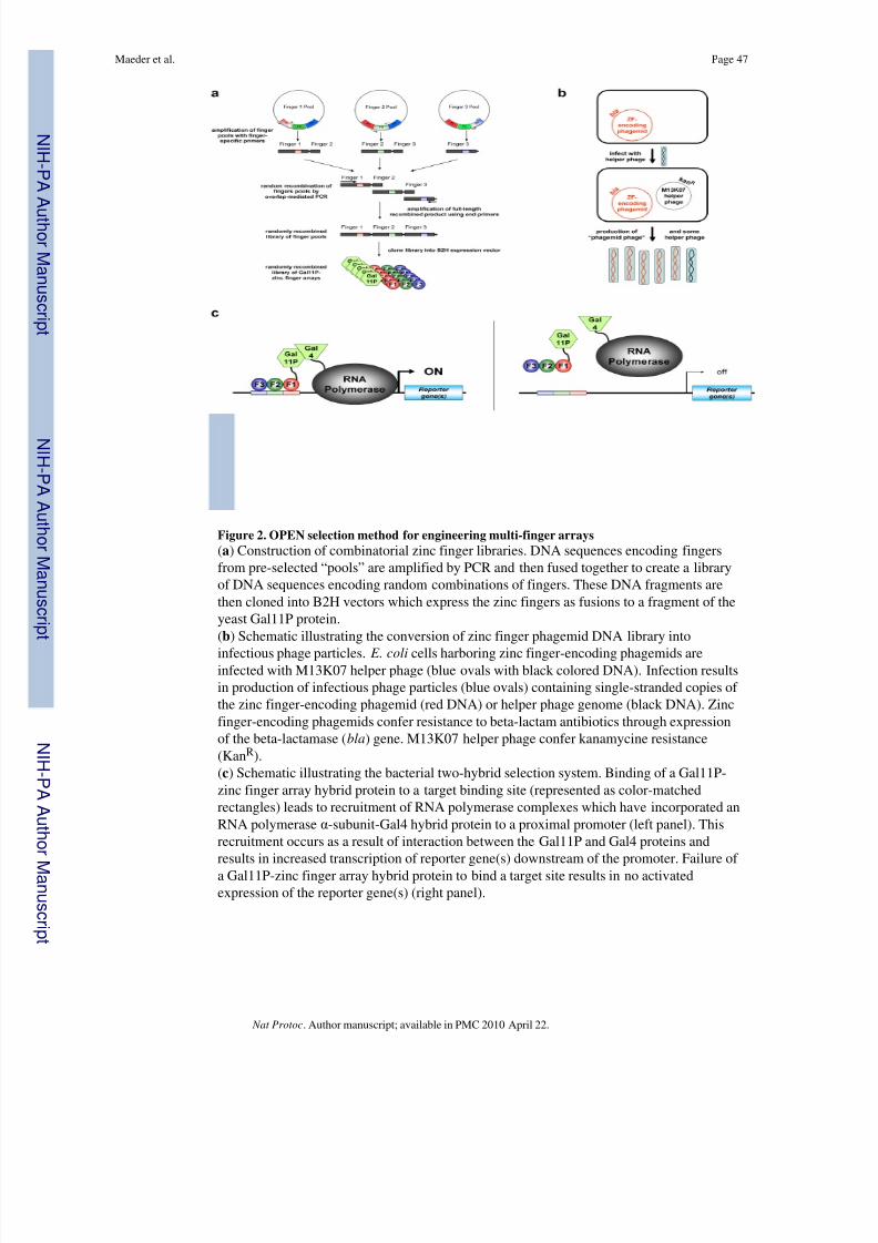
8/8/2019 Maeder - Thibodeau-Beganny Et Al 2010
http://slidepdf.com/reader/full/maeder-thibodeau-beganny-et-al-2010 47/61
Figure 2. OPEN selection method for engineering multi-finger arrays(a ) Construction of combinatorial zinc finger libraries. DNA sequences encoding fingersfrom pre-selected “pools” are amplified by PCR and then fused together to create a libraryof DNA sequences encoding random combinations of fingers. These DNA fragments arethen cloned into B2H vectors which express the zinc fingers as fusions to a fragment of theyeast Gal11P protein.(b) Schematic illustrating the conversion of zinc finger phagemid DNA library into
infectious phage particles. E. coli cells harboring zinc finger-encoding phagemids areinfected with M13K07 helper phage (blue ovals with black colored DNA). Infection resultsin production of infectious phage particles (blue ovals) containing single-stranded copies of the zinc finger-encoding phagemid (red DNA) or helper phage genome (black DNA). Zincfinger-encoding phagemids confer resistance to beta-lactam antibiotics through expressionof the beta-lactamase ( bla ) gene. M13K07 helper phage confer kanamycine resistance(Kan R).(c) Schematic illustrating the bacterial two-hybrid selection system. Binding of a Gal11P-zinc finger array hybrid protein to a target binding site (represented as color-matchedrectangles) leads to recruitment of RNA polymerase complexes which have incorporated anRNA polymerase α-subunit-Gal4 hybrid protein to a proximal promoter (left panel). Thisrecruitment occurs as a result of interaction between the Gal11P and Gal4 proteins andresults in increased transcription of reporter gene(s) downstream of the promoter. Failure of a Gal11P-zinc finger array hybrid protein to bind a target site results in no activatedexpression of the reporter gene(s) (right panel).
Maeder et al. Page 47
Nat Protoc . Author manuscript; available in PMC 2010 April 22.
N I H -P A A
ut h or Manus c r i pt
N I H -P A A ut h or Manus c r i pt
N I H -P A A ut h or
Manus c r i pt
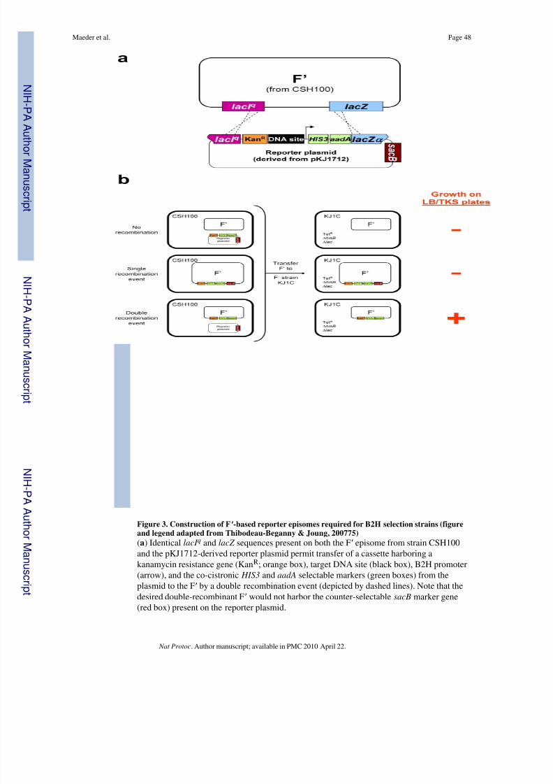
8/8/2019 Maeder - Thibodeau-Beganny Et Al 2010
http://slidepdf.com/reader/full/maeder-thibodeau-beganny-et-al-2010 48/61
Figure 3. Construction of F ′-based reporter episomes required for B2H selection strains (figureand legend adapted from Thibodeau-Beganny & Joung, 200775)(a ) Identical lacI q and lacZ sequences present on both the F ′ episome from strain CSH100and the pKJ1712-derived reporter plasmid permit transfer of a cassette harboring akanamycin resistance gene (Kan R; orange box), target DNA site (black box), B2H promoter(arrow), and the co-cistronic HIS3 and aadA selectable markers (green boxes) from theplasmid to the F ′ by a double recombination event (depicted by dashed lines). Note that thedesired double-recombinant F ′ would not harbor the counter-selectable sacB marker gene(red box) present on the reporter plasmid.
Maeder et al. Page 48
Nat Protoc . Author manuscript; available in PMC 2010 April 22.
N I H -P A A
ut h or Manus c r i pt
N I H -P A A ut h or Manus c r i pt
N I H -P A A ut h or
Manus c r i pt

8/8/2019 Maeder - Thibodeau-Beganny Et Al 2010
http://slidepdf.com/reader/full/maeder-thibodeau-beganny-et-al-2010 49/61
(b) Schematic depicting the various types of cells described in the bacterial mating of step24. The left side of the figure depicts CSH100 cells in which a double, single, or norecombination event has occurred between the F ′ and the reporter plasmid. The right side of the figure depicts KJ1C cells that have received a double, single, or non-recombinant F ′from the CSH100 cells and indicates whether or not these various cells will grow on LB/ TKS plates containing tetracycline, kanamycin, and sucrose.
Maeder et al. Page 49
Nat Protoc . Author manuscript; available in PMC 2010 April 22.
N I H -P A A
ut h or Manus c r i pt
N I H -P A A ut h or Manus c r i pt
N I H -P A A ut h or
Manus c r i pt

8/8/2019 Maeder - Thibodeau-Beganny Et Al 2010
http://slidepdf.com/reader/full/maeder-thibodeau-beganny-et-al-2010 50/61
Figure 4. Strategy for designing binding site oligonucleotides (figure from Wright et al., 200676)
Schematic illustrating the design of target zinc finger binding site oligonucleotides asdescribed in Box 1.
Maeder et al. Page 50
Nat Protoc . Author manuscript; available in PMC 2010 April 22.
N I H -P A A
ut h or Manus c r i pt
N I H -P A A ut h or Manus c r i pt
N I H -P A A ut h or
Manus c r i pt
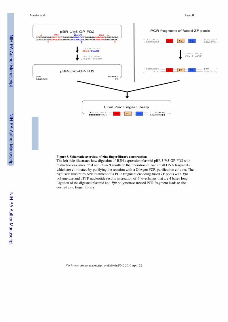
8/8/2019 Maeder - Thibodeau-Beganny Et Al 2010
http://slidepdf.com/reader/full/maeder-thibodeau-beganny-et-al-2010 51/61
Figure 5. Schematic overview of zinc finger library constructionThe left side illustrates how digestion of B2H expression plasmid pBR-UV5-GP-FD2 withrestriction enzymes Bbs I and BamH I results in the liberation of two small DNA fragments
which are eliminated by purifying the reaction with a QIAgen PCR purification column. Theright side illustrates how treatment of a PCR fragment encoding fused ZF pools with Pfupolymerase and dTTP nucleotide results in creation of 5 ′ overhangs that are 4 bases long.Ligation of the digested plasmid and Pfu polymerase-treated PCR fragment leads to thedesired zinc finger library.
Maeder et al. Page 51
Nat Protoc . Author manuscript; available in PMC 2010 April 22.
N I H -P A A
ut h or Manus c r i pt
N I H -P A A ut h or Manus c r i pt
N I H -P A A ut h or
Manus c r i pt

8/8/2019 Maeder - Thibodeau-Beganny Et Al 2010
http://slidepdf.com/reader/full/maeder-thibodeau-beganny-et-al-2010 52/61
Figure 6. Schematic depicting triplicate serial dilution and triplicate spotting of dilutions on agarplates100 μl of each sample to be diluted is placed in triplicate into three wells in the top row of a96-well plate. 10-fold serial dilutions are performed of each sample and then 5 μl of eachdilution is plated in replicate on half of an agar plate.
Maeder et al. Page 52
Nat Protoc . Author manuscript; available in PMC 2010 April 22.
N I H -P A A
ut h or Manus c r i pt
N I H -P A A ut h or Manus c r i pt
N I H -P A A ut h or
Manus c r i pt
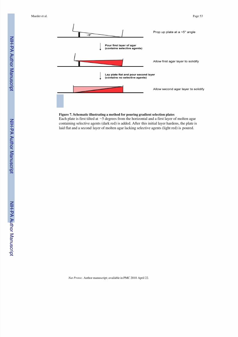
8/8/2019 Maeder - Thibodeau-Beganny Et Al 2010
http://slidepdf.com/reader/full/maeder-thibodeau-beganny-et-al-2010 53/61
Figure 7. Schematic illustrating a method for pouring gradient selection plates
Each plate is first tilted at ∼ 5 degrees from the horizontal and a first layer of molten agarcontaining selective agents (dark red) is added. After this initial layer hardens, the plate islaid flat and a second layer of molten agar lacking selective agents (light red) is poured.
Maeder et al. Page 53
Nat Protoc . Author manuscript; available in PMC 2010 April 22.
N I H -P A A
ut h or Manus c r i pt
N I H -P A A ut h or Manus c r i pt
N I H -P A A ut h or
Manus c r i pt

8/8/2019 Maeder - Thibodeau-Beganny Et Al 2010
http://slidepdf.com/reader/full/maeder-thibodeau-beganny-et-al-2010 54/61
Figure 8. Appearance of a typical gradient selection plate with a successful OPEN selectionThe density of bacterial colonies (beige circles) on the gradient plate (grey square) rangesfrom confluent (on the end of the plate containing the lowest concentrations of selectiveagents) to sparse (on the end of the plate with maximal concentrations of selective agents).We typically pick colonies from the end of the plate where the colony density is most sparse.
Maeder et al. Page 54
Nat Protoc . Author manuscript; available in PMC 2010 April 22.
N I H -P A A
ut h or Manus c r i pt
N I H -P A A ut h or Manus c r i pt
N I H -P A A ut h or
Manus c r i pt
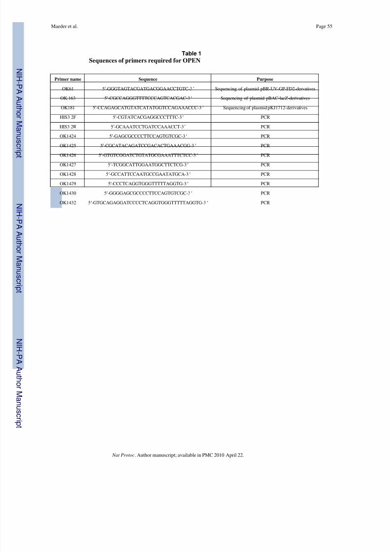
8/8/2019 Maeder - Thibodeau-Beganny Et Al 2010
http://slidepdf.com/reader/full/maeder-thibodeau-beganny-et-al-2010 55/61
N I H -P A A ut h or Manus c r i pt
N I H -P A A ut h or Manus c r i pt
N I H -P A A ut h or Manus c r i pt
Maeder et al. Page 55
Table 1Sequences of primers required for OPEN
Primer name Sequence Purpose
OK61 5 ′-GGGTAGTACGATGACGGAACCTGTC-3 ′ Sequencing of plasmid pBR-UV-GP-FD2-dervatives
OK.163 5 ′-CGCCAGGGTTTTCCCAGTCACGAC-3 ′ Sequencing of plasmid pBAC-lacZ-derivatives
OK181 5 ′-CCAGAGCATGTATCATATGGTCCAGAAACCC-3 ′ Sequencing of plasmid pKJ1712-derivatives
HIS3 2F 5 ′-CGTATCACGAGGCCCTTTC-3 ′ PCR
HIS3 2R 5 ′-GCAAATCCTGATCCAAACCT-3 ′ PCR
OK1424 5 ′-GAGCGCCCCTTCCAGTGTCGC-3 ′ PCR
OK1425 5 ′-CGCATACAGATCCGACACTGAAACGG-3 ′ PCR
OK1426 5 ′-GTGTCGGATCTGTATGCGAAATTTCTCC-3 ′ PCR
OK1427 5 ′-TCGGCATTGGAATGGCTTCTCG-3 ′ PCR
OK1428 5 ′-GCCATTCCAATGCCGAATATGCA-3 ′ PCR
OK1429 5 ′-CCCTCAGGTGGGTTTTTAGGTG-3 ′ PCR
OK1430 5 ′-GGGGAGCGCCCCTTCCAGTGTCGC-3 ′ PCR
OK1432 5 ′-GTGCAGAGGATCCCCTCAGGTGGGTTTTTAGGTG-3 ′ PCR
Nat Protoc . Author manuscript; available in PMC 2010 April 22.

8/8/2019 Maeder - Thibodeau-Beganny Et Al 2010
http://slidepdf.com/reader/full/maeder-thibodeau-beganny-et-al-2010 56/61
N I H -P A A ut h or Manus c r i pt
N I H -P A A ut h or Manus c r i pt
N I H -P A A ut h or Manus c r i pt
Maeder et al. Page 56
Table 2PCR conditions for amplifying Finger 1 pools (steps 41 and 42)
Step number Denature Anneal Extend
1 94°C, 5 min
2-6 94°C, 30 s 55°C, 30 s 72°C, 30 s
7-26 94°C, 30 s 59°C, 30 s 72°C, 30 s
27 72°C, 2 min
Nat Protoc . Author manuscript; available in PMC 2010 April 22.

8/8/2019 Maeder - Thibodeau-Beganny Et Al 2010
http://slidepdf.com/reader/full/maeder-thibodeau-beganny-et-al-2010 57/61
N I H -P A A ut h or Manus c r i pt
N I H -P A A ut h or Manus c r i pt
N I H -P A A ut h or Manus c r i pt
Maeder et al. Page 57
Table 3PCR conditions for amplifying Finger 2 pools (steps 41 and 42)
Step number Denature Anneal Extend
1 94°C, 5 min
2-6 94°C, 30 s 52°C, 30 s 72°C, 30 s
7-26 94°C, 30 s 57°C, 30 s 72°C, 30 s
27 72°C, 2 min
Nat Protoc . Author manuscript; available in PMC 2010 April 22.

8/8/2019 Maeder - Thibodeau-Beganny Et Al 2010
http://slidepdf.com/reader/full/maeder-thibodeau-beganny-et-al-2010 58/61
N I H -P A A ut h or Manus c r i pt
N I H -P A A ut h or Manus c r i pt
N I H -P A A ut h or Manus c r i pt
Maeder et al. Page 58
Table 4PCR conditions for amplifying Finger 3 pools (steps 41 and 42)
Step number Denature Anneal Extend
1 94°C, 5 min
2-6 94°C, 30 s 41°C, 30 s 72°C, 30 s
7-26 94°C, 30 s 56°C, 30 s 72°C, 30 s
27 72°C, 2 min
Nat Protoc . Author manuscript; available in PMC 2010 April 22.

8/8/2019 Maeder - Thibodeau-Beganny Et Al 2010
http://slidepdf.com/reader/full/maeder-thibodeau-beganny-et-al-2010 59/61
N I H -P A A ut h or Manus c r i pt
N I H -P A A ut h or Manus c r i pt
N I H -P A A ut h or Manus c r i pt
Maeder et al. Page 59
Table 5Zinc Finger Consortium vectors for expression of zinc finger arrays as ZFNs
Plasmid name For expression in… Fok I cleavage domain type
pST1374 a mammalian cells, zebrafish (RNA) wild-type
pMLM290 b mammalian cells, zebrafish (RNA) “+” obligate heterodimer 15
pMLM292 b mammalian cells, zebrafish (RNA) “− ” obligate heterodimer 15
pDW1775 a plants wild-type
Zinc finger arrays selected by OPEN can be directly cloned into the ZFC expression plasmids listed using unique Xba I and BamH I sites. Theresulting plasmids express a ZFN with a linker of sequence LRGS between the last conserved histidine of the carboxy-terminal zinc finger and thefirst amino acid of the Fok I nuclease domain.
aAvailable through Addgene (http://www.addgene.org/zfc)
bAvailable by request through the Joung lab
Nat Protoc . Author manuscript; available in PMC 2010 April 22.

8/8/2019 Maeder - Thibodeau-Beganny Et Al 2010
http://slidepdf.com/reader/full/maeder-thibodeau-beganny-et-al-2010 60/61
N I H -P A A ut h or Manus c r i pt
N I H -P A A ut h or Manus c r i pt
N I H -P A A ut h or Manus c r i pt
Maeder et al. Page 60
Table 6Troubleshooting table
Step(s) Problem Possible Reason Solution
6 No ZFN target sites identified Insufficient length of sequenceto identify an OPEN target site
Consider expanding the DNA search length
6 Too many ZFN target sitesidentified
Consider using only sites that contain two or moreGNN subsites
16, 58 No transformants obtained fromligation
Low efficiency of transformation
Consider re-making competent cells or purchasingcommercially available competent cells.
16, 58 No transformants obtained fromligation
Insufficient concentration of vector backbone or insert
Check DNA concentrations using 260 nm absorbancereadings on a spectrophotometer.
16, 58 Too many transformants fromvector backbone control ligation
Incomplete digestion of vectorbackbone
Screen additional colonies from the actual ligation/ transformation plate or consider re-digesting vectorusing higher concentration of enzyme or less DNA
26 Number of colonies on TK and TKSplates are the same
sacB gene in the reporterplasmid acquired a mutation
Repeat steps 20-26 using another plasmid candidate
36 Failure to obtain transformants Low efficiency of transformation
Consider re-making competent cells (step 35)
36 Failure to obtain transformants Insufficient amount of pAC-alphaGal4 plasmid used fortransformation
Use larger amount of DNA for transformation and/orplate a larger portion of the transformation
43 Failure to obtain PCR product ongel
PCR reaction was unsuccessful Verify that you are using Expand PCR enzyme and thatPCR cycling conditions are correct
43 Failure to obtain PCR product ongel
Low amounts of DNA templatein PCR reaction
Verify that the pools (obtained from the Joung lab)were fully resuspended by checking DNA concentrationon a spectrophotometer.
65 Number of transformants from pre-amplification quantification is toolow
Low efficiency of transformation
Consider re-making electrocompetent cells orpurchasing commercially available electrocompetentXL1-blue cells.
65 Number of transformants from thepositive control is adequate butnumber of library transformants istoo low
Insufficient quantity of libraryDNA
Repeat step 60 with remaining half of ligation andrepeat electroporation. If this fails, try repeating theligation.
71 Titers show that <50% of cells wereinfected by helper phage
Titer of helper phage was toolow to successfully superinfectthe culture
Repeat steps 66-71 using a larger volume of helperphage or consider re-making high-titer helper phagestock.
74 Failure of selection strain culture togrow to correct density
Colonies on plate were olderthan 1 week
Re-streak strain and repeat innoculation with freshcolonies
74 Failure of selection strain culture togrow to correct density
Glassware used for innoculationcontained residual soapdetergent
Thoroughly rinse glassware with distilled water beforeautoclaving to remove any residual detergent.
74 Failure of selection strain culture togrow to correct density
Insufficient colony dispersalduring innoculation
Repeat innoculation making sure to completely dispersecolony in media
74 Failure of selection strain culture togrow to correct density
Incorrect composition of NMmedia
Verify that M9 salt stock is actually 10× (not 5× asdescribed by recipe on the bottle supplied by Difco)
74 Failure of selection strain culture togrow to correct density
Incorrect composition of NMmedia
Verify that glucose used was obtained fromMallinckrodt-Baker
74 Failure of selection strain culture togrow to correct density
Incorrect composition of NMmedia
Verify composition of all other NM reagents
74 Failure of selection strain culture togrow to correct density
Inability of strain to grow inNM media
Go back to the “B” candidate from step 35, repeat step36 and proceed with selections using this strain.
80 Total number of cells in 250 μl of resuspended cells from step 77 is
Too much phage library wasused for the selection
Repeat steps 72-80 using an appropriately decreasednumber of CTUs (carbenicillin-transducing units)
Nat Protoc . Author manuscript; available in PMC 2010 April 22.

8/8/2019 Maeder - Thibodeau-Beganny Et Al 2010
http://slidepdf.com/reader/full/maeder-thibodeau-beganny-et-al-2010 61/61
N I H -P A A ut h or Manus c r i pt
N I H -P A A ut h or Manus c r i pt
N I H -P A A ut h or Man
Maeder et al. Page 61
Step(s) Problem Possible Reason Solution
not at least three-fold more than thetotal number of infected cells.
80 The total number of infected cells in250 μl of resuspended cells fromstep 77 is <2.5 × 10 6
Too little phage library wasused for the selection
Repeat steps 72-80 using an appropriately increasednumber of CTUs (carbenicillin-transducing units)
90 Titer of the enriched zinc fingerlibrary phage stock is not >1×10⁴ATU/ μl
Too little phage library wasused for the selection
Concentrate the enriched zinc finger library phage stock and re-titer the concentrated stock as described in Box8.
97 The total number of cells in theculture of step 94 is less than three-fold the number of infected cells
Too much enriched zinc fingerlibrary phage was used forinfection
Check calculation of enriched zinc finger library phagetiter. Consider repeating steps 85-90 to obtain anaccurate titer.
97 The total number of infected cellsplated on the gradient plate is <2.5 ×10⁶
Too little enriched zinc fingerlibrary phage was used forinfection
Check calculation of enriched zinc finger library phagetiter. Consider repeating steps 85-90 to obtain anaccurate titer.
97 The total number of infected cellsplated on the gradient plate is <2.5 ×10⁶
Too little enriched zinc fingerlibrary phage was used forinfection
Consider PEG precipitation to concentrate enriched zincfinger library (Box 8).
97 The total number of infected cellsplated on the gradient plate is <2.5 ×10⁶
Too little enriched zinc fingerlibrary phage was used forinfection
If colonies appear to be growing on gradient plate,proceed with protocol despite the fact that the numberswere not high enough to three-fold oversample the
library.115 Failure to obtain colonies One of the recognition helix
sequences selected in the zincfinger expression plasmidcontains a Pst I site
Isolate zinc finger-encoding plasmid from pAC-alphaGal4 plasmid by an alternative method. Diluteundigested plasmid isolated in step 98 by 100-fold inwater and transform into chemically competent XL-1Blue cells. Patch streak transformants onto LB/TC andLB+30mg/ml chloramphenicol plates. Pick candidatesthat fail to grow on LB+30mg/ml chloramphenicolplates (as these were transformed only by the zincfinger expression plasmid and not the pAC-alpha-Gal4expression plasmid) and isolate miniprep DNA. Repeatsteps 114-116.
116 Failure to obtain zinc finger arraysthat mediate at least 3-foldactivation
Target site may not be suitablefor OPEN
Consider other targets
116 Failure to obtain zinc finger arraysthat mediate at least 3-foldactivation
High basal level of transcription If the absolute ß-galactosidase activity units for theGal11P control are particularly high, considerproceeding with zinc finger arrays that activate >2-fold.