mammalian target of rapamycin complex 1 (mtorc1) is ... · mouse spermatogonial differentiation in...
TRANSCRIPT

Developmental Biology 407 (2015) 90–102
Contents lists available at ScienceDirect
Developmental Biology
http://d0012-16
n CorrGreenvi
E-m
journal homepage: www.elsevier.com/locate/developmentalbiology
Mammalian target of rapamycin complex 1 (mTORC1) Is required formouse spermatogonial differentiation in vivo
Jonathan T. Busada a, Bryan A. Niedenberger a, Ellen K. Velte a, Brett D. Keiper b,Christopher B. Geyer a,c,n
a Department of Anatomy and Cell Biology, East Carolina University, Greenville, NC, USAb Department of Biochemistry and Molecular Biology, East Carolina University, Greenville, NC, USAc East Carolina Diabetes and Obesity Institute, East Carolina University, Greenville, NC, USA
a r t i c l e i n f o
Article history:Received 28 April 2015Received in revised form2 August 2015Accepted 3 August 2015Available online 5 August 2015
Keywords:TranslationSpermatogoniaRetinoic acidTestisSpermatogenesismTOR
x.doi.org/10.1016/j.ydbio.2015.08.00406/& 2015 Elsevier Inc. All rights reserved.
espondence to: Brody School of Medicinelle, NC 27858-4353. Fax: þ 1 252 744 2850.ail address: [email protected] (C.B. Geyer).
a b s t r a c t
Spermatogonial stem cells (SSCs) must balance self-renewal with production of transit-amplifying pro-genitors that differentiate in response to retinoic acid (RA) before entering meiosis. This self-renewal vs.differentiation spermatogonial fate decision is critical for maintaining tissue homeostasis, as imbalancescause spermatogenesis defects that can lead to human testicular cancer or infertility. A great deal ofeffort has been exerted to understand how the SSC population is maintained. In contrast, little is knownabout the essential program of differentiation initiated by retinoic acid (RA) that precedes meiosis, andthe pathways and proteins involved are poorly defined. We recently reported a novel role for RA instimulating the PI3/AKT/mTOR kinase signaling pathway to activate translation of repressed mRNAs suchas Kit. Here, we examined the requirement for mTOR complex 1 (mTORC1) in mediating the RA signal todirect spermatogonial differentiation in the neonatal testis. We found that in vivo inhibition of mTORC1by rapamycin blocked spermatogonial differentiation, which led to an accumulation of undifferentiatedspermatogonia. In addition, rapamycin also blocked the RA-induced translational activation of mRNAsencoding KIT, SOHLH1, and SOHLH2 without affecting expression of STRA8. These findings highlight dualroles for RA in germ cell development – transcriptional activation of genes, and kinase signaling to sti-mulate translation of repressed messages required for spermatogonial differentiation.
& 2015 Elsevier Inc. All rights reserved.
1. Introduction
Retinoic acid (RA) is required for inducing both spermatogonialdifferentiation and subsequent entry into meiosis (Bowles et al.,2006; Koubova et al., 2006). After birth in the mouse, subsets ofspermatogonia begin to differentiate in response to RA at �P3-4,as indicated by their expression of STRA8 and KIT (Busada et al.,2014; Niedenberger et al., 2015; Snyder et al., 2010; Zhou et al.,2008). However, germ cells do not enter meiosis in response to RAuntil �P10 (Hogarth and Griswold, 2013). This reveals that theprocesses of differentiation and meiotic entry are temporally se-parated by �7 days in the neonate (which lengthens to 8.6 days inthe adult). It has been proposed that the reduced time in theneonate may be because some of the steps are skipped or becausecell proliferation is accelerated by the increased temperature atwhich spermatogenesis proceeds in the neonate (37°C in neonates
at East Carolina University,
versus 33°C in adults) (Kluin et al., 1982). During this approxi-mately week-long differentiation period, type A1 spermatogoniasuccessively become type A2, A3, A4, In, and B before enteringmeiosis as preleptotene spermatocytes (de Rooij, 2001; de Rooijand Griswold, 2000; Kluin et al., 1984). Spermatogonia apparentlycannot be induced to precociously enter meiosis in response toexogenous RA (Busada et al., 2014; Niedenberger et al., 2015; Endoet al., 2015), which implies that these sequential spermatogonialdivisions are required. However, little is known about the cellularprocesses that must occur or the molecular pathways that regulatethem in differentiating spermatogonia prior to meiotic initiation. Aprimary reason for this lack of knowledge is that there are fewchanges in steady-state mRNA levels during differentiation (Chanet al., 2014; Shima et al., 2004; Zhou et al., 2008). Without dra-matic changes in the transcriptome, scientists have lacked iden-tified targets (pathways, proteins) for focused studies.
A number of recent genome-wide studies have revealed thatthe transcriptome imperfectly predicts the proteome (estimatesrange from �40% to 80% agreement) (Li et al., 2014; Schwan-hausser et al., 2011; Vogel and Marcotte, 2012), and there is likelyto be considerable variation in this level of disconnect in different
J.T. Busada et al. / Developmental Biology 407 (2015) 90–102 91
cell types and under different conditions. Our previous studiessuggest that a majority of gene expression changes required forspermatogonial differentiation occur at the posttranscriptionallevel (Busada et al., 2015; Chappell et al., 2013). We previouslyshowed that RA treatment leads to increased phosphorylation ofthe master regulatory kinase mammalian target of rapamycin(mTOR) (Busada et al., 2015), indicating it is activated in differ-entiating spermatogonia. This is accompanied by enhancedtranslation of repressed Kit mRNAs through activation of the PI3K/PDPK1 (also termed PDK1)/AKT signaling network (Busada et al.,2015; Hermann et al., 2015). MTOR exists in functionally distinctprotein complexes (mTORC1 and mTORC2), which integrate nu-merous cues to regulate, in broad terms, cellular growth and dif-ferentiation (mTORC1) or the actin cytoskeleton and insulin sig-naling (mTORC2) (reviewed by (Fingar and Blenis, 2004; Hay andSonenberg, 2004; Laplante and Sabatini, 2012; Li et al., 2014;Wullschleger et al., 2006)). A primary role of mTORC1 is to reg-ulate cap-dependent mRNA translation initiation, which is the ratelimiting and regulated step of eukaryotic protein synthesis.MTORC1 performs this function in part by phosphorylatingdownstream targets EIF4EBP1 and RPS6KB1/2 (also termedp70S6K). Enhanced phosphorylation of EIF4EBP1 by activatedmTOR releases EIF4E to associate with the 5’-cap of mRNAs, thusallowing recruitment of translationally controlled mRNAs to ri-bosomes, particularly during germ cell differentiation in lowerorganisms (Dinkova et al., 2005; Henderson et al., 2009; Kimbleand Crittenden, 2007; Mendez and Richter, 2001; Song et al.,2010). Activated RPS6KB1/2 phosphorylates the 40S ribosomalsubunit RPS6, leading to activation of ribosomes and enhancedmRNA translation. This activity is essential for the translationalcontrol of the TOP mRNAs, which have 5′ oligopyrimidine tracts,are activated by changes in cellular metabolism, and often encodecomponents of the translational machinery (Hamilton et al., 2006;Meyuhas and Kahan, 2014).
The mTOR complexes differ in their sensitivity to rapamycin, amacrolide antifungal compound originally isolated from the soilbacterium Streptomyces hygroscopicus that has an expandingnumber of clinical uses. Rapamycin acutely inhibits mTORC1 ac-tivity, but has also been shown to inhibit mTORC2 in some con-texts following prolonged exposure (Laplante and Sabatini, 2012;Brown et al., 1994; Sabatini et al., 1994). The effects of rapamycinvary significantly depending on the cell type involved. Some celltypes appear to be quite rapamycin-insensitive, while others slowor cease proliferating, fail to differentiate, and/or undergo apop-tosis (Laplante and Sabatini, 2012; Li et al., 2014; Shimobayashiand Hall, 2014). This has been suggested to be due in part to dif-ferential effects on downstream substrates such as EIF4EBP1 and/or RPS6KB1/2 (Choo et al., 2008). Rapamycin can be administeredin vivo, and actually extends the lifespan of lower organisms andmice ((Harrison et al., 2009; Anisimov et al., 2011), reviewed in Liet al., 2014). In addition, studies have suggested a link betweenmTORC1 and spermatogonial cell fate regulation both in vitro andin vivo (Busada et al., 2015; Chappell et al., 2013; Feng et al., 2000;Hobbs et al., 2015; Hobbs et al., 2010; Kofman et al., 2013).
Studying the effects of mTORC1 inhibition by rapamycin hasdirect relevance for human male reproductive health. Rapamycinanalogs (Sirolimus and Everolimus) are currently used to reducecellular proliferation as part of immunosuppressive and che-motherapeutic regimens given to organ transplant, cardiology, andcancer patients (Laplante and Sabatini, 2012; Li et al., 2014; Ash-worth and Wu, 2014; de Pablo et al., 2013; Eyre et al., 2014;Ganschow et al., 2014; Ng et al., 2014; Peddi et al., 2013). Thesedrugs can cause reversible human male infertility with unclearetiology (Boobes et al., 2010; Framarino-dei-Malatesta et al., 2013;Huyghe et al., 2007; Zaza et al., 2013; Zuber et al., 2008). Specifi-cally, rapamycin analog treatments caused a block in
spermatogonial differentiation in a human patient (Deutsch et al.,2007) as well as in a study using rats, although no detailed ana-lyses were performed (Chen et al., 2013). Both treated humans andrats exhibit reduced testosterone (T) levels due to inhibition of thehypothalamic–pituitary–gonadal axis. However, this may notcause a defect in spermatogonial differentiation, but rather inprogression through meiosis, at least in rodents (Walker, 2011).Infertility is a significant quality of life concern for reproductive-aged male organ transplant and cancer patients, and to-date nocomprehensive studies have explored the mechanism of action ofmTOR inhibition during spermatogenesis.
Here, we examine the effects of rapamycin-mediated mTORC1inactivation on spermatogonial differentiation in vivo in themouse. Our results reveal that mTORC1 activation is dispensablefor the maintenance of undifferentiated spermatogonia, but that itis required for spermatogonial proliferation and differentiationprior to meiotic initiation. In addition, we find that rapamycininhibition of mTORC1 blocks the RA-induced translation of re-pressed mRNAs encoding KIT, SOHLH1, and SOHLH2, which areessential regulators of spermatogonial differentiation. However,rapamycin treatment did not block the expression of STRA8, adirect transcriptional target of RA. This reveals that spermatogoniaexhibit dual responses to RA in the form of transcriptional acti-vation and kinase signaling-enhanced translation of repressedmRNAs. In addition, these results provide critical insight into themale infertility that can result as an adverse side effect of theclinical use of rapamycin analogs.
2. Results
2.1. Rapamycin treatment reduces testicular size and arrests germcell development
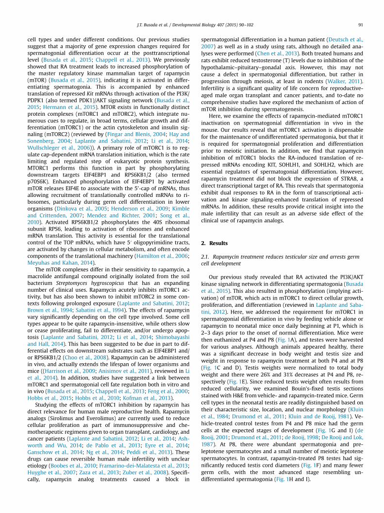
Our previous study revealed that RA activated the PI3K/AKTkinase signaling network in differentiating spermatogonia (Busadaet al., 2015). This also resulted in phosphorylation (implying acti-vation) of mTOR, which acts in mTORC1 to direct cellular growth,proliferation, and differentiation (reviewed in Laplante and Saba-tini, 2012). Here, we addressed the requirement for mTORC1 inspermatogonial differentiation in vivo by feeding vehicle alone orrapamycin to neonatal mice once daily beginning at P1, which is2–3 days prior to the onset of normal differentiation. Mice werethen euthanized at P4 and P8 (Fig. 1A), and testes were harvestedfor various analyses. Although animals appeared healthy, therewas a significant decrease in body weight and testis size andweight in response to rapamycin treatment at both P4 and at P8(Fig. 1C and D). Testis weights were normalized to total bodyweight and there were 26% and 31% decreases at P4 and P8, re-spectively (Fig. 1E). Since reduced testis weight often results fromreduced cellularity, we examined Bouin's-fixed testis sectionsstained with H&E from vehicle- and rapamycin-treated mice. Germcell types in the neonatal testis are readily distinguished based ontheir characteristic size, location, and nuclear morphology (Kluinet al., 1984; Drumond et al., 2011; Kluin and de Rooij, 1981). Ve-hicle-treated control testes from P4 and P8 mice had the germcells at the expected stages of development (Fig. 1G and I) (deRooij, 2001; Drumond et al., 2011; de Rooij, 1998; De Rooij and Lok,1987). At P8, there were abundant spermatogonia and pre-leptotene spermatocytes and a small number of meiotic leptotenespermatocytes. In contrast, rapamycin-treated P8 testes had sig-nificantly reduced testis cord diameters (Fig. 1F) and many fewergerm cells, with the most advanced stage resembling un-differentiated spermatogonia (Fig. 1H and I).

Fig. 1. MTORC1 inhibition blocks spermatogonial differentiation. (A) Experimental design for treating mice with rapamycin in vivo. Mice were treated with vehicle orrapamycin once daily starting at P1 and euthanized 24 h after last treatment at P4 or P8. (B) Representative images of testes from vehicle (left) and rapamycin (right) treatedmice euthanized at P4 (top) and P8 (bottom). (C–E) Mice treated with vehicle or rapamycin were euthanized at P4 or P8. Prior to euthanasia total body weights werecollected (C), following euthanasia total testis weights were collected (D). Testis weights were normalized to body weights and expressed as a ratio (E). (F) Quantitation oftestis cord diameter of mice treated with vehicle or rapamycin and euthanized at P4 or P8. (G–J) H&E staining of mice treated with vehicle (G and I) or rapamycin (H and J)and euthanized at P4 (G and H) or P8 (I and J). Yellow arrows indicate spermatogonia, and green and orange lines encircle preleptotene and leptotene spermatocytes,respectively (I). Scale bar¼40 μM. Asterisks indicate statistical significance with Po0.01.
J.T. Busada et al. / Developmental Biology 407 (2015) 90–10292
2.2. Rapamycin inhibits mTORC1 activity in spermatogonia
Before carefully examining the phenotype of rapamycin-treatedtestes, we verified that rapamycin treatment inhibited mTORC1activity as expected. We examined the phosphorylation of mTOR aswell as RPS6, which is an indirect target downstream of activatedmTORC1. There was a near-complete loss of p-mTOR and p-RPS6 inresponse to rapamycin (Fig. 2A–D). We recently reported that RAcaused a dramatic nuclear-to-cytoplasmic relocalization of FOXO1(Busada et al., 2015), which is indicative of AKT activation (Brunetet al., 1999; Goertz et al., 2011). Since this signaling step is generallyupstream of mTOR phosphorylation, we hypothesized that rapa-mycin inhibition would not alter FOXO1 localization. Indeed, therewas no appreciable difference in the ratios of cytoplasmic: nuclearFOXO1 in vehicle- or rapamycin-treated germ cells (Fig. 2E and F).
Therefore, we conclude that rapamycin treatment inhibitedmTORC1 activation in spermatogonia without affecting upstreamactivation of signaling components such as AKT, which implies thatmTORC2 signaling was not perturbed.
2.3. Undifferentiated spermatogonia accumulate in rapamycin-treated testes
Rapamycin treatment from P1 to P4 and P1 to P8 resulted in anapparent reduction in germ cells (Fig. 1H and J). We quantified thisin vehicle- and rapamycin-treated mice by immunostaining forDDX4, a pan germ cell marker in the neonatal testis. There werereduced numbers of DDX4þ germ cells at P4 (�1.6370.03-fold)and P8 (�2.7470.04-fold) (Fig. 3A–D, M and N). The decreasednumber of germ cells could result from a rapamycin-induced

Fig. 2. Treatment with rapamycin inhibits mTORC1 activity without affecting AKT. (A–F) Immunostaining of testis sections from mice treated with vehicle (A, C, and E) orrapamycin (B, D, and F) and euthanized at P4. Sections were stained with anti-phosphorylated RPS6 (A and B), anti-phosphorylated EIF4EBP1 (C and D), or total FOXO1 (E andF). F-actin was stained with phalloidin (red) to visualize testis cords. Scale bar¼50 μM.
J.T. Busada et al. / Developmental Biology 407 (2015) 90–102 93
increase in apoptosis, although we did not see evidence for this inH&E-stained sections (Fig. 1G–J, data not shown). We nonethelessassessed this possibility by immunostaining for cleaved PARP1, anaccepted marker for apoptotic cells. As expected, there was nosignificant increase in cleaved-PARP1þ cells in rapamycin-treatedtestes at P4 or P8 (P1-4 vehicle-treated¼0.1770.06 cleaved-PARP1þ cells/testis cord, P1-4 rapamycin-treated¼0.1370.06cleaved-PARP1þ cells/testis cord, Fig. 3E–H), indicating thatapoptosis was not responsible for the reduced germ cell numbers.We next assessed whether there was a reduction in germ cellproliferation in response to rapamycin. We first immunostained
using an antibody against MKI67, which marks actively pro-liferating cells in all stages of the cell cycle except in G0 (Gerdeset al., 1983; Lopez et al., 1991). There was no difference in MKI67staining at P4 in response to rapamycin treatment. However, therewere significantly fewer MKI67þ/DDX4þ cells at P8 in rapamycintreated testes. (�1.8570.37-fold, Fig. S1A–E). We next injectedvehicle- and rapamycin-treated mice with BrdU 10 h prior to eu-thanasia on P4 and P8. There were E1.9-fold fewer BrdUþ germcells in response to rapamycin at P4 and P8 (Fig. 3I–L, O and P). Wealso assessed effects of rapamycin on Sertoli cell numbers by im-munostaining for the Sertoli cell marker GATA4, and found that

Fig. 3. MTORC1 is required for postnatal expansion of the germ cell population. (A–L) Immunostaining of testis sections frommice treated with vehicle (A, C, E, G, I, and K) orrapamycin (B, D, F, H, J, and L) and euthanized at P4 (A, B, E, F, I, and J) or P8 (C, D, G, H, K, and L). Sections were stained with anti-DDX4 (A–D), anti-cleaved PARP1 (E–H), ordouble labeled with anti-BrdU (green, I–L) and anti-DDX4 (red, I–L). F-actin was stained with phalloidin (red, A–D or blue, E–H). Quantitation of the number of DDX4þ (Mand N) or the number of BrdUþ/DDX4þ (O and P) cells from testes treated starting at P1 with vehicle or rapamycin and then euthanized at P4 (M and O) or at P8 (N and P).Asterisks indicate statistical significance with Pr0.01. Scale bar¼50 μM.
J.T. Busada et al. / Developmental Biology 407 (2015) 90–10294
there was no appreciable change at P4 (P1-4 vehicle¼18.370.6GATA4þ cells/testis cord, P1-4 rap-treated¼19.6773.3 GATA4þcells/testis cord) or P8 (P1-8 vehicle¼18.874.0 GATA4þ cells/testis cord, P1-8 rap-treated¼17.1715.5 GATA4þ cells/testis cord,Fig. S2). Therefore, we conclude that rapamycin inhibited germ cellproliferation without increasing apoptosis or changing Sertoli cellnumbers.
We next tested whether mTORC1 inhibition by rapamycinprevented the normal differentiation of spermatogonia. Based onthe apparent accumulation of undifferentiated spermatogonia atP4 and P8 in response to rapamycin (Fig. 1G–J), it appeared thatspermatogonial differentiation was blocked by rapamycin treat-ment. We assessed whether spermatogonia upregulated markersof differentiation (KIT, SOHLH1, and SOHLH2) in the absence of

Fig. 4. MTOR activation is required for induction of SOHLH1, SOHLH2, and KIT protein. Immunostaining of mice treated with vehicle (A, C, and E) or rapamycin (B, D, and F)and euthanized at P4. Sections were stained with anti-SOHLH1 (A and B), anti-SOHLH2 (C and D), or anti-KIT (E and F). Phalloidin (red) was added to visualize testis cords.Scale bar¼50 μM.
J.T. Busada et al. / Developmental Biology 407 (2015) 90–102 95
mTORC1 function. Each marker was readily detectable in sper-matogonia in vehicle-treated testes at P4 (Fig. 4A, C, and E). Incontrast, KIT, SOHLH1, and SOHLH2 were barely detectable in afew spermatogonia in rapamycin-treated testes (Fig. 4B, D, and F).The absence of differentiation markers suggests that the germ cellsremained in an undifferentiated state in the absence of mTORC1activity. We tested this by immunostaining for GFRA1, an estab-lished marker of undifferentiated spermatogonia, which togetherwith RET forms the receptor for GDNF (Kubota et al., 2004; Menget al., 2000). We found that at P8 66% of DDX4þ spermatogoniawere GFRA1þ in response to rapamycin, which represented a 5.1-
fold increase over vehicle-treated controls (Fig. 5A–C). We furtherexplored this using a recently created transgenic mouse line, inwhich the spermatogonial stem cell (SSC) population is marked bythe expression of eGFP under the control of the Id4 promoter(Chan et al., 2014). Recent work has demonstrated that SSC activityresides within the Id4-GFPþ cell population (Chan et al., 2014). Totest if treatment with rapamycin affects formation or size of theSSC population, we treated Id4-GFP pups with vehicle or rapa-mycin from P1 through P7 and euthanized them on P8. The resultsdemonstrated that 19% of the total germ cell population was GFP-bright in both the vehicle- and rapamycin-treated testes (Fig. 5D–

Fig. 5. Inhibiting mTORC1 activation increases the number of undifferentiated spermatogonia. (A, B, D, and E) Immunostaining was performed on testis sections from micetreated with vehicle (A and D) or rapamycin (B and E) and euthanized at P8. (A–C) Testis sections from CD-1 mice were stained with anti-GFRA1 (green, A and B) and F-actinwas stained with phalloidin (blue) to visualize testis cords. The number of GFRA1þ germ cells in vehicle- and rapamycin-treated testes were quantitated and reported as afold change (C). (D–F) Transgenic Id4-GFP mice were treated with vehicle or rapamycin, and immunostaining was performed on testes. Green represents GFP epifluoresence,and sections were labeled with anti-DDX4 (red). White arrows indicate GFP bright spermatogonia (D and E). The number of GFP bright cells were quantitated and re-presented as a percentage of the DDX4þ cells (C). Scale bar¼40 μM. Asterisks indicate statistical significance with Po0.01.
J.T. Busada et al. / Developmental Biology 407 (2015) 90–10296
F), indicating that the size of the SSC pool was not affected bymTORC1 inhibition.
2.4. Rapamycin blocks RA-enhanced translation of repressed mRNAs
Finally, we investigated how KIT, SOHLH1, and SOHLH2 proteinlevels were dramatically reduced following inhibition of mTORC1.We previously found that mRNAs for Kit, Sohlh1, and Sohlh2 be-came associated with heavy polysomes at P4 (Chappell et al.,2013), which coincided with the appearance of detectable protein.In a separate study, we reported that RA activated translation ofrepressed Kit mRNAs during differentiation by inducing heavypolysome occupancy without a dramatic increase in their abun-dance (Busada et al., 2015). We therefore examined whetherSohlh1 and Sohlh2 mRNAs were regulated similarly in response toRA. In response to RA, both SOHLH1 and SOHLH2 protein levelsincreased dramatically (Fig. 6A–D), and as previously shown, RAtreatment also induced Kit translation (Fig. S3 A and B, (Busadaet al., 2014, 2015)). This increase in protein was not accompaniedby an increase in steady-state mRNA levels (Fig. 6E and F). We thenperformed polysome gradient analysis to test whether RA inducedSohlh1 and Sohlh2 mRNA heavy polysome occupancy for efficienttranslation, as we recently showed for Kit (Busada et al., 2015).Polysome gradients allow for the fractionation of mRNAs based ontheir association with ribonucleoprotein particles (RNPs), ribo-some subunits, and light and heavy polysomes. The identificationof specific mRNAs within sedimenting fractions reflects their
translational efficiency, with those in heavy polysomes being mostefficiently translated (reviewed in (Masek et al., 2011)). We pooledheavy polysome fractions in testes from vehicle- and RA-treatedmice and discovered that Sohlh1, Sohlh2, and Kit mRNAs becameenriched in heavy polysomes in response to RA (Fig. 6E and F andFig. S3C). We conclude that, like Kit, Sohlh1 and Sohlh2 mRNAs arenot efficiently translated in undifferentiated spermatogonia, andbecome activated at the level of translation in response to RA.
We next examined whether rapamycin treatment would inhibitthis RA-induced translational activation. We utilized P4 rapamy-cin-treated testis, as they contained more similar numbers of germcells to vehicle-treated controls than at P8 (Fig. 3A–D). In responseto rapamycin, there was a small but statistically significant de-crease in steady-state mRNA levels for Sohlh1, Sohlh2, and Kit(Fig. 7A), which corresponded closely with the decrease in thegerm cell population at P4 (Fig. 3A and B). We found that rapa-mycin treatment caused a significant decrease in polysome occu-pancy for each of these mRNAs in comparison with vehicle-treatedcontrols (Fig. 6A).
Lastly, because rapamycin treatment could affect other cells inthe testis and reduce endogenous RA levels, we tested whetherexogenous RA could induce STRA8 a known transcriptional targetrequired for meiotic initiation (Busada et al., 2014; Zhou et al.,2008; Anderson et al., 2008; Griswold et al., 2012; Mark et al.,2008), in testes of rapamycin-treated mice. We assessed this usingtestes from mice euthanized at P4 following treatment with ve-hicle or rapamycin from P1-4 and injected with RA at P3 (Fig. 7B).

Fig. 6. RA induces expression of SOHLH1 and SOHLH2 protein. (A–D) Immunostaining of testis sections from mice treated at P1 with vehicle (A and C) or RA (B and D) andeuthanized 24 h later (at P2). Sections were stained with anti-SOHLH1 (A and B) or anti-SOHLH2 (C and D), and F-actin was stained with phalloidin (red) to visualize testiscords. QRT-PCR was performed on RNA isolated from whole testis lysate of mice treated with vehicle or RA, and total Sohlh1 and Sohlh2 mRNA levels were measured (leftside, E and F). Messenger RNAs were separated by ribosome occupancy, fractions containing heavy polysomes were pooled, and qRT-PCR was performed to quantifypolysome-associated Sohlh1 and Sohlh2 (right side, E and F). Scale bar¼30 μM. Asterisks indicate statistical significance with Po0.01.
J.T. Busada et al. / Developmental Biology 407 (2015) 90–102 97
We found that RA induced STRA8 and KIT in vehicle-treated con-trol testes, as expected (Fig. 7C). In rapamycin-treated testes, KITwas not induced, as shown above (Fig. 4F and Fig. 7D). However,STRA8 was induced in response to RA. As expected, exogenous RAtreatment induced Stra8 mRNA in rapamycin-treated testes (Fig.S3D). This indicates that RA activates expression of STRA8 and KITin spermatogonia through distinct mechanisms (mTORC1-in-dependent for STRA8, mTORC1-dependent for KIT). Taken to-gether, results from the current study indicate that mTORC1 acti-vation is a critical step downstream of RA in the translational ac-tivation of essential regulators of spermatogonial differentiationsuch as KIT, SOHLH1, and SOHLH2.
3. Discussion
3.1. Summary
Here, we show that inhibition of mTORC1 activity in the testisby rapamycin has profound effects on spermatogonial develop-ment. Rapamycin inhibited spermatogonial proliferation and dif-ferentiation, which reduced the germ cell population overall, butincreased the percentage of undifferentiated spermatogonia(GFRA1þ) and did not affect the SSC pool (Id4-GFP bright cells). Atthe molecular level, we found that rapamycin changed the sper-matogonial response to RA. While Stra8 mRNA and protein werestill induced in rapamycin-treated testes in response to RA, theenhanced translation of Sohlh1, Sohlh2, and Kit mRNAs wasblocked. This supports the concept that RA exerts dual roles in theactivation of transcription and translation in spermatogonia, andthat blocking mTORC1 activation can functionally decouple theseactions. Altogether, our results reveal an essential role for mTORC1
activation in RA induced enhanced translation of genes requiredfor spermatogonia proliferation and differentiation, and provideinsight into the male infertility phenotype observed followingadministration of rapamycin to both rodents and humans.
3.2. The diverse effects of rapamycin in various cell types
Rapamycin has wide-ranging cell- and tissue-specific effects. Inanimal models, rapamycin can exert a variety of positive con-sequences including lifespan extension, reduction of cancer pro-gression, improved organ transplant retention, neuroprotectionfrom damage caused by diseases such as Alzheimer's, Hunting-ton's, and Parkinson's, and suppression of high fat diet-inducedobesity (reviewed in Li et al., 2014). These diverse outcomeshighlight the observations from many laboratories that the mole-cular signaling through mTORC1 varies in a cell-dependent con-text. In particular, mTORC1 inhibition can alternatively lead toreduced proliferation, increased apoptosis, and blocked cellulardifferentiation (reviewed in Laplante and Sabatini, 2012). In thisstudy, treatment with rapamycin prior to the onset of differ-entiation (at P3-4) did not result in germ cell loss by apoptosis, butrather a near-complete inhibition of differentiation such that nocells were seen preparing to enter meiosis as preleptotene sper-matocytes at P8. Our results also indicate that Sertoli cell numberswere unaffected by the treatment, and there were no significantchanges in their position or appearance. The prolonged exposureto rapamycin can also inhibit the function of mTORC2, althoughthis varies widely with cell type (Sarbassov et al., 2006). This is notlikely occurring in this study; since activated mTORC2 phosphor-ylates and activates AKT (Hay, 2011), we would expect that itsinhibition would lead to an increase in cytoplasmic FOXO1, whichwe did not observe.

Fig. 7. RA signaling through mTORC1 is required for induction of KIT but not STRA8. (A) Message levels for Sohlh1, Sohlh2, and Kit were measured by qRT-PCR using wholetestis RNA of mice treated with vehicle or rapamycin and then euthanized at P4. Sohlh1, Sohlh2, and Kit mRNAs associated with polysomes were pooled, isolated, andquantitated by qRT-PCR. (B) Mice were treated daily starting at P1 with vehicle or rapamycin. At P3, mice were given a single exogenous injection of RA and euthanized 24 hlater at P4. (C and D) Immunostaining of testis sections of mice treated with vehicle (C) or rapamycin (D) and RA and then euthanized at P4. Sections were labeled with KIT(green) and STRA8 (red), F-actin was stained with phalloidin to visualize testis cords (in blue). Scale bar¼40 μM. Asterisks indicate statistical significance with Po0.01.
J.T. Busada et al. / Developmental Biology 407 (2015) 90–10298
3.3. The role of RA in mTORC1 activation and enhanced translationin vivo
During differentiation, spermatogonia respond to RA by pro-liferating and undergoing largely unknown cellular changes thatprecede meiosis. A primary reason for this lack of knowledgeabout spermatogonial differentiation is that there are very fewchanges in steady-state mRNA levels between undifferentiatedand differentiating spermatogonia (Chan et al., 2014; Shima et al.,2004; Zhou et al., 2008). Without dramatic changes in the tran-scriptome, scientists have lacked targets (pathways, proteins) forfocused studies. The classic mechanism by which RA controls geneexpression is through modulating transcription of RA-responsivegenes such as Stra8 and Rec8, which are required for entry into andprogression through meiosis (Anderson et al., 2008; Mark et al.,2008; Koubova et al., 2014; Xu et al., 2005). Here and in a previousreport (Busada et al., 2015), we identify a novel mechanism bywhich RA signals through the PI3K/AKT/mTOR signaling pathwayto initiate the efficient translation of mRNAs required for sper-matogonia differentiation. This reveals that RA can regulate geneexpression by multiple mechanisms. In addition to Kit, we reporthere that the mRNAs for Sohlh1 and Sohlh2, which also encodeessential determinants of spermatogonial differentiation, are sti-mulated by RA to become recruited into polysomes, resulting in adramatic increase in protein levels without significant increases insteady-state mRNA abundance. This discrepancy between abun-dant mRNA and undetectable or barely detectable protein waspreviously alluded to in studies from the Rajkovic laboratory (D.Ballow et al., 2006; D.J. Ballow et al., 2006). Additional evidence forthe transcription of these genes in undifferentiated spermatogonia
comes from whole tubule explant cultures, in which GDNF in-creased mRNA levels for both Kit and Sohlh1, but did not induceprotein expression (Grasso et al., 2012). Therefore, it is possiblethat a subset of genes is transcribed in undifferentiated sperma-togonia, and that these mRNAs are poorly translated until RA ac-tivates the PI3K/AKT/mTOR kinase-signaling pathway to directtheir mobilization into heavy polysomes for efficient translation.Our data supports a model whereby mTORC1 activation by RAleads to the translational activation of specific mRNAs required forspermatogonial differentiation (Fig. 8). There is precedent for asimilar posttranscriptional regulation downstream of RA in localtranslation at neuronal dendritic and axonal termini. In thatparadigm, RA binds RARA to activate the PI3K/AKT signalingpathway and stimulate translation of repressed mRNAs includingGria1/Glur1 (Aoto et al., 2008; Chen and Napoli, 2008; Chen et al.,2008; Maghsoodi et al., 2008; Masia et al., 2007). Furthermore, astudy found using the F9 cell line that RARG associated with thep-85 regulatory subunit of PI3K, and that PI3K–AKT activation byRA was required for F9 cell differentiation (Masia et al., 2007;Lopez-Carballo et al., 2002).
It is clear that certain mRNAs are exceedingly sensitive totranslational suppression by rapamycin (Thoreen et al., 2012).These disproportionately affected mRNAs may have a strongerreliance on cap-dependent translation. Previous studies haveshown that mTORC1-sensitive mRNAs generally contain complex5′ UTRs that are positively regulated by phosphorylation of EI-F4EBP1 (Hay and Sonenberg, 2004) or are members of the class of5′ TOP mRNAs which contain a 5′ terminal oligopyrimidine tract(Meyuhas, 2000). It is clear that Kit, Sohlh1, and Sohlh2 mRNAsappear to be sensitive to translational repression during

Fig. 8. RA signaling through PI3K/AKT/mTOR is required for spermatogonia differentiation. Specific mRNAs are inefficiently translated (repressed) in undifferentiated germcells. RA signaling through a kinase (non-genomic) signaling pathway activates the PI3K/AKT/mTORC1 signaling network to induce efficient translation of genes (e.g. Kit,Sohlh1, and Sohlh2) that are required for differentiation. Rapamycin inhibition of mTORC1 prevents RA induced efficient translation, and blocks spermatogoniadifferentiation.
J.T. Busada et al. / Developmental Biology 407 (2015) 90–102 99
spermatogenesis in vivo. This effect is mimicked by mTORC1 in-hibition. Future studies will be aimed at identifying featureswithin the UTRs that regulate translational repression and acti-vation during spermatogenesis.
Germ cells are exposed to high levels of RA within discretesegments of the seminiferous cords (in the neonate and juvenile)and tubules (in the adult). In the adult, RA levels are highest alongthe seminiferous tubules at stages VII–VIII of the epithelial cycle(Hogarth et al., 2015). This provides an explanation for how RA canregulate three distinct events simultaneously in different celltypes, as they all occur at these stages: differentiation of types Apr
and Aal into A1 spermatogonia, meiotic initiation of preleptotenespermatocytes, and spermiation of condensed spermatids. Indeed,RA has been shown to be required for each of these processes(reviewed in de Rooij, 2001; Griswold et al., 2012; Bowles andKoopman, 2007; O’Donnell et al., 2011).
One interesting point to consider is that, although RA is re-quired for the initiation of spermatogonial differentiation (to A1
spermatogonia), the subsequent divisions (A2, A3, A4, In, B) occurin levels of low or absent RA (Hogarth et al., 2015). It is possible,then, that an important role of RA in spermatogonia is to stimulatethe translation of mRNAs encoding KIT, which binds KITL to signalthrough the same PI3K/AKT/mTOR signaling pathway in sperma-togonia in culture (Feng et al., 2000; Kissel et al., 2000). By doingso, RA may signal through PI3K/AKT initially, and then the newlysynthesized KIT receptor will bind KITL and maintain mTORC1activation during these later differentiation stages when levels ofRA are low or absent. This scenario would explain how the mRNAsfor Kit, Sohlh1, and Sohlh2 would remain efficiently translateddespite low levels of RA.
3.4. MTORC1 activity in germ cells
Previous studies have suggested a role for mTORC1 in sper-matogonial fate determination. In the first study, rapamycinblocked proliferation (incorporation of BrdU) in cultured sperma-togonia and prevented KITL-induced phosphorylation of RPS6KB1,suggesting that PI3K/AKT signaling was required in vivo (Fenget al., 2000). In the second study using spermatogonia isolatedfrom juvenile mice, it was concluded that ZBTB16/PLZF repressedmTORC1 activity by maintaining modestly higher steady-state
mRNA levels (�3-fold) of an indirect negative regulator, REDD1(Hobbs et al., 2010). However, it was recently reported that REDD1KO mice are viable and fertile, with no apparent defects in sper-matogenesis (Notini et al., 2012). In addition, proliferating/differ-entiating mTORC1-active neonatal spermatogonia contain abun-dant ZBTB16 (Niedenberger et al., 2015), suggesting this modeldoes not fully explain mTORC1 regulation in differentiating sper-matogonia in vivo. In a third study, rapamycin was administered toadult testis tubules maintained in hanging drop cultures for 24 h(Sahin et al., 2014). As expected, this reduced levels of p-mTOR,p-RPS6KB1, and p-EIF4EBP1 in spermatogonia and preleptotenespermatocytes. In addition, steady-state levels of PCNA and STRA8were reduced, although these were assessed by western blotanalysis of whole tubule lysates (Sahin et al., 2014). Since STRA8levels are highest in preleptotene spermatocytes within stage VIIItubules (Zhou et al., 2008; Endo et al., 2015; Hogarth et al., 2015),it is unclear whether the reduction in STRA8 protein levels fol-lowing short-term rapamycin treatment was from impaired ex-pression in spermatogonia (implying impaired differentiation) orin preleptotene spermatocytes. In a fourth study, Hobbs and col-leagues generated germ cell KO mice for Tsc2, which encodes anindirect repressor of mTORC1 activation. Therefore, Tsc2 KO germcells would be predicted to have elevated mTORC1 activity. WhenTsc2 was deleted beginning in fetal prospermatogonia (by Ddx4-Cre), there were fewer undifferentiated spermatogonia(ZBTB16þ), and an increased number of atrophic tubules in theadult. However, this incomplete effect suggests that loss of TSC2either did not increase mTORC1 activation in all undifferentiatedspermatogonia, or that a subset of spermatogonia differ in theirresponse to mTORC1 activation. Here, we did not see a change inthe Id4-GFPþ SSC-containing population in response to rapamycintreatment, which indicates that mTORC1 activity is low or notrequired in SSCs. Hobbs et al. found that Tsc2 deletion in differ-entiating spermatogonia (using Stra8-Cre deletion), resulted in nodiscernable phenotype, presumably because mTORC1 was alreadyactivated in these differentiating cells. Taken together, those re-sults complement our findings here that mTORC1 is suppressed inundifferentiated spermatogonia, and that its activation is neces-sary for differentiation.
In summary, this study provides the first examination of therequirement for mTORC1 activation in spermatogonial

Table 2Antibodies.
Protein Vendor (Catalog number) Dilution
DDX4 Abcam (ab13480) 1:250
J.T. Busada et al. / Developmental Biology 407 (2015) 90–102100
differentiation in vivo. Our results indicate that inhibition ofmTORC1 blocked the RA-induced translational activation of re-pressed mRNAs, repressed spermatogonial differentiation, andresulted in an accumulation of undifferentiated progenitorspermatogonia.
DDX4 R&D Systems (AF2030) 1:800RET Cell Signaling Technology (#3223) 1:200STRA8 Abcam (ab49602) 1:3000KIT Santa Cruz Biotechnology (sc-1494) 1:1000KIT Cell Signaling Technology (3074) 1:1000SOHLH1 Alexsandar Rajkovic (Pangas et al., 2006) 1:200SOHLH2 Alexsandar Rajkovic (Ballow et al., 2006) 1:200c-PARP1 Cell Signaling Technology (#9544) 1:100p-RPS6 Cell Signaling Technology (#5364) 1:800p-MTOR Cell Signaling Technology (#2880) 1:100GFRA1 R&D Systems (AF560) 1:800GATA4 Santa Cruz Biotechnology (sc-1237) 1:100
4. Materials and methods
4.1. Animal treatments and tissue collection
All animal procedures were performed in accordance with theNational Research Council Guide for the Care and Use of Labora-tory Animals and approved by the Animal Care and Use Committeeof East Carolina University (AUPs #A178a and #A193). Analyseswere done using CD-1 mice (Charles River Laboratories) or Id4-GFPmice, which are on a C57Bl/6 background (Chan et al., 2014). Ra-pamycin was dissolved in DMSO and then diluted to a final con-centration of 5 μg/μl in a solution of 5% polyethylene glycol 400(Sigma-Aldrich) and 5% polysorbate 80 (Sigma-Aldrich). Rapamy-cin was fed daily using a 24-gauge feeding needle, in two regi-mens: (1) from P1-P3 and then euthanized at P4 or (2) from P1-P7and then euthanized at P8. Using dosages similar to a previousstudy (Puighermanal et al., 2009), we fed CD-1 mice rapamycin at20 μg/g body weight, and Id4-GFP mice received 10 μg/g. Theadministration of exogenous RA was done as previously described(Busada et al., 2014). Briefly, neonatal mice received one sub-cutaneous injection of 100 μg all-trans RA (#R2625, Sigma-Al-drich) dissolved in 10 μl dimethyl sulfoxide (DMSO) or DMSOalone at P3, and were euthanized by decapitation 24 h later.
4.2. Polysome gradient analysis
Polysome gradients were performed as previously described(Chappell et al., 2013). Briefly, total testis lysates from at least 22P4 vehicle- or rapamycin-treated mice were loaded onto 15–45%linear sucrose gradient in polysome lysis buffer (100 mM KCl,5 mM MgCl2, 10 mM HEPES pH 7.4, 0.5% NP-40, and 100 μg/mlcycloheximide). Gradients were fractionated, and 14 successivefractions were collected. RNA was isolated using TRIzol reagentbased on manufacturer’s protocol from pooled heavy polysomefractions (9–14).
4.3. Quantitative RT-PCR
Quantitative RT-PCR (qRT-PCR) was performed in triplicate ontotal RNA isolated from pooled polysomal fractions and on RNAisolated from whole testis lysates from at least 3 different mice asbefore (Busada et al., 2015; Chappell et al., 2013). Briefly, genera-tion of cDNA and amplification were performed in the same re-action tube using One-Step SYBR green and iScript polymerase(Bio-Rad) in an Applied Biosystems ViiA 7 Real-Time PCR System(Life Technologies). Primers were designed to span introns for Kit,Sohlh1, Sohlh2, Stra8, and B2m (Table 1). QRT-PCR was performedto measure the abundance of specific mRNAs within whole testistotal RNA. Fold changes were calculated using the delta-delta Ct
Table 1Primer sequences.
Gene Upstream primer (5′–3′) Downstream primer (5′–3′)
Kit CATGGCGTTCCTCGCCT GCCCGAAATCGCAAATCTTTSohlh1 GGGCCAATGAGGATTACAGA AAGTTTGCAGCAGCCACAGSohlh2 TCTCAGCCACATCACAGAGG GGGGACGCGAGTCTTATACAStra8 TCACAGCCTCAAAGTGGCAGG GCAACAGAGTGGAGGAGGAGTB2m CCGTGATCTTTCTGGTGCTT CGTAGCAGTTCAGTATGTTCG
(ddCt) method using the reference gene B2m. Polysome occupancyof specific mRNAs was determined using qRT-PCR to amplify RNAisolated from pooled polysomal fractions. Relative mRNA levelswere assessed using the dCt method from the lowest Ct in thegroup, and reported as a fold change.
4.4. Indirect Immunofluorescence (IIF)
IIF was performed as previously reported. Briefly, testes from atleast 3 different mice were fixed in 4% PFA at 4°C. Testes wereembedded in O.C.T., frozen, and cut into 5 μm sections. Sectionswere incubated with primary antibodies (see Table 2) for 1 h atroom temperature. Following stringency washes, sections wereincubated with either Alexa Fluor anti-goat or anti-rabbit sec-ondary antibody (1:2000, Invitrogen) and phalloidin-635 or -594(1:1000, Invitrogen). Coverslips were mounted with Vectastaincontaining DAPI (Vector Laboratories). Images were captured usinga Fluoview FV1000 confocal laser-scanning microscope (OlympusAmerica).
4.5. Cell quantitation
Immunostaining was performed on 5 mm frozen testis sections.Quantitation was carried out as previously described (Nie-denberger et al., 2015), and immunostaining was performed forDDX4 to mark all prospermatogonia and spermatogonia in theneonatal testis. Germ cells within 21–30 testis cords were countedfrom 3 different animals. Cells were identified as positive for amarker if selected by the threshold tool in Image J (U.S. NationalInstitutes of Health) using the default algorithm. Intensitythresholds were as follows: DDX4¼100–255, KIT¼40–255,SOHLH1¼90–255, SOHLH2¼90–255. Testis sections from Id4-Gfpmice were immunostained with anti-DDX4 antibodies. Photo-micrographs were captured with an Axio Observer A1 microscope(Carl Zeiss Microscopy, LLC) equipped with an XL16C digitalcamera and Exponent version 1.3 software (Dage-MTI). Bright Id4-GFPþ cells were selected by Image J software with intensitythresholds set at 0–60. At least 400 DDX4þ cells were selectedfrom 4 testis sections, and the number of GFP-bright cells re-corded. Testes were analyzed from at least 3 mice.
4.6. Statistics
Statistical analyses of the qRT-PCR results and cell counts wereperformed using Student's t-test, and the level of significance wasset at pr0.01.

J.T. Busada et al. / Developmental Biology 407 (2015) 90–102 101
Conflicts of interest
The authors do not have any conflicts to disclose.
Acknowledgments
The authors thank Joani Zary-Oswald for technical assistance,and Brian Hermann (University of Texas at San Antonio) for criti-cally reading the manuscript. Antibodies against SOHLH1 andSOHLH2 were kindly provided by Alexsandar Rajkovic (Universityof Pittsburgh, Magee-Womens Research Institute), and Id4-GFPtransgenic mice were kindly provided by Jon Oatley (WashingtonState University). This work was supported by a Grant from theNIH/NICHD (HD072552 to C.B.G.).
Appendix A. Supplementary material
Supplementary data associated with this article can be found inthe online version at http://dx.doi.org/10.1016/j.ydbio.2015.08.004.
References
Anderson, E.L., Baltus, A.E., Roepers-Gajadien, H.L., Hassold, T.J., de Rooij, D.G., vanPelt, A.M., Page, D.C., 2008. Stra8 and its inducer, retinoic acid, regulate meioticinitiation in both spermatogenesis and oogenesis in mice. Proc. Natl. Acad. Sci.USA 105, 14976–14980.
Anisimov, V.N., Zabezhinski, M.A., Popovich, I.G., Piskunova, T.S., Semenchenko, A.V., Tyndyk, M.L., Yurova, M.N., Rosenfeld, S.V., Blagosklonny, M.V., 2011. Rapa-mycin increases lifespan and inhibits spontaneous tumorigenesis in inbredfemale mice. Cell Cycle 10, 4230–4236.
Aoto, J., Nam, C.I., Poon, M.M., Ting, P., Chen, L., 2008. Synaptic signaling by all-transretinoic acid in homeostatic synaptic plasticity. Neuron 60, 308–320.
Ashworth, R.E., Wu, J., 2014. Mammalian target of rapamycin inhibition in hepa-tocellular carcinoma. World J. Hepatol. 6, 776–782.
Ballow, D., Meistrich, M.L., Matzuk, M., Rajkovic, A., 2006. Sohlh1 is essential forspermatogonial differentiation. Dev. Biol 294, 161–167.
Ballow, D.J., Xin, Y., Choi, Y., Pangas, S.A., Rajkovic, A., 2006. Sohlh2 is a germ cell-specific bHLH transcription factor. Gene Expr. Patterns 6, 1014–1018.
Boobes, Y., Bernieh, B., Saadi, H., Raafat Al Hakim, M., Abouchacra, S., 2010. Gonadaldysfunction and infertility in kidney transplant patients receiving sirolimus. Int.Urol. Nephrol. 42, 493–498.
Bowles, J., Knight, D., Smith, C., Wilhelm, D., Richman, J., Mamiya, S., Yashiro, K.,Chawengsaksophak, K., Wilson, M.J., Rossant, J., Hamada, H., Koopman, P., 2006.Retinoid signaling determines germ cell fate in mice. Science 312, 596–600.
Bowles, J., Koopman, P., 2007. Retinoic acid, meiosis and germ cell fate in mammals.Development 134, 3401–3411.
Brown, E.J., Albers, M.W., Shin, T.B., Ichikawa, K., Keith, C.T., Lane, W.S., Schreiber, S.L., 1994. A mammalian protein targeted by G1-arresting rapamycin-receptorcomplex. Nature 369, 756–758.
Brunet, A., Bonni, A., Zigmond, M.J., Lin, M.Z., Juo, P., Hu, L.S., Anderson, M.J., Arden,K.C., Blenis, J., Greenberg, M.E., 1999. Akt promotes cell survival by phosphor-ylating and inhibiting a Forkhead transcription factor. Cell 96, 857–868.
Busada, J.T., Chappell, V.A., Niedenberger, B.A., Kaye, E.P., Keiper, B.D., Hogarth, C.A.,Geyer, C.B., 2015. Retinoic acid regulates Kit translation during spermatogonialdifferentiation in the mouse. Dev. Biol. 397, 140–149.
Busada, J.T., Kaye, E.P., Renegar, R.H., Geyer, C.B., 2014. Retinoic acid induces mul-tiple hallmarks of the prospermatogonia-to-spermatogonia transition in theneonatal mouse. Biol. Reprod. 90, 64.
Chan, F., Oatley, M.J., Kaucher, A.V., Yang, Q.E., Bieberich, C.J., Shashikant, C.S.,Oatley, J.M., 2014. Functional and molecular features of the Id4þ germline stemcell population in mouse testes. Genes Dev. 28, 1351–1362.
Chappell, V.A., Busada, J.T., Keiper, B.D., Geyer, C.B., 2013. Translational activation ofdevelopmental messenger RNAs during neonatal mouse testis development.Biol. Reprod. 89, 61.
Chen, N., Napoli, J.L., 2008. All-trans-retinoic acid stimulates translation and in-duces spine formation in hippocampal neurons through a membrane-asso-ciated RARalpha. FASEB J. 22, 236–245.
Chen, N., Onisko, B., Napoli, J.L., 2008. The nuclear transcription factor RARalphaassociates with neuronal RNA granules and suppresses translation. J. Biol.Chem. 283, 20841–20847.
Chen, Y., Zhang, Z., Lin, Y., Lin, H., Li, M., Nie, P., Chen, L., Qiu, J., Lu, Y., Chen, L., Xu, B.,Lin, W., et al., 2013. Long-term impact of immunosuppressants at therapeuticdoses on male reproductive system in unilateral nephrectomized rats: a com-parative study. Biomed. Res. Int. 2013, 690382.
Choo, A.Y., Yoon, S.O., Kim, S.G., Roux, P.P., Blenis, J., 2008. Rapamycin differentially
inhibits S6Ks and 4E-BP1 to mediate cell-type-specific repression of mRNAtranslation. Proc. Natl. Acad. Sci. USA 105, 17414–17419.
Deutsch, M.A., Kaczmarek, I., Huber, S., Schmauss, D., Beiras-Fernandez, A.,Schmoeckel, M., Ochsenkuehn, R., Meiser, B., Mueller-Hoecker, J., Reichart, B.,2007. Sirolimus-associated infertility: case report and literature review ofpossible mechanisms. Am. J. Transplant. 7, 2414–2421.
Dinkova, T.D., Keiper, B.D., Korneeva, N.L., Aamodt, E.J., Rhoads, R.E., 2005. Trans-lation of a small subset of Caenorhabditis elegans mRNAs is dependent on aspecific eukaryotic translation initiation factor 4E isoform. Mol. Cell. Biol. 25,100–113.
Drumond, A.L., Meistrich, M.L., Chiarini-Garcia, H., 2011. Spermatogonial mor-phology and kinetics during testis development in mice: a high-resolution lightmicroscopy approach. Reproduction 142, 145–155.
Endo, T., Romer, K.A., Anderson, E.L., Baltus, A.E., de Rooij, D.G., Page, D.C., 2015.Periodic retinoic acid-STRA8 signaling intersects with periodic germ-cellcompetencies to regulate spermatogenesis. Proc. Natl. Acad. Sci. USA 112,E2347–E2356.
Eyre, T.A., Collins, G.P., Goldstone, A.H., Cwynarski, K., 2014. Time now to TORC theTORC? New developments in mTOR pathway inhibition in lymphoid malig-nancies. Br. J. Haematol. 166, 336–351.
Feng, L.X., Ravindranath, N., Dym, M., 2000. Stem cell factor/c-kit up-regulatescyclin D3 and promotes cell cycle progression via the phosphoinositide 3-ki-nase/p70 S6 kinase pathway in spermatogonia. J. Biol. Chem. 275,25572–25576.
Fingar, D.C., Blenis, J., 2004. Target of rapamycin (TOR): an integrator of nutrientand growth factor signals and coordinator of cell growth and cell cycle pro-gression. Oncogene 23, 3151–3171.
Framarino-dei-Malatesta, M., Derme, M., Manzia, T.M., Iaria, G., De Luca, L., Fazzo-lari, L., Napoli, A., Berloco, P., Patel, T., Orlando, G., Tisone, G., 2013. Impact ofmTOR-I on fertility and pregnancy: state of the art and review of the literature.Expert Rev. Clin. Immunol. 9, 781–789.
Ganschow, R., Pollok, J.M., Jankofsky, M., Junge, G., 2014. The role of everolimus inliver transplantation. Clin. Exp. Gastroenterol. 7, 329–343.
Gerdes, J., Schwab, U., Lemke, H., Stein, H., 1983. Production of a mouse monoclonalantibody reactive with a human nuclear antigen associated with cell pro-liferation. Int. J. Cancer 31, 13–20.
Goertz, M.J., Wu, Z., Gallardo, T.D., Hamra, F.K., Castrillon, D.H., 2011. Foxo1 is re-quired in mouse spermatogonial stem cells for their maintenance and the in-itiation of spermatogenesis. J. Clin. Invest. 121, 3456–3466.
Grasso, M., Fuso, A., Dovere, L., de Rooij, D.G., Stefanini, M., Boitani, C., Vicini, E.,2012. Distribution of GFRA1-expressing spermatogonia in adult mouse testis.Reproduction 143, 325–332.
Griswold, M.D., Hogarth, C.A., Bowles, J., Koopman, P., 2012. Initiating meiosis: thecase for retinoic acid. Biol. Reprod. 86, 35.
Hamilton, T.L., Stoneley, M., Spriggs, K.A., Bushell, M., 2006. TOPs and their reg-ulation. Biochem. Soc. Trans. 34, 12–16.
Harrison, D.E., Strong, R., Sharp, Z.D., Nelson, J.F., Astle, C.M., Flurkey, K., Nadon, N.L.,Wilkinson, J.E., Frenkel, K., Carter, C.S., Pahor, M., Javors, M.A., et al., 2009.Rapamycin fed late in life extends lifespan in genetically heterogeneous mice.Nature 460, 392–395.
Hay, N., 2011. Interplay between FOXO, TOR, and Akt. Biochim. Biophys. Acta 1813,1965–1970.
Hay, N., Sonenberg, N., 2004. Upstream and downstream of mTOR. Genes Dev. 18,1926–1945.
Henderson, M.A., Cronland, E., Dunkelbarger, S., Contreras, V., Strome, S., Keiper, B.D., 2009. A germ line-specific isoform of eIF4E (IFE-1) is required for efficienttranslation of stored mRNAs and maturation of both oocytes and sperm. J. CellSci. 122, 1529–1539.
Hermann, B.P., Mutoji, K.N., Velte, E.K., Ko, D., Oatley, J.M., Geyer, C.B., Mc.Carrey, J.R., 2015. Transcriptional and translational heterogeneity among neonatalmouse spermatogonia. Biol. Reprod. 92, 329–338.
Hobbs, R.M., La, H.M., Makela, J.A., Kobayashi, T., Noda, T., Pandolfi, P.P., 2015. Dis-tinct germline progenitor subsets defined through Tsc2-mTORC1 signaling.EMBO Rep.
Hobbs, R.M., Seandel, M., Falciatori, I., Rafii, S., Pandolfi, P.P., 2010. Plzf regulatesgermline progenitor self-renewal by opposing mTORC1. Cell 142, 468–479.
Hogarth, C.A., Arnold, S., Kent, T., Mitchell, D., Isoherranen, N., Griswold, M.D., 2015.Processive pulses of retinoic acid propel asynchronous and continuous murinesperm production. Biol. Reprod. 92, 37.
Hogarth, C.A., Griswold, M.D., 2013. Retinoic acid regulation of male meiosis. Curr.Opin. Endocrinol. Diabetes Obes.
Huyghe, E., Zairi, A., Nohra, J., Kamar, N., Plante, P., Rostaing, L., 2007. Gonadalimpact of target of rapamycin inhibitors (sirolimus and everolimus) in malepatients: an overview. Transpl. Int. 20, 305–311.
Kimble, J., Crittenden, S.L., 2007. Controls of germline stem cells, entry into meiosis,and the sperm/oocyte decision in Caenorhabditis elegans. Annu. Rev. Cell Dev.Biol. 23, 405–433.
Kissel, H., Timokhina, I., Hardy, M.P., Rothschild, G., Tajima, Y., Soares, V., Angeles,M., Whitlow, S.R., Manova, K., Besmer, P., 2000. Point mutation in kit receptortyrosine kinase reveals essential roles for kit signaling in spermatogenesis andoogenesis without affecting other kit responses. EMBO J. 19, 1312–1326.
Kluin, P.M., Kramer, M.F., de Rooij, D.G., 1982. Spermatogenesis in the immaturemouse proceeds faster than in the adult. Int. J. Androl. 5, 282–294.
Kluin, P.M., Kramer, M.F., de Rooij, D.G., 1984. Proliferation of spermatogonia andSertoli cells in maturing mice. Anat. Embryol. 169, 73–78.
Kluin, P.M., de Rooij, D.G., 1981. A comparison between the morphology and cell

J.T. Busada et al. / Developmental Biology 407 (2015) 90–102102
kinetics of gonocytes and adult type undifferentiated spermatogonia in themouse. Int. J. Androl. 4, 475–493.
Kofman, A.E., Huszar, J.M., Payne, C.J., 2013. Transcriptional analysis of histonedeacetylase family members reveal similarities between differentiating andaging spermatogonial stem cells. Stem Cell Rev. 9, 59–64.
Koubova, J., Hu, Y.C., Bhattacharyya, T., Soh, Y.Q., Gill, M.E., Goodheart, M.L., Hogarth,C.A., Griswold, M.D., Page, D.C., 2014. Retinoic acid activates two pathwaysrequired for meiosis in mice. PLoS Genet. 10, e1004541.
Koubova, J., Menke, D.B., Zhou, Q., Capel, B., Griswold, M.D., Page, D.C., 2006. Re-tinoic acid regulates sex-specific timing of meiotic initiation in mice. Proc. Natl.Acad. Sci. USA 103, 2474–2479.
Kubota, H., Avarbock, M.R., Brinster, R.L., 2004. Growth factors essential for self-renewal and expansion of mouse spermatogonial stem cells. Proc. Natl. Acad.Sci. USA 101, 16489–16494.
Laplante, M., Sabatini, D.M., 2012. mTOR signaling in growth control and disease.Cell 149, 274–293.
Li, J., Kim, S.G., Blenis, J., 2014. Rapamycin: one drug, many effects. Cell Metab. 19,373–379.
Li, J.J., Bickel, P.J., Biggin, M.D., 2014. System wide analyses have underestimatedprotein abundances and the importance of transcription in mammals. PeerJ 2,e270.
Lopez, F., Belloc, F., Lacombe, F., Dumain, P., Reiffers, J., Bernard, P., Boisseau, M.R.,1991. Modalities of synthesis of Ki67 antigen during the stimulation of lym-phocytes. Cytometry 12, 42–49.
Lopez-Carballo, G., Moreno, L., Masia, S., Perez, P., Barettino, D., 2002. Activation ofthe phosphatidylinositol 3-kinase/Akt signaling pathway by retinoic acid isrequired for neural differentiation of SH-SY5Y human neuroblastoma cells. J.Biol. Chem. 277, 25297–25304.
Maghsoodi, B., Poon, M.M., Nam, C.I., Aoto, J., Ting, P., Chen, L., 2008. Retinoic acidregulates RARalpha-mediated control of translation in dendritic RNA granulesduring homeostatic synaptic plasticity. Proc. Natl. Acad. Sci. USA 105,16015–16020.
Mark, M., Jacobs, H., Oulad-Abdelghani, M., Dennefeld, C., Feret, B., Vernet, N., Co-dreanu, C.A., Chambon, P., Ghyselinck, N.B., 2008. STRA8-deficient spermato-cytes initiate, but fail to complete, meiosis and undergo premature chromo-some condensation. J. Cell Sci. 121, 3233–3242.
Masek, T., Valasek, L., Pospisek, M., 2011. Polysome analysis and RNA purificationfrom sucrose gradients. Methods Mol. Biol. 703, 293–309.
Masia, S., Alvarez, S., de Lera, A.R., Barettino, D., 2007. Rapid nongenomic actions ofretinoic acid on phosphatidylinositol-3-kinase signaling pathway mediated bythe retinoic acid receptor. Mol. Endocrinol. 21, 2391–2402.
Mendez, R., Richter, J.D., 2001. Translational control by CPEB: a means to the end.Nat. Rev. Mol. Cell Biol. 2, 521–529.
Meng, X., Lindahl, M., Hyvonen, M.E., Parvinen, M., de Rooij, D.G., Hess, M.W.,Raatikainen-Ahokas, A., Sainio, K., Rauvala, H., Lakso, M., Pichel, J.G., Westphal,H., et al., 2000. Regulation of cell fate decision of undifferentiated spermato-gonia by GDNF. Science 287, 1489–1493.
Meyuhas, O., 2000. Synthesis of the translational apparatus is regulated at thetranslational level. Eur. J. Biochem. 267, 6321–6330.
Meyuhas, O., Kahan, T., 2014. The race to decipher the top secrets of TOP mRNAs.Biochim. Biophys. Acta.
Ng, V.C., Johnson, J.J., Cuellar, S., 2014. Targeting the mammalian target of rapa-mycin pathway with everolimus: Implications for the management of meta-static breast cancer. J. Oncol. Pharm. Pract.
Niedenberger, B.A., Busada, J.T., Geyer, C.B., 2015. Marker expression reveals het-erogeneity of spermatogonia in the neonatal mouse testis. Reproduction 149,329–338.
Notini, A.J., McClive, P.J., Meachem, S.J., van den Bergen, J.A., Western, P.S., Gustin, S.E., Harley, V.R., Koopman, P., Sinclair, A.H., 2012. Redd1 is a novel marker oftestis development but is not required for normal male reproduction. Sex. Dev.6, 223–230.
O’Donnell, L., Nicholls, P.K., O’Bryan, M.K., McLachlan, R.I., Stanton, P.G., 2011.Spermiation: the process of sperm release. Spermatogenesis 1, 14–35.
de Pablo, A., Santos, F., Sole, A., Borro, J.M., Cifrian, J.M., Laporta, R., Monforte, V.,Roman, A., de la Torre, M., Ussetti, P., Zurbano, F., 2013. Recommendations onthe use of everolimus in lung transplantation. Transplant. Rev. 27, 9–16.
Pangas, S.A., Choi, Y., Ballow, D.J., Zhao, Y., Westphal, H., Matzuk, M.M., Rajkovic, A.,2006. Oogenesis requires germ cell-specific transcriptional regulators Sohlh1
and Lhx8. Proc. Natl. Acad. Sci. USA 103, 8090–8095.Peddi, V.R., Wiseman, A., Chavin, K., Slakey, D., 2013. Review of combination ther-
apy with mTOR inhibitors and tacrolimus minimization after transplantation.Transplant. Rev. 27, 97–107.
Puighermanal, E., Marsicano, G., Busquets-Garcia, A., Lutz, B., Maldonado, R., Ozaita,A., 2009. Cannabinoid modulation of hippocampal long-term memory ismediated by mTOR signaling. Nat. Neurosci. 12, 1152–1158.
de Rooij, D.G., 1998. Stem cells in the testis. Int. J. Exp. Pathol. 79, 67–80.de Rooij, D.G., 2001. Proliferation and differentiation of spermatogonial stem cells.
Reproduction 121, 347–354.de Rooij, D.G., Griswold, M.D., 2000. Questions about spermatogonia posed and
answered since. J. Androl. 2012 (33), 1085–1095.De Rooij, D.G., Lok, D., 1987. Regulation of the density of spermatogonia in the
seminiferous epithelium of the Chinese hamster: II. Differentiating spermato-gonia. Anat. Rec. 217, 131–136.
Sabatini, D.M., Erdjument-Bromage, H., Lui, M., Tempst, P., Snyder, S.H., 1994.RAFT1: a mammalian protein that binds to FKBP12 in a rapamycin-dependentfashion and is homologous to yeast TORs. Cell 78, 35–43.
Sahin, P., Sahin, Z., Gungor-Ordueri, N.E., Donmez, B.O., Celik-Ozenci, C., 2014. In-hibition of mammalian target of rapamycin signaling pathway decreases re-tinoic acid stimulated gene 8 expression in adult mouse testis. Fertil. Steril. 102(1482–1490), e1483.
Sarbassov, D.D., Ali, S.M., Sengupta, S., Sheen, J.H., Hsu, P.P., Bagley, A.F., Markhard,A.L., Sabatini, D.M., 2006. Prolonged rapamycin treatment inhibits mTORC2assembly and Akt/PKB. Mol. Cell 22, 159–168.
Schwanhausser, B., Busse, D., Li, N., Dittmar, G., Schuchhardt, J., Wolf, J., Chen, W.,Selbach, M., 2011. Global quantification of mammalian gene expression control.Nature 473, 337–342.
Shima, J.E., McLean, D.J., McCarrey, J.R., Griswold, M.D., 2004. The murine testiculartranscriptome: characterizing gene expression in the testis during the pro-gression of spermatogenesis. Biol. Reprod. 71, 319–330.
Shimobayashi, M., Hall, M.N., 2014. Making new contacts: the mTOR network inmetabolism and signalling crosstalk. Nat. Rev. Mol. Cell Biol. 15, 155–162.
Snyder, E.M., Small, C., Griswold, M.D., 2010. Retinoic acid availability drives theasynchronous initiation of spermatogonial differentiation in the mouse. Biol.Reprod. 83, 783–790.
Song, A., Labella, S., Korneeva, N.L., Keiper, B.D., Aamodt, E.J., Zetka, M., Rhoads, R.E.,2010. A C. elegans eIF4E-family member upregulates translation at elevatedtemperatures of mRNAs encoding MSH-5 and other meiotic crossover proteins.J. Cell Sci. 123, 2228–2237.
Thoreen, C.C., Chantranupong, L., Keys, H.R., Wang, T., Gray, N.S., Sabatini, D.M.,2012. A unifying model for mTORC1-mediated regulation of mRNA translation.Nature 485, 109–113.
Vogel, C., Marcotte, E.M., 2012. Insights into the regulation of protein abundancefrom proteomic and transcriptomic analyses. Nat. Rev. Genet. 13, 227–232.
Walker, W.H., 2011. Testosterone signaling and the regulation of spermatogenesis.Spermatogenesis 1, 116–120.
Wullschleger, S., Loewith, R., Hall, M.N., 2006. TOR signaling in growth and meta-bolism. Cell 124, 471–484.
Xu, H., Beasley, M.D., Warren, W.D., van der Horst, G.T., McKay, M.J., 2005. Absenceof mouse REC8 cohesin promotes synapsis of sister chromatids in meiosis. Dev.Cell 8, 949–961.
Zaza, G., Tomei, P., Ria, P., Granata, S., Boschiero, L., Lupo, A., 2013. Systemic andnonrenal adverse effects occurring in renal transplant patients treated withmTOR inhibitors. Clin. Dev. Immunol. 2013, 403280.
Zhou, Q., Li, Y., Nie, R., Friel, P., Mitchell, D., Evanoff, R.M., Pouchnik, D., Banasik, B.,McCarrey, J.R., Small, C., Griswold, M.D., 2008. Expression of stimulated byretinoic acid gene 8 (Stra8) and maturation of murine gonocytes and sperma-togonia induced by retinoic acid in vitro. Biol. Reprod. 78, 537–545.
Zhou, Q., Nie, R., Li, Y., Friel, P., Mitchell, D., Hess, R.A., Small, C., Griswold, M.D.,2008. Expression of stimulated by retinoic acid gene 8 (Stra8) in spermatogeniccells induced by retinoic acid: an in vivo study in vitamin A-sufficient postnatalmurine testes. Biol. Reprod. 79, 35–42.
Zuber, J., Anglicheau, D., Elie, C., Bererhi, L., Timsit, M.O., Mamzer-Bruneel, M.F.,Ciroldi, M., Martinez, F., Snanoudj, R., Hiesse, C., Kreis, H., Eustache, F., et al.,2008. Sirolimus may reduce fertility in male renal transplant recipients. Am. J.Transplant. 8, 1471–1479.