management of complex cases
TRANSCRIPT

Management of Complex Cases Grand Rounds from HSS | Rheumatology
November 2021Volume 10 Issue 2
HSS Authors
Anne R. Bass, MD Attending Physician Professor of Clinical Medicine Weill Cornell Medicine
David R. Fernandez, MD, PhD Assistant Attending Physician Assistant Professor of Medicine Weill Cornell Medicine
Roberto A. Garcia, MD Associate Attending Pathologist Associate Professor of Clinical Pathology and Laboratory Medicine Weill Cornell Medicine
Susan M. Goodman, MD Attending Physician Professor of Clinical Medicine Weill Cornell Medicine
Lauren Robinson, MD Rheumatology Fellow
Sarah Faith Taber, MD Assistant Attending Physician Assistant Professor of Pediatrics Weill Cornell Medicine
Kevin Yip, MD Rheumatology Fellow
Diane Zisa, MD Rheumatology Fellow
The Art and Science of RheumatologyKarmela Kim Chan, MD
It is my honor to serve as editor of this issue of Grand Rounds from HSS: Management of Complex Cases focused on rheumatology. We at HSS take pride in our thoughtful and thorough approach to caring for patients, and we are excited to share with you lessons we have learned in 4 cases involving complex rheumatologic conditions.
One principle encountered in medical training is that when a diagnosis is elusive, consider the triad of pathologies of malignancy, infection, or rheumatic disease. The cases in this issue confirm that there is a good reason this heuristic endures.
Case 1 from Kevin Yip, MD, and Anne R. Bass, MD, suggests that in a patient with a previously treated hepatitis B virus infection, vaccination against SARS-CoV-2 may have activated the immune system in an unexpected way. Case 2 from Diane Zisa, MD, Roberto A. Garcia, MD, and Susan M. Goodman, MD, demonstrates the challenge of identifying new-onset inflammatory arthritis after total joint arthroplasty. Case 3 from David R. Fernandez, MD, PhD, details the diagnostic curveballs encountered in a patient with proximal muscle weakness, rash, and nailfold capillary changes. Finally, Case 4 from Lauren Robinson, MD, and Sarah Taber, MD, presents a 2-year-old boy with macrophage activation syndrome, whose underlying diagnosis of Kikuchi–Fujimoto disease was revealed several years later.
I hope you find the cases in this issue as stimulating as I did. These cases show the complexities and uncertainties that rheumatologists face routinely as we practice this art form we call medicine. As ever, we welcome your feedback, at [email protected].
In This Issue
Case 1Recurrent Leukocytoclastic Vasculitis Following mRNA COVID-19 Vaccination in a 76-Year-Old Woman with Previously Treated Hepatitis B Virus Infection
Case 2Incident Rheumatoid Arthritis in a 64-Year-Old Woman with a Prosthetic Joint
Case 3An Overlap of Drug-Induced Subacute Cutaneous Lupus Erythematosus and Dermatomyositis in an 80-Year-Old Woman
Case 4Kikuchi–Fujimoto Disease in a 6-Year-Old Boy with a History of Macrophage Activation Syndrome
Case 1: Recurrent Leukocytoclastic Vasculitis Following mRNA COVID-19 Vaccination in a 76-Year-Old Woman with Previously Treated Hepatitis B Virus Infection
Case presented by:
Kevin Yip, MD Rheumatology Fellow
Anne R. Bass, MD Attending Physician Professor of Clinical Medicine Weill Cornell Medicine
Case Report The coronavirus disease 2019 (COVID-19) messenger RNA (mRNA) vaccines have proven largely safe and highly effective in preventing hospitalizations and deaths from infection with severe acute respiratory syndrome coronavirus 2 (SARS-CoV-2) [1]. In early 2021 we treated a 76-year-old woman with chronic hepatitis B virus (HBV) infection who developed a recurrence of leukocytoclastic vasculitis (LCV) within days of receiving her first dose of the Pfizer-BioNTech mRNA COVID-19 vaccine.
The patient was diagnosed with HBV in 1994 when she presented with hepatocellular carcinoma. She was started on lamivudine but in 2002 developed lamivudine resistance and LCV requiring corticosteroids. Adefovir and azathioprine were added in 2004, but in 2008, after developing adefovir resistance, she was switched to tenofovir, with suppression of HBV and resolution of LCV. Corticosteroids and azathioprine were discontinued, and in 2019 her HBV viral load was undetectable.
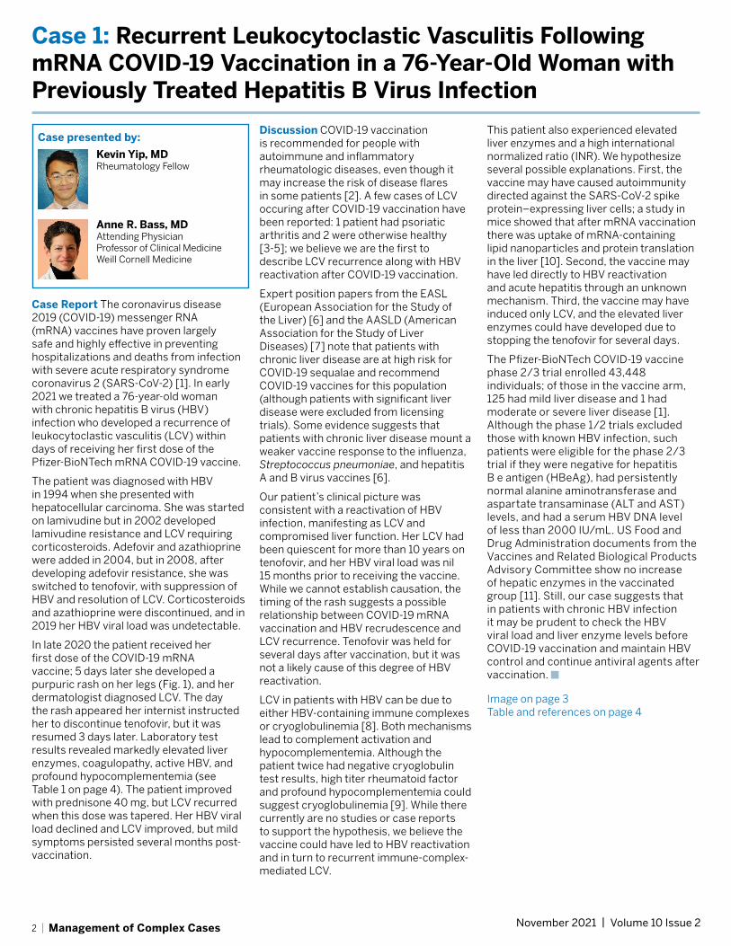
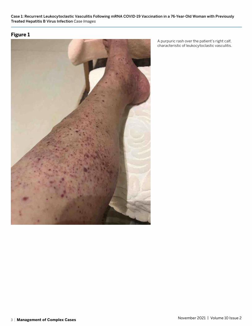
In late 2020 the patient received her first dose of the COVID-19 mRNA vaccine; 5 days later she developed a purpuric rash on her legs (Fig. 1), and her dermatologist diagnosed LCV. The day the rash appeared her internist instructed her to discontinue tenofovir, but it was resumed 3 days later. Laboratory test results revealed markedly elevated liver enzymes, coagulopathy, active HBV, and profound hypocomplementemia (see Table 1 on page 4). The patient improved with prednisone 40 mg, but LCV recurred when this dose was tapered. Her HBV viral load declined and LCV improved, but mild symptoms persisted several months post-vaccination.
Discussion COVID-19 vaccination is recommended for people with autoimmune and inflammatory rheumatologic diseases, even though it may increase the risk of disease flares in some patients [2]. A few cases of LCV occuring after COVID-19 vaccination have been reported: 1 patient had psoriatic arthritis and 2 were otherwise healthy [3-5]; we believe we are the first to describe LCV recurrence along with HBV reactivation after COVID-19 vaccination.
Expert position papers from the EASL (European Association for the Study of the Liver) [6] and the AASLD (American Association for the Study of Liver Diseases) [7] note that patients with chronic liver disease are at high risk for COVID-19 sequalae and recommend COVID-19 vaccines for this population (although patients with significant liver disease were excluded from licensing trials). Some evidence suggests that patients with chronic liver disease mount a weaker vaccine response to the influenza, Streptococcus pneumoniae, and hepatitis A and B virus vaccines [6].
Our patient’s clinical picture was consistent with a reactivation of HBV infection, manifesting as LCV and compromised liver function. Her LCV had been quiescent for more than 10 years on tenofovir, and her HBV viral load was nil 15 months prior to receiving the vaccine. While we cannot establish causation, the timing of the rash suggests a possible relationship between COVID-19 mRNA vaccination and HBV recrudescence and LCV recurrence. Tenofovir was held for several days after vaccination, but it was not a likely cause of this degree of HBV reactivation.
LCV in patients with HBV can be due to either HBV-containing immune complexes or cryoglobulinemia [8]. Both mechanisms lead to complement activation and hypocomplementemia. Although the patient twice had negative cryoglobulin test results, high titer rheumatoid factor and profound hypocomplementemia could suggest cryoglobulinemia [9]. While there currently are no studies or case reports to support the hypothesis, we believe the vaccine could have led to HBV reactivation and in turn to recurrent immune-complex-mediated LCV.
This patient also experienced elevated liver enzymes and a high international normalized ratio (INR). We hypothesize several possible explanations. First, the vaccine may have caused autoimmunity directed against the SARS-CoV-2 spike protein–expressing liver cells; a study in mice showed that after mRNA vaccination there was uptake of mRNA-containing lipid nanoparticles and protein translation in the liver [10]. Second, the vaccine may have led directly to HBV reactivation and acute hepatitis through an unknown mechanism. Third, the vaccine may have induced only LCV, and the elevated liver enzymes could have developed due to stopping the tenofovir for several days.
The Pfizer-BioNTech COVID-19 vaccine phase 2/3 trial enrolled 43,448 individuals; of those in the vaccine arm, 125 had mild liver disease and 1 had moderate or severe liver disease [1]. Although the phase 1/2 trials excluded those with known HBV infection, such patients were eligible for the phase 2/3 trial if they were negative for hepatitis B e antigen (HBeAg), had persistently normal alanine aminotransferase and aspartate transaminase (ALT and AST) levels, and had a serum HBV DNA level of less than 2000 IU/mL. US Food and Drug Administration documents from the Vaccines and Related Biological Products Advisory Committee show no increase of hepatic enzymes in the vaccinated group [11]. Still, our case suggests that in patients with chronic HBV infection it may be prudent to check the HBV viral load and liver enzyme levels before COVID-19 vaccination and maintain HBV control and continue antiviral agents after vaccination. ■Image on page 3 Table and references on page 4
2 | Management of Complex Cases November 2021 | Volume 10 Issue 2
Case 1: Recurrent Leukocytoclastic Vasculitis Following mRNA COVID-19 Vaccination in a 76-Year-Old Woman with Previously Treated Hepatitis B Virus Infection Case Images
Figure 1A purpuric rash over the patient’s right calf, characteristic of leukocytoclastic vasculitis.
3 | Management of Complex Cases November 2021 | Volume 10 Issue 2
Case 1: Recurrent Leukocytoclastic Vasculitis Following mRNA COVID-19 Vaccination in a 76-Year-Old Woman with Previously Treated Hepatitis B Virus Infection Table and references
Table 1Timeline and laboratory results, before and after first dose of the Pfizer-BioNTech mRNA COVID-19 vaccine
16 months prior to vaccine
5 weeks prior to vaccine
20 days prior to vaccine
Day 0: COVID-19 vaccine
Day 4 Day 9 Day 13 Day 21 Day 27 Day 39 Day 55 Day 71
Clinical event LCV onset LCV ongoing
LCV ongoing
LCV worse
LCV fading, no new lesions
LCV flare LCV ongoing
LCV improved
Tenofovir (mg/day)
300 300 300 300 Withheld Resumed 300
300 300 300 300 300 300
Prednisone (mg/day)
40 60 40 2.5 ->10 10 10
Alkaline phosphatase (IU/L)
66 71 99 247 245 118 168 188 177 188
Aspartate transaminase (IU/L)
29 28 335 495 85 45 58 40 58
Alanine aminotransferase (IU/L)
21 11 13 260 314 56 56 39 56
INR 1.0 1.08 1.1 6 1.1 0.6 1.02
Complement 3 (mg/dL)
100 91.2 67 69 76 67.1
Complement 4 (mg/dL)
22 16.5 <2 <2 <2 <2
HBV viral load Not Det 86,500 35,000 18,000 6,380 1,690 1,060
Other RF 19 IU/L
RF 28.8 IU/L
Cryo negative
RF 579 IU/mL
RF 105 IU/mL, Cyro negative
RF rheumatoid factor, Cryo cryoglobulin, LCV leukocytoclastic vasculitis, INR international normalized ratio, HBV hepatitis B virus, Not Det not detected
REFERENCES:
1. Polack FP, Thomas SJ, Kitchin N, et al.; C4591001 Clinical Trial Group. Safety and efficacy of the BNT162b2 mRNA Covid-19 Vaccine. N Engl J Med. 2020;383(27):2603–2615.
2. Curtis JR, Johnson SR, Anthony DD, et al. American College of Rheumatology Guidance for COVID-19 Vaccination in Patients With Rheumatic and Musculoskeletal Diseases: Version 3. Arthritis Rheumatol. 2021 Aug 4. doi: 10.1002/art.41928.
3. Cohen SR, Prussick L, Kahn JS, Gao DX, Radfar A, Rosmarin D. Leukocytoclastic vasculitis flare following the COVID-19 vaccine. Int J Dermatol. 2021 Aug;60(8):1032-1033. doi: 10.1111/ijd.15623
4. Erler, A, Fiedler, J, Koch, A., Schütz, A. and Heldmann, F. A case of leukocytoclastic vasculitis after vaccination with a SARS-CoV2-vaccine – a case report. Arthritis Rheumatol. 2021. Accepted Author Manuscript. https://doi.org/10.1002/art.41910
5. Bostan E, Gulseren D, Gokoz O. New-onset leukocytoclastic vasculitis after COVID-19 vaccine. Int J Dermatol. 2021 Jul 9. doi: 10.1111/ijd.15777.
6. Bornberg M, Buti M, Eberhardt CS, Grossi PA, Shouval D. EASL position paper on the use of COVID-19 vaccines in patients with chronic liver diseases, hepatobiliary cancer and liver transplant recipients. J Hepatol. 2021 Apr;74(4):944–951.
7. Fix OK, Blumberg EA, Chang KM, et al; AASLD COVID-19 Vaccine Working Group. AASLD Expert Panel Consensus Statement: Vaccines to Prevent COVID-19 Infection in Patients with Liver Disease. Hepatology. 2021 Feb 12:10.1002/hep.31751. doi: 10.1002/hep.31751
8. Sunderkötter C, Bonsmann G, Sindrilaru A, Luger T. Management of leukocytoclastic vasculitis. J Dermatolog Treat. 2005;16(4):193–206.
9. Chen KR, Carlson JA. Clinical approach to cutaneous vasculitis. Am J Clin Dermatol. 2008;9(2):71–92.
10. Pardi N, Tuyishime S, Muramatsu H, et al Expression kinetics of nucleoside-modified mRNA delivered in lipid nanoparticles to mice by various routes. J Control Release. 2015;217:345–351.
11. Vaccines and Related Biological Products Advisory Committee. December 10, 2020, meeting: sponsor briefing document. Silver Spring, MD: US Department of Health and Human Services, Food and Drug Administration; 2020. https://www.fda.gov/media/144246/download
4 | Management of Complex Cases November 2021 | Volume 10 Issue 2
Case 2: Incident Rheumatoid Arthritis in a 64-Year-Old Woman with a Prosthetic Joint
Case presented by:
Diane Zisa, MD Rheumatology Fellow
Roberto A. Garcia, MD Associate Attending Pathologist Associate Professor of Clinical Pathology and Laboratory Medicine Weill Cornell Medicine
Susan M. Goodman, MD Attending Physician Professor of Clinical Medicine Weill Cornell Medicine
Case Report A 64-year-old woman presented 16 months after bilateral total knee arthroplasty (TKA) with acute pain and swelling in her left knee that began during exercise and worsened; she also developed fatigue and malaise. X-rays demonstrated a well-fixed TKA in good position. Laboratory test results revealed an erythrocyte sedimentation rate (ESR) of 128 mm/hr and a C-reactive protein (CRP) level of 5.8 mg/dL, mild anemia and thrombocytosis, and a normal white blood cell (WBC) count. Aspiration of the left knee yielded opaque fluid with an elevated WBC count of 17,457/µL (89% neutrophils) and a negative Gram stain without crystals.
Acute periprosthetic joint infection (PJI) was the presumed diagnosis, prompting urgent irrigation and debridement of the left knee with a liner exchange. Aspiration of her asymptomatic right knee was also performed, given the concern for a hematogenous infection, and revealed a WBC count of 6,700/µL (62% neutrophils). She began antibiotics (daptomycin and ceftriaxone) for an anticipated 6-week course, without significant improvement. Synovial fluid samples taken from both knees were cultured and found to be negative for bacteria, acid-fast bacilli, and fungi.
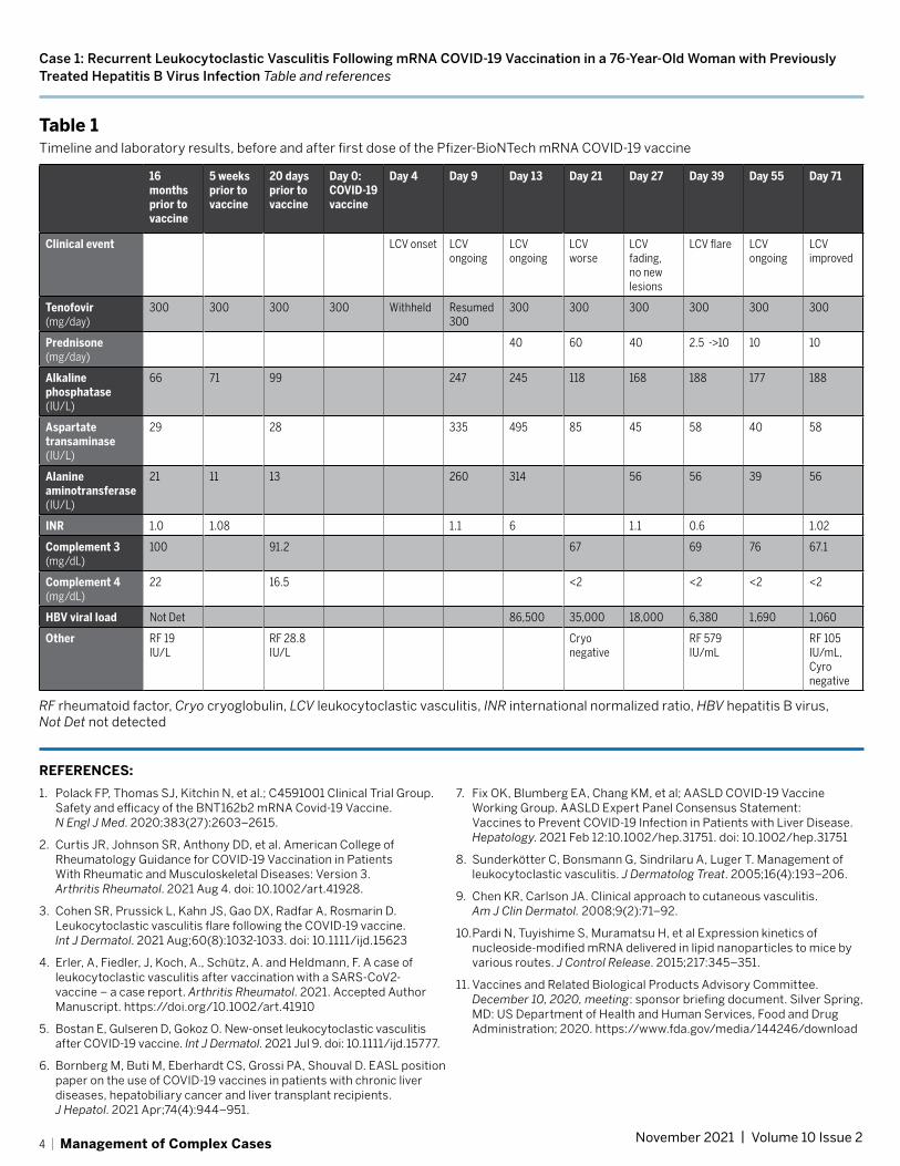
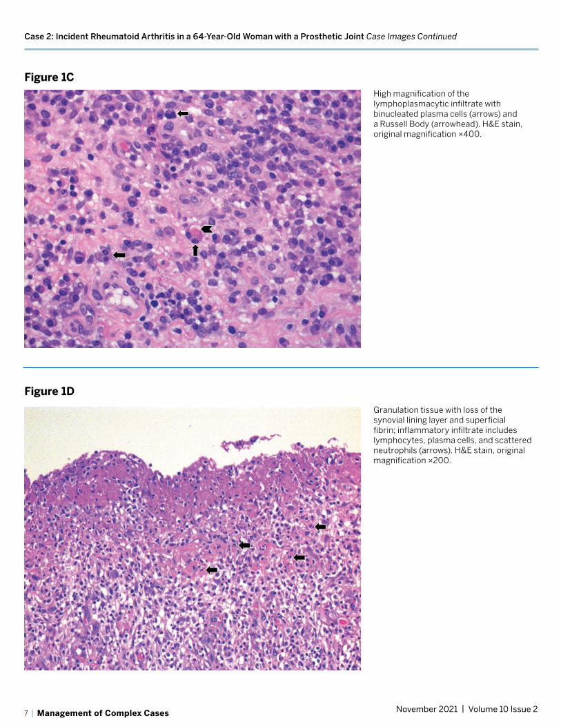
Histopathologic examination of the left knee tissue showed a proliferative and exudative synovitis with lymphoplasmacytic inflammation and scattered superficial neutrophils (Fig. 1A, 1B), as well as binucleated plasma cells and Russell bodies (Fig. 1C). These findings were suggestive of rheumatoid arthritis (RA).
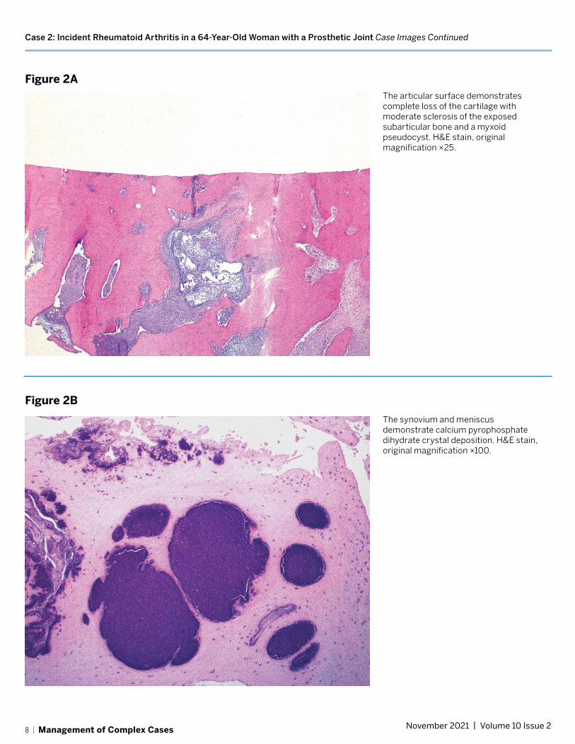
Focal granulation tissue with loss of the synovial lining layer and increased numbers of superficial neutrophils were suggestive but not diagnostic of infection (Fig. 1D). Review of the histopathology from the initial surgery confirmed degenerative joint disease without inflammatory features and moderate calcium pyrophosphate deposition (Fig. 2A, 2B).
Additional serologic testing revealed a high-titer rheumatoid factor (RF) of 168.3 IU/mL and an anticyclic citrullinated peptide (anti-CCP) antibody level of more than 250 units, with persistent elevations in ESR and CRP. A rheumatology review of her history revealed no symptoms suggestive of inflammatory joint disease. However, based on serology and histopathology results and subsequent swelling of several metacarpophalangeal and proximal interphalangeal joints, RA was diagnosed. Antibiotics were stopped 4 weeks after surgery and methotrexate was begun. She has had a partial response to antirheumatic therapy, and her regimen is being adjusted to achieve better control.
Discussion It is challenging to differentiate PJI from a flare of inflammatory arthritis, given the overlapping clinical and laboratory criteria for each diagnosis, especially when definitive microbiologic data are not available [1,2,3]. Furthermore, patients with rheumatoid arthritis are at increased risk for PJI and may have more frequent culture-negative infections [3]. Following any arthroplasty, elevated serum inflammatory markers and synovial fluid leukocyte levels are always concerning for PJI. Microbiology results can take time; because a delay in diagnosis may worsen outcomes, surgery should be performed promptly.
The patient’s initial monoarticular pain and swelling in a prosthetic joint was appropriately treated urgently as a presumed PJI. Histopathologic examination of the surgical specimen, showing polymorphonuclear leukocytes superimposed on the chronic inflammatory changes, was critical in pointing to the diagnosis of inflammatory arthritis. The RA diagnosis was corroborated by the patient’s lack of response to antibiotics, the subsequent serology results, and her clinical evolution to polyarthritis.
We know of no other reported cases of incident RA in a prosthetic joint. This case illustrates the ambiguity that often confronts rheumatologists. Tools to differentiate an inflammatory arthritis flare from PJI are needed to avoid the morbidity and expense associated with delay in making a definitive diagnosis and treating PJI. At HSS, research is under way to investigate serum and synovial biomarkers and next-generation microbial sequencing in arthroplasty patients with and without inflammatory arthritis to help diagnose PJI expeditiously in patients with inflammatory arthritis. ■Images on pages 6–8
REFERENCES:
1. Premkumar A, Morse K, Levack AE, Bostrom MP, Carli AV. Periprosthetic joint infection in patients with inflammatory joint disease: prevention and diagnosis. Curr Rheumatol Rep. 2018;20(11):68.
2. Mirza SZ, Richardson SS, Kahlenberg CA, et al. Diagnosing prosthetic joint infections in patients with inflammatory arthritis: a systematic literature review. J Arthroplasty. 2019;34(5):10321036.e2.
3. Goodman S, Kapadia M, Miller A, et al. Clinical features of prosthetic joint infections in patients with rheumatic diseases vs osteoarthritis [abstract]. Arthritis Rheumatol. 2019; 71 (suppl 10). Presented at: 2019 ACR/ARP Annual Meeting; November 8-13, 2019; Atlanta, GA.
5 | Management of Complex Cases November 2021 | Volume 10 Issue 2

Case 2: Incident Rheumatoid Arthritis in a 64-Year-Old Woman with a Prosthetic Joint Case Images
Figure 1ASynovial tissue with marked lymphoplasmacytic inflammation, abundant superficial fibrinous exudate, and scattered neutrophils (arrows). Hematoxylin-eosin (H&E) stain, original magnification ×200.
Figure 1B
The synovial lining layer is well preserved in this area with marked lymphoplasmacytic inflammation and increased number of superficial neutrophils (arrows). H&E stain, original magnification ×200.
6 | Management of Complex Cases November 2021 | Volume 10 Issue 2
Case 2: Incident Rheumatoid Arthritis in a 64-Year-Old Woman with a Prosthetic Joint Case Images Continued
Figure 1CHigh magnification of the lymphoplasmacytic infiltrate with binucleated plasma cells (arrows) and a Russell Body (arrowhead). H&E stain, original magnification ×400.
Figure 1D
Granulation tissue with loss of the synovial lining layer and superficial fibrin; inflammatory infiltrate includes lymphocytes, plasma cells, and scattered neutrophils (arrows). H&E stain, original magnification ×200.
7 | Management of Complex Cases November 2021 | Volume 10 Issue 2
Case 2: Incident Rheumatoid Arthritis in a 64-Year-Old Woman with a Prosthetic Joint Case Images Continued
Figure 2AThe articular surface demonstrates complete loss of the cartilage with moderate sclerosis of the exposed subarticular bone and a myxoid pseudocyst. H&E stain, original magnification ×25.
Figure 2B
The synovium and meniscus demonstrate calcium pyrophosphate dihydrate crystal deposition. H&E stain, original magnification ×100.
8 | Management of Complex Cases November 2021 | Volume 10 Issue 2

Case 3: An Overlap of Drug-Induced Subacute Cutaneous Lupus Erythematosus and Dermatomyositis in an 80-Year-Old Woman
Case presented by:
David R. Fernandez, MD, PhD Assistant Attending Physician Assistant Professor of Medicine Weill Cornell Medicine
Case Report An 80-year-old woman developed a pruritic rash on her scalp, prompting a dermatology evaluation after several weeks. A biopsy found interface dermatitis and prominent tissue eosinophils, perhaps consistent with a drug reaction. Her past medical history included Hashimoto’s thyroiditis, hypertension, knee osteoarthritis, collagenous colitis, mitral regurgitation requiring mitral valve replacement, atrial fibrillation for which she was taking coumadin, and a distant history of breast cancer, with no evidence of active disease. Several medications were discontinued, including diltiazem 2 months after the rash appeared, losartan–hydrochlorothiazide a month after that, followed by atorvastatin, guaifenesin, loperamide, and omeprazole.
Nonetheless, her rash progressed. She developed periorbital edema and erythema, rash of the anterior chest and lateral thighs, periungual erythema, diffuse erythema of the dorsum of her hands, and coarseness and fissuring of the skin of her lateral fingers. Hydroxychloroquine 200 mg twice daily was added 5 months after symptoms appeared, with little benefit. Given the increasingly classic presentation of the rash, she was thought to have dermatomyositis (DM). A skin biopsy taken from the thigh showed interface dermatitis with karyorrhexis, a finding consistent with subacute cutaneous lupus erythematosus (SCLE), although DM or a drug reaction could not be ruled out. Serologic testing was notable for a positive antinuclear antibody titer (1:2560) and antibodies to single-stranded DNA and thyroglobulin, but testing was otherwise negative, including for anti-Ro/SSA and anti-histone antibodies and a limited myositis panel.
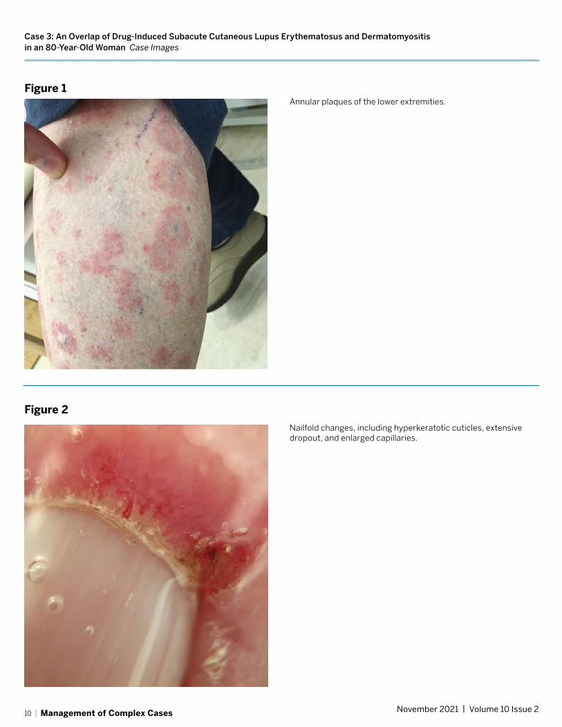
A re-introduction of diltiazem several months later to treat hypertension resulted in a dramatic surge in her pruritic rash, with more diffuse erythema covering most of her body. The rash was mostly confluent in the upper body but consisted of coalescing papules and annular plaques of the lower extremities (Fig. 1). She also reported
severe fatigue and subtle weakness, most noticeable as modest difficulty in climbing steps and dyspnea on exertion.
Examination confirmed proximal muscle weakness, rash, and marked nailfold capillary changes (Fig. 2), with hyperkeratotic cuticles, extensive dropout, and enlarged capillaries. Laboratory testing revealed normal creatine kinase but elevated lactate dehydrogenase levels, lymphopenia, and mild eosinophilia.
Based on these findings, she was diagnosed with DM and a second, distinct rash provoked by diltiazem exposure, suggestive of SCLE. She was treated with tapering prednisone, beginning at 40 mg daily, and hydroxychloroquine.
Later, the annular rash and peripheral eosinophilia faded with diltiazem discontinuation, but the DM rashes and weakness persisted. Two months later she was found to be positive for antibodies to transcription intermediary factor 1-γ, which was not part of the initial myositis assessment. This antibody can be associated with malignancy in patients with DM, but no cancer has been identified on subsequent annual screenings. Four years after her initial symptoms, the patient is still experiencing mild skin and muscle symptoms despite prednisone 5 mg daily and hydroxychloroquine. Trials of azathioprine and methotrexate were complicated by repeated infections and so were discontinued. She is also managing her symptoms through structured physical therapy.
Discussion This case presents an interesting overlap of SCLE and DM. Drug-induced lupus is a well-characterized phenomenon, classically associated with anti-histone antibodies; management involves removing the offending agent. SCLE can be associated with medications, most commonly calcium channel blocker therapy [1].
Drug-induced DM is less well characterized but can occur in the setting of a variety of medications [2], including statins, hydroxyurea, or supplements [3], although diltiazem has not been reported as a potential cause. In this case, diltiazem may have had a role in the onset of DM and SCLE, although removing it did not resolve all clinical findings, even with the addition of hydroxychloroquine and prednisone.
This case highlights the value of nailfold capillary assessment in the diagnosis of DM [4], as some other features of the patient’s presentation were suggestive of systemic lupus erythematosus (SLE), and skin biopsy was insufficient for distinguishing between SLE and DM. Additionally, while the annular rash of the lower extremities appearing as an atypical manifestation of DM cannot be ruled out, its resolution with diltiazem cessation, along with the persistence of other DM manifestations, suggests that these were separate phenomena. ■Images on page 10
REFERENCES:
1. Crowson AN, Magro CM. Subacute cutaneous lupus arising in the setting of calcium channel blocker therapy. Hum Pathol. 1997;28(1):67–73.
2. Seidler AM, Gottlieb AB. Dermatomyositis induced by drug therapy: A review of case reports. J Am Acad Dermatol. 2008;59(5):872–880.
3. Zeidi M, Chansky PB, Werth VP. Acute onset/flares of dermatomyositis following ingestion of IsaLean herbal supplement: Clinical and immunostimulatory findings. J Am Acad Dermatol. 2019;80(3):801–804.
4. Cassius C, Le Buanec H, Bouaziz JD, Amode R. Biomarkers in adult dermatomyositis: tools to help the diagnosis and predict the clinical outcome. J Immunol Res. 2019;2019:9141420.
9 | Management of Complex Cases November 2021 | Volume 10 Issue 2
Case 3: An Overlap of Drug-Induced Subacute Cutaneous Lupus Erythematosus and Dermatomyositis in an 80-Year-Old Woman Case Images
Figure 1Annular plaques of the lower extremities.
Figure 2
Nailfold changes, including hyperkeratotic cuticles, extensive dropout, and enlarged capillaries.
10 | Management of Complex Cases November 2021 | Volume 10 Issue 2

Case 4: Kikuchi–Fujimoto Disease in a 6-Year-Old Boy with a History of Macrophage Activation Syndrome
Case presented by:
Lauren Robinson, MD Rheumatology Fellow
Sarah Faith Taber, MD Assistant Attending Physician Assistant Professor of Pediatrics Weill Cornell Medicine
Case Report A 6-year-old boy with a remote history of massive cervical lymphadenopathy and macrophage activation syndrome (MAS) presented with 1 month of fever and cervical lymphadenopathy suspicious for recurrent MAS. After his birth he had been well until age 2, when he developed fever, rash, lymphadenopathy, elevated inflammatory markers, and cytopenias. Extensive infectious and oncologic workups, including cervical lymph node biopsy, at that time were unrevealing. His symptoms resolved with treatment with steroids and anakinra, which were discontinued within 5 months.
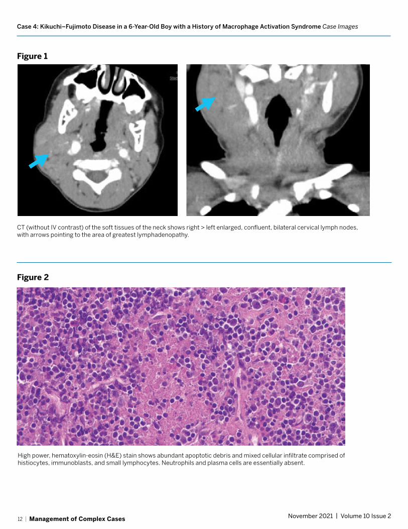
Having been off medication for several years, at age 6 he developed daily fever and massive bilateral cervical lymphadenopathy. Laboratory test results showed elevated erythrocyte sedimentation rate (ESR) (peak: 130 mm/hr), hyperferritinemia (peak: 1228 µg/L), and mild cytopenias (hemoglobin nadir: 8.5 g/dL; platelet nadir: 191 × 109/L; white blood cell count nadir: 2.9 × 10⁹/L). Computed tomography (CT) scanning
lymphadenopathy with a confluent of the neck showed bilateral cervical
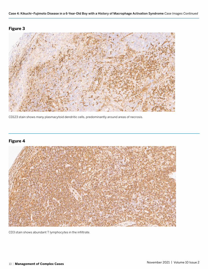
appearance (Fig. 1). Infectious workup was again unrevealing, and repeat excisional cervical node biopsy was performed. The pathology findings showed areas of necrosis with surrounding CD123+ plasmacytoid dendritic cells and mixed cellular infiltrates, consistent with Kikuchi–Fujimoto disease (Figs. 2, 3, 4). Review of his initial biopsy performed at age 2 showed similar mixed cellular infiltrate with high staining for CD123+ plasmacytoid dendritic cells but did not show the necrosis characteristic of Kikuchi–Fujimoto disease. It was determined that his current presentation was likely a recurrence of his earlier illness, most consistent with recurrent Kikuchi–Fujimoto disease
complicated by MAS. Genetic testing for underlying autoinflammatory disease or immunodeficiency was unrevealing.
He was treated initially with 2 mg/kg prednisone daily and 100 mg anakinra daily for 3 days, with rapid resolution of fever and stabilization of cytopenias. He was discharged on a slow steroid taper, but his fever returned and daily anakinra was re-initiated and titrated up to 100 mg BID. He subsequently remained afebrile; all laboratory parameters normalized and lymphadenopathy remitted, allowing for gradual discontinuation of steroids. After 2 months, treatment with hydroxychloroquine was begun for recurrent Kikuchi–Fujimoto disease. After an additional 6 months he remains well and is tolerating a gradual taper of anakinra.
Discussion Kikuchi–Fujimoto disease is seen increasingly in children, who often do not fit the classical disease presentation. Among children there is predominance in boys (1.4:1 male to female) compared to a predominance in women among adults. Additionally, children with Kikuchi–Fujimoto disease are more likely to present with fever and leukopenia and less likely to have a positive antinuclear antibody test result than adults with the disease [1].
It is hypothesized that Kikuchi–Fujimoto disease is driven by a cytotoxic T-cell mediated inflammatory response to a viral trigger in genetically predisposed individuals. About 3% of patients later develop systemic autoimmune disease, which may be triggered by immune system exposure to autoantigens via the apoptotic debris created in affected lymph nodes [2]. Diagnosis is based on excisional lymph node biopsy, which shows areas of necrosis surrounded by histiocytes and plasmacytoid dendritic cells.
Kikuchi–Fujimoto disease is widely believed to be a benign and self-limited process. However, retrospective studies have shown that up to 30% of hospitalized patients with this diagnosis may develop MAS, often requiring immunosuppressive therapy [3,4].
Kikuchi–Fujimoto disease in children is often recurrent (10%-42% of cases), with up to 3 recurrences in a single patient [5]. Treatment consists of supportive care for mild disease and glucocorticoids for severe or persistent disease, with no treatment trials available.
Hydroxychloroquine has been suggested for use in severe or recurrent disease in children [6]. ■Image on pages 12–13
REFERENCES:
1. Kim TY, Ha KS, Kim Y, Lee J, Lee K, Lee J. Characteristics of Kikuchi-Fujimoto disease in children compared with adults. Eur J Pediatr. 2014;173(1):111–116.
2. Ogata S, Bando Y, Saito N, Katsuoka K, Ishii M. Kikuchi-Fujimoto disease developed into autoimmune disease: a report of two cases. Mod Rheumatol. 2010;20(3):301–305.
3. Ahn SS, Lee B, Kim D, et al. Evaluation of macrophage activation syndrome in hospitalised patients with Kikuchi-Fujimoto disease based on the 2016 EULAR/ACR/PRINTO classification criteria. PLoS One. 2019;14(7):e0219970.
4. Duan W, Xiao ZH, Yang LG, Luo HY. Kikuchi’s disease with hemophagocytic lymphohistiocytosis: a case report and literature review. Medicine (Baltimore). 2020;99(51):e23500.
5. Selvanathan SN, Suhumaran S, Sahu VK, Chong CY, Tan NW, Thoon KC. Kikuchi-Fujimoto disease in children. J Paediatr Child Health. 2020;56(3):389–393.
6. Lin YC, Huang HH, Nong BR, Liu PY, Chen YY, Huang YF, Chiou YH, Lee HS. Pediatric Kikuchi-Fujimoto disease: a clinicopathologic study and the therapeutic effects of hydroxychloroquine. J Microbiol Immunol Infect. 2019;52(3):395–401.
11 | Management of Complex Cases November 2021 | Volume 10 Issue 2
Case 4: Kikuchi–Fujimoto Disease in a 6-Year-Old Boy with a History of Macrophage Activation Syndrome Case Images
Figure 1
CT (without IV contrast) of the soft tissues of the neck shows right > left enlarged, confluent, bilateral cervical lymph nodes, with arrows pointing to the area of greatest lymphadenopathy.
Figure 2
High power, hematoxylin-eosin (H&E) stain shows abundant apoptotic debris and mixed cellular infiltrate comprised of histiocytes, immunoblasts, and small lymphocytes. Neutrophils and plasma cells are essentially absent.
12 | Management of Complex Cases November 2021 | Volume 10 Issue 2
Case 4: Kikuchi–Fujimoto Disease in a 6-Year-Old Boy with a History of Macrophage Activation Syndrome Case Images Continued
Figure 3
CD123 stain shows many plasmacytoid dendritic cells, predominantly around areas of necrosis.
Figure 4
CD3 stain shows abundant T lymphocytes in the infiltrate.
13 | Management of Complex Cases November 2021 | Volume 10 Issue 2

Management of Complex Cases Grand Rounds from HSS | Rheumatology
HSS Editorial BoardEditorsKarmela Kim Chan, MDAssistant Attending PhysicianAssistant Professor of MedicineWeill Cornell MedicineMemorial Sloan Kettering Cancer Center
David M. Dines, MDAttending Orthopaedic Surgeon Clinical Professor of Orthopaedic SurgeryWeill Cornell Medicine
ConsultantsAndy O. Miller, MDChief, Division of Infectious DiseasesAssociate Attending PhysicianAssociate Professor of Clinical MedicineWeill Cornell Medicine
Sarah Faith Taber, MDAssistant Attending PhysicianAssistant Professor of PediatricsWeill Cornell Medicine
BoardDalit Ashany, MDAssistant Attending PhysicianAssistant Professor of Clinical MedicineWeill Cornell Medicine
Anne R. Bass, MDAttending PhysicianProfessor of Clinical MedicineWeill Cornell Medicine
Bryan T. Kelly, MD, MBASurgeon-in-Chief and Medical DirectorChief Emeritus, Sports Medicine InstituteAttending Orthopaedic SurgeonProfessor of Orthopaedic SurgeryWeill Cornell Medicine
Carolyn M. Sofka, MD, FACRAttending RadiologistDirector of EducationDepartment of Radiology and ImagingProfessor of Radiology Weill Cornell Medicine
Laura Robbins, DSWSenior Vice PresidentEducation Institute & Global PartnershipsAssociate ProfessorGraduate School of Medical SciencesClinical Epidemiology andHealth Services Research Weill Cornell Medicine
Joy Jacobson, MFADirector, Academic PublicationsManaging Editor, HSS JournalHSS Education Institute
Design/ProductionMarcia EnnisSenior Creative DirectorEducation Marketing & Digital CommunicationsHSS Education Institute
Randy HawkeAssociate DirectorEducation Marketing & Digital CommunicationsHSS Education Institute
Produced by Education Marketing & Digital Communications
©2021 Hospital for Special Surgery. 535 East 70th Street, New York, NY 10021. Hospital for Special Surgery, HSS and the HSS logo are trademarks or registered trademarks of Hospital for Special Surgery in the United States and other countries.
HSS Education Institute
14 | Management of Complex Cases November 2021 | Volume 10 Issue 2