complex cases in_peripheral_vascular_interventions
TRANSCRIPT

Complex Cases in Peripheral Vascular Interventions
Edited byMartin Schillinger
Erich Minar
Co
mp
lex C
ase
s in Perip
hera
l Va
scula
r Interve
ntions
Sch
illing
er •
Min
ar
www.informahealthcare.com
Telephone House, 69-77 Paul Street, London EC2A 4LQ, UK
52 Vanderbilt Avenue, New York, NY 10017, USA
About the book
This new title provides a systematic review of complex cases in peripheral vascular interventions (PVI), a minimal invasive endovascular approach to treating peripheral arteries. This increasingly evolving area in interventional therapies offers reduced risk for complications compared to conventional surgery and takes advantage of advanced techniques to treat more complex lesions in severely diseased patients.
This book edited by two highly respected professors from the University of Vienna, Medical School describes typical and atypical complex cases for all peripheral vessel areas, and outlines methods to successfully and safely handle these interventions. The text is divided into two sections. The first reviews general aspects on managing complex patients in PVI, and the second discusses complex cases in specific vessel areas, examining the interventions from puncture to closure, head to toe. Each of the chapters in this section is further divided into three parts and provides:
• An introduction to each vessel, specific information on thecomplexities of this lesion and the hazards of treatment, and a checklist for readers of recommended equipment when treating this specific vessel area.
• Selected teaching cases, whereby specific vessel areas aredescribed step-by-step.
• A summary of the key issues for the successful and safetreatment of complex lesions in this vessel area.
Complex Cases in Peripheral Vascular Interventions offers readers the opportunity to tap into the knowledge of those working on complex cases in this cutting edge field and receive both theoretical information and practical tips on how to approach specific vascular lesions. This book is a highly recommended read for beginners in endovascular interventions who want to get an impression on the advanced scope of treatment options as well as for experienced interventionists who want to extend their knowledge on difficult cases.
About the editors
MArTIN SCHILLINgEr, MD is a Professor and Senior Physician at the Department of Internal Medicine at general Hospital Vienna, Austria. Professor Schillinger gained his Medical Degree with honours in 1998 from the University of Vienna and in 2002 was appointed Associate Professor at the University. He has held his position at general Hospital Vienna since July 2008 where he also specialises in cardiology and angiology. Professor Schillinger has been lead investigator of numerous clinical trials and collaborated on many multi-center trials. He is on the advisory board of Boston Scientific, Abbott Vascular and Amaranth, among others, and editorial board of numerous societies and journals. Professor Schillinger has published and presented his work extensively.
ErICH MINAr, MD is a Professor of Internal Medicine at the Medical University of Vienna. He graduated from the Medical School at the University of Vienna in 1977 and following a position as an assistant in Internal Medicine at the University Clinic Vienna, was appointed as Assistant Professor at the Department of Angiology in 1984, and Professor of Internal Medicine in 1993. Since 1998 he has served as Deputy Head of the Department of Angiology at the University. Professor Minar has published more than 350 papers in peer-reviewed journals. He is co-editor of the European Journal of Vascular Diseases and editor of several books, including Complications in Peripheral Vascular Interventions (2007).
Complex Cases in Peripheral Vascular Interventions

fm IHBK065-Minar May 5, 2010 21:13 Specs: 7x10 tight 7in×10in Char Count=
Complex Cases inPeripheral Vascular
Interventions

fm IHBK065-Minar May 5, 2010 21:13 Specs: 7x10 tight 7in×10in Char Count=

fm IHBK065-Minar May 5, 2010 21:13 Specs: 7x10 tight 7in×10in Char Count=
Complex Cases inPeripheral Vascular
Interventions
Edited by
Martin SchillingerMedical University of Vienna
General HospitalVienna, Austria
Erich MinarMedical University of Vienna
General HospitalVienna, Austria
fm IHBK065-Minar May 5, 2010 21:13 Specs: 7x10 tight 7in×10in Char Count=
This edition published in 2010 by Informa Healthcare, Telephone House, 69-77 Paul Street, London EC2A 4LQ,UK.
Simultaneously published in the USA by Informa Healthcare, 52 Vanderbilt Avenue, 7th Floor, New York,NY 10017, USA.
Informa Healthcare is a trading division of Informa UK Ltd. Registered Office: 37–41 Mortimer Street,London W1T 3JH, UK. Registered in England and Wales number 1072954.
C© 2010 Informa Healthcare, except as otherwise indicated
No claim to original U.S. Government works
Reprinted material is quoted with permission. Although every effort has been made to ensure that all owners ofcopyright material have been acknowledged in this publication, we would be glad to acknowledge in subsequentreprints or editions any omissions brought to our attention.
All rights reserved. No part of this publication may be reproduced, stored in a retrieval system, or transmitted, inany form or by any means, electronic, mechanical, photocopying, recording, or otherwise, unless with the priorwritten permission of the publisher or in accordance with the provisions of the Copyright, Designs and PatentsAct 1988 or under the terms of any licence permitting limited copying issued by the Copyright Licensing Agency,90 Tottenham Court Road, London W1P 0LP, UK, or the Copyright Clearance Center, Inc., 222 Rosewood Drive,Danvers, MA 01923, USA (http://www.copyright.com/ or telephone 978-750-8400).
Product or corporate names may be trademarks or registered trademarks, and are used only for identificationand explanation without intent to infringe.
This book contains information from reputable sources and although reasonable efforts have been made to publishaccurate information, the publisher makes no warranties (either express or implied) as to the accuracy or fitnessfor a particular purpose of the information or advice contained herein. The publisher wishes to make it clearthat any views or opinions expressed in this book by individual authors or contributors are their personal viewsand opinions and do not necessarily reflect the views/opinions of the publisher. Any information or guidancecontained in this book is intended for use solely by medical professionals strictly as a supplement to the medicalprofessional’s own judgement, knowledge of the patient’s medical history, relevant manufacturer’s instructionsand the appropriate best practice guidelines. Because of the rapid advances in medical science, any informationor advice on dosages, procedures, or diagnoses should be independently verified. This book does not indicatewhether a particular treatment is appropriate or suitable for a particular individual. Ultimately it is the soleresponsibility of the medical professional to make his or her own professional judgements, so as appropriately toadvise and treat patients. Save for death or personal injury caused by the publisher’s negligence and to the fullestextent otherwise permitted by law, neither the publisher nor any person engaged or employed by the publishershall be responsible or liable for any loss, injury or damage caused to any person or property arising in any wayfrom the use of this book.
A CIP record for this book is available from the British Library.
Library of Congress Cataloging-in-Publication Data available on application
ISBN-13: 9781841847313
Orders may be sent to: Informa Healthcare, Sheepen Place, Colchester, Essex CO3 3LP, UKTelephone: +44 (0)20 7017 5540Email: [email protected]: http://informahealthcarebooks.com/
For corporate sales please contact: [email protected] foreign rights please contact: [email protected] reprint permissions please contact: [email protected]
Typeset by Aptara, Delhi, IndiaPrinted and bound in the United Kingdom.

fm IHBK065-Minar May 5, 2010 21:13 Specs: 7x10 tight 7in×10in Char Count=
Foreword
Vascular intervention represents a unique corner of medical care. While the potentially anal-ogous field of coronary intervention is practiced by a single, nonsurgical specialty and hasprogressed in a fairly orderly fashion with a reasonably standardized set of techniques and pro-duced a wealth of randomized data, vascular intervention has followed a decidedly differentpath. It is practiced by physicians from at least four different medical, surgical, and radiologicspecialty training backgrounds with variable skill sets that nevertheless converge on a signif-icant overlap of procedures, but which cover a myriad of vascular territories, both visceraland peripheral. And the techniques and tools in vascular intervention have evolved quickly inspite of the challenges of diverse practitioners, and in concert with industry partners working toimprove its tools. As a result, over the past several years, endovascular intervention has evolvedas the preferred first approach in many vascular territories, supplanting decades-old surgicalapproaches. This progress, however, has not produced nearly the same volume or quality ofdata, nor standardization of technique, largely as a result of a lack of a regulatory impetus butit had been related to an overall smaller volume of activity than that of coronary work.
Owing to the rapid pace of the prior and current evolution in this field, the lack of asingular specialty training “track,” the many interventionalists who “grandfathered” into thisfield, the paucity of data to support decision-making in the vascular patient vis-a-vis medicalor surgical options, and fractured training programs often split between endovascular skillsand other specialty training needs (interventional cardiology, vascular surgery, nonvascularinterventional radiology, etc.), the operator practicing endovascular intervention may havelimited exposure to the variety of techniques and approaches available for the care of thevascular patient.
On this background, we are fortunate to have two world-renowned interventionalistswho between them have more than 35 years of experience and nearly 600 publications in thisfield, which in addition to covering intervention in almost every conceivable vascular territory,but also nondevice intervention ranging from the pharmacology through brachytherapy. In aword, Drs. Minar and Schillinger have been at the forefront and instrumental in shaping thisfield through their tireless research and investigation. Here they have assembled an all-stargroup of multidisciplinary interventionalists, and they employ a unique and effective methodof transferring those experts’ knowledge to the reader. While the case-based style of teachingis not new, the editors here have the experts’ focus not only on the specific case at hand butalso the broader aspects of the vascular territory; so often the contextual facet of the potentialintervention is either unclear or not fully considered, which makes this formatting even morevital. In addition, more practical information such as the choice of tools and variety of approachesare detailed. Finally, the authors perform a critique of the case and discuss alternate approachesto complete the case successfully.
The editors have sectioned the book into vascular territories, which help highlight thedistinctive aspects of each intervention. Some will be more sensitive to embolization, othersaggressive to interventional techniques, and some in which residual stenosis may not be con-sidered a critical determinant of success. The tool sets, imaging, access, as well as the frequencyand consequences of adverse outcomes for each are unique and require a nuanced understand-ing. Save for intracranial interventions, this book should enable most operators to approach thesignificant majority of vascular syndromes requiring intervention.

fm IHBK065-Minar May 5, 2010 21:13 Specs: 7x10 tight 7in×10in Char Count=
vi Foreword
Publications like this are important in the field of intervention practiced by such a disparategroup of physicians in order to maximize the transfer of knowledge from among our ranks.Eleanor Roosevelt, herself an interventionalist of sorts, once advised, “Learn from the mistakesof others. You can’t live long enough to make them all yourself.” In this book, the reader will findthat Drs. Minar and Schillinger have assembled a wealth of accumulated knowledge intendedto minimize the interventionalist’s need for discovery at the bedside, and enable them to bemore effective interventionalists with greater options at their disposal. I believe Eleanor wouldhave heartily approved.
William A. Gray, MD

fm IHBK065-Minar May 5, 2010 21:13 Specs: 7x10 tight 7in×10in Char Count=
Preface
Indications for endovascular treatment of peripheral arterial diseases are dramatically expand-ing during the recent years, and peripheral vascular interventions (PVI) today are a well-accepted therapeutic approach for virtually all vessel areas. Rapidly advancing technologiesenable treatment of more and more complex lesions. The minimal invasive approach offers theadvantage of a reduced risk for complications compared to conventional surgery and enablestreatment even of severely diseased patients who otherwise would have been considered unfitfor surgical procedures.
With increasing numbers of heavily diseased patients treated in the cath lab, several cen-ters have gained an exceptional expertise in treatment of complex morphologies. Sophisticatedinterventional strategies and meticulous techniques have been developed to handle morpholo-gies that would have been considered not feasible for PVI in earlier years. Furthermore, industryhas recognized the utmost importance of PVI and meanwhile offers a multitude of technical solu-tions dedicated to specific peripheral vessel areas and specific vascular morphologies. Today,particularly for complex scenarios, the concept of “one fits all” has been abandoned and hasbeen replaced by a patient- and lesion-tailored treatment strategy.
The present book aims to systematically review complex cases in PVI. Typical and atypicalcomplex cases are described for all peripheral vessel areas, and methods how to successfullyand safely handle these interventions are outlined.
The book is divided in two sections:Section I—General considerations: This section briefly reviews general aspects on managing
complex patients in PVI.Section II—Complex Cases in Specific Vessel Areas: Section II covers the interventions from
puncture to closure, from the head to the toe. Each of the chapters on the specific vessel areas isdivided into three parts:
(Part 1) The Introduction to each vessel area addresses the following questions:
(a) What makes a lesion “complex” in this vessel area?(b) What are the clinical indications for endovascular treatment of complex lesions in this
vessel area?(c) What are the potential hazards of treating complex lesions in this vessel area?(d) Which specific interventional strategies can be considered for this vessel area?(e) Which specific interventional tools are available to handle complex lesions in this vessel
area?(f) Summary on technical success rates, acute and long-term outcomes after endovascular
treatment of complex lesions in this vessel area.(g) Checklist for the authors’ recommended equipment for complex interventions in this spe-
cific vessel area.
(Part 2) Selected teaching cases in the specific vessel area are then described step-by-stepfrom puncture to closure addressing the same issues as listed above for the specific cases:
(a) What made the lesion “complex” in this specific case?(b) What was the clinical indication for treatment of this specific case?(c) What were the potential hazards of treating this specific patient and this lesion?(d) Which interventional strategies were considered for this lesion?(e) Which interventional tools were used to handle this lesion?

fm IHBK065-Minar May 5, 2010 21:13 Specs: 7x10 tight 7in×10in Char Count=
viii Preface
(f) Summary: How was the case managed, is there anything that could have been improved,what was the immediate and long-term outcome?
(g) List of equipment used in this case.(h) Key learning issues of this specific case.
(Part 3) Authors’ summary of key issues for successful and safe treatment of complexlesions in this vessel area.
In summary, the book intends to focus on practical tips for the interventionist in the cathlab, to review complex cases and outline the different strategies theoretically and in real-lifecases, thus to share the experience of high-volume interventionists with the reader.
Martin Schillinger, MDErich Minar, MD

fm IHBK065-Minar May 5, 2010 21:13 Specs: 7x10 tight 7in×10in Char Count=
Contents
Foreword William A. Gray . . . . vPreface . . . . viiContributors . . . . xi
Section I: Complex Cases—General Considerations
1. Why Should We Tackle Complex Lesions by Endovascular Techniques? 1Martin Schillinger
2. Pharmacological Prerequisites for Treatment of Complex Patientswith Peripheral Artery Disease 10Erich Minar
3. Preprocedural Imaging in Peripheral Arterial Disease 22Thomas J. Kiernan, Bryan P. Yan, and Michael R. Jaff
Section II: Complex Cases in Specific Vessel Areas
4. Complex Vascular Access and Vessel Closure 41I. Baumgartner, N. Diehm, and D. D. Do
5. Intracranial Interventions 62Celine Rahman, Johanna T. Fifi, and Philip M. Meyers
6. Complex Cases in Vascular Interventions—Carotid Arteries 77Klaus Mathias
7. Subclavian Interventions 88Edward B. Diethrich
8. Vertebral Interventions 102J. Stephen Jenkins
9. Thoracic Aortic Dissection and Nondissective Thoracic Aortic Disease 120Martin Czerny and Michael Grimm
10. Abdominal Aortic Disease 131Yuji Kanaoka, Takao Ohki, and Frank J. Veith
11. Renal Arteries 143Thomas Zeller
12. Chronic Mesenteric Ischemia: Mesenteric Vascular Intervention 158Rajan A. G. Patel, Stephen R. Ramee, and Christopher J. White

fm IHBK065-Minar May 5, 2010 21:13 Specs: 7x10 tight 7in×10in Char Count=
x Contents
13. Leriche’s Disease 172Hans Krankenberg, Thilo Tubler, and Michael Schluter
14. Iliac Arteries 183Patrick Peeters, Jurgen Verbist, Koen Deloose, and Marc Bosiers
15. The Femoropopliteal Segment 205Andrej Schmidt and Dierk Scheinert
16. The Tibioperoneal Segment 220Lanfroi Graziani and Hubert Wallner
17. Dialysis Fistulas 239Patrick Haage and Dierk Vorwerk
18. Venous Interventions for Iliofemoral DVT 257Anthony J. Comerota and Marilyn H. Gravett
19. The Complex Patient with Multivessel Disease 272N. Diehm, Barry Katzen, Jennifer Franke, and Horst Sievert
Index . . . . 287

fm IHBK065-Minar May 5, 2010 21:13 Specs: 7x10 tight 7in×10in Char Count=
Contributors
I. Baumgartner Swiss Cardiovascular Center, Clinical and Interventional Angiology,Inselspital, University Hospital Bern, Bern, Switzerland
Marc Bosiers Department of Vascular Surgery, A.Z. Sint-Blasius, Dendermonde, Belgium
Anthony J. Comerota Jobst Vascular Center, Toledo, Ohio, U.S.A.
Martin Czerny Department of Cardiothoracic Surgery, University of Vienna Medical School,Vienna, Austria
Koen Deloose Department of Vascular Surgery, A.Z. Sint-Blasius, Dendermonde, Belgium
N. Diehm Swiss Cardiovascular Center, Clinical and Interventional Angiology, Inselspital,University Hospital Bern, Bern, Switzerland
Edward B. Diethrich Arizona Heart Institute and Arizona Heart Hospital, Phoenix, Arizona,U.S.A.
D. D. Do Swiss Cardiovascular Center, Clinical and Interventional Angiology, Inselspital,University Hospital Bern, Bern, Switzerland
Johanna T. Fifi Department of Radiology, St. Luke’s-Roosevelt Hospital, New York, NewYork, U.S.A.
Jennifer Franke CardioVascular Center Frankfurt Sankt Katharinen, Frankfurt, Germany
Marilyn H. Gravett Jobst Vascular Center, Toledo, Ohio, U.S.A.
Lanfroi Graziani Invasive Cardiology Unit, Istituto Clinico Citta di Brescia, Brescia, Italy
Michael Grimm Department of Cardiothoracic Surgery, University of Vienna MedicalSchool, Vienna, Austria
Patrick Haage Department of Diagnostic and Interventional Radiology, HELIOS KlinikumWuppertal, University Hospital Witten/Herdecke, Wuppertal, Germany
Michael R. Jaff Section of Vascular Medicine, Division of Cardiology, Massachusetts GeneralHospital, Boston, and The Vascular Center, Massachusetts General Hospital, Boston,Massachusetts, U.S.A.
J. Stephen Jenkins Interventional Cardiology Research, Ochsner Heart and VascularInstitute, New Orleans, Louisiana, U.S.A.
Yuji Kanaoka Department of Surgery, Jikei University School of Medicine, Nishishimbashi,Minatoku, Tokyo, Japan
Barry Katzen Baptist Cardiac and Vascular Institute, Miami, Florida, U.S.A.
Thomas J. Kiernan Section of Vascular Medicine, Division of Cardiology, MassachusettsGeneral Hospital, Boston, Massachusetts, U.S.A.
Hans Krankenberg Medical Care Center, Prof. Mathey, Prof. Schofer, Hamburg, Germany

fm IHBK065-Minar May 5, 2010 21:13 Specs: 7x10 tight 7in×10in Char Count=
xii Contributors
Klaus Mathias Department of Radiology, Klinikum Dortmund, Academic Teaching Hospitalof the University of Muenster, Dortmund, Germany
Philip M. Meyers Departments of Radiology and Neurological Surgery, ColumbiaUniversity, College of Physicians and Surgeons, Neurological Institute of New York, NewYork, New York, U.S.A.
Erich Minar Department of Internal Medicine II, Division of Angiology, Medical UniversityVienna, General Hospital, Vienna, Austria
Takao Ohki Department of Surgery, Jikei University School of Medicine, Nishishimbashi,Minatoku, Tokyo, Japan
Rajan A. G. Patel Department of Cardiology, Ochsner Heart and Vascular Institute, OchsnerClinic Foundation, New Orleans, Louisiana, U.S.A.
Patrick Peeters Department of Cardiovascular and Thoracic Surgery, Imelda Hospital,Bonheiden, Belgium
Celine Rahman Department of Neurology, Columbia University College of Physicians andSurgeons, New York, New York, U.S.A.
Stephen R. Ramee Department of Cardiology, Ochsner Heart and Vascular Institute,Ochsner Clinic Foundation, New Orleans, Louisiana, U.S.A.
Dierk Scheinert Parkkrankenhaus Leipzig, Medizinische Klinik I, Angiologie, Kardiologie,Herzzentrum Leipzig, Abteilung fur Angiologie, Leipzig, Germany
Martin Schillinger Department of Internal Medicine II, Division of Angiology, MedicalUniversity Vienna, General Hospital, Vienna, Austria
Michael Schluter Medical Care Center, Prof. Mathey, Prof. Schofer, Hamburg, Germany
Andrej Schmidt Parkkrankenhaus Leipzig, Medizinische Klinik I, Angiologie, Kardiologie,Herzzentrum Leipzig, Abteilung fur Angiologie, Leipzig, Germany
Horst Sievert CardioVascular Center Frankfurt Sankt Katharinen, Frankfurt, Germany
Thilo Tubler Medical Care Center, Prof. Mathey, Prof. Schofer, Hamburg, Germany
Frank J. Veith The Cleveland Clinic, Cleveland, Ohio, and New York University, New York,New York, U.S.A.
Jurgen Verbist Department of Cardiovascular and Thoracic Surgery, Imelda Hospital,Bonheiden, Belgium
Dierk Vorwerk Department of Diagnostic and Interventional Radiology, KlinikumIngolstadt, Ingolstadt, Germany
Hubert Wallner Kardinal Schwarzenberg´sches Krankenhaus, Schwarzach, Salzburg,Austria
Christopher J. White Department of Cardiology, Ochsner Heart and Vascular Institute,Ochsner Clinic Foundation, New Orleans, Louisiana, U.S.A.
Bryan P. Yan Section of Vascular Medicine, Division of Cardiology, Massachusetts GeneralHospital, Boston, Massachusetts, U.S.A.
Thomas Zeller Department of Angiology, Herz-Zentrum Bad Krozingen, Bad Krozingen,Germany

IHBK065-01 IHBK065-Minar April 26, 2010 13:44 Specs: 7x10 tight 7in×10in Char Count=
1 Why Should We Tackle Complex Lesions byEndovascular Techniques?Martin SchillingerDepartment of Internal Medicine II, Division of Angiology, Medical University Vienna,General Hospital, Vienna, Austria
INTRODUCTIONMinimally invasive endovascular treatment of peripheral arteries is one of the most rapidlyevolving techniques in interventional therapies. In the early days of endovascular therapy, whenit mainly consisted of plain balloon angioplasty, treatment was offered exclusively for short andeasy lesions, whereas more complex and longer lesions generally were considered indications foropen vascular surgery. This concept is still reflected by the guidelines of the Transatlantic inter-society consensus (TASC) I document published in the year 2000 (1). However, with increasingexperience and confidence in the minimally invasive approach and upcoming advanced tech-nologies, treatment of more complex lesions was becoming clinical routine. Seven years afterpublication of the first Transatlantic guidelines for treatment of patients with peripheral arterydisease, the second version of the TASC document (2) defined more widespread indications forendovascular therapy. Meanwhile clinical practice has further advanced, and in many centersTASC II surgical indications are routinely and successfully treated by endovascular means.However, with lesions getting more complex, interventionists are also confronted with morediseased patients and thus there is an increased risk for complications. Nevertheless, increas-ing evidence suggests that particularly in heavily diseased high-risk patients, endovascularsolutions may offer substantial advantages compared to vascular surgical procedures. Properphysician training, education of complex scenarios, and awareness of potential complicationsare key issues for running a peripheral interventions program, which offers the full range ofwhat is technically possible today.
WHY ENDOVASCULAR FIRST?Primarily, all vascular conditions are amenable for two treatment options: conservative medicaltreatment and revascularization. The first and the most important clinical question for eachpatient is whether revascularization is necessary. Discussing the specific indications for revas-cularization of each vessel area exceeds the scope of this book, but each chapter provides abrief overview on clinical indications for the respective procedures. If revascularisation is indi-cated, we have to decide between open vascular surgery or endovascular therapy. Most highvolume centers have adopted an “endovascular-first” approach whenever technically possible.Importantly, interventionists always have to keep the clinical indication in mind, particularlywhen treating complex patients. An example for clinical decision-making and its impact onrevascularization strategy is given in Figure 1. The patient presented with an occlusion of thesuperficial femoral artery (SFA) and an occlusion of all three tibioperoneal vessels [Fig. 1(A)].First, because of a flow-limiting dissection, the SFA was successfully recanalized, dilated, andstented [Fig. 1(B)]. In earlier days, interventions were stopped at this stage, which is todayconsidered adequate only in patients with intermittent claudication. However, since this wasa patient with critical limb ischemia, complete revascularization was anticipated and two ofthe three occluded tibioperoneal arteries were re-opened by long-segment balloon angioplasty[Fig. 1(C)].
In general, three major factors determine the decision for endovascular therapy or openvascular surgery: technical success, procedural complications, and patency rates. Targetingcomplex lesions and complex patients by endovascular techniques, major advances have beenmade in recent years in the improvement of technical success and avoidance of complications.In general, technical success rates even in complex anatomies exceed 90% in all vessel areas

IHBK065-01 IHBK065-Minar April 26, 2010 13:44 Specs: 7x10 tight 7in×10in Char Count=
2 SCHILLINGER
(A)
(B) (C)
Figure 1 (A) Occlusion of the superficial femoral artery (SFA) and all tibioperoneal arteries in a patient withcritical limb ischemia. (B) Successful recanalization, balloon angioplasty, and stenting of the SFA lesions. (C)Recanalization and long-segment balloon angioplasty of the posterior tibial and fibular arteries.
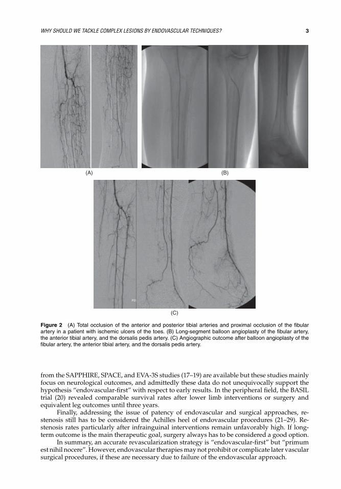
(3–11). Tailored strategies for chronic total occlusions like re-entry devices enable successfulrecanalization of long-segment and heavily calcified lesions. Adoption of coronary chronic totalocclusion technology has significantly advanced success rates particularly in below-the-kneeinterventions (Fig. 2).
Addressing the issue of complications, endovascular therapy traditionally is considereda minimally invasive and safe approach with respect to morbidity and mortality (12–16). Thereduction of device diameters and sheath sizes, introduction of monorail technology, and themore careful application of anticoagulant substances, have certainly contributed to safety ofendovascular therapy. A negligible mortality risk, no relevant immobilization, a shorter hospi-tal stay, and a significantly lower risk for in-hospital complications compared to open vascularsurgery, remain the major arguments for the “endovascular-first” approach. Unfortunately, sci-entifically hard data comparing technical success and complication rates between endovascularand surgical approaches are scarce. In the field of carotid revascularization, comparative data
IHBK065-01 IHBK065-Minar April 26, 2010 13:44 Specs: 7x10 tight 7in×10in Char Count=
WHY SHOULD WE TACKLE COMPLEX LESIONS BY ENDOVASCULAR TECHNIQUES? 3
(C)
(A) (B)
Figure 2 (A) Total occlusion of the anterior and posterior tibial arteries and proximal occlusion of the fibularartery in a patient with ischemic ulcers of the toes. (B) Long-segment balloon angioplasty of the fibular artery,the anterior tibial artery, and the dorsalis pedis artery. (C) Angiographic outcome after balloon angioplasty of thefibular artery, the anterior tibial artery, and the dorsalis pedis artery.
from the SAPPHIRE, SPACE, and EVA-3S studies (17–19) are available but these studies mainlyfocus on neurological outcomes, and admittedly these data do not unequivocally support thehypothesis “endovascular-first” with respect to early results. In the peripheral field, the BASILtrial (20) revealed comparable survival rates after lower limb interventions or surgery andequivalent leg outcomes until three years.
Finally, addressing the issue of patency of endovascular and surgical approaches, re-stenosis still has to be considered the Achilles heel of endovascular procedures (21–29). Re-stenosis rates particularly after infrainguinal interventions remain unfavorably high. If long-term outcome is the main therapeutic goal, surgery always has to be considered a good option.
In summary, an accurate revascularization strategy is “endovascular-first” but “primumest nihil nocere”. However, endovascular therapies may not prohibit or complicate later vascularsurgical procedures, if these are necessary due to failure of the endovascular approach.
IHBK065-01 IHBK065-Minar April 26, 2010 13:44 Specs: 7x10 tight 7in×10in Char Count=
4 SCHILLINGER
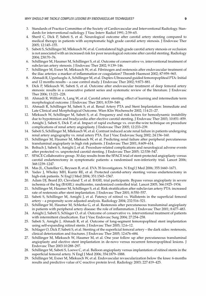
WHAT MAKES A LESION “COMPLEX”?Characteristics of complex lesions may vary in between different vessel areas, but some generalconsiderations can be made.
� Length of the lesion. Usually, the longer the more complex. Very long lesions typically areencountered in the femoropopliteal and below-the-knee segments (Fig. 3). The most criticalpart of the revascularization process, irrespective of the length, remains the re-entry point.
� Occlusions across bifurcations. Re-entry to the true lumen can be complex in these usuallylong lesions when bifurcations are included. This may typically occur at the level of thetibioperoneal trifurcation (Fig. 3) or if a pelvic occlusion includes the femoral bifurcation(Fig. 4).
� Diseased re-entry segments. Recanalization of occlusions frequently is done (whether onpurpose or not) by subintimal wire passage. This technique, as described by Bolia, is donewith a loop in the wire. As soon as the loop has passed the lesion and enters a healthyvascular segment, re-entry in the true lumen usually is achieved. In patients with heavilydisease landing zones (Fig. 5) re-entry can be very complex because the loop continues todissect the artery instead of re-entering the true lumen.
Figure 3 Long-segment occlusion of the distal superficial femoral artery, entire popliteal artery, and tibioper-oneal trifurcation. Below the knee only the fibular artery can be detected. Successful recanalization and balloonangioplasty.

IHBK065-01 IHBK065-Minar April 26, 2010 13:44 Specs: 7x10 tight 7in×10in Char Count=
WHY SHOULD WE TACKLE COMPLEX LESIONS BY ENDOVASCULAR TECHNIQUES? 5
Figure 4 Occlusion of both external iliac and common femoral arteries.
� Heavy calcification. Calcium can cause problems both during the recanalization phase,inhibiting wire passage, as well as after successful wire passage when residual stenosis andeven stent compression endanger a technical success (Fig. 6).
� Thrombus. Thrombotic lesions always have to be considered as potentially complex becauseof the evident risk of embolization. Particularly in carotid interventions (Fig. 7), thromboticlesions have to be tackled extremely carefully, but also peripheral interventions can getcomplex following embolization (Fig. 8).
� Bifurcated lesions. Unlike in coronary arteries, recommendations of how to handle bifur-cation lesions are missing (Fig. 9). Plaque shift and side-branch occlusion can have seriousnegative long-term effects, particularly if the “side branch” is a vessel of major importance,like the deep femoral artery.
� Flush ostial occlusions or strong collaterals originating at the beginning of the occlusion.Particularly in cases when the occlusion has a hard fibrous or calcified cap, inserting the wireinto the occlusion can be tricky because the wire likely will take the way of less resistance tothe collateral.
� Dissections. If dissection (spontaneous or due to a prior revascularization attempt) is present,re-entry to the true lumen may be difficult, especially when the false lumen expands becauseof contrast injections.
Figure 5 Long-segment occlusion of thesuperficial femoral artery with heavily diseaseand stenosed landing zone for re-entry.

IHBK065-01 IHBK065-Minar April 26, 2010 13:44 Specs: 7x10 tight 7in×10in Char Count=
6 SCHILLINGER
Figure 6 Suboptimal expansion of self-expanding stents in the superficial femoralartery due to heavy calcification.
WHAT ARE THE CLINICAL INDICATIONS FOR ENDOVASCULAR TREATMENTOF COMPLEX LESIONS?As stated above, interventionalists always have to keep in mind the clinical indication for revas-cularization. Complex lesions mean lower technical success rates, higher complication rates,and higher risk for recurrence. In this context, the indication for endovascular treatment is notonly driven by patients’ symptoms and need for revascularization, but also by the interventionalexpertise in the treating center.
WHAT ARE THE POTENTIAL HAZARDS OF TREATING COMPLEX LESIONS?Two major hazards of failed interventions in complex lesions have to be discussed. One isthe obvious risk for complications, which include systemic risks, like renal failure due to con-trast overload or heart failure due to fluid overload, and vascular complications, which arementioned in more detail in the respective chapters. The other and less obvious risk is thatfurther endovascular or surgical revascularization attempts are complicated or prohibited byan inadequate first attempt.
Thrombus
Figure 7 Thrombotic particles in a high-grade carotid stenosis.

IHBK065-01 IHBK065-Minar April 26, 2010 13:44 Specs: 7x10 tight 7in×10in Char Count=
WHY SHOULD WE TACKLE COMPLEX LESIONS BY ENDOVASCULAR TECHNIQUES? 7
Figure 8 Thrombotic occlusion of the ostiumof the superficial femoral artery (SFA) with distalembolization and occlusion of the crural trifurca-tion. Successful stenting of the SFA and aspira-tion thrombectomy of the tibioperoneal arteries.
Figure 9 Example of a bifurcated renal arterystenosis with two major segmental branches.

IHBK065-01 IHBK065-Minar April 26, 2010 13:44 Specs: 7x10 tight 7in×10in Char Count=
8 SCHILLINGER
WHICH SPECIFIC INTERVENTIONAL STRATEGIES AND TOOLS CAN BE CONSIDEREDFOR TREATMENT OF COMPLEX LESIONS?� Long lesions. Intraluminal or subintimal techniques can be applied, but especially in long
occlusions, these approaches frequently cannot be differentiated. Re-entry devices like theOutback catheter or the Pioneer catheter can be helpful in gaining access to the true lumen.
� Occlusions across bifurcations. Trying different choices of wires, re-entry devices, and retro-grade recanalization techniques are most frequently applied for this problem.
� Diseased re-entry segments. This clearly is the domain of the above-mentioned re-entrydevices. Alternatively, the “bad end” of the Terumo glidewire or a denuded tip of a V18-Control Wire can be used to have a needle-like device, which enables puncture of theprohibitive membrane between the false and the true lumen. Again, if these techniques fail,retrograde recanalization can be considered.
� Heavy calcification. In terms of wire passage, subintimal passage around the calcium mostfrequently is the only way to overcome “rocky” segments. Before stenting, these segmentsshould be pretreated with atherectomy, and balloon-expanding stents clearly are superior inthis indication. But especially in the SFA, the risks and benefits of a balloon-expanding stenthave to be carefully weighed.
� Thrombus. Small thrombi should be aspirated before any other manipulation is anticipated.In cases with a high thrombus burden, pretreatment with thrombolysis has to be considereddepending on the clinical indication and vessel area treated.
� Bifurcated lesions. The strategy mainly depends on the sizing or the vessels, plaque burden,and angle of the vessel. Preservation of both branches with wires and kissing balloon tech-nique is frequently applied in all vessel areas. Placing a stent in the main vessel, across theside branch, is defined as provisional stenting. Several two-stent techniques are available,with various levels of complexity—the culotte technique, the T technique, the Y technique,the V or simultaneous kissing stents, and the crush technique and its variations (reverse andstep). The decision of which technique to apply will also be determined by the territory oftreatment. In carotids, the side branch (external carotid artery) is ignored. A different situa-tion is faced when treating the subclavian–vertebral bifurcation or a renal bifurcation, wheretwo stent techniques frequently seem adequate. For the aortoiliac bifurcation, the kissingstent technique also is a routine procedure, whereas overstenting the femoral bifurcationremains a “don’t”. The tibioperoneal bifurcation is open to all above-mentioned approaches.However, when using two stents it is important to notice that the above-mentioned coronarytechniques make sense only when balloon-expanding stents are used.
� Flush ostial occlusions or strong collaterals originating at the beginning of the occlusion.Entry through the cap of the occlusion usually can be achieved by the use of different curvesof diagnostic catheters to support the direction of the tip of a stiff guidewire.
� Dissections. This also is the domain of a re-entry device. Again, alternatively the “bad end”of the Terumo glidewire or a denuded tip of a V18-Control Wire can be used to have aneedle-like device which enables puncture of the prohibitive membrane between the falseand the true lumen and retrograde recanalization can be considered.
KEY ISSUES FOR ENDOVASCULAR TREATMENT OF COMPLEX LESIONSThree main issues have to be considered when treating complex lesions and complex patients.First, the clinical indication has to justify the risk of a complex intervention. Second, prepro-cedural noninvasive diagnostic methods should adequately identify complex morphologies toallow precise planning of the procedure. Third, physician training and expertise and aware-ness for potential hazards and their solutions are crucial to guarantee good outcomes and lowcomplication rates.
REFERENCES1. Dormandy JA, Rutherford RB. Management of peripheral arterial disease (PAD). TASC Working
Group. Transatlantic inter-society consensus (TASC). J Vasc Surg 2000; 31:S1–S296.2. Norgren L, Hiatt WR, Dormandy JA, et al. TASC II Working Group. Inter-society consensus for the
management of peripheral arterial disease (TASC II). J Vasc Surg 2007; 26:S5–S67.
IHBK065-01 IHBK065-Minar April 26, 2010 13:44 Specs: 7x10 tight 7in×10in Char Count=
WHY SHOULD WE TACKLE COMPLEX LESIONS BY ENDOVASCULAR TECHNIQUES? 9
3. Standards of Practice Committee of the Society of Cardiovascular and Interventional Radiology. Stan-dards for interventional radiology. J Vasc Interv Radiol 1991; 2:59–65.
4. Sherif C, Dick P, Sabeti S, et al. Neurological outcome after carotid artery stenting compared tomedical therapy in patients with asymptomatic high grade carotid artery stenosis. J Endovasc Ther2005; 12:145–155.
5. Sabeti S, Schillinger M, Mlekusch W, et al. Contralateral high grade carotid artery stenosis or occlusionis not associated with an increased risk for poor neurological outcome after carotid stenting. Radiology2004; 230:70–76.
6. Schillinger M, Haumer M, Schillinger S, et al. Outcome of conservative vs. interventional treatment ofsubclavian artery stenosis. J Endovasc Ther 2002; 9:139–146.
7. Schillinger M, Exner M, Mlekusch W, et al. Fibrinogen and restenosis after endovascular treatment ofthe iliac arteries: a marker of inflammation or coagulation? Thromb Haemost 2002; 87:959–965.
8. Ahmadi R, Ugurluoglu A, Schillinger M, et al. Duplex-Ultrasound guided femoropopliteal PTA: Initialand 12 months results – a case control study. J Endovasc Ther 2002; 9:873–881.
9. Dick P, Mlekusch W, Sabeti S, et al. Outcome after endovascular treatment of deep femoral arterystenosis: results in a consecutive patient series and systematic review of the literature. J EndovascTher 2006; 13:221–228.
10. Ahmadi R, Willfort A, Lang W, et al. Carotid artery stenting: effect of learning and intermediate termmorphological outcome. J Endovasc Ther 2001; 8:539–549.
11. Ahmadi R, Schillinger M, Sabeti S, et al. Renal Artery PTA and Stent Implantation: Immediate andLate Clinical and Morphological Outcome. Wien Klin Wochenschr 2002; 114:21–27.
12. Mlekusch W, Schillinger M, Sabeti S, et al. Frequency and risk factors for hemodynamic instabilitydue to hypotension and bradycardia after elective carotid stenting. J Endovasc Ther 2003; 10:851–859.
13. Amighi J, Sabeti S, Dick P, et al. Impact of rapid exchange vs. over-the-wire technique on procedurecomplications of renal artery angioplasty. J Endovasc Ther 2005; 12:233–239.
14. Sabeti S, Schillinger M, Mlekusch W, et al. Contrast induced acute renal failure in patients undergoingrenal artery angiography vs. renal artery PTA. Eur J Vasc Endovasc Surg 2002; 24:156–160.
15. Schillinger M, Haumer M, Mlekusch W, et al. Predicting renal failure after peripheral percutaneoustransluminal angioplasty in high risk patients. J Endovasc Ther 2001; 8:609–614.
16. Boltuch J, Sabeti S, Amighi J, et al. Procedure-related complications and neurological adverse eventsafter protected vs. unprotected carotid stenting. J Endovasc Ther 2005; 12:538–547.
17. SPACE Collaborative group. 30 day results from the SPACE trial of stent-protected angioplasty versuscarotid endarterectomy in symptomatic patients: a randomised non-inferiority trial. Lancet 2006;368:1239–1247.
18. Mas JL, Chatellier G, Beyssen B, et al. EVA 3S Investigators. N Engl J Med 2006; 355:1660–1671.19. Yadav J, Wholey MH, Kuntz RE, et al. Protected carotid-artery stenting versus endarterectomy in
high-risk patients. N Engl J Med 2004; 351:1565–1567.20. Adam DJ, Beard JD, Cleveland T, et al. BASIL trial participants. Bypass versus angioplasty in severe
ischemia of the leg (BASIL): multicentre, randomized controlled trial. Lancet 2005; 366:1925–1934.21. Schillinger M, Haumer M, Schillinger S, et al. Risk stratification after subclavian artery PTA: increased
rate of restenosis after stent implantation. J Endovasc Ther 2001; 8:550–557.22. Sabeti S, Schillinger M, Amighi J, et al. Patency of nitinol vs. Wallstents in the superficial femoral
artery – a propensity score adjusted analysis. Radiology 2004; 232:516–521.23. Schillinger M, Haumer M, Schlerka G, et al. Restenosis after percutaneous transluminal angioplasty
in patients with peripheral artery disease: the role of inflammation. J Endovasc Ther 2001; 8:477–483.24. Amighi J, Sabeti S, Schlager O, et al. Outcome of conservative vs. interventional treatment of patients
with intermittent claudication. Eur J Vasc Endovasc Surg 2004; 27:254–258.25. Sabeti S, Amighi J, Ahmadi R, et al. Outcome of long-segment femoropopliteal stent implantation
using self-expanding nitinol stents. J Endovasc Ther 2005; 12:6–12.26. Schlager O, Dick P, Sabeti S, et al. Stenting of the superficial femoral artery – the dark sides: restenosis,
clinical deterioration and fractures. J Endovasc Ther 2005; 12:676–684.27. Schillinger M, Mlekusch W, Haumer M, et al. One year follow up after percutaneous transluminal
angioplasty and elective stent implantation in de-novo versus recurrent femoropopliteal lesions. JEndovasc Ther 2003:10:288–297.
28. Schillinger M, Sabeti S, Loewe C, et al. Balloon angioplasty versus implantation of nitinol stents in thesuperficial femoral artery. N Engl J Med 2006; 354:1879–1888.
29. Schillinger M, Exner M, Mlekusch W, et al. Endovascular revascularization below the knee: 6-monthsresults and predictive value of C-reactive protein level. Radiology 2003; 227:419–425.

IHBK065-02 IHBK065-Minar April 24, 2010 10:50 Specs: 7x10 tight 7in×10in Char Count=
2 Pharmacological Prerequisites for Treatment ofComplex Patients with Peripheral Artery DiseaseErich MinarDepartment of Internal Medicine II, Division of Angiology, Medical University Vienna, General Hospital,Vienna, Austria
INTRODUCTIONA patient can be characterized as a “complex” patient either by the complex morphology ofhis vascular disease, as described in the different chapters of this book, or by the differentunfavorable medical comorbidities. Both factors are responsible for an increased complicationrate in such complex patients. Complex morphological features often lead to a longer durationof an intervention, even with an experienced interventionist, and each prolonged interventioncan be associated with adverse events. Otherwise, vascular or nonvascular comorbidities canin most cases be identified easily by history and preinterventional examination of the patient,and this enables effective risk evaluation. The main goal should then be to reduce the overallperi- and postinterventional risk of the patient by adequate treatment, which is most oftenpharmacological.
While revascularization and in some cases also additional pharmacologic therapy withvasoactive drugs are used for improvement of symptoms and quality of life, the main goal ofimprovement of overall prognosis and survival of these patients (by reducing cardiovascularmorbidity and mortality) can mainly be achieved by different pharmacologic treatment. Becauseof the presence of mostly multiple risk factors and because of the systemic nature of atheroscle-rosis and the high risk of ischemic events, patients with peripheral artery disease (PAD) shouldbe candidates for aggressive secondary prevention strategies including antiplatelet therapy andaggressive risk factor modification, e.g., with lipid-lowering and antihypertensive treatment.
ANTIPLATELET THERAPYPAD is a serious medical problem and an indicator of systemic atherosclerosis. Concern-ing development and manifestation of PAD, atherosclerosis and in further consequenceatherothrombosis are the main etiopathogenic factors. In patients with primary diagnosis ofPAD, the prevalence of atherosclerotic manifestations in other vessel areas is higher than inpatients with a primary diagnosis of cardio- or cerebrovascular disease. The diagnosis of PADis therefore a marker for mostly generalized and severe atherosclerosis (1). Many studies suchas the recent REACH (Reduction of Atherothrombosis for Continued Health) Registry, whichincluded 55,814 patients with known atherosclerotic disease (coronary artery disease (CAD),PAD, and cerebrovascular disease), have clearly demonstrated that patients with polyvasculardisease have a significant worse outcome as compared to patients with only CAD (2).
Patients with intermittent claudication (IC) have a relatively good prognosis concerningamputation-free survival with only 1% to 2% of them needing major amputation over a five-year period. Otherwise, the prognosis concerning overall survival is poor. The 5-, 10-, and15-year morbidity and mortality rates from all causes are approximately 30%, 50%, and 70%,respectively, with CAD being by far the most common cause of death (1). Patients with chroniccritical limb ischemia (CLI) have an even much worse prognosis with 20% mortality in the firstyear after presentation.
The main reason to administer antiplatelet therapy to patients with PAD is to preventsevere vascular events such as myocardial infarction, stroke, or vascular death. The Antithrom-botic Trialists’ Collaboration meta-analysis in 42 trials found that among 9214 patients withPAD, there was a 23% reduction in serious vascular events (P = 0.004) in patients treated withantiplatelet therapy (3).
The CAPRIE trial (Clopidogrel versus Aspirin in Patients at Risk of Ischemic Events)demonstrated that the combined risk of death from vascular causes, myocardial infarction, and

IHBK065-02 IHBK065-Minar April 24, 2010 10:50 Specs: 7x10 tight 7in×10in Char Count=
PHARMACOLOGICAL PREREQUISITES FOR TREATMENT OF COMPLEX PATIENTS 11
stroke was significantly, albeit moderately, lower with clopidogrel (75 mg/day) compared withaspirin (325 mg q.d.) (4). Despite this difference, in the recently published recommendationsof the eighth ACCP Conference on Antithrombotic and Thrombolytic Therapy it was recom-mended, by placing a relatively high value on avoiding large expenditures to achieve smallreductions in vascular events, that aspirin be used instead of clopidogrel (5). Unfortunately, therecent Transatlantic inter-society consensus (TASC) II paper does not give clear recommenda-tions concerning this point.
In the past few years, “resistance” to antiplatelet drugs has become a major point ofinterest. Interpatient variability in drug response has been shown to be, at least partially, theresult of polymorphisms in genes encoding drug targets, such as receptors or enzymes (6). Manytests of platelet function are now available for clinical use, and some of these tests have shownthat hyporesponsiveness to antiplatelet drugs in the laboratory (i.e., resistance) is associatedwith adverse clinical events in different patient populations (7).
However, in most of these studies, the number of major adverse clinical events was low.Furthermore, uniform definitions and standardized assays are not yet available, and thereare also no published studies addressing the clinical effectiveness of altering therapy basedon the results of monitoring antiplatelet therapy. Therefore, monitoring of antiplatelet ther-apy in patients with vascular disease cannot be generally recommended currently for clinicalroutine.
Dual Antiplatelet TherapyThe routine use of dual antiplatelet therapy of aspirin and clopidogrel seems not justified inthe routine patient with PAD without planned intervention. The CHARISMA trial (Clopido-grel for High Atherothrombotic Risk and Ischemic Stabilization, Management, and Avoidance)compared the effects of such a dual antiplatelet therapy with clopidogrel plus low-dose aspirinversus aspirin alone (75 to 162 mg/day) on the incidence of cardiovascular events in 15,603patients at high risk for atherothrombotic events followed up for a median of 28 months (8).The primary efficacy end point, a composite of MI, stroke, or death from cardiovascular causes,was not significantly different between the two treatment arms. However, in a subset of 9478patients with previous MI, ischemic stroke, or symptomatic PAD, combination therapy withclopidogrel plus aspirin showed a significant reduction in the rate of cardiovascular death, MI,or stroke (7.3% vs. 8.8%) but with a significantly higher risk of transfusion-requiring bleeding(8). Therefore, some specialists argue that the risk–benefit of dual antiplatelet therapy withaspirin and clopidogrel may be justified in high-risk symptomatic patients but definitely not inasymptomatic patients. Otherwise, according to most vascular specialists working in this field,the slight additive benefit is not justified by the increased economic cost and hemorrhagic risk.
Dual Antiplatelet Therapy in Patients with Planned InterventionIn contrast to the coronary field, there are nearly no sufficient studies dealing with the questionof optimal peri- and postinterventional treatment in noncoronary vessels. While it is generallyaccepted that dual antiplatelet therapy should be prescribed for at least one month—better,however, for three months—in patients with stent implantation in the carotid arteries, thisquestion is not clarified at all in patients with PAD. Unfortunately also TASC II as consensuspaper does not give any clear recommendations. It is only stated that antiplatelet therapy shouldbe started preoperatively and continued as adjuvant pharmacotherapy after an endovascularor surgical procedure. In our department we prescribe dual antiplatelet therapy after stentimplantation in the infrainguinal region for three months. However, as already mentioned, thisrecommendation is not evidence based on hard scientific data. Patients treated by angioplastyalone are not prescribed dual antiplatelet therapy on a regular basis.
In the near future, drug-eluting stents may help to improve the long-term patency afterintervention in the lower limb arteries. However, as yet, trials evaluating DES in the peri-pheral arteries were underpowered and demonstrated no improved patency compared to baremetal stents. Otherwise, the concept of combining the advantages of the mechanical scaffoldingproperties of nitinol stents with the antiproliferative action of drugs seems appealing, andsome groups reported promising results especially in the infrapopliteal vessels. Because ofthe experiences in the coronary field we recommend (despite lack of data) also prolonged

IHBK065-02 IHBK065-Minar April 24, 2010 10:50 Specs: 7x10 tight 7in×10in Char Count=
12 MINAR
dual antiplatelet therapy after peripheral DES implantation, especially in the smaller lower legarteries, for at least six months.
To guarantee an optimal antiaggregatory effect already at the time of the intervention,antiplatelet pretreatment should start at least seven days before the planned intervention.Otherwise, a loading dose has to be given at the beginning of the intervention: 500 mg of aspirinand 300 mg (or according to newer data from the cardiologists, even 600 mg) of clopidogrel,respectively.
Until now it has not been demonstrated that antiplatelet therapy with aspirin and/or clopi-dogrel has any positive influence concerning reduction of restenosis after angioplasty or stentimplantation in patients with PAD. Therefore, it is interesting that a recent randomized studyfrom a Japanese group demonstrated a significant reduction of re-stenosis after femoropoplitealendovascular therapy, by a combined therapy of aspirin with cilostazol (this is a vasoactivedrug with also antiplatelet effects) compared to a combination of aspirin with ticlopidine (athienopyridine replaced by clopidogrel in most centers) (9).
Combined Anticoagulant and Antiplatelet TherapyBecause the results of previous randomized trials evaluating oral anticoagulants in patients withPAD—who represent a group at a high risk for thrombotic events—have been inconclusive,recently a large international, randomized clinical trial has been performed to determine ifmoderate levels of oral anticoagulation (OAC) (with an INR of 2–3) improve upon antiplatelettherapy alone (10). In this Warfarin Antiplatelet Vascular Evaluation (WAVE) trial, after a meanfollow-up time of 35 months the combination of an oral anticoagulant and antiplatelet therapywas not more effective than antiplatelet therapy alone in preventing major cardiovascularcomplications and was associated with an increase in life-threatening bleeding. The outcome ofmyocardial infarction, stroke, or death from cardiovascular causes occurred in 12.2% of patientsreceiving combination therapy and in 13.3% of patients receiving antiplatelet therapy alone.Otherwise, life-threatening bleeding occurred in 4% of patients receiving combination therapyas compared to 1.2% in the antiplatelet therapy alone group.
PERI-INTERVENTIONAL ANTITHROMBOTIC THERAPY (TABLE 1)Because of the rapid advances in technology concerning endovascular procedures, more andmore complex interventions are carried out nearly on a routine basis. Otherwise, we are facedwith an increased thrombotic risk with treatment of such patients. Therefore, it is also nec-essary to prescribe intensive supplementary antithrombotic therapy for prophylaxis of acutethrombotic occlusion.
HeparinUnfractionated heparin (UFH) is the classic anticoagulant agent administered during endovas-cular interventions to prevent embolization or new thrombus formation. Its efficacy is onlylimited by the fact that it does not resolve already existing thrombi. The half-life is prolonged inpatients with renal insufficiency. Despite its widespread use, there are no clear recommendationsconcerning the optimal dose, because the benefit of any antithrombotic drug or regimen duringperipheral endovascular interventions has not been demonstrated in any randomized study (incontrast to coronary interventions). UFH is commonly administered with a bolus of about5000 IU following vascular access, and further doses—sometimes dependent on activatedclotting time measurements—are given during procedures with longer duration. Otherwise,some experienced investigators administer less than 5000 IU in less complex interventions withexpected short (e.g., < 30 minutes) duration. The main complications are of course bleeding—the risk is especially increased in combination with GP IIb/IIIa inhibitors—and an immune-mediated platelet activation causing heparin-induced thrombocytopenia with potentially severethromboembolic complications in the arterial and/or venous system (11). Low molecular weightheparin has a more favorable benefit/risk ratio and superior pharmacokinetic properties com-pared to UFH. Because there are nearly no data for their routine use in peripheral vascular inter-ventions, the use of UFH is preferred currently in most vascular centers performing peripheralinterventions. One of the main advantages of UFH in this situation is the possibility to neutralizethe anticoagulant activity by protamine in case of severe bleeding complications.

IHBK065-02 IHBK065-Minar April 24, 2010 10:50 Specs: 7x10 tight 7in×10in Char Count=
PHARMACOLOGICAL PREREQUISITES FOR TREATMENT OF COMPLEX PATIENTS 13
Table 1 Peri-interventional Pharmacotherapy
Antiplatelet therapy: starting ≥ 7 days before intervention, otherwise loading dose
– Low-dose aspirin (about 100 mg/day; loading dose: 500 mg); all patients– Clopidogrel: (75 mg/day; loading dose 300–600 mg; duration: 3–6 months); in special situations (see text)
Anticoagulant therapy:
– Heparin� Unfractionated heparin: Bolus of 5000 IU; with longer duration interventions: further dose according to
ACT measurement� Less complex intervention with short duration: 3000 IU (Low molecular weight heparin: no sufficient
data)– Bivalirudin: Bolus of 0.75 mg/kg; then infusion of 1.75 mg/kg/hr during intervention (dose recommendation
from Cardiology)
Glycoprotein IIb/IIIa receptor inhibitors:
– Abciximab: Bolus of 0.25 mg/kg; then infusion of 0.125 �g/kg/min for 12 hr
Thrombolytic agents: dosage “as high as necessary and as low as possible”
– Classical drugs� Streptokinase� Urokinase: recommended maximal dose: 500,000 IU� rt-PA: recommended maximal dose: 10 mg
– Newer drugs:� Reteplase� Tenecteplase� Alfimeprase
Vasodilating drugs:
– Prostanoids:� Iloprost: intra-arterial bolus of 3000 ng (according to surgical data, no experience after endovascular
interventions)� Alprostadil: intra-arterial infusion of 10 �g/30–60 min
– Nitroglycerin: Repeated intra-arterial bolus of 0.1–0.2 mg/10 mL solution– Papaverine: 30–60 mg intra-arterial– Tolazoline: 12.5 mg intra-arterial– Nifedipine: 10 mg orally
Abbreviations: ACT, activated clotting time; rt-PA, recombinant tissue plasminogen activator.
BivalirudinThis drug belongs to the group of direct thrombin inhibitors, which bind directly to thrombin.According to a lot of recent data from coronary trials, the main benefit of bivalirudin overheparin is a significant reduction in the incidence of major bleeding complications (12).
Unfortunately until now, sufficient data are not available concerning the use of bivalirudinin peripheral interventions so as to evaluate the safety and feasibility of using this drug asthe procedural anticoagulant in patients undergoing peripheral interventions. Only recentlyhave some interventionists started using this agent as the sole anticoagulant for peripheralinterventions. Observational results reported from single centers are promising (13). Ischemicand bleeding events are low and, compared with those reported in the literature, suggest thatbivalirudin is safe to be used in this population.
Glycoprotein IIb/IIIa Receptor InhibitorsIntravenous platelet glycoprotein (GP) IIb/IIIa receptor inhibitors have shown convincing clin-ical efficacy in percutaneous coronary interventions. In the past few years, there is increasingevidence that GP IIb/IIIa receptor blockade might also have an important role in peripheralarterial intervention (14). However, because the use of adjunctive therapy with GP IIb/IIIareceptor blockade in noncoronary interventions is rather new, the reported data of controlled,

IHBK065-02 IHBK065-Minar April 24, 2010 10:50 Specs: 7x10 tight 7in×10in Char Count=
14 MINAR
randomized, and prospective trials are mainly limited to cardiovascular disease. Otherwise,although peripheral arterial occlusions differ from coronary occlusions with respect to thesize and age of the occluding thrombus, the success of GP IIb/IIIa inhibition in the settingof acute coronary occlusion might translate to the peripheral vessels. However, indication for“off-label” use in peripheral interventions should be strictly restrictive, since its application hasto be balanced against bleeding complications and the rather high costs. The theory behind theuse of GP IIb/IIIa antagonists during peripheral arterial interventions in chronic lesions is toprevent early thrombosis in lesions at high risk for such an occurrence. The phenomenon of“de-thrombosis,” that is the dissolution of thrombus by GP IIb/IIIa inhibitors without concomi-tant plasminogen activators, further increased the interest in peripheral arterial application ofthese agents.
Abciximab, the most widely studied of all GP IIb/IIIa inhibitors, is a monoclonal anti-body, while eptifibatide and tirofiban are small-molecule antagonists. The drugs are given intra-venously starting with a bolus followed by intravenous infusion for 12 to 36 hours. Althoughthere are extensive data for all three GP IIb/IIIa receptor antagonists available for coronaryindication, in PAD the literature is mainly limited to the use of abciximab. It was demonstratedin the PROMPT trial (15) that the speed of thrombolysis could be enhanced by adjunctiveGP IIb/IIIa blockade with abciximab. A further study demonstrated a favorable effect of abcix-imab on patency and clinical outcome in patients undergoing endovascular revascularizationof long-segment femoropopliteal occlusions not hampered by serious bleeding complications(16). Very recently, Keo et al. (17) reported favorable results of provisional abciximab therapy incomplex peripheral catheter interventions with imminent risk of early re-thrombosis. This riskwas defined by revascularization of arterial occlusions in association with one or more of thefollowing circumstances: time-consuming intervention >3 hours; compromised contrast flownot solved by stenting; distal embolization not solved by mechanical thromboembolectomy;peri-interventional notice of thrombus evolution despite adequate heparin adjustment. In theRELAX trial (18), a multicenter prospective comparison of reteplase alone versus reteplasewith abciximab, the GP IIb/IIIa inhibitor reduced the incidence of distal embolization duringthrombolysis from 31% to 5% (P = 0.01).
In our laboratory, at present, abciximab is used during peripheral interventions only inpatients at high risk of early re-occlusion, such as patients with treated long-segment occlusionsand poor run-off or residual intraluminal thrombus. Tepe et al. (14) have recently recommendedthat until data from larger prospective clinical trials are available, adjunct GP IIb/IIIa inhibitorsshould be used only in selected patients with a higher risk of failure during thrombolysis orre-thrombosis during time-consuming and technically complicated vascular interventions, i.e.,the complex patients.
Thrombolytic AgentsDifferent thrombolytic agents are currently available for patients with acute limb ischemia, andvarious techniques of intra-arterial thrombolysis are used for application of these agents (19).The efficacy of several fibrinolytic drugs, such as urokinase, streptokinase, and recombinanttissue plasminogen activator, has been reviewed in a recent meta-analysis (20). Alfimeprase is anewer direct-acting fibrinolytic agent that does not require activation of plasminogen, and thismechanism may potentially reduce the frequency of bleeding complications. However, clinicaldata with this new drug are limited until now (21).
While data show that intrathrombus delivery of thrombolytic agents is more effective thannonselective intra-arterial catheter-directed infusion, no particular thrombolytic regime has yetbeen proven to be clearly superior. Because thrombolysis in general is limited by a relativelyhigh rate of bleeding complications as well as long infusion times, many interventionists try toreduce the overall amount of applied thrombolytic drug and to decrease the procedural time.This goal can be reached by advanced endovascular methods such as selective catheter-directedthrombolysis with intra-arterial application of the agent with the so-called pulse-spraytechnique or by combination of mechanical thrombectomy with low-dose thrombolysis.These techniques have substantial benefits concerning efficacy with quicker recanalization andsafety with less bleeding complications compared to former studies. Therefore, a combinationof aspiration thromboembolectomy or any kind of mechanical thromboembolectomy with

IHBK065-02 IHBK065-Minar April 24, 2010 10:50 Specs: 7x10 tight 7in×10in Char Count=
PHARMACOLOGICAL PREREQUISITES FOR TREATMENT OF COMPLEX PATIENTS 15
selective intra-arterial low-dose local thrombolysis—in contrast to nonselective catheter-directed thrombolysis alone—can be recommended today as the preferred treatment option inthese patients.
PERI- AND POSTINTERVENTIONAL MANAGEMENT OF ANTITHROMBOTIC THERAPYIN PATIENTS WITH ATRIAL FIBRILLATIONAtrial fibrillation (AF) is the most common clinically relevant arrhythmia among adults, affect-ing over 1 in 25 adults, 60 years or older and 1 in 10 persons, 80 years or older (22). Furthermore,AF is the most potent common risk factor for ischemic stroke.
Numerous clinical trials performed over the past several decades have focused on testingthe efficacy and safety of OAC with vitamin K antagonists (i.e., warfarin and other coumarins) orantiplatelet drugs, especially aspirin, for the prevention of stroke and other systemic embolismin persons with AF. With recent advances in our understanding of the coagulation process anddrug development, new therapeutic strategies that intervene in different parts of the coagulationpathways (such as tissue factor/factor VIIa, factor VIIIa, factor Xa, and thrombin/factor IIa)are being evaluated as alternative antithrombotic therapies. Across all primary prevention trialsand despite variation in anticoagulation intensity target ranges, participants randomly assignedto OAC therapy experienced large consistent reductions in the risk of ischemic stroke comparedwith control subjects, with relative risk reductions of stroke ranging from about 50% to as high as85% (23). Otherwise, randomized trials have provided much weaker support for the efficacy ofaspirin compared with oral vitamin K antagonists. Therefore, OAC is strongly recommended inall high-risk AF patients (defined as having one or more risk factors including diabetes mellitus,congestive heart failure or left ventricular systolic dysfunction, coronary artery disease, a historyof stroke/transient ischemic attack or systemic embolism, hypertension, the female gender, andadvancing age). Current evidence supports an international normalized ratio (INR) target of 2.0to 3.0 (23).
It has also been demonstrated that OAC therapy was superior to dual antiplatelet therapywith clopidogrel and aspirin for prevention of vascular events in patients with AF at high risk ofstroke, especially in those already on OAC therapy (24). Therefore, it is often a difficult problemto handle peri- and postinterventional antithrombotic treatment in patients on long-term OAC,such as patients with AF or after venous thromboembolism with further need for antiplatelettherapy after an endovascular intervention.
There is lack of published evidence on the optimal peri- and postinterventionalantithrombotic management strategy in anticoagulated AF patients. There are only avail-able expert committee reports or opinions and/or clinical experiences of respected author-ities mainly concerning coronary interventions (25). A delicate balance between the risk ofthromboembolic complications or thrombotic re-occlusion and the risk of major bleeding isneeded (26).
After angioplasty without stent implantation or after stent implantation in the aortoil-iac vessels, antiplatelet therapy with aspirin alone is the current practice. Otherwise, dualantiplatelet therapy, for at least one month, is the current practice in most centers for the pre-vention of in-stent thrombosis after infrainguinal stent implantation. Unfortunately, such dualantiplatelet therapy does not adequately protect against thromboembolic complications causedby AF (24). Otherwise, not all patients requiring chronic anticoagulation have a similar riskof thromboembolic complications. Therefore, it appears to be reasonable to adjust the need orextent of anticoagulation, in case of antithrombotic triple therapy with dual antiplatelet therapy,to the perceived thromboembolic risk in patients after infrainguinal stenting. More frequent lab-oratory analyses might also be helpful to ensure INR values are well within the recommendedevidence-based ranges.
According to data from the cardiologists, the recommended duration of dual antiplatelettherapy varies, ranging from four weeks following bare-metal stent implantation during electiveangioplasty to at least 6 to 12 months with drug-eluting stents, in view of the risk of late stentthrombosis. The 2006 American College of Cardiology/American Heart Association/EuropeanSociety of Cardiology guidelines on AF management suggest that the maintenance regimenshould be a combination of clopidogrel and coumarin derivatives for 9 to 12 months, afterwhich the coumarin may be continued as monotherapy (25).

IHBK065-02 IHBK065-Minar April 24, 2010 10:50 Specs: 7x10 tight 7in×10in Char Count=
16 MINAR
PERI-INTERVENTIONAL APPLICATION OF VASODILATORSDuring and/or after complex peripheral endovascular interventions, a slow flow can beobserved sometimes, causing suspicion of severe spasm. This problem can be observed espe-cially during intervention of infrapopliteal arteries, and it can also be a severe problem duringstent angioplasty of the carotid arteries mainly caused by the embolic protection device. In thecarotid arteries, an intra-arterial bolus application of 0.1–0.2 mg nitroglycerin is mainly used asan antispasmodic agent, while different vasodilating agents are used in the peripheral circula-tion. However, there are no widespread recommendations in the literature mainly because ofthe lack of any study data.
A strong vasodilation can be induced after intra-arterial administration of prostanoidssuch as iloprost or alprostadil. These drugs are mainly used for treatment of chronic CLI, andbecause of their pharmacological profile, prostanoids also represent an interesting group ofdrugs for adjuvant treatment of patients with acute limb ischemia. Furthermore, small studieswith prostanoids in distal bypass surgery have shown that intragraft injection at the end of thebypass procedure can reduce the distal resistance and produce a sustained increase in blood flowthrough femorodistal vein grafts in the early postoperative period (27,28). We use these drugsmainly peri- and post-interventionally in patients with CLI. Otherwise they can, in accordancewith the above-mentioned surgical data, also be given as bolus intra-arterially to reduce theperipheral resistance in patients with slow flow and suspicion of vasospasm. Positive effectscan also be expected by an antiplatelet effect of these substances.
Other antispasmodic agents are intravenous papaverine or tolazoline and oral nifedipine.
RISK FACTOR MODIFICATIONPAD is considered a CAD equivalent, and the American Heart Association (AHA), NationalHeart, Lung and Blood Institute, and the National Cholesterol Education Program (NCEP)recommend identical atherosclerotic risk-reduction strategies for PAD and CAD patients (29).From studies of patients with PAD, and also generalizing from other trials in patients withatherosclerotic diseases, there is good evidence that by addressing the key risk factors—smoking,dyslipidemia, and hypertension—the mortality and morbidity of cardiovascular ischemic eventscan be definitely reduced.
Otherwise, there is also a growing body of evidence that treatment of these modifiable riskfactors is often neglected in PAD patients compared to cohorts of patients presenting with CADor stroke. The above-mentioned REACH registry has also clearly demonstrated that despitetheir well-known adverse effects, the classic cardiovascular risk factors have been still largelyundertreated and undercontrolled in patients with PAD in many regions of the world, alsoduring the past few years (30).
Despite a demonstrated positive effect of exercise and diet on many of the risk factors,most patients will require additive pharmacotherapy to achieve the therapeutic goals.
StatinsMultiple epidemiological studies have persuasively correlated chronic statin use with a favor-able decrease in cardiovascular events. The relative risk reduction is 20% to 30% in all subgroupsfor the primary outcome of the combined incidence of coronary death and MI over a three- tosix-year follow-up period.
The Heart Protection Study was the first large study that demonstrated the positive effectsof statins also for the subgroup (N = 6748) of patients with PAD. In the PAD subgroup, therewas a 25% risk reduction over five years (31).
Dietary modification and pharmacological therapy with regard to dyslipidemia should betailored to meet the current guidelines for high-risk patients. Concerning low-density lipopro-tein (LDL) cholesterol, a value of less than 100 mg/dL or even less than 70 mg/dL for thoseat very high risk should be the goal. The ACC/AHA guidelines define “very high-risk” PADpatients as those with multiple risk factors (especially diabetes), severe or poorly controlled riskfactors (especially smoking), and risk factors of the metabolic syndrome (especially elevatedtriglycerides in the face of an elevated non-HDL cholesterol). A recent prospective observationalcohort study of nearly 1400 PAD patients showed that intensive therapy with statins targetingan LDL ≤70 mg/dL improved overall as well as cardiac mortality at a mean follow-up of 6 years

IHBK065-02 IHBK065-Minar April 24, 2010 10:50 Specs: 7x10 tight 7in×10in Char Count=
PHARMACOLOGICAL PREREQUISITES FOR TREATMENT OF COMPLEX PATIENTS 17
(32). For the patient who fails to achieve LDL goals, options include switching to a more potentstatin or adding ezetimibe at 10 mg/day. A fasting lipid profile should be obtained six weeksafter changes in medication or dose are made.
A recent meta-analysis of postoperative mortality and morbidity in patients on preoper-ative statin therapy gives a 1.2% and 4.4% absolute reduction in 30-day mortality for cardiacand vascular surgery, respectively (33). The positive effects observed perioperatively may alsobe expected in patients treated by an endovascular method.
Statins are also promoted as an important perioperative risk reduction strategy because oftheir so-called pleiotropic effects. Among many others, possible mechanisms for the so-calledpleiotropic effects include improvement of vasomotor regulation of blood flow [LDL-C worsensendothelial function and bioavailability of nitric oxide (NO)], neoangiogenesis (collateral circu-lation), reduction in thrombus formation and platelet activation, antioxidant properties, reduc-tion of the inflammatory burden with better plaque stability, or even a reduction of plaque size(34,35). Statin therapy was especially associated with improved survival and event-free survivalrates in patients with severe PAD in states of high inflammatory activity (36). Statins thereforeshould be rigorously prescribed in these patients. The observed interaction with inflammationsupports the view that statins exert their beneficial effects in patients with advanced atheroscle-rosis partly via an anti-inflammatory pathway or, alternatively, by attenuating the deleteriouseffects of inflammation.
Furthermore, a protective effect of statins on renal function has been reported in patientswith CAD and PAD.
In patients with PAD, statins seem also to favorably influence leg function (37), in additionto reducing cardiovascular events and improving survival rates.
Antihypertensive TreatmentIn patients presenting with PAD, hypertension is a major associated cardiovascular risk factor.Hypertension also increases the risk of cardiovascular disease complications and mortality inpatients with established PAD. Unfortunately, blood pressure management in PAD tends tobe poor. In the PARTNERS (The PAD Awareness, Risk, and Treatment: New Resources forSurvival) program, hypertension was less often treated in new (84%) and prior (88%) PADpatients compared to treatment of hypertension in subjects with cardiovascular disease (95%)(38). Furthermore, underdiagnosis of PAD in primary care practice may also be a barrier toeffective secondary prevention of the high ischemic cardiovascular risk associated with PAD.
It is also important to identify possible underlying causes for secondary hypertensionso that specific treatment may be targeted against causes of hypertension. Atheromatous renalartery stenosis is one of these important common underlying causes of hypertension in PAD.
Current evidence supports a low threshold for blood pressure treatment in PAD andintensive blood pressure control to reduce the high risk of cardiovascular disease and death inpatients with PAD. Optimal treatment targets should be <140/85 mmHg, with the lower targetof <130/80 mmHg in the presence of diabetes mellitus or chronic renal disease. The importanceof intensive blood pressure control for reduction of cardiovascular events has especially beendemonstrated in PAD patients with diabetes (39).
An important goal to be reached in hypertensive patients with PAD is to make an appro-priate choice among the available antihypertensive drug classes.
According to recent guidelines, most drugs classes can be used to treat hypertension insymptomatic PAD. Class-specific selection of antihypertensive treatment in PAD should takeinto consideration indications and contraindications based on other significant comorbidity.
There has been some doubt whether �-blocking agents should be the first-line drugs inuncomplicated hypertensive patients with PAD. These drugs have until recently been consid-ered relatively contraindicated in patients with PAD because of the perceived risk that thesedrugs could reduce the peripheral circulation by inhibition of �2 receptors and thus worsenintermittent claudication. However, �-blockers are definitely not contraindicated in patientswith intermittent claudication (40). On the contrary, because of their cardioprotective effectthey are associated with improved survival in patients with PAD (41). Furthermore, �-blockingagents should be used in patients with PAD and further accepted indications for �-blockingagents such as angina pectoris, post infarction, chronic heart failure, and aortic dissection (where

IHBK065-02 IHBK065-Minar April 24, 2010 10:50 Specs: 7x10 tight 7in×10in Char Count=
18 MINAR
it is a cornerstone of medical approach). Otherwise, these drugs should be prescribed cautiouslyin patients with CLI.
If the indication is systolic hypertension in the elderly (which is frequently associated withPAD), diuretics and calcium channel blocking agents are often recommended. However, thereis little published hard evidence for calcium antagonists in the specific setting of hypertensionin patients with PAD. There are also no large outcome studies specifically addressing treatmentwith (low-dose) diuretics in PAD with hypertension, but there is little doubt that diuretics areeffective antihypertensive agents both for lowering blood pressure and for the reduction ofcardiovascular morbidity and mortality in uncomplicated hypertension.
Blockade of the renin-angiotensin system is an important approach to the prevention ofcardiovascular events. In the Heart Outcomes Prevention Evaluation (HOPE) study, includingpatients with PAD, the risk of heart attack, stroke, and death from vascular causes was reducedby 22% for patients given the (ACE) inhibitor, ramipril (42). This effect was independent ofits antihypertensive effect. In cases where ACE inhibitors are not well tolerated, angiotensin IIreceptor blockers may offer an alternative. In the largest angiotensin receptor blocker cardio-vascular outcome study to date, the ONTARGET trial compared the efficacy of therapy withthe angiotensin II receptor blocker, telmisartan and the ACE inhibitor, ramipril as well as thecombination of the two drugs in reducing cardiovascular events in patients at high risk (historyof CAD, stroke or transient ischemic attack, PAD, or diabetes with evidence of end-organ dam-age) (43). Telmisartan was equivalent to ramipril in patients with vascular disease or high-riskdiabetes and was associated with less angioedema. The combination of the two drugs wasassociated with more adverse events without an increase in benefit.
DiabetesMany patients with PAD have diabetes. Epidemiologic studies have shown a relationshipbetween glycated hemoglobin levels and cardiovascular events in patients with type 2 diabetes.Otherwise, previous randomized trials evaluating the effects of glycemic control in patientswith diabetes have provided inconsistent evidence of effects on vascular disease. For example,the incidence of amputation for PAD was not reduced by tight glycemic control in the U.K.Prospective Diabetes Study (44). Therefore, there are little data to support that aggressivecontrol of blood glucose levels improves risk of MI, stroke, vascular death, or amputation.Otherwise, according to the consensus paper, TASC II, patients with diabetes and PAD shouldhave an aggressive control of blood glucose levels with a hemoglobin A1c goal of <7.0% oras close to 6% as possible (1). In the new TASC paper this recommendation is graded asC, meaning that it is based on evidence obtained from expert committee reports or opinionsand/or clinical experiences of respected authorities, while there are no applicable studies of goodquality.
The importance of strict diabetic control concerning major cardiovascular events was alsoquestioned by the recently published ACCORD-study (45). About one-third of the patients inthis study had a history of macrovascular disease. The findings of this study indicated thata comprehensive, customized, therapeutic strategy that targeted glycated hemoglobin levelsbelow 6.0% increased the rate of death from any cause after a mean of 3.5 years, as compared witha strategy that targeted levels of 7.0% to 7.9% in patients with a median glycated hemoglobinlevel of 8.1%, and with either previous cardiovascular events or multiple cardiovascular riskfactors. These findings identify a previously unrecognized harm of intensive glucose loweringin high-risk patients with type 2 diabetes.
In another similar, recently published large trial with more than 11,000 patients a strat-egy of intensive glucose control, involving gliclazide (modified release) and other drugs asrequired that lowered the glycated hemoglobin value to 6.5%, yielded a 10% relative reductionin the combined outcome of major macrovascular and microvascular events, primarily as aconsequence of a 21% relative reduction in nephropathy (46).
It is beyond any doubt that in high-risk patients with type 2 diabetes, intensive interventionconcerning therapy of risk factors, such as hypertension and hyperlipidemia, with multiple drugcombinations and behavior modification has sustained beneficial effects with respect to vascularcomplications and on rates of death from cardiovascular causes (47).

IHBK065-02 IHBK065-Minar April 24, 2010 10:50 Specs: 7x10 tight 7in×10in Char Count=
PHARMACOLOGICAL PREREQUISITES FOR TREATMENT OF COMPLEX PATIENTS 19
PATIENTS WITH IMPAIRED KIDNEY FUNCTIONPatients with chronic kidney disease are at higher risk of cardiovascular morbidity and mortal-ity because of higher prevalence of traditional cardiovascular risk factors, such as hypertension,diabetes mellitus, dyslipidemia, and ischemic heart disease. The level of kidney function is anindependent predictor of short- and long-term, all-cause cardio- and cerebrovascular mortal-ity (48). Renal insufficiency was reported to be a strong independent predictor of mortality inCLI patients, and mortality risk was highest among patients with a glomerular filtration rate<30 mL/min/1.73 m2 (49). Renal insufficiency is also a risk factor for poor outcome (poorerpostoperative survival and higher amputation rates) after infrainguinal bypass in patients withCLI, because dialysis-dependent patients usually have heavily calcified distal arteries. Further-more, wound healing is poor due to anemia, malnutrition, impaired immunity, and tendency toinfection. The increased risks of surgery in these mostly high-risk patients have lead—togetherwith the improved technology—to a steadily increasing application of endovascular interven-tions as well as in patients with chronic kidney disease.
It is important to avoid contrast media–induced acute renal failure or deterioration ofkidney function, since mortality is increased if patients with underlying chronic kidney dis-ease develop contrast media–induced acute renal failure after coronary interventions (50). Themost effective preventive measure is adequate hydration of the patient, mainly with electrolytesolutions. Alternatively, isotonic sodium bicarbonate solution may be used. Acetylcysteine(600 mg b.i.d. before and on the day of the intervention) may be used since there are somepositive studies and it has almost no side effects. However, its efficacy is unclear to date (50).
Statin therapy also seems to be associated with improved outcomes in patients with renalimpairment, as it was demonstrated in vascular surgery patients (51).
SUMMARYPatients with PAD can mostly be judged as “complex” patients due to different medical comor-bidities, mainly due to the underlying atherothrombotic disease. Therefore, these patients needintensive antithrombotic therapy, mostly by antiplatelet agents, and aggressive risk factor modi-fication including especially lipid-lowering and antihypertensive treatment. Special care has tobe taken considering peri-interventional antithrombotic management in patients on long-termOAC treatment, such as patients with AF.
REFERENCES1. Norgren L, Hiatt WR, Dormandy JA, et al. TASC II Working Group. Inter-society consensus for the
management of peripheral arterial disease (TASC II). Eur J Vasc Endovasc Surg 2007; 33(suppl 1):S1–S75.
2. Steg PG, Bhatt DL, Wilson PW, et al. One-year cardiovascular event rates in outpatients withatherothrombosis. JAMA 2007; 297:1197–1206.
3. Antithrombotic Trialists’ Collaboration. Collaborative metaanalysis of randomised trials of antiplatelettherapy for prevention of death, myocardial infarction, and stroke in high risk patients. BMJ 2002;324:71–86.
4. CAPRIE Steering Committee. A randomised, blinded, trial of clopidogrel versus aspirin in patients atrisk of ischaemic events (CAPRIE). Lancet 1996; 348: 1329–1339.
5. Sobel M, Verhaeghe R. Antithrombotic therapy for peripheral artery occlusive disease: AmericanCollege of Chest Physicians Evidence- Based Clinical Practice Guidelines (8th Edition). Chest 2008;133:815S–843S.
6. Undas A, Brummel-Ziedins KE, Mann KG. Antithrombotic properties of aspirin and resistance toaspirin: beyond strictly antiplatelet actions. Blood 2007; 109:2285–9220.
7. Eikelboom JW, Hirsh J, Weitz JI. Aspirin-resistant thromboxane biosynthesis and the risk of myocardialinfarction, stroke, or cardiovascular death in patients at high risk for cardiovascular events. Circulation2002; 105:1650–1655.
8. Bhatt DL, Flather MD, Hacke W, et al. Patients with prior myocardial infarction, stroke, or symptomaticperipheral arterial disease in the CHARISMA trial. J Am Coll Cardiol 2007; 49:1982–1988.
9. Iida O, Nanto S, Uematsu M, et al. Cilostazol reduces restenosis after endovascular therapy in patientswith femoropopliteal lesions. J Vasc Surg 2008; 48:144–149.
10. The Warfarin Antiplatelet Vascular Evaluation Trial Investigators. Oral anticoagulant and antiplatelettherapy and peripheral arterial disease. N Engl J Med 2007; 357:217–227.

IHBK065-02 IHBK065-Minar April 24, 2010 10:50 Specs: 7x10 tight 7in×10in Char Count=
20 MINAR
11. Keo HH, Baumgartner I. Complications of pharmacologic interventions: antiplatelet, antithrombotic,and anticoagulant agents. In: Schillinger M, Minar E, eds. Complications in Peripheral Vascular Inter-ventions. London: Informa Healthcare, 2007:71–80.
12. Kastrati A, Neumann FJ, Mehilli J, et al. Bivalirudin versus unfractionated heparin during percuta-neous coronary intervention. N Engl J Med 2008; 359:688–696.
13. Katzen BT, Ardid MI, MacLean AA, et al. Bivalirudin as an anticoagulation agent: safety and efficacyin peripheral interventions. J Vasc Interv Radiol 2005; 16:1183–1187.
14. Tepe G, Wiskirchen J, Pereira P, et al. GP IIb/IIIa blockade during peripheral artery interventions.Cardiovasc Intervent Radiol 2008; 31:8–13.
15. Duda SH, Tepe G, Luz O, et al. Peripheral artery occlusion: treatment with abciximab plus uroki-nase versus with urokinase alone—a randomized pilot trial (the PROMPT Study). Platelet ReceptorAntibodies in Order to Manage Peripheral Artery Thrombosis. Radiology 2001; 221:689–696.
16. Doerffler-Melly J, Mahler F, Do DD, et al. Adjunctive abciximab improves patency and functionaloutcome in endovascular treatment of femoropopliteal occlusions: initial experience. Radiology 2005;237: 1103–1109.
17. Keo H, Diehm N, Baumgartner, et al. Single center experience with provisional abciximab therapy incomplex lower limb interventions. VASA 2008; 37:257–264.
18. Ouriel K, Castaneda F, McNamara T, et al. Reteplase monotherapy and reteplase/abciximab combi-nation therapy in peripheral arterial occlusive disease: Results from the RELAX trial. J Vasc InterventRadiol 2004; 15:229–238.
19. Working Party on Thrombolysis in the Management of Limb Ischemia. Thrombolysis in the manage-ment of lower limb peripheral arterial occlusion–a consensus document. J Vasc Interv Radiol. 2003;14:S337–S349.
20. Kessel D, Berridge D, Robertson I. Infusion techniques for peripheral arterial thrombolysis. CochraneDatabase Syst Rev 2004; (1):CD000985.
21. Moise MA, Kashyap VS. Alfimeprase for the treatment of acute peripheral arterial occlusion. ExpertOpin Biol Ther 2008; 8: 683–689.
22. Go AS, Hylek EM, Phillips JA, et al. Prevalence of diagnosed atrial fibrillation in adults: nationalimplications for rhythm management and stroke prevention: The AnTicoagulation and Risk Factorsin Atrial Fibrillation (ATRIA) study. JAMA 2001; 285:2370–2375.
23. Singer DE, Albers GW, Dalen JE. Antithrombotic therapy in atrial fibrillation: American College ofChest Physicians Evidence-Based Clinical Practice Guidelines (8th Edition). Chest 2008; 133; S546–S592.
24. The ACTIVE Writing Group of the ACTIVE Investigators. Clopidogrel plus aspirin versus oral antico-agulation for atrial fibrillation in the Atrial fibrillation Clopidogrel Trial with Irbesartan for preventionof Vascular Events (ACTIVE W): a randomised controlled trial. Lancet 2006; 367:1903–1912.
25. Fuster V, Ryden LE, Cannom DS, et al. ACC/AHA/ESC 2006 Guidelines for the management ofpatients with atrial fibrillation—executive summary: a report of the American College of Cardiol-ogy/American Heart Association Task Force and the European Society of Cardiology Committee forPractice Guidelines (Writing Committee to Revise the 2001 Guidelines for the management of patientswith atrial fibrillation). J Am Coll Cardiol 2006; 48:854–906.
26. Sinnaeve PR. The good, the bad, and the ugly: triple therapy after PCI in patients requiring chronicanticoagulation. Eur Heart J 2007; 28:657–658.
27. Hickey NC, Shearman, CP, Crowson MC, et al. Iloprost improves femoro-distal graft flow after a singlebolus injection. Eur J Vasc Surg 1991; 5:19–22.
28. Thul R, Heckenkamp J, Gawenda M, et al. The role of intra-operative prostavasin application duringcrural bypass surgery. Zentralbl Chir 2007; 132:485–490.
29. Smith SC Jr, Allen J, Blair SN, et al. AHA/ACC guidelines for secondary prevention for patients withcoronary and other atherosclerotic vascular disease: 2006 update: endorsed by the National Heart,Lung, and Blood Institute. Circulation 2006; 113:2363–2372.
30. Bhatt DL, Steg PG, Ohman EM, et al. International prevalence, recognition, and treatment of cardio-vascular risk factors in outpatients with atherothrombosis. JAMA 2006; 295:180–189.
31. Heart Protection Study Collaborative Group. MRC/BHF Heart Protection Study of cholesterol low-ering with simvastatin in 20.536 high-risk individuals: a randomized placebo-controlled trial. Lancet2002; 360:7–22.
32. Feringa HH, Karagiannis SE, van Waning VH, et al. The effect of intensified lipid-lowering therapyon long term prognosis in patients with peripheral arterial disease. J Vasc Surg 2007; 45:936–943.
33. Hindler K, Shaw AD, Samuels J, et al. Improved postoperative outcomes associated with preoperativestatin therapy. Anesthesiology 2006; 105:1260–1272.
34. Paraskevas KI, Mikhailidis DP, Athyros VG. Additional effects of statins in surgical patients. Ann Surg2008; 248:140–141.

IHBK065-02 IHBK065-Minar April 24, 2010 10:50 Specs: 7x10 tight 7in×10in Char Count=
PHARMACOLOGICAL PREREQUISITES FOR TREATMENT OF COMPLEX PATIENTS 21
35. Williams TM, Harken AH. Statins for surgical patients. Ann Surg 2008; 247:30–37.36. Schillinger M, Exner M, Mlekusch W, et al. Statin therapy improves cardiovascular outcome of patients
with peripheral artery disease. Eur Heart J 2004; 25:742–748.37. McDermott MM, Guralnik JM, Greenland P, et al. Statin use and leg functioning in patients with and
without lower-extremity peripheral artery disease. Circulation 2003; 107:757–761.38. Hirsch AT, Criqui MH, Treat-Jacobson D, et al. Peripheral arterial disease detection, awareness, and
treatment in primary care. JAMA 2001; 286:1317–1324.39. Mehler PS, Coll JR, Estacio R, et al. Intensive blood pressure control reduces the risk of cardiovascular
events in patients with peripheral arterial disease and type 2 diabetes. Circulation 2003; 107:753–756.40. Radack K, Deck C. ß-Adrenergic blocker therapy does not worsen intermittent claudication in subjects
with peripheral arterial disease: a meta-analysis of randomized controlled trials. Arch Intern Med 1991;151:1769–1776.
41. Feringa HH, van Waning VH, Bax JJ, et al. Cardioprotective medication is associated with improvedsurvival in patients with peripheral arterial disease. J Am Coll Cardiol 2006; 47: 1182–1187.
42. Yusuf S, Sleight P, Pogue J, et al. Effects of an angiotensin-converting-enzyme inhibitor, ramipril, oncardiovascular events in high-risk patients. N Engl J Med 2000; 342:145–153.
43. The ONTARGET Investigators. Telmisartan, ramipril, or both in patients at high risk for vascularevents. N Engl J Med 2008; 358:1547–1559.
44. UK Prospective Diabetes Study (UKPDS) Group. Intensive blood-glucose control with sulphonylureasor insulin compared with conventional treatment and risk of complications in patients with type 2diabetes (UKPDS 33). Lancet 1998; 352:837–853.
45. The Action to Control Cardiovascular Risk in Diabetes Study Group. Effects of intensive glucoselowering in type 2 diabetes. N Engl J Med 2008; 358:2545–2559.
46. The ADVANCE Collaborative Group. Intensive Blood Glucose Control and Vascular Outcomes inPatients with Type 2 Diabetes. N Engl J Med 2008; 358:2560–2572.
47. Gaede P, Lund-Andersen H, Parving HH, et al. Effect of a multifactorial intervention on mortality intype 2 diabetes. N Engl J Med 2008; 358:580–591.
48. O’Hare AM, Berthenthal D, Shlipak MG, et al. Impact of renal insufficiency on mortality in advancedlower extremity peripheral arterial disease. J Am Soc Nephrol 2005; 16:514–519.
49. Dangas G, Iakovou O, Nikolsky E, et al. Contrast-induced nephropathy after percutaneous coronaryinterventions in relation to chronic kidney disease and hemodynamic variables. Am J Cardiol 2005;95:13–19.
50. Oberbauer R. Contrast media associated complications. In: Schillinger M, Minar E, eds. Complicationsin Peripheral Vascular Interventions. London: Informa Healthcare, 2007:27–38.
51. Welten GM, Chonchol M, Hoeks SE, et al. Statin therapy is associated with improved outcomes invascular surgery patients with renal impairment. Am Heart J. 2007; 154: 954–961.

IHBK065-03 IHBK065-Minar April 24, 2010 11:8 Specs: 7x10 tight 7in×10in Char Count=
3 Preprocedural Imaging in PeripheralArterial DiseaseThomas J. Kiernan and Bryan P. YanSection of Vascular Medicine, Division of Cardiology, Massachusetts General Hospital, Boston,Massachusetts, U.S.A.
Michael R. JaffSection of Vascular Medicine, Division of Cardiology, Massachusetts General Hospital, Boston,and The Vascular Center, Massachusetts General Hospital, Boston, Massachusetts, U.S.A.
INTRODUCTIONEndovascular therapy for peripheral vascular disease has become the mainstay for the majorityof revascularization procedures. This is largely due to dramatic advances in device technology, aswell as increasing skills of interventionists. Multiple specialists are committed to advancing thisaspect of the care of patients with peripheral vascular disease. However, in order to determinethe appropriate therapy for patients with vascular disease, accurate diagnostic testing must beavailable before invasive procedures are attempted. This chapter highlights current strategies forthe diagnosis of peripheral artery disease (PAD) of the lower extremities, renal and extracranialcarotid arteries.
IMAGING FOR PERIPHERAL ARTERIAL DISEASE OF THE LOWER EXTREMITIESThere are two components for the diagnosis of PAD: physiologic testing to determine thepresence, severity, and function impact of PAD, and anatomic assessment to determine optimalrevascularization options.
Noninvasive Vascular Testing
Ankle–Brachial IndexThe ankle–brachial index (ABI) is the basis for the diagnosis of PAD. This safe, simple, andhighly accurate test should follow the performance of a comprehensive history and physicalexamination. The ABI is performed by determining the ratio of systolic blood pressure of theankle arteries relative to that of the brachial arteries using a hand-held, continuous wave Dopplerdevice with a 5- to 10-MHz transducer. This test requires a blood pressure cuff and acousticgel. Measurements for the ABI should be obtained after the patient has been supine for 5 to10 minutes. The test requires that the systolic blood pressure be recorded in both brachial arteriesand in both the dorsalis pedis and posterior tibial arteries of each limb. The ABI is calculated foreach leg by dividing the highest ankle systolic pressure by the highest brachial systolic pressure,recording the value to two decimal places. In general, the ankle pressure will exceed the brachialpressure by 10 to 15 mm Hg in healthy individuals as a result of higher peripheral resistance atthe ankles. The ABI is interpreted according to recent practice guidelines for PAD managementfrom the American College of Cardiology and the American Heart Association (ACC/AHA)(1) (Table 1).
An ABI of 0.90 or less has a sensitivity of 95% and a specificity of 100%, relative to the goldstandard, contrast angiography, for detecting a stenosis of at least 50% in the lower extremityarteries. Significant medial artery calcification will prevent arterial compression and result inelevated ABI values. This finding is most commonly seen in diabetic patients but may also bepresent in elderly individuals, patients with chronic kidney disease who require dialysis, andpatients receiving chronic steroid therapy. An ABI >1.30 suggests medial artery calcificationand has been shown to be associated with increased mortality similar to a low ABI (2).

IHBK065-03 IHBK065-Minar April 24, 2010 11:8 Specs: 7x10 tight 7in×10in Char Count=
PREPROCEDURAL IMAGING IN PERIPHERAL ARTERIAL DISEASE 23
Table 1 Index of ABI Values
ABI Interpretation
>1.3 Noncompressible0.91–1.29 Normal0.41–0.90 Mild-to-moderate PAD0–0.40 Severe PAD
The ABI correlates with cardiovascular outcomes. Epidemiologic studies have shownan association between the ABI and cardiovascular morbidity and mortality as well as anassociation between a low ABI and reduced limb function. In a cohort study of 154 patientswith an ABI less than 0.90, Sikkink et al. (3) reported the following five-year cumulative survivalrates, according to patients’ resting ABIs:
� 63% for those with an ABI less than 0.50,� 71% for those with an ABI of 0.50 to 0.69, and� 91% for those with an ABI of 0.70 to 0.89.
A normal ABI in the face of symptoms suggestive of intermittent claudication should notdiscourage the clinician from pursuing the diagnosis of PAD. Physicians should then pursueexercise testing, including waveform and pressure measurements at rest and after formal tread-mill exercise testing. This phenomenon of a normal resting ABI, which deteriorates followingtreadmill exercise testing, is most often found among patients with aortoiliac atherosclerosis.
Pulse Volume RecordingsThe ABI only provides information suggesting artery disease between the arm and the ankle. Ifthe clinician requires more specific information describing the segment of artery(ies) involved,other noninvasive tests should be performed.
The location and extent of PAD can be further defined by segmental limb systolic bloodpressure measurements, recorded with a Doppler instrument while plethysmographic cuffsare placed at various points on the lower limb, including the upper thigh, the lower thigh, theupper calf just below the knee, the ankle, metatarsal, and digit (Fig. 1). The high thigh pressure isnormally >30 mm Hg higher than the brachial pressure, largely due to the relationship betweencuff and thigh sizes, as well as increasing peripheral arterial resistance. Moving distally along thelimb, a drop in pressure >20 mm Hg between sequential cuff levels indicates arterial disease ofthe segment proximal to the lower cuff. For example, a segmental limb pressure of 120 mm Hgat the lower thigh and 80 mm Hg at the upper calf would suggest distal superficial femoralartery or popliteal artery disease. In addition, comparison of the same cuff pressure betweenlimbs can be helpful as a confirmatory diagnostic tool.
Figure 1 Patient scheduled to undergo physio-logic noninvasive vascular testing for peripheralartery disease.

IHBK065-03 IHBK065-Minar April 24, 2010 11:8 Specs: 7x10 tight 7in×10in Char Count=
24 KIERNAN ET AL.
Segmental limb pressure measurements have the same limitation as the ABI with regardto noncompressible vessels. Although segmental limb pressures can be measured alone, theyare commonly obtained with pulse volume recordings; the combination of the two measureshas a reported diagnostic accuracy of 97% (4). Pulse volume recordings are obtained witha cuff system that incorporates a pneumoplethysmograph to detect volume changes in thelimb throughout the cardiac cycle. Changes in pulse contour and amplitude can be ana-lyzed, providing additional information on the status of the limb arteries. Cuffs are inflated to∼65 mm Hg, and with each volume of arterial blood passing beneath the cuff, the change insize of the artery is detected and recorded. A normal waveform has a steep upstroke, a sharpsystolic peak, a narrow pulse width, a dicrotic notch, and a downslope bowing to the baseline(5) [Fig. 2(A)]. In the presence of PAD, the slope of the upstroke flattens, the peak becomes morerounded and has a wider pulse width, the dicrotic notch disappears, and the downslope bowsaway from the baseline [Fig. 2(B)].
Exercise Stress TestingExercise treadmill testing with the measurement of pre- and postexercise ABIs and waveformsare used to determine whether one’s lower extremity symptoms are due to PAD (claudication)or due to an alternate cause (pseudoclaudication) as well as to assess the functional status ofan individual with PAD. It is also an important method of noninvasively detecting PAD whenthe ABI is normal, but there is a high clinical suspicion for arterial disease. Finally, treadmilltesting may be the first opportunity to diagnose occult coronary artery disease using electrocar-diographic tracings. Once a baseline ABI is obtained, the patient is placed on a treadmill usinga preset speed and grade (commonly 2 mph at 12% grade). The patient’s leg symptoms, theirintensity, and their location should be recorded at symptom-onset, with changes during theexamination, and at the time of maximal discomfort when the patient must stop ambulating.When the patient has walked until maximal discomfort or the test has reached a prespecifiedendpoint (e.g., 5 minutes), the ABI is remeasured; a reduction in ankle pressure >20 mm Hgor a decrease in the postexercise ABI >0.15 suggests hemodynamically significant arterial dis-ease. Some laboratories also repeat ankle pressures at one minute intervals until reaching thepreexercise baseline, suggesting that the longer the time to normalization (pulse reappearancetime), the worse the arterial ischemia. Exercising produces significant peripheral vasodilatation,which, in the presence of arterial stenosis, results in a significant blood pressure gradient. Anormal individual will have no change or a slight increase in the ABI; the ABI will drop inpatients with PAD.
Duplex UltrasonographyArterial duplex ultrasonography (DUS) of the lower extremities is commonly used to diagnosePAD. It is especially helpful in determining the exact location of disease and in delineatingbetween stenotic and occlusive lesions, an added benefit when preparing for an endovascular.DUS combines color Doppler imaging, Doppler spectral waveform analysis, and Doppler-derived systolic and diastolic flow velocities. A normal peripheral arterial Doppler waveformis triphasic [Fig. 3(A)]. Cardiac systole results in initial forward flow of blood, followed by abrief period of flow reversal in early diastole and subsequent forward flow in late diastole.The flow-reversal component, a result of high peripheral vascular resistance, is absent in thepresence of hemodynamically significant stenosis due to damping of pulse wave reflection,along with a marked increase in systolic flow velocities [Fig. 3(B)]. Doppler waveform analysiscan be used to identify other indicators of disease, including the presence of turbulence.
Koelemay et al. performed a meta-analysis of studies on the utility of DUS for detectingocclusion or stenosis greater than or equal to 50% of peripheral arteries (6). The sensitivityand specificity rates were 86% and 97%, respectively, for aortoiliac arteries; 80% and 96% forthe femoropopliteal arteries; and 83% and 84% for the infragenicular arteries. DUS is widelyaccepted and recommended for surveillance of surgical vein grafts despite mixed results inpublished studies of its clinical utility (1). Although ultrasound surveillance of synthetic grafts orarteries after angioplasty are often performed, their value remains unproven. Despite accuracy,some limitations exist. Although DUS is an accurate noninvasive test for PAD, it requirestechnical expertise that may be lacking in many centers. Other limitations are diminished

IHBK065-03 IHBK065-Minar April 24, 2010 11:8 Specs: 7x10 tight 7in×10in Char Count=
PREPROCEDURAL IMAGING IN PERIPHERAL ARTERIAL DISEASE 25
(B)
(A)
Figure 2 (A) Normal physiologic lower extremity arterial study at rest. Note the normal pulse volume recordings(see text), segmental pressures, and ankle–brachial indices (Right: 1.07; Left 1.02). (B) Pulse volume recordingssuggest left femoropopliteal artery disease. Note the absence of augmentation of the left calf PVR compared tothe right. The left ABI is 0.64.

IHBK065-03 IHBK065-Minar April 24, 2010 11:8 Specs: 7x10 tight 7in×10in Char Count=
26 KIERNAN ET AL.
(A)
(B)
Figure 3 (A) (see color insert) Normal triphasic Doppler waveform performed during Duplex ultrasonography.However, note the incorrect Doppler angle, demonstrating lack of parallel placement compared to the directionof arterial blood flow. (B) Arterial duplex ultrasonography demonstrating a severe (>75%) stenosis of the distalanastomosis of a left lower extremity arterial bypass graft. Peak systolic velocity 484.5 cm/sec. Note the correctDoppler angle orientation.
accuracy in assessing the aortoiliac vessels due to body habitus and bowel gas, signal “dropout”in heavily calcified vessels, and reduced sensitivity for significant stenosis in the presence ofmultiple lesions within close proximity (tandem lesions).
Magnetic Resonance AngiographyThree-dimensional (3D) gadolinium-enhanced magnetic resonance angiography (MRA) facili-tates noninvasive assessment of the peripheral arteries without sedation, catheterization, ion-izing radiation, or potentially nephrotoxic iodinated contrast agents. Koelemay et al. publisheda meta-analysis of 34 studies (1090 patients) between January 1985 and May 2000, reportinghigh accuracy for the assessment of the lower extremity arteries using MRA (7). Furthermore,3D gadolinium-enhanced MRA improved diagnostic performance compared with 2D MRA;

IHBK065-03 IHBK065-Minar April 24, 2010 11:8 Specs: 7x10 tight 7in×10in Char Count=
PREPROCEDURAL IMAGING IN PERIPHERAL ARTERIAL DISEASE 27
the estimated points of equal sensitivity and specificity were 94% and 90% for 3D gadolinium-enhanced MRA and 2D MRA, respectively.
MRA is a particularly useful imaging tool in PAD. It does not expose patients to ionizingradiation, and the addition of gadolinium contrast offers advantages in evaluating revascular-ization options for patients. Time of flight (2D-TOF) imaging can be performed but is time-consuming and is unreliable in imaging vessels out of axis of the magnetic pulse (such as theproximal anterior tibial artery). Many investigators routinely use gadolinium-enhanced MRA.The large field of view required to image the lower extremities requires three to four overlap-ping stations with separate contrast boluses. Alternatively, use of bolus chase techniques withfloating tables is possible.
Contrast-enhanced MRA has replaced X-ray angiography for diagnosis and proceduralplanning at many institutions. Early experience comparing contrast-enhanced MRA with DUSshowed MRA to be impressively accurate in planning peripheral arterial revascularization. Aretrospective series of 100 patients who underwent both imaging methods found that MRA wasmore effective than DUS in planning revascularization (8). The ACC/AHA guidelines on PADsuggest that MRA may be useful in determining the location and severity of stenosis and mayaid in decisions between endovascular and surgical revascularization (1) (Fig. 4).
Figure 4 Gadolinium-enhanced magnetic resonance arteriogram, demonstrating diffuse aortic atherosclerosis,occlusion of the left common and external iliac arteries, and right superficial femoral artery.

IHBK065-03 IHBK065-Minar April 24, 2010 11:8 Specs: 7x10 tight 7in×10in Char Count=
28 KIERNAN ET AL.
From a technology standpoint, MR often classifies moderate stenoses as severe and severestenoses as occlusions. This tendency to overestimate the extent of stenosis may be avoided byclose postprocessing of images and by improved timing of contrast agent administration. Whenareas of stenoses are seen on 3D reconstructed images, cross-sectional source images shouldbe examined, recognizing that the Fourier transformations required to construct 3D imagesdegrade spatial resolution. In addition, MRA cannot reliably detect arterial calcification, whichis a potential limitation when revascularization options are being considered. Finally, the metalalloys used in current endovascular stents result in signal dropout, which precludes imagingof segments within stents, although MRA can reliably determine the presence of flow proximaland distal to the stent. With newer alloys, imaging within stents using MRA may become areality.
The limits of MRA center on patient factors and technology issues. Patients withimplantable defibrillators and permanent pacemakers may not undergo MR studies, for fear ofcausing these devices to malfunction. Patients with intracranial aneurysm clips also are deemedto be at high risk if exposed to the magnetic environment. Claustrophobia is also a major issue,precluding approximately 10% of patients from completing MR studies. It must be stated, how-ever, that the U.S. Food and Drug Administration (FDA) has issued a warning on the use ofgadolinium in patients with renal impairment because it has been linked to the development ofnephrogenic systemic fibrosis, also known as nephrogenic fibrosing dermopathy (9).
Computed Tomography AngiographyComputed tomography angiography (CTA) is increasingly attractive due to rapid technicaldevelopments. Shorter acquisition times, thinner slices, higher spatial resolution, and improve-ment of multidetector computed tomographic (CT) scanners enable scanning of the wholevascular tree in a limited period with a decreasing (but still substantial) amount of contrastmedium. Recent studies on CTA report sensitivity and specificity rates of approximately 98%for detecting PAD (10,11). The diagnostic accuracy of CTA seems to compare well with MRA(12), although studies directly comparing these imaging modalities are lacking.
A recent meta-analysis by Met et al. analyzed 957 patients with symptoms of peripheralarterial disease. Overall, the sensitivity of CTA for detecting more than 50% stenosis or occlusionwas 95% [95% confidence interval (CI), 92–97%) and specificity was 96% (95% CI, 93%–97%)when compared with digital subtraction angiography (DSA). CAT correctly identified occlu-sions in 94% of segments, the presence of more than 50% stenosis in 87% of segments, and theabsence of significant stenosis in 96% of segments. Overstaging occurred in 8% of segments andunderstaging in 15% (13).
Venous opacification and asymmetric filling of the arteries in the legs may degrade vesseldetail. Evaluation of peripheral arterial stents can be performed with CTA, as there is nosignal dropout during CTA scanning. However, the true degree of instent stenosis has not beenadequately quantified with current technology and scanning algorithms (Fig. 5).
Because of the need for large volumes of iodinated contrast media administered via aperipheral intravenous cannula, CTA cannot be performed in patients with azotemia or in indi-viduals at increased risk of contrast-induced acute tubular necrosis. In addition, repetitive CTAstudies are not recommended, because patients will receive considerable exposure to ionizingradiation. Furthermore, CTA has its limitations in patients with heavily calcified arteries. Par-ticularly, evaluation of below the knee arteries of diabetic patient with excessive mediasclerosisis problematic.
INDICATIONS FOR ARTERIOGRAPHY AND INTERVENTION IN PERIPHERAL ARTERIALDISEASE OF THE LOWER EXTREMITIESDiagnostic DSA is currently reserved for patients in whom revascularization is planned or forthose situations in which the results from noninvasive imaging are ambiguous. Digital sub-traction angiography is preferred for its enhanced imaging capabilities compared with unsub-tracted imaging techniques. Technically, DSA imaging of the abdominal aorta with bilaterallower-extremity runoff angiography requires an image intensifier large enough to include bothlegs in the same field (≥15 in.) and has road-mapping capability and stepping-table acquisition.

IHBK065-03 IHBK065-Minar April 24, 2010 11:8 Specs: 7x10 tight 7in×10in Char Count=
PREPROCEDURAL IMAGING IN PERIPHERAL ARTERIAL DISEASE 29
Figure 5 Computed tomographic arteriogram of the right lower extremity, demonstrating long superficial femoralartery stent. Note the “cut through” image demonstrating in-stent restenosis.
Imaging for Renal Artery DiseaseAtherosclerotic renal artery stenosis is common in patients with coronary and peripheral arterydisease. However, once identified, the true benefits of revascularization for renal artery disease(RAS) remain uncertain (14). Before proceeding to revascularization, an accurate diagnosis ofRAS must be made.
DUSRenal artery DUS is a safe and accurate test to determine the presence and severity of RAS(Fig. 6). However, this is a technically challenging examination, which requires skill and perse-verance by the technologist and supervising physician.
Several investigators have demonstrated the validity of renal artery DUS to diagnoserenal artery stenosis. In one prospective series, 29 patients (58 renal arteries) underwent DUSand contrast arteriography. The sensitivity of renal artery DUS was 84%, specificity of 97%,and positive predictive value of 94% for a detection of >60% stenosis (15). Utilizing criteria ofpeak systolic velocity within the renal artery >180 cm/sec, duplex scanning was able to discernbetween normal and diseased renal arteries with sensitivity of 95% and specificity of 90% (16).The ratio of peak systolic velocity (PSV) in the area of renal artery stenosis compared to the PSVwithin the aorta [renal-to-aortic ratio [RAR]) of >3.5 predicts the presence of >60% renal arterystenosis. Using this criterion, renal artery duplex demonstrated a sensitivity of 92%.
In a prospective series of 102 consecutive patients who underwent both DUS and contrastarteriography, there was a higher sensitivity for detecting stenoses of <60% and 80–99% com-pared with a stenosis of 60–79% using DUS. Occluded renal arteries were correctly identifiedby ultrasonography in 22 of 23 cases. The overall sensitivity of DUS was 98%, specificity 99%,positive predictive value 99%, and negative predictive value 97% (17).
One of the greatest utilities of renal artery DUS is surveillance of renal artery stents. Arecent publication identified excellent utility of this in following patency versus stenosis withinstents in the renal arteries. Chi et al. studied 67 patients with renal stents and 55 patients withoutrenal stents. In this study, a statistically significant correlation was found for both PSV and RAR

IHBK065-03 IHBK065-Minar April 24, 2010 11:8 Specs: 7x10 tight 7in×10in Char Count=
30 KIERNAN ET AL.
Figure 6 (See color insert) Renal artery duplex ultrasonography, demonstrating severe right renal artery steno-sis. Note the dramatic elevation in peak systolic (972 cm/sec) and end-diastolic velocities (308 cm/sec).
in detecting renal instent restenosis (ISR) (p = 0.02). For any level of angiographic stenosis≥50%, the ISR group had relatively higher PSV and RAR compared with the nonstented group.Receiver operating characteristic curves indicated that PSV ≥395 cm/sec or RAR ≥5.1 was themost predictive of angiographically significant ISR ≥70% (18) (Fig. 7).
Limitations of direct visualization of the renal arteries include body habitus and overlyingbowel gas obscuring identification of the renal arteries. Given that many patients have both mainrenal artery disease and parenchymal renal disease, the addition of resistive indices within theparenchyma may help predict which patients will benefit from revascularization (19). However,renal resistive indices have not been consistently identified as a reliable predictor of clinicaloutcomes following renal artery stent revascularization (20).
Magnetic Resonance AngiographyAlthough conventional DSA remains the gold standard for the diagnosis of renal artery steno-sis, it is typically not used as a screening study because of the invasive nature and associatedcomplication rate (21). Of the less-invasive alternatives, contrast-enhanced MR angiography(CE-MRA) has emerged as a promising technique for evaluating the renal arteries (22–24), witha meta-analysis (25) showing it performs significantly better than DUS, captopril renal scintigra-phy, or the captopril test (Fig. 8). This same study showed CE-MRA and CT angiography (CTA)to be roughly equivalent, although CTA has the distinct disadvantages of ionizing radiation andadministration of nephrotoxic contrast material. However, approximately 10% of patients can-not undergo MRA because of implanted metal (i.e., permanent pacemakers) or claustrophobia.Finally, in patients who have undergone renal revascularization with metallic endoluminalstents, MRA cannot be used to determine patency of the stent due to signal dropout from themetal.

IHBK065-03 IHBK065-Minar April 24, 2010 11:8 Specs: 7x10 tight 7in×10in Char Count=
PREPROCEDURAL IMAGING IN PERIPHERAL ARTERIAL DISEASE 31
Figure 7 (See color insert) Duplex ultrasonography of right renal artery stent, demonstrating peak systolicvelocity 390 cm/sec, suggestive of >60% in-stent restenosis.
Computerized Tomographic AngiographyCTA has recently gained attention as a less-invasive method of imaging the renal arteries.With improving technology, multidetector CTA images have demonstrated excellent sensitivityto determine renal artery stenosis (Fig. 9). This examination requires the administration ofiodinated contrast via a peripheral intravenous catheter and significant external beam radiation.A normal CTA virtually excludes the presence of significant RAS (26).
Figure 8 Magnetic resonance arteriography, demonstrating left renal artery stenosis.

IHBK065-03 IHBK065-Minar April 24, 2010 11:8 Specs: 7x10 tight 7in×10in Char Count=
32 KIERNAN ET AL.
Figure 9 Computed tomographic arteriogramof the renal arteries, using surface-shadedtechnology.
In a recent prospective comparison trial of 356 patients who underwent CTA, MRA, andDSA because of high clinical suspicion for RAS, the sensitivity and specificity for MRA was 62%and 84%, and for CTA 64% and 92%, respectively (27).
The limitations of CT mainly concern the use of iodinated contrast agents that can increasethe risk for contrast-induced nephropathy. Patients who are considered at highest risk are thosewith preexisting chronic kidney disease, especially elderly patients and those with diabetes mel-litus. Other risk factors for contrast-induced nephropathy include multiple myeloma, protein-uria, concomitant nephrotoxic drug use, hypertension, congestive heart failure, hyperuricemia,and dehydration.
INDICATIONS FOR ARTERIOGRAPHY AND INTERVENTIONIN RENAL ARTERY STENOSISSelective digital subtraction renal angiography is currently reserved for patients in whom revas-cularization is planned or for those situations in which the results from noninvasive imaging areuncertain. Intervention is only advised for specific clinical manifestations (hypertension, renalfailure, and pulmonary edema) in which the patient has failed maximal medical therapy andthe likelihood of clinical improvement is strong. Revascularization for “asymptomatic” renalartery stenosis is strongly discouraged (28). In some patients, severe renal artery stenosis maythreaten renal function, particularly in those patients with global renal ischemia (bilateral renalartery stenosis or stenosis to a solitary functioning kidney). The level of hypertension and theseverity of the baseline stenosis may predict progression from stenosis to occlusion. This riskapproximates 50% over five years for lesions with baseline stenosis >60% at the time of initialdetection (29). The future of percutaneous renal revascularization will depend on the resultsof the prospective multicenter trial, the Cardiovascular Outcomes for Renal Artery Lesions(CORAL) trial.
Preintervention Imaging for Internal Carotid Artery RevascularizationExtracranial carotid artery revascularization, with either surgical endarterectomy or percuta-neous stenting, relies on accurate and safe imaging for diagnosis and assessment prior to theprocedure. Although contrast angiography is still considered to be the reference standard forimaging the carotid artery, most patients evaluated for carotid disease today do not undergoangiography. DUS, MRA, and CTA have emerged as comparable, if not superior, imagingmodalities for the assessment of the carotid artery disease in potential candidates for carotidrevascularization.

IHBK065-03 IHBK065-Minar April 24, 2010 11:8 Specs: 7x10 tight 7in×10in Char Count=
PREPROCEDURAL IMAGING IN PERIPHERAL ARTERIAL DISEASE 33
Severity of Stenosis
DUSCarotid DUS is the initial diagnostic test for evaluating extracranial carotid artery disease. Itis a safe, noninvasive, and inexpensive imaging test. Meta-analyses of published criteria forcarotid DUS have demonstrated sensitivities of 98% and specificities of 88% for detecting ≥50%internal carotid artery (ICA) stenosis, and 94% and 90%, respectively, for detecting ≥70% ICAstenosis (30).
Spectral and color Doppler imaging are ideally recorded with an angle of insonationof ≤60◦. The spectral Doppler waveform provides measurements of peak systolic and end-diastolic velocities, as well as ratios of the systolic velocities, to quantify degree of stenosis(Fig. 10). Abnormal flow patterns evident at sites of arterial narrowing are shown by changes(i.e., aliasing or reversal of flow) in color Doppler signal.
Limitations of carotid DUS include acoustic shadowing from calcific atheroscleroticplaque, compensatory elevation in peak systolic velocity with contralateral ICA occlusion, andincreased peak systolic velocities in tortuous vessels without stenosis. The inability to visualizeintracranial lesions, distal tortuosity, proximal disease, and arch type may hamper decisionmaking prior to carotid artery stenting (CAS). A stenosis of greater than 95% can cause the flowand the peak systolic velocity at the site of the stenosis to decrease. Low velocities consistentwith a narrowed lumen assessed by color Doppler are referred to as the “string sign.” Thistrickle of flow is consistent with subtotal occlusion.
Accuracy of diagnostic criteria may vary between laboratories and technicians. Therefore,it is essential for individual vascular laboratories to maintain a strict quality-assurance programand proper accreditation (www.icavl.org).
Figure 10 (See color insert) Carotid duplex ultrasonography, demonstrating critical (90–99%) left internal carotidartery stenosis. Note the peak systolic velocity elevation (710 cm/sec) and end-diastolic velocity (198 cm/sec).

IHBK065-03 IHBK065-Minar April 24, 2010 11:8 Specs: 7x10 tight 7in×10in Char Count=
34 KIERNAN ET AL.
Figure 11 Magnetic resonance arteriography of the right carotid bifurca-tion, demonstrating a severe right internal carotid artery stenosis.
MRAMRA of the cervical carotid arteries yields a 3D rendition of the entire carotid artery and offersthe possibility of additional imaging of thoracic aorta and arch, intracranial circulation, andevaluation of cerebral parenchyma and other soft tissue (Fig. 11).
The accuracy of MRA in assessing severity of carotid artery stenosis has been shown to becomparable to that of other noninvasive imaging modalities. Using DSA as the gold standard,contrast-enhanced MRA (CE-MRA) showed sensitivity of 87% to 95% and specificity of 46%to 88%, 3D Time-of-flight MRA (TOF-MRA) showed sensitivity of 86% to 94% and specificityof 73% to 100%, and 2D TOF-MRA showed sensitivity of 84% to 94% and specificity of 94%to 97% (31–36). Recently developed 3D gradient echo CE-MRA sequences, which use k-spacesegmentation and temporal interpolation of k-space views, such as TRICKS (time-resolvedimaging of contrast kinetics), allow imaging to be done in a fraction of the time required forconventional CE-MRA, and also permit time-resolved images showing distinct arterial andvenous phases (37).
Disadvantages of MRA include cost, well-documented contraindications, administrationof intravenous gadolinium in patients with renal impairment, and certain technical limitations.In all MR imaging, patient motion (including swallowing and vessel pulsation) can degradeimage quality. Although the presence of signal dropout roughly corresponds to severe stenosisaccording to NASCET criteria (38), this relationship is inconsistent. Moreover, signal dropoutoccurs both with true complete occlusion and with slow flow distal to a severe stenosis so thatTOF-MRA is unreliable for distinguishing these two conditions. CE-MRA is significantly lesssensitive than TOF-MRA to flow artifacts but can still reduce its accuracy (39,40). CE-MRA hasbeen shown to overestimate the degree of carotid stenosis for reasons that may be intrinsic tothe technique (41), although the rate of misclassification of stenosis is low (42). CE-MRA mayalso be inaccurate in the presence of metallic surgical clips that introduce susceptibility artifacts(43), although this occurs with TOF imaging as well.

IHBK065-03 IHBK065-Minar April 24, 2010 11:8 Specs: 7x10 tight 7in×10in Char Count=
PREPROCEDURAL IMAGING IN PERIPHERAL ARTERIAL DISEASE 35
Figure 12 Computed tomographic arteriography of the right carotid bifurcation, suggesting ∼70% internal carotidartery stenosis.
CTAAlthough CTA exposes a patient to ionizing radiation and iodinated contrast, it has severaladvantages over DUS and MRA for carotid imaging. First, CTA has better overall spatial res-olution than MRA, with smaller pixels providing more exquisite detailed vascular anatomy. Arecent meta-analysis of the diagnostic accuracy of CTA for the assessment of carotid stenosisreviewed 28 studies that compared CTA with DSA (44). CTA was found to be highly accuratefor diagnosing degree of carotid occlusion, with an overall sensitivity of 97% and specificity of99%. For severe stenoses (70–99% range), CTA was found to be reliable, with sensitivity of 85%and specificity of 93%. Measurement of residual lumen diameter by CTA compares favorablyto DSA, MRA, and ultrasonography (45–49) (Fig. 12).
There are several limitations of CTA for evaluation of ICA stenosis. Heavy, circumferentialcalcification of the ICA can cause beam-hardening artifact that can result in overestimation ofstenosis (50). This may represent an advantage to CE-MRA, where dense calcification does notcause image degradation to the same extent as that seen with CTA. There are well-establishedrisks associated with the use of iodinated CT contrast agents, as well as radiation exposure tosuch potentially radiosensitive tissues as the lenses and thyroid gland. For these reasons, CTAis not typically suggested as a first-line screening examination for asymptomatic or outpatientcohorts, but rather as a “problem-solving” tool when the results of screening DUS and MRAare discordant or when tandem lesions or other confounding factors that may influence surgeryare suspected.
Plaque CharacterizationAlthough the degree of carotid stenosis is the most widely accepted predictor of stroke risk,plaque morphology and ulceration are becoming recognized as significant risk factors for stroke(51) (Fig. 13). Intraplaque hemorrhage, commonly found in unstable plaques, has been associ-ated with higher likelihood of recurrent ischemic events (hazard ratio = 4.8; 95% confidenceinterval, 1.1–20.9; p < 0.05) in patients with high-grade symptomatic carotid stenosis (52).
Ultrasound, MRA, and CTA have all shown some efficacy in detecting and characterizingplaque morphology (53). A study of 44 carotid arteries found both CTA (100% sensitive andspecific) and CE-MRA (93% sensitive and 100% specific) to be highly accurate in detecting

IHBK065-03 IHBK065-Minar April 24, 2010 11:8 Specs: 7x10 tight 7in×10in Char Count=
36 KIERNAN ET AL.
Figure 13 Gray scale carotid duplex ultrasonography, demonstrating lipid-laden plaque with a thin fibrous cap.
ulcerated plaques (54). DUS was less sensitive (38%) and specific (92%) in differentiating plaquecomposition and ulceration (53).
Knowledge of plaque morphology may influence the choice of stent and embolic protec-tion systems used in CAS. A closed-cell stent may be preferable for the friable high-risk plaque,whereas an open-cell stent design would be acceptable for a more stale plaque with tortuousICA anatomy. Furthermore, a “vulnerable” lipid-laden plaque may be more suited for proximalbrain protection rather than a distal embolic filter, which involves crossing the lesion.
Anatomic ConsiderationUnfavorable anatomy and significant contralateral stenosis (≥50%) are among some of thefactors associated with increased complication risk during CAS. Aortic arch type (type 2 and3), vascular anomalies (e.g., bovine anatomy), proximal and distal tortuosity, as well as occulttandem lesions, all increases the degree of difficulty for CAS and may be under-appreciatedif DUS is used as the sole preoperative imaging test. By contrast, CE-MRA and CTA providedetailed images of arch, cervical, and intracranial anatomy. The identification of distal ICAtortuosity may preclude safe positioning of a distal neuroprotection device and a flow reversalsystem may be more appropriate. Assessment of intracranial circulation may be useful to predictpatients with incomplete isolated hemispheres who are intolerant of reversal of flow and thusare not good candidates for this type of cerebral protection.
Complete signal dropout can occur on MRA with severe proximal ICA stenosis, despitethe presence of a hairline residual lumen (“pseudo-occlusion”). Distinguishing between pseudo-occlusion and true complete occlusion has important prognostic implications. CTA is the mostappropriate noninvasive method for differentiating these two entities because CT has higherspatial resolution and is unaffected by slow flow (55). A delayed CTA scan immediately follow-ing the arterial phase bolus administration is often helpful for the detection of hairline lumenwhen pseudo-occlusion is indeterminate secondary to very slow flow in the presence of a criticalfocal proximal ICA stenosis.
INDICATIONS FOR ARTERIOGRAPHY AND INTERVENTIONIN CAROTID ARTERY STENOSISConventional contrast angiography has long been considered the standard reference for imagingthe cerebrovascular system. Single-plane angiography may underestimate the tortuosity of the

IHBK065-03 IHBK065-Minar April 24, 2010 11:8 Specs: 7x10 tight 7in×10in Char Count=
PREPROCEDURAL IMAGING IN PERIPHERAL ARTERIAL DISEASE 37
Table 2 High-Risk Anatomic Features for Carotid Artery Stenting and Endarterectomy
Carotid artery stenting Carotid endarterectomy
Severe tortuosity of aortic arch Lesion at C-2 or higherSevere tortuosity of CCA or ICA Lesion below clavicleIntracranial aneurysm or AVM requiring treatment Prior radical neck surgery or external beam radiationDense lesion calcification Contralateral carotid occlusionVisible thrombus in lesion Prior ipsilateral carotid endarterectomyTotal occlusion Contralateral laryngeal nerve palsyLong subtotal occlusion (string sign) Tracheostoma
great vessels, so orthogonal views, biplane angiography, or rotational acquisition is preferred.Although angiography is highly accurate in determining the degree of stenosis, it is less helpfulin assessing plaque morphology. One study showed a sensitivity of 46% and specificity of74% in detecting ulcerated plaques (56,57). There are inherent risks associated with invasiveangiography, such as access site bleeding, contrast-induced nephropathy, and atheroembolism.The risk of neurologic deficits following cerebral angiography has been reported to rangefrom 0.3% to 5.7% (58). In most cases, noninvasive imaging has come to replace conventionalangiography in guiding treatment for carotid artery disease.
Cerebral angiography defines the aortic arch type, the configuration of the great ves-sels, the presence of tortuosity and atherosclerotic disease in the arch and great vessels, andthe condition of the intracranial circulation, particularly with respect to intracranial stenosis,aneurysm, arteriovenous malformations, and patterns of collateral blood flow. Such informationwill influence choice of therapy and the interventional strategy. Anatomic high-risk features forcarotid artery stenting and endarterectomy are shown in Table 2. Although decisions aboutthe need for carotid endarterectomy are often made based on noninvasive imaging withoutcarotid angiography, all patients being considered for carotid artery stenting must undergoangiography.
CONCLUSIONSImaging for peripheral artery disease has become sophisticated, safe, and accurate. For each ofthe three vascular beds discussed, we suggest that all imaging begin in the vascular diagnosticlaboratory, utilizing physiologic testing (in the case of lower extremity artery disease), DUS,and if needed, axial imaging with either MRA or CTA. The choice of specific imaging strategiesis largely based on the expertise by the technologists and physicians who perform and interpretthe examinations.
DUS is the first line of diagnostic investigations following endovascular revascularization,as it is a reliable and useful tool and is able to image within metallic stents. MRA is hindered bymetal artifact within stents, and although CTA may ultimately become the ideal test for stentsurveillance, data is currently lacking for this indication. Organized surveillance of vascularstents may be clinically helpful in predicting adverse clinical outcomes in the face of in-stentrestenosis.
REFERENCES1. Hirsch AT, Haskal ZJ, Hertzer NR, et al. ACC/AHA 2005 guidelines for the management of patients
with peripheral arterial disease (lower extremity, renal, mesenteric, and abdominal aortic): executivesummary. J Am Coll Cardiol 2006; 47:1239–1312.
2. Ankle Brachial Index Collaboration. Ankle brachial index combined with Framingham risk score topredict cardiovascular events and mortality: a meta-analysis. JAMA 2008; 300(2):197–208.
3. Sikkink CJ, van Asten WN, van’t Hof MA, et al. Decreased ankle/brachial indices in relation tomorbidity and mortality in patients with peripheral arterial disease. Vasc Med 1997; 2:169–173.
4. Rutherford RB, Lowenstein DH, Klein MF. Combining segmental systolic pressures and plethysmog-raphy to diagnose arterial occlusive d of the legs. Am J Surg 1979; 138:211–218.

IHBK065-03 IHBK065-Minar April 24, 2010 11:8 Specs: 7x10 tight 7in×10in Char Count=
38 KIERNAN ET AL.
5. Darling RC, Raines JK, Brener BJ, et al. Quantitative segmental pulse volume recorder: a clinical tool.Surgery 1972; 72:873–877.
6. Koelemay MJ, den Hartog D, Prins MH, et al. Diagnosis of arterial disease of the lower extremitieswith duplex ultrasonography. Br J Surg 1996; 83:404–409.
7. Koelemay M, Lijmer J, Stoker J, et al. Magnetic resonance angiography for the evaluation of lowerextremity arterial disease: a meta-analysis. JAMA 2001; 285:1338–1345.
8. Leiner T, Tordoir JH, Kessels AG, et al. Comparison of treatment plans for peripheral arterial diseasemade with multi-station contrast medium-enhanced magnetic resonance angiography and duplexultrasound scanning. J Vasc Surg 2003; 37:1255–1262.
9. US Food and Drug Administration CfDEaR, Public Health Advisory. Gadolinium-Containing ContrastAgents for Magnetic Resonance Imaging (MRI): Omniscan, OptiMARK, Magnevist, ProHance, andMultiHance. http://www.fda.gov/cder/drug/advisory/gadolinium agents.htm. Accessed Decem-ber 2006.
10. Laswed T, Rizzo E, Guntern D, et al. Assessment of occlusive arterial disease of abdominal aortaand lower extremities arteries: value of multidetector CT angiography using an adaptive acquisitionmethod. Eur Radiol 2008; 18(2):263–272.
11. Schernthaner R, Stadler A, Lomoschitz F, et al. Multidetector CT angiography in the assessment ofperipheral arterial occlusive disease: accuracy in detecting the severity, number, and length of stenoses.Eur Radiol 2008; 18(4):665–671.
12. Nelemans PJ, Leiner T, de Vet HCW, et al. Peripheral arterial disease: meta-analysis of the diagnosticperformance of MR angiography. Radiology 2000; 217(1):105–114.
13. Met R. Bipat S. Legemate DA. et al. Diagnostic performance of computed tomography angiogra-phy in peripheral arterial disease: a systematic review and meta-analysis. JAMA 2009; 301(4):415–424.
14. Mukherjee D. Renal artery revascularization: is there a rationale to perform? J Am Coll Cardiol Intv2009; 2:183–184.
15. Taylor DC, Kettler MD, Moneta GL, et al. Duplex ultrasound scanning in the diagnosis of renal arterystenosis: a prospective evaluation. J VascSurg 1988; 7:363–269.
16. Strandness DE. Duplex imaging for the detection of renal artery stenosis. Am J Kid Dis 1994; 24:674–678.
17. Olin JW, Piedmonte MR, Young JR, et al. The utility of duplex ultrasound scanning of the renal arteriesfor diagnosing significant renal artery stenosis. Ann Intern Med 1995; 122:833–838.
18. Chi YW, White CJ, Thornton S, et al. Ultrasound velocity criteria for renal instent restenosis. J VascSurg 2009; 50(1):119–123.
19. Cohn EJ, Benjamin ME, Sandager GP, et al. Can intrarenal duplex waveform analysis predict successfulrenal artery revascularization? J VascSurg 1998; 28:471–481.
20. Zeller T, Bonvini RF, Sixt S. Color-coded duplex ultrasound for diagnosis of renal artery stenosis andas follow-up examination after revascularization. Catheter Cardiovasc Interv 2008; 71(7):995–999.
21. Young N, Chi K, Ajaka J, et al. Complications with outpatient angiography and interventional proce-dures. Cardiovasc Intervent Radiol 2002; 25:123–126.
22. Schoenberg S, Rieger J, Weber C, et al. High-spatial-resolution MR angiography of renal arteries withintegrated parallel acquisitions: comparison with digital subtraction angiography and US. Radiology2005; 235:687–698.
23. Schoenberg S, Knopp M, Londy F, et al. Morphologic and functional magnetic resonance imaging ofrenal artery stenosis: a multireader tricenter study. J Am Soc Nephrol 2002; 13:158–169.
24. Mittal T, Evans C, Perkins T, et al. Renal arteriography using gadolinium-enhanced 3D MR angiog-raphy: clinical experience with the technique, its limitations and pitfalls. Br J Radiol 2001; 74:495–502.
25. Gilfeather M, Yoon H, Siegelman E, et al. Renal artery stenosis: evaluation with conventional angiog-raphy versus gadolinium-enhanced MR angiography. Radiology 1999; 210:367–372.
26. Fleischmann D. Multiple detector row CT angiography of the renal and mesenteric vessels. Eur JRadiol 2003; 45(suppl 1):S79–S87.
27. Vasbinder GBC, Nelemans PJ, Kessels AGH, et al. Accuracy of computed tomographic angiographyand magnetic resonance angiography for diagnosing renal artery stenosis. Ann Intern Med 2004;141:674–682.
28. Creager MA, White CJ, Hiatt WR, et al.; American Heart Association. Atherosclerotic PeripheralVascular Disease Symposium II: executive summary. Circulation 2008; 118(25):2811–2825.
29. Textor S. Ischemic nephropathy: where are we now? J Am Soc Nephrol 2004; 15:1974–1982.30. Jahromi AS, Cina CS, Liu Y, et al. Sensitivity and specificity of color duplex ultrasound measurement
in the estimation of internal carotid artery stenosis: a systematic review and meta-analysis. J Vasc Surg2005; 41(6):962–972.

IHBK065-03 IHBK065-Minar April 24, 2010 11:8 Specs: 7x10 tight 7in×10in Char Count=
PREPROCEDURAL IMAGING IN PERIPHERAL ARTERIAL DISEASE 39
31. Anzalone N, Scomazzoni F, Castellano R, et al. Carotid artery stenosis: intraindividual correlationsof 3D time-of-flight MR angiography, contrast-enhanced MR angiography, conventional DSA, androtational angiography for detection and grading. Radiology 2005; 236(1):204–213.
32. DeMarco JK, Huston J III, Bernstein MA. Evaluation of classic 2D time-of-flight MR angiography inthe depiction of severe carotid stenosis. AJR Am J Roentgenol 2004; 183(3):787–793.
33. JM UK-I, Trivedi RA, Cross JJ, et al. Measuring carotid stenosis on contrast-enhanced magnetic reso-nance angiography: diagnostic performance and reproducibility of 3 different methods. Stroke 2004;35(9):2083–2088.
34. Johnston DC, Eastwood JD, Nguyen T, et al. Contrast-enhanced magnetic resonance angiography ofcarotid arteries: utility in routine clinical practice. Stroke 2002; 33(12):2834–2838.
35. Nederkoorn PJ, Elgersma OE, van der Graaf Y, et al. Carotid artery stenosis: accuracy of contrast-enhanced MR angiography for diagnosis. Radiology 2003; 228(3):677–682.
36. Scarabino T, Carriero A, Magarelli N, et al. MR angiography in carotid stenosis: a comparison of threetechniques. Eur J Radiol 1998; 28(2):117–125.
37. Korosec FR, Frayne R, Grist TM, et al. Time-resolved contrast-enhanced 3D MR angiography. MagnReson Med 1996; 36(3):345–351.
38. Huston J III, Lewis BD, Wiebers DO, et al. Carotid artery: prospective blinded comparison of two-dimensional time-of-flight MR angiography with conventional angiography and duplex US. Radiol-ogy 1993; 186(2):339–344.
39. Cloft HJ, Murphy KJ, Prince MR, et al. 3D gadolinium-enhanced MR angiography of the carotidarteries. Magn Reson Imaging 1996; 14(6):593–600.
40. Muhs BE, Gagne P, Wagener J, et al. Gadolinium-enhanced versus time-of-flight magnetic resonanceangiography: what is the benefit of contrast enhancement in evaluating carotid stenosis? Ann VascSurg 2005; 19(6):823–828.
41. JM UK-I, Trivedi RA, Graves MJ, et al. Contrast-enhanced MR angiography for carotid disease: diag-nostic and potential clinical impact. Neurology 2004; 62(8):1282–1290.
42. Patel SG, Collie DA, Wardlaw JM, et al. Outcome, observer reliability, and patient preferences ifCTA, MRA, or Doppler ultrasound were used, individually or together, instead of digital subtractionangiography before carotid endarterectomy. J Neurol Neurosurg Psychiatry 2002; 73(1):21–28.
43. Phan T, Huston J III, Bernstein MA, et al. Contrast-enhanced magnetic resonance angiography of thecervical vessels: experience with 422 patients. Stroke 2001; 32(10):2282–2286.
44. Koelemay MJ, Nederkoorn PJ, Reitsma JB, et al. Systematic review of computed tomographic angiog-raphy for assessment of carotid artery disease. Stroke 2004; 35(10):2306–2312.
45. Anderson GB, Ashforth R, Steinke DE, et al. CT angiography for the detection and characterization ofcarotid artery bifurcation disease. Stroke 2000; 31(9):2168–2174.
46. Berg M, Zhang Z, Ikonen A, et al. Multi-detector row CT angiography in the assessment of carotidartery disease in symptomatic patients: comparison with rotational angiography and digital subtrac-tion angiography. AJNR Am J Neuroradiol 2005; 26(5):1022–1034.
47. Josephson SA, Bryant SO, Mak HK, et al. Evaluation of carotid stenosis using CT angiography in theinitial evaluation of stroke and TIA. Neurology 2004; 63(3):457–460.
48. Link J, Brossmann J, Grabener M, et al. Spiral CT angiography and selective digital subtractionangiography of internal carotid artery stenosis. AJNR Am J Neuroradiol 1996; 17(1):89–94.
49. Schwartz RB, Tice HM, Hooten SM, et al. Evaluation of cerebral aneurysms with helical CT: correlationwith conventional angiography and MR angiography. Radiology 1994; 192(3):717–722.
50. Dix JE, Evans AJ, Kallmes DF, et al. Accuracy and precision of CT angiography in a model of carotidartery bifurcation stenosis. AJNR Am J Neuroradiol 1997; 18(3):409–415.
51. Wechsler LR. Ulceration and carotid artery disease. Stroke 1988; 19(5):650–653.52. Altaf N, MacSweeney ST, Gladman J, et al. Carotid intraplaque hemorrhage predicts recurrent symp-
toms in patients with high-grade carotid stenosis. Stroke 2007; 38:1633–1635.53. Saba L, Caddeo G, Sanfilippo R, et al. CT and ultrasound in the study of ulcerated carotid plaque
compared with surgical results: potentialities and advantages of multidetector row CT angiography.AJNR Am J Neuroradiol 2007; 28(6):1061–1066.
54. Randoux B, Marro B, Koskas F, et al. Carotid artery stenosis: prospective comparison of CT,three-dimensional gadolinium-enhanced MR, and conventional angiography. Radiology 2001; 220:179–185.
55. Lev MH, Romero JM, Goodman DN, et al. Total occlusion versus hairline residual lumen of theinternal carotid arteries: accuracy of single section helical CT angiography. AJNR Am J Neuroradiol2003; 24(6):1123–1129.
56. Streifler JY, Eliasziw M, Fox AJ, et al. Angiographic detection of carotid plaque ulceration. Comparisonwith surgical observations in a multicenter study. North American Symptomatic Carotid Endarterec-tomy Trial. Stroke 1994; 25:1130–1132.

IHBK065-03 IHBK065-Minar April 24, 2010 11:8 Specs: 7x10 tight 7in×10in Char Count=
40 KIERNAN ET AL.
57. American College of Cardiology Foundation, American Society of Interventional and TherapeuticNeuroradiology, et al. ACCF/SCAI/ SVMB/SIR/ASITN 2007 clinical expert consensus document oncarotid stenting: A report of the American College of Cardiology Foundation Task Force on ClinicalExpert Consensus Documents (ACCF/SCAI/ SVMB/SIR/ASITN Clinical Expert Consensus Docu-ment Committee on Carotid Stenting). J Am Coll Cardiol 2007; 49:126–170.
58. Connors JJ III, Sacks D, Furlan AJ, et al. Training, competency, and credentialing standards for diagnos-tic cervicocerebral angiography, carotid stenting, and cerebrovascular intervention: a joint statementfrom the American Academy of Neurology, American Association of Neurological Surgeons, AmericanSociety of Interventional and Therapeutic Radiology, American Society of Neuroradiology, Congressof Neurological Surgeons, AANS/CNS Cerebrovascular Section, and Society of Interventional Radi-ology. Radiology 2005; 234:26–34.

IHBK065-04 IHBK065-Minar May 1, 2010 7:25 Specs: 7x10 tight 7in×10in Char Count=
4 Complex Vascular Access and Vessel ClosureI. Baumgartner, N. Diehm, and D. D. DoSwiss Cardiovascular Center, Clinical and Interventional Angiology, Inselspital, University Hospital Bern,Bern, Switzerland
INTRODUCTION TO COMPLEX VASCULAR ACCESS AND VESSEL CLOSUREThe most convenient access route for peripheral arterial interventions is the common femoralartery. The femoral artery is a continuation of the external iliac artery. Its name changes asit passes deep into the inguinal ligament. The femoral artery is found at the midpoint of theinguinal ligament. The relationship of the femoral artery to other neurovascular structurespassing deep into the inguinal ligament is expressed by the phrase NAVEL, useful to directproper arterial access (Fig. 1). An arterial access without palpable pulse should not be performedbecause this is associated with increased complications such as bleeding, arteriovenous fistulas(AVF), or vasovagal reactions when repetitive punctures are needed.
The retrograde femoral access is the easiest and safest for peripheral arterial diagnosticand interventional procedures. The retrograde access becomes complex, with heavy calcifica-tions of the femoral bifurcation, because of the risk of distal embolization or vessel occlusiondue to plaque rupture. Moreover, heavy calcifications of the aortoiliac axis and an undersizedor hairpin bending of the aortic bifurcation also makes crossover positioning of a sheath trou-blesome (see interventional strategies). The ipsilateral antegrade access has been advocatedby many authorities as the best access site in patients with critical limb ischemia, especiallywhen there is extensive atherosclerotic involvement of infragenicular vessels (1). The approachis technically more demanding, fraught by an increased risk of access site failure or compli-cations (e.g., bleeding, dissection), and is associated with a steep learning curve as comparedwith the retrograde access (Fig. 2) (2). The antegrade access is challenging in obese patientsor in patients with a high femoral bifurcation (direct puncture of superficial femoral artery[SFA] with increased risk of bleeding due to poor hemostasis; Fig. 3), as well as in patientswith low body weight and with atherosclerotic lesions at the ostium of the SFA (compromisedinflow with wedge position of sheath). Besides the risk of access failure and local angiographiccomplications, antegrade femoral access has other drawbacks, which include the inability todetect or treat proximal ipsilateral inflow lesions and contralateral or aortorenal lesions. Finally,while not substantial, manual compression on the femoral artery might be associated with anincreased risk of ipsilateral vessel thrombosis, especially in those with extensive atheroscleroticdisease and poor run-in or run-off.
The transpopliteal retrograde approach is a safe and efficient alternative after failed ante-grade recanalization of the SFA or the use of re-entry devices (3). The antegrade popliteal accessis described in rare instances with previous long-standing SFA occlusion and well-developedcollateral reconstitution to approach infrapopliteal culprit lesions (4). The antegrade poplitealapproach to obtain access to tibial arteries challenges the traditional tenets that critical limbischemia can only be treated by establishing continuous in-line femorotibial flow. Below-kneelesions that could not be crossed by either of these techniques can also be approached via the tib-ial arteries. Even more technically demanding, and given the risk of iatrogenic vessel occlusionhampering bypass surgery, the retrograde tibial access should be limited to experienced oper-ators. Perfusion of either the anterior or posterior tibial artery by collaterals at the level of theankle is necessary, and these vessels have to be of sufficient caliber for percutaneous access (5).
The brachial approach is currently regarded as one of the most valuable low-risk alterna-tive to avoid groin puncture, equivalent and sometimes superior to the femoral access in patientswith absent femoral pulses, recent graft surgery, bilateral lesions of the common femoral artery,or occlusion of the iliac axis that does not allow for retrograde access of fragile femoral arteries(6). This approach gives high comfort to most patients and allows early ambulation. The brachialartery is found anteromedial to the upper arm. Access should be located about 1 to 2 cm above

IHBK065-04 IHBK065-Minar May 1, 2010 7:25 Specs: 7x10 tight 7in×10in Char Count=
42 BAUMGARTNER ET AL.
femoral nerve external
iliac arteryfemoral veinfemoral artery
NAVEL
Figure 1 Relationship of femoral nerve, artery,and vein in the groin (NAVEL).
the antecubital fossa. In close vicinity there are the median nerves below the aponeurosis ofthe biceps brachii muscle and the radial nerve above the tendon of the biceps brachii muscle.Potential limitations are the long distance from the puncture site to that of intervention andthat damage to the brachial artery can result in upper limb ischemia. To minimize, at leasttheoretically, the risk of cerebral complications, the left-sided approach should be preferred andheparin should be administered routinely.
HAZARDS AND COMPLICATIONSThe risk of complications for diagnostic catheterization is low (0.1%–1.1%), while in patientsundergoing therapeutic procedures, with larger, up to 8-F (French) access sheaths and/or anti-coagulation, this rate increases to 1.3% to 3.4%. The highest rate of complications 5.8% to 13.2%is seen in patients receiving a combination of heparin and multiple antiplatelet agents (7,8).Risk factors for vascular complications are patient-, procedure-, and drug related as given inTable 2. In a large, single-center, contemporary observational study, the female gender was the
10
11.7
20*
3.3*3.31.7
*P=0.008
%
25
20
15
10
5
0First 60 cases Last 60 cases
access failure complications composite end-point
Figure 2 Histogram displaying the occurrence of in-training operator access failure, complications, and thecomposite endpoint of failure or complications, according to the caseload. All complications were adjudicated asminor and self-limiting angiographic complications, were conservatively and successfully managed, and did notcause interruption of the diagnostic or interventional procedure (28).

IHBK065-04 IHBK065-Minar May 1, 2010 7:25 Specs: 7x10 tight 7in×10in Char Count=
COMPLEX VASCULAR ACCESS AND VESSEL CLOSURE 43
(A) (B)
Figure 3 Panel A shows a case of low bifurcation. In such cases, the CFA can be easily punctured and cannulatedeven in obese patients. Panel B shows a case of high bifurcation. In these cases the CFA is more challenging. Itmay be acceptable to selectively puncture the SFA if this appears without significant atherosclerotic disease andof adequate size.
strongest independent predictor of any vascular complications with a risk-adjusted odds ratiofor any vascular complication comparing women to men between 1.4 to 2.2
Most commonly observed and reported complications are access site bleedings, AVF, andpseudoaneurysm (Table 3). Most dangerous and lethal in 2% to 18% of patients is the occurrenceof a retroperitoneal bleeding with an overall incidence reported to be 0.15% to 0.74%. Featuresof retroperitoneal bleeding include abdominal pain (42%), groin pain (46%), back pain (23%),diaphoresis (58%), bradycardia (31%), and hypotension (92%). The incidence of retroperitonealbleeding is higher in patients with a low body surface area and in cases where the femoralpuncture site is relatively high, that is, close to the inguinal ligament (9). The operator shouldalways take measures to assure that the arterial puncture site is below the inguinal ligament(Fig. 4). Anatomy of the inferior epigastric artery has been pointed out to be an importantpredictor of femoral access site complications. The inferior epigastric artery is the last majorbranch of the external iliac artery before it crosses the inguinal ligament. Bleeding from theinferior epigastric artery can result from unintentional laceration of the artery during femoralaccess, high antegrade femoral artery puncture, or retrograde femoral artery access (10).
The transpopliteal access site is particularly prone to hematoma and AVF after sheathremoval with manual compression. The overall complication rate ranges between 4% and 10%,with formation of an AVF in up to 4% of cases, which seems to be higher as compared with thefemoral access. In a more recent series, complication rates were reported to be lower. Noory etal. reported 2.5% to 5.2% after manual compression (11). Yilmaz et al. reported an overall andpuncture-related complication rate of 6.4% and 4.3% with 234 procedures in 174 consecutivepatients with ultrasound-guided retrograde popliteal artery catheterization (12).
Major brachial access site complications are reported to be rare, with ≤0.6% in most studiesusing 6-F or smaller sized, armed sheaths (Table 4). In an earlier retrospective series, Heenan

IHBK065-04 IHBK065-Minar May 1, 2010 7:25 Specs: 7x10 tight 7in×10in Char Count=
44 BAUMGARTNER ET AL.
Table 1 Clinical Indications for Available Access Routes and Closure Techniques
Access route Closure technique Indication
Retrogradefemoral
Manual compression; plug-, clip-and suture-mediated closuredevices approved
Standard access for diagnostic angiography andperipheral arterial interventions
Antegradefemoral
Manual compression;plug-mediated andclip-mediated closure devicessafe in single-center caseseries (off-label use)
Enables access to very distal vessels (e.g., plantar orpedal arteries) remarks:
– careful techniques to be employed– learning curve (access site failure, minor
angiographic complications)
Brachial Manual compressionrecommended, immediateambulation; closure devicesused in single-center series(off-label use), ratherchallenging with shortsubcutaneous access tunnel,questionable advantages
Most valuable low-risk alternative to transfemoralarterial access remarks:
– intra-arterial administration of nitroglycerine toprevent arterial spasm
– heparin administration to prevent thromboemboliccomplications
– left-sides access preferred
Transpopliteal Manual compression; closuredevices (off-label use)
Retrograde popliteal access as alternative after failedantegrade recanalization of the SFA or to the use ofre-entry devices antegrade popliteal access in rareinstances with previous long-standing SFA occlusionand well-developed collateral reconstitution toapproach infrapopliteal culprit lesions
Tibial Manual compression Experienced operators only remark:
– interventional procedure from femoral access siteto prevent larger sized tibial access and vesseltrauma
Abbreviation: SFA, superficial femoral artery.
et al. reported on 62 brachial artery punctures in 53 patients over a five-year period. Cathetersize ranged from 3-F to 8-F, 82% of the procedures were diagnostic and 18% were interventional.Glyceryl trinitrate was routinely administered. The overall incidence of complications was 8%and included hematoma and arterial spasm. However, none required surgical intervention, andall resolved without permanent sequelae (6).
In patients treated with oral anticoagulants the INR should be 1.5 if there is no urgentindication for treatment. Low molecular heparin should be stopped six hours and unfractionatedheparin two hours before the intervention. In case of urgent need for treatment higher values upto an INR of 3 can be accepted if patients are not extremely obese. In these cases the crossoverapproach should be preferred as hemostasis is easier to be achieved.
Manual compression remains an unappealing, unpopular, and time-consuming part ofthe procedure. Noncompliance of the patient with lengthy immobilization may result in signif-icant bleeding. Arterial puncture closing devices (PCD) have been developed to avoid manualcompression and shorten bed rest (14). Relative risk of access site hematoma, local bleeding,
Table 2 Risk Factors for Vascular Complications
Patient related Procedure related Drug related
Female gender Level of puncture site Over anticoagulationAdvanced age Larger arterial sheath diameter GP IIb/IIIa antagonistsHypertension Prolonged sheath time ThrombolysisObesity Concomitant venous sheathLow weight Need for repeat interventionRenal failureLow platelet count

IHBK065-04 IHBK065-Minar May 1, 2010 7:25 Specs: 7x10 tight 7in×10in Char Count=
COMPLEX VASCULAR ACCESS AND VESSEL CLOSURE 45
Table 3 Potential Vascular Access Complications
Complication Frequency Remarks
Hematoma 1.5%–12.7%
– hematoma any size,isolated, and uneventful≈10%
– more severe withhemoglobin loss >2 g, needfor blood transfusion orvascular repair ≈1%
High variability due to patient-, procedure, anddrug related factors
Retroperitonealbleeding
0.15%–0.74% (mortality2%–18%)
Risk predictors: female gender, low body surfacearea, high femoral arterial puncture, ipsilateralantegrade femoral access, excessiveanticoagulation
Pseudoaneurysm 0.8%–2.2% Incidence lower with contemporary low-profilecatheter systems Pseudoaneurysm repair:
– ultrasound-guided compression (UGC)– duplex-guided thrombin injection (UGTI)– open operative repair
UGTI minimally painful, can be done asoutpatient procedure, anticoagulation therapydoes not hinder success
Arteriovenous fistula(AVF)
Unknown No systematic documentation in the literature
Dissection 0.42%–1.2% Treatment:
– not flow limiting → follow up– follow limiting → interventional treatment
(long balloon inflation, stenting)– surgery rarely needed
Infection Rare (< 0.3%) (related andnonrelated bacteremia 0.6%)
Septic complications, which included femoralartery mycotic aneurysm, septic arthritis, andseptic thrombosis (0.24%).
Risk predictors: duration of procedure, number ofcatheterizations at the same site, difficultvascular access, arterial sheath in place > 1day, congestive heart failure
Neuropathy Unknown Direct access related injury of nerve orcompression by large hematoma
Ischemia–thrombosis–emboli
0.1%–2% High variability due to patient-, procedure-, anddrug-related factors
AVF, and pseudoaneurysm using a percutaneous closure device (PCD) as compared to manualcompression is not significantly different with 1.40, 1.48, 0.83, and 1.19, respectively. Relative riskof need for surgery, blood transfusion, and arterial leg ischemia after deployment of an arterialPCD was described to be 1.61, 1.21, and 2.10, respectively. From a large sized meta-analysis itwas calculated that in a worst-case scenario for every 43 patients treated with a PCD, 1 patientwill need surgery (15). Women show a significantly increased risk of developing severe com-plications secondary to the application of collagen-based arterial PCD, which is most probablyrelated to the smaller arterial diameter at the puncture side (16). A serious complication withthe use of PCD is infection of the puncture site, which is avoidable if meticulous techniquescomprising resterilization of the access site and use of new draping and gloves are respected(17). Local infection, which can be fatal, has been described with the use of plug-mediatedand suture-mediated systems (18). Embolization (of parts) of plug-related PCDs (19–21), andstenosis or occlusion occurring at the level of the puncture site have been described with the

IHBK065-04 IHBK065-Minar May 1, 2010 7:25 Specs: 7x10 tight 7in×10in Char Count=
46 BAUMGARTNER ET AL.
(A) (B) (C)
Figure 4 Tibial approach. (A) Retrograde wire passage through the vessel occlusion. (B) Snaring the retrogradewire from the femoral access. (C) Result after intervention (5).
use of suture- and plug-mediated PCDs. The presence of a severely diseased femoral arteryseems to predispose to these complications (22,23). A femoral neuralgia syndrome caused byirritation of the anterior femoral cutaneous nerves is a rare condition that is related to the useof suture-mediated closure devices. Fascial closure, rather than arterial wall closure with use ofthe PercloseTM device has been described as leading to retroperitoneal hematoma.
Adverse events can be minimized by adherence to a few simple guidelines. PCDs shouldnot be deployed if the arterial puncture is above the inguinal ligament or below the femoralbifurcation, if the vessel is diseased, or if the artery is less than 5 mm in diameter. Luminalcompromise of 40% or more within 5 mm of the puncture site is an absolute contraindicationfor anchor systems such as the AngiosealTM. Puncture should be restricted to the anteriorvessel wall, as PCDs will not close the puncture of the posterior wall or multiple puncture sites.Relevant calcification that limits safe hemostasis using PCDs should be excluded by fluoroscopy.Some concerns have been raised about the possibility to re-puncture the access site after theuse of PCD. This concern, especially directed towards collagen plug–mediated closure devices,seems not to hold true and re-puncturing can safely be performed.
Table 4 Complications After Brachial Puncture Observed at 30 Days (ConsecutiveSeries of 88 Patients) (13)
Complication No (%)
Major complication 4 (4.5)Hematoma > 4 cm in diameter or bleeding, requiring transfusion 3 (3.4)Pseudoaneurysm 0Vessel occlusion or thrombosis 1 (1.1)
Minor complications 7 (8.0)Hematoma < 4 cm or bleeding, not requiring transfusion 2 (2.3)Oozing of blood 2 (2.3)Arteriovenous fistula 1 (1.1)Pain at the puncture site (up to 24 hr) 2 (2.3)
Overall complications 12 (12.5)
IHBK065-04 IHBK065-Minar May 1, 2010 7:25 Specs: 7x10 tight 7in×10in Char Count=
COMPLEX VASCULAR ACCESS AND VESSEL CLOSURE 47
Table 5 Currently Commercially Available Devices
Closure device Type Puncture size (F) Manufacturer
AngioLink EVS Staple 6–8 MedtronicAngioseal Plug-wire 6–8 St. Jude MedicalBoomerang Patch 5–6 Cardiva MedicalChito-Seal Patch All Abbott VascularClo-Sur PAD Patch All MedtronicD-Stat Patch All Vascular SolutionsDuett Plug 5–9 Vascular SolutionsElite Plug 5–8 DatascopeMatrix VSG Plug 5–7 AccessClosureNeptune Patch All BiotronikPerclose Suture 5–10 Abbott VascularQuickSeal Plug 6–8 Sub-QStarclose Staple 6 Abbott VascularStasysPatch Patch All St. Jude MedicalSuperStitch Suture 6–8 SuturaSyvek Patch Patch All Marine Polymer TechnologiesVasoseal Plug 4–8 DatascopeX-site Suture 6 Datascope
No superiority of any specific device has been demonstrated so far (27).
Many trials comparing PCD and manual compression used the earliest versions of suchdevices, a learning curve for the interventionist using such devices can be assumed and bleedinghas not been further characterized in some studies. Major complications such as infection,hemorrhage, and vessel occlusion have been uncommon, ranging from 0.5% to 1.9% similar tothose seen with manual compression (24–26). PCDs appear to be effective in terms of reducingtime to hemostasis and allow early ambulation. Whether this translates into a clinically relevantbenefit such as reduced hospital stay remains to be seen (Table 5).
INTERVENTIONAL STRATEGIES—TIPS AND TRICKSAccess material recommended to be routinely available in a catheter laboratory should includesheaths sized F4 to F9 (11 cm and 25 cm), sheaths with removable hemostatic valve for catheteraspiration F6, F8, F9 (Argon R©, Maxim, Medical), long sheaths F5 to F10 (45 cm, 90 cm, optional65 cm, and 100 cm; straight and shaped) for crossover and brachial approach as well as for renaland carotid interventions.
Access site complications can be prevented by a perfect puncture technique, use of differentpuncture sites for staged procedures, and nurse staff training for early recognition as well aspatient education (e.g., coughing, heavy lifting).
The site of femoral puncture is localized manually, in the region of maximal arterialpulsatility at the level of the inguinal ligament, appreciated, whenever possible, by palpatingit along the line drawn between the anterior superior iliac spine and the pubic tubercle [Fig.4(A) and 4(B)]. Difficult femoral access in heavily calcified, stiff femoral arteries or in obesepatients can be guided by fluoroscopy, centering the direction of puncture slightly medial tothe center of the femoral head. Crossover access can be troublesome in patients with heavycalcifications at the aortic bifurcation. In these cases, access should be supported by stiff wiresand armed crossover sheaths. Under rare circumstances, anchoring of an inflated balloon canbe helpful to advance a sheath (Fig. 5). The antegrade femoral approach can be performed,thereby minimizing risk of major complications if stringent patient triage and careful techniqueare employed. In case of puncture of the deep femoral artery a road map using a small-sizeddilator can guide safe direct puncture of the SFA.
The retrograde puncture of the popliteal artery is usually performed under fluoroscopicguidance (calcification) or with the assistance of duplex ultrasound due to the missing postoc-clusive popliteal pulse [Fig. 6(A) and 6(B)]. The posterior tibial artery can be accessed next tothe medial malleolus and the anterior tibial on the dorsum of the foot. Most interventionistssnare the wire after lesion passage from the femoral access site and the procedure is continued

IHBK065-04 IHBK065-Minar May 1, 2010 7:25 Specs: 7x10 tight 7in×10in Char Count=
48 BAUMGARTNER ET AL.
(A)
b
a
A2
C
A1
(B)
Retrograde
Antegrade
1
23
Figure 5 Femoral access route for peripheral arterial interventions below a line drawn between the anteriorsuperior iliac spine and the pubic tubercle (inguinal ligament). (A) a, anterior superior iliac spine; b, inguinalligament; c, pubic tubercle; A1, external iliac artery; A2, inferior epigastric artery. (B) 1, exteral iliac artery; 2,common femoral artery; 3, femoral bifurcation.
in a standard fashion from the original femoral access site to prevent larger sized access andvessel trauma distally (Fig. 7). Puncture size should be small using, for example, a 21-G microp-uncture set. Usually a 0.018-inch guidewire can be advanced retrograde across the occlusion,followed by introduction of small-sized atraumatic dilator, and finally capturing of the wirewith a gooseneck snare to be exteriorized at the original femoral access site.
CASE IA 64-year-old diabetic female was referred from an outside hospital for treatment of severerecurrent claudication of the right calf. During the last six months prior to admission, thepatient had undergone balloon dilatation of the SFA and popliteal artery twice [Fig. 8(A) and8(B)], with the anterior tibial artery as the only run-off vessel [Fig. 8(C)].
What Made This Case “Complex”?This case was considered to be complex because of severe calcifications and due to diffuse neoin-timal hyperplasia, resulting in a comparatively small diameter of the entire femoropopliteal
Figure 6 Complex crossover access. Ballooninflation at the level of the contra-lateral externaliliac artery helps to advance an armed larger-sized sheath non-obstructive, excentric calcifiedplaques at the level of the aortic bifurcation. a,stiff wire; b, armed sheath; c, inflated balloon; d,crossover advancement.
IHBK065-04 IHBK065-Minar May 1, 2010 7:25 Specs: 7x10 tight 7in×10in Char Count=
COMPLEX VASCULAR ACCESS AND VESSEL CLOSURE 49
ultrasound devicetibial nervepopliteal veinpopliteal artery
med. lat.
Figure 7 Transpopliteal puncture technique. (A) With the patient prone, the popliteal fossa is examined withgrayscale ultrasound, and the popliteal artery (A) and vein (V) are identified on a transverse image. The twovessels can be easily differentiated based on their typical course and the degree of compressibility, thus the useof color duplex Doppler is not necessary. Note that the popliteal vein is posterior to the artery, increasing therisk of arteriovenous fistula if the puncture is performed in this position. (B) When the ultrasound probe is movedmedially, the popliteal vessels can generally be visualized side by side, which helps avoid the popliteal vein. Notethat the vessels are now superimposed by the semi-membranous muscle (SM) that forms the medial border ofthe popliteal fossa. (C) When the ultrasound probe is moved several centimeters caudally and tilted cranially,the semi-membranous muscle can be avoided because it tapers distally. The popliteal artery can now be safelypunctured with a needle. Compression of the popliteal vein over the artery should be avoided to decrease risk ofpuncture of the vein and induction of an arteriovenous fistula (12).
arterial segment. Moreover, fibrosis of the puncture site in the groin following repeated arterialpunctures rendered this case potentially challenging.
What Was the Clinical Indication for Treatment of This Patient?Desired improvement of quality of life limited by severe calf claudication.
What Were the Potential Hazards of Treating This Patient and Anatomy?Arterial rupture or flow-limiting dissection due to diffuse neointimal hyperplasia resulting in acomparatively small diameter of the entire femoral and popliteal arterial segments.
Which Interventional Strategies Were Considered?� Re-intervention (redo plain balloon angioplasty and subsequent oral off-label use of
rapamycin aimed at suppression of neointimal hyperplasia)� Antegrade femoral approach allowing the use of a small sheath (4-F)� Road mapping by injection of contrast media through the needle for advancement of a 0.035′′
guidewire� Introduction of 4-F sheath over the 0.035′′ guidewire� Percutaneous transluminal angioplasty (PTA) with low-profile balloon catheter using a
0.018′′ guidewire� Manual compression of the puncture site
Which Interventional Tools Were Used to Manage This Case?� Antegrade puncture of the right common femoral artery resulted in a long dissection of the
proximal superficial femoral artery (SFA) despite mild hand injection of contrast mediumfor road map (Fig. 9).

IHBK065-04 IHBK065-Minar May 1, 2010 7:25 Specs: 7x10 tight 7in×10in Char Count=
50 BAUMGARTNER ET AL.
(A) (B) (C)
Figure 8 Baseline angiography showing severe neointimal hyperplasia of the femoropopliteal arteries and singlevessel runoff via the anterior tibial artery.
Figure 9 Long-segment dissection of the very frail super-ficial femoral artery after manual contrast injection.
IHBK065-04 IHBK065-Minar May 1, 2010 7:25 Specs: 7x10 tight 7in×10in Char Count=
COMPLEX VASCULAR ACCESS AND VESSEL CLOSURE 51
(A) (B)
Figure 10 Crossover-access via the left common femoral artery (A) and catheterization of the true lumen of theright superficial femoral artery (B).
� Frustraneous attempt to catheterize the true lumen by advancement of a hydrophilic 0.035′′Terumo guidewire through the puncture needle using road-map technique.
� Therefore crossover access after retrograde puncture of the left common femoral arteryand subsequently difficult but successful catheterization of the true lumen of the right SFA[Fig. 10(A) and 10(B)] using a 4 F single-curved supportive catheter along with a hydrophilic0.035′′ Terumo guidewire.
� Exchange of the hydrophilic 0.035′′ Terumo guidewire by a 0.018′′ guidewire� Crossover balloon angioplasty of the entire femoropopliteal segment using a low profile
4–100 mm OTW-sv Fox balloon catheter [Figure 11(A) and 11(B)].� The final angiogram demonstrated wide patency of the treated SFA and popliteal artery
[Fig. 12(A) to 12(C)].
Summary: How Was the Case Managed, Is There Anything That Could Have BeenImproved, and What Was the Immediate and Long-Term Outcome?Iatrogenic dissection of a heavily calcified and thin SFA after antegrade femoral puncture wassuccessfully managed using a crossover access and plain balloon angioplasty of the entirefemoropopliteal segment.
On the basis of these experiences, we aim to use crossover approach in all patients pre-senting with heavily calcified ipsilateral common femoral artery and with proximally locatedfemoropopliteal lesions and small arterial diameters.
List of Equipment Used in This Case� Puncture needle (ANGIOKARD Medizintechnik GmbH, D-2947 Friedeburg, Germany)� 4 F sheath (Terumo Medical Corporation, Tokyo, Japan)

IHBK065-04 IHBK065-Minar May 1, 2010 7:25 Specs: 7x10 tight 7in×10in Char Count=
52 BAUMGARTNER ET AL.
(A) (B)
Figure 11 Crossover PTA of the entire femoropopliteal segment using a 4–100 mm balloon.
(A) (B) (C)
Figure 12 Wide patency of the femoro-popliteal axis after PTA.

IHBK065-04 IHBK065-Minar May 1, 2010 7:25 Specs: 7x10 tight 7in×10in Char Count=
COMPLEX VASCULAR ACCESS AND VESSEL CLOSURE 53
� 0.035′′ guidewire (Terumo Medical Corporation, Tokyo, Japan)� 0.018′′ guidewire (Pointer Nitinol guidewire, Angiotech, PBN Medicals, Stenlose, Denmark)� 4 F RIM catheter (Cook Incorporated, Bloomington, IN)� Balloon catheter 4–100 mm (svFox balloon catheter, Abbott Laboratories Vascular Enteprises
Ltd. CH-8222 Beringen, Switzerland)� Inflation device (Atrion QL2030, Atrion Medical Products, Inc. Arab. AL)
Key Learning Issues of This Specific CaseAntegrade femoral approach is associated with a considerable risk of dissection of access vessel,either by injection of contrast agent or advancement of the guidewire through the punctureneedle.
In case of anterograde dissection, new access either femoral contralateral or retrogradepopliteal should be used for better catheterization of the true lumen.
Chose contralateral retrograde crossover access in case of presence of ipsilateral heavilycalcified access arteries and/or small caliber arteries.
CASE IIA 72-year-old male patient had undergone successful balloon angioplasty and stenting of theSFA for the treatment of severe lifestyle-limiting claudication [Fig. 13(A) to 13(C)]. Hemostasisat the arterial puncture site for which a 6-F sheath had been used was achieved using anAngioSeal R© (St. Jude Medical GmbH, Eschborn, Germany) closure device. Postprocedurally,the clinical findings as well as oscillometry and measurement of ankle-brachial index revealed a
(A) (B) (C)
Figure 13 Angiographic result of successful balloon angioplasty and stenting of the femoropopliteal tract oneday prior to readmission. The puncture site had been sealed using a closure device (AngioSeal).

IHBK065-04 IHBK065-Minar May 1, 2010 7:25 Specs: 7x10 tight 7in×10in Char Count=
54 BAUMGARTNER ET AL.
Figure 14 (See color insert) Duplex sonography one day postprocedurally showing a tight stenoses at the originof the superficial femoral artery.
deterioration of lower limb perfusion at rest despite successful angiographic stenting of the SFA.Duplex sonography showed a tight stenosis with a significant increase in arterial flow velocityat the off-take of the SFA (Fig. 14). The lesion had not been visualized on the post-proceduralangiogram the day before. It was suggested that the stenosis was caused by the dislocatedanchor of the AngioSeal R© closure device [Fig. 15(A)].
(A) (B) (C)
Figure 15 Angiographic depiction of a short-segment stenosis at the offtake of the superficial femoral arterymost likely caused by a dislocated vascular closure device (A). Primary stenting using a Nitinol stent (B and C).

IHBK065-04 IHBK065-Minar May 1, 2010 7:25 Specs: 7x10 tight 7in×10in Char Count=
COMPLEX VASCULAR ACCESS AND VESSEL CLOSURE 55
What Made This Case “Complex”?Endovascular management of complications associated with the use of arterial closure deviceshas rarely been described. Bailout in the present case was considered complex due to the factthat the anchor of the closure device was located in close proximity to the orifice of the deepfemoral artery.
What Was the Clinical Indication for Treatment of This Patient?The remaining part of the closure device led to a severe stenosis at the beginning of the SFAresulting in a relevant impairment of arterial flow of the left leg.
What Were the Potential Hazards of Treating This Patient and Anatomy?Endovascular treatment of the stenotic lesion of the arterial puncture site caused by the dislo-cated anchor of the closure device could have resulted in distal embolization potentially leadingto further deterioration of limb ischemia.
Which Interventional Strategies Were Considered?Surgical retrieval of the maldeployed closure device was considered. After interdisciplinarydiscussion, however, treating physicians agreed on an endovascular treatment attempt.
Which Interventional Tools Were Used to Manage This Case?To secure inflow into the SFA and to prevent embolization of the anchor of the closure device,primary stenting with a self-expanded Nitinol stent in the distal common femoral, and theproximal SFA across the orifice of the deep femoral artery was performed [Figs. 15(B) and 15(C)and 16(A)]. Placement of this stent in that location led to a substantial narrowing of the deepfemoral artery [Fig. 16(A)] that was managed by balloon angioplasty of the deep femoral arterythrough the stent struts [Fig. 16(B)], resulting in a decrease in the grade of the residual stenosis[Fig. 16(C)].
Summary: How Was the Case Managed, Is There Anything That Could Have BeenImproved, and What Was the Immediate and Long-Term Outcome?A dislocated anchor that had led to a subtotal occlusion of the proximal superficial artery wassuccessfully treated by placement of a Nitinol stent across that device.
(A) (C)(B)
Figure 16 Placement of the Nitinol stent resulted in a stenosis of the orifice of the superficial femoral artery(A). Balloon angioplasty of the deep femoral artery through the stent (B). Final angiogram showing significantdecrease of the stenosis of the deep femoral artery (C).

IHBK065-04 IHBK065-Minar May 1, 2010 7:25 Specs: 7x10 tight 7in×10in Char Count=
56 BAUMGARTNER ET AL.
List of Equipment Used in This Case� Puncture needle (ANGIOKARD Medizintechnik GmbH, D-2947 Friedeburg, Germany)� 6-F 45-cm crossover sheath (Destination, Terumo Medical Corporation, Tokyo, Japan)� 0.035′′ guidewire (Terumo Medical Corporation, Tokyo, Japan)� 0.018′′ guidewire (Pointer Nitinol guidewire, Angiotech, PBN Medicals, Stenlose, Denmark)� 0.014′′ guidewire (Galeo Hydro Biotronik GmbH, Berlin, Germany)� Nitinol stent 8–30 mm (X-pert, Abbott Laboratories Vascular Enteprises Ltd. CH-8222 Berin-
gen, Switzerland)� Balloon catheter 6–20 mm (svFox balloon catheter, Abbott Laboratories Vascular Enteprises
Ltd. CH-8222 Beringen, Switzerland)� Balloon catheter 5–20 mm (Viatrac, Guidant, USA)� Inflation device (Atrion QL2030, Atrion Medical Products, Inc. Arab. AL)
Key Learning Issues of This Specific CaseUse of closure devices containing an intraluminal anchor can result in an obstruction of thepuncture site due to the dislocated anchor. Successful endovascular treatment can be achievedby stent placement across the anchor. Balloon angioplasty of a stent-covered collateral vessel,such as the deep femoral artery, can be performed if modern Nitinol stents are used.
CASE IIIA 70-year-old female presented with severe lifestyle-limiting claudication of the right lowerlimb. Ten years prior to admission, she had undergone kissing balloon angioplasty with stentplacement of both common iliac arteries.
Present ischemic symptoms were attributed to a high-grade in-stent restenosis of the rightcommon iliac artery [Fig. 17(A)] as well as to a significant de-novo stenosis of the right SFA [Fig.17(B) and 17(C)].
After retrograde puncture of the left common femoral artery, a short 6-F sheath wasintroduced. The stenosis in the right SFA was dilated successfully using a 5–20 mm balloon.
(B) (C)(A)
Figure 17 High-grade restenosis of the right common iliac artery (A). Widely patent proximal superficial femoralartery (B). Significant short-segment de novo stenosis in the middle superficial femoral artery (C).

IHBK065-04 IHBK065-Minar May 1, 2010 7:25 Specs: 7x10 tight 7in×10in Char Count=
COMPLEX VASCULAR ACCESS AND VESSEL CLOSURE 57
Subsequently, the iliac stenosis was dilated using a 9–20 mm balloon which ruptured at nominalpressure in an oblique fashion. As a consequence, the catheter could not be retrieved.
What Made This Case “Complex”?The oblique rupture of the balloon led to an umbrella-like configuration of the balloon so thatit could not be pulled back through the crossover access.
What Were the Potential Hazards of Treating This Patient and Anatomy?A forced pull-back of the ruptured balloon could have resulted in a severe damage or ruptureof the common femoral artery.
Which Interventional Strategies Were Considered?Besides an endovascular management of the present case, surgical extraction of the rupturedballoon would have been the only solution.
Which Interventional Tools were used to Manage this Case?After retrograde puncture of the right common femoral artery the 0.035′′ wire was retrievedusing a snare (Fig. 18). A 45-cm 8-F sheath was introduced over the same wire and advancedover the aortic bifurcation in the distal segment of the left external iliac artery [Fig. 19(A)].The same snare was advanced through the 8-F sheath to the ruptured part of the balloon [Fig.19(B)], which was then retracted into the sheath after the shaft of the balloon catheter had beendissected using a blade. The sheath along with the ruptured part of the balloon could then beremoved (Fig. 20). Figure 21 demonstrates the removed part of the balloon with the dissectedcatheter shaft (lower right corner), the snare, as well as the 8-F over the 0.035′′ guidewire. Thefinal angiograms showed patent femoral, popliteal, and tibial arteries on both sides withoutevidence of dissection and/or distal embolization (Fig. 22). The puncture sites on both siteswere successfully closed with the STARCLOSE device.
Figure 18 Retrieval of the 0.035 wire using a snare.
IHBK065-04 IHBK065-Minar May 1, 2010 7:25 Specs: 7x10 tight 7in×10in Char Count=
58 BAUMGARTNER ET AL.
(A) (B)
Figure 19 Insertion of a 45-cm-8F cross-over sheath to the left distal external iliac artery (A) and snaring of theruptured balloon (B).
(A) (B)
Figure 20 Removal of sheath and captured balloon (A and B).
Figure 21 Photographic depiction of removedballoon and dissected catheter shaft.

IHBK065-04 IHBK065-Minar May 1, 2010 7:25 Specs: 7x10 tight 7in×10in Char Count=
COMPLEX VASCULAR ACCESS AND VESSEL CLOSURE 59
(A) (B) (C)
Figure 22 Final angiogram showing absence of peripheral embolization (A,B,C).
Final angiography showed widely patent iliac and femoropopliteal arteries without evi-dence of extravazation or distal embolization (Fig. 22). The arterial access site in the groin wascompressed manually, whereas a PCD was used to obtain hemostasis in the left groin.
Summary: How Was the Case Managed, Is There Anything That Could Have BeenImproved, and What Was The Immediate And Long-Term Outcome?Oblique rupture of a PTA balloon in the right common iliac artery that could not be removed overthe crossover access of the left groin because of an umbrella-like configuration was managed byextraction through the ipsilateral right side groin using an ENSnare system.
List of Equipment Used in This Case� Puncture needle (ANGIOKARD Medizintechnik GmbH, D-2947 Friedeburg, Germany)� 6-F, 10-cm sheath (Terumo Medical Corporation, Tokyo, Japan)� 0.035′′ guidewire (Terumo Medical Corporation, Tokyo, Japan)� 4-F RIM catheter (Cook Incorporated, Bloomington, IN)� 0.018′′ guidewire (Pointer Nitinol guidewire, Angiotech, PBN Medicals, Stenlose, Denmark)� Balloon catheter 5–20 mm (Fox Plus balloon catheter, Abbott Laboratories Vascular Enter-
prises Ltd. CH-8222 Beringen, Switzerland)� Balloon catheter 9–20 mm (Fox Plus balloon catheter, Abbott Laboratories Vascular Enter-
prises Ltd. CH-8222 Beringen, Switzerland)� Inflation device (Atrion QL2030, Atrion Medical Products, Inc. Arab. AL)� ENSnare System 9–15 mm (Angiotech, Medical Device Technologies, INC. Gainesville, FL)� 8-F, 45-cm sheath (Arrow International, Inc., Reading, PA)
Key Learning Issues of This Specific CaseTreatment of in-stent restenosis and postdilatation of a stented segment can be associated withthe risk of balloon rupture due to friction between the balloon and stent struts. This risk seems

IHBK065-04 IHBK065-Minar May 1, 2010 7:25 Specs: 7x10 tight 7in×10in Char Count=
60 BAUMGARTNER ET AL.
to be high, especially in crossover approach for cases with in-stent stenosis after iliac kissingstenting.
During troubleshooting of interventional complications all endovascular means should beattempted prior to conversion to open surgery. Of note, the present case highlights that balloonrupture with consecutive difficulties in balloon retrieval can be managed by endovascularintervention. Awareness of adequate troubleshooting strategies and availability of dedicatedtools are warranted.
Summary of the Key Issues for Successful and Safe Treatment Access/Vessel ClosureHeavy calcification at the access site and brittle small-sized arteries makes arterial access trou-blesome and complication prone. The most convenient access route for peripheral arterial inter-ventions is the retrograde femoral access. The ipsilateral antegrade femoral access is advocatedwhen there is extensive atherosclerotic involvement of infragenicular vessels, but it is techni-cally more demanding, fraught by an increased risk of access site failure, and complications.The transpopliteal retrograde approach is a safe and efficient alternative after failed antegraderecanalization of the SFA and for the use of re-entry devices. The retrograde tibial access shouldbe limited to experienced operators. The brachial approach is the most valuable low-risk alterna-tive in order to avoid groin puncture in patients with absent pulse or recent surgery. Risk factorsfor vascular complications are patient-, procedure-, and drug related. Arterial PCD appear tobe effective in terms of reducing time to hemostasis and allow early ambulation. Relative riskof access site complications such as hematoma, local bleeding, and formation of an AVF or apseudoaneurysm is comparable to that of manual compression.
REFERENCES1. Faglia E, Dalla Paola L, Clerici G, et al. Peripheral angioplasty as the first-choice revascularization
procedure in diabetic patients with critical limb ischemia: Prospective study of 993 consecutive patientshospitalized and followed between 1999 and 2003. Eur J Vasc Endovasc Surg 2005; 29:620–627.
2. Nice C, Timmons G, Bartholemew P, et al. Retrograde vs. antegrade puncture for infra-inguinalangioplasty. Cardiovasc Intervent Radiol 2003; 26:370–374.
3. Beyer-Enke SA, Adamus R, Loose R, et al. Indication and outcome effectiveness of transpoplitealangioplasty. Aktuelle Radiol 1997; 7:297–300.
4. Feiring AJ, Wesolowski AA. Antegrade popliteal artery approach for the treatment of critical limbischemia in patients with occluded superficial femoral arteries. Catheter Cardiovasc Interv 2007;69:665–670.
5. Botti CFJ, Ansel GM, Silver MJ, et al. Percutaneous retrograde tibial access in limb salvage. J EndovascTher 2003; 10:614–618.
6. Heenan SD, Grubnic S, Buckenham TM, et al. Transbrachial arteriography: indications and complica-tions. Clin Radiol 1996; 51:205–209.
7. Lincoff AM, Teheng JE, Califf RM, et al. Standard versus low dose weight-adjusted heparin in patientstreated with the platelet glycoprotein IIb/IIIa receptor antibody fragment abciximab (c7E3 Fab) duringpercutaneous coronary revascularization. Am J Cardiol 1997; 79:286–291.
8. Dorffler-Melly J, Mahler F, Do DD, et al. Adjunctive abciximab improves patency and functionaloutcome in patients undergoing endovascular treatment of femoropopliteal occlusions. Radiology2005; 237:1103–1109.
9. Farouque HM, Tremmel JA, Raissi SF, et al. Risk factors for the development of retroperitonealhematoma after percutaneous coronary intervention in the era of glycoprotein IIb/IIIa inhibitorsand vascular closure devices. J Am Coll Cardiol 2005; 45:363–368.
10. Kawamura A, Piemonte TC, Nesto RW, et al. Retroperitoneal hemorrhage from inferior epigastricartery: Value of femoral angiography for detection and management. Catheter Cardiovasc Interv2006; 68:267–270.
11. Noory E, Rastan A, Sixt S, et al. Arterial puncture closure using a clip device after transpoplitealretrograde approach for recanalization of the superficial femoral artery. J Endovasc Ther 2008; 15:310–314.
12. Yilmaz S, Sindel T, Luleci E. Ultrasound-guided retrograde popliteal artery catheterization: experiencein 174 consecutive patients. J Endovasc Ther 2005; 12:714–722.
13. Lupattelli T, Clerissi J, Clerici G, et al. The efficacy and safety of closure of brachial access using theAngioSeal closure device: Experience with 161 interventions in diabetic patients with critical limbischemia. J Vasc Surg 2008; 47:782–788.

IHBK065-04 IHBK065-Minar May 1, 2010 7:25 Specs: 7x10 tight 7in×10in Char Count=
COMPLEX VASCULAR ACCESS AND VESSEL CLOSURE 61
14. Abando A, Hood D, Weaver F, et al. The use of the Angioseal device for femoral artery closure. J VascSurg 2004; 40:287–290.
15. Koreny M, Riedmuller E, Nikfardjam M, et al. Arterial puncture closing devices compared withstandard manual compression after cardiac catheterization: systematic review and meta-analysis.JAMA 2004; 291:350–357.
16. Eggebrecht H, von Birgelen C, Naber C, et al. Impact of gender of femoral access complicationssecondary to application of a collagen-based vascular closure device. J Invasive Cardiol 2004; 16:247–250.
17. Kahn ZN, Kumar M, Hollander G, et al. Safety and efficacy of the Perclose suture-mediated closuredevice after diagnostic and interventional catheterizations in a large consecutive population. CatheterCardiovasc Interv 2002; 55:8–13.
18. Boston US, Panneton JM, Hofer JM, et al. Infectious and ischemic complications from percutaneousclosure devices used after vascular access. Ann Vasc Surg 2003; 17:66–71.
19. Carere RG, Webb JG, Miyagishima R, et al. Groin complications associated with collagen plug closureof femoral arterial puncture sites in anticoagulated patients. Cathet Cardiovasc Diagn 1998; 43:124–129.
20. Eidt JF, Habibipour S, Saucedo JF, et al. Surgical complications from hemostatic puncture closuredevices. Am J Surg 1999; 178:511–516.
21. Hoffer EK, Bloch RD. Percutaneous arterial closure devices. J Vasc Interv Radiol 2003; 14:865–885.22. Ferreira AC, Eton D, de Marchena E. Late clinical presentation of femoral artery occlusion after
deployment of the Angioseal closure device. J Invasive Cardiol 2002; 14:689–691.23. Katsouras CS, Michalis LK, Leontaridis I, et al. Treatment of acute lower limb ischemia following use
of the Duett sealing device: report of three cases and review of the literature. Cardiovasc InterventRadiol 2004; 27:268–270.
24. Cremonesi A, Castriota F, Tarantino F, et al. Femoral arterial hemostasis using the Angio-Seal systemafter coronary and vascular percutaneous angioplasty and stenting. J Invas Cardiol 1998; 10:464–469.
25. Shammas NW, Rajendran VR, Alldredge SG, et al. Randomized comparison of Vasoseal and Angiosealclosure devices in patients undergoing coronary angiography and angioplasty. Catheter CardiovascInterv 2002; 55:421–425.
26. Michalis LK, Rees MR, Patsouras D, et al. A prospective randomized trial comparing the safety andefficacy of three commercially available closure devices (Angio-Seal, Vaso-Seal and Duett). CardiovascIntervent Radiol 2002; 25:423–429.
27. Van den Berg JC. A close look at closure devices. J Cardiovasc Surg 2006; 47:285–295.

IHBK065-05 IHBK065-Minar May 5, 2010 21:17 Specs: 7x10 tight 7in×10in Char Count=
5 Intracranial InterventionsCeline RahmanDepartment of Neurology, Columbia University College of Physicians and Surgeons, New York, New York, U.S.A.
Johanna T. FifiDepartment of Radiology, St. Luke’s-Roosevelt Hospital, New York, New York, U.S.A.
Philip M. MeyersDepartments of Radiology and Neurological Surgery, Columbia University, College of Physicians and Surgeons,Neurological Institute of New York, New York, New York, U.S.A.
PART 1: INTRODUCTIONStroke exacts a great socioeconomic toll worldwide. It is the most common life-threateningneurological disease, the number one cause of adult disability, and the third leading cause ofdeath in Europe, the Americas, and Asia. More than 780,000 people have a stroke annually inthe United States alone, costing an estimated $65.5 billion in treatment and lost productivity(1,2). Intracranial atherosclerosis is a common cause of stroke, accounting for ∼8% to 10% (3) toup to 29% (4) of cases of all ischemic events, and is particularly prevalent in persons of Asian(Japanese, Chinese, Korean), African, and Hispanic descent. Type 1 diabetes mellitus, cigarettesmoking, and hypertension are risk factors associated with intracranial atherosclerosis (4–6).
Over the last two decades, the development of intra-arterial therapies for ischemic strokehas focused on acute recanalization therapies. More recently, angioplasty and stenting ofintracranial stenosis for secondary stroke prevention in patients with intracranial stenosis hasbeen studied. Though medical management has been the mainstay of treatment for both acuteand secondary stroke management, endovascular methods are increasingly being used to pre-vent and treat ischemic stroke acutely and in patients who have failed medical therapy. While itis now possible in many cases to use endovascular methods such as intracranial thrombolysis,thrombectomy, angioplasty, or stenting to improve blood flow to cerebral tissue, identificationof appropriate candidates remains a challenge.
WHAT MAKES A LESION “COMPLEX” IN INTRACRANIAL INTERVENTIONS?Intracranial atherosclerotic disease is complex and endovascular treatment remains challengingdue to anatomic and biomechanical differences between delicate cerebral arteries and muscularsystemic arteries, specifically coronary arteries. The anatomical and structural peculiarities ofcerebral arteries may in part explain the higher complication rates for intracranial revasculariza-tion procedures using techniques primarily developed for coronary applications. As such, somekey anatomic features of cerebral vessels should be underscored, including the relatively smallcaliber and wall thickness of intracranial vessels, the lack of surrounding support of intracranialvessels, tortuous access to the intracranial circulation, the more distal location of the disease, rel-atively poor collateral routes, the variable configuration of intracranial atherosclerotic disease,as well as the suitability of available devices (7).
The wall composition of cerebral arteries contributes to the complexity of endovascularinterventions. Cerebral arteries lack an external elastic membrane, and the boundary betweenmedia and adventitia is the outermost layer of the muscle cells (8,9). There is a dominance of thetunica media in intracranial arteries and a relative paucity of tunica adventitia. For this reason,cerebral arteries are prone to vasospasm, a potentially catastrophic complication if persistent.Furthermore, deficiency of the tunica adventitia predisposes cerebral arteries to rupture atmuch lower forces than coronary arteries. This has significant bearing on the revascularizationprocedures resulting in dilatation of cerebral arteries, where the arterial wall must be forciblystretched. The wall composition also renders cerebral arteries stiffer in circumferential and

IHBK065-05 IHBK065-Minar May 5, 2010 21:17 Specs: 7x10 tight 7in×10in Char Count=
INTRACRANIAL INTERVENTIONS 63
longitudinal dimensions than other arteries, predisposing to failure with rupture at lowerstretching forces (10–12).
Cerebral arteries are suspended in cerebrospinal fluid with little support from the sur-rounding perivascular tissue, the pia arachnoid. Furthermore, cerebral arteries are tethered tothe brain by branching arteries measuring <250 �m, many of which are below the resolutionof modern angiographic equipment. These fine branches can easily rupture with endovascularmanipulation, leading to a catastrophic subarachnoid hemorrhage. Despite their overall smallsize, perforator arteries, arising from the major intracranial arteries at the base of the brain,supply functionally eloquent brain structures and occlusion of one of these perforators can leadto a functionally debilitating stroke. Therefore, a detailed anatomical knowledge of vascularand brain anatomy is mandatory in order to safely perform these procedures.
Finally, cerebral arteries are very tortuous. Compared with coronary arteries, they arelocated more distally in the vascular tree during transfemoral catheterization for endovascularrevascularization. These anatomical features make precise and careful endovascular navigationthrough delicate cerebral vessels significantly more challenging.
CLINICAL INDICATIONS FOR TREATMENT OF COMPLEX LESIONSIN INTRACRANIAL INTERVENTIONS
Indications for Acute StrokeAfter acute stroke onset, the core ischemic zone is surrounded by the penumbra, a region ofhypoperfused brain tissue at high risk of infarction. The goal of endovascular intervention inacute stroke is to salvage the ischemic penumbra via reperfusion, thereby minimizing strokeburden and maximizing the patient’s long-term recovery. Various procedures are already avail-able for use and more are currently under investigation.
The PROACT II study, a randomized control trial, evaluated the use of intra-arterialthrombolysis with pro-urokinase, allowing for a six-hour window for stroke intervention (13).In addition, the Emergency Management of Stroke (EMS) Bridging Trial, a double blind, ran-domized placebo-controlled trial looked into the feasibility of combined intravenous throm-bolysis followed by intra-arterial thrombolysis (14). In this trial, intravenous (IV) and localintra-arterial (IA) recombinant tissue plasminogen activator (rtPA) therapy was given in suc-cession in patients with acute stroke who presented within a three-hour window. Bridging IArtPA with the IV formulation appeared to provide better recanalization, although it was notassociated with improved clinical outcomes in the EMS trial.
In addition, endovascular thrombectomy with the FDA-approved MERCI Retrieval Sys-tem has been studied and shown to be both safe and feasible up to eight hours after strokeonset (15). The MERCITM device is a flexible, tapered nitinol wire with a helical tip consistingof five loops of decreasing diameter, which is lodged into the clot akin to a corkscrew to allowfor clot removal. Recently, the Penumbra device also received FDA approval for use in acutestroke. Penumbra is a suction microcatheter for clot aspiration (16). The FDA has approved bothdevices for acute recanalization of occluded cerebral vessels. Similarly, little data is availableregarding the ability of the Penumbra device to improve clinical outcomes.
Basilar artery thrombosis is a special case of a devastating, potentially fatal, neurovas-cular insult in which acute endovascular recanalization may result in remarkable reduction inmorbidity and mortality. Centers have reported successful outcomes in carefully selected caseswith intervention up to 20 hours after stroke onset (17).
In the future, radiographic techniques to assess brain viability such as CT with CT per-fusion and MR with MR perfusion may be used to better identify patients with salvageableischemic penumbra. In fact, trials addressing this issue are currently ongoing. Such studies mayfurther help with appropriate selection of patients for interventional procedures.
Indications for Elective StentingPatients are usually referred for elective intracranial angioplasty if they have failed “maximalmedical therapy.” Failure of medical therapy is a poorly defined term but connotes somepersistent or recurrent ischemic neurological events, often transient ischemic attacks (TIAs)or recurrent ischemic stroke while on therapeutic doses of aspirin (≥ 81 mg/day), ticlopidine

IHBK065-05 IHBK065-Minar May 5, 2010 21:17 Specs: 7x10 tight 7in×10in Char Count=
64 RAHMAN ET AL.
(500 mg/day), clopidogrel (75 mg/day), warfarin [International Normalized Ratio (INR) ≥ 2.0],or IV heparin (prolongation of partial thromboplastin time > 1.2 × baseline value) (18–21)
The vascular evaluation of stroke patients often includes transcranial Doppler and carotidduplex ultrasonography, computerized tomography, angiography and/or magnetic resonanceimaging (MRI) with magnetic resonance angiography (MRA). Intracranial stenoses in the largearteries accessible to angioplasty and stenting are usually apparent with these modalities. Inindividual patients, verification of the noninvasive studies with conventional cerebral angiog-raphy may be necessary prior to treatment.
The alternative to an interventional procedure is medical management of stroke, which isaimed at preventing stroke recurrence but, with the exception of IV rTPA, it does not generallyaddress the initial presenting stroke. The current standard of medical management includesadministration of IV rtPA activator (rtPA: 0.9 mg/kg body weight) if symptoms are identifiedwithin three hours of onset based upon the Neurological Disorders and Stroke rtPA Stroke Study(22,23). Several studies suggest that there may be a wider window for safe administration ofIV rtPA; a pooled analysis of six randomized placebo-controlled trials showed that intravenousthrombolysis initiated within 4.5 hours of stroke onset resulted in better three-month outcomescompared with placebo (24). The safety and efficacy of IV rtPA, even in centers with little previ-ous experience with this therapy, was also demonstrated in the European Safe Implementationof Thrombolysis in Stroke-Monitoring Study, which was required in Europe to assess the safetyprofile of rtPA in routine clinical practice, and the study found rtPA to be safe and effective(Wahlgren et al., 25). Still, estimates suggest that only 1% to 2% of ischemic stroke patients arein fact treated with intravenous thrombolysis in the U.S. The most common reason is delayedrecognition of symptoms and patient presentation beyond the three-hour window. Large-scalepublic health initiatives have been undertaken to improve stroke awareness in an effort to getstroke victims to hospitals earlier. Stroke Center designation is a method to improve in-hospitalpolicies and procedure to rapidly triage stroke victims for treatment. For patients beyond thethree-hour time horizon for intravenous thrombolysis, modern neuroimaging such as MR dif-fusion and perfusion may help in the selection of those who may benefit from reperfusiontherapies, and trials are ongoing to clarify these issues.
Medical management also includes initiation of HMG-CoA inhibitors suggested by thePravastatin Therapy and the Risk of Stroke and SPARCL trials (26–28), and antiplatelet therapy,either with aspirin or aspirin combined with extended-release dipyridamole as suggested bynumerous trials, including Chinese Aspirin Stroke Trial (CAST) (29), American Aspirin Trial(AAT) (30), Swedish ASA low dose trial (SALT) (31), the UK-TIA (United Kingdom transientishemic attack)(32), and the ESPS 2 (European Stroke Prevention Study 2) (18) that demonstratedthe efficacy of the aspirin-extended dipyridamole combination in secondary stroke prevention.In the case of intracranial atherosclerosis, the WASID trial showed that treatment with aspirinwas preferable to warfarin (19,33). The CAPRIE trial showed a modest benefit of clopidogrel overaspirin in the secondary prevention of ischemic stroke (34). Angiotensin-converting enzymeinhibitors may also be used (35).
Particular attention must be paid to the differences in treating cerebrovascular infarctionand myocardial infarction. Combination therapy with aspirin and clopidogrel is a commonlyused regimen for cardiac patients. By contrast, dual antiplatelet therapy is not commonly usedin the medical management of stroke, and has in fact been shown to be harmful in the MATCHtrial (36). For acute myocardial infarction, glycoprotein IIb/IIIa inhibitors have been founduseful, but they may also be quite harmful in stroke patients, as evidenced by the Abestt-II trial(Adams et al., 37), and is currently contraindicated. Largely based on coronary stent trials, dualantiplatelet regimens are still used after intracranial stenting.
HAZARDS AND COMPLICATIONSComplications of intracranial stenting include embolic stroke, reperfusion injury, subarachnoidhemorrhage, and death. According to a recent study on the Wingspan stent, in cases with70% to 90% intracranial stenosis, the frequency of any stroke, intracerebral hemorrhage, ordeath within 30 days (or ipsilateral stroke beyond 30 days) was 14.0% at 6 months, and thefrequency of ≥50% restenosis on follow-up angiography was 25% (38). Another recent studyinvestigated the durability of endovascular therapy for symptomatic intracranial atherosclerosis

IHBK065-05 IHBK065-Minar May 5, 2010 21:17 Specs: 7x10 tight 7in×10in Char Count=
INTRACRANIAL INTERVENTIONS 65
by studying 53 patients with a total of 69 intracranial lesions greater than 70% stenosis that wereeither angioplastied or stented. In this study, the 30-day death/stroke rate was 10.1% withone death, and within a median follow-up of 24 months, the TIA or stroke rate reached 5.8%.Restenosis rate at one year was 15.9% and was symptomatic in 18.2%. The restenosis rate was50% for angioplasty and 7.5% for stenting (39). In a multicenter registry reporting 78 patientsundergoing endovascular revascularization of intracranial stenosis, the major complication ratewas 6.1% (40). High rates of early in-stent restenosis nearing 30% have raised questions aboutthe viability of this therapy (41). An NIH-funded trial to address these and other issues isscheduled to begin in 2008.
Current data suggest a procedural mortality and morbidity in the range of 4% to 5% forintracranial angioplasty and stenting (42–63). Although one-year stroke rates differ a little fromthe medically treated population in patients having greater than 50% stenosis, more substantialimprovements may be found in patients having greater than 70% stenosis.
INTERVENTIONAL STRATEGIESPrimary evaluation including neurological examination and noninvasive imaging is required.Generally, catheter angiography is not the primary diagnostic procedure for this condition,and it has been largely supplanted by cross-sectional imaging with CT and MRI. Conventionalcerebral angiography may become necessary to assess the patient’s vasculature as well as targetstenotic lesions, when a revascularization procedure is contemplated. Then, the appropriateapproach and devices are chosen in advance to facilitate the reperfusion procedure. A briefsummary of procedure management follows.
PROCEDURE MANAGEMENT
PremedicationCombination therapy using aspirin with other antiplatelet agents has been shown in the coro-nary literature to help prevent early restenosis (64). The benefits of combination therapy havebeen extrapolated to the revascularization of cerebrovascular disease. Antiplatelet agents, typ-ically clopidogrel or ticlopidine in addition to aspirin, are routinely administered one to threedays before the procedure, if stent placement is anticipated. Some operators may administer anIV antiplatelet agent, such as abciximab before stent deployment if antiplatelet premedication isnot used. However, there is both randomized trial data and anecdotal evidence that intravenousIIb/IIIa inhibitors may increase the risk of hemorrhagic complications in the ischemic brain.
Oral nimodipine may provide a neuroprotective effect, yet the benefits of this calciumchannel blocking agent have been extrapolated from the subarachnoid hemorrhage literature.Intra-arterial injection of isosorbide dinitrate or calcium channel blockers such as verapamilvia the guide catheter may help limit catheter-induced vasospasm. Preoperative administrationof 10 mg IV dexamethasone and antibiotics have also been proposed, although the benefitsremain unknown. Intraprocedural use of heparin as an IV bolus is common in neurovascularprocedures, although there is little agreement on the loading dose or rate of infusion or the levelof anticoagulation needed when high-level platelet inhibition is also present.
AnesthesiaCerebral revascularization procedures have been performed successfully under both generaland local anesthesia. Basilar artery lesions should be treated under general anesthesia becauseocclusion of the artery during balloon inflation can result in loss of consciousness and apnea.Patient immobilization facilitates visualization of severe stenoses in delicate cerebral arteries,possibly improving procedure safety.
Most intracranial procedures are often done under general anesthesia with use of paralyt-ics to maximize safety and reduce procedure time. Complete cerebral angiography is importantto determine the optimal working projection, assess collateral blood supply to the territory atrisk, identify tandem lesions, characterize the length and geometry of the stenotic area, andguide further therapy if the revascularization procedure fails.
Care must be taken to limit the use of contrast during the procedure. The injured brain canbe less tolerant of iodinated contrast than the uninjured brain. Nonionic, iso-osmolar contrast

IHBK065-05 IHBK065-Minar May 5, 2010 21:17 Specs: 7x10 tight 7in×10in Char Count=
66 RAHMAN ET AL.
Rotatinghemostaslsvalve
Innerbody hub Outer
body hub
Proximalouter shaft
Distalflexibleshaft Stent
Dualtaperedtip
Distalmarkerband
Proximalmarkerband
Figure 1 The Wingspan Stent System http://www.fda.gov/cdrh/MDA/DOCS/h050001.gif.
dosage should be limited as much as possible, despite good renal function with a maximumdose of 6 mg/kg.
Postprocedure ManagementIdeally, the patient is extubated postprocedure, allowing for clinical assessment prior to transferto intensive care unit with neurological specialists. However, for unstable patients, particularlythose with blood pressure management concerns, extubation in the intensive care setting isprudent. The problems encountered by the neurologically injured patient are different fromthose seen in a standard medical–surgical ICU and warrant special attention.
Strict blood pressure control is mandatory in patients considered at risk for hyperper-fusion injury. After revascularization and catheter removal, systolic and mean arterial bloodpressure may need to be reduced by 25% to 30% to prevent hyperperfusion hemorrhage, whichmay complicate up to 5% of technically successful procedures (65), resulting in possible seizures,brain edema, hemorrhage, and even death. Cerebral perfusion imaging remains under develop-ment; however, many CT and MRI systems now offer perfusion imaging software to assess forthis potentially fatal condition. Transcranial Doppler may provide surrogate information whencompared with pretreatment measurements (65).
Acute or subacute stent thrombosis may result from platelet aggregation on stent struts anddamaged intima. As such, clopidogrel or ticlopidine are used for up to 12 weeks postprocedureor longer. It must be stressed that these agents should be used judiciously, because both theMATCH and CHARISMA trials showed that the dangers of dual antiplatelet therapy in a strokepopulation outweighed their benefits.
INTERVENTIONAL TOOLSThe MERCI Retrieval System and the Penumbra device are available for acute stroke inter-vention while the GatewayTM balloon-WingspanTM stent system, coronary angioplasty ballooncatheters, and balloon-mounted stents are available for stroke prevention (Figs. 1–4). Angio-plasty and stenting in acute stroke is experimental and not the standard of care.
SUMMARY OF OUTCOMESRecent studies quote a high technical success rate of angioplasty and stent deployment of96.7% to 98.8% (38–40). When compared with medical management alone (as determined inthe WASID trial), no significant difference in the stroke risk at one year with treatment usinga Wingspan stent was observed for the 50% to 99% stenosis group. In the 70% to 99% stenosis
Figure 2 The Wingspan stent in place http://www.clevelandclinic.org/clevelandclinicmagazine/articles/stent.jpg.

IHBK065-05 IHBK065-Minar May 5, 2010 21:17 Specs: 7x10 tight 7in×10in Char Count=
INTRACRANIAL INTERVENTIONS 67
(A) (B)
Figure 3 The MERCI Clot Retrieval System (A) The MERCI Device. (B) The MERCI Device in action http://www.nature.com/ncpneuro/journal/v3/n1/images/ncpneuro0372-f1.jpg.
group, there was a trend towards decreased risk of stroke in the stent-treated group, suggestingthat the Wingspan stent may well represent a significant improvement over medical therapy inpatients having a great degree of stenosis (i.e., 70%–99%).
EQUIPMENT CHECKLISTThere are numerous devices used to treat intracranial stenosis. Historically, many neurointer-ventionists have used devices designed for coronary revascularization on an “off-label” basis.
Figure 4 The Penumbra Device http://www.medgadget.com/archives/img/penumbra system new.jpg.

IHBK065-05 IHBK065-Minar May 5, 2010 21:17 Specs: 7x10 tight 7in×10in Char Count=
68 RAHMAN ET AL.
More recently, devices specific to the delicate cerebral circulation have reached the U.S. marketor are under development. Currently, the only FDA-approved devices include the Gateway bal-loon and the Wingspan stent (Boston Scientific, Fremont, CA), the MERCI device (ConcentricMedical, Inc, Mountain View, CA), and the Penumbra device (Penumbra, Inc., Alameda, CA).
PART 2: CASE PRESENTATIONS
Case 1: Intra-arterial ThrombolysisA 62-year-old woman with multiple cardiovascular risk factors presented with quadriplegia,followed by loss of consciousness. After neurological assessment and head CT to excludeintracranial hemorrhage, she was brought directly to the interventional suite for angiographyand emergent intervention. Given the short time between stroke onset and intervention, the clotis likely to be a soft, red one amenable to thrombolysis with an intra-arterial tissue plasminogenagent. However, it is hard to know in advance if the clot is a plasminogen-poor thromboembolusfrom the heart.
What Made the Case “Complex”?The procedure is complex because vertebrobasilar occlusion is a life-threatening condition witha high risk of permanent morbidity and death. Patients suffer from autonomic instability dueto brainstem dysfunction. Delays in diagnosis often lead to difficult decisions about treatmentand risk of resultant hemorrhage. Brainstem and cerebellar infarction may require emergentneurosurgical decompression to prevent herniation syndromes and irreversible damage. Tech-nically, the vertebrobasilar system, which supplies the brainstem, is tortuous, impeding accessvia transfemoral catheterization.
What was the Clinical Indication for Treatment of this Patient?The acute life-threatening nature of basilar occlusion warrants emergent intervention in thispatient. The risk of poor outcome and death following acute basilar artery occlusion is high andmay exceed 80% (66).
What were the Potential Hazards of Treating this Patient and Anatomy?The basilar system supplies the brainstem. Manipulation of this vessel system entails the risk ofembolic complication as well as potential vessel rupture. Damage to the brainstem could lead tocranial neuropathies, respiratory compromise, locked-in syndrome, and potentially brain death.
Which Interventional Strategies were Considered?Potential therapies for this lesion include intra-arterial thrombolysis and mechanical clotretrieval with the MERCI or penumbra devices.
Which Interventional Tools were used to Manage this Case?Initially, a cerebral angiogram was performed to assess for obstructive vessel lesions. Intra-arterial thrombolysis using tissue plasminogen activator was used to dissolve the clot.
SummaryCerebral arteriography via transfemoral approach demonstrated a distal occlusion of the basilarartery [Fig. 5 (A) and 5(B)] with absence of the bilateral posterior cerebral arteries. On high-magnification angiography, the filling defect resulting from the occlusive thromboembolismwas evident [Fig. 5(B)]. A microcatheter was guided to the site of the occlusion. Microcatheterarteriography in the lateral projection further delineated the site of occlusion prior to throm-bolysis with the plasminogen activator, urokinase [Fig. 5(C)]. Following lysis of the occlusivethrombus, complete restoration of flow in the vertebrobasilar system was present [Fig. 5(D)].
List of Equipment� Prowler-14 microcatheter (Cordis Neurovascular, Miami Lakes, FL)� Transcend EX guidewire (Boston Scientific, Fremont, CA)� Recombinant tissue plasminogen activator (Activase, Genetec, San Francisco, CA)

IHBK065-05 IHBK065-Minar May 5, 2010 21:17 Specs: 7x10 tight 7in×10in Char Count=
INTRACRANIAL INTERVENTIONS 69
(A) (B)
(C) (D)
Figure 5 (A) Frontal view of left vertebral arteriography demonstrates distal occlusion of the basilar artery. (B)High-magnification angiography showing the filling defect. (C) Microcatheter arteriography in the lateral projectionfurther delineates the site of occlusion. (D) Following intra-arterial thrombolysis with urokinases, flow in thevertebrobasilar system has been completely restored.
Key Learning IssuesA soft clot causing neurological deficits in an acute stroke patient can be successfully lysed withintra-arterial urokinase, without need for further vessel and catheter manipulation. However,mechanical thrombectomy may avoid potential hemorrhagic complications associated withthrombolytic agents.
Case 2: MERCI Retrieval SystemA 75-year-old, right-handed woman with new-onset atrial fibrillation developed sudden righthemiplegia, neglect, and global aphasia during transesophageal echocardiography for evalua-tion for elective cardioversion. After neurological assessment and head CT showing no hemor-rhage, he was brought to the interventional suite.
What Made the Case “Complex”?Acute middle cerebral artery occlusion s associated with a high risk of major morbidity, evendeath. The time horizon for emergency revascularization is very brief. In general, intravenous

IHBK065-05 IHBK065-Minar May 5, 2010 21:17 Specs: 7x10 tight 7in×10in Char Count=
70 RAHMAN ET AL.
thrombolysis may be considered for acute ischemic strokes under three hours duration. How-ever, large cerebral artery occlusions respond poorly to intravenous treatment. Intra-arterialthrombolysis in patients with large cerebral artery occlusions with less than six-hour durationhas demonstrated improved patient outcomes at 90 days compared to placebo; however, thistechnique remains unproven. Mechanical thrombectomy has been shown to be effective to revas-cularize large cerebral arteries in 54% (MERCI device) and 80% (Penumbra), but trial end-pointshave not been evaluated for improved patient outcomes. Technically, the tortuous vasculaturemade the intervention complex. In addition, the vessel wall composition of intracranial vesselsmakes them more susceptible to rupture than the systemic arteries, with such intervention.
What was the Clinical Indication for Treatment of this Patient?Emergent thrombectomy was undertaken due to the morbidity of sustaining a completed leftholo-MCA stroke.
What were the Potential Hazards of Treating this Patient and Anatomy?Given the patient’s age and obvious potential for thromboembolic events, care had to be takennot to cause further embolic brain damage. As manifested by the patient’s symptoms, anacute ischemic stroke involving the dominant cerebral hemisphere profoundly affected herlanguage (expression and comprehension) and mobility (right-sided paralysis). Complicationsof intervention include further damage to her dominant hemisphere, potentially causing moremorbidity than her initial symptoms. Reperfusion hemorrhage or malignant brain edema in theinfracted tissue could require emergency hemicraniectomy to prevent cerebral herniation.
Which Interventional Strategies were Considered?Intra-arterial thrombolysis is usually beneficial for soft clots only and is relatively contraindi-cated in patients with recent surgeries and those on anticoagulants. A mechanical thrombectomydevice, such as the MERCI or Penumbra device, can be used to extract clots, without worseninga bleeding diathesis.
Which Interventional Tools were used to Manage this Case?The Concentric MERCI device was used to extract the clot.
SummaryLeft carotid arteriography showed intracranial occlusion of the left internal carotid artery[Fig. 6(A)]. The MERCI device, a mechanical thrombectomy device, was deployed througha microcatheter [Fig. 6(B)] using standard transfemoral catheterization technique across the siteof the occlusion in the left carotid terminus and proximal left middle cerebral artery. Speci-men photograph demonstrates the retrieval device and the occluding thromboembolus in twofragments [Fig. 6(C)]. Left common carotid arteriography post thrombectomy demonstratesrestoration of flow in the left internal carotid, the anterior, and the middle cerebral arteries [Fig.6(D)].
List of Equipment used in this Case� 9-French Balloon-tip Guiding Catheter (Concentric Medical, Mountain View, CA)� 14X Microcatheter (Concentric Medical)� Transcend EX guidewire (Boston Scientific, Fremont, CA)� X4 Thrombectomy Device (Concentric Medical)� X5 Thrombectomy Device (Concentric Medical)
Key Learning IssuesMechanical clot extraction devices can successfully revascularize acutely obstructed vessels.These devices should be considered in patients with acute large cerebral artery occlusions,contraindications to intra-arterial thrombolysis, or in those presenting for treatment between 3and 8 hours post ictus.

IHBK065-05 IHBK065-Minar May 5, 2010 21:17 Specs: 7x10 tight 7in×10in Char Count=
INTRACRANIAL INTERVENTIONS 71
(A) (B)
(C)
(D)
Figure 6 (A) Left carotid arteriography with opacification of the left external carotid artery branches but intracra-nial occlusion of the left internal carotid artery (arrow). (B) A mechanical thrombectomy device was deployedacross the site of the occlusion in the left carotid terminus and proximal left middle cerebral artery (arrow). (C)Specimen photograph demonstrates the retrieval device and the occluding thromboembolus in two fragments. (D)Left common carotid arteriography post thrombectomy demonstrates restoration of flow in the left internal carotid,the anterior, and middle cerebral arteries.
Case 3: Wingspan StentA 68-year-old, right-handed woman presented with stereotyped episodic attacks consisting ofspeech arrest occurring over several weeks. She underwent a complete neurological work-up.MRA of brain revealed a critical left M1 stenosis [Fig. 7(A)], and diffusion-weighted imagingsequence showed an acute left frontal stroke [Fig. 7(B)]. She was started on antiplatelet agentsand statins, but continued to suffer speech arrest TIAs. Her history was significant for ischemiccardiomyopathy with ejection fraction of 42% with diastolic dysfunction, 75-pack year tobaccohistory, dyslipidemia, poorly controlled hypertension, and daily alcohol use. Her examinationwas notable for some right/left confusion. Transcranial Doppler studies with CO2 reactivityshowed reduced cerebrovascular reserve in the left hemisphere and a risk factor for recurrentstroke (67). Because of continued symptoms despite maximal medical therapy, treatment withangioplasty and stenting of the symptomatic lesion was planned.
What made the Case “Complex”?Intracranial revascularization using angioplasty and stenting remains an experimental therapy.On the basis of experiments performed on a small series of patients treated in Europe, BostonScientific received provisional approval for their Gateway balloon catheter and Wingspan stentto treat symptomatic intracranial atherosclerosis with >70% stenosis refractory to medical ther-apy. The device has an investigational exemption and must be used with investigational review

IHBK065-05 IHBK065-Minar May 5, 2010 21:17 Specs: 7x10 tight 7in×10in Char Count=
72 RAHMAN ET AL.
(A) (B)
(C) (D)
Figure 7 (A) MRA of brain shows a critical left M1 stenosis (arrow). (B) DWI sequence shows acute left frontalstroke (arrow). (C) Left internal carotid angiography demonstrates critical left M1 stenosis (arrow). (D) Restorationof the M1 luminal diameter after angioplasty and stenting with the Gateway balloon and Wingspan stent.
board approval. Because data on the Wingspan device remain limited, the U.S. Centers forMedicare and Medicaid Services (CMS) do not reimburse its usage. It is not yet clear that thenatural history risk of intracranial atherosclerotic disease exceeds the procedural risk. Table 1compares stroke risk at one year between medical therapy alone and stenting with the Wingspanstent.
Table 1 Comparison of Medical Management of Intracranial stenosis with Self-expanding Stents (WingspanStent System). Wingspan Humanitarian Device Exemption Study and WASID Study for Treatment of IntracranialAtherosclerosis
DesignSelf-expanding stent(Wingspan)
Medical therapy (Aspirinversus warfarin)
Stroke risk at 1 year for:50%–99% baseline stenosis 9.3% (3%–22%)
(4.5% procedural)N = 45
11% (9%–14%)N = 569
70%–99% baseline stenosis 10.3% (2%–27%)(4.5% procedural)N = 29
19% (13%–24%)N = 206

IHBK065-05 IHBK065-Minar May 5, 2010 21:17 Specs: 7x10 tight 7in×10in Char Count=
INTRACRANIAL INTERVENTIONS 73
Table 2 Published Case Series of Intracranial Angioplasty
References (Angioplasty) Number Complications (%)
Higashida, RT (43) 8 38Clark, WM (44) 22 12Terada, T (45) 12 33Takis, C (46) 10 40Mori, T (47) 35 9Callahan, AS III (48) 15 13Marks, MP (49) 23 9Connors, JJ III (50) 70 VariableNahser, HC (51) 20 5Alazzazz, A (52) 16 13Marks, MP (62) 37 8.3TOTAL 259
What was the Clinical Indication for Treatment of this Patient?Elective stenting was undertaken in this case because the patient had failed medical therapywith antithrombotic medications, and a severe intracranial stenosis in the distribution of strokewas identified.
What were the Potential Hazards of Treating this Patient and Anatomy?Given this patient’s history of recurrent stroke and intracranial atherosclerosis, manipulationof her vessels has the potential of causing more embolic damage, particularly by perforatorbranch or target vessel occlusion, which may lead to a larger area of stroke and more severedisability. This would result in a permanent deficit in her dominant hemisphere in contrast toher reversible, though markedly symptomatic, ischemia during her TIAs. Vessel rupture couldresult in immediate death.
Which Interventional Strategies were Considered?Angioplasty alone or angioplasty with stent placement were considered. Table 2 shows pub-lished complication rates for angioplasty alone and Table 3 shows those for combined angio-plasty and stenting. A stent may provide improved short-term patency than angioplasty alone.This remains a topic of debate in the neurointerventional literature.
Which Interventional Tools were used to Manage this Case?The Wingspan stent, specially designed for the intracranial vasculature, was chosen to keepthe vessel walls open after balloon angioplasty. No residual stenosis was present followingstent-supported angioplasty.
SummaryCerebral angiography demonstrated critical left M1 stenosis [Fig. 7(C)]. A Gateway balloonand Wingspan stent were utilized to restore the M1 luminal diameter [Fig. 7(D)]. The patientrecovered well and remains asymptomatic at three years clinical follow-up.
Table 3 Published Case Series of Treatment of IntracranialAtherosclerosis with Angioplasty and Balloon-mounted Stents
References Number Complications (%)
Mori, T (53) 12 0Gomez, CR (54) 12 0Levy, EI (55) 11 36Lylyk, P (56) 34 6De Rochemont, Rdu M (57) 18 6Jiang, WJ (58) 40 8Suh, DC (59) 35 11Jiang, WJ (63) 213 7.98

IHBK065-05 IHBK065-Minar May 5, 2010 21:17 Specs: 7x10 tight 7in×10in Char Count=
74 RAHMAN ET AL.
List of Equipment used in this Case� 6-French Envoy Guiding Catheter (Cordis Neurovascular, Miami Lakes, FL)� Prowler-14 microcatheter (Cordis Neurovascular)� Synchro-2 guidewire (Boston Scientific, Fremont, CA)� 1.5 × 9-mm Gateway balloon catheter (Boston Scientific, Fremont, CA)� 2.5 × 9-mm Wingspan stent (Boston Scientific, Fremont, CA)
Key Learning Issues of this Specific CaseIntracranial stenting is indicated in patients who have failed maximal medical therapy forsecondary stroke prevention and can successfully revascularize severely stenotic vessels toprevent artery-to-artery embolism and improve cerebral perfusion.
PART 3: SUMMARY OF THE KEY ISSUES FOR SUCCESSFUL AND SAFE TREATMENTOF COMPLEX LESIONS IN THIS VESSEL AREAThe goal of all the described intracranial interventions is the reduction in the morbidity andmortality of ischemic strokes. Challenges to these goals include choosing the appropriate timingand type of intervention as well as identifying the appropriate candidates for the procedures.Significant advances have been made in the treatment of cerebrovascular disease, particularlythrough the advent of more specialized intracranial endovascular devices. However, the rolesof these devices and interventions remain to be proven.
REFERENCES1. Rosamond W, Flegal K, Furie et al. Heart disease and stroke statistics 2008 update: a report from
the American Heart Association Statistics Committee and Stroke Statistics Subcommittee. Circulation2008; 117:e25–e146.
2. Taylor TN, Torner JC, Holmes J, et al. Lifetime cost of stroke in the United States. Stroke 1996; 27:1459–1466.
3. Sacco RL Kargman DE, Gu Q, et al. Race-ethnicity and determinants of intracranial atheroscleroticcerebral infarction. The Northern Manhattan Stroke Study. Stroke 1995; 26:14–20.
4. Rundek T, Chen X. Increased early stroke recurrence among patients with extracranial and intracranialatherosclerosis: Northern Manhattan Stroke Study. Neurology 1998; 50:A75.
5. Caplan LR, Hier DB. Race, sex and occlusive cerebrovascular disease: a review. Stroke 1986; 17;648–655.
6. Ingall TJ, Horner D, Baker HL Jr, et al. Predictors of intracranial carotid artery atherosclerosis. Durationof cigarette smoking and hypertension are more powerful than serum lipid levels. Arch Neurol 1991;48:687–691.
7. Hurst R. Interventional Neuroradiology in Intracranial Lesions. Medlink 2007. www.medlink.com.8. Stehbens W. Pathology of Cerebral Blood Vessels. St. Louis, MO: Mosby, 1972.9. Ross MH, Romrell LJ, Kaye GI. Histology: a text and Atlas. Baltimore, MD: Williams & Wilkins, 1995.
10. Busby DE, Burton AC. The effect of age on the elasticity of the major brain arteries. Can J PhysiolPharmacol 1965; 43:185–202.
11. Hayashi K, Nagasawa S, Naruo Y, et al. Mechanical properties of human cerebral arteries. Biorheology1980; 17:211–218.
12. Monson KL Goldsmith W, Barbaro NM, et al. Axial mechanical properties of fresh human cerebralblood vessels. J Biomech Eng 2003; 125:288–294.
13. Furlan A, Higashida R, Wechsler L, et al. Intraarterial prourokinase for acute ischemic stroke. ThePROACT II study: a randomized controlled trial. Prolyse in Acute Cerebral Thromboembolism. JAMA1999; 282(21):2003–2011.
14. Lewandowski CA, Frankel M, Tomsick TA, et al. Combined intravenous and intraarterial r-tpa versusintraarterial therapy of acute ischemic stroke: emergency management of stroke (EMS) bridging trial.Stroke 1999; 30:2598–2605.
15. Gobin PY, Starkman S, Duckwiler GR, et al. MERCI 1. Stroke 2004; 35:2848–2854.16. US Food and Drug Administration. Summary for the Penumbra Device. US Food and Drug Admin-
istration, 2006. www.fda.gov/cdrh/pdf5/K053491.pdf. Accessed February 12, 2006.17. Yu W, Foster-Barber A, Malek R, et al. Endovascular embolectomy of acute basilar artery occlusion.
Neurology 2003; 61:1421–1423.18. Diener HC, Cunha L, Forbes C, et al. European stroke prevention study 2. Dipyridamole and acetyl-
salicylic acid in the secondary prevention of stroke. J Neurol Sci 1996; 143:1–13.

IHBK065-05 IHBK065-Minar May 5, 2010 21:17 Specs: 7x10 tight 7in×10in Char Count=
INTRACRANIAL INTERVENTIONS 75
19. Chimowitz MI, Lynn MJ, Howlett-Smith H, et al. Comparison of warfarin and aspirin for symptomaticintracranial arterial stenosis. N Engl J Med 2005; 352:1305–1316.
20. Thijs VN. Symptomatic intracranial atherosclerosis: outcome of patients who fail antithrombotic ther-apy. Neurology 2000; 55:490–497.
21. Mohr JP, Thompson JL, Lazar RM, et al. A comparison of warfarin and aspirin for the prevention ofrecurrent ischemic stroke. N Engl J Med 2001; 345:1444–1451.
22. The National Institute of Neurological Disorders and Stroke rt-PA Stroke Study Group. Tissue plas-minogen activator for acute ischemic stroke. N Engl J Med 1995; 333:1581–1587.
23. Kwiatkowski TG, Libman R, Frankel M, et al. Effects of tissue plasminogen activator for acute ischemicstroke at one year. National institute of neurological disorders and stroke recombinant tissue plas-minogen activator stroke study group. N Engl J Med 1999; 340:1781–1787.
24. Schellinger PD, Hacke W. An update on thrombolytic therapy for acute stroke. Curr Opin Neurol2004; 17:69–77.
25. Wahlgren N, Davalos A, Ford GA, et al. SITS-MOST investigators. Thrombolysis with alteplase foracute ischaemic stroke in the safe implementation of thrombolysis in stroke-monitoring study (SITS-MOST): an observational study. Lancet 2007; 369:275–282.
26. White HD, Simes RJ, Anderson NE, et al. Pravastatin therapy and the risk of stroke. N Engl J Med2000; 343:317–326.
27. Amarenco P, Callahan A III, Goldstein LB, et al. Stroke prevention by aggressive reduction in choles-terol levels (SPARCL) investigators. N Engl J Med 2006; 355:549–559.
28. Heart Protection Study Collaborative Group. MRC/BHF heart protection study of cholesterol loweringwith simvastatin in 20,536 high-risk individuals: a randomised placebo-controlled trial. Lancet 2002;360:7–22.
29. CAST (Chinese Acute Stroke Trial) Collaborative Group. CAST: randomised placebo-controlled trialof early aspirin use in 20,000 patients with acute ischaemic stroke. Lancet 1997; 349:1641.
30. Fields WS, Frankowski RF, Hardy RJ. Controlled trial of aspirin in cerebral ischemia. Stroke 1977;8:301.
31. The SALT Collaborative Group. Swedish aspirin low-dose trial (SALT) of 75 mg aspirin as secondaryprophylaxis after cerebrovascular ischaemic events. Lancet 1991; 338:1345.
32. Farrell B, Richards S, Warlow C. The United Kingdom transient ischaemic attack (UK-TIA) aspirintrial: final results. J Neurol Neurosurg Psychiatry 1991; 54:1044.
33. Chimowitz MI, Kokkinos J, Strong J, et al. The warfarin-aspirin symptomatic intracranial diseasestudy. Neurology 1995; 45:1488–1493.
34. CAPRIE Steering Committee. A randomised, blinded, trial of clopidogrel versus aspirin in patients atrisk of ischaemic events (CAPRIE). Lancet 1996; 348:1329–1339.
35. The Heart Outcomes Prevention Evaluation Study Investigators. Effects of an angiotensin-converting–enzyme inhibitor, ramipril, on cardiovascular events in high-risk patients. N Engl J Med 2000; 342:145–153.
36. Diener HC, Bogousslavsky J, Brass LM, et al. Aspirin and clopidogrel compared with clopidogrel aloneafter recent ischaemic stroke or transient ischaemic attack in high-risk patients (MATCH): randomised,double-blind, placebo-controlled trial. Lancet 2004; 364:331–337.
37. Adams HP Jr, Effron M, Torner J, et al. Emergency administration of abciximab for treatment ofpatients with acute ischemic stroke: results of an international phase III trial: Abciximab in EmergencyTreatment of Stroke Trial (AbESTT-II). Stroke 2008; 39(1):87–99.
38. Zaidat OO, Klucznik R, Alexander MJ, et al. The NIH registry on use of the Wingspan stent forsymptomatic 70–99% intracranial arterial stenosis. Neurology 2008; 70(17):1518–1524.
39. Mazighi M, Abou-Chebl A. Durability of endovascular therapy for symptomatic intracranialatherosclerosis. Stroke 2008; 39(6):1766–1769.
40. Fiorella D, Levy E, Turk AS, et al. US multicenter experience with the wingspan stent system for thetreatment of intracranial atheromatous disease: periprocedural results. Stroke 2007; 38:881–887.
41. Levy EI, Turk AS, Albuquerque FC, et al. Wingspan in-stent restenosis and thrombosis: incidence,clinical presentation, and management. Neurosurgery 2007; 61:644–650.
42. Eckard DA, Zarnow DM, McPherson CM, et al. Intracranial internal carotid artery angioplasty:technique with clinical and radiographic results and follow-up. AJR Am J Roentgenol 1999; 172:703–707.
43. Higashida RT, Tsai FY, Halback VV, et al. Transluminal angioplasty for atherosclerotic disease of thevertebral and basilar arteries. J Neurosurg 1993; 78:268–272.
44. Clark WM, Nesbit G, O’Neill OR, et al. Safety and efficay of percutaneous transluminal angiolasty forintracranial atherosclerotic stenosis. Stroke 1995; 26:1200–1204.
45. Terada T, Higashida RT, Halbach VV, et al. Transluminal angioplasty for atherosclerotic disease of thedistal vertebral and basilar arteries. J Neurol Neurosurg Psychiatry 1996; 60:377–381.

IHBK065-05 IHBK065-Minar May 5, 2010 21:17 Specs: 7x10 tight 7in×10in Char Count=
76 RAHMAN ET AL.
46. Takis C, Pessin MS, Jacobs DH, et al. Intracranial angioplasty: experience and complications. Am JNeuroradiol 1997; 18:1661–1668.
47. Mori T, Kazita K, Mori K. Percutaneous transluminal cerebral angioplasty: serial angiographic follow-up after successful dilatation. Neuroradiology 1997; 39:111–116.
48. Callahan AS III. Balloon angioplasty of intracranial arteries for stroke prevention. J Neuroimaging1997; 7:323–325.
49. Marks MP, Norbash AM, Steinberg GK, et al. Outcome of angioplasty for atherosclerotic intracranialstenosis. Stroke 1999; 30:1065–1069.
50. Connors JJ III. Percutaneous transluminal angioplasty for intracranial atherosclerotic lesions: evolutionof technique and short-term results. J Neurosurg 1999; 91:415–423.
51. Nahser HC, Henkes H, Weber W, et al. Intracranial vertebrobasilar stenosis: angioplasty and follow-up.Am J Neuroradiol 2000; 21:1293–1301.
52. Alazzazz A, Aletich VA, Debrun GM, et al. Intracranial percutaneous transluminal angioplasty forarteriosclerotic stenosis. Arch Neurol 2000; 51:1625–1630.
53. Mori T, Chokyu K, Mima T, et al. Short-term arteriographic and clinical outcome after cerebral angio-plasty and stenting for intracranial vertebrobasilar and carotid atherosclerotic occlusive disease. AmJ Neuroradiol 2000; 21:249–254.
54. Gomez CR, Misra VK, Liu MW, et al. Elective stenting of symptomatic basilar artery stenosis. Stroke2000; 31:95–99.
55. Levy EI, Horowitz MB, Koebbe CJ, et al. Transluminal stent-assisted angioplasty of the intracranialvertebrobasilar system for medically refractory posterior circulation ischemia: early results. Neuro-surgery 2001; 48:1215–1221.
56. Lylyk P, Ceratto R, Ferrario A, et al. Angioplasty and stent placement in intracranial atheroscleroticstenoses and dissections. Am J Neuroradiol 2002; 23:430–436.
57. De Rochemont Rdu M, Buchkremer M, Sitzer M, et al. Recurrent symptomatic high-grade intracranialstenoses: safety and efficacy of undersized stents – initial experience. Radiology 2004; 231:45–49.
58. Jiang WJ, Wang YJ, Du B, et al. Stenting of symptomatic M1 stenosis of middle cerebral artery: aninitial experience of 40 patients. Stroke 2004; 40:6.
59. Suh DC, Kim SJ, Lee DH, et al. Outcome of endovascular treatment in symptomatic intracranialvascular stenosis. Korean J Radiol 2005; 6:1–7.
60. Mori T, Kazita K, Mori K. Follow-up study after percutaneous transluminal cerebral angioplasty. EurRadiol 1998; 8:403–408.
61. Song JK. Intracranial angioplasty and thrombolysis. Neurosurg Clin N Am 2000; 11:49–65.62. Marks M. Intracranial angioplasty without stenting for symptomatic atherosclerotic stenosis: long-
term follow-up. AJNR Am J Neuroradiol 2005; 26:525–530.63. Jiang W. Comparison of elective stenting of severe vs moderate intracranial atherosclerotic stenosis.
Neurology 2007; 68:420–426.64. Berger P. Clopidogrel after coronary stenting. Curr Interv Cardiol Rep 1999; 1:263–269.65. Meyers PM, Higashida R, Phatouros, et al. Cerebral hyperperfusion syndrome after percutaneous
transluminal stenting of the craniocervical arteries. Neurosurgery 2000; 47:335–343.66. Schonewille WJ, et al. Outcome in patients with basilar artery occlusion treated conventionally. J
Neurol Neurosurg Psychiatry 2005; 76(9):1238–1241.67. Marshall R. Monitoring of cerebral vasodilatory capacity with transcranial Doppler carbon dioxide
inhalation in patients with severe carotid artery disease. Stroke 2003; 34:945–949.

IHBK065-06 IHBK065-Minar May 1, 2010 7:27 Specs: 7x10 tight 7in×10in Char Count=
6 Complex Cases in VascularInterventions—Carotid ArteriesKlaus MathiasDepartment of Radiology, Klinikum Dortmund, Academic Teaching Hospital of the University of Muenster,Dortmund, Germany
PART 1: INTRODUCTION AND ACTUAL EVIDENCESeveral randomized trials comparing surgery with medical therapy have shown that prophylac-tic carotid endarterectomy (CEA) reduces the incidence of stroke in properly selected patientswith high-grade symptomatic and asymptomatic carotid artery stenosis (1,2). Carotid angio-plasty has a history of 30 years and can be considered an alternative treatment option aftera period of technical development and growing clinical experience (3,4). Recent trials andnumerous registries have shown a similar outcome of CEA and carotid artery stenting (CAS)concerning morbidity, mortality, and long-term results (5,6). Presently, it is not evident what forpatients would have more benefit, whether from one or the other treatment modality. But thehuge amount of CAS data, delivers some information about subgroups of patients who have anincreased risk of endovascular therapy. Old age, anatomical variations, additional pathologicfindings besides the carotid artery stenosis, may influence the choice of treatment. Some specialcases will be presented and the technical peculiarities will be described.
CAS is a particularly complex intervention, and although skills acquired in other vascularterritories are helpful, specific training is necessary to achieve personal competence and goodresults. Although it is difficult to set exact thresholds for competence in CAS due to the relativepaucity of data, several reports in the literature give a hint that the learning curve extendsmuch longer than some might be inclined to think (7). In evaluating the applications of stentoperators who wanted to participate in the CREST trial, it was found that 94 operators whohad performed fewer than 15 CAS procedures in NASCET-equivalent cases had a stroke anda death rate of 7.1%, as compared to 3.7% of the 9 operators who had performed 15 or moreprocedures (8). However, probably the learning curve for CAS extends even far beyond the first15 cases, because an analysis of the learning curve for the use of distal protection devices inthe “global carotid artery stent registry” (7,9) revealed that the incidence of procedure-relatedcomplications was clearly lower in institutions where several hundred procedures had beenperformed as compared to centres with less experience.
This is in line with data from Pro-CAS, the prospective registry of CAS organized by theGerman Societies of Angiology and Radiology. A multivariate analysis of 5341 interventionsthat had been documented in Pro-CAS between 1 July 1999 and 30 June 2005, was carriedout in search of predictors of peri-interventional complications. It was found that neurologicalcomplications and/or death occurred 1.77 times more often (95% confidence interval 1.11 to2.81) in the first 50 interventions of an institution as compared to later, when experience with150 or more interventions had been accumulated (7,10).
TECHNIQUE OF CASOver the years, carotid angioplasty and stenting went through several steps of technicalimprovement. The first interventions were performed in an “over-the-wire-technique” with-out sheath or guiding catheter. Presently, we use three different techniques for the access to thelesion:
anchoring technique aortic arch type I and IItelescoping technique aortic arch type I and IIguiding catheter technique aortic arch type I to III

IHBK065-06 IHBK065-Minar May 1, 2010 7:27 Specs: 7x10 tight 7in×10in Char Count=
78 MATHIAS
The anchoring technique uses the external carotid artery (ECA) for placement of a stiffguidewire, which serves for the positioning of the sheath in the common carotid artery (CCA).First the CCA is probed with a diagnostic catheter with a vertebral, sidewinder, head hunter,Vitek, or right Judkins tip configuration. The guidewire—preferably a Terumo J-wire—is navi-gated into the ECA and the diagnostic catheter follows the wire in this artery. The guidewire isexchanged for a stiffer wire like the Amplatz stiff guidewire, the diagnostic catheter is removed,and the sheath is advanced over the wire in the CCA.
The telescoping technique saves some steps in comparison to the anchoring technique.The sheath is introduced into the femoral artery and placed in the descending thoracic aorta.The guidewire and mandrin are pulled out. The sheath is connected with a permanent salineflushing. A Vitek catheter with a stiff Terumo wire is inserted in the sheath. The curve ofthe catheter tip is formed in the descending thoracic aorta. The catheter is then gently pushedforward until it enters the orifice of the left common carotid or innominate artery. An angiogramshows the carotid bifurcation and the stenosis. The guidewire is now navigated into the ECA,the catheter is pushed into the artery, and afterwards the sheath is advanced into the CCA. Thediagnostic catheter and guidewire are pulled out.
The guiding catheter technique is the third method to place a “working channel” in the CCA.Some interventionists routinely use a guiding catheter, often combined with a short 8-Frenchsheath. The puncture holes in the femoral artery are larger with guiding catheters than with a6-French sheath.
We prefer a guiding catheter with a sidewinder (1 or 2 configuration) in patients with atype III aortic arch. The shape of the guiding catheter is formed in the iliac arteries in a crossovermaneuver. The catheter is then pushed up into the aortic arch and hooked in the CCA. Thistechnique avoids the shaping of the tip of the catheter in the aortic arch, the left subclavianartery, or at the aortic valve. The risk of embolization in the cerebral circulation is prevented(Fig. 1).
Cerebral protection is used routinely with different embolic protection devices.
Filter protectionFixed basket
Symmetrical Accunet, Emboshield ProAsymmetrical Filterwire EZ
Bare wireSymmetrical Emboshield ProAsymmetrical Spider
Balloon protectionProximal Mo.Ma, NPSDistal Percusurge
Filters are used in more than 80% of the cases, but have their limits in tortuous internalcarotid arteries (ICAs), in which they cannot be placed properly and may induce spasm. Thelesion is crossed unprotected, which can release embolic particles. Balloon protection is technicallymore demanding and causes transient ischemic reactions in patients with exhausted neurovas-cular reserve capacity. With the reversed flow system, thrombolysis and thromboaspiration aremade possible in acute carotid artery occlusions.
The debate is still on about the ideal stent. At the level of the carotid bifurcation onlyself-expandable stents should be used. These stents must meet several requirements like radialforce, conformability, flexibility, visibility, and scaffolding. In symptomatic patients a smallmesh size of the stent has been shown to have a lower complication rate compared to large freecell areas, which can be explained by the fact that less plaque material protrudes through thestent meshes. On the other hand, stents with small meshes are less flexible, straighten the carotidartery, and remodel the bifurcation. In asymptomatic carotid artery stenosis the mesh size seemsto be unimportant. A hybrid stent (CristalloTM; Invatec) was developed with closed cells in its

IHBK065-06 IHBK065-Minar May 1, 2010 7:27 Specs: 7x10 tight 7in×10in Char Count=
COMPLEX CASES IN VASCULAR INTERVENTIONS 79
(A)
(B)
(C)
(D)
Figure 1 Guiding catheter technique for CCAaccess. (A) The tip of the SIM-2 or SIM-3 guidingcatheter is formed in a crossover maneuvre inthe contralateral iliac artery. (B) The catheter isgently pushed forward through the aorta into theaortic arch. (C) The guiding catheter is movedforward until it hooks in the CCA. (D) The guidingcatheter is carefully pulled to insert its tip in theCCA as deep as possible.
middle part and open cells at its ends. More than three-quarters of the stents are placed acrossthe bifurcation. Drug-eluting stents are not necessary in the treatment of bifurcational diseasebecause the recurrence rate is low at 3% to 7%, and the re-intervention rate is below 3% ofall CAS.
INDICATIONSThe indications for CAS are derived from the experiences of vascular surgery and the random-ized trials on CEA and CAS (1,2,5,6). Symptomatic older men with a recent event and a stenosisof more than 70% according to the NASCET method have the highest benefit. The numbersneeded to treat to prevent an ipsilateral stroke are between three and four patients when thecomplication rate stays below 5%.
Asymptomatic patients should have a degree of stenosis exceeding 80%. Rapid progressionof the degree of stenosis, an incomplete Circle of Willis, a contralateral carotid occlusion, andan insufficient neurovascular reserve capacity are strong arguments for removal of the stenosisin these patients. CAS will prevent one stroke out of 17 at risk when the complication rate isbelow 3%.
The risk of CAS is lower than that of CEA in patients with an occluded contralateral carotidartery, and in patients with recurrent stenosis after endarterectomy (1). On the other hand the riskof CAS increases in patients older than 80 years due to generalized atherosclerosis, elongationof the vessels, and diminished biological reserve. Also severe calcifications of the stenosis andkinks of the CCA or ICA may be reasons to abstain from an endovascular procedure and to

IHBK065-06 IHBK065-Minar May 1, 2010 7:27 Specs: 7x10 tight 7in×10in Char Count=
80 MATHIAS
prefer CEA. The table that follows lists medical findings that are in favor of CAS in comparisonto CEA.
Tandem stenosesBifurcation at the C2 levelRecurrent stenosis after CEA or CASRadical neck dissectionRadiotherapyInflammation or tumor of the neckParesis of the contralateral laryngeal nerveTracheostomyBechterew’s diseaseSevere obesity (weight limits of angiographic tables 160–200 kg)
PART 2: UNUSUAL CASES
CASE 1: CAS after CEACEA has a recurrence rate between 5% and 10% in the first year after the procedure (11). Re-operations have an increased risk of stroke and cranial nerve injury, which amounts to up to10%. Patients have a better outcome with CAS because cranial nerve injuries do not occur andstrokes are rare when the recurrent stenosis is due to myointimal proliferation.
The 71-year-old woman suffered from a symptomatic stenosis of the right ICA and wastreated by CEA. Nine months later, a recurrent stenosis was detected by duplex ultrasoundwith a systolic peak flow velocity in the ICA of 322 cm/sec and only 44 cm/sec in the CCA. Thefinding of a recurrent ICA stenosis was confirmed by CT angiography (CTA), which revealedadditionally a stenosis of the CCA (Fig. 2). The diagnostic results were discussed with thepatient. The risk of the disease with an annual stroke rate of about 1% to 3% and the chancesof endovascular treatment were explained. The patient desired removal of the stenoses despitethe fact that she was asymptomatic.
CAS was performed with a femoral access in local anaesthesia. An aortic arch angiogramrevealed a type III arch with a deeply seated origin of the innominate artery, a 3 cm longstenosis of the proximal CCA, an 80% carotid stenosis at the bifurcation, and no opacification ofthe ECA. The lesion was approached by a telescoping technique using a 90-cm long, 6-FrenchCook shuttle sheath, a 5-French Vitek catheter, and a 0.035′′ stiff Terumo guidewire. An anchortechnique was not considered because the ECA seemed to be occluded. It was decided to treatfirst the bifurcational recurrent stenosis and then the CCA stenosis under cerebral protectionwith a filter system (Acculink; Abbott). The innominate artery was probed with a Vitek catheter,and the guidewire was navigated into the CCA and advanced close to the bifurcation. In thatmanner enough support was given to place the sheath at the orifice of the CCA. After crossingof both lesions with the delivery sheath of the filter and deployment of the filter a 3 × 40-mmballoon catheter was introduced and the distal lesion predilated. The idea was to facilitate thecrossing of the stenosis with the stent catheter; pushing the stent catheter against a tight stenosismay otherwise displace the sheath. After predilatation, a self-expandable nitinol stent (Acculink10 × 40 mm; Abbott) was placed, and an in-stent dilatation with a balloon catheter (8 × 40 mm)was performed. The stenosis could be completely removed, but the flow was still slow with thesheath in the orifice of the CCA. A second stent (Acculink 10 × 40 mm) was then deployed inthe CCA without predilatation ending proximally at the origin of the artery. The balloon of 8 ×40 mm was used again. Control angiograms demonstrated a good result of CCA and ICAstenting, a normal opacification of the ECA due to a normalized perfusion pressure, and afilling of the middle cerebral artery (MCA), but not of the anterior cerebral artery territory.
The Acculink stents were chosen because of their large cell size and high flexibility. Thearterial segment covered by a stent becomes mechanically stiffer. Two stents in a neck artery,which follows every head movement, will increase the mechanical stress between them. Themechanical stress may induce a myointimal proliferation or, with a short gap between the twostents, even aneurysm formation. Using stents with good conformability and sufficient distance

IHBK065-06 IHBK065-Minar May 1, 2010 7:27 Specs: 7x10 tight 7in×10in Char Count=
COMPLEX CASES IN VASCULAR INTERVENTIONS 81
(A) (B) (C)
(D) (E)
Figure 2 71-year-old woman with recurrent stenosis of the right CCA artery and occlusion of the ECA eightmonths after CEA. (A) CT-angiography reveals a distal CCA stenosis. (B) Additional stenosis of the proximal CCA.(C) Elongated aorta: type III aortic arch. (D) Removal of the distal CCA stenosis. (E) Removal of proximal CCAstenosis.

IHBK065-06 IHBK065-Minar May 1, 2010 7:27 Specs: 7x10 tight 7in×10in Char Count=
82 MATHIAS
between the stents, CAS may have a good long-term prognosis. Follow-up examinations haveshown no recurrence up to 18 months.
What is special about this case?
– recurrent stenosis after CEA– combined proximal and distal CCA stenosis– selection of stent
CASE 2: CAS—Vascular Surgery ImpossibleA 74-year-old patient was referred to our department with a symptomatic stenosis of the leftICA and an occlusion of the right ICA. He observed a weakness of his left side, which persistedfor half an hour and disappeared within four hours completely, five weeks prior to admission.Normally, such a combination of bilateral carotid artery disease should be treated by CASbecause vascular surgery has a higher stroke rate as shown in the NASCET trial with 14.7%,and the SPACE trial with 13% (1,5). The stroke rate of carotid artery stenting in a patient with anoccluded contralateral carotid artery is not increased in comparison to unilateral stenosis withendovascular treatment.
But our patient presented an additional problem and which ruled out surgery as a treat-ment option. He suffered for several decades from severe Bechterew’s disease with a completelyfrozen neck (Fig. 3). There was no surgical access to the carotid artery with the chin in a fixedposition close to the sternum. He had coronary artery disease with an older anteroseptal myocar-dial infarction. Duplex ultrasound detected a peak systolic flow velocity of 344 cm/sec in theICA stenosis and a flow of 53 cm/sec in the CCA. Cranial CT revealed moderate brain atrophyand a lacunar infarct in the left basal ganglia.
CAS was performed with a SIM-2 guiding catheter (Cordis), a FilterWire EZ (BostonScientific), and a Precise stent (Cordis). The guiding catheter was chosen for smooth atraumaticprobing of the left CCA in a type III aortic arch and an innominate artery of the bovine type.The SIM-2 tip was formed in a crossover maneuver in the iliac artery. The guiding catheterwas then connected with the saline line for permanent flushing, was gently pushed up into theaorta, and hooked in the innominate artery. A Terumo wire was then placed in the left CCA andthe guiding catheter pulled back to let the catheter tip glide in the left CCA. The FilterWire EZwas then navigated through the stenosis, a 6 × 20-mm Precise stent was deployed in the ICA,and an in-stent dilatation was carried out with a 5 × 20-mm balloon catheter.. We placed thestent only into the ICA, because the lesion was very short and was located 1.5 cm distal to thenormally wide bifurcation. After angiographic documentation of the treatment result we closedthe puncture site with 8-French Angioseal (St. Jude). The patient received 5000 IU of heparin and1 mg atropine during the intervention. He was under aspirin and clopidogrel before, during,and after the procedure. Clopidogrel was prescribed for eight weeks after CAS.
The special features of the case were:
– the positioning of the C-arm with 50% caudocranial tilting– the type III aortic arch– the bovine type origin of the left CCA– stenting of only the ICA
In more than 80% of CAS the stent is placed across the bifurcation.
CASE 3: CAS in Tandem StenosisA 74-year-old man suffered an acute stroke (modified Rankin Scale 3) and was referred to usfor treatment of his symptomatic left ICA stenosis. MRI showed two fresh separate smallerinfarctions in the MCA territory, and color-coded Duplex ultrasound confirmed the diagnosisof a severe ICA stenosis with a peak systolic flow velocity of 488 cm/sec. A medication with100 mg aspirin and 75 mg clopidogrel was started, and CAS planned three days later. Selectivecarotid angiography confirmed the ICA stenosis at the level of the bifurcation, but revealed anadditional stenosis in the petrous part of the distal ICA (Fig. 4).
The intervention was planned with a shuttle sheath (Cook), cerebral protection withFilterWire EZ (Boston Scientific), predilatation with a balloon diameter of 3 mm, a Carotid

IHBK065-06 IHBK065-Minar May 1, 2010 7:27 Specs: 7x10 tight 7in×10in Char Count=
COMPLEX CASES IN VASCULAR INTERVENTIONS 83
(A) (B) (C)
(D) (E) (F) (G)
Figure 3 74-year-old man with far advanced Bechterew’s disease. Symptomatic stenosis of the left ICA. (A)Head in vertical position with the patient on the angiographic table. (B) Frozen cervical neck, nor surgical windowfor CEA. (C) Occluded right ICA. (D) Type III aortic arch and bovine trunk; the SIM-2 guiding catheter is in place.(E) Short 88% left ICA stenosis. (F) Stent placement and in-stent angioplasty with good result. (G) Left ICAsupplies ACA and MCA of both hemispheres.
Wallstent (Boston Scientific) for the bifurcational stenosis, and a Herculink stent (Abbott) ofthe distal ICA stenosis. The sheath was brought in position with the aid of a Vitek diagnosticcatheter and a stiff Terumo J wire. The filter was placed below the distal ICA stenosis to protectthe cerebral circulation during treatment of the bifurcational stenosis. After removal of thatstenosis and retrieval of the filter, a 0.014′′ guidewire was used (Choice PT, Boston Scientific)for probing of the distal ICA stenosis. The stent was placed without predilatation and cerebralprotection. Control angiography showed a normal width of the ICA at both treatment sites. Thepuncture site was closed with 6-French Angioseal (St. Jude). The blood pressure was monitored,and the systolic pressure was kept below 120 mm Hg to prevent a hyperperfusion syndrome.
What is special about this case?
– CAS in acute stroke– blood pressure monitoring– selection of stent– second stenosis of the distal ICA– partial cerebral protection

IHBK065-06 IHBK065-Minar May 1, 2010 7:27 Specs: 7x10 tight 7in×10in Char Count=
84 MATHIAS
(A) (B) (C)
(D) (E) (F)
Figure 4 Acute stroke in a 74-year-old man. (A and B) Acute infarction of the left MCA territory in the T2- andDiffusion Weighted MR scans. (C) Short 90% stenosis of the left ICA. (D) Additional stenosis of the petrous partof the ICA. (E) ICA stenosis treated with stent placement and angioplasty with slight residual stenosis of less than20%. (F) Distal ICA stenosis removed by balloon-expandable stent.
The risk from a second stroke after an acute cerebral ischemic event is highest in thefirst few weeks and decreases within six months to a risk level of an asymptomatic stenosis.Early treatment is therefore desirable, but larger strokes may endanger patients with cerebralhemorrhage. A degree of stenosis of about 90%, as in this case, reduces the pressure of thecerebral circulation of the affected side. Cerebrovascular autoregulation will open cerebralarteries and arterioles as much as possible. With removal of the stenosis, a sudden increasein pressure and flow occurs and leads to hyperperfusion. The patient may develop head ache,brain edema, and in the worst case cerebral bleeding. The risk of cerebral hemorrhage is growingconsiderably with the combination of hyperperfusion, double platelet function inhibition, andinfarction necrosis. In such cases we keep the blood pressure as low as reasonably possible withsystolic pressures between 90 and 120 mm Hg. The blood pressure is not only measured duringthe stay in the angiographic unit, but pressure monitoring continues for the next few hours inthe ward. We could keep the incidence of cerebral hemorrhage in more than 4000 CAS in therange of 1% to 2%.
In symptomatic patients the fibrous cap of the plaque is eroded and thrombus forms onthe plaque surface. This soft material will protrude through the wide stent meshes and mayembolize in the cerebral circulation. Narrow meshes reduce the risk of plaque and thrombus

IHBK065-06 IHBK065-Minar May 1, 2010 7:27 Specs: 7x10 tight 7in×10in Char Count=
COMPLEX CASES IN VASCULAR INTERVENTIONS 85
(A) (B) (C)
(D) (E)
Figure 5 Irregular symptomatic stenosis of the right ICA in a 63-year-old man. (A) 75% ICA stenosis containingthrombus. (B) Removal of the stenosis under cerebral protection with stenting and angioplasty. (C) The cerebralangiogram reveals an aneurysm of the anterior communicating artery 5 × 5 × 10 mm. (D) A microcatheter isintroduced and its tip is navigated into the aneurysmal sack. (E) Complete exclusion of the aneurysm after coiling.
prolapse. The Wallstent has the smallest cell size of all carotid stents has with about 1-mm2 freecell area. This fact determined our choice of stent in that case.
It is well known from the surgical literature that patients benefit from endarterectomy ofa bifurcational stenosis despite an additional distal ICA stenosis. CAS is more advantageous insuch cases of tandem stenoses because both lesions can be treated in one session with completenormalization of blood flow.

IHBK065-06 IHBK065-Minar May 1, 2010 7:27 Specs: 7x10 tight 7in×10in Char Count=
86 MATHIAS
We routinely use cerebral protection devices. In this case we had a filter device in placeperforming CAS at the bifurcation. We thought that this tight lesion may be the cause forthe stroke. The embolic risk of placing a balloon-expandable stent in the distal ICA seemedmuch lower, and the procedure was performed without cerebral protection. It can be discussedwhether proximal balloon protection (Mo.Ma, Invatec; NPS, Gore) would have increased thesafety of the intervention.
CASE 4: CAS and Cerebral Artery AneurysmThe incidence of cerebral artery aneurysms is about 50 to 60 out of 100,000 people (12). Therisk of aneurysmal rupture peaks in the sixth decade. That is the same age group in whichcarotid artery stenosis is encountered most frequently. The incidental finding of cerebral arteryaneurysms and carotid artery stenosis must be expected from time to time. The question ariseshow to treat patients with a tight ICA stenosis when the cerebral aneurysm is located at theaffected side, and how the risk of subarachnoidal hemorrhage is influenced by removal of apressure-reducing ICA stenosis.
A 63-year-old man was referred for CAS after a right hemispheric transient ischemic attackthree weeks ago. An irregular ulcerated ICA stenosis was found by CTA and DSA. Additionally,an aneurysm of the anterior communicating artery was detected. We determined the size of theaneurysm as 5 × 5 × 10 mm. It was decided to treat both lesions in one session to preventsubarachnoidal hemorrhage (Fig. 5).
A 6-French sheath was introduced in the right CCA with a telescoping technique. Undercerebral protection with a FilterWire EZ a Protege stent (7–10 × 40 mm) was placed and anin-stent dilation was performed with a balloon 5-mm in diameter. The control angiogramdemonstrated a good stenting result.
Immediately afterwards a microcatheter was guided via the sheath in the aneurys-mal sack. Seven GDC coils (Boston Scientific) with decreasing diameter and length wereplaced in the aneurysmal sack until the aneurysm was completely excluded from the bloodstream.
What is special about this case?
– combination of a high-degree ICA stenosis with an ipsilateral cerebral artery aneurysm– treatment of both lesions in one session– prevention of subarachnoidal hemorrhage
REFERENCES1. North American Symptomatic Carotid Endarterectomy Trial Collaborators (NASCET). Beneficial effect
of carotid endarterectomy in symptomatic patients with high-grade carotid stenosis. N Engl J Med1991; 325:445–453.
2. European Carotid Surgery Trialists’ Collaborative Group. Randomised trial of endarterectomy forrecently symptomatic carotid stenosis: final results of the MRC European Carotid Surgery Trial (ECST).Lancet 1998; 351:1379–1387.
3. Mathias K. Ein neuartiges Katheter-System zur perkutanen transluminalen Angioplastie von Karotis-stenosen. Fortschr Med 1977; 95:1007–1009.
4. Mathias K, Mittermayer Ch, Ensinger H, et al. Perkutane Katheterdilatation von Karotisstenosen.Fortschr Rontgenstr 1980; 133:258–261.
5. Ringleb PA, Allenberg J. Bruckmann H, et al. 30 day results from the SPACE trial of stent-protectedangioplasty versus carotid endarterectomy in symptomatic patients: a randomised non-inferioritytrial. Lancet 2006; 368:1239–1247.
6. Mas JL, Chatellier G, Beyssen B, et al. Endarterectomy versus stenting in patients with symptomaticsevere carotid stenosis. N Engl J Med. 2006; 355:1660–1671.
7. Theiss W, Hermanek P, Mathias K, et al. Predictors of death and stroke after carotid angioplasty andstenting. A subgroup analysis of the Pro-CAS data. Stroke 2008; 39:2325–2330.
8. Al-Mubarak N, Roubin GS, Hobson RW, et al. Credentialing of Stent Operators for the Carotid Revas-cularization Endarterectomy vs. Stenting Trial (CREST). Stroke 2000; 31:292.
9. Wholey MH. Al-Mubarek N, Wholey MH. Updated review of the global carotid artery stent registry.Catheter Cardiovasc Interv 2003; 60:259–266.

IHBK065-06 IHBK065-Minar May 1, 2010 7:27 Specs: 7x10 tight 7in×10in Char Count=
COMPLEX CASES IN VASCULAR INTERVENTIONS 87
10. Mudra H, Buchele W, Mathias K, et al. Interventional treatment of extracranial carotid stenoses: currentstatus, requirements and indications. Vasa 2006; 35:125–131.
11. Rugonfalvi-Kiss S, Dosa E, Madsen HO, et al. High rate of early restenosis after carotid eversionendarterectomy in homozygous carriers of the normal mannose-binding lectin genotype. Stroke 2005;36:944–948.
12. Taylor AD, Byrne JV. Cerebral arterial aneurysm formation and rupture in 20767 elderly patients:hypertension and other risk factors. J Neurosurg 1995; 83:812–819.

IHBK065-07 IHBK065-Minar April 24, 2010 13:39 Specs: 7x10 tight 7in×10in Char Count=
7 Subclavian InterventionsEdward B. DiethrichArizona Heart Institute and Arizona Heart Hospital, Phoenix, Arizona, U.S.A.
PART 1: INTRODUCTION
What Makes a Lesion “Complex” in Subclavian Interventions?While most interventional procedures in the subclavian region are fairly straightforward, thereare conditions that increase the complexity of intervention. Flush occlusions of the subclavianartery, usually occurring on the left side, can make passage of a wire difficult, particularlyfrom the retrograde femoral approach. The retrograde brachial approach is more satisfactoryin this setting, since a wire and guiding catheter can be placed to assist in penetration of theplaque. Nonetheless, the wire may still deviate, producing a dissection or procedural failure.The relationship of the vessel orifice and the occlusion or stenosis can create the potential forvertebral artery compromise. Aortic arch configurations are variable and, in some cases, it canbe difficult to manipulate the wire and catheter. Any or all of these types of complications (aswell as others) can add to the complexity of subclavian intervention.
Although carotid–subclavian bypass and transposition of the carotid artery to the subcla-vian for revascularization of the upper extremity and correction of subclavian steal syndromewere areas of particular interest in earlier days (1–3), endovascular treatment of these conditionsis the norm today. The left subclavian artery is most frequently diseased, and intervention mayinvolve antegrade and retrograde access. Right subclavian artery intervention is often the mostdifficult because fluoroscopic visualization is harder to achieve, and there is a tendency forstenoses to develop in the very short segment between the origin and take-off of the right verte-bral artery. The subclavian artery is a thin-walled vessel and is susceptible to closed trauma andoperative injury, even during conventional procedures. Although perforation is rare, the tortuos-ity in subclavian arteries may contribute to the potential for wire injury when there is an attemptto maneuver through the lesion or manipulation occurs inadvertently at the level of the branches.
Clinical Indications for Treatment of Complex Lesions in Subclavian InterventionsSubclavian stenting is frequently used for treatment of stenotic and occlusive disease; patho-logic lesions of the subclavian arteries may be the result of Takayasu’s arteritis, Behcet’s disease,fibromuscular dysplasia, radiation-induced stenosis, true aneurysms, mycotic aneurysms, post-traumatic pseudoaneurysms, arteriovenous fistulas, and spontaneous or iatrogenic dissections.Most of the procedures, however, are for the treatment of atherosclerotic stenotic or occlusivearterial lesions, producing subclavian steal or upper extremity claudication. The common clin-ical indications for intervention are described in Table 1. Stents, usually covered, are also usedto repair traumatic injuries following motor vehicle accidents and such; however, interventioncan be extremely challenging, and endovascular procedures to treat these injuries should beperformed only by those who are able to convert quickly to an open procedure. Subclavianstenting has also been used to salvage left internal mammary artery (LIMA) grafts, and to treatvertebrobasilar ischemia, which results from severe atherosclerosis or dissection of the verte-bral or subclavian artery. For atherosclerotic lesions, the results of balloon angioplasty aloneare not as satisfactory as those of stenting. Therefore, our preference is to use stents routinelyunder these conditions. In general, the results of these procedures have been highly successful;complications such as stent deformation, fracture, and infection yielding aneurysm formation,have been reported very infrequently.
Hazards and ComplicationsAlthough lesions in the subclavian arteries tend to be firm, concentric, localized, and relativelysmooth, potential complications of treatment still include dissection, perforation, disruption,

IHBK065-07 IHBK065-Minar April 24, 2010 13:39 Specs: 7x10 tight 7in×10in Char Count=
SUBCLAVIAN INTERVENTIONS 89
Table 1 Indications for Endovascular Treatment of SubclavianDisease
Subclavian steal syndromeTransient ischemic attack (TIA)StrokeAmaurosis fugaxArm claudicationUpper limb ischemiaVertebrobasilar ischemiaAngina in patients with LIMA graftLeg claudication in patients with axillofemoral graftsAnticipation of coronary artery bypass (+ need for a patent IMA)Pulsatile tinnitusAneurysmTraumaRupture
thrombosis, and embolization (Table 2); the rates of these complications are extremely variable.Calcific atherosclerotic plaques affecting either the subclavian arteries are prone to dissection,which is probably the most common complication related to angioplasty, and a potential causeof extensive damage and procedural failure. In occlusive lesions, creation of false channels canalso complicate the procedure and even cause it to be unsuccessful. When contrast migration isindicative of dissection outside the central channel, the operator must determine if the procedureshould be abandoned. In our experience, coaxial passage of the wire is more difficult when afalse channel has been created but most lesions can be crossed safely. Though entering under(subintimally) a plaque in high-grade lesions and re-entering into the true lumen may be asuccessful strategy, it is not always possible, especially in an occluded subclavian artery. Whileit is tempting to proceed in a subintimal plane, balloon angioplasty and stenting here may resultin perforation of the vessel or even a dissection across the aortic arch.
The nearly universal use of the LIMA as a conduit for bypassing coronary artery obstruc-tions may lead to complications in subclavian endovascular procedures. When there is anobstructed lesion at the origin of the left subclavian artery (the side usually preferred with theleft internal mammary bypass), flow is limited and can result in graft failure. Stent deploymentat the origin of the left subclavian can ensure uncompromised flow to the LIMA prior to coro-nary revascularization. Given that restenosis is unlikely, long-term success can be anticipated.In some patients, angina may resolve initially after a LIMA bypass to the left anterior descend-ing artery; later, however, progressive occlusive disease may develop in the left subclavian
Table 2 Potential Complications of Subclavian Endovascular Intervention
ThrombosisBleedingAV fistulaPeripheral nerve injuryPeripheral atheroembolismPeripheral atheroembolism: to limbs, visceral, cerebralSubintimal dissectionPerforationDissectionPseudoaneurysmPlaque ruptureVertebral artery compromiseObstruction of left subclavian artery origin limits LIMA bypass flow → graft failureStent blocks artery orificeRestenosis (rare)Persistence of symptoms

IHBK065-07 IHBK065-Minar April 24, 2010 13:39 Specs: 7x10 tight 7in×10in Char Count=
90 DIETHRICH
artery and cause a recurrence of symptoms. Stenting under these circumstances restores normalcoronary flow through the internal mammary artery.
A complication that can occur relates to the origin of the internal mammary artery andpositioning of the stent in the left subclavian artery. Atherosclerotic plaque may be present nearthe internal mammary artery orifice, and an improperly positioned stent can actually compro-mise the vessel. Proper sizing and positioning of the device using adequate imaging techniquesare required to eliminate this complication. Another potential and similar complication of sub-clavian stenting is related to the position of obstructing plaque and the vertebral artery origin.Frequently, there are several millimeters of artery free of significant disease, and a stent can besafely deployed. If vertebral artery compromise is a potential problem, a second, protective wirecan be inserted in the vertebral artery. This permits access for ballooning if the subclavian stentimpinges upon the vertebral artery during the procedure. We have occasionally seen cases inwhich a stent blocked an artery orifice, and it was necessary to thread a wire through the stentand dilate it to restore flow to the branched vessel. Overall, restenosis is relatively rare and canbe effectively treated using additional endovascular intervention. Small-size stent diameters,more than one stent implantation, and a difference in upper limb systolic blood pressures afterintervention are independent predictors of restenosis (4).
Interventional StrategiesSubclavian lesions frequently affect the left side more often than the right because of the intratho-racic segment and are generally limited to the proximal segment. Overall, endovascular manip-ulation of the subclavian is relatively safe. Disease in the subclavian is most commonly foundat the arch origins, and there often remains a 1 to 2 cm stump of subclavian artery from theaortic lumen. Normally, subclavian lesions are concentric, although the proximal aortic stumpmay present some asymmetry secondary to flow disturbances caused by plaque formation,particularly at the 90-degree origin of the left subclavian artery.
The best way to access the target lesion is still controversial, and the risks and benefitsof different approaches (femoral, brachial) have to be assessed on a case-by-case basis. Ourpreference, especially in a tight or occluded lesion, is the percutaneous brachial approachthrough a 5- to 6-F sheath because proximity to the site of the lesion allows better “pushability”and directing of the device. In general, the closer the puncture site is to the area of therapy, theeasier it is to maneuver and access the area of interest.
Noninvasive assessment with duplex imaging of the arch, vertebral, and extracranial arter-ies is important, and computed tomography (CT) examination of the brain is often performedas well. Duplex imaging of the arch vessels is sometimes technically difficult because of theintrathoracic location of left subclavian artery origin. Nevertheless, it can reveal hemodynamicpatterns of subclavian steal. Using several imaging modalities, including angiography and mag-netic resonance angiography, help ensure adequate procedural planning and often assist theclinician in identifying patients who may be at high risk for a perioperative cerebrovascularaccident during a stenting procedure.
Although angiography is still the main imaging method during endovascular procedures,the ostial location of most subclavian artery lesions may be difficult to visualize using standardarch views because of superimposed images of the subclavian artery origin over the aorta.Additional oblique views are of utmost importance for accurate diagnosis. The newer 64-sliceCT angiography is extremely useful for high-quality resolution of occlusive lesions, and wehave also found that intravascular ultrasound (IVUS) is particularly helpful in assessing lesionsand determining accurate stent placement at the level of the arch vessel origin. IVUS produceshigh-resolution, real-time images and reassembled three-dimensional reconstructions that pro-vide longitudinal or volume views. Longitudinal views are assembled rapidly by specializedcomputer software and are available to the clinician in the operating room, facilitating rapidclinical decisions following a catheter pull-through and permitting exact positioning of theintravascular stent.
Oral antiplatelet therapy is optimal for these procedures. Some teams prefer to use Plavixbefore the operation; however, there are no data to support this treatment. We have routinelyused antiplatelet therapy for 30 days following stent deployment.
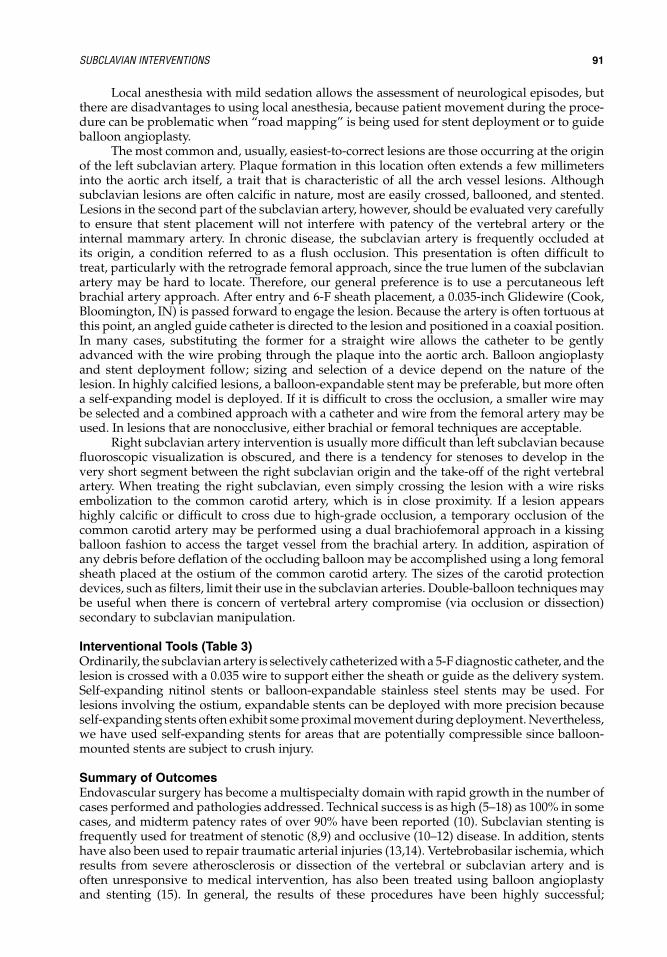
IHBK065-07 IHBK065-Minar April 24, 2010 13:39 Specs: 7x10 tight 7in×10in Char Count=
SUBCLAVIAN INTERVENTIONS 91
Local anesthesia with mild sedation allows the assessment of neurological episodes, butthere are disadvantages to using local anesthesia, because patient movement during the proce-dure can be problematic when “road mapping” is being used for stent deployment or to guideballoon angioplasty.
The most common and, usually, easiest-to-correct lesions are those occurring at the originof the left subclavian artery. Plaque formation in this location often extends a few millimetersinto the aortic arch itself, a trait that is characteristic of all the arch vessel lesions. Althoughsubclavian lesions are often calcific in nature, most are easily crossed, ballooned, and stented.Lesions in the second part of the subclavian artery, however, should be evaluated very carefullyto ensure that stent placement will not interfere with patency of the vertebral artery or theinternal mammary artery. In chronic disease, the subclavian artery is frequently occluded atits origin, a condition referred to as a flush occlusion. This presentation is often difficult totreat, particularly with the retrograde femoral approach, since the true lumen of the subclavianartery may be hard to locate. Therefore, our general preference is to use a percutaneous leftbrachial artery approach. After entry and 6-F sheath placement, a 0.035-inch Glidewire (Cook,Bloomington, IN) is passed forward to engage the lesion. Because the artery is often tortuous atthis point, an angled guide catheter is directed to the lesion and positioned in a coaxial position.In many cases, substituting the former for a straight wire allows the catheter to be gentlyadvanced with the wire probing through the plaque into the aortic arch. Balloon angioplastyand stent deployment follow; sizing and selection of a device depend on the nature of thelesion. In highly calcified lesions, a balloon-expandable stent may be preferable, but more oftena self-expanding model is deployed. If it is difficult to cross the occlusion, a smaller wire maybe selected and a combined approach with a catheter and wire from the femoral artery may beused. In lesions that are nonocclusive, either brachial or femoral techniques are acceptable.
Right subclavian artery intervention is usually more difficult than left subclavian becausefluoroscopic visualization is obscured, and there is a tendency for stenoses to develop in thevery short segment between the right subclavian origin and the take-off of the right vertebralartery. When treating the right subclavian, even simply crossing the lesion with a wire risksembolization to the common carotid artery, which is in close proximity. If a lesion appearshighly calcific or difficult to cross due to high-grade occlusion, a temporary occlusion of thecommon carotid artery may be performed using a dual brachiofemoral approach in a kissingballoon fashion to access the target vessel from the brachial artery. In addition, aspiration ofany debris before deflation of the occluding balloon may be accomplished using a long femoralsheath placed at the ostium of the common carotid artery. The sizes of the carotid protectiondevices, such as filters, limit their use in the subclavian arteries. Double-balloon techniques maybe useful when there is concern of vertebral artery compromise (via occlusion or dissection)secondary to subclavian manipulation.
Interventional Tools (Table 3)Ordinarily, the subclavian artery is selectively catheterized with a 5-F diagnostic catheter, and thelesion is crossed with a 0.035 wire to support either the sheath or guide as the delivery system.Self-expanding nitinol stents or balloon-expandable stainless steel stents may be used. Forlesions involving the ostium, expandable stents can be deployed with more precision becauseself-expanding stents often exhibit some proximal movement during deployment. Nevertheless,we have used self-expanding stents for areas that are potentially compressible since balloon-mounted stents are subject to crush injury.
Summary of OutcomesEndovascular surgery has become a multispecialty domain with rapid growth in the number ofcases performed and pathologies addressed. Technical success is as high (5–18) as 100% in somecases, and midterm patency rates of over 90% have been reported (10). Subclavian stenting isfrequently used for treatment of stenotic (8,9) and occlusive (10–12) disease. In addition, stentshave also been used to repair traumatic arterial injuries (13,14). Vertebrobasilar ischemia, whichresults from severe atherosclerosis or dissection of the vertebral or subclavian artery and isoften unresponsive to medical intervention, has also been treated using balloon angioplastyand stenting (15). In general, the results of these procedures have been highly successful;
IHBK065-07 IHBK065-Minar April 24, 2010 13:39 Specs: 7x10 tight 7in×10in Char Count=
92 DIETHRICH
Table 3 List of Tools for Subclavian Intervention
Equipment for Subclavian Stenting (Brachial Approach)
– 18-gauge Cook (Cook Bloomington, IN) needle / Cook micropuncture set– Indeflator (ACS, Santa Clara, CA)– Guidewire: 0.035-inch × 180-cm angled Glidewire (Terumo, Somerset, NJ) or 0.035-inch, 180-cm straight
Glidewire (Terumo)– Guidewire: 0.035-inch × 180-cm Amplatz (Cook)– Guidewire: 0.035-inch × 180-cm straight-stiff Glidewire (Terumo)– Catheter: 5-F × 65-cm Berenstein (Boston Scientific, Natick, MA)– Sheath: 6-F × 11-cm Cordis Brite-tip (Cordis, Miami Lakes, FL)– Sheath: 6-F × 55-cm Cordis Brite-tip (Cordis)– Balloons: Cordis Opta-Pro 80-cm (Cordis), or Abbott Fox PTA 75-cm (Abbott Laboratories, Abbott Park, IL)– Balloon-expandable stent (size/type to be determined by physician)
Equipment for Subclavian Stenting (Femoral Approach)
– 18-gauge Cook needle (Cook, Bloomington, IL)– Indeflator (ACS, Santa Clara, CA)– Guidewire: 0.035-inch × 260-cm angled Glidewire (Terumo, Somerset, NJ)– Guidewire: 0.035-inch × 260-cm Amplatz (Cook)– Catheter: 5-F × 100-cm Bentson 2 (Cook), or 5 F × 100-cm Headhunter (Terumo)– Sheath: 7-F × 11-cm Cordis Brite-tip (Cordis, Miami Lakes, FL)– Sheath: 7-F × 90-cm Cook Flexor Shuttle (Cook)– Balloons: Cordis Opta-Pro 110-cm (Cordis), or Abbott Fox PTA 135-cm (Abbott Laboratories, Abbott Park,
IL)– Balloon-expandable stent (size/type to be determined by physician)
complications, such as stent deformation (16), fracture (17), and infection yielding aneurysmformation (18) are rare.
Equipment ChecklistEquipment for Subclavian Stenting (Brachial Approach)
– 18-gauge Cook (Cook Bloomington, IN) needle / Cook micropuncture set– Indeflator (ACS, Santa Clara, CA)– Guidewire: 0.035-inch × 180-cm angled Glidewire (Terumo, Somerset, NJ) or 0.035-inch,
180-cm straight Glidewire (Terumo)– Guidewire: 0.035-inch × 180-cm Amplatz (Cook)– Guidewire: 0.035-inch × 180-cm straight-stiff Glidewire (Terumo)– Catheter: 5-F × 65-cm Berenstein (Boston Scientific, Natick, MA)– Sheath: 6-F × 11-cm Cordis Brite-tip (Cordis, Miami Lakes, FL)– Sheath: 6-F × 55-cm Cordis Brite-tip (Cordis)– Balloons: Cordis Opta-Pro 80-cm (Cordis), or Abbott Fox PTA 75-cm (Abbott Laboratories,
Abbott Park, IL)– Balloon-expandable stent (size/type to be determined by physician)
Equipment for Subclavian Stenting (Femoral Approach)
– 18-gauge Cook needle (Cook, Bloomington, IL)– Indeflator (ACS, Santa Clara, CA)– Guidewire: 0.035-inch × 260-cm angled Glidewire (Terumo, Somerset, NJ)– Guidewire: 0.035-inch × 260-cm Amplatz (Cook)– Catheter: 5-F × 100-cm Bentson 2 (Cook), or 5-F × 100-cm Headhunter (Terumo)– Sheath: 7-F × 11-cm Cordis Brite-tip (Cordis, Miami Lakes, FL)– Sheath: 7-F × 90-cm Cook Flexor Shuttle (Cook)– Balloons: Cordis Opta-Pro 110-cm (Cordis), or Abbott Fox PTA 135-cm (Abbott Laboratories,
Abbott Park, IL)– Balloon-expandable stent (size/type to be determined by physician)

IHBK065-07 IHBK065-Minar April 24, 2010 13:39 Specs: 7x10 tight 7in×10in Char Count=
SUBCLAVIAN INTERVENTIONS 93
PART 2: COMPLEX CASES
Case #1: Long Total Occlusion of the Left Subclavian ArteryAn 80-year-old female presented with dizziness and headaches when using her left arm forextended periods. She was evaluated with duplex ultrasound of the left arm and carotids,revealing total occlusion of her left subclavian artery and reversal of flow in her left vertebralartery.
Procedure:Access to the left brachial artery was obtained in a retrograde fashion, with an 18-gauge Cookneedle and a 0.035-inch × 180-cm angled Glidewire using Seldinger technique. A 6-F × 1-cmsheath was then placed in the artery, and 5000 units of heparin were given intravenously; theactivated clotting time was maintained at 200 to 250 for the remainder of the case. A retro-grade hand-injected arteriogram was performed, showing a long-segment total occlusion of theprevertebral left subclavian artery, with a large, patent left vertebral artery in close proximity.To obtain better working leverage, the short sheath was exchanged for a 6-F × 55-cm Cordissheath. A repeat arteriogram was performed. Once again, the regular 0.035-inch × 180-cmangled Glidewire was advanced through the sheath and attempts were made to cross thelesion. These were unsuccessful, so a 5-F × 90-cm Berenstein catheter was advanced overthe wire to obtain even greater working leverage. Multiple attempts at subintimal dissec-tion were made using the regular-angled 0.035-inch × 180-cm Glidewire. Eventually, a tightloop was formed but re-entry was not established. To obtain greater leverage, the Berensteincatheter was exchanged for a 5-F LIMA catheter. New attempts to cross the lesion with theregular angled 0.035-inch × 180-cm Glidewire failed, so it was exchanged for a straight stiff0.035-inch × 180-cm Glidewire. Using the combination of the long sheath, LIMA catheter, andstraight-stiff Glidewire, we were able to cross the lesion subintimally and re-enter the true lumenat the ostium of the subclavian artery. After crossing the lesion, confirmation of re-entry into thetrue lumen was obtained by back bleeding through the catheter and injection of a puff of contrast.The wire and catheter were then advanced into the descending thoracic aorta. To gain supportfor the planned intervention, the wire was exchanged for an Amplatz 0.035-inch × 180-cm wire.The lesion was then predilated with a 5-mm × 7-cm semi-compliant Fox balloon. An angiogramwas done to mark the landing zone. A Bridge Assurant 8-mm × 30-mm balloon-expandablestent was then deployed, with care not to cover the vertebral artery (Figs. 1 and 2). A secondstent was required to obtain sufficient overhang into the aorta. Pressure gradients after balloonangioplasty were 60 mm Hg, but this resolved with stenting. A completion angiogram was thenobtained, demonstrating completely patent subclavian, vertebral, and mammary arteries. Thesheath was then removed under pressure in the recovery room.
Figure 1 Angiogram showing occlusion of the left subclavian artery and post-stent deployment with goodarterial flow.

IHBK065-07 IHBK065-Minar April 24, 2010 13:39 Specs: 7x10 tight 7in×10in Char Count=
94 DIETHRICH
Figure 2 Photo of Bridge Assurant stent, 8mm × 30 mm.
What Made the Case Complex?The hazards involved in treating this patient were primarily anatomical. The lesion wascomplex—a long total occlusion of the left subclavian artery, requiring stent extension nearthe vertebral vessel. Great precision in landing the stent was required to prevent coverageof this vessel. Further, the risk of failure in treating a long subclavian artery occlusion hashistorically been high.
What was the Clinical Indication?On the basis of the patient’s symptoms and the noninvasive tests, a diagnosis of subclaviansteal syndrome was made.
What were the Potential Hazards of Treating this Patient and Anatomy?The hazards involved in treating this patient were primarily anatomical as described earlier.
Which Interventional Strategies were Considered?We chose a brachial approach on the basis of our extensive experience with such complex lesions.A femoral approach was not appropriate due to the inherent lack of catheter and wire supportit affords. Once the lesion has been crossed using a retrograde brachial approach, placement ofa stiff guidewire (such as the Amplatz wire used in this case) provides sufficient support formost interventions. However, in treating long, highly calcified lesions, we sometimes employ abrachiofemoral wire when additional support is needed.
Which Interventional Tools were used to Manage this Case?(See list of equipment used in this case)
Summary: How was the Case Managed, is There Anything That Could Have Been Improved, and Whatwas the Immediate and Long-Term Outcome?In treating proximal branch vessels arising from the aorta, we almost always employ balloon-expandable stents; it is our belief that their greater radial force is of benefit. In this particular case,we could have judged better by selecting the length of the initial stent, and therefore avoidedthe need for a second stent. The patient is currently 12 months out from her intervention and isdoing well. No recurrent symptoms have been noted.
List of Equipment used in this Case18-gauge Cook needle (Cook, Bloomington, IN)6-F × 11-cm Cordis sheath (Cordis, Miami Lakes, FL)6-F × 55-cm Cordis sheath (Cordis)0.035-inch × 180-cm regular angled Glidewire (Terumo, Somerset, NJ)0.035-inch × 180-cm straight stiff Glidewire (Terumo)0.035-inch × 180-cm Amplatz wire (Cook)5-F × 90-cm Berenstein catheter (Boston Scientific, Natick, MA)5-F × 90-cm IMA catheter (Cordis)7-mm × 4-cm Fox balloon (Abbott Labs, Abbott Park, IL)(2) 8-mm × 30-mm Bridge Assurant balloon-expandable stents (Medtronic, Santa Rosa, CA)

IHBK065-07 IHBK065-Minar April 24, 2010 13:39 Specs: 7x10 tight 7in×10in Char Count=
SUBCLAVIAN INTERVENTIONS 95
Key Learning Issues of this Specific CaseAs mentioned earlier, in this particular case, we could have judged better by selecting the lengthof the initial stent, and therefore avoided the need for a second stent.
Case # 2: Heavily Calcified High-Grade Stenosis of the Left Subclavian Artery WithTotal Occlusion of the Innominate Artery and High-Grade Stenosis of the Left CommonCarotidA 72-year-old male who presents with coronary artery bypass grafting with three vein grafts inthe remote past.
Procedure:After addressing the right common carotid lesion from a femoral approach using an I-Castcovered stent, we proceeded with intervention on the subclavian lesion. Access to the leftbrachial artery was obtained in a retrograde fashion with an 18-gauge Cook needle and a0.035-inch × 180-cm angled Glidewire using Seldinger technique. A 6-F × 11-cm sheath wasthen placed in the artery. The Glidewire was then advanced across the lesion with ease. Theshort sheath was exchanged for a 7-F × 55-cm Cordis sheath, which was advanced into thepostvertebral left subclavian artery. A hand-injected retrograde arteriogram was then obtained,demonstrating a short-segment, highly calcified ostial lesion of >90% stenosis, with a largeand patent vertebral artery. With the dilator re-inserted into the sheath, it was advanced acrossthe lesion into the aorta. A puff of contrast was then used to confirm placement in the aorta.A pull-back arteriogram was then obtained to mark the landing zones. The sheath was re-advanced into the aorta, using the dilator. Because of the lesion’s heavy calcification and therisk of embolizing into the patient’s only vertebral artery, we chose to proceed with primarystenting using a covered, balloon-expandable stent. A 7-mm × 22-mm I-Cast stent was selected.The stent was advanced over the wire into the sheath, which had been advanced distal tothe lesion. The sheath was pulled back to uncover the stent and prepare it for deployment.A predeployment angiogram was obtained through the sheath to confirm correct positioning.The stent-graft was slowly deployed, allowing the balloon to form an hourglass shape. Afterdeployment, the stent’s proximal overhang was flared by advancing the balloon partially intothe aorta and inflating it. The wire and balloon were removed and a completion arteriogramwas obtained, showing excellent results (Figs. 3 and 4). A Boomerang device was then used toclose the artery (Fig. 5).
What made the Case Complex?Because of innominate artery occlusion, severe coronary disease, and highly calcific carotid andsubclavian lesions, the patient is considered a complex case. In addition, the subclavian lesionin this case was considered complex, because of the lesion’s high degree of calcification and
Figure 3 Angiogram showing 90% ostial lesion and post-stent deployment.

IHBK065-07 IHBK065-Minar April 24, 2010 13:39 Specs: 7x10 tight 7in×10in Char Count=
96 DIETHRICH
Figure 4 Photo of I-Cast stent, 7 mm × 20 mm(note covering of stent).
because the left vertebral artery appeared to be the only patent vessel connecting to the patient’sposterior cerebral circulation.
What was the Clinical Indication for Treatment of this Patient?He had a recent cardiac event that lead to repeat catheterization, revealing coronary disease notamenable to revascularization, total occlusion of the innominate artery, and high-grade stenosis(> 90%) of both the left common carotid and left subclavian arteries.
What were the Potential Hazards of Treating this Patient and Anatomy?Stenting was considered important in this case, as balloon angioplasty would not have beena viable long-term treatment in this very calcified lesion. Embolization appears to be a truerisk in highly calcified lesions and, given the importance of this patient’s left vertebral artery,we believed steps should be taken to minimize this risk. Therefore, we chose to proceed withprimary stenting in the hope this would minimize the potential for free fragmentation of debris.
Which Interventional Strategies were Considered?In similar cases, one option might have been the use of a protection device such as theEmboshield (Abbbott Labs, Abbott Park, IL) but, in this case, the vertebral artery was smallerthan the device was designed for.
Which Interventional Tools were used to Manage this Case?We selected a covered stent (I-Cast, Atrium Medical, Hudson, NH), believing this would furtherreduce the possibility of embolization.
Summary: How was the case Managed, is There Anything that Could Have Been Improved, and What wasthe Immediate and Long-Term Outcome?The stent-graft was slowly deployed, allowing the balloon to form an hourglass shape. Afterdeployment, the stent’s proximal overhang was flared by advancing the balloon partially into
Figure 5 Photograph of the BoomerangDevice for arterial closure.

IHBK065-07 IHBK065-Minar April 24, 2010 13:39 Specs: 7x10 tight 7in×10in Char Count=
SUBCLAVIAN INTERVENTIONS 97
the aorta and inflating it. The wire and balloon were removed, and a completion arteriogramwas obtained, showing excellent results (Figs. 3 and 4). A Boomerang device was then used toclose the artery. The patient is now six months out from his procedure and doing well.
List of Equipment used in this Case18-gauge Cook needle (Cook, Bloomington, IL)6-F × 11-cm Cordis sheath (Cordis, Miami Lakes, FL)7-F × 55-cm Cordis sheath (Cordis)0.035-inch × 180-cm regular angled Glidewire (Terumo, Somerset, NJ)7-mm × 22-mm I-Cast stent graft (Atrium Medical, Hudson, NH)5/6 Boomerang closure device (Cardiva, Mountain View, CA)
Key Learning Issues of this Specific CaseThe vertebral artery is very fragile, so risk of dissection is of great concern. Another optionmight have been abandoning endovascular therapy altogether and proceeding with carotid–subclavian bypass after stenting the left common carotid. This was not a good option for thispatient because of his severe cardiac disease and recent cardiac event.
Case #3: Difficult Left Carotid–Subclavian Bypass StenosisThis is a 50-year-old male, who had undergone an ascending aortic graft for a Type I dis-section three years ago. He then developed a large aortic arch aneurysm that was treated bya rerouting procedure, with grafts from the ascending aorta (anastomosed to the previousgraft) to the innominate and left common carotid arteries. The arch aneurysm was excludedwith a Gore TAG thoracic endoprosthesis (WL Gore Associates, Flagstaff, AZ). The left sub-clavian artery was intentionally covered with this graft. The patient did well until six weekspostprocedure, at which time he developed left upper extremity claudication necessitating aleft carotid–subclavian bypass. Recurrent symptoms developed two months later, and duplexscanning showed increased velocities at the carotid anastomosis.
Procedure:Access to the left brachial artery was obtained in a retrograde fashion with an 18-gauge Cookneedle and a 0.035-inch × 180-cm angled Glidewire, using Seldinger technique. A 6-F × 11-cmsheath was then placed in the artery. Heparin was given intravenously, and the activatedclotting time was kept at 200 to 250 seconds for the remainder of the case. A 4-F × 90-cm angledGlide catheter was advanced over the wire to the proximal subclavian, and a hand-injectedarteriogram obtained. This revealed an occluded proximal left subclavian artery, a patent leftvertebral artery, and a patent carotid–subclavian bypass. The bypass was selectively cannulated,and an arteriogram of the bypass performed. This showed significant evidence of a carotid-to-graft anastomotic stenosis, greater than 50%. The bypass was crossed with a 0.035-inch × 180-cmGlidewire. Because of the angle and severity of stenosis, a Glide catheter would not advanceover the wire into the common carotid artery. A Spectranetics “Quick-cross” catheter was placedover the wire into the common carotid and rerouting bypass graft. The wire was removed, andpull-back gradients obtained across the carotid–subclavian bypass, demonstrating a 40 mm Hgsystolic gradient with significant dampening of the waveform. The bypass was re-crossed witha 0.035-inch × 180-cm regular angled Glidewire, advanced to the proximal rerouting bypassand into the ascending aorta. The Spectranetics catheter was used to exchange this for a 0.035-inch × 450-cm angled stiff Glidewire. Fortunately, the Glidewire tracked easily into the thoracicendograft and down to the aorta into the left iliac artery. Left femoral access was obtained usingan 18-gauge Cook needle with a 0.035-inch × 260-cm regular angled Glidewire and a 9-F ×11-cm Cordis sheath placed in a retrograde fashion. The 450-cm Glidewire was snared andpulled out of the left femoral sheath, establishing a left-brachial-to-left-femoral wire.
After establishing the left-brachial-to-left-femoral wire, tension was applied at each endby grasping the wire with hemostats and pulling. The left brachial sheath was exchanged for a7-F × 11-cm Cordis sheath, and a Multi-track 7-F monorail angiocatheter was advanced over thebrachial aspect of the wire into the left carotid–subclavian bypass. A power injector angiogramwas obtained showing the stenosis. Next, a balloon angioplasty was performed using first a

IHBK065-07 IHBK065-Minar April 24, 2010 13:39 Specs: 7x10 tight 7in×10in Char Count=
98 DIETHRICH
Figure 6 Spectranetics Multi-track angio-graphic catheter.
4-mm × 2-cm, then a 6-mm × 2-cm OptiPro balloon. The Multi-track (Spectranetics) catheter(Fig. 6) was used for a postangioplasty arteriogram, which showed significant rebound of thelesion. Due to the rebound, a 6-mm × 17-mm Express LD stent was deployed, leaving a 2-mmoverhang into the common carotid artery (Figs. 7 and 8). The Multi-track catheter was againused to obtain a completion angiogram, which now showed resolution of the stenosis. Gradientswere re-evaluated, showing improved waveform and resolution of pressure differences. Thebrachial and femoral sheaths were removed in the recovery room.
What Made the Case Complex?The complexity of this case is reflected in the inability to use traditional techniques, to evaluateand treat the lesion because of the lack of “pushability” of the catheters in relation to the lesion.
What was the Clinical Indication for Treatment of this Patient?This patient had undergone an ascending aortic graft for a Type I dissection three years ago.He then developed a large aortic arch aneurysm that was treated by a rerouting procedure,with grafts from the ascending aorta (anastomosed to the previous graft) to the innominate andleft common carotid arteries. He developed left upper extremity claudication necessitating aleft carotid–subclavian bypass. Recurrent symptoms developed two months later, and duplexscanning showed increased velocities at the carotid anastomosis.
Figure 7 Angiogram showing stenosis of the carotid–subclavian bypass and post-stent deployment.

IHBK065-07 IHBK065-Minar April 24, 2010 13:39 Specs: 7x10 tight 7in×10in Char Count=
SUBCLAVIAN INTERVENTIONS 99
Figure 8 Photo of Express LD stent (6 ×17 mm) used to correct subclavian bypassstenosis.
What were the Potential Hazards of Treating this Patient and Anatomy?Because of the angulation and stenosis of the bypass, we were unable to pass even a simplecatheter over a wire into the common carotid. This was initially overcome using the Spectranetics“quick-cross” catheter, with its very low-profile tapered tip. This is one of our workhorsecatheters for difficult lesions and total occlusions. Next, we were faced with a defined problemthat we could not treat because no balloon/stent would cross the lesion from the brachialapproach.
Which Interventional Strategies were Considered?One option that we considered was the placement of a very stiff wire, such as an Amplatz wire,across the lesion for support. The downside of such an approach is the potential for injury ordissection of the left common carotid artery. Ultimately, we chose to obtain femoral access andestablish a brachial-to-femoral wire for the added support needed, with less risk of injury to thecarotid artery.
Which Interventional Tools Were Used to Manage this Case?Given the nature of the patient’s previous rerouting, the intervention was challenging. Themaneuver would not have been possible without the extended-length (450-cm) Glidewire.
Summary: How was the Case Managed, is There Anything that Could Have Been Improved, and What wasthe Immediate and Long-Term Outcome?After establishing the brachial-to-femoral wire, the final challenge was how to performangiograms while maintaining the wire in place. This problem was easily overcome usingthe Multi-track angiocatheter, which has a monorail design, allowing power-injection imag-ing while maintaining the wire. The patient is now two months postprocedure and is free ofsymptoms.
List of Equipment used in this Case(2) 18-gauge Cook needle (Cook, Bloomington, IN)6-F × 11-cm Cordis sheath (Cordis, Miami Lakes, FL7-F × 11-cm Cordis sheath (Cordis)9-F × 11-cm Cordis sheath (Cordis)0.035-inch × 180-cm regular angled Glidewire (Terumo, Somerset, NJ)0.035-inch × 260-cm regular Glidewire (Terumo)

IHBK065-07 IHBK065-Minar April 24, 2010 13:39 Specs: 7x10 tight 7in×10in Char Count=
100 DIETHRICH
0.035-inch × 450-cm angled stiff Glidewire (Terumo)Spectranetics “Quick-cross” catheter (Spectranetics, Colorado Springs, CO)7-F Multi-track angiographic catheter (NuMed, Hopkinton, NY)4-mm × 2-cm OptiPro Balloon (Cordis, Miami Lakes, FL)6-mm × 2-cm OptiPro Balloon (Cordis)6-mm × 17-mm Express LD Stent (Boston Scientific, Natick, MA)
Key Learning Issues of this Specific CaseThis was a patient with significant disease and at risk from prior intervention. Because ofangulation and stenosis of the bypass, we were unable to pass even a simple catheter over awire into the common carotid. It is important to consider all options in such a case and to havea variety of tools at hand. Considerable experience with these tools and an experienced team isa must in these situations.
SUMMARY OF KEY ISSUES FOR SAFE, SUCCESSFUL TREATMENTOF SUBCLAVIAN LESIONSEndovascular therapy of the subclavian arteries has been in wide use for a number of years andhas proven safe and effective. Complications associated with endovascular therapy are oftenpreventable, and the use of appropriate technique is one of the most important ways to limitcomplications. Preoperative imaging is extremely important in determining the indication andoptimal approach for subclavian intervention. Although angiography remains the gold stan-dard, CT and magnetic resonance angiography offer additional diagnostic value, as does IVUSimaging. IVUS may be particularly useful in vessel diameter determination so that overdilata-tion and arterial injury can be prevented. It is also helpful in accurate stent placement at theorigins of the subclavian or aortic arch. Gentle manipulation of endovascular equipment is veryimportant in reducing the risk of embolization and other complications. While restenosis doesnot represent a considerable problem in the subclavian arteries, it may present as a complica-tion later. As such, ongoing clinical follow-up and imaging is mandatory in those who haveundergone endovascular intervention.
REFERENCES1. Diethrich EB, Garrett HE, Ameriso J, et al. Occlusive disease of the common carotid and subclavian
arteries treated by carotid-subclavian bypass. Analysis of 125 cases. Am J Surg 1967; 114:800–808.2. DeBakey ME, Diethrich EB, Garrett HE, et al. Surgical treatment of cerebrovascular disease. Postgrad
Med 1967; 42:218–230.3. Diethrich EB, Koopot R. Simplified operative procedure for proximal subclavian arterial lesions: direct
subclavian anastomosis. Am J Surg 1981; 142:416–421.4. Przewlocki T, Kablak-Ziembicka A, Pieniazek P, et al. Determinants of immediate and long-term results
of subclavian and innominate artery angioplasty. Catheter Cardiovasc Interv 2006; 67(4):519–526.5. Motarjeme A. Percutaneous transluminal angioplasty of supra-aortic vessels. J Endovasc Surg 1996;
3(2):171–181.6. Martinez R, Rodriguez JA, Torruella L, et al. Stenting for occlusion of the subclavian arteries: Technical
aspects and follow-up results. Tex Heart Inst J 1997; 24:23–27.7. Mathias KD, Luth I, Haarmann P. Percutaneous transluminal angioplasty of proximal subclavian
artery occlusions. Cardiovasc Intervent Radiol 1993; 16(4):214–218.8. Nomura M, Kida S, Yamashima T, et al. Percutaneous transluminal angioplasty and stent placement
for subclavian and brachocephalic artery stenosis in aortitis syndrome. Cardiovasc Intervent Radiol1999; 22:427–432.
9. Maskovic J, Jankovic S, Lusic I, et al. Subclavian artery stenosis caused by non-specific arteritis(Takayasu disease): treatment with Palmaz stent. Eur J Radiol 1999; 31:193–196.
10. Rodriguez-Lopez J, Werner A, Martinez R, et al. Stenting for atherosclerotic occlusive disease of thesubclavian artery. Ann Vasc Surg 1999; 13:254–260.
11. Al-Mubarak N, Liu MW, Dean LS, et al. Immediate and late outcomes of subclavian artery stenting.Catheter Cardiovasc Interv 1999; 46:169–172.
12. Korner M, Baumgartner I, Do DD, et al. PTA of the subclavian and innominate arteries: long-termresults. Vasa 1999; 28:117–122.

IHBK065-07 IHBK065-Minar April 24, 2010 13:39 Specs: 7x10 tight 7in×10in Char Count=
SUBCLAVIAN INTERVENTIONS 101
13. d’Othee BJ, Rousseau H, Otal P, et al. Noncovered stent placement in a blunt traumatic injury of thesubclavian artery. Cardiovasc Intervent Radiol 1999; 22:424–427.
14. Bruninx G, Wery D, Dubois E, et al. Emergency endovascular treatment of an acute traumatic ruptureof the thoracic aorta complicated by a distal low-flow syndrome. Cardiovasc Intervent Radiol 1999;22:515–518.
15. Malek AM, Higashida RT, Phatouros CC, et al. Treatment of posterior circulation ischemia withextracranial percutaneous balloon angioplasty and stent placement. Stroke 1999; 30:2073–2085.
16. Sitsen ME, Ho GH, Blankensteijn JD. Deformation of self-expanding stent-grafts complicatingendovascular peripheral aneurysm repair. J Endovasc Surg 1999; 6:288–292.
17. Phipp LH, Scott DJ, Kessel D, et al. Subclavian stents and stent-grafts: cause for concern? J EndovascSurg 1999; 6:223–226.
18. Malek AM, Higashida RT, Reilly LM, et al. Subclavian arteritis and pseudoaneurysm formation sec-ondary to stent infection. Cardiovasc Intervent Radiol 2000; 23:57–60.

IHBK065-08 IHBK065-Minar May 1, 2010 7:33 Specs: 7x10 tight 7in×10in Char Count=
8 Vertebral InterventionsJ. Stephen JenkinsInterventional Cardiology Research, Ochsner Heart and Vascular Institute, New Orleans, Louisiana, U.S.A.
COMPLEX VERTEBRAL INTERVENTIONSThe complexity of vertebral artery intervention is determined by the anatomical location of thelesion being treated (1). The anatomic course of the vertebral artery is divided into four segments(V1–V4) (Fig. 1). The complexity of ostial vertebral artery angioplasty presents a challenge asdoes angioplasty in the ostial location of any vessel. Precise stent placement covering the ostiumwithout protruding excessively into the parent artery can always be a challenge. One to two mil-limeters of stent hanging into the parent artery is necessary to prevent missing ostial disease (2).
The V1 segment originates at subclavian artery and extends to the transverse foraminaof either the fifth or sixth cervical vertebrae. This segment can be treated percutaneously withrelative ease provided it is not tortuous or redundant, as it tends to be in the more elderlypatient. Percutaneous angioplasty is relatively simple in straight V1 segment arteries (3).
The V2 segment courses through the bony canal of the transverse foramina from C2 to C6.This is the easiest vertebral segment to treat percutaneously due to favorable anatomic featuressuch as its short distance from the subclavian artery and its straight course.
The V3 segment continues as the artery exits the transverse foramina at C2 and endsas the vessel penetrates the dura mater and becomes an intracranial vessel. The complexityof angioplasty in the V3 segment increases as this segment is extremely tortuous to allowmobility of the alanto-axial and the alanto-occipital joint. Care should be taken to avoid balloonexpandable stents in this flexible joint region. Short self-expanding stents and avoiding extremetortuousity increase success in this segment.
The V4 segment courses along the inferior portion of the pons until joining the contralateralvertebral artery to form the basilar artery. The anterior spinal communicator arteries originatefrom the V4 segments bilaterally and join in the midline to supply the anterior two-thirds of thespinal cord. For this reason, angioplasty in the V4 segment should be approached with extremecaution, as occlusion of the anterior spinal communicators can cause major deficits.
CLINICAL INDICATIONS FOR TREATMENT OF COMPLEX LESIONSIN VERTEBRAL INTERVENTIONSVertebral artery revascularization is indicated when medical therapy has failed to alleviatesymptoms of vertebral basilar insufficiency (VBI) (4,5). Accepted posterior circulation (verte-brobasilar) ischemic symptoms include: diplopia, dizziness, drop attacks, gait disturbance, dys-phasia, and bilateral homonymous hemianopia (6). Prior to consideration for revascularization,the anatomy of the anterior and posterior circulation and the circle of Willis should be defined.A diagnostic aortic arch and four-vessel angiography including selective carotid and vertebralartery angiography is required. The anatomy is then correlated with the posterior circulationsymptom complex to determine which artery requires revascularization. In the absence of prox-imal atherosclerotic occlusive disease, unilateral vertebral artery stenosis or occlusion is welltolerated. Revascularization therapy for symptomatic VBI should be pursued in patients with:
1. bilateral vertebral stenosis greater than 70% luminal diameter stenosis;2. unilateral stenosis greater than 70% luminal diameter stenosis in the presence of an occluded
or hypoplastic contralateral vertebral artery;3. artery-to-artery embolism from a stenosis in the vertebral artery even in the presence of a
unilateral stenosis (7,8).
Additional indications include instances where vertebral angioplasty would increase totalcerebral blood flow to patients with diffuse atherosclerotic disease involving occlusions of both

IHBK065-08 IHBK065-Minar May 1, 2010 7:33 Specs: 7x10 tight 7in×10in Char Count=
VERTEBRAL INTERVENTIONS 103
Figure 1 Vertebral artery anatomy.
carotid arteries (9). Vertebral angioplasty should not be attempted once the vessel is totallyoccluded. Although some percentage of total occlusions could technically be treated, the distalembolic debris would be clinically devastating due to territory supplied by V4 and basilar arterycausing cerebellar, midbrain, pons, medullary or brainstem infarctions. Total occlusion of thevertebral artery is a contraindication to endovascular treatment (10).
HAZARDS AND COMPLICATIONSProcedural complications of vertebral artery stenting include major and minor stroke, TransientIschemic Attacks (TIA), and all other complications that accompany percutaneous proceduresincluding death, access site bleeding, vessel rupture, and renal failure (11,12) (Table 1).
Important branches of the V4 segment include the posterior inferior cerebellar artery(PICA) and the anterior spinal artery. The anterior spinal artery from each V4 artery joins in themidline and supplies the anterior two-thirds of the spinal cord. It is because of these branchesthat angioplasty of the V4 segment is extremely risky and is rarely attempted except for acutestroke intervention or severe symptoms unresponsive to medical treatment.
INTERVENTIONAL STRATEGIESContrast angiography using digital subtraction techniques is the gold standard to identifyvertebral basilar atherosclerotic disease. Angiographic evaluation includes an aortic arch and
Table 1
Peri-interventional complications
Proximal vertebral artery Distal vertebral artery
TIA 1–2% 1%Major stroke 1–2% 10%MI 0 0Death <1% 3%

IHBK065-08 IHBK065-Minar May 1, 2010 7:33 Specs: 7x10 tight 7in×10in Char Count=
104 Jenkins
four-vessel study with selective angiography of bilateral carotid and vertebral arteries includingintracranial imaging. The intracranial distribution of the anterior and posterior circulation andthe circle of Willis should also be defined in several views making certain to identify anycollateral blood supply.
A 5-F Berenstein is the diagnostic catheter of choice to perform selective angiography ofthe vertebral artery. A 4-F, 5-F or 6-F Judkins right, internal mammary, or Vitek curved cathetersare acceptable if the vertebral artery cannot be cannulated with a Berenstein catheter. A mul-tipurpose curve is useful when procedures are performed using a brachial access. A 0.035-in.hydrophilic wire or J-wire should be used to place the diagnostic catheter in the subclavianartery distal to the vertebral artery ostium. A manifold is connected and continuous pressuremonitored while engaging the vertebral artery ostium. A 0.014-in. soft guidewire or filterwireis recommended for use with a rapid exchange or monorail balloon to predilate the stenoticlesion. Embolic protection devices are not FDA approved for use in the vertebral artery; how-ever, it should be strongly considered if there is a comfortable landing zone distal to the indexlesion. The predilation balloon should be 0.5 mm less than that of the reference vessel diameter.One must be very diligent to keep the tip of the guidewire within view at all times during theprocedure as guidewire perforation can cause fatal intracranial hemorrhage. This can happenwith any guidewire, but is of particular concern when a hydrophilic guidewire is used. Balloonexpandable coronary or peripheral stents on a 0.014-in. platform work well in the vertebralartery ostium. The proximal stent should be extended 1 to 2 mm into the subclavian artery toensure coverage of the vertebral artery ostium. Both balloon expandable and self-expandingstents are acceptable in the V1 and V2 segments of the vertebral artery. Balloon expandablestents should be used when the stenosis is in proximity of the vertebral artery ostium (13).
INTERVENTIONAL TOOLS
Size Device
Guide catheters 6-Fr EnvoyJudkins rightInternal mammaryMultipurpose
Guidewires 0.014 in. Balance Middle Weight (Abbott)Wisper (Abbott)Choice PT (Cordis)
Embolic protection 4–6 mm Filterwire (BSC)Stents balloon expandable 3–6 mm Vision (Abbott)
Liberte (BSC)Express Biliary SD (BSC)Palmaz Blue (Cordis)
Stents self-expanding 4–7 mm Xpert (Abbott)
SUMMARY OF OUTCOMESSymptomatic vertebral artery stenosis (VAS) has a five-year stroke risk of 30% to 35%. Mortalityassociated with posterior circulation (PC) strokes is high, ranging from 20% to 30% (10,14,15).Surgical revascularization is rarely performed due to high morbidity and mortality. Surgicalmorbidity includes recurrent laryngeal nerve palsy, Horner’s syndrome, lymphocele, chylotho-rax, and thrombosis (Table 2). Endovascular revascularization with stents offers a potentialtreatment option for these patients (Table 2) (16,17).
Percutaneous angioplasty and stent placement of PC, symptomatic, vertebrobasilaratherosclerotic disease is a safe and effective approach that avoids the morbidity associatedwith major surgery. Balloon angioplasty alone or combined with stenting is associated withhigh success rates and low to moderate restenosis rates when bare metal stents are used. Mostavailable data are limited to single-center retrospective reports. There is a scarcity of data usingdrug-eluting stents in PC territory and it will not be included in this review (Table 3) (2,12,18–20).

IHBK065-08 IHBK065-Minar May 1, 2010 7:33 Specs: 7x10 tight 7in×10in Char Count=
VERTEBRAL INTERVENTIONS 105
Table 2 Surgical Morbidity
Recurrent Laryngeal nerve palsy 2%Horner’s syndrome 15%Lymphocele 4%Chylothorax 5%Thrombosis 1%
The only randomized data to date comparing vertebral artery stenting to medical ther-apy was reported by Coward in 2007 (21). The CAVATAS study (Carotid and Vertebral ArteryTransluminal Angioplasty Study) included a total of 16 vertebral stenosis patients random-ized equally to endovascular or medical therapy. Endovascular treatment was successful in alleight patients with two procedural TIAs giving a 25% morbidity in the percutaneous arm. Atmean follow-up of 4.7 years there were no vertebral strokes in either group. The compositeof myocardial infarction, stroke, and death occurred in three patients in the medical treatmentarm and in four patients in the endovascular arm. The authors concluded from this study thatthere was no benefit from endovascular therapy and stroke was more common in the carotidterritory. Although the data benefit from being randomized, the numbers are too small to drawconclusions.
In the Cochrane Review reported by Coward et al. (22), the literature was searched forEnglish papers that reported at least three cases of vertebral artery intervention. He identified313 cases of vertebral artery intervention, which included 173 cases of primary stent placement.In these 20 studies combined, there was a 30-day major stroke and death rate of 3.2% and a30-day TIA and nondisabling stroke rate of 3.2%. These published case series combined suggestthat endovascular treatment of the vertebral artery is safe and effective although selection biasexists. Although this data is nonrandomized, it is much stronger than the one randomizedvertebral trial published to date.
EQUIPMENT CHECKLIST (TABLE 4)
Interventional Guidewires0.014′′ soft guidewire is recommended for use with a rapid exchange (RX) or monorail balloon.A filterwire should be used in all vertebral arteries with an adequate landing zone distal to theindex lesion. Although there is no data to support this statement, the author feels the obligatoryembolization of atherosclerotic debris with balloon angioplasty in a territory as sensitive as thePC can potentially be minimized with distal protection devices.
Interventional Guide CathetersA 6-F or 5-F Judkins right, multipurpose or envoy guiding catheter can be used to perform theintervention. A curve similar to the diagnostic imaging catheter should be chosen.
PTA BalloonsThe balloon length is determined by the lesion length and should cover the lesion completely.The balloon diameter should be at least 0.5 mm less than the reference vessel diameter.
Table 3 Studies of Vertebral Artery Stenting
References NTechnical
success rate (%)Proceduralcomplications
Improvementin symptoms
Meanfollow-up
Latestroke Restenosis
12 32 100 TIA (1) 31/32 10.6 mo 0/32 1/322 33 97 CVA (1) 27/33 16.2 mo 1/33 43%18 50 98 None 48/50 25 mo 1/50 10%19 58 100 CVA (3) 56/58 31.3 mo 0/58 25%20 38 100 TIA (1) 23/26 11 mo 0/38 36%
CVA = cerebral vascular event.
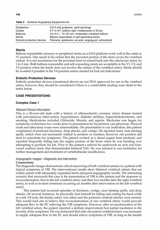
IHBK065-08 IHBK065-Minar May 1, 2010 7:33 Specs: 7x10 tight 7in×10in Char Count=
106 Jenkins
Table 4 Vertebral Angioplasty Equipments list
Guidewires 0.014 soft guidewire, rapid exchangeGuides 5-F or 6-F Judkins right, multipurpose or EnvoyBalloons 3.0–5.0 × 15–20 mm, moderately compliant balloonStents Balloon expandable or self-expanding stentsEmbolic protection devices Filterwire, spiderwire, accunet, angioguard, emboshield
StentsBalloon expandable coronary or peripheral stents on a 0.014 platform work well in the ostial orV1 position. One needs to be certain that the proximal portion of the stent covers the vertebralostium. It is not uncommon for the proximal stent to extend back into the subclavian artery by1 to 2 mm. Both balloon expandable and self-expanding stents are acceptable in the V1, V2, andV3 position when the lesion does not involve the ostium of the vertebral artery. Stents shouldbe avoided if possible in the V4 position unless needed for bail out indications.
Embolic Protection DevicesEmbolic protection devices (mentioned above) are not FDA approved for use in the vertebralartery, however, they should be considered if there is a comfortable landing zone distal to theindex lesion.
CASE PRESENTATIONS
Complex Case 1
Relevant Clinical InformationThis is a 50-year-old male with a history of atherosclerotic coronary artery disease treatedwith percutaneous intervention, hypertension, diabetes mellitus, hypercholesterolemia, andsmoking. Medications included Glyburide, Maxide, and aspirin. Meclozine was begun forsymptoms of dizziness two weeks prior to presentation by his primary care physician. Physicalexam and laboratory exam were unremarkable. On presentation to our institution, this patientcomplained of profound dizziness, drop attacks, and vertigo. He reported many near faintingspells, which were not necessarily related to position or exertion; however, one position didtend to exacerbate his symptoms. The patient worked as a diesel engine boat mechanic andreported frequently falling into the engine portion of the boats when he was bending overattempting to perform his job. Prior to the patient’s referral he underwent an arch and four-vessel cerebral study that demonstrated bilateral VAS. He was referred to our institution forfurther management and treatment of vertebrobasilar insufficiency.
Angiographic Images—Diagnostic and InterventionCommentaryThe diagnostic images demonstrate critical narrowing of both vertebral arteries in a patient withtypical symptoms of VBI. The interventional results show bilateral vertebral ostium that arewidely patent with adequately expanded stents and good angiographic results. The interestingscenario that surrounds this case is the presentation of VBI in this patient and the response torevascularization, first in the left vertebral artery and then two months later the right vertebralartery, as well as in-stent restenosis occurring six months after intervention on the left vertebralartery.
This patient had recurrent episodes of dizziness, vertigo, near fainting spells, and dropattacks. On several instances, he physically hurt himself by falling and cutting his head whileat work. Of note, the basilar artery was intact and the posterior cerebral arteries were normal.This would lead one to believe that revascularization of one vertebral artery would provideadequate flow to the PC relieving the VBI symptoms. However, after revascularization of theleft vertebral artery, the patient reported a definite improvement but partial resolution in theseverity of his symptoms. He was instructed that only one patent vertebral artery was necessaryto supply adequate flow to the PC and should relieve symptoms of VBI, as long as the basilar

IHBK065-08 IHBK065-Minar May 1, 2010 7:33 Specs: 7x10 tight 7in×10in Char Count=
VERTEBRAL INTERVENTIONS 107
Figure 2 This image demonstrates a high-grade ostialstenosis of the left vertebral artery. Approximately 2 cmdistal to the ostium the vessel makes a 90◦ turn cephalad.This gives an adequate window for angioplasty and stentingwith a short stent.
artery and posterior cerebrals are normal. Only partial resolution of his symptoms occurredwith a patent left vertebral artery and he requested on multiple occasions that intervention beperformed on the other vertebral artery. After much persistence, the right vertebral artery wasangioplastied and stented two months later with complete resolution of symptoms. Althoughit has been reported that one vertebral artery is adequate to prevent symptoms of VBI in man(23,24), this was clearly not adequate to relieve symptoms in this patient.
Figure 3 The final result demonstrates a well-deployedstent with a distal step down. Angioplasty was performedusing 8-Fr coronary equipment. Anticoagulation with 8000units of intravenous heparin was given to maintain an acti-vated clotting time (ACT) > 300 seconds and a diagnostic6-Fr JR4 catheter was used to selectively cannulate theleft vertebral artery ostium. A 0.035-in guidewire was theninserted into the V2 segment of the artery and the diag-nostic catheter was exchanged for an 8-Fr JR4 coronaryguiding catheter. A 4 × 2 peripheral angioplasty balloonwas used to perform predilation. A J&J P104 biliary stent(Cordis Corporation, Miami, FL) was then mounted on a5 × 2 balloon and deployed in the ostium of the left ver-tebral artery. Note that the short stent has allowed thetortuous segment of this artery to remain unchanged.

IHBK065-08 IHBK065-Minar May 1, 2010 7:33 Specs: 7x10 tight 7in×10in Char Count=
108 Jenkins
Figure 4 This image represents a 95% stenosis of theright vertebral artery ostium. Transition to the bony canalof the V2 segment is smooth, lending this vessel morefavorable anatomy for percutaneous angioplasty with lessrisk of distal dissection.
The patient remained asymptomatic and free of VBI symptoms with two patent vertebralarteries and could once again perform his duties as a diesel boat mechanic. Four months later,he presented again with VBI symptoms that were consistent with the symptoms he experiencedwith one patent vertebral artery. There was definite improvement but only partial resolutionin severity of the VBI symptoms when compared to his initial presentation. Repeat angiog-raphy was performed demonstrating in-stent restenosis of the left vertebral artery. Simple
Figure 5 This image demonstrates the final angioplastyresult in the ostial right vertebral artery. Anticoagulationwith heparin was given to maintain an ACT > 300 sec-onds and a 5-Fr Van Andle catheter was used to cannulatethe right vertebral artery ostium. A 0.035-in. guidewire wasthen advanced into the V2 segment of the artery takingcare not to traumatize the V4 segment with the wire. Twoimportant branches, the posterior inferior cerebellar arteryand anterior spinal communicators, arise from this seg-ment and care should be exercised to protect these smallvessels from the guidewire. The diagnostic catheter wasthen exchanged for an 8-Fr multipurpose coronary guidingcatheter. Predilation was performed with a 5 × 2 balloon.A J&J P104 biliary stent was mounted on the balloon anddeployed in the ostium of the right vertebral artery withhigh-pressure inflations. The final result demonstrates awidely patent ostium with a step down seen at the distalend of the stent.

IHBK065-08 IHBK065-Minar May 1, 2010 7:33 Specs: 7x10 tight 7in×10in Char Count=
VERTEBRAL INTERVENTIONS 109
Figure 6 This image of the left vertebral arterywas taken six months after stent implantation. Itdemonstrates a critical 90% in-stent restenosislesion.
balloon angioplasty was performed with adequate results and the patient’s symptoms onceagain resolved completely.
Contrary to the published literature with regards to the number of vertebral arteriesnecessary to relieve VBI symptoms, this patient clearly needed two patent vertebral arteries torelieve his symptoms. Whether the right vertebral was patent with an occluded left vertebralartery or vice versa, symptoms persisted that prevented this patient from performing his job.
(a) What made the case “complex”? This case is considered complex as it was necessary totreat both vertebral arteries in order to render this patient asymptomatic.
(b) What was the clinical indication for treatment of this patient? The indication to treat thispatient was drop attacks and dizziness.
(c) What were the potential hazards of treating this patient and anatomy? The hazards includedissection, stroke, and TIA.
(d) Which interventional strategies were considered? A strategy that would allow theguidewire to sit in the subclavian artery without intubating the vertebral artery was neces-sary to successfully treat both of these ostial lesions.
Figure 7 Left vertebral artery is shown aftertreatment of in-stent restenosis with a 5 × 2balloon at 18 atm. There is approximately a 10%residual stenosis.

IHBK065-08 IHBK065-Minar May 1, 2010 7:33 Specs: 7x10 tight 7in×10in Char Count=
110 Jenkins
(e) Which interventional tools were used to manage this case? A Judkins right coronary guideand sportwire were used to image and cross these lesions. Palmaz P154 stents were mountedon a 5 × 20 balloon and deployed at 12 atm.
(f) Summary: How was the case managed, is there anything that could have been improved,what was the immediate and long-term outcome? The outcome was suboptimal with onlypartial improvement after treatment on only one vertebral artery. The symptoms in thispatient resolved completely after stenting of both vertebral arteries.
(g) List of equipments used in this case: Plamaz P154 stent, 5 × 20 angioplasty balloon,sportwire, and Judkins right 4 guide.
(h) Key learning issues of this specific case: Two patent vertebral arteries are necessary in somepatients to provide adequate PC flow.
Complex Case 2This patient is an 83-year-old male with a history of dilated cardiomyopathy, peripheral vas-cular disease, and non-Hodgkin’s lymphoma treated with chemotherapy 10 years prior whopresented with two transient ischemic attacks in the previous month. The symptoms weredescribed as dizziness and blurred vision that lasted between 15 to 20 minutes and resolved.Initial work-up included a computed tomography scan of the head that was normal and carotidduplex scan that demonstrated 20% to 39% stenosis bilaterally. The patient was initially treatedwith coumadin maintaining a therapeutic INR for two months before the identical symptomsas above returned.
Physical examination revealed a blood pressure of 154/70 mm Hg and heart rate 60 beatsper minute. The patient was alert and oriented × 3. There was no carotid bruit, no neurologicfindings, and the rest of the physical exam was normal.
At this point, he was referred for an arch and four-vessel angiography with intracranialimaging due to break through symptoms on coumadin with a therapeutic INR. Angiographicimaging revealed an occluded right V4 artery that ended in PICA. The left vertebral artery hada 90% stenosis in the V4 segment at the vertebral basilar junction.
This scenario of angiographic findings represents a high-risk case with potential for severalcomplications. First, one should remember that angioplasty in the basilar artery can causemajor stroke if plaque shift occurs into anterior inferior cerebellar or pontine arteries thatarise from the basilar artery. Likewise, the posterior inferior cerebellar and anterior spinalcommunicator arteries arise from the V4 segment of the vertebral artery and can cause majorstroke if balloon inflations compromise these branches. It is therefore wise to attempt plain oldballoon angioplasty in these segments with an undersized balloon if an adequate angiographicresult can be obtained without a flow limiting dissection. Stents should only be used for bailout with suboptimal balloon angioplasty results.
Angiographic ImagesCommentaryThis case represents successful percutaneous balloon angioplasty in the most risky segment ofthe vertebral artery. The vertebral basilar junction deserves respect due the tiny branches thatarise from the V4 segment and the basilar artery. Major deficits or life-threatening cerebral vascu-lar accidents can occur if these small branches are compromised with percutaneous angioplasty.With the use of an undersized balloon and no stent, this patient was successfully treated withno complications. Conservatism should be practiced when considering percutaneous therapyin this segment of the vertebral artery, and percutaneous therapy should not be attempted ifthere is resolution of symptoms with antiplatelet or anticoagulant therapy alone.
(a) What made the case “complex”? This case was complex because of the location of thestenosis in the V4 segment.
(b) What was the clinical indication for treatment of this patient? There were symptoms ofdizziness and blurred vision while the patient was taking coumadin with therapeutic INRlevels. Failure of medical therapy to relieve symptoms constitutes an indication for archand four-vessel angiography to determine if percutaneous therapy is a therapeutic option.

IHBK065-08 IHBK065-Minar May 1, 2010 7:33 Specs: 7x10 tight 7in×10in Char Count=
VERTEBRAL INTERVENTIONS 111
Figure 8 This image demonstrates anoccluded right V4 at PICA.
(c) What were the potential hazards of treating this patient and anatomy? First, one shouldremember that angioplasty in the basilar artery can cause major stoke if plaque shift occursinto anterior inferior cerebellar or pontine arteries that arise from the basilar artery. Like-wise, the posterior inferior cerebellar and anterior spinal communicator arteries arise fromthe V4 segment of the vertebral artery and can cause major stroke if balloon inflationscompromise these branches.
(d) Which interventional strategies were considered? The only interventional strategy thatshould be considered in the basilar artery is provisional stenting. If an adequate result canbe obtained with plain old balloon angioplasty then this is the best approach.
(e) Which interventional tools were used to manage this case? A guiding catheter, guidewire,and angioplasty balloon were used to manage this case.
(f) Summary: How was the case managed, is there anything that could have been improved,what was the immediate and long-term outcome? The procedure was completed
Figure 9 There is a 90% stenosis of the leftvertebral artery in the V4 segment at the verte-bral basilar junction.
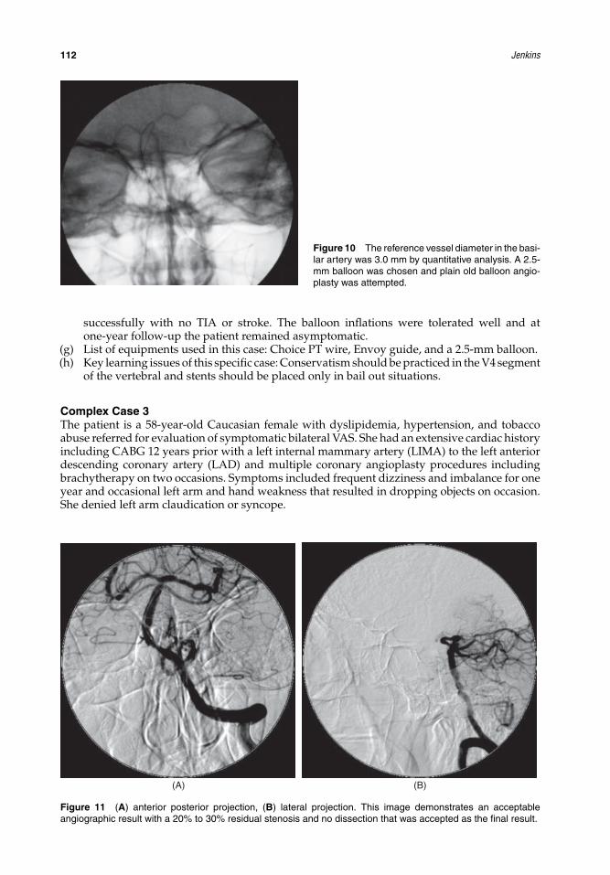
IHBK065-08 IHBK065-Minar May 1, 2010 7:33 Specs: 7x10 tight 7in×10in Char Count=
112 Jenkins
Figure 10 The reference vessel diameter in the basi-lar artery was 3.0 mm by quantitative analysis. A 2.5-mm balloon was chosen and plain old balloon angio-plasty was attempted.
successfully with no TIA or stroke. The balloon inflations were tolerated well and atone-year follow-up the patient remained asymptomatic.
(g) List of equipments used in this case: Choice PT wire, Envoy guide, and a 2.5-mm balloon.(h) Key learning issues of this specific case: Conservatism should be practiced in the V4 segment
of the vertebral and stents should be placed only in bail out situations.
Complex Case 3The patient is a 58-year-old Caucasian female with dyslipidemia, hypertension, and tobaccoabuse referred for evaluation of symptomatic bilateral VAS. She had an extensive cardiac historyincluding CABG 12 years prior with a left internal mammary artery (LIMA) to the left anteriordescending coronary artery (LAD) and multiple coronary angioplasty procedures includingbrachytherapy on two occasions. Symptoms included frequent dizziness and imbalance for oneyear and occasional left arm and hand weakness that resulted in dropping objects on occasion.She denied left arm claudication or syncope.
(A) (B)
Figure 11 (A) anterior posterior projection, (B) lateral projection. This image demonstrates an acceptableangiographic result with a 20% to 30% residual stenosis and no dissection that was accepted as the final result.

IHBK065-08 IHBK065-Minar May 1, 2010 7:33 Specs: 7x10 tight 7in×10in Char Count=
VERTEBRAL INTERVENTIONS 113
Figure 12 One-year follow-up angiography.
The physical exam was pertinent for a soft bruit over the right carotid artery and equalpulses bilaterally. The systolic blood pressure was 140/70 mm Hg bilaterally. There were no othersignificant physical findings. The patient was placed on dual antiplatelet therapy 18 monthsprior for coronary stenting with no improvement in her mentioned symptoms. She was referredfor evaluation after nonselective coronary angiography of the LIMA graft demonstrated mod-erate left subclavian stenosis and significant left VAS.
Angiographic Images
Equipments and StrategiesA 5-Fr Bernstein diagnostic catheter was used to engage the brachieocephalic trunk and placea 0.035-guidewire into the right subclavian artery. The diagnostic catheter was exchanged fora 6-Fr Judkins right coronary guiding catheter that was withdrawn to select the ostium of the
Figure 13 Ostial angiography of the left verte-bral artery demonstrates an 80% stenosis with a25-mm Hg gradient. There was a 50% stenosisof the left subclavian artery with no significantgradient.
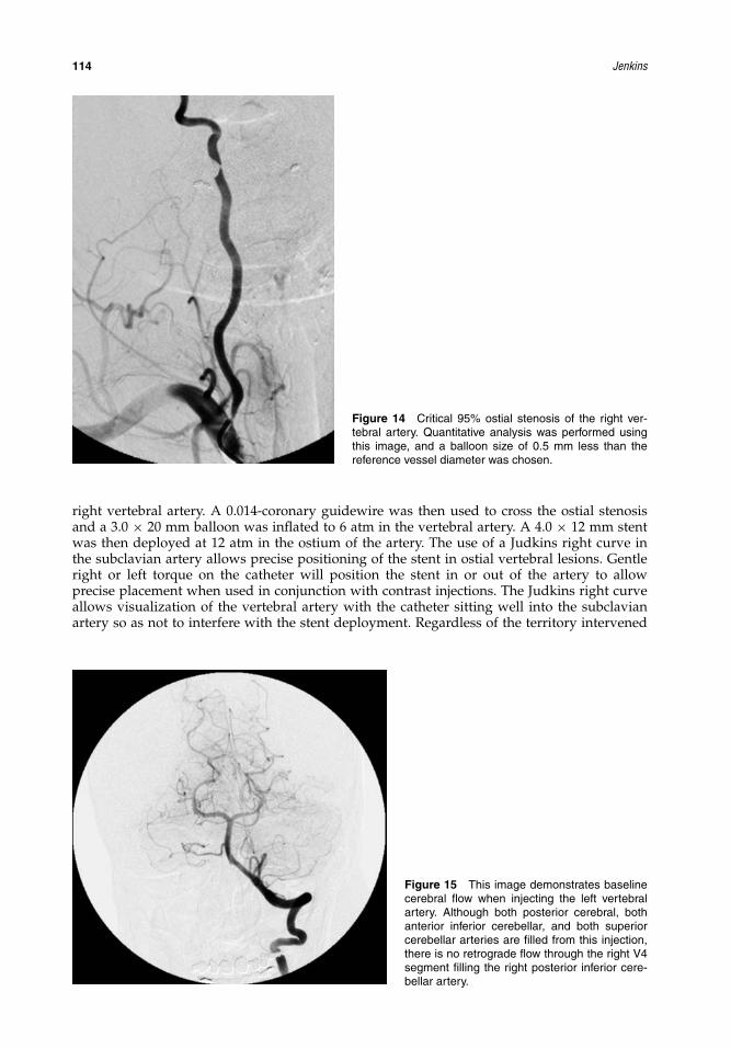
IHBK065-08 IHBK065-Minar May 1, 2010 7:33 Specs: 7x10 tight 7in×10in Char Count=
114 Jenkins
Figure 14 Critical 95% ostial stenosis of the right ver-tebral artery. Quantitative analysis was performed usingthis image, and a balloon size of 0.5 mm less than thereference vessel diameter was chosen.
right vertebral artery. A 0.014-coronary guidewire was then used to cross the ostial stenosisand a 3.0 × 20 mm balloon was inflated to 6 atm in the vertebral artery. A 4.0 × 12 mm stentwas then deployed at 12 atm in the ostium of the artery. The use of a Judkins right curve inthe subclavian artery allows precise positioning of the stent in ostial vertebral lesions. Gentleright or left torque on the catheter will position the stent in or out of the artery to allowprecise placement when used in conjunction with contrast injections. The Judkins right curveallows visualization of the vertebral artery with the catheter sitting well into the subclavianartery so as not to interfere with the stent deployment. Regardless of the territory intervened
Figure 15 This image demonstrates baselinecerebral flow when injecting the left vertebralartery. Although both posterior cerebral, bothanterior inferior cerebellar, and both superiorcerebellar arteries are filled from this injection,there is no retrograde flow through the right V4segment filling the right posterior inferior cere-bellar artery.

IHBK065-08 IHBK065-Minar May 1, 2010 7:33 Specs: 7x10 tight 7in×10in Char Count=
VERTEBRAL INTERVENTIONS 115
Figure 16 Intracranial imaging of the rightvertebral artery demonstrates a hypoplastic,atherosclerotic V4 segment that isolates theright posterior inferior cerebellar artery. This rep-resents the most likely ischemic territory perangiography.
upon, ostial stent positioning can always be a challenge and this is no different in the vertebralartery.
CommentaryThere are several factors that make this case complex. First, one needs to determine clinicallywhich lesion is the culprit when both subclavian arteries are moderately stenosed and both verte-bral arteries have significant stenoses. A team approach is necessary with neurology evaluationto determine which lesion is the most symptomatic. In this case, the neurologist determined thatthe left PICA was the most likely cause of the patient’s symptoms. Angiographically this seemedappropriate as there was a 5-mm Hg gradient across both subclavian stenoses and the patienthad no anterior ischemia on nuclear stress imaging, and there was no claudication symptomsin either arm.
Figure 17 Balloon predilation is performedto both optimize stent positioning and to ver-ify appropriate vessel diameter determined byquantitative analysis. Adequate predilation isalso very important in facilitating stent place-ment in ostial lesions, as in this patient.

IHBK065-08 IHBK065-Minar May 1, 2010 7:33 Specs: 7x10 tight 7in×10in Char Count=
116 Jenkins
Figure 18 Contrast injections are performedto position the stent 1 to 2 mm into the subcla-vian artery. The vertebral artery ostium can bemissed if this step is omitted.
The second complex factor in this case was the critical stenoses that were truly ostial inlocation. Careful stent positioning and hanging the stent 1 to 2 mm into the subclavian ostiumwill prevent missing the ostium and the need for an additional stent.
There was complete resolution of this patient’s symptoms on neurologic exam at threemonths post procedure. This demonstrates that the untreated 80% stenosis of the left vertebralartery was not contributing to the patient’s symptoms and that the right posterior inferiorcerebellum was the ischemic territory. It is also possible that angioplasty of the left vertebralwould allow enough retrograde flow through the hypoplastic V4 segment to render the patientasymptomatic. This approach was not chosen as it would be less likely to provide flow to theright PICA and because of the possibility of interfering with the LIMA graft. This patient was
Figure 19 During deployment, the stent should beinflated slowly to prevent watermelon seeding forward orbackward. This allows repositioning if necessary by plac-ing the indeflator on neutral and making small movementswith the stent delivery system.
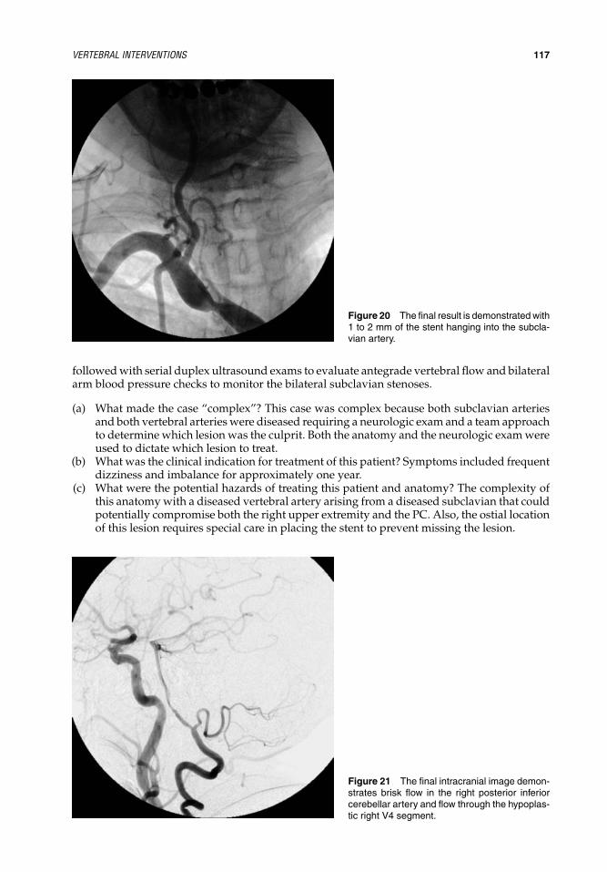
IHBK065-08 IHBK065-Minar May 1, 2010 7:33 Specs: 7x10 tight 7in×10in Char Count=
VERTEBRAL INTERVENTIONS 117
Figure 20 The final result is demonstrated with1 to 2 mm of the stent hanging into the subcla-vian artery.
followed with serial duplex ultrasound exams to evaluate antegrade vertebral flow and bilateralarm blood pressure checks to monitor the bilateral subclavian stenoses.
(a) What made the case “complex”? This case was complex because both subclavian arteriesand both vertebral arteries were diseased requiring a neurologic exam and a team approachto determine which lesion was the culprit. Both the anatomy and the neurologic exam wereused to dictate which lesion to treat.
(b) What was the clinical indication for treatment of this patient? Symptoms included frequentdizziness and imbalance for approximately one year.
(c) What were the potential hazards of treating this patient and anatomy? The complexity ofthis anatomy with a diseased vertebral artery arising from a diseased subclavian that couldpotentially compromise both the right upper extremity and the PC. Also, the ostial locationof this lesion requires special care in placing the stent to prevent missing the lesion.
Figure 21 The final intracranial image demon-strates brisk flow in the right posterior inferiorcerebellar artery and flow through the hypoplas-tic right V4 segment.

IHBK065-08 IHBK065-Minar May 1, 2010 7:33 Specs: 7x10 tight 7in×10in Char Count=
118 Jenkins
(d) Which interventional strategies were considered? The strategy was to treat the vertebralartery without compromising the subclavian artery. A primary stenting approach to theright vertebral artery ostium was planned.
(e) Which interventional tools were used to manage this case? A 6-Fr Judkins right guidingcatheter, a 0.014 coronary guidewire, and a balloon expandable stent were used to primarilystent the vertebral artery.
(f) Summary: How was the case managed, is there anything that could have been improved,what was the immediate and long-term outcome? There was complete resolution of thispatient’s symptoms on neurologic exam at three months post procedure. This demon-strates that the untreated 80% stenosis of the left vertebral artery was not contributing tothe patient’s symptoms and that the right posterior inferior cerebellum was the ischemicterritory.
(g) List of equipments used in this case: A 6-Fr Judkins right guiding catheter, a 0.014 wisperwire, and a 4.0 × 12 balloon expandable stent were used to perform this procedure.
(h) Key learning issues of this specific case: This case reinforces the need to take a team approachwhen performing vertebral angioplasty. Only the lesions that are symptomatic should berevascularized and the rest should be managed medically with routine surveillance.
SUMMARY OF THE KEY ISSUES FOR SUCCESSFUL AND SAFE TREATMENTOF COMPLEX VERTEBRAL ARTERY LESIONSIn summary, it is necessary to first know the anatomy of the PC by performing an arch andfour-vessel diagnostic angiogram. A complete neurologic exam is also necessary to determinethe symptomatic vessel that will provide the best revascularization to the PC. Proper guideselection and the use of coronary angioplasty equipment will allow safe and effective treatmentof complex vertebral lesions. When anatomy allows, embolic protection devices should be usedto prevent distal embolic complications. Following these rules will improve the chances ofsuccessful angioplasty in complex vertebral lesions.
REFERENCES1. Jenkins JS, Subramanian R. Endovascular treatment for vertebrobasilar insufficiency. Curr Treat
Options Cardiovasc Med 2002; 4(5):385–391.2. Albuquerque FC, Fiorella D, Han P, et al. A reappraisal of angioplasty and stenting for the treatment
of vertebral origin stenosis. Neurosurgery 2003; 53(3):607–614; discussion 14–16.3. Nomura M, Hashimoto N, Nishi S, et al. Percutaneous transluminal angioplasty for intracranial
vertebral and/or basilar artery stenosis. Clin Radiol 1999; 54(8):521–527.4. Diener HC, Bogousslavsky J, Brass LM, et al. Management of atherothrombosis with clopidogrel in
high-risk patients with recent transient ischaemic attack or ischaemic stroke (MATCH): study designand baseline data. Cerebrovasc Dis 2004; 17(2–3):253–261.
5. Diener HC, Bogousslavsky J, Brass LM, et al. Aspirin and clopidogrel compared with clopidogrel aloneafter recent ischaemic stroke or transient ischaemic attack in high-risk patients (MATCH): randomised,double-blind, placebo-controlled trial. Lancet 2004; 364(9431):331–337.
6. Whisnant JP, Cartlidge NE, Elveback LR. Carotid and vertebral-basilar transient ischemic attacks:effect of anticoagulants, hypertension, and cardiac disorders on survival and stroke occurrence–apopulation study. Ann Neurol 1978; 3(2):107–115.
7. Jenkins JS, White CJ, Ramee SR, et al. Vertebral insufficiency: when to intervene and how? Curr IntervCardiol Rep 2000; 2(2):91–94.
8. Henry M, Polydorou A, Henry I, et al. Angioplasty and stenting of extracranial vertebral arterystenosis. Int Angiol 2005; 24(4):311–324.
9. Wehman JC, Hanel RA, Guidot CA, et al. Atherosclerotic occlusive extracranial vertebral artery dis-ease: indications for intervention, endovascular techniques, short-term and long-term results. J IntervCardiol 2004; 17(4):219–232.
10. Chimowitz MI, Lynn MJ, Howlett-Smith H, et al. Prognosis of patients with symptomatic vertebralor basilar artery stenosis. The Warfarin-Aspirin Symptomatic Intracranial Disease (WASID) StudyGroup. Stroke 1998; 29(7):1389–1392.
11. Eberhardt O, Naegele T, Raygrotzki S, et al. Stenting of vertebrobasilar arteries in symptomaticatherosclerotic disease and acute occlusion: case series and review of the literature. J Vasc Surg 2006;43(6):1145–1154.

IHBK065-08 IHBK065-Minar May 1, 2010 7:33 Specs: 7x10 tight 7in×10in Char Count=
VERTEBRAL INTERVENTIONS 119
12. Jenkins JS, White CJ, Ramee SR, et al. Vertebral artery stenting. Catheter Cardiovasc Interv 2001;54(1):1–5.
13. Mukherjee D, Roffi M, Kapadia SR, et al. Percutaneous intervention for symptomatic vertebral arterystenosis using coronary stents. J Invasive Cardiol 2001; 13(5):363–366.
14. Chimowitz MI, Lynn MJ, Howlett-Smith H, et al. Comparison of warfarin and aspirin for symptomaticintracranial arterial stenosis. N Engl J Med 2005; 352(13):1305–1316.
15. Fields WS, North RR, Hass WK, et al. Joint study of extracranial arterial occlusion as a cause of stroke.I. Organization of study and survey of patient population. JAMA 1968; 203(11):955–960.
16. Berguer R, Flynn LM, Kline RA, et al. Surgical reconstruction of the extracranial vertebral artery:management and outcome. J Vasc Surg 2000; 31(1, pt 1):9–18.
17. Thevenet A, Ruotolo C. Surgical repair of vertebral artery stenoses. J Cardiovasc Surg (Torino) 1984;25(2):101–110.
18. Chastain HD II, Campbell MS, Iyer S, et al. Extracranial vertebral artery stent placement: in-hospitaland follow-up results. J Neurosurg 1999; 91(4):547–552.
19. Lin YH, Juang JM, Jeng JS, et al. Symptomatic ostial vertebral artery stenosis treated with tubularcoronary stents: clinical results and restenosis analysis. J Endovasc Ther 2004; 11(6):719–726.
20. Weber W, Mayer TE, Henkes H, et al. Stent-angioplasty of intracranial vertebral and basilar arterystenoses in symptomatic patients. Eur J Radiol 2005; 55(2):231–236.
21. Coward LJ, McCabe DJ, Ederle J, et al. Long-term outcome after angioplasty and stenting for symp-tomatic vertebral artery stenosis compared with medical treatment in the Carotid and Vertebral ArteryTransluminal Angioplasty Study (CAVATAS): a randomized trial. Stroke 2007; 38(5):1526–1530.
22. Coward LJ, Featherstone RL, Brown MM. Percutaneous transluminal angioplasty and stenting forvertebral artery stenosis. Cochrane Database Syst Rev 2005 (2):CD000516.
23. Berguer R, Feldman AJ. Surgical reconstruction of the vertebral artery. Surgery 1983; 93(5):670–675.24. Molnar RG, Naslund TC. Vertebral artery surgery. Surg Clin North Am 1998; 78(5):901–913.

IHBK065-09 IHBK065-Minar April 26, 2010 13:50 Specs: 7x10 tight 7in×10in Char Count=
9 Thoracic Aortic Dissection and NondissectiveThoracic Aortic DiseaseMartin Czerny and Michael GrimmDepartment of Cardiothoracic Surgery, University of Vienna Medical School, Vienna, Austria
INTRODUCTIONWithin the last decade, thoracic endovascular aortic repair (TEVAR) has been increasingly per-formed on patients with more advanced age as well as on thoracic aortic pathology beinglocated within the aortic arch as supra-aortic transpositions have become an established, safe,and reproducible procedure (1–7). Indications for treatment contain dilatative as well as oblit-erative pathology, primarily aneurysms, being followed by a steadily increasing number ofpenetrating atherosclerotic ulcers (8). Furthermore, TEVAR has become the modality of choicein patients with complicated type B dissections (9,10). By a better understanding of the mecha-nisms of intramural hematoma (IMH), TEVAR is also applied in patients presenting with IMHof the entire thoracic aorta (11).
Atherosclerotic Thoracic Aortic AneurysmsMore than 50% of patients presenting with thoracic aortic aneurysms have to be regarded ascomplex, being defined by the need of an additional procedure prior to stent-graft placement inorder to pave the route for a successful and durable endovascular procedure (12,13). The majorityof these additional procedures are rerouting procedures primarily of the supra-aortic branchesin order to gain a sufficient proximal landing zone for extensive sealing and prevention ofendoleak formation. The length of the proximal landing zone has been shown to independentlypredict avoidance of early as well as late endoleak formation (5). The same is true for the distallanding zone; nevertheless visceral rerouting procedures are complex and invasive and it is stillcontroversial if visceral rerouting prior to TEVAR really is less invasive than a conventionalsurgical approach with all safety nets available (14,15).
Penetrating Atherosclerotic UlcersPenetrating atherosclerotic ulcers (PAU) have been recently defined as a distinct entity of acuteaortic syndromes gaining increasing importance by improved diagnostic means and by sensibi-lization of the scientific community to the pathology itself (16). Being the result of an advancedobliterative atherosclerotic process, PAU may also be associated with other severe manifesta-tions of the underlying disease such as coronary artery disease as well as peripheral arterialocclusive disease. The natural history of PAU, especially when being symptomatic, is dismal(17,18). PAU may be complex due to their location, as at least one-third of them are located inthe aortic arch as well as to the fact that-–in contrast to aneurysms—many intercostal arteriesare still patent and the risk of spinal cord injury is higher. Furthermore, vascular access maybe challenging as more than 50% of patients require a retroperitoneal approach to the commoniliac artery or even to the infrarenal aorta in order to be able to advance the introduction sheathof the stent graft (8).
Type B DissectionsMerely 1 out of 10 type B dissections is or turns complicated (19). Due to the results from theINSTEAD trial (yet unpublished), it is now generally accepted that uncomplicated type B dis-sections should be treated with a primary conservative approach. Nevertheless, complicatedtype B dissections are the ideal target for TEVAR as procedural morbidity and mortality aresubstantially less than any other approach (9,10). Complicated type B dissections define them-selves by rupture or malperfusion irrespective of the affected vascular bed, that is, intercostal,visceral, renal or peripheral.

IHBK065-09 IHBK065-Minar April 26, 2010 13:50 Specs: 7x10 tight 7in×10in Char Count=
THORACIC AORTIC DISSECTION AND NONDISSECTIVE THORACIC AORTIC DISEASE 121
Intramural HematomaIMH of the aorta has been recently defined as a subtype of acute aortic wall dissection withno detectable intimal tear and consequently, lack of false lumen flow (20). Lack of presenceof intimal disruption or flap has been the prerequisite for IMH diagnosis (16). With the recentadvances in imaging, in particular via ECG-gating techniques, a more precise evaluation ofIMH is possible. The presumed mechanism of IMH, rupture of vasa vasorum (21), however,may be doubted since in many IMH patients a small primary entry tear can be found (11).Rupture of a small atherosclerotic plaque at the free lateral wall or at the concavity of thedistal aortic arch may be found as the underlying disease mechanism. Following plaque rup-ture, antegrade and/or retrograde progression of the pathology affects the aortic wall. If theprimary lesion is located at the convexity of the aortic arch, supra-aortic branches may serveas a natural anatomical barrier against retrograde progression, thus resulting in predominantantegrade development as observed in type B dissection. However, in case of a primary lesionlocated at the free lateral wall or at the concavity of the distal aortic arch, the lack of a nat-ural anatomical barrier may permit retrograde progression down to the level of the aorticroot. This mechanism seems valid for both variants of acute aortic syndromes, IMH as well asdissection (11).
IMH is regarded as complex when the entire thoracic aortic up to the level of the aortic rootis affected and empiric surgical replacement of the ascending aorta as well as of the concavity ofthe aortic arch is the mainstay of therapy if the described mechanism of plaque rupture cannotbe retraced.
CLINICAL INDICATIONS
Atherosclerotic Thoracic Aortic AneurysmsTreatment of thoracic aortic aneurysms is indicated when the risk of the yearly rate of ruptureexceeds the remaining risk of treatment. It is generally accepted that the cutoff point is at amaximum diameter of 6 cm being adjusted for age, sex, and body mass index. TEVAR hasbecome the therapy of first choice due to its clearly lower procedural morbidity and mortality.
Penetrating Atherosclerotic UlcersIn contrast to the decision making in patients with atherosclerotic thoracic aortic aneurysms, themaximum diameter of PAU plays a minor role. Morphology and, if applicable and traceable,progression of the lesion are the cornerstones of decision making besides clinical symptoms oralready sustained rupture.
Type B DissectionsTEVAR in type B dissections is indicated in contained rupture as well as in patients with anykind of malperfusion in downstream vascular beds.
Intramural HematomaTreatment of IMH is indicated in contained rupture as well as if the ascending aorta is affected.Malperfusion in IMH is rarely an issue.
HAZARDS AND COMPLICATIONS
Atherosclerotic Thoracic Aortic AneurysmsAs the underlying disease is atherosclerotic, manipulation especially within the aortic arch maycause embolization of debris. As a consequence, manipulation should be kept to a minimum.As a long covered segment may be necessary for total exclusion of the lesion, a high number ofintercostal arteries may have to be sacrificed. Therefore, a high level of alertness for spinal cordmalperfusion has to be there. Cerebrospinal fluid drainage may add as an important adjunct toprevent this dreadful complication. Overstenting of the left subclavian artery may add to spinalcord malperfusion and should be done only in emergency situations.

IHBK065-09 IHBK065-Minar April 26, 2010 13:50 Specs: 7x10 tight 7in×10in Char Count=
122 CZERNY AND GRIMM
Penetrating Atherosclerotic UlcersPatients with PAU are frail and are generally in an advanced stage of their aggressive underlyingdisease. Furthermore, the rough intravascular surface of the lesions may cause tear of the stent-graft fabric as well as atheroembolic embolization.
Type B DissectionsIt is of utmost importance to reaffirm to beside within the true lumen, as stent-graft placementin the false lumen may have dreadful consequences. Therefore, serial angiographies should beperformed and the CT scans have to be studied in detail in advance in order to know about theindividual functional anatomy. Furthermore, balloon dilatation after TEVAR has to be avoided,as rupture of the dissecting membrane might be the consequence. Furthermore, the so-calledfloating visceral sign has to be ruled out, as despite filling of the visceral bed with contrastmedium, significant malperfusion may be present.
Intramural HematomaAs in dissections, balloon dilatation should not be performed due to the risk of rupture. Fur-thermore, oversizing of the stent graft should not be more than 10% in order not to expose thefriable aortic wall to a high amount of radial force.
IMAGING, INTERVENTIONAL STRATEGIES, AND TOOLS
ImagingAll CT examinations prior to treatment should be performed on a 64-row CT scanner. To allowpulsation-free visualization of the thoracic aorta up to the aortic root, a retrospective ECG-gatingtechnique should be used.
In our setting, all CT angiograms are performed in the arterial phase during the intra-venous administration of nonionic iodinated contrast material. A bolus triggering techniqueassesses the contrast medium transit time. A threshold of 150 HU (absolute) is used. One hun-dred and thirty milliliters of contrast agent is administered in a biphasic fashion: the first 30 mLis injected at a flow rate of 6 mL/sec followed by the second 100 mL administered at 5 mL/sec.A saline flush of 40 mL is administered to all the patients after the contrast medium injectionto optimize contrast utilization. With a post-threshold delay of eight seconds, ECG-gated CTangiography of the entire aorta is performed using the following imaging parameters: a slicecollimation of 64 × 0.625 and a pitch of 0.29 is used. The matrix size is 512 × 512. Images arereconstructed with a slice thickness of 1.4 and a slice increment of 1 mm as well as with a slicethickness of 3 and a slice increment of 2 mm without using the ECG-triggering (“untaggedimages”). Additionally, image redistribution according to the heart action is performed afterscanning (retrospective ECG-triggering). Usually, images are triggered in a midphase at 55% ofa RR-interval. If pulsation artifacts occur, other phases of the heart cycle are reconstructed. Forevery phase, two series are reconstructed (1.4/1 as well as 3/2 mm).
Interventional StrategyStent grafts are placed under anesthesia. Access vessel exposure is being performed surgically. A5-French pigtail catheter is advanced via the right brachial artery into the aortic arch to reconfirmcharacterization of the morphology and extent of the lesions. After systemic heparinizationwith 80 IU/kg, arteriotomy is performed and the catheter is advanced under fluoroscopicguidance. Afterwards, a change to an ultrastiff guidewire (Backup Meier) is being performed.Stent grafts are deployed during systemic hypotension with a systolic pressure of 70 mm Hg.The more proximal the stent graft is being deployed the more liberal we do use rapid pacingvia a temporary pacemaker electrode being inserted via the right subclavian vein into the rightventricle. Pacing is performed up to 180 beats per minute. With this method blood pressurecontrol seems most effective.
Interventional ToolsThree different, commercially available stent-graft systems are used namely, the modifiedValiant endovascular stent graft (Medtronic, Santa Rosa, CA), the Relay stent graft (BoltonMedical, Sunrise, FL) as well as the Gore TAG stent graft (Gore Inc. Phoenix, AZ).

IHBK065-09 IHBK065-Minar April 26, 2010 13:50 Specs: 7x10 tight 7in×10in Char Count=
THORACIC AORTIC DISSECTION AND NONDISSECTIVE THORACIC AORTIC DISEASE 123
Quality ControlPatients undergo a completion CT scan to verify the effectiveness of the procedure beforedischarge, then at one, three, and six months and annually thereafter.
SUMMARY OF OUTCOMES
Atherosclerotic Thoracic Aortic AneurysmsResults after supra-aortic transpositions in various extents followed by endovascular stent-graft placement for the treatment of various pathology affecting the aortic arch are promising.Endoleak formation is directly related to the number of prostheses and may be reduced by longerdevices (5). Each type of arch rerouting, irrespective of extent, has turned out to be effective(1–8). Therefore, extended applications of these combined treatment strategies substantiallyaugment the therapeutic options.
Penetrating Atherosclerotic UlcersEndovascular stent-graft placement in patients with PAU is an effective palliation for a life-threatening sign of a severe systemic process. Hemodynamic instability at referral and a highpreoperative risk score predict adverse outcome. During midterm follow-up, patients are mainlylimited by sequelae of their underlying disease (8).
Type B DissectionsTEVAR has emerged as the therapy of first choice in patients sustaining complicated type Bdissections. Nevertheless, these patients have to be subjected to serial follow-up CT scans asaneurysm formation requiring treatment in downstream aortic segments occurs in up to 20%of patients. Uncomplicated type B dissection should be initially managed with best medicaltreatment. TEVAR remains an option in these patients if a progression of the underlying diseaseoccurs (9,10,22).
Intramural HematomaPlaque rupture may be identified as the cause of IMH in a previously unrecognized subgroupof patients. If at the convexity of the distal arch, supra-aortic branches prevent retrogradeextension toward the ascending aorta. If at the free lateral wall or at the concavity, IMH mayaffect the entire thoracic aorta, due to the lack of the natural barrier of the supra-aortic branches.Endovascular stent-graft placement of this plaque-associated IMH may be more effective andless invasive than conventional surgery in order to treat the entire thoracic aortic disease (11).
EQUIPMENTS CHECKLISTThe following chart shall supply the reader with a detailed list of all medical supplies neededin TEVAR as well as with the costs of these supplies (23).
Ordering # Costs/unit (€) N Total costs (€)
Introduction sheath PTA Angio 407850 9-F 12 cm Ulti 30031427 10.67 2 21.34Introduction sheath Angio Terumo RSB50 N 5-F 10 cm 30201542 10.40 1 10.40FD PTCA SCH 30601 0.35′′ 300 cm ger. Backup 30022227 154.38 1 154.38Kath. Diagn. Pigtail 5-F 451503V5 65 cm 5 SL 30018107 15.04 1 15.04Abd. Angio. t. femor 203 × 280 cm 30004841 9.21 1 9.21Abd. BV cover 85 × 95 30014695 1.16 1 1.16Abd. draping nsk 100 × 150 251220/4 30020443 1.44 1 1.44Flushing set Angiofill II MDD520 H 30032630 9.51 1 9.51Cannula arterial 099326 18G Seldinger 30012286 3.98 1 3.98Syringe 20 mL 30001692 0.04 6 0.27Syringe 10 mL 30001691 0.03 4 0.11Cannula inject 20G × 1-1/2 0.90 × 40 Nr. 01 30001700 0.01 2 0.02Scalpel No. 11 sterile 30003225 0.21 1 0.21Gazin MK 9/7.5 16-F 13682 1 Set 113314 0.41 4 1.65

IHBK065-09 IHBK065-Minar April 26, 2010 13:50 Specs: 7x10 tight 7in×10in Char Count=
124 CZERNY AND GRIMM
Gloves OP Derma Plus Gr. 7.5 30042051 0.26 4 1.05Gown OP large 133 cm 30016949 2.61 4 10.44Angiography coil San 1939 117802 2.39 2 4.78Phys. NaCl Inffl. 200 mL 114960 0.32 1 0.32Xenetix 300 mg/mL Inffl. 200 mL 168650 62.95 1 62.95Fentanyl Amp. 10 mL 183313 0.54 1 0.54Dormicum Amp. 5 mg/1 mL 110797 0.15 1 0.15Heparin-Imm Dstfl. 5000 IE/mL 5 mL 115120 3.44 1 3.44Ro.Film Drystar TM 1B NIF 35 × 43 1 sheath 30097999 1.49 6 8.93Introduction sheath RCF 18.0-P-38-30-JRB Checkflo 30093713 200.79 1 200.79Introduction sheath XLCFW 24.0-38-30 Endostent 30092185 274.51 1 274.51FD Bentson 035′′ 7/260 cm M001491031 ger. 30067544 7.93 1 7.93Pressure tube angio 1520010 1200PSI 200 30095803 4.81 1 4.81Syringe 200 mL for Illumena 900105 30209174 9.17 1 9.17Stent graft 30207964 10,045.00 1 10,045.00Cath. Ballon Reliant AB46 10–46 mm (TAA) 30117306 226.64 1 226.64Abd. C arm multipart DBL 990716 30099509 41.23 1 41.23
11,131.40
COMPLEX CASES
Case 1—Atherosclerotic Thoracic Aortic AneurysmA 79-year-old male was referred for treatment of a huge aortic arch aneurysm with a maximumdiameter of 8 cm (Fig. 1). Due to his frail general status and due to his risk factors (extrac-ardiac arteriopathy as well as COPD) a conventional surgical replacement of the aortic archwas deemed high risk. Median numeric EuroSCORE was 12 and median logistic EuroSCOREwas 37.
Therefore, a combined vascular and endovascular approach was chosen. As a first step,total arch rerouting was performed. Access was gained via an upper median sternotomy andthe pericardium was opened. As a next step, the supra-aortic branches were circumferentiallydissected. Afterwards, 80 IU of heparin per kilogram bodyweight was applied. For total archrerouting, a 18/9 mm Dacron prosthesis was chosen. The ascending aorta was tangentiallyclamped and an end-to-side anastomosis between the proximal portion of the Dacron prosthesisand the ascending aorta was performed. Afterwards the brachiocephalic trunk was clampedat its origin, the trunk was transected and an end-to-end anastomosis between the first branch
Figure 1 Aortic arch aneurysm at the time ofdiagnosis.

IHBK065-09 IHBK065-Minar April 26, 2010 13:50 Specs: 7x10 tight 7in×10in Char Count=
THORACIC AORTIC DISSECTION AND NONDISSECTIVE THORACIC AORTIC DISEASE 125
Figure 2 Final intraoperative result after totalarch rerouting.
of the Dacron prosthesis and the trunk was being performed. Then, the left subclavian arterywas transected and an end-to-end anastomosis between the second branch of the prosthesisand the left subclavian artery was done. Finally, the left common carotid artery was reinsertedinto the branch to the left subclavian. Figure 2 shows the final intraoperative result. Afterwards,the wound was closed in layers.
After a recovery period of one week, the patient underwent stent graft placement. Theright common femoral artery was exposed and 80 IU of heparin per kilogram bodyweight wasapplied. A pigtail catheter was advanced via the right subclavian artery for serial angiography.After insertion of an ultrastiff Backup-Meier guidewire, a 42/250 mm stent graft (Bolton, Relay,Sunrise, FA) was advanced into the aortic arch. Rapid pacing was begun and after havingreached functional circulatory arrest, the stent graft was deployed. Afterwards, rapid pacing wasgradually reduced and normal circulation was thereby restored. All catheters and guidewireswere removed, the wound was closed in layers and the patient woke up on the operating table.Recovery was uneventful. Completion CT scan showed regular supra-aortic perfusion, the stentgraft in place as well as no signs of any kind of endoleak. Follow-up is regular for three years(Fig. 3).
The complexity of this case is underlined by a limitating thoracic aortic pathology in avery delicate anatomical region encomprising all brain and upper extremity supplying greatvessels. By this combined concept, a durable result could be achieved and the patient was ableto reassume a normal life soon after the procedure. Quality of life was preserved and the threatof aneurysmal rupture was averted with a stable and durable result.
Case 2—Penetrating Atherosclerotic UlcersA 64-year-old male was referred for treatment of multiple PAU of the thoracic aorta originatingfrom the level of the left common carotid artery to below the thoracoabdominal transition(Fig. 4). The patient suffered from severe generalized atherosclerosis, was on dialysis forglomerulonephritis, and had substantial COPD. Median numeric EuroSCORE was 13 andmedian logistic EuroSCORE was 41. The patient was symptomatic with new onset of chestpain corresponding to the multisegmental location of the lesions.
Therefore, a combined vascular and endovascular approach was chosen. As a first step,autologous double transposition of the supra-aortic branches was performed in order to gaina sufficient proximal landing zone for stent-graft insertion. A median upper hemisternotomyaccess was chosen and the supra-aortic branches were circumferentially dissected (Fig. 5).Eighty IU of heparin per kilogram bodyweight was applied. The common carotid artery wastransected at the level of the arch and an end-to-side anastomosis between the brachiocephalictrunk and the left common carotid artery was performed. Afterwards, the left subclavian arterywas transected and an end-to-side anastomosis between the already transposed left commoncarotid artery and the subclavian artery was performed (Fig. 6). Then, the wound was closed inlayers.

IHBK065-09 IHBK065-Minar April 26, 2010 13:50 Specs: 7x10 tight 7in×10in Char Count=
126 CZERNY AND GRIMM
Figure 3 Completion CT scan after stent-graft placement.
After a recovery period of one week, stent-graft placement was performed. Vascular accesswas gained via the right common iliac artery, as the external iliac artery was very narrow due tothe underlying obliterative disease. A 9 mm Dacron graft was sewn onto the native vessel in anend-to-side fashion. After heparinization, two stent grafts were deployed (Bolton, Relay, Sunrise,FA) beginning at the level of the brachiocephalic trunk down to the level of the celiac trunk.Afterwards, due to severe narrowing of the iliac bifurcation as well as of the external iliac artery,the Dacron graft was extended to the right groin as an iliaco-femoral bypass. Then, the woundwas closed in layers. The patient woke up uneventfully. After two hours the patient becameparaplegic. Mean arterial pressure was elevated at once and cerebrospinal fluid drainage wasinstalled. Symptoms disappeared and further recovery was uneventful. The final complicationCT scan is depicted in Figure 7.
The complexity of this case is underlined by multisegmental thoracic aortic disease, requir-ing extension of the proximal landing zone by rerouting as well as by symptomatic spinal cordmalperfusion. In this context, it is essential to remind onto the four spinal cord supplying vas-cular territories, the left subclavian artery, the intercostal arteries, the lumbar arteries as well asthe hypogastric arteries. Normally, sacrifice of one supplying territory does not subject thesepatients to a higher risk of symptomatic spinal cord malperfusion. Previous or procedure-relatedsacrifice of two territories, however, may highly likely result in malperfusion. Cerebrospinalfluid (CSF) drainage as well as elevation of mean arterial blood pressure are highly valu-able tools in reversing this complication whereas CSF drainage acts like a fasciotomy to thespinal cord.
Case 3—Intramural HematomaA 72-year-old male was referred with a symptomatic IMH of the entire thoracic aorta with anaccompanying pericardial effusion (Fig. 8). The general condition was weak and conventional

IHBK065-09 IHBK065-Minar April 26, 2010 13:50 Specs: 7x10 tight 7in×10in Char Count=
THORACIC AORTIC DISSECTION AND NONDISSECTIVE THORACIC AORTIC DISEASE 127
Figure 4 Multiple PAU extending from the left commoncarotid artery to the celiac trunk.
surgical treatment was deemed high risk. Numeric EuroSCORE was 13 and logistic EuroSCOREwas 45. By imaging via gated 64-row CT scanning, a small atherosclerotic plaque could beidentified as the primary entry site at the concavity of the distal arch.
Therefore an endovascular approach was chosen. After induction of general anesthesia aswell as standard insertion of catheters and guidewires, three stent grafts (Medtronic, Valiant,Santa Rosa, CA) were inserted from the offspring of the left common carotid artery to the distalthird of the descending aorta. The left subclavian artery had to be covered in this situation.Afterwards, the hemodynamically significant pericardial effusion was treated by subxyphoidaldrainage. The patient recovered uneventfully and the CT scan showed complete resorption ofthe IMH in the ascending aorta as well as complete remodeling of the entire thoracic aorta sofar within a two-year follow-up period (Fig. 9).
Figure 5 Intraoperative situs before doubletransposition.

IHBK065-09 IHBK065-Minar April 26, 2010 13:50 Specs: 7x10 tight 7in×10in Char Count=
128 CZERNY AND GRIMM
Figure 6 Intraoperative situs after doubletransposition.
The complexity in this case lies in the multisegmental affection of the aorta by this acuteaortic syndrome with a high risk of rupture into the pericardium such as in classical type Aaortic dissection. To date rupture of vasa vasorum was held responsible for the mechanism ofIMH development. We strongly encourage every physician dealing with this entity to carefullylook for a small entry tear such as the plaque in that very case. By defining such an entry tear,treatment can be performed by TEVAR and results may be favorable.
SUMMARY OF THE KEY ISSUESIn summary, a combined vascular and endovascular approach for the treatment of thoracicaortic pathology has emerged as an established procedure with excellent long-term results.Vascular transposition is safe and—by avoiding cardiopulmonary bypass as well as hypothermiccirculatory arrest—the method proofs its less invasiveness.
Figure 7 Completion CT scan after stent-graft placement.

IHBK065-09 IHBK065-Minar April 26, 2010 13:50 Specs: 7x10 tight 7in×10in Char Count=
THORACIC AORTIC DISSECTION AND NONDISSECTIVE THORACIC AORTIC DISEASE 129
Figure 8 IMH of the entire thoracic aorta at the time ofdiagnosis.
By adhering to the principle of a long landing zone, a sufficient overlap as well as a lownumber of prostheses, the rate of early and late endoleak formation can be kept very low.
A broader application of these techniques will enable treatment of a patient group who afew years ago would have been sent home with mere antihypertensive therapy.
Perforating ulcers have to be regarded as a distinct clinical entity being the result of avery aggressive underlying obliterative disease. Indication for treatment is not based on themaximum diameter of the lesion but on morphology, dynamics as well as clinical symptoms.
Figure 9 Complete resorption of IMH in the ascendingaorta after stent-graft placement into the distal aortic arch.

IHBK065-09 IHBK065-Minar April 26, 2010 13:50 Specs: 7x10 tight 7in×10in Char Count=
130 CZERNY AND GRIMM
Complicated type B dissections have become a domain of endovascular therapy andshow excellent midterm results. Aneurysm development in downstream segments remains anunsolved issue and requires continuous follow-up in patients with chronic type B dissectionsirrespective of if after TEVAR or with best medical therapy.
IMH may be the result of plaque rupture in many cases and may therefore turn out to bea suitable target for TEVAR. If the mechanism of plaque rupture cannot be retraced, surgeryshould remain the mainstay of therapy.
REFERENCES1. Czerny M, Fleck T, Zimpfer D, et al. Combined repair of an aortic arch aneurysm by sequential trans-
position of the supra-aortic branches and endovascular stent-graft placement. J Thorac CardiovascSurg 2003; 126:916–918.
2. Czerny M, Zimpfer D, Fleck T, et al. Initial results after combined repair of aortic arch aneurysmsby sequential transposition of the supra-aortic branches and consecutive endovascular stent-graftplacement. Ann Thorac Surg 2004; 78:1256–1260.
3. Czerny M, Gottardi R, Zimpfer D, et al. Transposition of the supraaortic branches for extendedendovascular arch repair. Eur J Cardiothorac Surg 2006; 29:709–713.
4. Czerny M, Gottardi R, Zimpfer D, et al. Mid-term results of supraaortic transpositions for extendedendovascular repair of aortic arch pathologies. Eur J Cardiothorac Surg 2007; 31:623–1627.
5. Gottardi R, Funovics M, Eggers N, et al. Supra-aortic transposition for combined vascular and endovas-cular repair of aortic arch pathology. Ann Thorac Surg 2008; 86:1524–1529.
6. Brinkman WT, Szeto WY, Bavaria JE. Stent graft treatment for transverse arch and descending thoracicaorta aneurysms. Curr Opin Cardiol 2007; 22:510–516.
7. Weigang E, Parker J, Czerny M, et al. Endovascular aortic arch repair after aortic arch de-branching.Ann Thorac Surg 2009; 87:603–607.
8. Gottardi R, Zimpfer D, Funovics M, et al. Mid-term results after endovascular stent-graft placementdue to penetrating atherosclerotic ulcers of the thoracic aorta. Eur J Cardiothorac Surg 2008; 33:1019–1024.
9. Szeto WY, McGarvey M, Pochettino A, et al. Results of a new surgical paradigm: endovascular repairfor acute complicated type B aortic dissection. Ann Thorac Surg 2008; 86:87–93.
10. Schoder M, Czerny M, Cejna M, et al. Endovascular repair of acute type B aortic dissection: long-termfollow-up of true and false lumen diameter changes. Ann Thorac Surg 2007; 83:1059–1066.
11. Grimm M, Loewe C, Gottardi R, et al. Novel insights into the mechanisms and treatment of intramuralhematoma affecting the entire thoracic aorta. Ann Thorac Surg 2008;86:453–456.
12. Czerny M, Cejna M, Hutschala D, et al. Stent-graft placement in atherosclerotic descending thoracicaortic aneurysms: midterm results. J Endovasc Ther 2004; 11:26–32.
13. Czerny M, Grimm M, Zimpfer D, et al. Results after endovascular stent graft placement in atheroscle-rotic aneurysms involving the descending aorta. Ann Thorac Surg 2007; 83:450–455.
14. Donas KP, Czerny M, Guber I, et al. Hybrid open-endovascular repair for thoracoabdominal aorticaneurysms: current status and level of evidence. Eur J Vasc Endovasc Surg 2007; 34:528–533.
15. Coselli JS, Bozinovski J, LeMaire SA. Open surgical repair of 2286 thoracoabdominal aortic aneurysms.Ann Thorac Surg 2007; 83:S862–S864.
16. Erbel R, Alfonso F, Boileau C, et al. Task force on aortic dissection, European Society for Cardiology.Diagnosis and management of aortic dissection. Eur Heart J 2001; 1642–1681.
17. Sundt TM. Intramural hematoma and penetrating atherosclerotic ulcer of the aorta. Ann Thorac Surg2007; 83:S835–S841.
18. Cho KR, Stanson AW, Potter DD, et al. Penetrating atherosclerotic ulcer of the descending thoracicaorta and arch. J Thorac Cardiovasc Surg 2004; 127:1393–1401.
19. Verhoye JP, Miller DC, Sze D, et al. Complicated acute type B aortic dissection: midterm results ofemergency endovascular stent-grafting. J Thorac Cardiovasc Surg 2008; 136:424–430.
20. Evangelista, A, Mukherjee, D, Mehta RH, et al; International Registry of Aortic Dissection (IRAD)Investigator. Acute intramural hematoma of the aorta: a mystery in evolution. Circulation 2005;111:1063.
21. Tsai TT, Nienaber CA, Eagle KA. Acute aortic syndromes. Circulation 2005; 112:3802.22. Czerny M, Zimpfer D, Rodler S, et al. Endovascular stent-graft placement of aneurysms involving the
descending aorta originating from chronic type B dissections. Ann Thorac Surg 2007; 83:1635–1639.23. Schuster I, Dorfmeister M, Scheuter- Mlaker S, et al. Endovascular and conventional treatment of
thoracic aortic Aneurysms: a comparison of costs. Ann Thorac Surg 2009; 87:1801–1805.
IHBK065-10 IHBK065-Minar May 6, 2010 20:7 Specs: 7x10 tight 7in×10in Char Count=
10 Abdominal Aortic DiseaseYuji Kanaoka and Takao OhkiDepartment of Surgery, Jikei University School of Medicine, Nishishimbashi, Minatoku, Tokyo, Japan
Frank J. VeithThe Cleveland Clinic, Cleveland, Ohio, and New York University, New York, New York, U.S.A.
PART 1: INTRODUCTIONThe majority of endovascular treatment for the abdominal aorta is for abdominal aorticaneurysms (AAAs). Endovascular aortic aneurysm repair (EVAR) has gained wide acceptance,and there has been a significant increase in the number of AAAs treated by EVAR (1,2). How-ever, not all AAA cases are indicated to undergo EVAR. Patient selection of EVAR involvescareful assessment of the anatomical features of aneurysm. The indications vary according todevice; however, to summarize the preferable anatomy for EVAR (3,4), (i) the proximal neck’slength should be long enough (15 mm or longer), orientation should be relatively straight (60◦or less), and diameter should be 28 mm or less; (ii) as an access route, the iliac artery shouldbe large enough (6–7 mm or more) without extreme tortuosity, bending, or calcification; and(iii) the distal neck should be 10 mm or longer. Conversely, it can be said that cases that donot include these anatomical criteria involve factors that make EVAR difficult. In addition, thepresence of neck thrombus and calcification is known to affect the ability to achieve adequateseal between the device and the aortic wall.
Indications for an endovascular treatment of even complex, potentially complication-associated lesions are summarized in Table 1. The potential to eliminate the majority of opensurgical aneurysm repairs, limit procedural mortality, and offer a treatment option to patientswho have long been deemed inoperable is enormous.
The potential risks of endovascular treatment of complex abdominal aortic lesions are listedin Table 2.
Preinterventional diagnostic workup of patients with complex AAAs consists primarily ofcomputed tomography angiography (CTA). When considering EVAR, preoperative evaluationand treatment strategies are very important to get good results. Treatment strategies withpreoperative CTA can prevent various intraoperative complications. Otherwise, it may leadto occlusion of the abdominal branches, embolism, a type I endoleak, a damaged aorta, or aninjury of access route, which may result in open conversion in some cases. Moreover, if EVAR isperformed for cases that are not anatomically preferable, initial complications as well as chronicstent-graft migration and endoleak may be expected to increase (3,4), so a careful follow-upis required. One important matter in establishing a treatment strategy is the selection of adevice. Several devices for treating AAAs are currently available (Table 3) and are further beingdeveloped (6,7). Among them, the Zenith (Cook Medical, Bloomington, IN) and the Excluder(Gore & Associates, Flagstaff, AZ) are popular and are frequently used, so it is important tobecome fully acquainted with their respective features and disadvantages.
First, the best feature of a Zenith stent graft is the minimal migration because of asuprarenal stent (4). Therefore, a Zenith is better for cases with a short and/or flare proxi-mal neck. In addition, the Zenith has a wide range of iliac diameter and infrarenal aortic length,allowing treatment of AAAs with large iliac arteries and/or short infrarenal aorta. On the otherhand, it has a disadvantage in that it has a Z stent base and is less responsive to flexure. Inaddition, the suprarenal stent is very effective for the endograft fixation, although it is diffi-cult to apply in cases with a mural thrombus in the suprarenal aorta, cases with an angulatedsuprarenal aorta, or cases that involve aortic dissection. Moreover, because the tip of the Zenithdevice reaches the thoracic descending aorta, the appearance of the thoracic descending aortashould always be checked. On the other hand, because the Excluder is small in all the sizes, withthe main body being 18-F and leg being 12-F, it is useful for cases with small access vessels and

IHBK065-10 IHBK065-Minar May 6, 2010 20:7 Specs: 7x10 tight 7in×10in Char Count=
132 KANAOKA ET AL.
Table 1 Indications for Complex Abdominal Aortic Intervention
Patients of poor health status considered unfit for open repair under general anesthesiaPatients who have severe COPD with forced expiratory volume in 1 minute (%FEV1.0) of
less than 70%, congestive heart failure, unstable angina, coagulopathy, and cerebralvascular disease
Patients who have a history of abdominal aortic surgeryPatients who have hostile abdomenPatients who have malignancy that needs treatment (operation and/or chemotherapy) (5)
with stenosis. In addition, because the stent graft is flexible, it can be advanced in cases withextremely tortuous access route and cases with significant angulation of the aneurysm neck.
After a device has been selected, further detailed treatment strategies will be establishedwith a preoperative computed tomography (CT). Most complications of EVAR commonly resultfrom the access route, and it should especially be carefully checked as to whether the access routeis adequate. The judgment regarding the side from which the main body should be inserted ismade based on the feature of the iliac artery. The main body should be inserted from the sidewithout advanced tortuosity, stenosis, and calcification. If the access routes on the left and rightare the same, the access route should be decided based on the angle of the neck. The side onwhich the guidewire is nearly parallel to the angle of the neck is preferable. If one renal arteryis clearly lower, insertion of the main body via the femoral artery opposite to the lower renalartery makes alignment easier. In addition, in the case of the Zenith, because overlapping ofthe leg on the ipsilateral side is wide from 1 to 3 stents, it is easier to insert the main bodyfrom the side on which alignment of the iliac artery is difficult. On the other hand, it is easierfor the Excluder to access the side with a more difficult alignment of the iliac artery than thecontralateral leg. In addition, it is also useful to check the angle of the proximal neck, the clockposition of the renal arteries, the angle of the hypogastric and external iliac arteries, and soon in a preoperative CT to estimate the cranial–caudal and lateral angle of the C-arm in eachsituation in EVAR. Information obtained via fluoroscopy is only two-dimensional, and all threedimensions should always be considered. We have created a schematic image (diagram) fromthis information obtained via a CT and have also recorded points to be noted (Fig. 1). Therefore,the possibilities that can occur during EVAR may be predicted before the EVAR as much aspossible. The device is introduced without arterial clamping and creating an arteriotomy. Onemay encounter difficulties while introducing the device. This can be caused by several reasons.First, the iliac artery may be diseased or too small. In such cases, balloon angioplasty of thelesion may be helpful. Stenting should be avoided, as insertion of the endograft may dislodgethe stent, and also because the stent may damage the delivery system.
If the difficulty is related to tortuousity, the push-and-pull technique should be used.This is done by simultaneously pulling on the super stiff wire while introducing the endograft.
Table 2 Potential Risks of Endovascular Treatment of Complex Abdominal Aortic Lesions
Clinical and anatomical presentation Potential complication
Small and/or calcified iliac accessDiffuse iliac stenosisExtremely tortuous iliac access
Injury of the access vessels (perforation, rupture,dissection, surgical conversion)
Kinking and occlusion of endograft limbsInadequate proximal neck
Short and/or flareMural thrombusAngulated and/or crooked neck
Type I endoleak, graft migrationEmbolizationType I endoleak, cover the visceral arteries
Shaggy aorta EmbolizationVisceral artery stenosis Occlusion of the visceral arteriesHorseshoe kidneyAccessory renal artery
Kidney infarction
AAA and/or iliac aneurysm with an A-V fistel Pulmonary thromboembolismBorderline renal dysfunction End stage renal insufficiency, contrast-induced
nephropathy or embolism

IHBK065-10 IHBK065-Minar May 6, 2010 20:7 Specs: 7x10 tight 7in×10in Char Count=
ABDOMINAL AORTIC DISEASE 133
Table 3 Commercially Available AAA Endografts
Device(company) Material
Main bodydelivery systemprofile (F)
Iliac legdeliverysystemprofile(F) Features
Zenith Flex(Cook Medical)
Stainlesssteel
Wovenpolyester
18,20-F sheath20,22 OD
14,16-F ModularSuprarenal fixation
(suprarenal stentwith 16 barbs)
Various sizesFenestrated and
branchedstentgraft
Excluder (Gore &Associates)
NitinolePTFE
18-F Sheath20 OD
18-F Sheath20 OD
12-F ModularSmaller crossing
profileFlexibleSimple deployment
sysytem
Powerlink(Endologix)
Cobalt-chromiumalloy
ePTFE
21 OD Unibody Unibody
AneuRxAAAdvantage(Medtronic)
NitinolWoven
polyester
21 OD ModularVersatile
Having maximum purchase of wire, distally, should help the operator perform this maneuver.Alternatively, one may stabilize the tortuous vessel by applying external pressure onto the accessvessel of the aneurysm. If such maneuvers are ineffective, pull-through method may be effective.This method is applied after placing 4Fr short sheath at right brachial artery; the radifocusguidewire is inserted into aneurysm from this sheath. Similarly, a 6Fr EN Snare is inserted fromthe ipsilateral side into aneurysm. After the Snare is used to catch the guidewire, pull it from theipsilateral side, so that it can pass the guidewire from the right brachial artery to the ipsilateralartery, and do the push-and-pull technique, one may obtain better purchase (Table 4).
Finally, a limited retroperitoneal exposure of the common iliac artery may be made tobypass the diseased external iliac artery. A temporary conduit may be made by anastomosinga vascular graft to the common iliac artery, or an arteriotomy may be made in the iliac artery,for direct insertion. One should also consider the option of performing a standard, open repair,if difficulties are encountered and if the patient is a reasonable candidate for open surgery.Because it would be worse to damage the access route or exacerbate the aneurysm, thus leadingto an emergent conversion to a surgical operation. When the main body can be inserted andthe contralateral leg cannot be advanced, conversion to the aorto-uni-iliac (AUI) should beconsidered. For AUI, a converter is used in the case of the Zenith, but two aortic cuffs of thesame size are used in the case of the Excluder.

IHBK065-10 IHBK065-Minar May 6, 2010 20:7 Specs: 7x10 tight 7in×10in Char Count=
134 KANAOKA ET AL.
Proximal neck
15°/35°
Cranial
18°
9
Rt. iliac
Lt. iliac
LAO 30CAU 30
RAO 20CAU 30
9
10
8
Short neck
Rt. RA
Rt. RALt. RALt. RA
SMA
SMA
CA
20 OD18 ID
20 OD19 ID20 OD
19 ID
AB
C
F
D
E
Shaggy (muralthrombus)Celiac Upper
edge
Graftedge
Lower
Zenith
Fenestrated
SMA scallop
bilateral RA
Small fene
9
11
73 × 65
8
10
141
62
38
2.1
2.8
7.1
2.1
5.2
10:30
2:15
12:15
12:3020
19
19
20
5.4
6.2
5.5
Figure 1 Preoperative schema (this schema is for case 3 of this chapter) (A) Main image of AAA. OD meansouter diameter of aorta (mm). ID means inner diameter of aorta (mm). Numbers in the quadrilateral show length(mm). (B) Clock position of visceral branches. Diameter of the aorta and visceral branches. (C) Proximal neckangulation. (D) C arm angle when hypogastric artery is well identified. (E) Image of EVAR. (F) The reason whythis device was chosen.
PART 2: TEACHING CASESCase 1: Extremely tortuous iliac arteries and acute angle of the proximal neck
a. What made the lesion “complex” in this specific case? The complexity of the case results inthe excessive tortuosity of the proximal aortic neck. Tortuosity of the proximal neck is notuncommon, and is more often seen in patients with a huge aneurysm.
b. What was the clinical indication for treatment of this specific case? The indication for thetreatment was a large AAA greater than 10 cm. Unfortunately, the patient was unfit for openrepair due to severe COPD with forced expiratory volume in 1 minute (FEV1.0) of less than500 ml and was given up by other doctors as an inoperable patient.

IHBK065-10 IHBK065-Minar May 6, 2010 20:7 Specs: 7x10 tight 7in×10in Char Count=
ABDOMINAL AORTIC DISEASE 135
Table 4 Recommended Devices for Complex Abdominal Aortic Interventions
Special situation Tool Device example(s)
Tortuous access Stiff type guidewire
push- and- pull techniqueexternal pressurepull-through wire
Flexible endograft
Asmplatz Super Stiff (Boston ScientificCorp.)
Amplatz Ultra Stiff (Cook Medical)Lunderquist (Cook Medical)
Glidewire (TERUMO) 260 cmEN Snare (Angiotech)Excluder (Gore & Associates)
Segmental stenosisof iliac access
PTA balloonIliac approach
Short and/or flareproximal neck
Suprarenal fixation deviceBalloon expandable stent
Zenith (Cook Medical)Palmaz P4010 + Maxi LD PTA balloon
(Cordis)Fenestrated and branched endograft Zenith (Cook Medical)
7-F Flexor Check-Flo introducers, Anselmodification (Cook)
Palmaz stent (Cordis)Advanta V12 covered stent (Atrium Medical)iCAST covered stent (Atrium Medical)
Angulated proximalneck
Flexible endograftBalloon expandable stent
Excluder (W.L. Gore)Palmaz P4010 + MAXI LD 25 mm (Cordis)
Difficulty tocannulate thecontralateral gate
Brachial wireIpsilateral wire with SH catheter
Snare wire EN Snare (Angiotech)Goose Neck Snare (ev3, Inc)
Aorto-uni-iliac Zenith Converter and Occluder (Cook)Main body extension (Aortic cuff) (Gore)
Small terminal aorta Kissing balloon
Self-expandable stent
Equalizer (Boston Scientfic), Maxi LD(Cordis)
Luminexx (BARD)
c. What were the potential hazards of treating this specific patient and this lesion? The poten-tial hazards were, first, incomplete exclusion of the aneurysm sac with type I endoleakAnd, second, the risk of aortic wall injury and embolism caused by extensive wire andcatheter manipulations. Moreover, stent migration or stent-graft kinking had to be takeninto consideration.
d. Which interventional strategies were considered and which interventional tools were usedto handle this lesion? The procedure was performed in an operating room equipped witha fixed Flat Panel Detector C-arm system (Innova 4100, GE Healthcare, Waukesha, WI).The procedure was performed under epidural anesthesia. Bilateral common femoral arter-ies were exposed with oblique groin incision just below the inguinal ligament. Aortog-raphy showed an extremely tortuous AAA and left common iliac aneurysm [Fig. 2(A)].Prior to the endograft deployment, coil embolization of the left hypogastric artery wasperformed with IDC coil [Fig. 2(B)]. Right brachial access was established percutaneouslyusing a 6-F sheath. A 0.035-in. glidewire was inserted through the right femoral arteryto the ascending aorta, and the tip of the wire was caught and pulled by the snare wireinserted via the right brachial artery to straighten the guidewire [pull through snare wire;Fig. 2(C) and 2(D)]. Heparin (100 U/kg) was injected intravenously. An 18-F sheath wasinserted via the right femoral artery pulling both sides of the glidewire. A 26-mm Excluder

IHBK065-10 IHBK065-Minar May 6, 2010 20:7 Specs: 7x10 tight 7in×10in Char Count=
136 KANAOKA ET AL.
(B) (D)(C)(A)
(H)(G)(F)(E)
Figure 2
trunk-ipsilateral device (main body) was inserted through the right femoral artery sheathand was deployed at appropriate position in the infrarenal abdominal aorta [Fig. 2(E)].Because of the tortuosity of the proximal neck, precise deployment was difficult. Therefore,compromised deployment of the main body device should be performed. An iliac extenderwas deployed via the right femoral artery not to cover the right hypogastric artery. Afterthat, the aortic extender was precisely deployed at the proximal neck via the right femoralartery. At that time, appropriate cranial and Left Anterior Oblique (LAO) angulation of theC-arm provided best visualization of the neck. Furthermore, the extra large Palmaz stent(P4010) with a Maxi LD balloon (25 × 40 mm) was deployed to straighten the proximal neckand to reinforce the proximal fixation [Fig. 2(F)]. Because tortuosity and huge aneurysmsac made it difficult to cannulate the contralateral gate, the glidewire via the brachial arterywas advanced to the left femoral artery [Fig. 2(G)], which was changed for a 0.035′′ AmplazSuper Stiff Wire Guide (Boston Scientific) under fluoroscopic guidance. A 12-F sheath wasinserted via the left femoral artery and subsequently a contralateral iliac limb and two iliacextenders were deployed through the left femoral artery. After touch-up balloon inflationto the attachment site and junction, completion angiography showed successful exclusionof the aneurysm sac without type I and III endoleaks [Fig. 2(H)]. The sheaths were removedfrom the femoral arteries, and femoral access site was closed with 5-0 polypropylene sutures.
e. Summary: How was the case managed, is there anything that could have been improved,what was the immediate and long-term outcome? The Excluder device is low profile andflexible and can track the tortuous aneurysm. When a main body device is deployed, appro-priate cranial and LAO angulation of the C-arm provides best visualization of the proximalneck perpendicularly. Furthermore, deployment of the extra large Palmaz stent (P4010)with a Maxi LD balloon is quite effective to straighten the proximal neck and to reinforce theproximal fixation. Because tortuosity and huge aneurysm sac make it difficult to cannulatethe contralateral gate, the glidewire via the brachial artery is easy access to advance the leftfemoral artery. A completion angiography showed successful exclusion of the aneurysmsac without endoleak. Six months after EVAR, CT revealed shrinking aneurysm sac with noevidence of an endoleak.
f. List of contemporary equipments that should be used in this case:� InterlockTM Fibered IDC Occlusion System (Boston Scientific)� Renegade HI-FLO Microcatheter (Boston Scientific)� Excluder (Gore & Associates)
IHBK065-10 IHBK065-Minar May 6, 2010 20:7 Specs: 7x10 tight 7in×10in Char Count=
ABDOMINAL AORTIC DISEASE 137
(B)(A) (C)
(F)(E)(D)
Figure 3
� Glidewire 0.035′′ (TERUMO)� Amplaz Super Stiff Wire Guide 0.035′′ (Boston Scientific)� Amplatz GOOSE NECK Snare (ev3)� Equalizer balloon catheter (Boston Scientific)� Maxi LD PTA dilatation catheter (Cordis)� Palmaz XL P4010 (Cordis)
g. Key learning issues of this specific case: If the difficulty of device insertion is related totortuousity, pull-through method may be effective. Even though flexible Gore Excluderdevice does not fit a tortuous proximal neck, Palmaz XL with Maxi LD balloon can providethe complete exclusion of the aneurysm sac.
Case 2: Total occlusion of iliac vessel: EVAR after thrombectomy of iliac access
a. What made the lesion “complex” in this specific case? The right iliac artery was totallyoccluded [Fig. 3(A)]. Therefore, the patient complained about a severe intermittent clau-dication (50 m). Usually the AUI device should be indicated; however, the Gore Excluderbifurcated endoprosthesis was applied.
b. What was the clinical indication for treatment of this specific case? The patient with a 5.0-cmAAA complained about a 50-m claudication. He was unfit for open repair because he alsohad a COPD and an uncontrolled angina pectoris.
c. What were the potential hazards of treating this specific patient and this lesion? The majorhazard was to injure an iliac access and embolization.
d. Which interventional strategies were considered and which interventional tools were usedto handle this lesion? The procedure was performed in an operating room equipped with afixed Flat Panel Detector C-arm system (Innova 4100, GE Healthcare, Waukesha, WI). Theprocedure was performed under epidural anesthesia. Bilateral common femoral arterieswere exposed with oblique groin incision just below the inguinal ligament. Heparin (100U/kg) was injected intravenously. First, right femoral artery was punctured and 0.035′′

IHBK065-10 IHBK065-Minar May 6, 2010 20:7 Specs: 7x10 tight 7in×10in Char Count=
138 KANAOKA ET AL.
glidewire was tried to advance into the aorta. It seemed to be impossible to cross theocclusive iliac artery. After a 9-F sheath was inserted via the left femoral artery, angiographywas performed through the 9-F sheath. An angiography revealed the AAA with occlusion ofthe right common iliac artery [Fig. 3(A)]. After the right femoral approach was abandoned,a pigtail catheter through the left femoral artery was changed to the RIM catheter. Usingthe RIM catheter, the glidewire was passed down to the right femoral artery [Fig. 3(B)]. Itcould be retrieved in the right femoral artery through a small arteriotomy. Thrombectomyof right iliac access was performed using a 5.5-F Fogarty over-the-wire catheter [Fig. 3(C)and 3(D)]. A KMP catheter was then passed up the wire on the right iliac access and asthis was done the wire on the left iliac side was released so that it flicked up into theaorta. A glidewire was changed for a 0.035-in. Amplaz Super Stiff Wire Guide (BostonScientific) under fluoroscopic guidance. An 18-F sheath was inserted via the right femoralartery after thrombectomy. A 23-mm Excluder trank-ipsilateral (main body) device wasinserted through the right femoral artery sheath [Fig. 3(E)] and was deployed at appropriateposition in the infrarenal abdominal aorta. If a main body device is completely deployed,occupying ipsilateral leg makes it difficult to cannulate the contralateral gate in the casethat has a small terminal aorta. Therefore, a deployment knob was pulled about 40 cmuntil the contralateral gate was open in this case. After halfway deployment of the trank-ipsilateral device, cannulation to the contralateral gate was conducted with a KMP catheterand a glidewire. After the contralateral gate cannulation, appropriate exchanges were madefor Super Stiff Wire Guide, and a 12-F sheath was then passed through the contralaterallimb. Once the sheath was placed, the contralateral log was positioned and the contralaterallimb was deployed. Finally, the ipsilateral limb was completely deployed. An Equalizerballoon and a 15-mm Maxi LD were used to seal the attachment sites and junction. After theprocedure was completed, angiography was performed to confirm exclusion of aneurysm[Fig. 3(F)]. Arterial access sites were closed according to standard manner. Angiography ofthe right leg was performed to confirm that no embolization had occurred to his leg.
e. Summary: How was the case managed, is there anything that could have been improved,what was the immediate and long-term outcome? An 18-F sheath was inserted via thefemoral artery after thrombectomy with Fogarty catheter because of occluded iliac access inthis case. When the patient has a segmental stenosis of access vessels, balloon angioplastywith 6 to 7 mm diameter balloon will be helpful to insert the device. A deployment of stentshould be avoided, as insertion of the device may dislodge the stent, and also because thestent may damage the device. If there is a residual stenosis at the access vessels, stentingshould be performed after deployment of endograft. If a trank-ipsilateral device is com-pletely deployed, occupying ipsilateral leg makes it difficult to cannulate the contralateralgate in the case that has a small terminal aorta. Therefore, a deployment knob was pulledabout 40 cm until the contralateral gate is open (two-stage deployment). In the case ofsmall or diseased access vessels, placement of a 9-F sheath into the femoral artery shouldbe performed prior to removing the guidewire to perform a retrograded arteriography. Ifdissection or perforation is detected, self-expandable stent or covered stent may treat thesetroubles. In the case of prior thrombectomy, final angiography of the leg should be per-formed to check embolization. The short-term outcome of this case was excellent, with anevanescence of 50 m-claudication. He had a good DP pulse and remained stable during thefollow-up period.
f. List of contemporary equipments that should be used in this case:� RIM catheter� 5.5-F Fogarty catheter� 0.035′′ Amplatz Super Stiff Wire Guide (Boston Scientific)� Excluder (Gore & Associates)� Equalizer balloon catheter (Boston Scientific)� Maxi LD PTA dilatation catheter (Cordis)
g. Key learning issues of this specific case: When terminal aorta is small, the ipsilateral legshould be deployed after contralateral gate cannulation is completed. It seems to be difficultto cannulate the contralateral gate after the ipsilateral leg occupies a terminal aorta. Kissingballoon technique can be used for optimizing vessel wall apposition of the stent.

IHBK065-10 IHBK065-Minar May 6, 2010 20:7 Specs: 7x10 tight 7in×10in Char Count=
ABDOMINAL AORTIC DISEASE 139
Case 3: No proximal neck aneurysm involving visceral arteries
a. What made the lesion “complex” in this specific case? Proximal seal is dependent on asuitable infrarenal neck with straight parallel wall that is long enough to allow an adequatearea for a complete and durable seal. The most unfavorable feature of proximal neck is ashort neck. The complexity of this case results in the AAA involving with visceral arteries.
b. What was the clinical indication for treatment of this specific case? The patient had a largeAAA greater than 7 cm that was expanding 1 cm/yr. He had a history of thoracoplasty dueto tuberculosis. Therefore, he was suffering from dyspnea despite the home oxygen therapy.
c. What were the potential hazards of treating this specific patient and this lesion? The potentialhazards were as follows: first, incomplete exclusion of aneurysm sac with type I endoleakand, second, the risk of visceral ischemia and acute renal failure caused by covering visceralarteries and/or embolism including renal arteries. Moreover, stent migration had to be takeninto consideration.
d. Which interventional strategies were considered for this lesion? The problem of inadequateshort neck can be solved by custom-made grafts with fenestrations or branches (8). Repairwith fenestrated or branched grafts serves two purposes: exclusion of aneurysm sac andmaintenance of blood flow to the aortic branches.
e. Which interventional tools were used to handle this lesion? Prior to the EVAR, a graft witha scallop for the superior mesenteric artery (SMA) and fenestrations for the bilateral renalarteries (RAs) was designed according to the preoperative CT (Cook Medical) [Fig. 1 & Fig.4(A)]. Accurate planning and construction requires high-quality 3D-CT.
The procedure was performed in an operating room equipped with a fixed Flat PanelDetector C-arm system (Innova 4100, GE Healthcare, Waukesha, WI). The procedure was per-formed under epidural anesthesia. Bilateral common femoral arteries were exposed with obliquegroin incision just below the inguinal ligament. Right brachial access was established percu-taneously using a 5-F sheath for an angiographic catheter. Angiography showed a large AAAinvolved with RAs as the preoperative CT showed [Fig. 4(B)]. Heparin (100 U/kg) was injectedintravenously. First, we tried to insert the Zenith endograft with a scallop and two fenestrations(proximal body) via the left femoral artery over the Amplaz Super Stiff Wire Guide. However, itwas impossible to reach the proximal neck because of iliac angulation. Subsequently, a proximalbody was inserted from the right femoral artery [Fig. 4(C)], and a short 20-F sheath was insertedfrom the left femoral artery to advance 7-F Ansel sheaths (Cook Medical, Bloomington, IN). Tofacilitate intervention, three 7-F Ansel sheaths were then placed over the aortic bifurcation. Theuse of fenestration required controlled and accurate deployment, especially rotational maneu-verability, to allow exact positioning of the fenestration over the target orifice. Controlled andaccurate positioning of the fenestrated device was based on partial deployment of the graftto allow fine adjustment to position the fenestrations over the orifice of the visceral arteries[Fig. 4(D)]. According to angiography, the device was positioned and partially deployed; thevessels were cannulated from inside the graft through the fenestration to the vessels with shep-herd hook (SH) catheter and glidewire via 7-F Ansel sheaths. The left RA was catheterized, anda 7-F Ansel 2 introducer sheath was inserted. At that time, the patient complained of abdominaldiscomfort and nausea. Angiography revealed that a SMA had disappeared, consequently cov-ering a SMA, or embolic occlusion was highly suspected. The remaining procedure of EVAR wasperformed at a quick pace. A proximal body was completely deployed including suprarenalstent deployment. A left renal stent (Palmaz P1506) was deployed through the fenestration via7-F Ansel introducer sheath [Fig. 4(E)]. After that distal bifurcated body was deployed, thecontralateral limb was deployed in a similar manner. Following touch-up ballooning for theattachment sites and every junction, angiography showed successful exclusion of aneurysmwithout a SMA flow [Fig. 4(F)]. The sheaths were removed from the femoral arteries, and thefemoral access site was closed with 5-0 polypropylene sutures.
We decided to save SMA with retrograde SMA stenting. After the patient was sedated,midline laparotomy was performed. The intestine was pale, and a distal SMA that had nopulsation was exposed. The distal SMA was punctured with micropuncture kit and a 6-F sheathwas inserted toward the aorta. Contrast injection from the SMA sheath revealed that the distalSMA was covered by the endograft fabric [Fig. 4(G) and 4(H)]. A 0.035′′ glidewire was easily

IHBK065-10 IHBK065-Minar May 6, 2010 20:7 Specs: 7x10 tight 7in×10in Char Count=
140 KANAOKA ET AL.
Scallop for SMA Small fenestration for right RA
(A)
Small fenestrationfor left RA
(B) (C) (E)(D)
(G)(F) (H) (I)
(L)(K)(J) (M)
Figure 4

IHBK065-10 IHBK065-Minar May 6, 2010 20:7 Specs: 7x10 tight 7in×10in Char Count=
ABDOMINAL AORTIC DISEASE 141
advanced to the aorta [Fig. 4(I)], and Palmaz stent (P2006) was deployed as a final procedure[Fig. 4(J)]. The SMA stent made an intestine color vital, and an angiography showed successfulexclusion of aneurysm with a good SMA flow [Fig. 4(K)–4(M)]. The laparotomy was closedin a multilayer fashion. The patient was discharged from hospital without any complication10 days after the procedure.
f. Summary: How was the case managed, is there anything that could have been improved,what was the immediate and long-term outcome? EVAR with stented fenestrated graft isfeasible, and good short-term and midterm results are reported. Despite the preoperativeprecise evaluation of aneurysm and branches, loss of visceral artery patency is one of themost important complications. When visceral artery was covered and lost, this retrogradestenting may be one of the ultimate troubleshooting. This retrograde stenting technique maybe applied to TEVAR for aortic arch aneurysm.
g. List of contemporary equipments that should be used in this case:� 9-F, 7-F, 6-F, and 5-F sheaths� Zenith fenestrated graft (Cook Medical)� Flexor Check-Flo introducer Ansel Modification (Cook Medical)� Palmaz stent (P1506 for RA) (P2006 for SMA)� Micropuncture set (Cook Medical)
h. Key learning issues of this specific case: Fenestrated and branched endografts expand theapplication of EVAR to a wider range of patients. However, these new devices present newcomplications. Important requirements for successful fenestrated EVAR are careful planning,precise measurement, appropriate device making, and extremely experienced deploymenttechnique. Fenestrated and branched endografts are associated with relatively high risks ofvisceral branch loss and renal complications. When visceral artery was covered and lost,this retrograde stenting may quickly recover the visceral ischemia.
PART 3: AUTHOR’S SUMMARY OF KEY ISSUES FOR SUCCESSFUL AND SAFETREATMENT OF COMPLEX LESIONS IN THIS VESSEL AREABasis for a successful and safe treatment of complex abdominal aortic lesions is to understandthe features of some endografts. In order to avoid EVAR complications, the important issueis patient selection. Successful EVAR mandates precise measurement and evaluation prior toprocedure so that an appropriate endograft can be chosen. Key features to avoid complicationsare as follows:
� Patient selection involves careful assessment of anatomical features of the aneurysm.– Proximal neck length, diameter, angulation, shape, presence of mural thrombus, calcifica-
tion, etc.– Access vessels diameter, tortuosity, stenosis, thrombus, calcification, etc.
� Precise measurement and device selection.� Preoperative determination of the cranial–caudal and lateral angle of the C-arm in each
situation in EVAR and execution.� Withdrawal should be considered in some cases. Perfect is enemy of good.� Confirm the access route before the guidewire is removed. In the case of small or diseased
access vessels, placement of a 9-F sheath into the femoral artery is highly recommendedprior to removing the guidewire to perform a retrograded arteriography.
� Avoid unnecessary interventional steps. Every step makes sense.� Reduce the amount of contrast.� Check the patient status. Vital sign, consciousness, pain, etc.
REFERENCES1. McPhee JT, Hill JS, Eslami MH. The impact of gender on presentation, therapy and mortality of
abdominal aortic aneurysm in the US, 2001–2004. J Vasc Surg 2007; 45:891–899.2. Anderson PL, Arons RR, Moskowitz AJ, et al. A statewide experience with endovascular abdominal
aortic aneurysm repair: rapid diffusion with excellent early results. J Vasc Surg 2004; 39(1):10–19.

IHBK065-10 IHBK065-Minar May 6, 2010 20:7 Specs: 7x10 tight 7in×10in Char Count=
142 KANAOKA ET AL.
3. Stanley BM, Semmens JB, Mai Q, et al. Evaluation of patient selection guidelines for endoluminal AAArepair with the Zenith Stent-Graft: the Australasian experience. J Endovasc Ther 2001; 8(5):457–464.
4. Abbruzzese TA, Kwolek CJ, Brewaster DC, et al. Outcomes following endovascular abdominal aorticaneurysm repair (EVAR): an anatomic and device-specific analysis [published online ahead of printApril 25, 2008]. J Vasc Surg. 2008; 48(1):19–28.
5. Porcellini M, Nastro P, Bracale U, et al. Endovascular versus open surgical repair of abdominal aorticaneurysm with concomitant malignancy. J Vasc Surg 2007; 46(1):16–23.
6. Ohki T, Deaton DH, Condado JA. Aptus Endovascular AAA Repair System: report of the 1-year follow-up in a first-in-man study. Endovascular Today. 2006:29–36.
7. Saratzis N, Melas N, Saratzis A, et al. Anaconda aortic stent-graft: single-center experience of a newcommercially available device for abdominal aortic aneurysms. J Endovasc Ther 2008; 15(1):33–41.
8. O’Neill S, Greenberg RK, Haddad F, et al. A prospective analysis of fenestrated endovascular grafting:intermediate-term outcomes. Eur J Vasc Endovasc Surg 2006; 32(2):115–123.

IHBK065-11 IHBK065-Minar May 1, 2010 8:5 Specs: 7x10 tight 7in×10in Char Count=
11 Renal ArteriesThomas ZellerDepartment of Angiology, Herz-Zentrum Bad Krozingen, Bad Krozingen, Germany
PART 1: INTRODUCTION
What Makes a Lesion “Complex” in Renal Interventions?In the past decade, stent-supported angioplasty of atherosclerotic renal artery stenosis (RAS)became the treatment of choice and replaced surgery in most cardiovascular centers (1). Recentadvances in catheter technology have made the procedure a supposed simple procedure (2).However, anatomical variations and concomitant aortic–pelvic disease can translate the inter-vention into a challenging procedure.
Clinical Indications for Treatment of Complex Lesions in Renal InterventionsIndications for an endovascular treatment of even complex, potentially complication-associatedlesions are summarized in Table 1.
Hazards and ComplicationsThe potential risks of endovascular treatment of complex renal artery lesions are listed inTable 2.
Interventional StrategiesPreinterventional diagnostic workup of patients with RAS consists primarily of color duplex ultra-sound. In experienced hands, this technology allows the reliable diagnosis of a severe RAS,defined as at least 70% stenosis (3). Furthermore, it is the ideal diagnostic tool to examine themorphology of the abdominal aorta in terms of detecting aneurysm formation and plaque bur-den. However, this technology frequently misses atypically located accessory renal arteries andusually does not allow visualizing the course of the renal artery in detail and the division intothe side branches. For these purposes, magnetic resonance angiography (MRA) or computedtomography angiography (CTA) are the diagnostic tools of choice. These methods allow thethree-dimensional reconstruction of the renal artery anatomy and in case of CTA the evaluationof the plaque composition. This can be helpful for planning the interventional access route evenif nowadays the majority of the renal interventions can be managed from the femoral access.
The most frequently used access routes are the femoral and brachial access, few physiciansprefer the radial access. The main indication for a brachial or—if preferred a radial access—isan acute angle of the off-take of the renal artery (Fig. 1). Another more rare indication is anuntreated total occlusion of the infrarenal aorta (Fig. 2). The most frequently used guidingcatheter configuration for this approach is the multipurpose shape; alternative shapes are theright “Amplatz 1” and “Judkins right 4” configurations.
In particular cases of ulcerated and bended course of the renal artery in which theguidewire—usually a 0.014′′ stiff uncoated type—the telescoping technique can manage tocross the lesion with wire. And at least 125-cm long 4-F angled diagnostic catheter (e.g., “Multi-purpose”) has to be introduced through the 6-F guiding catheter (100 cm in length) to navigatethe guidewire through the lesion (Fig. 3).
In the majority of the cases, the femoral approach is feasible even in unfavorable anatomicsituations, as shown in Figure 4 using the telescoping technique.
Usually, a 6-F guiding catheter is large enough to introduce stent devices up to a diameter of7 mm. However, in bifurcation lesions requiring kissing balloon angioplasty or kissing stenting,8-F guiding catheters might be indicated. In small aortic diameters, the “IMA” or “Judkins right”configuration is appropriate, whereas in larger diameters (>20 mm) the “Renal Double Curve”or the “Hockey Stick” configuration fits the best. In extremely caudal angulated off-takes of the

IHBK065-11 IHBK065-Minar May 1, 2010 8:5 Specs: 7x10 tight 7in×10in Char Count=
144 ZELLER
Table 1 Indications for Complex Renal Artery Interventions
Every patient with an absolute indication for renal artery revascularization with contraindications for surgicalrevascularization
Insufficient medical blood pressure associated with a RAS > 70%Recurrent flush pulmonary edema associated with (bilateral) RASDecreasing renal function associated with either unilateral RAS of a functional single kidney or bilateral RASRecurrent unstable angina in severe coronary artery disease associated with hypertensive crises not
manageable with coronary revascularization procedures or medication
renal artery the telescoping technique must be used to introduce the guidewire: a 5-F “Simmonds(SIM) 1” or “SOS Omni” diagnostic catheter has to be introduced through a 6-F guiding cathetersuch as an “IMA” shaped one to engage the renal artery origin with the guidewire. Once thewire is in place, the guiding catheter is advanced over the diagnostic catheter close to theostium of the renal artery and the diagnostic catheter can be withdrawn (Fig. 4). The standardguidewire is a 0.014′′ extra support wire with a floppy radio opaque tip, such as Galeo ESTM
(Biotronik, Berlin, Germany). Hydrophilic-coated wires should only be used in difficult-to-passlesions because of the increased risk of perforation using such types of wire. Predilation is onlyindicated in subtotal occlusions or highly calcified lesions when it is uncertain if the stenosis canbe dilated with a balloon. In situations of a nondilatable stenosis, a Cutting BalloonTM (BostonScientific, Rangendingen, Germany) or a Scoring BalloonTM (Biotronik, Berlin, Germany) mightbe indicated.
Interventional ToolsIn bended arteries, dedicated renal stent devices with a long so-called “anti-flip-tip” of theballoon catheter facilitate the introduction of the stent into the lesion even without predilation.The HippocampusTM 0.014 Balloon Expanding Rapid Exchange Renal Stent System (InvatecCorp., Concesio, Brescia, Italy) device consists of a long “non-flip-tip” with progressive flexibilityand minimal entry profile. Since the system shows a progressive flexibility, coming from thelong tip, followed by the long balloon cone, the guidewire will not be straightened and possiblyflipped out of the origin (Fig. 5). As soon as the balloon segment with the crimped stent isadvanced through the curve of the guiding catheter or introducer sheath, the long tip is alreadyinserted in the renal artery origin so that the position cannot be lost again.
Currently, no dedicated distal protection devices for renal use are on the market. Therefore,their general use cannot yet be recommended. The most important step potentially leading torenal embolism is the engagement of the renal artery with the guiding catheter. During thismanipulation, a lot of debris can be collected from the aortic wall at the tip of the guidingcatheter. With the first selective contrast agent injection into the renal artery, all this captured
Table 2 Potential Risks of Endovascular Treatment of Complex Lesions
Clinical and anatomicalpresentation Potential complication
Frequency ofcomplication
AAA Micro- and macro embolismAortic wall dissection
Up to 4%anecdotic
Diffuse atheroscleroticplaques of abdominalaorta
Micro- and macro embolismCholesterol embolism
Up to 4%
Acute angle of renal arteryoff-take
Wire-induced dissection or perforationStent misplacement
Up to 1% each
Early renal arterybifurcation
Stenosis or occlusion of one of the side branches No reports
Common renal artery trunk Plaque shift into one of the side branches No reportsAccessory renal artery Lesion can be overseen, dissection, plaque shift No reportsBorderline renal function End-stage renal insufficiency > contrast-induced
nephropathy or embolismUp to 0.5%
Stent protrusion into theaorta
Stent dislocation when passing the abdominal aorta withwires or catheters
Case reports

IHBK065-11 IHBK065-Minar May 1, 2010 8:5 Specs: 7x10 tight 7in×10in Char Count=
RENAL ARTERIES 145
(B)(A)
(C) (D)
Figure 1 (A,B) Drawing of brachial access to an acutely angled off-take of a right renal artery. (C–E) Acute angleof the right and the left cranial RA, regular origin of the left caudal RA. (F–H) Transbrachial angioplasty of the leftcranial renal artery (6-F multipurpose guiding catheter).
(Continued on page 146)
debris is already dislodged into the kidney before any distal protection device works. To avoidembolism during this crucial step of the intervention the “no-touch” technique (4; Fig. 6) shouldbe used or at least before the first dye injection, the catheter should be cleaned from debris byaspirating blood through a Y-connector (Fig. 7).
In ostial atherosclerotic RAS balloon, expandable stents are mandatory to resist the vesselrecoil at the origin. Several stents show a progressive radial strength toward the proximal end,which covers the renal artery origin (e.g., Hippocampus TM). In more distally located lesion,low-profile self-expanding stents fitting through a 6-F guiding catheter (e.g., XpertTM, AbbottVascular, Diegem, Belgium or Maris deepTM, Invatec) should be used because of the highmechanical forces that are applied to the renal artery trunk by respiration synchronic vesselbending, which can lead to stent fracture in rigid balloon expandable stents with consecutiveabrupt vessel occlusion resulting in functional kidney loss. Neither passive stent coating (5,6)nor drug-eluting stent technology (7) could yet prove superior technical outcome compared tobare metal stent technology.
Summary of OutcomesOverall, including complex lesions, in experienced hands technical success rates of endovascularrenal artery interventions are close to 100% and one-year restenosis rates reported, depending
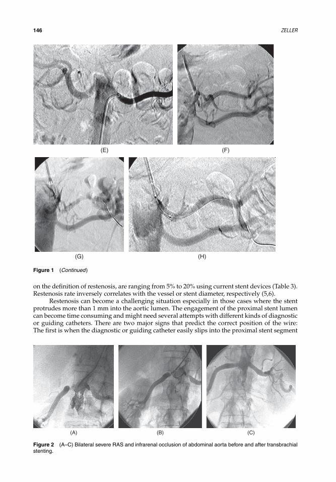
IHBK065-11 IHBK065-Minar May 1, 2010 8:5 Specs: 7x10 tight 7in×10in Char Count=
146 ZELLER
(E) (F)
(G) (H)
Figure 1 (Continued)
on the definition of restenosis, are ranging from 5% to 20% using current stent devices (Table 3).Restenosis rate inversely correlates with the vessel or stent diameter, respectively (5,6).
Restenosis can become a challenging situation especially in those cases where the stentprotrudes more than 1 mm into the aortic lumen. The engagement of the proximal stent lumencan become time consuming and might need several attempts with different kinds of diagnosticor guiding catheters. There are two major signs that predict the correct position of the wire:The first is when the diagnostic or guiding catheter easily slips into the proximal stent segment
(A) (B) (C)
Figure 2 (A–C) Bilateral severe RAS and infrarenal occlusion of abdominal aorta before and after transbrachialstenting.

IHBK065-11 IHBK065-Minar May 1, 2010 8:5 Specs: 7x10 tight 7in×10in Char Count=
RENAL ARTERIES 147
(A) (B) (C)
(D)
(G) (H)
(E) (F)
Figure 3 (A) Right RAS, unfavorable angle of the origin and bended course of the trunk. (B) Cannulation of theostium with a 5-F JR 4 125-cm long diagnostic catheter to place the guidewire. (C) Guiding sheath pushed intothe origin, diagnostic catheter withdrawn. (D) Predilation with a 5/20 mm balloon. (E) Positioning of the stent. (F)Result after 5/13 mm Multilink ultra stent. (G and H) Final result after bilateral RA stenting.
after the lesion is passed with wire. If this is not possible, the wire went through the stentmashes from outside. The second sign is, when the balloon catheter or in case of intended directstenting the stent device can easily be placed in the restenosed stent. In some rare cases, it canbe helpful to use an orthogonal view for the engagement of the stent. Rare data are availableabout the appropriate treatment strategy of restenosis. It seems that plain balloon angioplasty,cutting balloon angioplasty, and restenting are associated with recurrent restenosis rates of about30%. Covered stents (JostentgraftTM, Abbott Vascular) or drug-eluting stents (TaxusTM, BostonScientific) seem to considerably reduce this recurrence rate (17). However, no randomized dataare yet available to proof this concept.

IHBK065-11 IHBK065-Minar May 1, 2010 8:5 Specs: 7x10 tight 7in×10in Char Count=
148 ZELLER
Equipment ChecklistTable 4 summarizes access and lesion-specific tools that should be available on stock for complexrenal interventions.
PART 2: TEACHING CASESCase 1: Telescoping technique (Fig. 4): This case shows an acute angle of the off-take of the leftrenal artery of approximately 180◦ with an origin near subtotal eccentric RAS.
a. What made the lesion “complex” in this specific case? The complexity of the case results inthe difficult angulations of the course of the renal artery associated with the eccentric subtotalstenosis making it nearly impossible to cross the lesion with the wire guided simply througha guiding catheter [Fig. 4(E)]. The second challenge was the navigation of the stent into thestenosis.
(C) (D)
6-F Sidewinder1 or Soft VUDiagnostic catheter
7–9-F guiding catheter
60–90°
α
(A) (B)
Figure 4 (A–D) Step by step technique of transfemoral telescoping technique for engagement of an acutelyangled off-take of a right renal artery. (E) Tight stenosis of the left RA, RDC guiding catheter. (F) SOS omnicatheter through RDC guiding catheter. (G) Guiding catheter advanced over the diagnostic catheter. (H) Left RAafter first stent. (I) Final result after second stent.

IHBK065-11 IHBK065-Minar May 1, 2010 8:5 Specs: 7x10 tight 7in×10in Char Count=
RENAL ARTERIES 149
(E) (F) (G)
(H) (I)
Figure 4 (Continued)
(C) (D)
(A) (B)
Figure 5 Unique crossing design of the HippocampusTM stent system: The long anti-flip tip (A) with progressiveflexibility and minimal entry profile. As soon as the balloon segment with the crimped stent is advanced throughthe curve of the guiding catheter the long tip is already inserted in the ostium. Thus the device cannot flip out therenal artery (B). Figure (C) and (D) show the release of the stent slightly protruding into the aortic lumen.

IHBK065-11 IHBK065-Minar May 1, 2010 8:5 Specs: 7x10 tight 7in×10in Char Count=
150 ZELLER
(A) (B)
(C) (D)
(E)
Figure 6 (A) Severe right RAS. (B) One 0.014′′ wire each in the renal artery and the suprarenal aorta. (C)Placement of the stent. (D) Release of the stent, aortic wire still in place. (E) Final result.
Figure 7 Atheromatous debris after back-bleeding through the guiding catheter following can-ulation of the renal artery ostium in a patient withdiffusely diseased aorta.

IHBK065-11 IHBK065-Minar May 1, 2010 8:5 Specs: 7x10 tight 7in×10in Char Count=
RENAL ARTERIES 151
Table 3 Restenosis Rates for Ostial Stent Angioplasty, Literature Review
References n pts Stent typeTechnique and definition of
restenosisRestenosis rate and
follow-up period
8 68 Palmaz Duplex; PSV > 2.0 m/sec +dRI > 0.05 followed byangiography (> 50%)
11% (mean FU 27 months)
9 29 Palmaz Angiography, ≥ 50% 16% (mean FU 6.7 months)10 100 Mixed Angiography, > 50% 19% (mean FU 8.7 months)11 30 Palmaz, InFlow,
beStentAngiography, not defined 12.5% (6 months)
12 42 Palmaz Angiography; ≥ 50% 14% (6 months)13 63 Not documented Angiography, ≥ 50% 30% (median FU 23 months)14 300 Mixed Angiography, ≥ 50% 21% (median 16 months)15 51 Palmaz Duplex; ≥ 60% (PSV >
2 m/sec, RAR > 3.5) orangiography (≥ 50%)
14.3% (mean FU 13.1months)
16 215 Mixed Duplex; ≥ 70% (RAR > 3.5 +dRI > 0.05)
11.2% (1 year)
5 143 Palmaz Genesis,Radix
Duplex; ≥ 70% (RAR > 3.5 +dRI > 0.05)
6.4% (mean FU 22 months)
b. What was the clinical indication for treatment of this specific case? The indication for thetreatment was a poorly controlled hypertension despite five antihypertensive drugs. Thepatient presented with a diffuse type of atherosclerosis including coronary artery diseasewith already twice done percutaneous coronary intervention and peripheral artery dis-ease with no lifestyle-limiting claudication of the left leg. Baseline creatinine concentrationwas slightly elevated to 1.5 mg/dl. Risk factors besides arterial hypertension included allaspects of a metabolic syndrome.
c. What were the potential hazards of treating this specific patient and this lesion? The potentialhazards were first inducing a dissection during the crossing attempt of the lesion with thewire potentially leading to acute vessel occlusion and second the risk of embolism causedby extensive wire and catheter manipulations. Moreover, stent loss or misplacement had tobe taken into consideration.
Table 4 Recommended Devices for Complex Renal Interventions
Special situation Tool Device example(s)
Brachial access Diagnostic (4/5-F) orguiding catheter (6-F)
Multipurpose, Amplatz right, Judkins right
Femoral access � Diagnostic (4/5-F)� Guiding catheter
(6-F)
SIM 1 or SOS Omni (diagnostic catheter) IMA,Judkins right, Hockey Stick, RDC (guiding catheter)
Subtotal occlusion Guidewire Hydrophilic-coated 0.014′′ extra support guide-wire(e.g., PilotTM 150, Abbott Vascular)
“Undilatable” lesion Plaque modulationdevice
Scoring Balloon (Biotronic)Cutting Balloon (Boston Scientific)
Acute angle of the renalartery origin
“Anti-flip-tip”balloon/stent device
HippocampusTM stent (Invatec)
In-stent restenosis � Drug-eluting stent� Covered stent
TaxusTM (Boston Scientific)Graftmaster (Abbott Vascular)
Chronic total occlusion Crossing device Excimer laser (Spectranetics)Thrombus containing
lesionDistal protection
devicesFilters (e.g., Emboshield proTM, Abbott Vascular,
FibernetTM, Lumen Biomedical, Plymouth, MN)Distal occlusion balloon (PercusurgeTM, Medtronic)
Renal artery perforation � Main artery� Segmental artery
Covered stent (GraftmasterTM)Microcoils (e.g., Trufill, Cordis J&J)

IHBK065-11 IHBK065-Minar May 1, 2010 8:5 Specs: 7x10 tight 7in×10in Char Count=
152 ZELLER
d. Which interventional strategies were considered and which interventional tools were usedto handle this lesion? The easiest approach would have been the brachial access usinga “Multipurpose”-shaped guiding catheter. However, at the time this intervention wasperformed, stents had to be crimped onto the balloon by hand and 8-F guiding cathetershad to be used. The risk of an access site complication inserting an 8-F sheath into thebrachial artery was considered to be high. Therefore, the femoral approach was chosen.Because no 0.014′′ wire compatible devices were available at the time of the intervention,a stiff uncoated 0.018′′ guidewire had to be used. This wire was unable to cross the lesionnavigated through the guiding catheter. This lead to the decision for a selective engagementof the renal artery origin using a 5-F SOS Omni diagnostic catheter, which was introducedthrough the 55-cm long 8-F “RDC” guiding catheter [Fig. 4(A, B, and F)]. Once the origin wascannulated with the diagnostic catheter with its soft atraumatic tip, the 0.018′′ guidewirecould be easily passed through the lesion and the guiding catheter was advanced acrossthe diagnostic catheter and positioned at the origin of the renal artery [Fig. 4(C, D, and G)].Following predilation with a 3/20-mm balloon catheter, a 6/17-mm AVE bridgeTM stent wasplaced [Fig. 4(H)]. However, this stent did not cover the renal artery origin, which was alsodiseased. Therefore, a second 6/10 mm stent was placed without leaving a residual stenosis[Fig. 4(I)].
e. Summary: How was the case managed, is there anything that could have been improved,what was the immediate and long-term outcome? The first stent was placed slightly toofar distal. Either, this stent should have been placed 3 mm more proximal or a longer(20 mm) stent should have been used to avoid the placement of a second stent. The long-term outcome in this particular patient was favorable; the stents are still patent after nineyears of serial annual duplex ultrasound follow-up. Renal function slightly improved withserum creatinine values dropping to 1.0 mg/dl immediately after the intervention and beingstable during follow-up with 1.1 mg/dl after nine years. Blood pressure control persistentlyimproved by a reduction of two antihypertensive drugs.
f. List of contemporary equipments that should be used in this case:� Guiding catheter 6-F (RDC-1 or IMA)� Diagnostic catheter 5-F (SOS Omni or SIM-1)� Guidewire 0.014′′ hydrophilic coated (PilotTM 150) or uncoated (Galeo ESTM)� Coronary balloon catheter 3/20 mm (e.g., MaverickTM) for predilation� 6/20 mm renal stent device (e.g., HippocampusTM)
g. Key learning issues of this specific case: Even if appropriately angled guiding catheters arenot available to engage a renal artery origin due to an acute angle of the renal artery off-take,the telescoping technique can avoid the need of an additional brachial approach.
Case 2: Bifurcation lesion: Early bifurcation of the renal artery, kissing balloon technique.
a. What made the lesion “complex” in this specific case? The right renal artery was acutelyoccluded one year before the index procedure by a cardioembolic event and there wasonly 6% residual function despite successful, but delayed stent recanalization [Fig. 8(A)].Therefore, the patient presented with a functional single kidney (the left one) and a stenosisof the left renal artery, which presented with a short trunk of about 8 mm length beforethe division in the two major side branches [Fig. 8(B) and 8(D)]. Because of the functionalsingle kidney situation, none of the both side branches should have been compromised bythe intervention. Interestingly, the left renal artery was unsuspicious for stenosis one yearbefore during the recanalization of the acutely occluded right renal artery.
b. What was the clinical indication for treatment of this specific case? The patient presentedwith deterioration in blood pressure control and decrease of glomerular filtration rate(GFR) of 15 mL/min within one year. Major concomitant diseases were moderate coronary1-vessel disease, intermittent atrial fibrillation, and nonsignificant peripheral artery disease(Fontaine class I, Rutherford category 1). Risk factors besides arterial hypertension werehistory of smoking and hypercholesterolemia.
c. What were the potential hazards of treating this specific patient and this lesion? The majorhazard was to compromise one of the side branches by plaque shift. Moreover, the largevessel diameter made the use of a noncoronary stent device, in this case a Palmaz stent

IHBK065-11 IHBK065-Minar May 1, 2010 8:5 Specs: 7x10 tight 7in×10in Char Count=
RENAL ARTERIES 153
(A) (B)
(C) (D)
Figure 8 (A) Right RA 12 months after recanalization. (B) Ostial 75% left RA stenosis. Short trunk, origin nearbifurcation. (C and D) One 0.014′′ Galeo ES guidewire in each side branch. (E) Stent positioning, two wires stentPTRA of the RA trunk. (F) Result after stent placement. (G) Additional double balloon angioplasty. (H) Final resultafter Palmaz P 104 stent.
(Continued on page 154)
necessary. Covering one of the side branches with this stent type it would have been impos-sible to dilate the connections between the struts performing kissing balloon angioplastyafter stent deployment.
d. Which interventional strategies were considered for this lesion? First, one 0.014′′ extra sup-port guidewire had to be placed into each main side branch [Fig. 8(C) and 8(D)]. Thereafter,the shortest available stent (Palmaz P 104) had to be positioned with the distal stent endreaching into the bifurcation but not covering completely the cranial side branch to enableflaring the distal stent end into both side branches using kissing balloon technique followingstent placement [Fig. 8(E) and (8F)].
e. Which interventional tools were used to handle this lesion? The intervention was performedvia an 8-F “RDC” guiding catheter using two 0.014′′ Galeo ESTM guidewires. The PalmazP 104TM stent was hand crimped onto a 6/20-mm Bypass SpeedyTM balloon and positionedinto the short common renal artery trunk covering the dummy wire that was placed intothe cranial side branch [Fig. 8(E) and (8F)]. After repositioning the guidewire through thestent into the cranial side branch [Fig. 8(F)], a 3.5/20-mm coronary balloon was positionedreaching from the main trunk into the cranial side branch and a 6/12-mm balloon from thecommon trunk into the caudal main side branch. Performing prolonged kissing balloonangioplasty for one minute [Fig. 8(G)], the distal stent ends were flared into both sidebranches to achieve an optimized vessel wall apposition [Fig. 8(H)].
f. Summary: How was the case managed, is there anything that could have been improved,what was the immediate and long-term outcome? The immediate and long-term technicaloutcome was excellent with a patent vessel for more than eight years. Clinically, GFRincreased immediately after the intervention by 12 mL/min and remained stable during
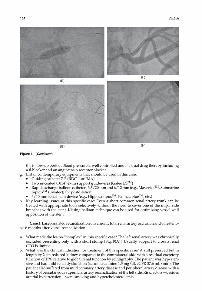
IHBK065-11 IHBK065-Minar May 1, 2010 8:5 Specs: 7x10 tight 7in×10in Char Count=
154 ZELLER
(E)(F)
(G) (H)
Figure 8 (Continued)
the follow-up period. Blood pressure is well controlled under a dual drug therapy includinga ß-blocker and an angiotensin receptor blocker.
g. List of contemporary equipments that should be used in this case:� Guiding catheter 7-F (RDC-1 or IMA)� Two uncoated 0.014′′ extra support guidewires (Galeo ESTM)� Rapid exchange balloon catheters 3.5/20 mm and 6/12 mm (e.g., MaverickTM, Submarine
rapidoTM (Invatec)) for postdilation� 6/10 mm renal stent device (e.g., HippocampusTM, Palmaz blueTM, etc.)
h. Key learning issues of this specific case: Even a short common renal artery trunk can betreated with appropriate tools selectively without the need to cover one of the major sidebranches with the stent. Kissing balloon technique can be used for optimizing vessel wallapposition of the stent.
Case 3: Laser-assisted recanalization of a chronic total renal artery occlusion and of resteno-sis 6 months after vessel recanalization.
a. What made the lesion “complex” in this specific case? The left renal artery was chronicallyoccluded presenting only with a short stump [Fig. 9(A)]. Usually, support to cross a renalCTO is limited.
b. What was the clinical indication for treatment of this specific case? A still preserved but inlength by 2 cm reduced kidney compared to the contralateral side with a residual excretoryfunction of 15% relative to global renal function by scintigraphy. The patient was hyperten-sive and had mild renal dysfunction (serum creatinine 1.5 mg/dl, eGFR 37.6 mL/min). Thepatient also suffered from mild coronary artery disease and peripheral artery disease with ahistory of percutaneous superficial artery recanalization of the left side. Risk factors—besidesarterial hypertension—were smoking and hypercholesterolemia.

IHBK065-11 IHBK065-Minar May 1, 2010 8:5 Specs: 7x10 tight 7in×10in Char Count=
RENAL ARTERIES 155
(A) (B)
(C)
(D)
(E)
Figure 9 (A) Left renal artery 100%. (B) Result after laser recanalization and implantation of a Magic Wallstent.(C) In-stent restenosis after six months after the index procedure. (D) Two-millimeter eccentric excimer lasercatheter, 8-F RDC guiding catheter. (E) Left RA after laser, 6 mm balloon angioplasty, and Palmaz P 104 at theorigin.
c. What were the potential hazards of treating this specific patient and this lesion? Majorrisks of recanalization of renal artery CTOs are dissections of the aorta and renal artery,perforation, and embolism (renal and lower limbs).
d. Which interventional strategies were considered for this lesion? Most important was thechoice of the appropriate guiding catheter or diagnostic catheter configuration to guaran-tee a stable catheter position for sufficient wire backup. As guiding catheter, the “RDC”configuration or “Hockey Stick” shape would offer the best support. Alternatively, the tele-scoping technique using a “SIM-1” or “SOS Omni” diagnostic catheter introduced throughthe guiding catheter would offer adequate wire support to cross the lesion.

IHBK065-11 IHBK065-Minar May 1, 2010 8:5 Specs: 7x10 tight 7in×10in Char Count=
156 ZELLER
e. Which interventional tools were used to handle this lesion? To facilitate the passage of theCTO and for debulking of potentially embolic occlusive material, an excimer laser catheterwas used. Because of the length of the lesion, a self-expanding stent was chosen, even thoughthe origin of the renal artery was also affected.
f. Summary: How was the case managed, is there anything that could have been improved,what was the immediate and long-term outcome? The recanalization procedure was per-formed via an 8-F “RDC” guiding catheter. The lesion could be penetrated with a 0.014′′hydrophilic-coated coronary guidewire. Using the step-by-step technique, a 2-mm eccentriclaser probe was passed through the lesion with reconnection of the wire to the perfusedlumen at the level of the division of the renal artery trunk into the segmental arteries.A 6/24-mm Easy WallstentTM was placed covering the whole length of the former occlu-sion [Fig. 9(B)]. Postdilation was performed with a 6/20-mm coronary balloon catheter.Six months later, severe restenosis was detected by duplex ultrasound and confirmed byangiography [Fig. 9(C)]. Restenosis included the origin with incomplete expansion of theproximal stent end, diffuse in-stent neointima hyperproliferation, and restenosis at the divi-sion of the segmental arteries. Using again an 8-F “RDC” guiding catheter, four passes withan eccentric 2-mm excimer laser probe were performed [Fig. 9(D)], followed by postdilationwith a 6/20-mm monorail balloon catheter and the placement of a 10-mm long balloonexpandable stent into the renal artery origin without residual stenosis [Fig. 9(E)]. Nine yearslater, the artery is still patent as shown by duplex ultrasound. To avoid restenosis at leastat the origin, a balloon expandable stent should have been placed already during the indexprocedure. Nowadays, a self-expanding low-profile nitinol stent device should be preferredfitting through a 6-F guiding catheter.
Clinically, even after nine years, renal function was preserved with a serum creati-nine concentration of 1.0 mg/dl and blood pressure is well controlled with an angiotensinconverting enzyme blocker only.
g. List of contemporary equipments that should be used in this case:� Guiding catheter 7-F (“RDC”-1 or “Hockey Stick”)� Hydrophilic-coated 0.014′′ extra support guidewire (PilotTM or Choice PTTM)� 1.4 or 1.7 mm Turbo eliteTM laser catheter (Spectranetics)� For occasional predilation, RX balloon catheter 3/20 mm (e.g., MaverickTM)� 6/20 mm or 7/20 mm low-profile nitinol stent device, for example, XpertTM (Abbott
Vascular), Astron pulsarTM (Biotronik) for the renal artery trunk� 6/20 mm RX balloon catheter for postdilation of the nitinol stent (e.g., Submarine
rapidoTM)� 6/10 mm balloon expandable stent for the renal artery origin (e.g., HippocampusTM)
h. Key learning issues of this specific case: Recanalization of even chronic total renal arteryocclusions is feasible provided a renal artery stump can be identified. Photoablation fordebulking with the excimer laser can facilitate the recanalization procedure and potentiallyprevent distal embolism during the recanalization attempt. Excimer laser technology canalso be used for debulking of in-stent restenosis. In long lesions, self-expanding stents canbe used to cover the renal artery trunk. However, if the origin is involved into the occlusion,radial strength of the self-expanding stent might be insufficient to keep the proximal stentsegment fully expanded. The overlapping additional placement of a balloon expandableshould be considered.
PART 3: KEY ISSUES FOR SUCCESSFUL AND SAFE TREATMENT OF COMPLEXRENAL LESIONSThe basis for a successful and safe treatment of complex renal artery lesions is the exact knowl-edge of the pathoanatomy and the presence of a predefined treatment strategy. If not all neededtools are on the shelf, reschedule the patient for a later date. Because complex interventionsincorporate an increased periinterventional risk, the indication for the intervention must beundisputed. Key features to avoid complications are as follows:
� No guiding catheter manipulation along the aortic wall without aspiration of potentiallytrapped debris.

IHBK065-11 IHBK065-Minar May 1, 2010 8:5 Specs: 7x10 tight 7in×10in Char Count=
RENAL ARTERIES 157
� Use the appropriate access to the lesion. If indicated change from a femoral to a brachialapproach.
� Avoid unnecessary interventional steps. Every additional interventional step increases therisk of complication.
� Reduce the amount of contrast agent to what is absolutely necessary.� Use the whole cardiovascular–endovascular toolbox and techniques. In doubt, ask a col-
league of another specialty for support.
REFERENCES1. Safian RD, Textor SC. Renal-artery stenosis. N Engl J Med 2001; 344:431–442.2. Zeller T, Frank U, Muller C, et al. Technological advances in the design of catheters and devices used
in renal artery interventions: impact on complications. J Endovasc Ther 2003; 10:1006–1014.3. Zeller T, Bonvini RF, Rastan A, et al. Colour-coded duplex ultrasound for diagnosis of renal artery
stenosis and as follow-up examination after revascularization. Catheter Cardiovasc Interv 2008;71:995–999.
4. Feldman RL, Wargovich TJ, Bittl JA. No-touch technique for reducing aortic wall trauma during renalartery stenting. Catheter Cardiovasc Interv 1999; 46:245–248.
5. Zeller T, Rastan A, Kliem M, et al. Impact of carbon coating on restenosis rate after stenting ofatherosclerotic renal artery stenosis. J Endovasc Ther 2005; 12:605–611.
6. Zeller T, Muller C, Frank U, et al. Gold-coating and restenosis after primary stenting of ostial renalartery stenosis. Catheter Cardiovasc Interv 2003; 60:1–6.
7. Zahringer M, Sapoval M, Pattynama PM, et al. Sirolimus-eluting versus bare-metal low-profile stentfor renal artery treatment (GREAT Trial): angiographic follow-up after 6 months and clinical outcomeup to 2 years. J Endovasc Ther 2007; 14(4):460–468.
8. Blum U, Krumme B, Flugel P, et al. Treatment of ostial renal-artery stenoses with vascular endoprothe-ses after unsuccessful balloon angioplasty. N Engl J Med 1997; 336:459–465.
9. Taylor A, Sheppard D, MacLeod MJ, et al. Renal artery stent placement in renal artery stenosis:Technical and early clinical results. Clin Radiology 1997; 52:451–457.
10. White CJ, Ramee SR, Collins TJ, et al. Renal artery stent placement: utility in lesions difficult to treatwith balloon angioplasty. J Am Coll Cardiol 1999; 30:1445–1450.
11. Gross CM, Kramer J, Waigand J, et al. Ostial renal artery stent placement for atherosclerotic renalartery stenosis in patients with coronary artery disease. Cathet Cardiovasc Diagn 1998; 45:1–8.
12. Van de Ven PJG, Kaatee R, Beutler JJ, et al. Arterial stenting and balloon angioplasty in ostial atheroscle-rotic renovascular disease: a randomised trail. Lancet 1999; 353:282–286.
13. Beutler JJ, van Ampting JM, van de Ven PJ, et al. Long-term effects of renal artery stenting on kidneyfunction for patients with ostial atherosclerotic renal artery stenosis and renal insufficiency. J Am SocNephrol 2001; 1475–1481.
14. Lederman RJ, Mendelsohn FO, Santos R, et al. Primary renal artery stenting: characteristics andoutcomes after 363 procedures. Am Heart J 2001; 142:314–323.
15. Rocha-Singh KJ, Ahuja RK, Sung CH, et al. Long-term renal function preservation after renal arterystenting in patients with progressive ischemic nephropathy. Catheter Cardiovasc Interv 2002; 57:135–141.
16. Zeller T, Frank U, Muller C, et al. Predictors of improved renal function after primary stenting ofsevere atherosclerotic ostial renal artery stenosis. Circulation 2003; 108:2244–2249.
17. Zeller T, Rastan A, Schwarzwalder U, et al. Treatment of instent restenosis following stent-supportedrenal artery angioplasty. Catheter Cardiovasc Interv 2007; 70:454–459.

IHBK065-12 IHBK065-Minar May 1, 2010 14:47 Specs: 7x10 tight 7in×10in Char Count=
12 Chronic Mesenteric Ischemia: MesentericVascular InterventionRajan A. G. Patel, Stephen R. Ramee, and Christopher J. WhiteDepartment of Cardiology, Ochsner Heart and Vascular Institute, Ochsner Clinic Foundation, New Orleans,Louisiana, U.S.A.
PART 1: INTRODUCTIONAtherosclerosis of the mesenteric vessels may result in chronic mesenteric ischemia (CMI). Thisuncommon condition is often amenable to percutaneous intervention by experienced operators.Percutaneous endovascular procedures have been applied to the mesenteric arterial systemsince the early 1980s. In most symptomatic patients, at least two splanchnic vessels have severestenosis. Moawad and Gewertz (1) report at least two-vessel disease in 91% of CMI patientsand three-vessel disease in 55% of CMI patients. In this review, only 7% of CMI patients had anoccluded superior mesenteric artery (SMA) and 2% had isolated celiac disease. Abrupt occlusionof mesenteric blood vessels can result in acute mesenteric ischemia. In a review of the topic, Old-enburg et al. (2) report the incidence of various etiologies of acute mesenteric ischemia as follows:arterial embolism 40% to 50%, arterial thrombosis 25%, nonocclusive arterial disease resulting inischemia secondary to other factors such as low cardiac output or �adrenergic agonist use 20%,and venous thrombosis 10%. The patient with acute mesenteric ischemia has often developedinfarcted bowel by the time of presentation. These patients generally require surgical treatmentto remove necrotic tissue. Therefore, the focus of this chapter is the percutaneous treatmentof CMI.
What Makes a Lesion “Complex” in Mesenteric Artery Interventions?Endovascular treatment of mesenteric artery atherosclerotic lesions is made complex by thelesion location. These lesions are typically located near the aortic origin of the affected vessel.Furthermore, proximal segments of the mesenteric vessels frequently have an acute caudalorientation. The proximal location requires balloon expandable stents for precise positioning.The vessels are moderate in size usually 5 to 7 mm in diameter. These factors can make bal-loon expandable stent delivery from the femoral artery challenging. Additionally, angiographicimaging of mesenteric vessels, which arise from the anterior aspect of the aorta, requires steeplateral angulation with cranial or caudal orientation of the image intensifier. With steep angu-lation, X-rays must penetrate a larger volume of tissue that may decrease the image quality.Additionally, the patient is exposed to a higher dose of X-rays and the operators may be exposedto greater scatter radiation. Finally, the incidence of restenosis after stent placement in mesentericarteries is higher than that reported for renal or coronary arteries.
Clinical Indications for Treatment of Complex Lesionsin Mesenteric Artery InterventionsAtherosclerotic disease of the aorta with associated aorto-ostial stenosis of the mesenteric vesselsis a common finding. Of 552 healthy Medicare beneficiaries that were screened with abdominalultrasound, 17.5% had significant narrowing of a mesenteric vessel detected (3). However, lessthan 1% of patients with atherosclerotic narrowing of a mesenteric artery require revascular-ization (4). The most likely explanation for the infrequent occurrence of CMI in clinical practiceis the redundancy of the splanchnic circulation with multiple interconnections (collaterals)between the visceral vessels.
Patients with CMI avoid food because of associated abdominal discomfort with eating and,therefore, almost always demonstrate significant weight loss. Abdominal pain in the absenceof weight loss is more likely a result of a “functional” complaint, rather than bowel ischemia.Postprandial abdominal pain from CMI typically persists for one to three hours (5). However,

IHBK065-12 IHBK065-Minar May 1, 2010 14:47 Specs: 7x10 tight 7in×10in Char Count=
CHRONIC MESENTERIC ISCHEMIA: MESENTERIC VASCULAR INTERVENTION 159
Table 1 Clinical Indications for Treatment of Mesenteric Artery Stenosis Are Defined by the Syndromes ofChronic Mesenteric Ischemia
Typical chronic mesenteric ischemia Clinical triad: postprandial symptoms, pain or bloatingWeight loss over time“Food fear”—avoidance of food due to abdominal pain associated with
eatingIschemic gastropathy Nausea
VomitingFullnessAbdominal painWeight loss
Ischemic colitis Abdominal painGastrointestinal bleedingHematochezia
patients with ischemic gastropathy may present with atypical symptoms, such as vomiting,diarrhea, constipation, ischemic colitis, and lower gastrointestinal bleeding. CMI may be verylow on the list of differential diagnoses for patients with atypical symptoms. However, a highdegree of suspicion for mesenteric ischemia is warranted for patients with other manifestationsof atherosclerosis and unexplained weight loss. Table 1 summarizes the symptoms associatedwith CMI syndromes.
Among patients with the aforementioned symptoms and weight loss, the diagnosis of CMIis often delayed while evaluation for possible malignancy is conducted. Patients with functionalbowel complaints rarely experience significant weight loss, which helps to differentiate themfrom CMI patients. Evidence of significant obstruction of two or more vessels is often foundwhen classic symptoms and endoscopy suggest bowel ischemia (6). However, cases of single-vessel disease, usually of the SMA, have been described, particularly if collateral connectionshave been disrupted by prior abdominal surgery. When critical stenosis (≥70%) is found insymptomatic patients with weight loss, revascularization is appropriate. Unfortunately, forpatients with borderline lesions or atypical symptoms, there is no noninvasive stress test toelicit an ischemic response.
Hazards and ComplicationsRevascularization for CMI has traditionally been accomplished with open surgery using bypassgrafting, endarterectomy, or aortic reimplantation of the SMA. This patient group has a high inci-dence of associated coronary artery disease. The reported perioperative mortality ranges from3.5% to 15% (1,7–16). The highest incidence of complications during surgical revascularizationreported is in the cohort of patients over the age of 70 (17).
At the Ochsner Clinic, we have reported the largest series of patients with CMI treated withendovascular techniques (18). A total of 79 vessels were treated in 59 patients. One patient in thisseries died from complications of multiorgan failure postprocedure. Table 2 lists the clinical eventrates from this series. Two patients in this cohort experienced vascular access site complications.Both patients experienced brachial artery thombosis that was treated percutaneously. In anothercase series of 25 patients with CMI treated using stents, 2 (8%) cases of pseudoaneursym and1 (4%) case of acute renal failure were reported (19).
Interventional StrategiesAtherosclerotic aorto-ostial obstructions of the visceral vessels are similar to those of the renalarteries and the technical considerations for percutaneous transluminal angioplasty (PTA) withstent placement are similar to those for renal artery intervention. As with renal artery interven-tions, stent placement offers superior patency compared to PTA alone (20). The endovascularapproach circumvents the need for general anesthesia and the operative trauma associated withopen surgery. Retrospective data suggest that the endovascular approach results in lower acutemortality and morbidity relative to surgical revascularization (6,16,18,20,21).
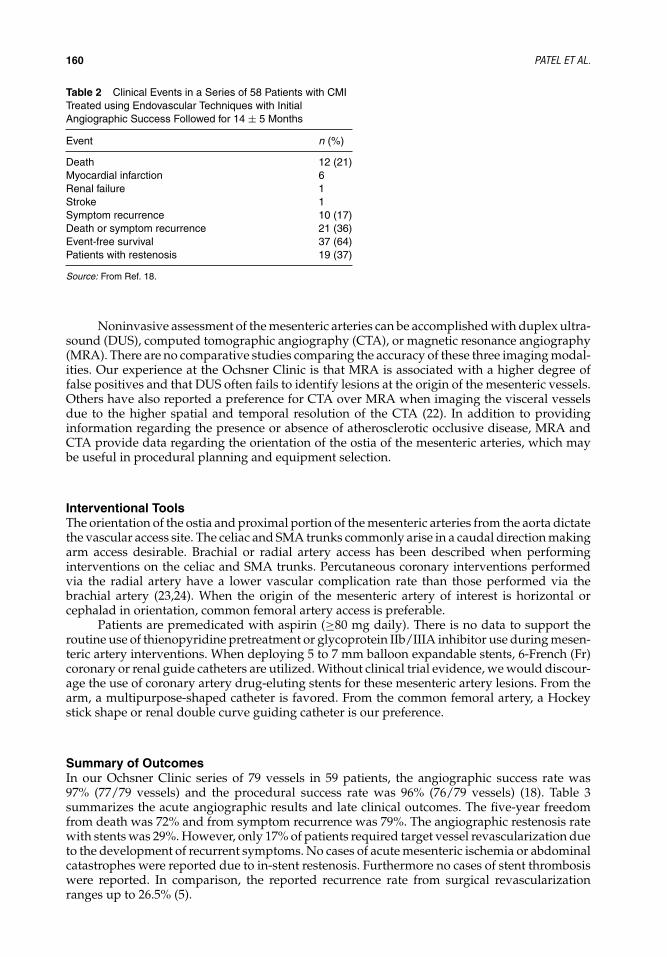
IHBK065-12 IHBK065-Minar May 1, 2010 14:47 Specs: 7x10 tight 7in×10in Char Count=
160 PATEL ET AL.
Table 2 Clinical Events in a Series of 58 Patients with CMITreated using Endovascular Techniques with InitialAngiographic Success Followed for 14 ± 5 Months
Event n (%)
Death 12 (21)Myocardial infarction 6Renal failure 1Stroke 1Symptom recurrence 10 (17)Death or symptom recurrence 21 (36)Event-free survival 37 (64)Patients with restenosis 19 (37)
Source: From Ref. 18.
Noninvasive assessment of the mesenteric arteries can be accomplished with duplex ultra-sound (DUS), computed tomographic angiography (CTA), or magnetic resonance angiography(MRA). There are no comparative studies comparing the accuracy of these three imaging modal-ities. Our experience at the Ochsner Clinic is that MRA is associated with a higher degree offalse positives and that DUS often fails to identify lesions at the origin of the mesenteric vessels.Others have also reported a preference for CTA over MRA when imaging the visceral vesselsdue to the higher spatial and temporal resolution of the CTA (22). In addition to providinginformation regarding the presence or absence of atherosclerotic occlusive disease, MRA andCTA provide data regarding the orientation of the ostia of the mesenteric arteries, which maybe useful in procedural planning and equipment selection.
Interventional ToolsThe orientation of the ostia and proximal portion of the mesenteric arteries from the aorta dictatethe vascular access site. The celiac and SMA trunks commonly arise in a caudal direction makingarm access desirable. Brachial or radial artery access has been described when performinginterventions on the celiac and SMA trunks. Percutaneous coronary interventions performedvia the radial artery have a lower vascular complication rate than those performed via thebrachial artery (23,24). When the origin of the mesenteric artery of interest is horizontal orcephalad in orientation, common femoral artery access is preferable.
Patients are premedicated with aspirin (≥80 mg daily). There is no data to support theroutine use of thienopyridine pretreatment or glycoprotein IIb/IIIA inhibitor use during mesen-teric artery interventions. When deploying 5 to 7 mm balloon expandable stents, 6-French (Fr)coronary or renal guide catheters are utilized. Without clinical trial evidence, we would discour-age the use of coronary artery drug-eluting stents for these mesenteric artery lesions. From thearm, a multipurpose-shaped catheter is favored. From the common femoral artery, a Hockeystick shape or renal double curve guiding catheter is our preference.
Summary of OutcomesIn our Ochsner Clinic series of 79 vessels in 59 patients, the angiographic success rate was97% (77/79 vessels) and the procedural success rate was 96% (76/79 vessels) (18). Table 3summarizes the acute angiographic results and late clinical outcomes. The five-year freedomfrom death was 72% and from symptom recurrence was 79%. The angiographic restenosis ratewith stents was 29%. However, only 17% of patients required target vessel revascularization dueto the development of recurrent symptoms. No cases of acute mesenteric ischemia or abdominalcatastrophes were reported due to in-stent restenosis. Furthermore no cases of stent thrombosiswere reported. In comparison, the reported recurrence rate from surgical revascularizationranges up to 26.5% (5).

IHBK065-12 IHBK065-Minar May 1, 2010 14:47 Specs: 7x10 tight 7in×10in Char Count=
CHRONIC MESENTERIC ISCHEMIA: MESENTERIC VASCULAR INTERVENTION 161
Table 3 Acute Angiographic Results and Five-Year ClinicalOutcomes from a Series of 59 Patients with CMI Treated withEndovascular Techniques Including Stent Placement
Angiographic findings:Reference vessel diameter, baseline (mm) 6.20 ± 1.1Minimal luminal diameter, baseline (mm) 1.26 ± 0.4Percent diameter stenosis, baseline (%) 80 ± 6Reference vessel diameter, postintervention (mm) 6.21 ± 1.1Minimal luminal diameter, postintervention (mm) 5.82 ± 1.0Percent diameter stenosis, postintervention (%) 7 ± 155-year clinical outcomes:Freedom from death (%) 72Freedom from symptom recurrence (%) 79Freedom from death or symptom recurrence (%) 57
Source: From Ref. 18.
Equipment Checklist
Table 4 Equipment Checklist
Upper extremity accessGuide catheter 6–8-Fr Multipurpose/Hockey stickGuidewire 0.014-in Choice of moderate to extra-supportStents 5–7 mm balloon expandable (off-label biliary) stentsInflation deviceRotating hemostatic valve
Femoral accessGuide catheter 6–8-Fr Hockey stick/internal mammaryGuidewire 0.014-in Extra-support 0.014-in guidewireStents 5–7 mm balloon expandable (off-label biliary) stentsInflation deviceRotating hemostatic valve
PART 2: CASE PRESENTATIONS
Mesenteric Case #1The patient is a 32-year-old female with a one-month history of postprandial abdominal pain.The abdominal pain is associated with abdominal bloating. Abdominal ultrasound showed amildly enlarged liver and the abdominal DUS showed elevated velocities in the celiac trunksuggestive of a ≥70% stenosis. The patient underwent an abdominal aortogram with runoffthat showed a 90% stenosis involving the celiac trunk. The patient was therefore referred forpercutaneous intervention to the celiac stenosis.
On physical examination, blood pressure was 99/64 mm Hg and the heart rate was72 bpm. The patient was in no acute distress. There was no jugular venous distention (JVD) orcarotid bruit. The lungs were clear to auscultation bilaterally. Examination of the heart revealeda regular rate and rhythm with no murmurs. The abdomen was soft, mildly distended, andnontender. Bowel sounds were present. There was no hepatosplenomegaly. The extremitiesexam revealed no cyanosis, clubbing or edema. Peripheral pulses were as follows: femoral 2+bilaterally, dorsalis pedis and posterior tibial 2+ bilaterally, brachial 2+ bilaterally, and radialpulses 2+ with her Allen’s test was normal bilaterally. Laboratory results were normal.
What Made the Case “Complex”?Diagnosing CMI in this 32-year-old woman was challenging in that CMI is not typically highon the list of differential diagnoses for vague abdominal symptoms among patients in this agegroup. Figures 1 and 2 show a 4-Fr multipurpose catheter selectively engaged at the origin ofthe celiac artery placed via the brachial artery. Note the steep downward angulation of the celiac

IHBK065-12 IHBK065-Minar May 1, 2010 14:47 Specs: 7x10 tight 7in×10in Char Count=
162 PATEL ET AL.
Figure 1 Selective (LAT) celiac trunk angio-gram showing a tight stenosis in the proximalportion of the vessel prior to the bifurcation ofthe splenic and hepatic arteries.
artery. This case could be performed via femoral access, but it would be much more difficultgiven the angle at which the celiac artery leaves the aorta. Choosing the upper extremity forarterial access, either the radial or brachial artery, is a key decision point in this case. If theprocedure is performed via the radial artery, a 125-cm long guide catheter may be necessarydepending on the height of the patient.
What Was the Clinical Indication for the Treatment of This Patient?The clinical indication for the treatment of this patient was a history of postprandial pain andthe finding of severe celiac trunk disease on noninvasive ultrasound imaging confirmed withinvasive angiography. Interestingly, the SMA and inferior mesenteric arteries were patent. Thepatient was evaluated by both a gastroenterologist and a vascular surgeon who concurred inthe diagnosis and agreed that endovascular therapy was the treatment modality of choice forthis patient.
Figure 2 Selective (RAO cranial) celiac trunkangiogram with a 4-Fr multipurpose cathetershowing an 80% stenosis in the celiac trunk,just proximal to the bifurcation of the hepaticand splenic arteries.

IHBK065-12 IHBK065-Minar May 1, 2010 14:47 Specs: 7x10 tight 7in×10in Char Count=
CHRONIC MESENTERIC ISCHEMIA: MESENTERIC VASCULAR INTERVENTION 163
Figure 3 Placement of 6-Fr shuttle sheath(Cook, Bloomington, IN) over an 0.035-in. Who-ley guidewire (Guidant, Santa Clara, CA), withpredilation of the celiac artery stenosis with a6-mm balloon at 6 atm.
What Were the Potential Hazards of Treating This Patient and Anatomy?The primary potential hazards of treating this patient are vascular complications. First, thebrachial artery access site must be appropriately managed. Once the procedure is complete, thesheath should be removed. Manual pressure should be held over the brachial artery. However,as the brachial artery is an end artery the pressure should not be occlusive. Second, perform-ing PTA and stent placement at the aorto-ostial junction of the celiac trunk has the potentialcomplication of vessel dissection at the celiac trunk and its branches as well as of the aortaitself. Atheroembilization is another potential complication and concern when treating bulkyaorto-ostial plaques.
What Interventional Strategies Were Considered?The interventional strategy considered was to first predilate the lesion using a 6-mm PTA balloonand then place a balloon expandable stent across the lesion. Prior to crossing the lesion witha guidewire, an intravenous heparin bolus was administered to maintain an activated clottingtime (ACT) between 250 and 300 seconds.
Which Interventional Tools Were Used to Manage This Case?Figure 3 shows a 6-Fr 90-cm long Shuttle sheath (Cook Incorporated, Bloomington, IN)exchanged over the diagnostic catheter, with a 0.035-in Wholey guidewire (Mallinckrodt Inc.,Hazelwood, MO) across the lesion. Predilation of the lesion was performed with a 6 × 20 mmOpta 5 PTA balloon (Cordis, Warren, NJ). Figure 4 shows positioning of the 6 × 28 mm Omni-link balloon expandable stent (Guidant, Santa Clara, CA) across the celiac stenosis. Figure 5shows the final result with the stent deployed and relief of the stenosis.
SummaryProcedural, angiographic, and clinical success were achieved in this case. The brachial sheathwas removed after the case and manual pressure was held until hemostasis was achieved.An alternative to using heparin in this patient might have been to use bilvalirudin. However,bilvalirudin has not been studied during mesenteric intervention. The theoretical advantage ofusing bilvalirudin is that the ACT would not need to be monitored and the bilvalirudin infusionwould not need to be adjusted during the procedure. Additionally, the sheath could be removedone hour after discontinuing the infusion.

IHBK065-12 IHBK065-Minar May 1, 2010 14:47 Specs: 7x10 tight 7in×10in Char Count=
164 PATEL ET AL.
Figure 4 Placement of a 6 × 28 mm Omnilink(Guidant, Santa Clara, CA) stent at the lesionsite in the celiac artery.
List of Equipment6-French brachial artery sheath6-French multipurpose diagnostic catheter6-French 90-cm long Shuttle sheath (Cook Incorporated, Bloomington, IN)0.035-in Wholey guidewire (Mallinckrodt Inc., Hazelwood, MO)6 × 20 mm Opta 5 PTA balloon (Cordis, Warren, NJ)6× 28 mm Omni-link balloon expandable stent (Guidant, Santa Clara, CA)
Key Learning IssuesThe key learning issue in this case is recognition of the acute downward angle at which theceliac trunk originates from the aorta. Approaching the celiac trunk from above (i.e., arm access)facilitates engaging the vessel and delivering a stent. The vessel could have been treated via thefemoral artery. However, the steep angle would have made crossing the lesion with a balloon or
Figure 5 Final angiogram following stentplacement to the celiac artery.

IHBK065-12 IHBK065-Minar May 1, 2010 14:47 Specs: 7x10 tight 7in×10in Char Count=
CHRONIC MESENTERIC ISCHEMIA: MESENTERIC VASCULAR INTERVENTION 165
stent difficult and increased the risk of stent dislocation or embolization. When approaching amesenteric vessel with downward angulation from the arm, the multipurpose-shaped catheteroften provides an ideal curve.
Mesenteric Case #2The patient is a 49-year-old female with history of chronic abdominal pain during the last threeyears. The abdominal pain was not temporally related to food intake, but was associated withnausea and vomiting. The patient lost 40 pounds over one year. Color flow Doppler studies weresuggestive of celiac, superior mesenteric, and inferior mesenteric artery stenosis. The patientwas referred for mesenteric angiography to establish the presence and severity of splanchnicvascular disease.
On examination, the patient’s blood pressure was 100/70 mm Hg and her heart rate was64 bpm. There was no JVD or carotid bruit. Her lungs were clear. Auscultation of the heartrevealed normal S1 and S2 with no murmurs, gallop, rub, or heave. The patient had normalbowel sounds. Her abdomen was soft and nontender. Examination of her extremities demon-strated no clubbing or cyanosis. Peripheral pulses were as follows: femoral pulses 2+ bilaterally,dorsalis pedis and posterior tibial pulses 2+ bilaterally, brachial pulses 2+ bilaterally, and radialpulses 2+ bilaterally. Her Allen’s test was normal bilaterally. The patient’s medications includedvalsartan and lansoprazole.
What Made the Case “Complex”?The diagnostic angiogram was performed from the right brachial artery with a 6-Fr multipur-pose catheter. A critical narrowing [Fig. 6(A)] of the superior mesenteric artery was found. Theceliac artery was also occluded. An exchange length 0.014-in extrasupport coronary guidewire(Spartacore, Guidant/Boston Scientific, Santa Clara, CA) was advanced into the SMA acrossthe lesion. The 6-Fr multipurpose catheter was exchanged for a 6-Fr multipurpose coronaryguide catheter, which engaged the ostium of the SMA. This case was made “complex” by thelocation of the lesion. In order to gain adequate guide catheter support to cross the lesion, a multi-purpose catheter was utilized and the lesion was approached in antegrade manner via the arm.
What Was the Clinical Indication for the Treatment of This Patient?The clinical indication for the treatment of this patient was a history of abdominal symptomsand weight loss in the context of an occluded celiac artery and a critically narrowed SMAwithout another explanation for her symptoms.
(A) (B)
Figure 6 (A) Angiogram of superior mesenteric artery ostial stenosis with 6-Fr multipurpose angiographiccatheter from the brachial access. (B) Angiogram after balloon expandable stent deployment.

IHBK065-12 IHBK065-Minar May 1, 2010 14:47 Specs: 7x10 tight 7in×10in Char Count=
166 PATEL ET AL.
What Were the Potential Hazards of Treating This Patient and Anatomy?As this patient’s celiac artery is occluded, perfusion of her small bowel is dependent on herSMA. A procedural complication such as SMA dissection resulting in cessation of SMA arterialflow could theoretically result in life-threatening bowel necrosis.
What Interventional Strategies Were Considered?The keys to this case were engaging the SMA from above and crossing the lesion with adequateguide catheter support. The plan was to predilate the lesion with PTA, and then deploy a balloonexpandable stent.
Which Interventional Tools Were Used to Manage This Case?As stated above, after diagnostic angiography of the SMA, 2500 IU of heparin was administeredintravenously and a 0.014-in extra-support coronary guidewire was advanced into the SMAand across the lesion. Then the 6-Fr multipurpose catheter was exchanged for a 6-Fr multipur-pose coronary guide catheter. The ostium of the SMA was engaged with this catheter whileusing the coronary guidewire as a rail. The lesion was then predilated using a 3.0 × 20 mmQuantum Maverick balloon (Boston Scientific, Natick, MA). A 4.0 × 25 mm NIR balloon expand-able stent (Boston Scientific, Natick, MA) was then placed into the lesion. The stent was deployedwith the balloon inflated to 12 atm. A 5.0 × 20 Cross Sail balloon (Guidant, San Jose, Ca) wasutilized to postdilate the stent. The final angiographic result is illustrated in Figure 6(B). Thepatient tolerated the procedure well.
SummaryProcedural, angiographic, and clinical success were achieved in this case. An alternative to usinga multipurpose guide catheter in this case may have been to utilize a right coronary bypass graftguide catheter from the brachial artery. Both catheters provide support when working in vesselswith inferior angulation.
List of Equipment6-French brachial artery sheath6-French multipurpose diagnostic catheter6-French multipurpose guide catheter0.014-in extra-support coronary guidewire (Spartacore, Guidant, Santa Clara, CA)3.0 × 20 mm Quantum Maverick balloon (Boston Scientific, Natick, MA)4.0 × 25 mm NIR balloon expandable stent (Boston Scientific, Natick, MA)5.0 × 20 Cross Sail balloon (Guidant, San Jose, CA)
Key Learning IssuesThe key learning issues in this case are to recognize the importance of guide catheter supportfrom the arm to cross a critical stenosis as well as to successfully deliver angioplasty balloonsand stents. Another key issue is an appreciation of the collateral blood flow via the SMA whenthe celiac artery is occluded and the potential for massive bowel necrosis if this collateral supplyis jeopardized during revascularization.
Mesenteric Case #3The patient is a 62-year-old female with a history of mesenteric artery disease who was treatedwith PTA and stent placement in the celiac artery and SMA eight years ago after presentingwith symptoms of postprandial pain, weight loss, and food avoidance. Three years ago sheexperienced recurrence of these symptoms and was found to have in-stent restenosis of theceliac stent and stenosis distal to the SMA stent. The celiac artery in-stent restenosis was treatedwith sequential 4.0 × 23 and 4.0 × 13 bare metal stents (Duet, Guidant/Boston Scientific,San Jose, CA) with a good angiographic result. The SMA lesion was dilated with a 5.0 × 20balloon with a good angiographic result. A year later she experienced another recurrence ofabdominal symptoms. She underwent angiography and was found to have in-stent restenosis ofthe celiac artery stents with extension of the lesion into the ostia of both the hepatic and splenicarteries [Fig. 7(A)]. The patient underwent PTA of these lesions with a good angiographic

IHBK065-12 IHBK065-Minar May 1, 2010 14:47 Specs: 7x10 tight 7in×10in Char Count=
CHRONIC MESENTERIC ISCHEMIA: MESENTERIC VASCULAR INTERVENTION 167
(A) (B)
Figure 7 Before (A) and after (B) PTA for in-stent restenosis of the celiac artery and stenosis in the proximalhepatic and splenic arteries.
result [Fig. 7(B)]. One year later she again experienced recurrence of her abdominal symptoms.Angiography was performed. The patient was found to have recurrence of celiac artery in-stentrestenosis and stenosis of the ostia of the hepatic and splenic arteries [Fig. 8(A)]. The SMA stentwas widely patent. The patient underwent PTA of celiac, hepatic, and splenic arteries onceagain [Fig. 8(B)]. Fifteen months later, the patient presented with yet another recurrence of herabdominal symptoms. Ultrasound imaging demonstrated restenosis of the celiac artery stents.Repeat angiography was scheduled.
On examination, the patient’s blood pressure was 140/70 mm Hg and her heart rate is72 bpm. There was no JVD. She had a left carotid bruit and a left carotid endarterectomy(CEA) scar. Her lungs were clear. Auscultation of the heart revealed a normal S1 and S2, andan S4 gallop with no rub or heave. The patient had normal bowel sounds. Her abdomen was
(A) (B)
Figure 8 (A) Instent restenosis of the celiac artery and stenoses at the previous PTA sites in the proximal hepaticand splenic arteries. (B) Angiogram after PTA of the celiac, hepatic, and splenic arteries.
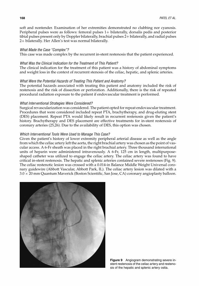
IHBK065-12 IHBK065-Minar May 1, 2010 14:47 Specs: 7x10 tight 7in×10in Char Count=
168 PATEL ET AL.
soft and nontender. Examination of her extremities demonstrated no clubbing nor cyanosis.Peripheral pulses were as follows: femoral pulses 1+ bilaterally, dorsalis pedis and posteriortibial pulses present only by Doppler bilaterally, brachial pulses 2+ bilaterally, and radial pulses2+ bilaterally. Her Allen’s test was normal bilaterally.
What Made the Case “Complex”?This case was made complex by the recurrent in-stent restenosis that the patient experienced.
What Was the Clinical Indication for the Treatment of This Patient?The clinical indication for the treatment of this patient was a history of abdominal symptomsand weight loss in the context of recurrent stenosis of the celiac, hepatic, and splenic arteries.
What Were the Potential Hazards of Treating This Patient and Anatomy?The potential hazards associated with treating this patient and anatomy included the risk ofrestenosis and the risk of dissection or perforation. Additionally, there is the risk of repeatedprocedural radiation exposure to the patient if endovascular treatment is performed.
What Interventional Strategies Were Considered?Surgical revascularization was considered. The patient opted for repeat endovascular treatment.Procedures that were considered included repeat PTA, brachytherapy, and drug-eluting stent(DES) placement. Repeat PTA would likely result in recurrent restenosis given the patient’shistory. Brachytherapy and DES placement are effective treatments for in-stent restenosis ofcoronary arteries (25,26). Due to the availability of DES, this option was chosen.
Which Interventional Tools Were Used to Manage This Case?Given the patient’s history of lower extremity peripheral arterial disease as well as the anglefrom which the celiac artery left the aorta, the right brachial artery was chosen as the point of vas-cular access. A 6-Fr sheath was placed in the right brachial artery. Three thousand internationalunits of heparin were administered intravenously. A 6-Fr, 125 cm in length, multipurpose-shaped catheter was utilized to engage the celiac artery. The celiac artery was found to havecritical in-stent restenosis. The hepatic and splenic arteries contained severe restenoses (Fig. 9).The celiac restenotic lesion was crossed with a 0.014-in Balance Middle Weight Universal coro-nary guidewire (Abbott Vascular, Abbott Park, IL). The celiac artery lesion was dilated with a3.0 × 20 mm Quantum Maverick (Boston Scientific, San Jose, CA) coronary angioplasty balloon.
Figure 9 Angiogram demonstrating severe in-stent restenosis of the celiac artery and resteno-sis of the hepatic and splenic artery ostia.

IHBK065-12 IHBK065-Minar May 1, 2010 14:47 Specs: 7x10 tight 7in×10in Char Count=
CHRONIC MESENTERIC ISCHEMIA: MESENTERIC VASCULAR INTERVENTION 169
Figure 10 Final angiogram after PTA of theceliac artery followed by placement of a 3.0 × 28Cypher DES (Cordis, Warren, NJ) in the splenicartery and a 3.5 × 23 Cypher (Cordis, Warren,NJ) in the celiac artery.
The splenic artery was stented with a 3.0 × 28 mm Cypher DES (Cordis, Warren, NJ). A second3.5 × 23 mm Cypher DES was overlapped with the splenic artery stent and deployed extendinginto the celiac artery. The hepatic artery stenosis was dilated with a 3.0 × 20 mm coronaryangioplasty balloon. The final angiographic result is shown in Figure 10.
SummaryProcedural and angiographic success were achieved in this case. Unfortunately within twoyears the patient developed recurrence of her symptoms and was found to have restenosisof the celiac artery with hepatic and splenic artery involvement. She then underwent surgicalrevascularization. She remained free of abdominal symptoms three years post surgery.
List of Equipment6-French brachial artery sheath6-French multipurpose guide catheter0.014-in Balance Middle Weight Universal coronary guidewire (Abbott Vascular, Abbott
Park, IL)3.0 × 20 mm Quantum Maverick balloon (Boston Scientific, San Jose, CA)3.0 × 28 and 3.5 × 23 mm Cypher stents (Cordis, Warren, NJ)
Key Learning IssuesThe key learning issues in this case are to understand the high rate of restenosis after PTA orstent placement in mesenteric arteries. Potential options for the treatment of in-stent restenosisin this vascular bed include PTA, brachytherapy, and DES placement. However, no randomizedclinical trials addressing in-stent restenosis in the mesenteric arteries have been published. Thisis in part due to the low incidence of CMI relative to coronary artery disease.
PART 3: KEY ISSUES FOR SUCCESSFUL AND SAFE TREATMENT OF COMPLEXMESENTERIC LESIONSThe key points in the management of patients with CMI are (i) to be as certain as possible thatsymptoms truly represent visceral ischemia, as the prevalence of mesenteric artery stenosis ismuch more common than the clinical syndrome of CMI and (ii) once a decision has been madeto proceed with endovascular treatment, selecting the appropriate vascular access and guidecatheter is necessary.

IHBK065-12 IHBK065-Minar May 1, 2010 14:47 Specs: 7x10 tight 7in×10in Char Count=
170 PATEL ET AL.
Mesenteric vascular disease is common in patients with atherosclerosis, but clinicalischemia is unusual given the redundancy of the mesenteric circulation. Patients with func-tional bowel complaints will not have lost weight and will not improve with mesenteric stent-ing. Before planning an intervention, it is important to elicit a history of significant weight loss,a history of postprandial discomfort, and anatomic evidence of occlusion or stenosis of at leasttwo of the three mesenteric vessels. Patients who do not meet these criteria can prove to be adiagnostic challenge.
Once the patient has been carefully selected, then the next issue is to decide upon the bestvascular access. The acute downward angle of most of these vessels as they arise from the aortais often most suitable to an upper extremity, antegrade approach. Many of these lesions can beapproached from the common femoral artery in retrograde fashion, but it is often a struggle andthe risk of stent embolization or dislocation is high. At this time, there is no clinical trial data tosupport a role for DES’s in mesenteric arteries. However, the restenosis rate of bare metal stentsin these arteries appears to be higher than that reported in renal arteries.
REFERENCES1. Moawad J, Gewertz BL. Chronic mesenteric ischemia. Clinical presentation and diagnosis. Surg Clin
North Am 1997; 77:357–369.2. Oldenburg WA, Lau LL, Rodenberg TJ, et al. Acute mesenteric ischemia: a clinical review. Arch Intern
Med 2004; 164:1054–1062.3. Hansen KJ, Wilson DB, Craven TE, et al. Mesenteric artery disease in the elderly. J Vasc Surg 2004;
40:45–52.4. Thomas JH, Blake K, Pierce GE, et al. The clinical course of asymptomatic mesenteric arterial stenosis.
J Vasc Surg 1998; 27:840–844.5. Brandt LJ, Boley SJ. AGA technical review on intestinal ischemia. American Gastrointestinal Associa-
tion. Gastroenterology 2000; 118:954–68.6. Matsumoto AH, Tegtmeyer CJ, Fitzcharles EK, et al. Percutaneous transluminal angioplasty of visceral
arterial stenoses: results and long-term clinical follow-up. J Vasc Interv Radiol 1995; 6:165–674.7. Calderon M, Reul GJ, Gregoric ID, et al. Long-term results of the surgical management of symptomatic
chronic intestinal ischemia. J Cardiovasc Surg (Torino) 1992; 33:723–728.8. Christensen MG, Lorentzen JE, Schroeder TV. Revascularisation of atherosclerotic mesenteric arteries:
experience in 90 consecutive patients. Eur J Vasc Surg 1994; 8:297–302.9. Cormier JM, Fichelle JM, Vennin J, et al. Atherosclerotic occlusive disease of the superior mesenteric
artery: late results of reconstructive surgery. Ann Vasc Surg 1991; 5:510–518.10. Cunningham CG, Reilly LM, Rapp JH, et al. Chronic visceral ischemia. Three decades of progress.
Ann Surg 1991; 214:276–287.11. Gentile AT, Moneta GL, Taylor LM Jr, et al. Isolated bypass to the superior mesenteric artery for
intestinal ischemia. Arch Surg 1994; 129:926–931.12. Johnston KW, Lindsay TF, Walker PM, et al. Mesenteric arterial bypass grafts: early and late results
and suggested surgical approach for chronic and acute mesenteric ischemia. Surgery 1995; 118:1–1187.13. Kieny R, Batellier J, Kretz JG. Aortic reimplantation of the superior mesenteric artery for atherosclerotic
lesions of the visceral arteries: sixty cases. Ann Vasc Surg 1990; 4:122–125.14. Mateo RB, O’Hara PJ, Hertzer NR, et al. Elective surgical treatment of symptomatic chronic mesenteric
occlusive disease: early results and late outcomes. J Vasc Surg 1999; 29:821–831.15. McAfee MK, Cherry KJ, Jr, Naessens JM, et al. Influence of complete revascularization on chronic
mesenteric ischemia. Am J Surg 1992; 164:220–224.16. Sivamurthy N, Rhodes JM, Lee D, et al. Endovascular versus open mesenteric revascularization:
immediate benefits do not equate with short-term functional outcomes. J Am Coll Surg 2006; 202:859–867.
17. Park WM, Cherry KJ Jr, Chua HK, et al. Current results of open revascularization for chronic mesentericischemia: a standard for comparison. J Vasc Surg 2002; 35:853–859.
18. Silva JA, White CJ, Collins TJ, et al. Endovascular therapy for chronic mesenteric ischemia. J Am CollCardiol 2006; 47:944–950.
19. Sharafuddin MJ, Olson CH, Sun S, et al. Endovascular treatment of celiac and mesenteric arteriesstenoses: applications and results. J Vasc Surg 2003; 38:692–698.
20. Landis MS, Rajan DK, Simons ME, et al. Percutaneous management of chronic mesenteric ischemia:outcomes after intervention. J Vasc Interv Radiol 2005; 16:1319–1325.
21. Hallisey MJ, Deschaine J, Illescas FF, et al. Angioplasty for the treatment of visceral ischemia. J VascInterv Radiol 1995; 6:785–791.

IHBK065-12 IHBK065-Minar May 1, 2010 14:47 Specs: 7x10 tight 7in×10in Char Count=
CHRONIC MESENTERIC ISCHEMIA: MESENTERIC VASCULAR INTERVENTION 171
22. Shih MC, Hagspiel KD. CTA and MRA in mesenteric ischemia: part 1, role in diagnosis and differentialdiagnosis. AJR Am J Roentgenol 2007; 188:452–461.
23. Agostoni P, Biondi-Zoccai GG, de Benedictis ML, et al. Radial versus femoral approach for percuta-neous coronary diagnostic and interventional procedures; Systematic overview and meta-analysis ofrandomized trials. J Am Coll Cardiol 2004; 44:349–356.
24. Alvarez-Tostado JA, Moise MA, Bena JF, et al. The brachial artery: a critical access for endovascularprocedures. J Vasc Surg 2009; 49:378–385.
25. Dibra A, Kastrati A, Alfonso F, et al. Effectiveness of drug-eluting stents in patients with bare-metalin-stent restenosis: meta-analysis of randomized trials. J Am Coll Cardiol 2007; 49:616–623.
26. Grise MA, Massullo V, Jani S, et al. Five-year clinical follow-up after intracoronary radiation: resultsof a randomized clinical trial. Circulation 2002; 105:2737–2740.

IHBK065-13 IHBK065-Minar April 26, 2010 7:48 Specs: 7x10 tight 7in×10in Char Count=
13 Leriche’s DiseaseHans Krankenberg, Thilo Tubler, and Michael SchluterMedical Care Center Prof. Mathey, Prof. Schofer, Hamburg, Germany
INTRODUCTIONThe total occlusion of the infrarenal aorta by atherosclerosis or a thrombus was first describedin 1814 by the physician Sir Gilbert Blane from Glasgow and almost 100 years later definedby the French surgeon Rene Leriche (1,2). The condition is generally diagnosed between 40and 60 years of age; its incidence is low (3). It is located at the aortic bifurcation obliteratingboth common iliac arteries (4). The main symptoms are fatigue in the lower limbs, cramps inthe calf area, ischemic pain of intermittent bilateral claudication, absent or diminished femoralpulse, pallor and coldness of the feet and legs, and erectile dysfunction. Symptoms related toischemia of the lower limbs may ultimately lead to severe impairment of walking capability,resting pains, and wheelchair dependence.
To date, the predominant treatment of patients with the Leriche syndrome has beensurgery with anatomical or extra-anatomical (mainly bilateral subclavian-femoral prosthetic)bypass grafting (3,5–7). The main drawback of this approach is a significant perioperativemortality, which ranges between 2.5% and 19% for anatomical bypasses (5,8–10). In patientswho require repeat surgery, the mortality risk rises up to 25% (11).
In recent years, endovascular equipment and experience have developed rapidly pro-viding a variety of tools for recanalization and enabling interventions in complex lesions andpatient subsets. Iliac and superficial femoral artery occlusions as well as aortic aneurysms arenowadays treated by endovascular means with satisfying results or even—particularly in aorticaneurysms—with equal results compared to surgery (12–15). In contrast to surgery, the periop-erative endovascular risk remains low and predictable in these patients with severe peripheralarterial disease (PAD).
An endovascular approach to treatment of the Leriche syndrome has only been describedanecdotally (8,16–19). The complexity of the lesion encountered in Leriche’s disease resultsfrom the fact that it is a chronic diffuse disease in which total vascular occlusion usuallyencompasses the infrarenal aorta as well as both common iliac arteries, often extending forseveral centimeters into the external iliac arteries. To complicate matters further, the occludedsegments are frequently severely calcified.
The indication for an endovascular attempt at revascularization is the expected relief ofsymptoms and improvement in walking capacity in severely handicapped patients who hadeither been denied surgery, experienced surgical failure, or refused to undergo surgery. Thus,endovascular intervention is the only therapeutic option left to these patients.
Potential hazards of catheter-based therapy comprise target vessel rupture and perfora-tion, aortic dissection, and visceral, in particular renal, artery occlusion, and/or dissection.
Preprocedural diagnostics include a standard treadmill test (2 mph, 12% incline), duplexultrasonography of the abdominal aorta and iliac/infrainguinal arteries; echocardiography toevaluate left ventricular function; an assessment of the ankle brachial pressure index (ABI); andmagnetic resonance angiography to estimate the occlusion lengths in the aorta and iliac arteries,as well as assess femoral, popliteal, and infrapopliteal runoff.
The interventional strategy adhered to at our institution consists of antegrade, trans-brachial, recanalization of the occluded vessel segments with subsequent retrograde, trans-femoral, angioplasty. The infrarenal aorta is stented electively, whereas primary nitinol stentingis the therapeutic end point for the iliac limbs.
All patients are premedicated with acetylsalicylic acid (aspirin, 100 mg/day) and clopi-dogrel (75 mg b.i.d.) for at least three days or receive a loading dose of 600 mg of clopidogrelimmediately before the intervention.

IHBK065-13 IHBK065-Minar April 26, 2010 7:48 Specs: 7x10 tight 7in×10in Char Count=
LERICHE’S DISEASE 173
CASE PRESENTATIONWe report on a 60-year-old female patient in whom Leriche’s disease was diagnosed in 1998.By the time she presented at our institution, she was wheelchair dependent and could walkno more than 5 m. Despite the presence of chronic obstructive pulmonary disease, the patientwas actively smoking 20 cigarettes per day; she was hypertensive (mean daytime pressure140 mm Hg) and hyperlipidemic (low-density lipoprotein 151 mg/dl). Left ventricular ejectionfraction was 40%; left and right ABIs were 0.50 and 0.45, respectively.
Following local anesthesia, 90-cm 6-F and 7-F sheaths were placed in the left and rightarm, respectively. Heparinization was begun with a bolus of 5000 units administered throughthe 6-F sheath; a second bolus of 5000 units of heparin was given after two hours. Throughoutthe procedure, the patient did not require general anesthesia.
Preprocedural magnetic resonance angiography had revealed an onset of the aortic occlu-sion immediately distal to the left renal artery (Fig. 1). Therefore, 0.014 in. coronary wires (IronMan
TM, Abbott Vascular, Abbott Park, IL) were introduced into both renal arteries and the supe-
rior mesenteric artery by way of a 5-F angled support catheter advanced through the 7-F sheathin the right arm (Fig. 2). This protective measure would allow accessing the three arteries incase of an unexpected plaque shift or a dissection compromising their blood supply. It is notdeemed necessary in patients in whom the distance between the left renal artery and the aorticocclusion is >2 cm.
Access to the target lesions was gained by means of the 6-F sheath placed in the left brachialartery. Through this sheath, diagnostic angiography was performed to verify the extent of thedisease from the aorta to the iliac/femoral arteries, which was followed by the advancement ofa 125-cm 5-F multipurpose catheter toward the aortic occlusion. Penetration of the occlusion byway of this catheter was attempted with a 260-cm hydrophilic, stiff, angled guidewire. Once theproximal fibrous cap of the occlusion was penetrated, the multipurpose catheter was exchangedfor a 135-cm, 0.035-in. support catheter (Quick-Cross R©, Spectranetics Corp., Colorado Springs,CO) that was passed through the occlusion either following the Terumo wire or “clearing theway” for it. Controlled wire passage through both the aortic and the iliac artery occlusion(Fig. 3) necessitated switching between angled and straight wires. Upon entering the femoralartery, an intraluminal position of the support catheter and wire was verified by a freely mobile
Figure 1 Preprocedural magnetic reso-nance angiography showing total occlusion ofinfrarenal aorta and both common iliac arteries.

IHBK065-13 IHBK065-Minar April 26, 2010 7:48 Specs: 7x10 tight 7in×10in Char Count=
174 KRANKENBERG ET AL.
Figure 2 Coronary wires placed in superiormesenteric artery and both renal arteries.
wire tip and ultimately by an injection of contrast media through the support catheter (Fig. 4).The described recanalization technique was then repeated for the other iliac limb (Fig. 5). Incases of antegrade recanalization failure, we generally do not attempt retrograde recanalizationbecause of the risk of an aortic dissection secondary to subintimal wire passage. Recanalizationof the aorta was monitored by fluoroscopy in anteroposterior and lateral projections, whereasrecanalization of the iliac arteries was preferentially monitored in the 30◦ contralateral anterioroblique projection.
Figure 3 Wire passage of right iliac artery.

IHBK065-13 IHBK065-Minar April 26, 2010 7:48 Specs: 7x10 tight 7in×10in Char Count=
LERICHE’S DISEASE 175
Figure 4 Angiography verifying intraluminalposition of distal tip of support catheter (arrow)in right common femoral artery.
When the two wires were successfully passed into the femoral arteries, each artery waspunctured under fluoroscopic guidance, targeting the endoluminal wire. By way of 8-F sheaths,the Terumo wires were snared with a 180-cm 0.035 in. looped guidewire (Fig. 6) and pulled out,resulting in single wires reaching from their entry point in the left brachial artery all the waythrough the aortoiliac occlusion to an exit point in either femoral artery. Both Terumo wireswere then exchanged for stiffer wires over which the subsequent angioplasties were performedretrogradely from both groins.
Starting from the aortic occlusion, the entire occluded segment was predilated with a4.0/80-mm balloon (Figs. 7–9). Balloon inflation pressure was restricted to 6 atm to minimizethe risk of vessel rupture. Since the infrarenal aorta was occluded for more than 3 cm andno major thrombus burden present in this patient, the 8-F sheath on the side of the shorter(right) iliac occlusion was replaced with a 12-F sheath of 25 cm length through which a balloonexpandable stent (Palmaz-SchatzTM, Cordis, Warren, NJ) was placed (Fig. 10). Its correct position
Figure 5 Wire passed through left iliac arteryinto left common femoral artery.
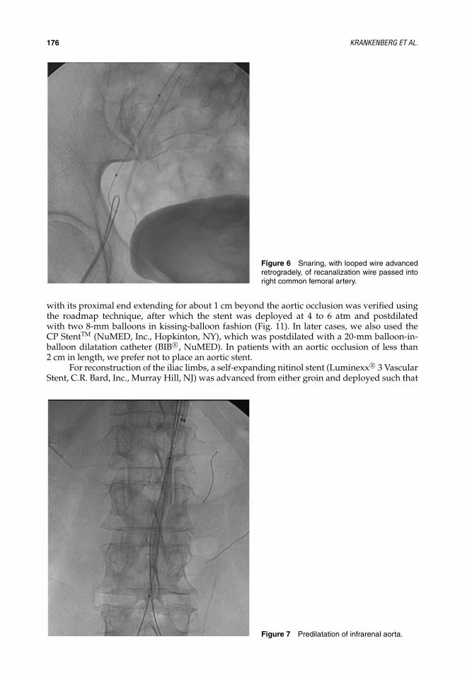
IHBK065-13 IHBK065-Minar April 26, 2010 7:48 Specs: 7x10 tight 7in×10in Char Count=
176 KRANKENBERG ET AL.
Figure 6 Snaring, with looped wire advancedretrogradely, of recanalization wire passed intoright common femoral artery.
with its proximal end extending for about 1 cm beyond the aortic occlusion was verified usingthe roadmap technique, after which the stent was deployed at 4 to 6 atm and postdilatedwith two 8-mm balloons in kissing-balloon fashion (Fig. 11). In later cases, we also used theCP StentTM (NuMED, Inc., Hopkinton, NY), which was postdilated with a 20-mm balloon-in-balloon dilatation catheter (BIB R©, NuMED). In patients with an aortic occlusion of less than2 cm in length, we prefer not to place an aortic stent.
For reconstruction of the iliac limbs, a self-expanding nitinol stent (Luminexx R© 3 VascularStent, C.R. Bard, Inc., Murray Hill, NJ) was advanced from either groin and deployed such that
Figure 7 Predilatation of infrarenal aorta.

IHBK065-13 IHBK065-Minar April 26, 2010 7:48 Specs: 7x10 tight 7in×10in Char Count=
LERICHE’S DISEASE 177
Figure 8 Predilatation of right iliac artery.
it invaded the distal end of the aortic stent by about 3 mm (Fig. 12). The nominal diameterof either iliac stent was chosen to exceed the respective iliac artery reference diameter by 1mm. Both self-expandable stents were postdilated in kissing-balloon fashion (Fig. 13). Becausethe iliac occlusions could not be completely covered with 12-cm stents in this patient, oneadditional self-expandable nitinol stent was implanted into each iliac artery. Unimpeded bloodflow through the entire reconstructed aortoiliac tract was documented by angiography (Fig. 14).In patients in whom no aortic stent is placed, the iliac artery stents are deployed such that theirproximal ends extend for about 1 cm beyond the occlusion into the aorta.
Figure 9 Predilatation of left iliac artery.

IHBK065-13 IHBK065-Minar April 26, 2010 7:48 Specs: 7x10 tight 7in×10in Char Count=
178 KRANKENBERG ET AL.
Figure 10 Placement of aortic stent.
Both groins were closed using a percutaneous suture device (Prostar XL, Abbott Vascular)to avoid vessel compression and ensure unimpeded distal runoff. Postprocedural computedtomography of the abdominal and pelvic region was performed to exclude retroperitonealbleeding.
The patient was discharged on an oral regimen of aspirin (100 mg/day indefinitely) andclopidogrel (75 mg/day for three months). Computed tomography angiography at 12 monthsshowed persistence of acute procedural success (Fig. 15).
Figure 11 Kissing-balloon postdilatation of aorticstent.

IHBK065-13 IHBK065-Minar April 26, 2010 7:48 Specs: 7x10 tight 7in×10in Char Count=
LERICHE’S DISEASE 179
Figure 12 Placement of nitinol stents in com-mon iliac arteries. Note proximal ends of stentsextending into aortic stent (arrows).
Figure 13 Simultaneous (kissing-balloon)postdilatation of iliac artery stents.

IHBK065-13 IHBK065-Minar April 26, 2010 7:48 Specs: 7x10 tight 7in×10in Char Count=
180 KRANKENBERG ET AL.
Figure 14 Composite figure of final result after place-ment of two stents in each iliac artery. There is unimpededblood flow through aortic bifurcation into iliac arteries.
Figure 15 Computed tomography angiogra-phy at 12 months.

IHBK065-13 IHBK065-Minar April 26, 2010 7:48 Specs: 7x10 tight 7in×10in Char Count=
LERICHE’S DISEASE 181
MATERIALS
n
Sheaths6-F/90 cm 17-F/90 cm 18-F/25 cm 212-F/25 cm 1
WiresIron Man
TM/0.014 in./300 cm 3
Terumo hydrophilic stiff angled/260 cm 1Terumo hydrophilic stiff straight/260 cm 1Supra Core R© 35/0.035 in./260 cm 2
CathetersMPA2 4-F/125 cm 1MPA 5-F/125 cm 1IMA 5-F/100 cm 1JR4 5-F/100 cm 1Quick-Cross R© 130 cm 1
Balloons4/80 mm OTW 26–8/80–120 mm OTW 28/40 mm OTW 220 mm BIB R© OTW 1
StentsPalmaz-SchatzTM 40/50 mm 1CP StentTM 1Luminexx R© 3 Vascular Stent 7–9/60–120 mm 6
Vascular Closure SystemsProstar XL 10-F 1
RESULTS IN 11 PATIENTSWe defined “procedural success” as restored vessel patency with a residual diameter stenosis<30% and a translesion pressure gradient <20 mm Hg. In case of a residual stenosis ≥30%,postdilatation was performed using an appropriately sized balloon. Another postdilatation ofthe stented segments was performed if the pressure gradient exceeded 20 mm Hg.
Between 2003 and 2008, we treated 11 consecutive patients [8 men (73%), 64 ± 12 years]according to the strategy presented above. Leriche’s disease had been present for a median of3 years in these patients, ranging up to 13 years. Six patients had been denied surgery and twopatients had previously undergone extra-anatomic bypass surgery with subsequent reocclusionof the grafts. The latter two patients as well as the remaining three patients all refused a surgicalintervention. Bilateral endovascular success was achieved in eight patients (73%), unilateral suc-cess in the other three patients. Seven patients received aortic stents; the total stented segmentlength in 19 iliac arteries successfully recanalized was a median of 18 cm (range, 12–26 cm).Attesting to the safety of our treatment strategy, we encountered only one periprocedural com-plication, an acute thrombotic aortoiliac occlusion managed by thrombolysis. One patient withunilateral endovascular success had to undergo subsequent femorofemoral crossover bypassgrafting. At a median of 14 months, significant hemodynamic improvement was observed insuccessfully revascularized legs (ABI, 0.79 ± 0.20 vs. 0.48 ± 0.08 at baseline; P = 0.0004). Walkingcapacity and Rutherford category of PAD had improved in all patients.
SUMMARYThe endovascular strategy for the treatment of the extended aortoiliac occlusions present inLeriche’s disease appears to be safe and efficacious. It is essentially a two-step procedure.
→ In the first step, antegrade, transbrachial, wire recanalization of all occluded vascular seg-ments usually encompassing the infrarenal aorta and both common/external iliac arteriesis attempted.

IHBK065-13 IHBK065-Minar April 26, 2010 7:48 Specs: 7x10 tight 7in×10in Char Count=
182 KRANKENBERG ET AL.
� As a protective measure, placement of coronary wires in the superior mesenteric arteryand both renal arteries must precede recanalization in patients in whom inflow intothese arteries may be compromised as a consequence of plaque shift or dissection.
→ Following successful antegrade recanalization, the second procedural step consists of ret-rograde, transfemoral, angioplasty of the previously occluded segments.� Predilatation of the aorta must be performed at a low pressure (≤6 atm) to avoid vessel
rupture. An aortic stent is placed electively in patients with an aortic occlusion exceeding3 cm in length and no major thrombus burden.
� The iliac arteries are also predilated at low pressure. Nitinol stents are implanted suchthat their proximal ends extend into the distal aorta and that they cover the entireoccluded iliac artery segments.
→ A percutaneous closure device should be used in both groins to avoid vessel compres-sion and ensure unimpeded distal runoff. Postprocedural computed tomography of theabdominal and pelvic region should be used to exclude retroperitoneal bleeding.
REFERENCES1. Graham R. Case of Obstructed Aorta. Communicated by Sir G. Blane. [Sir Gilbert Blane, 1747–1834].
London: Medico-Chirurgical Transactions, 1814.2. Leriche R. Des obliterations arterielles hautes (obliteration de la terminaison de l’aorte) comme causes
des insuffisances circulatoires des membres inferieurs. Bull Mem Soc Chir Paris 1923; 49:1404–1406.3. Erskine JM, Gerbode FL, French SW III, et al. Surgical treatment of thrombotic occlusion of aorta and
iliac arteries; the Leriche syndrome. AMA Arch Surg 1959; 79:85–93.4. Leriche R. De la resection du carrefour aortico-iliaque avec double sympathectomie lombaire pour
thrombose arteritique la l’aorte: le syndrome de l’obliteration termino-aortique par arterite. PresseMed 1940; 48:601–607.
5. Cooley DA, Creech O Jr, De Bakey ME. The Leriche syndrome and its surgical treatment by resectionand homograft replacement. Lyon Chir 1956; 52:402–411.
6. Burgess CM. Treatment of the Leriche syndrome: a technique of endarterectomy. Arch Surg 1959;79:487–492.
7. Sugimoto T, Ogawa K, Asada T, et al. Leriche syndrome. Surgical procedures and early and late results.Angiology 1997; 48:637–642.
8. Garcia FC, Gard PD Jr. Leriche syndrome: review of the literature and a case report. J La State MedSoc 1979; 131:263–265.
9. Stubbs DH, Kasulke RJ, Kapsch DN, et al. Populations with the Leriche syndrome. Surgery 1981;89:612–616.
10. Genoni M, von Segesser LK, Laske A, et al. Occlusion of the distal aorta. Helv Chir Acta 1994; 60:723–728.
11. Mavioglu I, Veli Dogan O, Ozeren M, et al. Surgical management of chronic total occlusion of abdom-inal aorta. J Cardiovasc Surg (Torino) 2003; 44:87–93.
12. Scheinert D, Schroder M, Balzer JO, et al. Stent-supported reconstruction of the aortoiliac bifurcationwith the kissing balloon technique. Circulation 1999; 100(suppl 19):II295–II300.
13. Schillinger M, Sabeti S, Loewe C, et al. Balloon angioplasty versus implantation of nitinol stents in thesuperficial femoral artery. N Engl J Med 2006; 354:1879–1888.
14. Krankenberg H, Schluter M, Steinkamp HJ, et al. Nitinol stent implantation versus percutaneoustransluminal angioplasty in superficial femoral artery lesions up to 10 cm in length: the femoral arterystenting trial (FAST). Circulation 2007; 116:285–292.
15. EVAR Trial Participants. Endovascular aneurysm repair versus open repair in patients with abdominalaortic aneurysm (EVAR trial 1): randomised controlled trial. Lancet 2005; 365:2179–2186.
16. Marty AT, Penkava R, Whitehead D, et al. Kissing balloon therapy for Leriche syndrome. Indiana Med1985; 78:288–289.
17. Bean WJ, Rodan BA, Thebaut AL. Leriche syndrome: treatment with streptokinase and angioplasty.AJR Am J Roentgenol 1985; 144:1285–1286.
18. Katzenschlager R, Ahmadi A, Koppensteiner R, et al. Leriche syndrome: treatment with local lysisand subsequent percutaneous transluminal angioplasty. Vasa 1996; 25:180–183.
19. Schroder M, Friedrich K, Zipfel B, et al. Acute painless paraplegia of the legs as a manifestation ofextensive acute Leriche syndrome. Clin Res Cardiol 2007; 96:240–242.

IHBK065-14 IHBK065-Minar May 1, 2010 8:11 Specs: 7x10 tight 7in×10in Char Count=
14 Iliac ArteriesPatrick Peeters and Jurgen VerbistDepartment of Cardiovascular and Thoracic Surgery, Imelda Hospital, Bonheiden, Belgium
Koen Deloose and Marc BosiersDepartment of Vascular Surgery, A.Z. Sint-Blasius, Dendermonde, Belgium
INTRODUCTION
What Makes a Lesion “Complex” in Iliac Interventions?Although over 90% of iliac lesions can be passed with simple guidewire techniques, eachendovascular attempt to treat an iliac occlusion might be compromised by failure of guidewirepassage. In order to achieve optimal success when treating complex iliac lesions, the operatorshould know when and how ipsilateral, contralateral, and brachial access techniques should beapplied. Later in section “Interventional Strategies,” we will discuss the different interventionalstrategies that can help to treat a complex iliac case successfully.
Clinical Indications for Treatment of Complex Iliac Lesions in Iliac InterventionsThe application of endovascular techniques for the treatment of short iliac stenoses and occlu-sion has been well accepted for several years now. A meta-analysis from 1997 by Bosch et al.(1) compared the results of six PTA studies (1300 patients) and eight stent studies (816) oniliac lesions. It revealed four-year primary patency rates of 67% after PTA and 81% after stent-ing. This means there is a 43% lower risk for long-term failure for stent placement over bal-loon angioplasty after four years. The randomized controlled Dutch Iliac Stenting trial failedin demonstrating superiority of primary stenting over PTA with additional stenting in shortlesions (2–4).
A similar spirit was reflected when in 2000 the TransAtlantic Inter-Society Consensus(TASC) working group published their recommendations for the management of peripheralarterial disease. Based on the literature available and drawn up by field specialists, they arewidely considered as the authoritative guidelines for use during everyday practice. One impor-tant aspect included in the TASC document is its lesion classification. It is based on response tointervention, independent of technology and techniques. The goal of this system is to indicatethe best form of treatment, endovascular (TASC A) or surgical (TASC D). The lesions withoutstrongly supportive evidence, but that are more likely to respond better to endovascular therapy(TASC B) or surgery (TASC C), need more evidence (Table 1) (5).
Owing to advancements in endovascular techniques and technology, there has been anevolution to tackle more and more complex iliac lesions and chronic iliac occlusions with mini-mally invasive procedures. Leville et al. (6) reported primary and secondary patency rates of 76%and 90% after three years after stenting of iliac occlusions. They concluded that endovasculartreatment for iliac occlusive disease should be extended to Type C and D lesions, as stratificationfor the different TASC lesion classifications did not show any significant differences in outcomefor primary and secondary patency rates (Fig. 1).
The TASC working group has revised and updated their recommendations, which resultedin their publication in 2007 of the Inter-Society Consensus for the Management of PeripheralArterial Disease TASC II document (7). A concise overview of the TASC aortoiliac lesion classi-fication and its evolution over the past few years is shown in Table 2. Lesions that are easier tobe tackled by endovascular means belong to TASC A and B categories. The more complex iliaclesions are listed under TASC C and D categories.

IHBK065-14 IHBK065-Minar May 1, 2010 8:11 Specs: 7x10 tight 7in×10in Char Count=
184 PEETERS ET AL.
Table 1 TASC Lesion Classification Categories
Treatment choice TASC category
• Endovascular therapy is the treatment of choice Type A• Endovascular therapy is the preferred treatment after consideration of the
patient’s comorbidities, fully informed patient preference, and the localoperator’s long-term success rates
Type B
• Surgery is the preferred treatment for good-risk patients after consideration ofthe patient’s comorbidities, fully informed patient preference, and the localoperator’s long-term success rates
Type C
• Surgery is the treatment of choice Type D
Hazards and Complications
Access site complications
– Groin hematoma– Retroperitoneal bleeding– Pseudoaneurysm– Arteriovenous fistula
These complications used to be more frequent in the early yearsdue to the need for 9-F or 10-F systems. With the current lowerFrench sizes, groin complications are now much less common
Dissection Hemodynamically significant dissection has been reported in7.1% after iliac PTA. The majority of these dissections can betreated by stent implantation If a retrograde dissection into theaorta occurs, more complex endovascular repair is necessary
Perforation/rupture This rare complication has been reported in 0.8% of the cases,especially in calcified lesion configurations. When it occurs,aggressive reversal of anticoagulants, balloon tamponade ofthe vessel, placement of a covered stent or surgery will haveto be performed to solve the problem
Late aneurysm formation pseudoaneurysm This complication can usually be treated with placement of acovered stent
Distal embolization Distal embolization has been reported in 8.8–24% of the casesCompromise/closure of internal iliac arteryDevice-related events
– Stent thrombosis– Stent embolization/migration– Stent crush– Septic infection– Septic endarteritis
Although these events are rare, operators should be familiar withtechniques associated with snaring and retrieval of embolizedstents
Primary patency
100
80
60
223037
71412
597
TASC-BTASC-CTASC-D
Per
cent
age
40
20
00 6 12 18 24 30
Months
(A) (B)
36
Secondary patency
100
80
60
223037
91513
510
8
TASC-BTASC-CTASC-D
Per
cent
age
40
20
00 6 12 18 24 30
Months
36
Figure 1 Kaplan–Meier curve estimates for endovascular iliac treatment stratified by TASC classification.Kaplan–Meier curve estimates for (A) primary and (B) secondary patencies in patients treated for iliac occlu-sion stratified by TASC. No significant difference was found between groups (6).

IHBK065-14 IHBK065-Minar May 1, 2010 8:11 Specs: 7x10 tight 7in×10in Char Count=
ILIAC ARTERIES 185
Table 2 TASC Classifications for Aortoiliac Lesions Evolution from 2000 to 2007
Interventional StrategiesAlthough the TASC stratification is an important tool to assess the extent of lesion morphology,successful endovascular treatment is not precluded for TASC C and D aortoiliac lesions. Inexperienced hands, endovascular treatment of complex iliac lesions is always possible, exceptwhen the common femoral artery (CFA) is occluded (for which a surgical aortobifemoral graftis indicated, preferably with profundaplasty); or when there is presence of an aortic occlusionup to the renal arteries of an aortic thrombosis. This chapter describes the different endovas-cular techniques and materials that can be used to successfully complete a minimally invasiveintervention.
Access TechniquesAlthough over 90% of iliac lesions can be passed with simple guidewire techniques, eachendovascular attempt to treat an iliac occlusion might be compromised by failure of guidewirepassage. In order to achieve optimal success when treating complex iliac lesions, the operatorshould know when and how ipsilateral, contralateral, and brachial access techniques should beapplied. Tables 3 and 4 give a schematic overview regarding which access technique should befollowed for each type of complex iliac lesion configuration.
Since the introduction of subintimal angioplasty by Reekers and Bolia (8), it has becomea common practice in many centers. However, this technique may compromise the result ofthe procedure, especially when an ipsilateral approach is performed, when re-entry to the truelumen is impossible or obtained at another site than the initially intended re-entry point (9).The use of re-entry catheters has recently been described by Ramjas et al. (10) as a means tosuccessfully achieve continuity of the dissected portion with the true lumen, and to succeed ina primary stenting of the common iliac arteries. In a case report by Cho et al. (11), subintimalguidewire lesion passage in a patient who previously had aortic bypass surgery resulted in asmall false lumen attached to the true aortic lumen with an intimal flap in between. A re-entry

IHBK065-14 IHBK065-Minar May 1, 2010 8:11 Specs: 7x10 tight 7in×10in Char Count=
186 PEETERS ET AL.
Table 3 Total Occlusion of Common and/or External Iliac Artery
Unilateral occlusion Bilateral occlusionContralateral approach Brachial access
Step 1: catheterization and crossoverAccess is made by puncturing the common
femoral artery at the contralateral side using theSeldinger technique. A hydrophilic guidewire anda crossover catheter are advanced into theaorta. Correct positioning is confirmed byangiographic control at the iliac bifurcation
Step 1: catheterization into common iliac arteryAccess is made by puncturing the brachial artery.Intraluminal lesion passage is done with a
hydrophilic guidewire. The wire is installed intoone of the CFA to obtain support for the catheter
MaterialsStraight sheath, 6-F, short
− Interventional Sheath (Arrow)− Brite-Tip (Cordis)− Performer (Cook)
Crossover catheter, 6-F, 65/100 cm− RIM (Cook)− Universal Flush (Cordis)− Simmons 1 (Terumo)− Simmons 2 (Terumo)− Berenstein (Cordis)
Hydrophilic guidewire, 0.035′′, 260 cm− GlideWire Flex (Terumo) (soft lesions)− GlideWire Stiff (Terumo) (calcified lesions)
MaterialsStraight sheath, 6-F, long (90/100 cm) (brachial)
− Super-Arrow Flex (Arrow)− Brite-Tip (Cordis)− Flexor (Cook)
Interventional catheter, 6-F, 150 cm− Quick-Cross (Spectranetics)
Hydrophilic guidewire, 0.035′′, 260 cm− GlideWire Flex (Terumo) (soft lesions)− GlideWire Stiff (Terumo) (calcified lesions)
Step 2: lesion passageThe hydrophilic guidewire and crossover catheter
are advanced into the superficial femoral artery(SFA) of the contralateral limb to gain support.Then a wire exchange is done to a stiff guidewireand careful intraluminal lesion passage isperformed
Step 2: guidewire capture and securing the accesssite
A short sheath is installed in the groin. Next, asnaring catheter is advanced to capture theguidewire, which is then partially pulled out ofthe contralateral (CFA). Next, the access site issecured by installing a 0.018′′ guidewire
MaterialsStiff guidewire, 0.035′′, 260 cm
− GlideWire Stiff (Terumo)− Amplatz (Boston Scientific)
MaterialsStraight sheath, 6-F, short (groin)
− Interventional Sheath (Arrow)− Brite-Tip (Cordis)− Performer (Cook)
Guidewire, 0.018′′, 260 cm− GlideWire Flex (Terumo)− GlideWire Stiff (Terumo)
Step 3: guidewire capture and exchange Step 3: guidewire installment in other groinAccess is made by puncturing the common
femoral artery at the ipsilateral side using theSeldinger technique. Next, a snaring catheter isadvanced to capture the guidewire, which is thenpartially pulled out of the contralateral commonfemoral artery (CFA). Next, the cross-overcatheter can be removed
Repeat the above steps 1 and 2 for the other groin;or use the technique described under “Unilateralocclusion—Contralateral approach”
MaterialsStraight sheath, 5-F, shortSnare kit
− Goose Neck (ev3)
site was created by puncturing the intimal flap under intravascular ultrasound (IVUS) with atransseptal needle and the patient was successfully stented afterwards.
Interventional ToolsIn order to obtain maximum and durable success, it is important to stent the entirety of the iliacsegment affected by stenotic or occlusive disease. Depending on the type and location of thelesion, different devices and techniques can be applied.

IHBK065-14 IHBK065-Minar May 1, 2010 8:11 Specs: 7x10 tight 7in×10in Char Count=
ILIAC ARTERIES 187
Table 4 Multisegmental Stenoses of Common and/or External Iliac Arteries
Unilateral stenoses Bilateral stenosesIpsilateral approach Ipsilateral approach
Step 1: sheath placement and lesion crossingAccess is made by puncturing the ipsilateral CFA,
using the Seldinger technique and under duplexguidance. Next, the lesion is crossed
intraluminallywith a hydrophilic guidewire
Step 1: sheath placement and lesion crossingAccess is made by puncturing the ipsilateral CFA,
using the Seldinger technique and under duplexguidance. Next, the lesion is crossed
intraluminallywith a hydrophilic guidewire
Materials− Straight sheath, 6-F, short− Interventional Sheath (Arrow)− Brite-Tip (Cordis)− Performer (Cook)
Hydrophilic guidewire, 0.035′′, 260 cm− GlideWire Flex (Terumo) (soft lesions)− GlideWire Stiff (Terumo) (calcified lesions)
MaterialsStraight sheath, 6-F, short
− Interventional Sheath (Arrow)− Brite-Tip (Cordis)− Performer (Cook)
Hydrophilic guidewire, 0.035′′, 260 cm− GlideWire Flex (Terumo) (soft lesions)− GlideWire Stiff (Terumo) (calcified lesions)
REMARK: If a dissection is created duringipsilateral approach, contralateral approach(as described in Table 3) is indicated
REMARK: If a dissection is created duringipsilateral approach, contralateral approach orbrachial access (as described in Table 3)is indicated.
Preferably self-expanding stents are used to treat iliac arteries, they offer good trackabilityneeded in tortuous anatomy and have an excellent radial force. It is advised to oversize anyself-expanding stent with 10% to 15% in order to obtain the best result. An overview of devicesfor iliac lesion treatment, with their indication for use, can be found in Table 5.
Primary Stenting Versus Elective StentingIn short iliac lesions, there is no difference in results between primary stenting and electivestenting. The randomized controlled Dutch Iliac Stenting trial failed in demonstrating superi-ority of primary stenting over PTA with additional stenting in short lesions (2–4). Yet, complexiliac lesions are best managed by primary stenting (Fig. 2). AbuRahma et al. (12) compared theirresults for primary stenting and elective stenting in short stenoses (TASC A and B) and in longstenoses (TASC C and D). In their patient cohort, there was no statistical difference in patencyrates for short lesions. In long lesions, however, results for primary stenting were significantlybetter.
As the stent is placed without predilation, potential embolic material is trapped betweenthe arterial wall and the stent mesh, which significantly reduces the risk of distal embolizations.Additional potential advantages of direct stenting include shorter procedural time and lessradiation exposure (13).
Stent GraftingEndovascular treatment of any lesion inherits a certain risk of vessel rupture and distal emboliza-tion. With the increasing complexity of the lesion to be treated, the risk of complications tendsto follow. When arterial rupture following iliac artery PTA occurs, stenting of the artery aloneresults in maintenance of flow through the ruptured segment and exsanguination. Treatmentwith balloon occlusion alone has been reported (14), however this complication may require fur-ther revascularization (15). More recently, treatment of these complications has been performed
Table 5 Devices for Complex Iliac Lesions
Special situation Device/example(s)
Tortuous lesion site Self-expanding stentCalcified lesion Balloon expandable stent Cutting balloonIn-stent restenosis Covered stent Cutting balloon

IHBK065-14 IHBK065-Minar May 1, 2010 8:11 Specs: 7x10 tight 7in×10in Char Count=
188 PEETERS ET AL.
(A) (B) (C)
Figure 2 Example of a primary iliac stenting case (13). (A) Total CIA occlusion with reinjection at the level ofexternal iliac artery (EIA) (arrow). (B) Direct stenting after retrograde recanalization by means of two self-expandingstents. (C) Good antegrade flow in the left iliac axis with preservation of the flow in the left IIA.
with covered stents (Fig. 3) with immediate exclusion obtained in 100% of patients and withprimary and secondary patency rates of 87% and 100%, respectively at just under two years (16).
Distal embolization with PTA alone is not common for uncomplicated lesions, but doesoccur with greater frequency, as high as 24%, in the treatment of ulcerated plaques (17), withthe recanalization of aortoiliac bifurcation lesions (18) or iliac occlusions (19). Stent grafts areused in our center for complex iliac lesions and have proven to effectively exclude the source ofembolization (Fig. 4). In our recently published series of 91 limbs with stenotic diseased iliacstreated with the balloon expandable Atrium Advanta V12 PTFE covered stent (107 stent graftsused), we reported a successful deployment in all patients without any procedural complicationsincluding distal embolization and vessel rupture and with a primary limb patency at one yearwas 91.1% (20). However, even with the placement of stent grafts, embolization may remain aconcern.
(A) (B)
Figure 3 Example of an endograft placement (16). (A) Right common iliac artery (CIA) dissection (horizontalarrows) and high-grade EIA stenosis (vertical arrow). (B) Result after endovascular repair of the dissection with astentgraft (8 × 60 mm) and of the stenosis with an additional Palmaz stent (7 × 30 mm).

IHBK065-14 IHBK065-Minar May 1, 2010 8:11 Specs: 7x10 tight 7in×10in Char Count=
ILIAC ARTERIES 189
(A) (B)
Figure 4 Example of a stentgraft placement. (A) Right CIA stenosis before treatment. (B) Result after stentgraft(8 × 38 mm) deployment.
Bifurcation StentingLesions at the aortic bifurcation are traditionally treated using the “kissing balloons” technique.Simultaneous balloon dilatation at the origins of both common iliac arteries (CIAs) is advocated,even in the presence of unilateral lesion, to protect the contralateral CIA from dissection orplaque embolization. Because calcified lesions typically occurring at the aortic bifurcation arenot amenable to balloon dilatation alone, “kissing stents” or “aortic reconstruction” techniqueis applied (Fig. 5). The aortic bifurcation reconstruction technique is technically very successful,although some have expressed fears that the proximal ends of the stents that extend into thedistal aorta may serve as a nidus for thrombus formation, or cause hemolysis. Nevertheless, thisfear has not been realized as has been shown by the low complication rates of this procedure.In the available study publications, different types of stent have been described, without anyindication of a significant outcome. It is, however, important that both the used stents are of thesame type and dimensions and they are deployed and dilated simultaneously.
Summary of OutcomesTable 6 gives a literature overview of study results for endovascular treatment of complex iliaclesions. It shows that endovascular treatment of iliac artery occlusions can be accomplished
(A) (B) (C)
Figure 5 Example of the kissing-stent technique (16). (A) Preoperative occlusion of the left CIA and significantstenosis at the origin of the right CIA. (B) Poststenting image showing two kissing balloon expandable stents inboth CIAs (arrow shows zone where kissing stents touch each other). (C) Postoperative good bilateral CIA flow.

IHBK065-14 IHBK065-Minar May 1, 2010 8:11 Specs: 7x10 tight 7in×10in Char Count=
190 PEETERS ET AL.
Tab
le6
Lite
ratu
reO
verv
iew
for
Com
plex
Iliac
Lesi
ons
Tech
nic
alsu
cces
s30
-mo
rtal
ity
PP
@1Y
PP
@3Y
PP
@5Y
PP
@10
YR
efer
ence
sL
esio
nty
pe/
dev
ice
typ
e(%
)(%
)(%
)(%
)(%
)(%
)
1A
llili
acle
sion
s—P
TA91
174
68–
–A
llili
acle
sion
s—st
ent
960.
886
80–
–23
Mul
tiseg
men
tilia
coc
clus
ive
dise
ase
972
6143
––
24K
issi
ngst
ents
100
0–
86–
–18
Kis
sing
sten
ts10
00
–79
.4–
–25
Kis
sing
sten
ts–
–94
––
–26
Poo
rdi
stal
runo
ff—ili
acst
entin
gal
one
97–
7666
55–
Poo
rdi
stal
runo
ff—ili
acst
entin
g+
bypa
ss97
–87
5442
–27
TAS
CC
and
D—
surg
ery
96–
8572
64–
TAS
CC
and
D—
endo
vasc
ular
96–
8986
86–
21TA
SC
A–D
––
–87
8349
22TA
SC
B–D
97.4
2.7
9489
77–
6TA
SC
Can
dD
with
conc
omita
ntin
frai
ngui
nald
isea
se91
––
76–
–28
Kis
sing
sten
ts10
0–
7663
63–
12A
llili
acle
sion
s;pr
imar
yst
entin
g10
0–
9887
77–
All
iliac
lesi
ons;
sele
ctiv
est
entin
g10
0–
8369
69–
TAS
CA
and
Bpr
imar
yst
entin
g10
0–
100
9887
–TA
SC
Aan
dB
sele
ctiv
est
entin
g10
0–
100
8585
–TA
SC
Can
dD
—pr
imar
yst
entin
g10
0–
9672
–-
TAS
CC
and
D—
sele
ctiv
est
entin
g10
0–
4628
––
20B
allo
onex
pand
able
cove
red
sten
ts10
00
91.1
––
–29
Kis
sing
sten
ts–
––
–82
6830
TAS
CC
and
D—
kiss
ing
sten
ts95
.4–
92–
80.7
–31
Cut
ting
ballo
onfo
rin
-ste
ntre
sten
osis
92.8
–10
0–
––

IHBK065-14 IHBK065-Minar May 1, 2010 8:11 Specs: 7x10 tight 7in×10in Char Count=
ILIAC ARTERIES 191
via endovascular means with little morbidity and acceptable patency rates. Although evidence-based proof from controlled randomized trials is lacking, several recent publications have shownthe excellent durability of iliac stenting in daily practice. Park et al. (21) described their long-term(up to 10 years) experience in their total cohort of iliac patients (TASC Type A–D lesions) andpresented impressive primary patency rates of 87%, 83%, 61%, and 49% after 3, 5, 7, and 10 yearsof follow-up. De Roeck et al. (22) published their results after stenting of different types of iliacocclusions (TASC Type B–D) and showed primary patency rates of 94%, 89%, and 77% after one,three, and five years. They also stated that in their cohort of patients with complex aortoiliaclesions, stent failures can always be rescued endovascularly. They reported secondary patencyrates of 100% after one year and 94% after three and five years. The success rate on midtermand long term is also heavily dependent on the presence of concomitant infrainguinal disease.Leville et al. (6) pointed out that surgical repair of iliac occlusions should only be referred towhen all endovascular attempts are exhausted, bearing in mind the decreased perioperativemorbidity and good mid-term durability.
Equipment Checklist
Contralateral approachStraight sheath, 6-F, short Interventional Sheath (Arrow)Crossover catheter, 6-F, 65/100 cm RIM (Cook)Snare kit Goose Neck (ev3)Hydrophilic guidewire, 0.035′′, 260 cm GlideWire (Terumo)Ipsilateral approachStraight sheath, 6-F, short Interventional Sheath (Arrow)Hydrophilic guidewire, 0.035′′, 260 cm GlideWire (Terumo)Brachial approachBrachial: Straight sheath, 6-F, long (90/100 cm) Super-Arrow Flex (Arrow)Groin: Straight sheath, 6-F, short Interventional Sheath Flex (Arrow)Hydrophilic guidewire, 0.035′′, 260 cm GlideWire (Terumo)Lesion passageStiff guidewire, 0.035′′, 260 cm GlideWire Stiff (Terumo) Amplatz (Boston Scientific)Lesion treatmentTortuous lesion site Self-expanding stentCalcified lesion Balloon expandable stent Cutting balloonIn-stent restenosis Covered stent Cutting balloon
TEACHING CASES
Case 1: Management of an Intraprocedural Complication
What Made the Case “Complex”?Although there was the presence of a bilateral occlusion at the level of the iliac arteries, thereal difficulty with this case was the rupture after stent placement and postdilation in the rightexternal iliac artery, which needed immediate action.
What Was the Clinical Indication for Treatment of This Patient?A 70-year-old male patient presented in our hospital service with symptomatic PAD. Hesuffered from the Leriche syndrome, with intermittent claudication in the buttocks and erectiledysfunction. As clinical vascular risk factors the patient had medically controlled hypertension,hypercholesterolemia, and previous nicotine abuse (he quit smoking more than five yearsbefore). A preoperative duplex investigation showed complete absence of femoral pulses. Thiswas confirmed by the preoperative MR Angiography, which showed a complete occlusion ofthe bilateral iliac arteries.
The lesion was a complete occlusion of the bilateral iliac arteries, as was confirmed by thepreoperative MR Angiography (Fig. 6).

IHBK065-14 IHBK065-Minar May 1, 2010 8:11 Specs: 7x10 tight 7in×10in Char Count=
192 PEETERS ET AL.
Figure 6 Complete occlusion of the bilateraliliac arteries.
What Were the Potential Hazards of Treating This Patient and Anatomy?Ruptures need to be addressed as soon as possible as the tear in the vessel wall can lengthen.During this procedure, this was actually the case when a dilation of a covered stent, which wasintended to seal the rupture, was performed.
Which Interventional Strategies Were Considered?For the rupture, urgent complication management was necessary and the option consideredwas placement of a covered stent to seal the rupture.
Which Interventional Tools Were Used to Manage This Case?– Regular PTA balloons– Stents
� Balloon expandable stents� Covered stents
– Endografts
Case SummaryAccess was done via a femoral puncture on the left side with a 6-F Glidesheath (Terumo MedicalCorporation, Somerset, NJ), after which intraluminal catheterization was performed into theaorta with a 0.035′′ GlideWire (Terumo) guidewire. Next, an angiogram of the iliac bifurcationwas done through a 6-F RIM catheter (Cook Incorporated, Bloomington, IN). Then, access wasperformed via a puncture in the right CFA with a short 6-F Arrow-Flex PSI sheath (ArrowInternational Inc.) and an X-ray-guided retrograde angiogram was taken. Subintimal antegradecatheterization and guide retrieval with an Amplatz Goose Neck R© Snare catheter (ev3 Inc.,Plymouth, MN) were then performed, followed by a bilateral guidewire exchange with two0.035′′ Amplatz (Boston Scientific Corporation, Natick, MA) wires.
Due to calcifications at the bifurcation, balloon expandable stents were selected to dilatethis region using the kissing-stent technique. Kissing predilation was performed with twoPheron balloons (Biotronik GmbH, Bulach, Switzerland) of 6 mm in diameter and 80 mm inlength. Following was kissing stenting procedure with two balloon expandable Primus GPS

IHBK065-14 IHBK065-Minar May 1, 2010 8:11 Specs: 7x10 tight 7in×10in Char Count=
ILIAC ARTERIES 193
stents (ev3 Inc.) of 8 mm in diameter and 57 mm long. Control angiography showed nice resultwith good flow. Next, the right external iliac artery was stented with a long self-expandingProtege stent (ev3) of 8 mm in diameter and 100 mm long, which was postdilated with a Pheronballoon (Biotronik) of 7 mm in diameter and 40 mm in length. Control angiography showedblood loss due to a rupture at the external iliac artery.
At this moment the complication management to control the rupture was initiated. Bloodloss was reduced by means of local control with a Pheron balloon (Biotronik) of 8 mm in diameterand 40 mm in length. Both sheaths were exchanged with an 11-F Brite Tip Interventional Sheath(Cordis Endovascular, Miami Lakes, FL) at the right side and a 9-F Arrow-Flex sheath (Arrow)at the left side, and a 33-mm aortic Equalizer balloon (Boston Scientific, Ratingen, Germany)was dilated to control the bleeding. In an attempt to seal the rupture, an Advanta V12 coveredstent (Atrium Medical Corporation, Hudson, NH) of 8 mm in diameter and 38 mm in length wasimplanted, but angiographic control still showed a remaining small contrast flush afterwards.Therefore, another Advanta V12 covered stent (Atrium Medical) of 8 mm by 38 mm wasplaced immediately distal to the first, but still a residual bleeding remained. Dilation with aPheron (Biotronik) balloon of 8 mm in diameter and 60 mm long to stop the remaining smallcontrast flush resulted in a new rupture. Therefore, a Viabahn (Gore & Associates, Flagstaff,AZ) endograft of 10 mm in diameter and 100 mm in length was placed. The bleeding wassuccessfully stopped, but the graft showed to be oversized.
After this, the original procedural plan could be continued by recanalization of the leftexternal iliac artery with primary placement of a self-expanding Protege stent (ev3) of 8 mmin diameter and 60 mm long. After postdilation with a Star PTA balloon (Abbott Vascular,Redwood City, CA) of 7 mm in diameter and 40 mm long, final postoperative control showed agood result. The patient left the hospital two days later without any complications.
List of Equipment– Sheaths and catheters:
� 6-F Glidesheath (Terumo Medical Corporation, Somerset, NJ)� 6-F Arrow-Flex PSI sheath (Arrow International, Inc.)� Amplatz Goose Neck R© Snare catheter (ev3 Inc., Plymouth, MN)� 6-F RIM Catheter (Cook Incorporated, Bloomington, IN)� 11-F Brite Tip Interventional Sheath (Cordis Endovascular, Miami Lakes, FL)� 9-F Arrow-Flex sheath (Arrow International, Inc.)
– Guidewires:� 0.035′′ Glidewire (Terumo Medical Corporation, Somerset, NJ)� 0.035′′ Amplatz (Boston Scientific Corporation, Natick, MA) (2×)
– Balloons:� 6 × 80 mm Pheron balloon (Biotronik GmbH, Bulach, Switzerland) (2×)� 7 × 40 mm Pheron balloon (Biotronik GmbH, Bulach, Switzerland)� 8 × 40 mm Pheron balloon (Biotronik GmbH, Bulach, Switzerland)� 33-mm aortic Equalizer balloon (Boston Scientific, Ratingen, Germany)� 8 × 60 mm Pheron balloon (Biotronik GmbH, Bulach, Switzerland)� 7 × 40 mm Star PTA balloon (Abbott Vascular, Redwood City, CA)
– Stents and endografts:� 8 × 57 mm Primus GPS stent (ev3 Inc., Plymouth, MN) (2×)� 8 × 38 mm Advanta V12 covered stent (Atrium Medical Corporation, Hudson, NH) (2×)� 10 × 100 Viabahn endoprosthesis (Gore & Associates, Flagstaff, AZ)� 8 × 60 Protege stent (ev3 Inc., Plymouth, MN)
Key Learning IssuesIn this case, the Viabahn endoprosthesis was oversized in diameter, resulting in the plicaturesthat can be seen on the images of the final procedure result. Despite this imperfect final result,however, it is imperative in this case to know when to quit instead of insisting too much onachieving the perfect result.

IHBK065-14 IHBK065-Minar May 1, 2010 8:11 Specs: 7x10 tight 7in×10in Char Count=
194 PEETERS ET AL.
Case 2: Patient Presenting with Multisegment Lesions, Impaired Outflow, andAmputated Contralateral Leg
What Made the Case “Complex”?This patient had a history of persisting vascular insufficiency, with several prior failed attemptsof recanalization. On the left side, she had an upper leg amputation and on the right side,above-knee blood flow was provided by collaterals only. This extensive lesion configurationadded significantly to this case’s complexity.
What Was the Clinical Indication for Treatment of This Patient?An 85-year-old female patient presented in our service with symptoms of mixed arterial andvenous pathology. Oedemic rubor of the right big toe was reported in combination with sev-eral painful ulcerative wounds at the level of the fourth and fifth toe in the same leg. Thepatient had a fungal infection at the bilateral groin. Furthermore, she suffered from chronicrenal insufficiency with serum creatinine levels of 2.6 mg/dl, which was treated by dialysistherapy. As further relevant clinical background, the patient did not smoke, nor had diabetesor nightly rest pain. She had had an upper leg amputation at the left side approximately twoyears earlier due to persisting vascular insufficiency, even after several failed attempts forrecanalization.
Doppler investigation of the right leg showed a diminished flow curve at the right ankle.Due to the renal insufficiency, an nuclear magnetic resonance (NMR) angiographic investiga-tion is done to evaluate the vascular status of the right limb. Several atheromatic vessel wallirregularities were reported at the right external iliac artery with an occlusion of the mid-third.Common and internal iliac arteries were patent. There was no opacification of the common andsuperficial femoral arteries, although some lateral collaterals had formed. This compromisedarterial outflow increased the complexity of the case.
What Were the Potential Hazards of Treating This Patient and Anatomy?If the endovascular intervention should fail, this patient is at high risk of another upper legamputation.
Which Interventional Strategies Were Considered?Considering the old age of this patient, the bad vascular circulation, the contralateral ampu-tation, and the unfavorable overall condition of this patient, endovascular therapy was thefirst-line treatment considered in order to alleviate the patient from her complaints.
Which Interventional Tools Were Used to Manage This Case?– Regular PTA balloons– Stents
� Balloon expandable stents� Covered stents
Case SummaryThe patient underwent an endovascular intervention under general anesthesia. The patientwas installed on the table in dorsal decubitus with the left arm on a side table. After antegradepuncture at the left brachial artery, a 5-F Glidesheath (Terumo) was brought into the left brachialartery by using the Seldinger technique under fluoroscopy.
Through a Headhunter catheter (Terumo), a 0.035′′ GlideWire (Terumo) guidewire ismaneuvered through the abdominal aorta up to the right common iliac artery. The guidewire isexchanged for a stiffer 0.035′′ Amplatz (Boston Scientific) guidewire to gain more support, afterwhich a 6-F Brite Tip Sheath (Cordis) of 90 cm is installed in the right common iliac. Angio-graphic control shows an occlusion of the right external iliac with distal reinjection of the deepfemoral artery (DFA). The right superficial femoral artery (SFA) is also occluded from its originonwards and has a heavily calcified aspect. Reinjection at the distal third of the SFA throughcollaterals formed from the DFA could be appreciated (Figs. 7–9).

IHBK065-14 IHBK065-Minar May 1, 2010 8:11 Specs: 7x10 tight 7in×10in Char Count=
ILIAC ARTERIES 195
Figure 7
Figure 8

IHBK065-14 IHBK065-Minar May 1, 2010 8:11 Specs: 7x10 tight 7in×10in Char Count=
196 PEETERS ET AL.
Figure 9
The Amplatz (Boston Scientific) guidewire is exchanged for a 0.018′′ SV Wire (Cordis) andthe common and proximal external iliac arteries are dilated with a 4 × 80 mm o.p.e.r.a. PTAcatheter (Abbott Vascular), resulting in a small dissection (Fig. 10), which would be addressedlater.
While the 0.018′′ SV (Cordis) guidewire is left in the DFA, a 0.035′′ GlideWire (Terumo)guidewire is inserted in the SFA through a long multipurpose catheter (Cook) for a selectiverecanalization up to popliteal level. After guidewire exchange to a 0.018′′ SV Wire (Cordis),the entire femoropopliteal length is dilated with a 5 mm by 60 mm Agiltrac (Abbott Vascular)
Figure 10 Dissection after dilation of the external iliacarteries.

IHBK065-14 IHBK065-Minar May 1, 2010 8:11 Specs: 7x10 tight 7in×10in Char Count=
ILIAC ARTERIES 197
Figure 11 Dilation of the entire length of thefemoropopliteal segment with a 5/60 Agiltracballoon (Abbott Vascular).
balloon and the previously used 4 mm by 80 mm o.p.e.r.a. PTA Catheter (Abbott Vascular)balloon (Fig. 11). Control angiography showed a reasonable patency over the entire dilatedtract, with the exception of a flow-limiting dissection at the segment situated in the mid-SFAand popliteal artery (Figs. 12, 13). Next, a Protege GPS (ev3) stent of 6 mm in diameter and150 mm long was implanted from Hunter’s canal to the popliteal artery, followed by a second6 mm by 150 mm Protege GPS (ev3) stent at the proximal SFA (Figs. 14, 15). The gap in betweenthese two stents was covered by an 8 mm by 100 mm Absolute (Abbott Vascular) stent, withadequate results. Postdilation was performed over the entire stented length of the SFA with aAgiltrac (Abbott Vascular) balloon of 5 mm in diameter and 60 mm in length. After checkingthe inflow, the second guidewire is removed from the SFA.
Figure 12 Flow-limiting dissection in the SFA and thepopliteal artery after dilation.

IHBK065-14 IHBK065-Minar May 1, 2010 8:11 Specs: 7x10 tight 7in×10in Char Count=
198 PEETERS ET AL.
Figure 13 Flow-limiting dissection in the SFA and the poplitealartery after dilation.
Figure 14 Stenting of the proximal SFA with a 6/150 mm ProtgegeGPS stent (ev3).

IHBK065-14 IHBK065-Minar May 1, 2010 8:11 Specs: 7x10 tight 7in×10in Char Count=
ILIAC ARTERIES 199
Figure 15 Stenting of the proximal SFA with a 6/150 mm ProtgegeGPS stent (ev3).
Next, the Amplatz (Boston Scientific) guidewire is exchanged for a stiff 0.035′′ GlideWire(Terumo) wire and, with the use of a Quick-Cross (Spectranetics, Colorado Springs, CO) catheter,the subintimal passage of the occluded segment in the DFA is achieved (Fig. 16). Next, themidportion of the DFA is treated with a self-expanding 6 mm by 40 mm Sinus-Superflex(Optimed, Ettlingen, Germany) stent and postdilation by means of the 5 mm by 60 mm Agiltrac(Abbott Vascular) balloon. Control angiography shows a good result with good flow, andwithout the presence of a dissection of peripheral embolization (Figs. 17, 18). Finally, a self-expanding Protege GPS (ev3) of 6 mm in diameter and 100 mm long is placed along the lengthof the external iliac artery in order to improve inflow.
Final control angiography showed a good result and sheath removal was performed. Thepuncture site was closed by means of the Starclose closure system (Abbott Vascular).
List of Equipment– Sheaths and catheters:
� 5-F Glidesheath (Terumo Medical Corporation, Somerset, NJ)� Headhunter catheter (Terumo Medical Corporation, Somerset, NJ)� 6-F Brite Tip Sheath (Cordis Endovascular, Miami Lakes, FL)� Quick-Cross (Spectranetics, Colorado Springs, CO)
– Guidewires:� 0.035′′ GlideWire (Terumo Medical Corporation, Somerset, NJ) (2×)� 0.035′′ Amplatz (Boston Scientific Corporation, Natick, MA)� 0.018′′ SV Wire (Cordis Endovascular, Miami Lakes, FL)
– Balloons:� 4 × 80 mm o.p.e.r.a. PTA catheter (Abbott Vascular, Redwood City, CA)� 5 × 60 mm Agiltrac (Abbott Vascular, Redwood City, CA)
– Stents:� 6 × 150 mm Protege GPS (ev3 Inc., Plymouth, MN) (2×)� Sinus-Superflex (Optimed, Ettlingen, Germany)� 6 × 100 mm Protege GPS (ev3 Inc., Plymouth, MN)
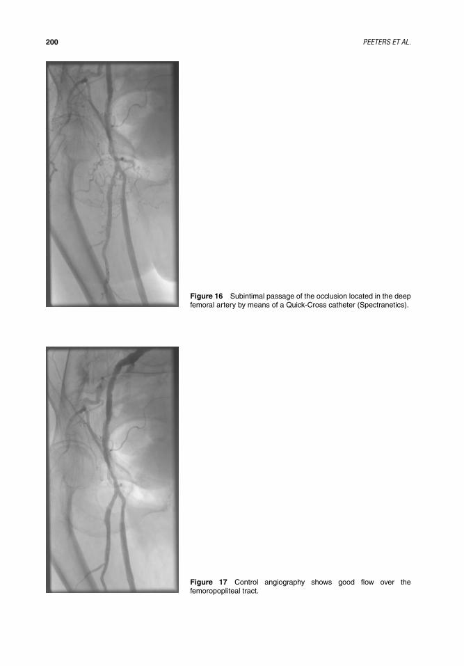
IHBK065-14 IHBK065-Minar May 1, 2010 8:11 Specs: 7x10 tight 7in×10in Char Count=
200 PEETERS ET AL.
Figure 16 Subintimal passage of the occlusion located in the deepfemoral artery by means of a Quick-Cross catheter (Spectranetics).
Figure 17 Control angiography shows good flow over thefemoropopliteal tract.
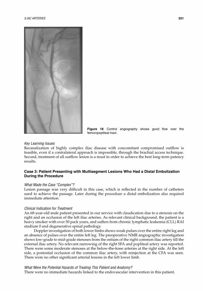
IHBK065-14 IHBK065-Minar May 1, 2010 8:11 Specs: 7x10 tight 7in×10in Char Count=
ILIAC ARTERIES 201
Figure 18 Control angiography shows good flow over thefemoropopliteal tract.
Key Learning IssuesRecanalization of highly complex iliac disease with concomitant compromised outflow isfeasible, even if a contralateral approach is impossible, through the brachial access technique.Second, treatment of all outflow lesion is a must in order to achieve the best long-term patencyresults.
Case 3: Patient Presenting with Multisegment Lesions Who Had a Distal EmbolizationDuring the Procedure
What Made the Case “Complex”?Lesion passage was very difficult in this case, which is reflected in the number of cathetersused to achieve the passage. Later during the procedure a distal embolization also requiredimmediate attention.
Clinical Indication for TreatmentAn 68-year-old male patient presented in our service with claudication due to a stenosis on theright and an occlusion of the left iliac arteries. As relevant clinical background, the patient is aheavy smoker with over 50 pack years, and suffers from chronic lymphatic leukemia (CLL) RAIstadium 0 and degenerative spinal pathology.
Doppler investigation of both lower limbs shows weak pulses over the entire right leg andan absence of pulses over the entire left leg. The preoperative NMR angiographic investigationshows low-grade to mid-grade stenoses from the ostium of the right common iliac artery till theexternal iliac artery. No relevant narrowing of the right SFA and popliteal artery was reported.There were some moderate stenoses at the below-the-knee arteries at the right side. At the leftside, a postostial occlusion of the common iliac artery, with reinjection at the CFA was seen.There were no other significant arterial lesions in the left lower limb.
What Were the Potential Hazards of Treating This Patient and Anatomy?There were no immediate hazards linked to the endovascular intervention in this patient.

IHBK065-14 IHBK065-Minar May 1, 2010 8:11 Specs: 7x10 tight 7in×10in Char Count=
202 PEETERS ET AL.
Which Interventional Strategies Were Considered?Given the bad overall health, open surgery was to be avoided. An endovascular interventionusing contralateral approach immediately seemed to be the best option.
Which Interventional Tools Were Used to Manage This Case?– Regular PTA balloons– Stents
� Self-expanding stents
Case SummaryAccess was done via a common femoral puncture on the right side with a 6-F Glidesheath(Terumo), after which intraluminal catheterization was performed into the aorta with a 0.035′′GlideWire (Terumo) guidewire. Next, an angiogram of the iliac bifurcation was done througha 6-F RIM catheter (Cook Incorporated). Although the crossover with the RIM catheter wassuccessful, it was impossible to obtain catheterization of the lesion in the left common iliac.Therefore, a SIM 1 catheter (Cook Incorporated) from the arch was placed and subintimallesion passage was commenced. In order to pass the distal portion of the lesion a catheterexchange was performed to a Quick-Cross (Spectranetics) catheter. Eventually, recanalizationis successful with the CFA as the re-entry site. Angiographic control shows the low bifurcationof the superficial and deep femoral arteries. Next, the guidewire is exchanged for a 0.035′′GlideWire stiff guidewire (Terumo) and a Balkin sheath (Cook Incorporated) is installed in theleft common iliac artery.
After predilation with a 7 mm by 80 mm Fox Plus balloon (Abbott Vascular), the con-trol angiography shows a dissection and a residual stenosis. Therefore, an 8 mm by 120 mmLuminexx stent (C.R. Bard) is placed at the level of the common and external iliac arteries up tothe CFA. Postdilation with a 7 mm by 80 mm Fox Plus balloon (Abbott Vascular) results in anadequate outcome of the treated segment, as confirmed by angiography.
Due to a remaining stenosis at the postostial common iliac artery, another Luminexx stent(C.R. Bard) of 8 mm by 50 mm is implanted. After postdilation with a 4 mm by 60 mm FoxPlus balloon (Abbott Vascular), angiographic control shows a good blood flow throughout theiliac trajectory. At the middle portion of the SFA, however, blood flow interruption can be seenby angiography, which could be related to either a stenotic lesion or a distal embolization. Anadditional dilation with a 5 mm by 40 mm Pheron balloon (Biotronik GmbH) at the middleportion of the SFA confirms the thesis of a distal embolization. At this stage, the dilated segmentin the SFA shows to be patent, but the anterior tibial artery and tibiofibular trunk are nowembolized. Several laborious attempts for thrombus aspiration fail and it is chosen to firstaddress a stenosis at the right external iliac artery. An 8 mm by 40 mm Luminexx stent (C.R.Bard) is implanted, followed by a dilation with a 7 mm by 80 mm Fox Plus balloon (AbbottVascular) at the same treatment area.
Next, distal embolization at the left lower leg was readdressed. This time, success wasfinally achieved after sheath exchange for a Destination sheath (Spectranetics) in combinationwith the Quick-Cross catheter (Spectranetics). The result shows a good outflow over all threebelow-the-knee vessels. In order to alleviate residual spasm, Cedocard is administered.
Final control angiography showed a good result, without embolization, and sheathremoval was performed. The puncture site was closed by manual compression.
List of Equipment– Sheaths and catheters:
� 6-F Glidesheath (Terumo Medical Corporation, Somerset, NJ)� 6-F RIM catheter (Cook Incorporated, Bloomington, IN)� SIM 1 catheter (Cook Incorporated, Bloomington, IN)� Quick-Cross (Spectranetics, Colorado Springs, CO)� Balkin sheath (Cook Incorporated, Bloomington, IN)� Destination sheath (Spectranetics, Colorado Springs, CO)
– Guidewires:� 0.035′′ GlideWire (Terumo Medical Corporation, Somerset, NJ)� 0.035′′ GlideWire stiff (Terumo Medical Corporation, Somerset, NJ)

IHBK065-14 IHBK065-Minar May 1, 2010 8:11 Specs: 7x10 tight 7in×10in Char Count=
ILIAC ARTERIES 203
– Balloons:� 7 × 80 mm Fox Plus balloon (Abbott Vascular, Redwood City, CA) (3×)� 4 × 60 mm Fox Plus balloon (Abbott Vascular, Redwood City, CA)� 5 × 40 mm Pheron balloon (Biotronik GmbH, Bulach, Switzerland)
– Stents:� 8 × 120 mm Luminexx stent (C.R. Bard GmbH, Karlsruhe, Germany)� 8 × 50 mm Luminexx stent (C.R. Bard GmbH, Karlsruhe, Germany)� 8 × 40 mm Luminexx stent (C.R. Bard GmbH, Karlsruhe, Germany)
Key Learning IssuesEven though endovascular iliac interventions may seem straightforward to perform, a compli-cated case always demands the presence of backup materials on the shelf. In this particular case,we needed the Destination sheath (Spectranetics) in combination with the Quick-Cross catheter(Spectranetics) in order to successfully treat the distal embolization.
REFERENCES1. Bosch JL, Hunink MG. Meta-analysis of the results of percutaneous transluminal angioplasty and
stent placement for aortoiliac occlusive disease. Radiology 1997; 204:87–96.2. Tetteroo E, van der Graaf Y, Bosch JL, et al. Randomised comparison of primary stent placement versus
primary angioplasty followed by selective stent placement in patients with iliac-artery occlusivedisease. Dutch Iliac Stent Trial Study Group. Lancet 1998; 351:1153–1159.
3. Klein WM, van der Graaf Y, Seegers J, et al. Long-term cardiovascular morbidity, mortality, andreintervention after endovascular treatment in patients with iliac artery disease: The Dutch Iliac StentTrial Study. Radiology 2004; 232:491–498.
4. Klein W, van der Graaf Y, Seegers J, et al. Dutch iliac stent trial: long-term results in patients randomizedfor primary or selective stent placement. Radiology 2006; 238:734–744.
5. Dormandy JA, Rutherford RB. Management of peripheral arterial disease (PAD). TASC WorkingGroup. TransAtlantic Inter-Society Consensus (TASC). J Vasc Surg 2000; 31:S1–S296.
6. Leville CD, Kashyap VS, Clair DG, et al. Endovascular management of iliac artery occlusions: extend-ing treatment to TransAtlantic Inter-Society Consensus class C and D patients. J Vasc Surg 2006;43:32–39.
7. Norgren L, Hiatt WR, Dormandy JA, et al. Inter-Society Consensus for the Management of PeripheralArterial Disease (TASCII). Eur J Vasc Endovasc Surg 2007; 33(suppl 1):S1–S75.
8. Reekers JA, Bolia A. Percutaneous intentional extraluminal (subintimal) recanalization: how to do ityourself. Eur J Radiol 1998; 28:192–198.
9. Lipsitz EC, Ohki T, Veith FJ, et al. Does subintimal angioplasty have a role in the treatment of severelower extremity ischemia ? J Vasc Surg 2003; 37:386–391.
10. Ramjas G, Thurley P, Habib S. The use of re-entry catheters in recanalization of chronic inflow occlu-sions of the common iliac artery. Cardiovasc Intervent Radiol 2008; 31(3):650–654.
11. Cho JR, Kim JS, Cho YH, et al. Subintimal angioplasty of an aortoiliac occlusion: re-entry site createdusing a transseptal needle under intravascular ultrasound guidance. J Endovasc Ther 2007; 14(6):816–822.
12. AbuRahma AF, Hayes JD, Flaherty SK, et al. Primary iliac stenting versus transluminal angioplastywith selective stenting. J Vasc Surg 2007; 5:965–970.
13. Brountzos EN, Kelekis DA. Iliac artery angioplasty : technique and results. Acta Chir Belg 2004;104:532–539.
14. Ballard JL, Sparks SR, Taylor FC, et al. Complications of iliac artery stent deployment. J Vasc Surg1996; 24:545–553.
15. Murphy TP, Ariaratnam NS, Carney WI Jr, et al. Aortoiliac insufficiency: long-term experience withstent placement for treatment. Radiology 2004; 231:243–249.
16. Scheinert D, Ludwig J, Steinkamp HJ, et al. Treatment of catheter-induced iliac artery injuries withself-expanding endografts. J Endovasc Ther 2000; 7:213–220.
17. Vorwerk D, Gunther RW. Percutaneous interventions for treatment of iliac artery stenoses and occlu-sions. World J Surg 2001; 25:319–326; discussion 326–327.
18. Haulon S, Mounier-Vehier C, Gaxotte V, et al. Percutaneous reconstruction of the aortoiliac bifurcationwith the ‘kissing stents’ technique: long-term follow-up in 106 patients. J Endovasc Ther 2002; 9:363–368.
19. Leu AJ, Schneider E, Canova CR, et al. Long-term results after recanalisation of chronic iliac arteryocclusions by combined catheter therapy without stent placement. Eur J Vasc Endovasc Surg 1999;18:499–505.

IHBK065-14 IHBK065-Minar May 1, 2010 8:11 Specs: 7x10 tight 7in×10in Char Count=
204 PEETERS ET AL.
20. Bosiers M, Iyer V, Deloose K, et al. Flemish experience using the Advanta V12 stent-graft for thetreatment of iliac artery occlusive disease. J Cardiovasc Surg (Torino) 2007; 48:7–12.
21. Park KB, Do YS, Kim JH, et al. Stent placement for chronic iliac arterial occlusive disease: the resultsof 10 years experience in a single institution. Korean J Radiol 2005; 6:256–266.
22. De Roeck A, Hendriks J, Delrue F, et al. Long-term results of primary stenting for long and complexiliac artery occlusions. Acta Chir Belg 2006; 106:187–192.
23. Powell RJ, Fillinger M, Bettmann M, et al. The durability of endovascular treatment of multisegmentiliac occlusive disease. J Vasc Surg 2000; 31(6):1178–1184.
24. d’Othee BJ, Haulon S, Mounier-Vehier C, et al. Percutaneous endovascular treatment for stenoses andocclusions of infrarenal aorta and aortoiliac bifurcation: midterm results. Eur J Vasc Endovasc Surg2002; 24(6):516–523.
25. Mohammed F, Sarkar B, Timmons G, et al. Outcome of ‘kissing stents’ for aortoiliac atheroscleroticdisease, including the effect on the non-diseased contralateral iliac limb. Cardiovasc Intervent Radiol2002; 25:472–475.
26. Timaran CH, Ohki T, Gargiulo NJ III, et al. Iliac artery stenting in patients with poor distal runoff:influence of concomitant infrainguinal arterial reconstruction. J Vasc Surg 2003; 38(3):479–484.
27. Timaran CH, Prault TL, Steven SL, et al. Iliac artery stenting versus surgical reconstruction for TASC(TransAtlantic Inter-Society Consensus) type B and type C iliac lesions. J Vasc Surg 2003; 38:272–278.
28. Yilmaz S, Sindel T, Golbasi I, et al. Aortoiliac kissing stents: long-term results and analysis of riskfactors affecting patency. J Endovasc Ther 2006; 13:291–301.
29. Houston BG, Bhat R, Ross R, et al. Long-term results after placement of aortic bifurcation self-expanding stents: 10 year mortality, stent restenosis, and distal disease progression. Cardiovasc Inter-vent Radiol 2007; 30(1):42–47.
30. Piffaretti G, Tozzi M, Lomazzi C, et al. Mid-term results of endovascular reconstruction for aortoiliacobstructive disease. Int Angiol 2007; 26:18–25.
31. Tsetis D, Belli AM, Morgan R, et al. Preliminary experience with cutting balloon angioplasty for iliacartery in-stent restenosis. J Endovasc Ther 2008; 15(2):193–202.

IHBK065-15 IHBK065-Minar May 1, 2010 8:13 Specs: 7x10 tight 7in×10in Char Count=
15 The Femoropopliteal SegmentAndrej Schmidt and Dierk ScheinertParkkrankenhaus Leipzig, Medizinische Klinik I, Angiologie, Kardiologie, Herzzentrum Leipzig,Abteilung fur Angiologie, Leipzig, Germany
PART 1: INTRODUCTION
What Makes a Lesion “Complex” in This Vessel Area?During the last years, endovascular treatment of the femoropopliteal segment has been moreand more accepted as a valuable treatment option. However, recanalization can be challengingin chronic total occlusions due to sever calcification, but recent development of dedicatedtechniques and devices has significantly increased the success rate for these lesions. Due to thehigh restenosis rate, long obstructions can be considered challenging as well and research tosolve this problem is ongoing.
Clinical Indications for Endovascular Treatment of Complex Lesions in This Vessel AreaThe decision for, whether to treat conservatively or to recanalize the vessel depends on theclinical situation and extent of the lesion.
The recent improvements in decreasing the restenosis rate of longer femoropopliteallesions have shifted the therapeutic approach from surgery to intervention as shown in theTASC recommendations (1,2) (Table 1).
Potential Hazards of Treating Complex Lesions in This Vessel Area?The potential risks are generally rare (Table 2). The frequency of peripheral embolization intothe tibial arteries is mainly dependent on the thrombus load of the obstruction and low inchronic lesions; however, it can be high in rather acute or subacute occlusions or in stentocclusions. When embolization occurred, aspiration is the technique of choice. Local thrombol-ysis, dedicated thrombectomy devices [e.g., Rotarex (Straub Medical)] (3), and finally stentingof the peripheral embolus to the arterial wall is reserved in case aspiration fails. Perforationis rare and hardly ever leads to sever bleeding. The treatment options are prolonged low-pressure balloon inflation, implantation of nitinol stents, and finally covered stents. Destructionof the infrapopliteal trifurcation can occur, if the position of the tip of the guidewire is notregularly controlled during the intervention. Meticulous endovascular reconstruction and closefollow-up, preferably by angiography is mandatory.
Specific Interventional Strategies and Tools for This Vessel AreaPreinterventional diagnostic workup should include duplex ultrasound. Several trials havedemonstrated a good correlation between duplex ultrasound and digital subtraction angiogra-phy or magnetic resonance (MR) (8). Computed tomography (CT)- or MR-angiography playsan ancillary role for planning the intervention, for example, to rule out additional lesions indifferent arterial segments. Ankle brachial index (ABI) measurements are mandatory beforeand after recanalization to verify the interventional success.
The most frequent approach to the femoropopliteal lesion is the contralateral crossoveraccess, mainly preferred by cardiologists, used in the retrograde femoral puncture or the ipsilat-eral antegrade approach, which is mainly familiar to radiologists. The contralateral approach isof advantage if the lesion involves the proximal superficial femoral artery (SFA), if an antegradepuncture is difficult as in obese patients or to avoid reduction of the inflow to the lesion bycompression of the artery for hemostasis after the intervention. Antegrade ipsilateral accessmight be easier to perform in case of significantly kinked and calcified iliac arteries and a steepaortic bifurcation. It is clearly the access of choice in case of peripheral embolization to be ableto perform embolus aspiration. A transpopliteal approach is reserved for situations, when an

IHBK065-15 IHBK065-Minar May 1, 2010 8:13 Specs: 7x10 tight 7in×10in Char Count=
206 SCHMIDT AND SCHEINERT
Table 1 Recommendations from the TASC I and II Consensus Paper (1,2)
antegrade recanalization fails and a second retrograde attempt seems promising (9). A brachialapproach is also possible if a femoral puncture is contraindicated as after aortobifemoral orbilateral femoropopliteal bypass surgery. The equipment for a brachial approach is howeverlimited (Table 3). A 6-Fr sheath, either from contralateral or ipsilateral, is usually large enoughfor a standard procedure. For crossover, a rather stiff sheath [e.g., Balkin crossover (Cook)]is most important to avoid friction and loss of steerability and pushability of the angioplastyequipment during recanalization. Also for a popliteal approach, a 6-Fr access is safe and easyto compress manually after the intervention. However, it can be limited to 4- or 5-Fr if theguidewire is snared after retrograde wire passage and the procedure is continued via the ante-grade access. Anticoagulation during the intervention usually consists of the administrationof 5000 IE Heparin. If thrombotic material is visualized during the procedure, the additionaladministration of 5000 IE Heparin or GP IIb/IIIa inhibitors (10) should be considered.
For recanalization of total occlusions of the femoropopliteal tract, the guidewire can eitherbe passed intraluminally through the occluded artery or via the subintimal space (11). Whetherone or the other technique is more successful in terms of primary success or patency has neverbeen proven. For an intraluminal recanalization, a hydrophilic tipped 0.018′′ guidewire (e.g.,V-18 Control, Boston Scientific) is the wire of choice. Other devices to intraluminally passan occlusion are the Excimer laser (Spectranetics) or the Frontrunner CTO catheter (Cordis)(Table 4). The later device is a catheter with a jaw-like blunt tip to create a channel prior
Table 2 Potential Risks of Angioplasty of Femoropopliteal Lesion
Clinical and anatomicpresentation Potential complications Frequency of events
Acute–subacute occlusion Peripheral embolization of thrombus 24% (3)Chronic total occlusion Peripheral embolization of plaque
materialDepending on embolus sizeAngiographically visible: <1%Filter analysis: 58% (4)
Heavily calcified lesion Perforation <1%Disease involving the femoral
bifurcationPlaque shift to the profunda artery No reports
Long segment stenting Stent fracture 2% to >50% (5–7)Long obstructions Early reocclusion <1%Nitinol stenting Clinical deterioration 3–5% (5)

IHBK065-15 IHBK065-Minar May 1, 2010 8:13 Specs: 7x10 tight 7in×10in Char Count=
THE FEMOROPOPLITEAL SEGMENT 207
Table 3 Recommended Devices for Complex Femoropopliteal Lesions
Special situation Tool Device example
Brachial access 6-F 90 cm sheath 6-F Flexor introducer sheath, 90cm (COOK)
Balloons with sufficient shaftlength for brachial access
Pacific Xtreme, 180 cm shaftlength (Invatec)
Stents with sufficient shaft lengthfor brachial access
Sinus stents, 150 cm shaft length(Optimed)
Femoral crossover access 6-F 40 cm sheath 6-F Balkin Up & OverContralateral sheath, 40 cm(COOK)
Guidewire passage Intraluminal passage V-18 Control wire (Boston S.)Subintimal passage Stiff angled Terumo (Terumo)Inability to enter the occlusion
with guidewireLaser catheter Excimer Turbo laser
(Spectranetics)Inability to re-enter true lumen
distal to occlusionCTO re-entry devices Outback catheter (Cordis)
Severe perforation Covered stents Fluency (Bard)Peripheral embolization Aspiration catheter Multipurpose 6–8-F
Straight 6–8-F
to further endovascular treatment. The subintimal passage is performed using a stiff angledhydrophilic 0.035′′ guidewire. A loop of the wire is used to dissect the subintimal space [Fig.3(B)]. Re-entry from the subintimal space into the true lumen distal to the occlusion can bedifficult. Dedicated re-entry catheters like the Outback (Cordis) or Pioneer catheter (Medtronic)are very successful in this regard (12,13). If the guidewire cannot be engaged into the occlusionat all, the laser can be very helpful, by creating an “entrance” for the wire into the occlusion.
After wire passage, balloon angioplasty is the standard technique. Balloons tracking overa 0.035′′ guidewire can be difficult to pass through an occlusion, low-profile balloons trackingover 0.018′′ or 0.014′′ guidewires are therefore preferred, potentially reducing also the riskof embolization. The cutting balloon or a dedicated high-pressure balloon can be necessaryin the rare case of a very rigid stenosis. Another device similar to the cutting balloon, theAngioSculpt (AngioScore/Biotronik) (Table 4) is a semicompliant balloon with an externalnitinol shape-memory helical scoring edge (14) and is currently under investigation. The laser orSilverHawk atherectomy device (ev3) is used in total occlusions to facilitate balloon angioplastyor to achieve a stand-alone result (15,16); however, the success of these devices is limited inhighly calcified lesions. A new laser device, the so-called “Booster laser” consisting of a catheter,which directs the laser tip into an eccentric position to increase the ablation area, is currentlyunder investigation (17).
Stenting seems to be superior to balloon angioplasty alone in terms of patency rate only forlonger lesions (6,18). Self-expanding nitinol stents are standard. More flexible stents, for examplewith a helical design [Everfelx (ev3), LifeStent (Bard)], are preferred in the popliteal region. Adisadvantage of rather flexible stents might be a lower radial force and after implantationof standard nitinol stents in highly calcified lesions recoil is not infrequent. An additionalimplantation of a short balloon expandable stent or nitinol stents with enhanced radial force,such as the Supera stent (IDEV-Technologies), might be a solution.
Stenting can be performed as full lesion coverage or spot stenting. In case of intentionalsubintimal recanalization, stenting of the proximal “entrance” to the subintimal channel anddistal re-entry into the true lumen might preserve the flexibility of the artery; however, whetherthe patency rate is superior to long segment stenting is not proven. In fact, if the artery reoccludes,entry into the occluded distal stent can be extremely difficult and failure to reopen the artery isnot infrequent.
Summary on Technical Success Rates, Acute and Long-Term OutcomesThe technical success rate in terms of possibility to pass the occlusion is, using the avail-able tools like devices to facilitate re-entry (Pioneer, Outback catheter), very high and up to
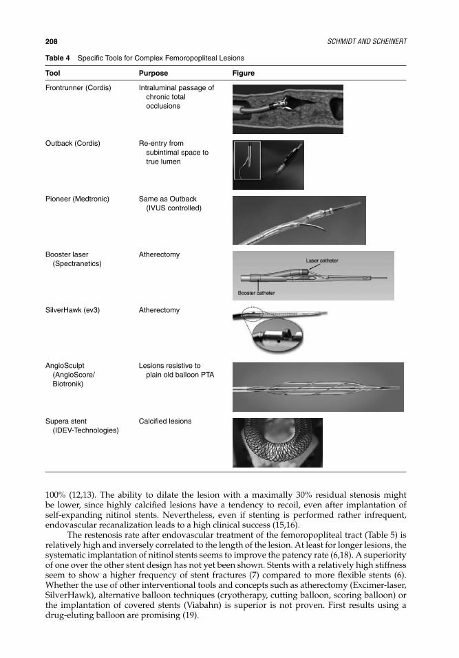
IHBK065-15 IHBK065-Minar May 1, 2010 8:13 Specs: 7x10 tight 7in×10in Char Count=
208 SCHMIDT AND SCHEINERT
Table 4 Specific Tools for Complex Femoropopliteal Lesions
Tool Purpose Figure
Frontrunner (Cordis) Intraluminal passage ofchronic totalocclusions
Outback (Cordis) Re-entry fromsubintimal space totrue lumen
Pioneer (Medtronic) Same as Outback(IVUS controlled)
Booster laser(Spectranetics)
Atherectomy
SilverHawk (ev3) Atherectomy
AngioSculpt(AngioScore/Biotronik)
Lesions resistive toplain old balloon PTA
Supera stent(IDEV-Technologies)
Calcified lesions
100% (12,13). The ability to dilate the lesion with a maximally 30% residual stenosis mightbe lower, since highly calcified lesions have a tendency to recoil, even after implantation ofself-expanding nitinol stents. Nevertheless, even if stenting is performed rather infrequent,endovascular recanalization leads to a high clinical success (15,16).
The restenosis rate after endovascular treatment of the femoropopliteal tract (Table 5) isrelatively high and inversely correlated to the length of the lesion. At least for longer lesions, thesystematic implantation of nitinol stents seems to improve the patency rate (6,18). A superiorityof one over the other stent design has not yet been shown. Stents with a relatively high stiffnessseem to show a higher frequency of stent fractures (7) compared to more flexible stents (6).Whether the use of other interventional tools and concepts such as atherectomy (Excimer-laser,SilverHawk), alternative balloon techniques (cryotherapy, cutting balloon, scoring balloon) orthe implantation of covered stents (Viabahn) is superior is not proven. First results using adrug-eluting balloon are promising (19).
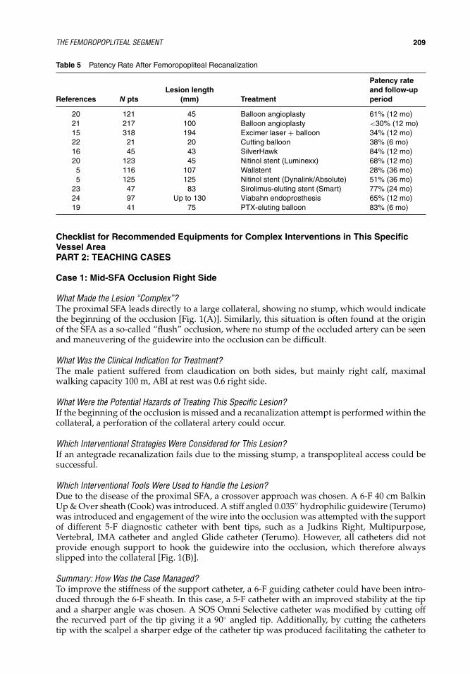
IHBK065-15 IHBK065-Minar May 1, 2010 8:13 Specs: 7x10 tight 7in×10in Char Count=
THE FEMOROPOPLITEAL SEGMENT 209
Table 5 Patency Rate After Femoropopliteal Recanalization
References N ptsLesion length
(mm) Treatment
Patency rateand follow-upperiod
20 121 45 Balloon angioplasty 61% (12 mo)21 217 100 Balloon angioplasty <30% (12 mo)15 318 194 Excimer laser + balloon 34% (12 mo)22 21 20 Cutting balloon 38% (6 mo)16 45 43 SilverHawk 84% (12 mo)20 123 45 Nitinol stent (Luminexx) 68% (12 mo)
5 116 107 Wallstent 28% (36 mo)5 125 125 Nitinol stent (Dynalink/Absolute) 51% (36 mo)
23 47 83 Sirolimus-eluting stent (Smart) 77% (24 mo)24 97 Up to 130 Viabahn endoprosthesis 65% (12 mo)19 41 75 PTX-eluting balloon 83% (6 mo)
Checklist for Recommended Equipments for Complex Interventions in This SpecificVessel AreaPART 2: TEACHING CASES
Case 1: Mid-SFA Occlusion Right Side
What Made the Lesion “Complex”?The proximal SFA leads directly to a large collateral, showing no stump, which would indicatethe beginning of the occlusion [Fig. 1(A)]. Similarly, this situation is often found at the originof the SFA as a so-called “flush” occlusion, where no stump of the occluded artery can be seenand maneuvering of the guidewire into the occlusion can be difficult.
What Was the Clinical Indication for Treatment?The male patient suffered from claudication on both sides, but mainly right calf, maximalwalking capacity 100 m, ABI at rest was 0.6 right side.
What Were the Potential Hazards of Treating This Specific Lesion?If the beginning of the occlusion is missed and a recanalization attempt is performed within thecollateral, a perforation of the collateral artery could occur.
Which Interventional Strategies Were Considered for This Lesion?If an antegrade recanalization fails due to the missing stump, a transpopliteal access could besuccessful.
Which Interventional Tools Were Used to Handle the Lesion?Due to the disease of the proximal SFA, a crossover approach was chosen. A 6-F 40 cm BalkinUp & Over sheath (Cook) was introduced. A stiff angled 0.035′′ hydrophilic guidewire (Terumo)was introduced and engagement of the wire into the occlusion was attempted with the supportof different 5-F diagnostic catheter with bent tips, such as a Judkins Right, Multipurpose,Vertebral, IMA catheter and angled Glide catheter (Terumo). However, all catheters did notprovide enough support to hook the guidewire into the occlusion, which therefore alwaysslipped into the collateral [Fig. 1(B)].
Summary: How Was the Case Managed?To improve the stiffness of the support catheter, a 6-F guiding catheter could have been intro-duced through the 6-F sheath. In this case, a 5-F catheter with an improved stability at the tipand a sharper angle was chosen. A SOS Omni Selective catheter was modified by cutting offthe recurved part of the tip giving it a 90◦ angled tip. Additionally, by cutting the catheterstip with the scalpel a sharper edge of the catheter tip was produced facilitating the catheter to

IHBK065-15 IHBK065-Minar May 1, 2010 8:13 Specs: 7x10 tight 7in×10in Char Count=
210 SCHMIDT AND SCHEINERT
(C) (D)
(B)(A)
(E) Figure 1

IHBK065-15 IHBK065-Minar May 1, 2010 8:13 Specs: 7x10 tight 7in×10in Char Count=
THE FEMOROPOPLITEAL SEGMENT 211
“hook” into the occluded artery [Fig. 1(C)]. It then was easy to engage an angled hydrophilicguidewire (Terumo) into the subintimal space, which was indicated by the loopform of the wire[Fig. 1(D)]. Also other catheters such as IMA, Judkins Right or Pigtail catheter can be modifiedin this manner. Re-entry of the guidewire into the distal artery was unproblematic, followedby balloon angioplasty using a 5-mm diameter balloon with a length of 120 mm. Due to thelength of the lesion, primary stenting was performed using two 6/120 mm nitinol stents andpostdilatation with the same balloon [Fig. 1(E)].
List of Equipment Used in This Case� 6-F Balkin Up & Over Contralateral introducer sheath, 40 cm (COOK)� 5-F SOS Omni Selective (Angiodynamics)� 0.035′′ angled stiff guidewire, 300 cm (TERUMO)� 5.0/120 mm Admiral Xtreme, 130 cm (Invatec)� 6.0/120 mm Everfelx nitinol stent (ev3)
Key Learning Issues of This Specific CaseA selection of support catheters and the modification of these by cutting off the tip at differentpoints can be helpful to engage an occlusion with the guidewire. Also, the tip of guidewires canbe gradually modified in their stiffness by cutting off the tip with a sterile scissor. However, therisk of perforation might be increased.
Case 2: Heavily Calcified Popliteal Occlusion
What Made the Lesion “Complex”?The severe calcification and not the length of an occlusion is the most important predictor offailure to recanalize CTOs of the SFA [Fig. 2(A)].
What Was the Clinical Indication for Treatment?The male patient suffered from severe claudication of the left calf, maximal walking capacity80 m, ABI at rest 0.55.
What Were the Potential Hazards of Treatment?In highly calcified lesions, the risk of perforation is increased just by the mere fact that higherforces have to be applied to pass the guidewire through the lesion. Another potential hazard incalcified CTOs is the risk of dissecting the artery distal to the occlusion, thus ending up witha much longer lesion to be dilated or stented. This is, however, only a risk using a subintimalrecanalization technique, where re-entry of the guidewire into the artery distal to the occlusioncan be difficult due to severe calcification of the arterial wall. An intraluminal recanalizationattempt is therefore preferable to avoid difficulties during re-entry; however, in case of severelycalcified lesions only feasible in rather short occlusions.
Which Interventional Strategies Were Considered for This Lesion?A crossover access can be chosen; however, pushability of the angioplasty tools is enhancedusing a more direct ipsilateral antegrade approach.
Catheter-based atherectomy is considered in this case to achieve a result without the needfor stenting. A laser catheter (Spectranetics) would be preferable over a SilverHawk device (ev3)in this case, because wire passage before atherectomy is not necessary using the laser and theability of debulking severely calcified plaque is estimated to be higher for a laser using highenergy (Turbo laser) compared to the SilverHawk.
If intraluminal passage of the occlusion fails, a subintimal passage could be performed.The Pioneer or the Outback catheter permits to re-enter the artery exactly distal to the occlusionto avoid ballooning and stenting further distal.
Which Interventional Tools Were Used to Handle the Lesion?A 6-F 45-cm Arrow sheath was introduced ipsilateral, thereby having the tip of the sheathdirectly in front of the occlusion. After several attempts to pass the occlusion intraluminally

IHBK065-15 IHBK065-Minar May 1, 2010 8:13 Specs: 7x10 tight 7in×10in Char Count=
212 SCHMIDT AND SCHEINERT
(A) (B) (C)
(D) (E) (F) (G) (H)
Figure 2
with a stiff straight 0.035′′ hydrophilic guidewire (Terumo) supported by a 5-F multipurposecatheter, the wire was introduced in a reversed way using the very stiff end [Fig. 2(B)]; however,this was also unsuccessful in the first attempt. A 2.0-mm Turbo laser (Spectranetics) [(Fig. 2(C)]was used to pass or at least weaken the plaque. The laser catheter did not pass, although themaximal energy (60 mJ/mm2) and a maximal pulse-repetition rate (60/sec) were used, butfacilitated passage of the reversed tip of the Terumo wire [Fig. 2(D)] through the occlusion.Since leaving the wire in a reversed mode in the artery during the next interventional stepsmight have been hazardous in terms of potential perforation, a 5-F diagnostic catheter waspositioned over the Terumo wire as far into the occlusion as possible. Thereafter, the Terumowire was withdrawn and a 300-cm 0.014′′ guidewire inserted, which easily passed the channelcreated by the Terumo wire [Fig. 2(E)]. Laser atherectomy was continued thereafter [Fig. 2(F)].

IHBK065-15 IHBK065-Minar May 1, 2010 8:13 Specs: 7x10 tight 7in×10in Char Count=
THE FEMOROPOPLITEAL SEGMENT 213
(I)
Figure 2 (Continued)
A high-pressure balloon, tracking over a 0.035′′ guidewire, chosen due to the calcification of thelesion, did not pass the channel created by the laser [Fig. 2(G)]. A PTCA balloon with a verylow crossing profile was then used for predilatation [Fig. 2(H)] followed by dilatation using alow-profile OTW balloon with a larger diameter [Fig. 2(I)].
Summary: How Was the Case Managed, Is There Anything That Could Have Been Improved?A direct antegrade approach improved the pushability of the tools used for recanalization. Alaser catheter was helpful to weaken the plaque and facilitate wire passage. If a laser device is notavailable, further ongoing attempts to pass the lesion with the wire might have been successfulas well. The reversed tip of the wire came rather close to the arterial wall after passage of theocclusion with the risk of perforation. The reversed tip of the Terumo wire can be bent usinga sterile clamp to be able to direct the wire into another position. If intraluminal wire passagewould have failed, a subintimal recanalization attempt could have been attempted.
List of Equipment Used in This Case� 6-F 45 cm Arrow sheath (Arrow)� 0.035′′ Radiofocus Terumo angled stiff guidewire, 300 cm (TERUMO)� 3.0 OTW Elite Turbo laser catheter (Spectranetics)� 0.014′′ PT2 hydrophilic 300 cm guidewire (Boston Scientific)� 5.0/40 mm Admiral Xtreme, 130 cm (Invatec)� 4.0/20 Mercury PTCA catheter (Abbott)� 5.0/40 mm Pacific Xtreme, 130 cm (Invatec)
Key Learning Issues of This CaseIn case of severely calcified short CTOs, an intraluminal recanalization attempt is justified usingthe stiff, reversed tip of guidewires. Laser atherectomy can be helpful to facilitate guidewirepassage into an occlusion.
Case 3: Severely Calcified Long SFA Occlusion
What Made the Lesion “Complex”?The severe calcification of the occlusion clearly makes the success of an endovascular approachquestionable [Fig. 3(A)].

IHBK065-15 IHBK065-Minar May 1, 2010 8:13 Specs: 7x10 tight 7in×10in Char Count=
214 SCHMIDT AND SCHEINERT
(A) (A2)
(B) (C)
(D) (E)
Figure 3

IHBK065-15 IHBK065-Minar May 1, 2010 8:13 Specs: 7x10 tight 7in×10in Char Count=
THE FEMOROPOPLITEAL SEGMENT 215
(F) (G)
(H) (H2)
(I) (J)
Figure 3 (Continued)(Continued on page 216)

IHBK065-15 IHBK065-Minar May 1, 2010 8:13 Specs: 7x10 tight 7in×10in Char Count=
216 SCHMIDT AND SCHEINERT
(K) (L)
(M)
Figure 3 (Continued)
What Was the Clinical Indication for Treatment?The male patient suffered from critical limb ischemia with minor gangrene of the toes. The severecalcification is typical for patients with endstage renal insufficiency. The patient was referred bya vascular surgical department for an endovascular recanalization attempt. Difficulty insertingthe bypass to the highly calcified arterial wall was anticipated.
What Were the Potential Hazards of Treating This Lesion?The risk of bleeding is clearly higher if guidewire perforation occurs right at the beginning of theocclusion, leading the blood flow from the proximal open artery directly into the surroundingtissue. In contrast, guidewire perforation within an occlusion is normally self-limiting either ifthe procedure is stopped at this stage or balloon angioplasty and/or stenting is performed aftersuccessful guidewire passage of the occlusion.
Dissection with the guidewire further distal of the end of the occlusion is frequent inhighly calcified lesions and makes ballooning and potentially stenting further distal necessary.This scenario should be avoided not to destroy an area for a future distal bypass anastomosis.
Which Interventional Strategies Were Considered for This Lesion?Generally two ways of endovascular recanalization can be attempted, an intraluminal or asubintimal approach. In this highly calcified artery, the subintimal way is the only chance topass the occlusion.

IHBK065-15 IHBK065-Minar May 1, 2010 8:13 Specs: 7x10 tight 7in×10in Char Count=
THE FEMOROPOPLITEAL SEGMENT 217
Which Interventional Tools Were Used to Handle the Lesion?After having introduced a 6-F crossover sheath, a 5-F Judkins Right catheter was used to engagethe tip of the 0.035′′ stiff angled hydrophilic guidewire into the subintimal space at the verybeginning of the occlusion, using the so-called “percutaneous intended subintimal recanaliza-tion technique (PIER)” (11). Creating a loop with the wire a subintimal passage of the occlusionwas possible [Fig. 3(B)]; however, re-entry into the patent distal artery was unsuccessful. There-fore, the Terumo wire was exchanged for a 0.014′′ guidewire and predilatation was performedusing a low-profile balloon (2.5/120 mm) [Fig. 3(C)] to facilitate the introduction of the Outbackcatheter. The marker at the tip of the catheter has to appear as a “T” in alignment with the arteryand should be “L”-shaped if the position is lateral to the artery [Fig. 3(D)] to direct the curvedneedle from the subintimal space back into the true lumen. Thereafter, the 0.014′′ guidewire isadvanced through the needle into the distal SFA [Fig. 3(E)]. It can be difficult to pass a balloonthrough the calcified membrane, low-profile PTCA catheters are successful, if other peripheralballoon catheters fail [Fig. 3(F)]. Thereafter, gradual angioplasty using a 4.0/120 mm and 5.0/120 mm balloon was performed [Fig. 3(G)]. Due to severe recoil, nitinol stents were implantedand postdilated with the 5.0-mm balloon [Fig. 3(H)]. However, thereafter also significant resid-ual stenoses remained due to recoil. A balloon expandable renal stent was implanted at thearea of most severe recoil [Fig. 3(I)] and finally a significantly improved outflow was achieved[Fig. 3(J)].
Summary: How Was the Case Managed, Is There Anything That Could Have Been Improved, What Wasthe Immediate and Long-Term Outcome?The combination of a subintimal recanalization technique and a re-entry device was successful.The Outback catheter is a very stiff device with an excellent pushability, therefore predilatationof the lesion might not have been necessary before introduction of the device. Whether a cuttingor scoring balloon would have reduced the recoil after ballooning is questionable. There wasa sustained improvement of the clinical situation during the next six months. Reangiographyafter six months showed a patent artery with severe stent fracture at the area of re-entry intothe true lumen [Fig. 3(K)] and stent-in-stenting was performed. Twenty-six months later thepatient suffered from recurrent claudication. A proximal subtotal de novo stenosis of the SFAand moderate restenosis in the stents with severe stent fractures not associated to restenosiswere seen [Fig. 3(L)]. Figure 3(M) shows the result after additional stenting of the proximallesion and re-PTA in the stents.
List of Equipment Used in This Case� 6-F Balkin Up & Over Contralateral sheath, 40 cm (COOK)� 5-F JR4 diagnostic catheter, 100 cm (Cordis)� 0.035′′ angled stiff guidewire, 300 cm (Terumo)� 0.014′′ ACS High-Torque Floppy II Extra Support, 300 cm (Abbott)� 2.0/120 mm Amphirion Deep balloon, 120 cm (Invatec)� Outback catheter (Cordis)� 2.0/20 mm Mercury PTCA catheter (Abbott)� 4.0/120 mm and 5.0/120 mm Pacific Xtreme, 130 cm (Invatec)� 6.0/120 mm SMART nitinol stents (Cordis)� 6.0/18 mm Genesis on Amiia renal stent (Cordis)
Key Learning Issues of This specific CaseIn severely calcified SFA occlusions, only a subintimal recanalization technique is feasible. A re-entry catheter is necessary in many cases. The radial force of nitinol stents might be suboptimalin heavily calcified lesions, an additional implantation of a short balloon expandable stent mightbe necessary. Recanalization of heavily calcified obstructions should probably be restricted topatients with critical ischemia; in claudicants, the residual stenoses even after stenting mightnot yield the desired clinical result.

IHBK065-15 IHBK065-Minar May 1, 2010 8:13 Specs: 7x10 tight 7in×10in Char Count=
218 SCHMIDT AND SCHEINERT
PART 3: KEY ISSUES FOR SUCCESSFUL AND SAFE TREATMENT OF COMPLEXFEMOROPOPLITEAL LESIONSSuccess and safety of the intervention starts with the thorough consideration of the treatmentoptions, which mainly depend on the clinical situation and morphology of the lesion. Althoughthe complication rate of angioplasty is generally low, the equipment and knowledge of tools fortreatment of complications is mandatory.
� Experience and tools for infrapopliteal interventions.� Appropriate aspiration catheters.� Experience in antegrade access for aspiration.� Covered stents.
The primary success depends largely on the availability and experience with dedicatedmaterials:
Crossover sheath that provide sufficient support.Variety of hydrophilic-coated guidewires.Variety of support catheters.Re-entry device.Nitinol stents with proven efficacy in terms of long-term patency.
Long-term success:
Close and continuous follow-up surveillance.
REFERENCES1. Dormandy JA, Rutherford RB. Management of peripheral arterial disease (PAD). TASC Working
Group TransAtlantic Inter-Society Consensus (TASC). J Vasc Surg 2000; 31(1 Pt 2):S1–S296.2. Norgren L, Hiatt WR, Dormandy JA, et al. Inter-society consensus for the management of peripheral
vascular disease (TASC II). J Vasc Surg 2007; 45(suppl S):S5–S67.3. Duc SR, Schoch E, Pfyffer M, et al. Recanalization of acute and subacute femoropopliteal artery occlu-
sions with the Rotarex catheter: one year follow-up, single center experience. Cardiovasc InterventRadiol 2005; 28(5):603–610.
4. Karnabatidis D, Katsanos K, Kagadis GC, et al. Distal embolism during percutaneous revascularizationof infra-aortic arterial occlusive disease: an underestimated phenomenon. J Endovasc Ther 2006;13(3):169–180.
5. Schlager O, Dick P, Sabeti S, et al. Long-segment SFA stenting—the dark side: in-stent restenosis,clinical deterioration, and stent-fractures. J Endovasc Ther 2005; 12:676–684.
6. Schillinger M, Sabeti S, Loewe C, et al. Balloon angioplasty versus implantation of nitinol stents in thesuperficial femoral artery. N Engl J Med 2006; 354(18):1879–1888.
7. Scheinert D, Scheinert S, Sax J, et al. Prevalence and clinical impact of stent fractures afterfemoropopliteal stenting. J Am Coll Cardiol 2005; 45:312–315.
8. Schlager O, Francesconi M, Haumer M, et al. Duplex sonography versus angiography for assessmentof femoropopliteal arterial disease in a “real-world” setting. J Endovasc Ther 2007; 14(4):452–459.
9. Yilmaz S, Sindel T, Luleci E. Ultrasound-guided retrograde popliteal artery catheterization: experiencein 174 consecutive patients. J Endovasc Ther 2005; 12(6):714–722.
10. Dorffler-Melly J, Mahler F, Do DD, et al. Adjunctive abciximab improves patency and functionaloutcomes in endovascular treatment of femoropopliteal occlusions: initial experiences. Radiology2005; 237(3):1103–1109.
11. Laxdal E, Jenssen GL, Pedersen G, et al. Subintimal angioplasty as a treatment of femoropoplitealartery occlusions. Eur J Vasc Endovasc Surg 2003; 25:578–582.
12. Jacobs DL, Motaganahalli RL, Cox DE, et al. True lumen re-entry devices facilitate subintimal angio-plasty and stenting of total chronic occlusions: initial report. J Vasc Surg 2006; 43(6):1291–1296.
13. Scheinert D, Braunlich S, Scheinert S, et al. Initial clinical experience with an IVUS-guided transmem-brane puncture device to facilitate recanalization of total femoral occlusions. EuroIntervention 2005;1:115–119.
14. Scheinert D, Peeters P, Bosiers M, et al. Results of the multicenter first-in-man study of a novel scoringballoon catheter for the treatment of infra-popliteal peripheral arterial disease. Catheter CardiovascInterv 2007; 70(7):1034–1039.

IHBK065-15 IHBK065-Minar May 1, 2010 8:13 Specs: 7x10 tight 7in×10in Char Count=
THE FEMOROPOPLITEAL SEGMENT 219
15. Scheinert D, Laird JR, Schroeder M, et al. Excimer laser-assisted recanalization of long chronic super-ficial femoral artery occlusions. J Endovasc Ther 2001; 8:156–8166.
16. Zeller T, Rastan A, Sixt S, et al. Long-term results after directional atherectomy of femoro-popliteallesions. J Am Coll Cardiol 2006; 48(8):1573–1578.
17. Rastan A, Sixt S, Schwarzwalder U, et al. Initial experience with directed laser atherectomy using theCLiRpath photoablation atherectomy system and bias sheath in superficial femoral artery lesions. JEndovasc Ther 2007; 14(3):365–373.
18. Schillinger M, Sabeti S, Dick P, et al. Sustained benefit at 2 years of primary femoropopliteal stentingcompared with balloon angioplasty with optional stenting. Circulation 2007; 115(21):2745–2749.
19. Tepe G, Zeller T, Albrecht T, et al. Local delivery of paclitaxel to inhibit restenosis during angioplastyof the leg. N Engl J Med 2008; 358(7):689–699.
20. Krankenberg H, Schluter M, Steinkamp HJ, et al. Nitinol stent implantation versus percutaneoustransluminal angioplasty in superficial femoral artery lesions up to 10 cm in length: the femoral arterystenting trial (FAST). Circulation 2007; 116(3):285–292.
21. Capek P, McLean GK, Berkowitz HD. Femoropopliteal angioplasty. Factors influencing long-termsuccess. Circulation 1991; 83:170–180.
22. Amighi J, Schillinger M, Dick P, et al. De novo superficial femoropopliteal artery lesions: periph-eral cutting balloon angioplasty and restenosis rates-randomized controlled trial. Radiology 2008;247(1):267–272.
23. Duda SH, Bosiers M, Lammer J, et al. Drug-eluting and bare nitinol stents for the treatment ofatherosclerotic lesions in the superficial femoral artery: long-term results from the SIROCCO trial. JEndovasc Ther 2006; 13(6):701–710.
24. Saxon RR, Dake MD, Volgelzang RL, et al. Randomized, multicenter study comparing expandedpolytetrafluoroethylene-covered endoprosthesis placement with percutaneous transluminal angio-plasty in the treatment of superficial femoral artery occlusive disease. J Vasc Interv Radiol 2008;19(6):823–832.

IHBK065-16 IHBK065-Minar May 11, 2010 16:58 Specs: 7x10 tight 7in×10in Char Count=
16 The Tibioperoneal SegmentLanfroi GrazianiInvasive Cardiology Unit, Istituto Clinico Citta di Brescia, Brescia, Italy
Hubert WallnerKardinal Schwarzenberg´sches Krankenhaus, Schwarzach, Salzburg, Austria
PART 1: INTRODUCTIONIschemic diabetic foot ulcer is the main cause of nontraumatic amputations in westerncountries (1).
For patients aged 65 to 74 years, diabetes heightens the risk of amputation morethan 20-fold, putting these patients at great risk of limb loss; 20% of subjects with diabetesdevelop a foot ulcer in the course of life, and 33% of them, if untreated, frequently undergoamputation (2–5).
The most common indication for intervention in the tibial–peroneal segment (Table 1) isrepresented by the condition of critical limb ischemia (CLI) (6).
Diagnosis of CLI is possible in the presence of pain at rest or ulceration or gangrene withor without ankle pressure less than 50 to 70 mm Hg or transcutaneous oxygen tension (TcPO2)values lesser than 30 to 50 mm Hg (7).
In general, the development of CLI condition strictly depends on the efficiency of collateralcirculation but, it has been fully demonstrated that in diabetics, the development and formationof collateral circulation is significantly reduced or absent and thus the majority of patients withCLI are diabetic (8–10).
As a consequence, it is also possible in a limited percentage of cases that diabetics maydevelop a condition of CLI, even in the presence of isolated obstruction of only one of theirtibial arteries.
This condition rarely recognized if not foreseen, makes it quite impossible in some casesto identify the CLI presence, simply for the mere existence of at least one valid tibial pulse.
Only in 1984, thanks to a group of American vascular surgeons, was it possible to establishthat the best treatment in diabetics with ischemic foot lesions was to provide direct flow to thefoot arteries (11), but even now peripheral ischemia remains frequently undiagnosed.
The frequent false negative result of the ankle brachial index (ABI) measurement com-plicates even more the possibility of recognizing the condition of CLI particularly in diabetics,due to the poor compression of leg arteries for the presence of diffuse calcifications along theircourse.
It has been noticed, in fact, that an increase of the ABI index greater than 1.3 must beconsidered as a suspected false negative value in subjects with CLI.
Poorly diffused and utilized only in the area of diabetic foot centers is the TcPO2 measure-ment that, however, represents one of the most important diagnostic tests in the demonstrationof the CLI condition.
From a procedural point of view, the condition that complicates the treatment further, isthe need to use the femoral approach for the endovascular recanalization of most tibial arteryocclusions.
The antegrade approach, which offers conditions of absolute advantage with respect tothe traditional crossover aortailiac technique, has not been sufficiently popularized over the lastfew years and limits the feasibility of extreme interventions, even today.
Moreover, the management of hemostasis subsequent to the antegrade approach is con-sidered by some as difficult and a possible cause of complications (Table 2).

IHBK065-16 IHBK065-Minar May 11, 2010 16:58 Specs: 7x10 tight 7in×10in Char Count=
THE TIBIOPERONEAL SEGMENT 221
Table 1 Clinical Indications for the Treatment of Complex Lesions in Tibial–Peroneal Interventions
1. The avoidance of limb amputation or reducing the level of amputation when this proves to be unavoidable2. The presence of pain at rest in patients with CLI, with or without ulceration. Due to the frequent
coexistence of diabetic neuropathy, the presence of pain can vary consistently (12)3. The presence of ischemic ulcerations that often represent the initial manifestation of a CLI condition4. Further indication is represented by the presence of a healed ischemic ulceration in a persistent CLI
condition to prevent ulcer recurrence5. Symptomatic claudication, often absent due to the associated neuropathy (13), does not represent a
possible indication in the absence of a CLI condition
CLI represents the most common indication for tibioperoneal interventions.
In reality, a correct antegrade puncture and catheterization technique and an appropriatelearning curve determine a rate of complications absolutely comparable to that of the classicfemoral retrograde approach.
Due to the reduced diameter of tibial vessels, often diffusely occluded and calcified,regular techniques in use for the femoral and popliteal recanalization segment do not havethe same applications, leaving ample possibility in the utilization of techniques derived fromangioplasty of coronary vessels.
Moreover, the requirement of sufficient experience in the use of coronary-type devicesrepresents a further element of complexity in the tibial–peroneal interventions (Table 3).
Finally, the frequent coexistence of necrosis and infection requires the competence of adiabetic foot center for appropriate surgical treatment promoting wound healing and limbsalvage.
Usually the revascularization precedes surgical foot care.Along with normal risk conditions of endovascular procedures, such as renal failure,
hematoma, bleeding, hemorrhage, dissection, and thrombosis at the puncture site, interventionsin the tibial and peroneal segment present additional risks of accidental vessel perforation andrupture.
The risk of significant hemorrhage due to perforation is very low, as well as the possibilityof partial vessel laceration or rupture even in case the dilatation is performed with a balloon ofa diameter slightly bigger than the regular size of the vessel.
These conditions can be easily managed and treated with the application of a tourni-quet around the trauma site, maintaining a pressure value greater than the systolic one andproceeding with reversed heparin therapy using protamine sulfate (14).
During the procedure, it is preferable to keep or maintain the guide inside the artery foran eventual continuation and completion of the treatment with balloon dilatation and stenting.
Other possible complications are embolization and thrombosis of the tibial–peroneal andfoot vessels.
The formation of emboli could be relatively frequent in the presence of a fresh thrombusin a femoral or popliteal site.
Its treatment could be selective transcatheter lysis by urokinase infusion, or more com-monly thrombus aspiration using a 5 to 6-F coronary guiding catheter.
In resistant cases, a stent implantation in the thrombus occluded site is advisable, for aquick re-establishment of the arterial flow.
Table 2 Hazards and Complications in Extreme Interventions
Complication Cause Incidence (%) Treatment
Perforation Guidewire 5 Tamponade (tourniquet)Rupture Balloon oversize 3 Tamponade (tourniquet)Thrombosis Insufficient heparinization 2 Full dose of heparin, frequent ACT
assessmentEmbolization Wire catheter manipulation 2 Lysis, catheter aspirationSpasm Balloon inflation, guidewire 20 Selective nitroglycerin

IHBK065-16 IHBK065-Minar May 11, 2010 16:58 Specs: 7x10 tight 7in×10in Char Count=
222 GRAZIANI AND WALLNER
Table 3 Interventional Tools
Special procedure Device/s
Long tibial stenosis/occlusion crossing (regular) Boston Sci. PT Graphic Super SupportTM.014 G.W., InvatecAmpirion DeepTM balloon
Long tibial stenosis/occlusion crossing (difficult) Terumo 3 mm J tipped 0.035 G.W., Cordis 4 FrBerensteinTM cath.
Long tibial stenosis/occlusion crossing (difficult) Boston Sci. 0.035 straight G.W., Cordis 4 Fr Berenstein cath.Stenting of long lesions (self-expandable nitinol
stents)Invatec Maris DeepTM, Abbott Vascular XpertTM,
Biotronic Astron PulsarTM
Stenting of proximal lesions (balloonexpandable coronary stents)
Medtronic DriverTM, Boston Sci. TaxusTM
Kissing-balloon dilatation of bifurcation lesions Medtronic Sprinter LegendTM coronary balloonAtherectomy of bifurcation/ostial lesions Ev3 Fox HollowTM
Focal hard lesions dilatation Boston Sci. Cutting BalloonTM and Cordis 4 Fr Berensteincath.
Re-entry maneuver after CTO subintimalcrossing
Boston Sci. V18TM G.W.
Extremely hard lesions dilatation Boston Sci. QuantumTM coronary balloonExtremely tight lesion dilatation Boston Sci. MaverickTM coronary balloon
Thrombosis of the tibial arterial vessels during the procedure is usually due to an insuffi-cient heparinization. Prevention of such a condition can be obtained by the frequent monitoringof the activated coagulation time (ACT) during the procedure.
Our data on procedural complications (15,16) (L.G., unpublished data) referred to 1500consecutive interventions in diabetics with CLI between 1998 and 2000 are:
Minor complications (no surgery or transfusion required): 6%Major complications: 2%Mortality: 0.2%
Preinterventional Diagnostic WorkupOnly in less than 28% of cases the condition of CLI is sustained by an exclusive involvement ofthe tibial–peroneal arteries (17).
CLI conditions, particularly in the presence of ischemic ulcerations, are usually caused bymultilevel involvement of the femoral, popliteal, and tibial–peroneal segments.
The approach to the endovascular treatment of tibial vessel obstruction must be chosen inorder to allow for easy manoeuvrability of catheters, guides, and other devices. The antegradefemoral approach is the best solution for this purpose.
MRI and TC angiography and Doppler ultrasound examinations are useful preliminarytests to detect the anatomical complexity of a planned procedure.
In the absence of iliac arteries lesions, since 1998 in our centers, the procedure has alwaysbegun with the performance of a direct antegrade femoral angiography using a 19 G single-wallneedle to establish the best entry site for intervention.
Then we proceed with the advancement of a short angled 0.035 Teflon-coated guidewirealong the first segment of the superficial femoral artery and then with the positioning of a 5 to6-F vascular introducer.
Interventional StrategiesIn the presence of multilevel lesions, the initial procedure is to cross the proximal lesions as faras reaching the dorsalis pedis artery, or plantar artery, or in cases where this is not possible, theperoneal artery (PA).
During the procedure, 5000 UI of sodium heparin is injected.Crossing the tibial vessel obstructions is usually performed by a 0.014 coronary-type
guidewire and low-profile over-the-wire (OTW) balloon catheters with the diameter of theballoon selected on the basis of the diameter of the most distal segment to be dilated (Fig. 1).

IHBK065-16 IHBK065-Minar May 11, 2010 16:58 Specs: 7x10 tight 7in×10in Char Count=
THE TIBIOPERONEAL SEGMENT 223
(A)
(D) (E)
(B)(C)
Figure 1 (A) Anterior tibial artery (AT) occlusion in a diabetic with ischemic foot ulcer. (B–D) The occlusion wascrossed with a 0.014 coronary wire followed by balloon angioplasty dilatation using a 3.0 × 120 mm AmphirionDeep balloon. (E) Optimal final result. The ulcer healed after some weeks.
The broad selections of .014-inch dedicated wire systems for chronic total occlusions incoronary systems offer versatile solutions in below the knee lesions and are utilized in followingtechniques:
Controlled drilling: the guide wire is advanced using gentle movements.Sliding technique with controlled drilling: very lubricious polymer covered guide wires are
used to slide through narrow lesions or functional occlusions.Penetration technique: penetrating the obstruction aiming at the target. The direction of the
guide wire is more precisely controlled. Tapered tip guide wires permit higher penetratingforces but the following balloon advancement could be very difficult due to increasedfriction under calcified plaques.

IHBK065-16 IHBK065-Minar May 11, 2010 16:58 Specs: 7x10 tight 7in×10in Char Count=
224 GRAZIANI AND WALLNER
Alternative techniques are the use of the 4-F diagnostic Berenstein catheter and a straight0.035 Teflon-coated guidewire, replaced by a 3 mm J tipped 0.035 Terumo wire if a subintimalrecanalization is required.
In all patients with multilevel lesions, the first vessel to be treated must be the most distal,which is usually represented by one of the two tibial vessels, to get direct flow to the foot at thecompletion of the procedure.
Ideally direct flow should be assured along the tibial and foot artery that supplies theischemic lesion, when present.
Alternative techniques such as plantar arch crossing, collateral dilatation (Fig. 2) or retro-grade tibial artery recanalization, can represent possible options (18,19).
Whenever possible, the goal of the procedure is to get direct flow to the dorsalis pedisartery in case of toe lesions or to the plantar artery in case of calcanear lesions.
The most common and effective technique for below-the-knee (BTK) revascularization istransluminal angioplasty using the new generation of OTW low-profile long balloons.
These very low profile and tapered to 0.014 balloons could be used over the several special-ized coronary-type guidewires available, with a 70% of successful recanalization of chronicallyoccluded tibial–peroneal arteries.
Elective or provisional stenting is used in case of:
1. complex lesions that involve the bifurcation of the popliteal artery (Fig. 3);2. lesions of tibial arteries ostium and popliteal bifurcation with plaque shifting (Fig. 4);3. persistent recoiling of proximal tibial arteries lesions;4. short embolus not amenable to selective thrombolysis or aspiration thrombectomy;5. extensive flow-limiting dissection;6. elective stenting in stenosis of bypass graft anastomosis and ostial lesions (Fig. 5).
The new generation of self-expandable nitinol stents designed for use in tibial vessels hasexpanded the indications in obstructions of considerable lengths, and particularly in the case oflesion recurrence after balloon angioplasty, but their long-term result is under evaluation.
Normally, balloon expandable stent implantation is avoided in foot arteries and mediumdistal tibial vessels due to the concrete risk of stent crushing after ABI measurement or physicalexamination with pulse palpation.
A possible alternative to stenting in proximal bifurcation lesion is the kissing-balloondilatation (Fig. 6)
Direct atherectomy using the Fox HollowTM device (20) can be considered appropriate inthe case of eccentric plaques, complex lesions of popliteal bifurcation involving the origin oftibial branches, and in-stent restenosis in BTK arteries (Fig. 7).
In these lesions, encouraging results have been achieved using drug-eluting coronary-typestent (DES) implantation (21). The frequently diffuse tibial artery obstructive involvement in atypically multilevel disease limits the use of DES to short lesions in proximal segments, withoutthe privilege of ascribing the clinical result to this otherwise crucial device as in the coronarytree.
The most effective technique in tibial–peroneal segments is balloon angioplasty dilatation.General rules for its performance are well established (22–31) and are represented by the
following:
1. Prolonged dilatations, greater than three minutes.2. Gradual high-pressure balloon inflation.3. The balloon diameter at the maximum inflation pressure must be similar or slightly exceed
the normal vessel size.4. The length of the balloon must approach or slightly exceed the length of the lesion.
Cutting balloon dilatation (Fig. 8), laser recanalization, and cryoplasty balloon dilatationare alternative techniques that have to be tested in large series of CLI patients (32–34).
The common multilevel disease of CLI also means that, in case of endovascular treatment,different techniques and devices are used during the same procedure.
Therefore, studies specifically addressing a single technique and/or device in subjectswith CLI could depend on the concomitant treatment of other vascular segments with different

IHBK065-16 IHBK065-Minar May 11, 2010 16:58 Specs: 7x10 tight 7in×10in Char Count=
THE TIBIOPERONEAL SEGMENT 225
(A) (B)
(C)
(D)
(E) (F)
(G)
Figure 2 Ischemic nonhealing calcanear ulcer in a 60-year-old diabetic patient. (A–D) Occlusion of the posteriortibial (PT) artery and stenosis of the peroneal artery (PA). Typical appearance of lack of collaterals formation inposterior aspect of the foot. (E–G) A 0.014-coronary support wire was advanced along the PA and its posteriorperforating branch to reach the plantar artery. Final balloon dilatation using the 3.0 × 120 mm Amphirion Deepballoon with direct flow to the plantar artery. The lesion healed after ulcerectomy.

IHBK065-16 IHBK065-Minar May 11, 2010 16:58 Specs: 7x10 tight 7in×10in Char Count=
226 GRAZIANI AND WALLNER
T-Stent
Figure 3 Complex femoral popliteal and ostial tibial recanalization in a 76-year-old man; diabetes, ischemiculcer, and dry gangrene of the right forefoot. To maintain the patency of both AT and PA ostium (arrow), a T-stentimplantation was performed using two Medtronic chromium–cobalt 3.5-mm coronary stents. The stent for theAT was implanted first followed by the second with final kissing-balloon inflation. A 12-cm long nitinol stent wasimplanted in the fem–pop segment. CLI was relieved and the patient underwent a transmetatarsal amputationwith limb salvage.
“T-stent”placementtechnique
(A) (B) (C) (D) (G)
(E) (F)
Figure 4 (A–G) A 49-year-old woman patient with diabetes, with smoking background, who had ischemicnonhealing ulcer of the great toe. Ostial, noncalcified soft stenosis of the AT, prone to prolapse into the tibioperoneal(TP) trunk. This was the case after 3.5 mm balloon dilatation, with plaque shifting into the trunk. This kind ofbifurcation lesion required a 4.0- and 3.5-mm Medtronic chromium–cobalt coronary stent in TP trunk and AT,respectively, with final kissing-balloon dilatation. The lesion healed in two weeks.

IHBK065-16 IHBK065-Minar May 11, 2010 16:58 Specs: 7x10 tight 7in×10in Char Count=
THE TIBIOPERONEAL SEGMENT 227
STENTING
Figure 5 Persistent CLI and foot lesion in a 69-year-old diabetic patient with a normally patent fem–pop bypassgraft. Occlusion of the distal anastomosis. Procedure: antegrade approach, AT occlusion was crossed with anextra support hydrophilic coronary wire and 3.5 mm coronary balloon, Magic Wallstent R© deployment. Final balloondilatation. The lesion healed after one month.
Kissing-balloonangioplasty
Ostial and bifurcating lesions
Figure 6 Kissing-balloon dilatation of a true bifurcate lesion of the TP trunk using two 3.0 × 40 mm AmphirionDeep balloons inflated at 14 atm. Perfect final angiographic result.

IHBK065-16 IHBK065-Minar May 11, 2010 16:58 Specs: 7x10 tight 7in×10in Char Count=
228 GRAZIANI AND WALLNER
Figure 7 Previous placement of a bare metal stent in the TP trunk of a 62-year-old claudicant, nondiabetic,smoker. The stent (arrows) occluded after three months. The sequence shows a 0.014 wire crossing the occlusionfollowed by intimal hyperplasia removal using the Fox Hollow atherotome (fragments in a box). Final 4.0 mmAmphirion Deep balloon dilatation.
techniques. This has to be considered when judging the real impact of a single technique/deviceon outcome.
The interest in testing new endovascular solutions to be used in the tibioperoneal territorymakes urgent the need of appropriate evaluation criteria to assess the benefit on clinical outcomeand the long-term patency of the treated vessels.
The new criteria should consider new morphologic classifications (17) to stratify thepatients in classes of homogeneous vascular involvement and appropriate clinical and instru-mental measurement of the result.
The goal of extreme intervention in patients with CLI is in fact not long-term patency, butrather the avoidance of a major amputation.
Even in subjects with end-stage renal disease (ESRD) and chronic dialysis, extreme inter-ventions showed to be feasible and effective (Table 4), with a limb salvage rate similar to thehighest reported in literature for surgical series (35). These patients have an increased risk ofCLI and limb loss and are accompanied by diffuse vascular calcifications and involvement ofdistal infrapopliteal and foot arteries that makes extreme interventions challenging.

IHBK065-16 IHBK065-Minar May 11, 2010 16:58 Specs: 7x10 tight 7in×10in Char Count=
THE TIBIOPERONEAL SEGMENT 229
2.5 mm cuttingballoon
Figure 8 ESRD, 61-year-old dialyzed male. CLI and dry gangrene of the foot. Multilevel disease with tibial andfoot arteries calcified stenoses and occlusions. Initial attempt of complete PT and plantar artery recanalizationusing the Amphirion Deep balloon and a high-pressure coronary balloon at 20 atm was ineffective, due to thepresence of an extremely hard focal lesion. A 2.5-mm cutting balloon was then inflated at 18 atm with good result.The patient underwent only a transmetatarsal amputation with limb salvage.
A strict cooperation with a specialized foot center is crucial in obtaining the best results.Although restenosis rates of 40% to 50% can be expected in these patients, due to lesion
diffusion in a typical multilevel disease, limb salvage can be achieved in more than 90% of cases,primarily dependent on restoration of straight-line flow to the foot.
Furthermore, as in surgical practice, clinical and instrumental follow-up surveillance andsubsequent early reintervention in case of restenosis can prolong long-term patency and theclinical benefit.
Repeat intervention, much less invasive if compared to surgical options, performed toenhance secondary patency is probably underused, even if well tolerated and often done on anoutpatient basis. A list of recommended devices for complex cases is reported in Table 5.
Table 4 Results of Endovascular Treatment of Combined Tibio-peroneal and Femoropopliteal Lesions inSubjects with CLI
Patients/ Technical Limb PrimaryAuthor lesions success % Mortality % salvage % patency
Faglia 2005 993 (1,191) 83 0.1 88 (5 yrs) —Dorros 2001 270 91 0.4 91 (5 yrs) —Lofberg 1996 94 88 2.4 72 (3 yrs) —Soder 2000 72 74 — 80 (18 mo) 48%, (18 mo)Brillu 2001 37 94.5 — 87 (2 yrs) —Rand 2006 37/57 — 2.0 98 (6 mo) 61-83%, (6 mo)Staffa 2003 18 — — — 78%, (6 mo)Matsagas 2003 67 88 4.0 98 (3 yrs) 52%, (2 yrs)Balmer 2002 66 — — 94 (12 mo) 44%, (1 yr)Ferraresi 2009 107 98 — 93 (3 yrs) 42%, (1 yr)
Source: Modified from Ref. 6.

IHBK065-16 IHBK065-Minar May 11, 2010 16:58 Specs: 7x10 tight 7in×10in Char Count=
230 GRAZIANI AND WALLNER
Table 5 Recommended Devices for Complex Tibioperoneal Interventions
Device Company Type Code
Needle Cordis 19 G 502–654Vascular introducer Terumo Regular RS B50K10MQMultipurpose
catheterCordis Berenstein 4-F 451–415 HO
Guidewire Boston Scientific Coronary stiff 14905-01Coronary intermediate 14902-01Short 0.035 49–2090.035 support exchange 46–5920.035 straight 49–1500.018 V18TM 46–852
Medtronic CouguarTM CGRXT190HSZingerTM ZNGRS180HSPersuaderTM 9PSDR180HS
Terumo 0.035 angled RFGA35153M0.035 angled stiff RFHR35183M0.035 3 mm J RFPA35183M
Guiding catheter Medtronic 6-F LA6SR60Angioplasty
balloonsInvatec OTW long balloons Amphirion DeepTM series
Boston Scientific RX coronary, regular andhigh pressure
MaverickTM series,QuantumTM series
Cutting BalloonTM BPM125015BMedtronic RX coronary, regular Sprinter LegendTM seriesAbbott OTW small vessels 0.018 82156-54-01
Stent Invatec Small vessels Maris DeepTM seriesAbbott Small vessels X-PertTM seriesBiotronic Small vessels Astron PulsarTM seriesBoston Scientific RX coronary DES TaxusTM coronary stent
seriesMedtronic RX coronary DriverTM series
Atherotome Ev3 Small vessels Fox HollowTM series
PART 2: TEACHING CASES
Case 1: Retrograde Recanalization of the Plantar and Distal Posterior Tibial ArteryThrough the Plantar Arch (18) [Fig. 9(A)–9(K)]. Case Description in the Text.a. What made the lesion “complex” in this specific case? The calcanear ischemic ulcer is a well-
established possible cause of limb loss in diabetics. The AT was occluded proximally, thedorsalis pedis artery was normal, and the PA was very thin and diffusely stenosed/occludedwith the absence of well-developed collaterals to be used in order to reach the plantarartery with alternative techniques. The posterior tibial (PT) was diffusely occluded, but theplantar branch was patent [Fig. 9(A)–9(C)]. It was impossible to reopen the PT by the classicretrograde technique. In this case, there was a typical lack of sufficient collateral formation(8–10) toward the posterior aspect of the foot so that it was urged PT and plantar arteriesrevascularization. The possibility of a femoral-distal saphenous bypass graft using the distalPT was also considered, but due to the presence of a large calcanear infected wound thishypothesis was excluded by vascular surgeons.
b. What was the clinical indication for treatment of this specific case? The clinical indicationwas the presence of a large ischemic infected calcanear ulcer in a diabetic subject. TcPO2 was20 at the dorsum of the foot but probably did not reflect the ischemia in the posterior aspectof the foot. There was a high risk of major amputation in eventual failure of intervention.
c. What were the potential hazards of treating this specific patient and this lesion? There is apotential risk of dissection during the plantar arch crossing using the coronary-type support

IHBK065-16 IHBK065-Minar May 11, 2010 16:58 Specs: 7x10 tight 7in×10in Char Count=
THE TIBIOPERONEAL SEGMENT 231
(D) (E) (F)
(I)(H)(G)
(B)(A) (C)
Figure 9 (A–K) Case 1, description in the text.

IHBK065-16 IHBK065-Minar May 11, 2010 16:58 Specs: 7x10 tight 7in×10in Char Count=
232 GRAZIANI AND WALLNER
(J) (K) Figure 9 (Continued)
wire. Crossing the plantar arch requires a complete angiographic study of the segment andcareful manipulation of the 0.014 wire in order to promptly reach the plantar artery withoutinducing spasm or dissection. Intra-arterial nitroglycerine injection could prevent the spasm.During the balloon inflation, there is a risk of rupture of the plantar artery with possibleplantar hematoma formation.
d. Which interventional strategies were considered and which interventional tools were usedto handle this lesion? The antegrade femoral approach was the only one considered. Thecontralateral crossover technique does not allow the precise manipulation maneuvers nec-essary to cross tibial arteries occlusions. A 5-F regular introducer sheath was placed at thegroin. The intraluminal attempt to reopen the PT was done using both the 2.0 × 80 mmAmphirion DeepTM balloon over a 0.014 PT Graphic Super SupportTM wire and the 0.035straight wire inside a 4-F Berenstein catheter. The procedure was unsuccessful due to theunfavorable anatomy. Subintimal technique was not taken into consideration in this case forthe risk of arterial rupture. The same balloon was then directed to the AT ostium and the0.014 wire carefully advanced along the AT occlusion in an intraluminal fashion. Followedby the balloon, the wire crossed part of the plantar arch and a direct angiogram of the plantarartery was taken injecting 50% dilute contrast through the balloon catheter [Fig. 9(D)].
The wire was then advanced up to the distal third of the PT and a dilatation wasperformed at 14 atm [Fig. 9(E)–9(I)]. Proximal TP was dilated using an Amphirion DeepTM
3 × 120 mm inflated at 14 atm. The control angiogram showed a good result with directflow to the distal PT and plantar artery and clear evidence of the inflammatory hyperemiaaround the wound [Fig. 9(J) and 9(K)].
e. Summary: How was the case managed, is there anything that could have been improved,what was the immediate and long-term outcome? Antiplatelet thyenopiridine therapy wasmaintained indefinitely. The pain at rest immediately ceased and two days after the patientwas admitted to a specialized foot center for ulcerectomy and conservative cares. The woundcompletely healed after three months.
f. List of contemporary equipments that should be used in this case:� Selective catheter (4-F Berenstein type)� Guidewire 0.035 (regular, teflon coated)� Guidewire 0.014 (PT Graphic Super SupportTM)� Long balloon (Amphirion DeepTM OTW)
g. Key learning issues of this specific case: Retrograde recanalization of the plantar artery is awell-established technique in case of failure of the antegrade PT recanalization.

IHBK065-16 IHBK065-Minar May 11, 2010 16:58 Specs: 7x10 tight 7in×10in Char Count=
THE TIBIOPERONEAL SEGMENT 233
Case 2: Retrograde Pedal Access (38) for Anterior Tibial Artery Recanalization [Fig.10(A)–10(M)]. Case Description in the Text.a. What made the lesion “complex” in this specific case? In this case of ischemic diabetic left
great toe ulcer, the PT was diffusely occluded with no visible stump to attempt an antegraderecanalization. The AT was occluded near its origin, showing a truly unfavorable anatomydue to the presence of a well-developed collateral branch running along the occlusion.There was diffuse disease of AT downward but a normally patent dorsalis pedis artery [Fig.10(A)–10(C)]. Alternative technique using transluminal angioplasty of PA branches was notconsidered in this case for the poor development of the anterior perforating branch of thePA (19). Several attempts made to cross the AT occlusion intraluminally had failed. Nosubintimal attempt was done for the risk of collateral branch rupture requiring therapeuticAT embolization and/or the procedure abortion.
b. What was the clinical indication for treatment of this specific case? In case of CLI and footlesions in diabetics, a normally perfused PA cannot improve the TcPO2 values significantly,due to the frequent lack of efficient collateral formation (8–10). In this case, TcPO2 was 28.Direct flow to one of the foot arteries is often necessary for limb salvage in diabetics (11),therefore every effort should be made to provide direct flow to foot arteries.
As an alternative, a possible femoral dorsalis pedis bypass graft surgery was not consid-ered due to the previous utilization of the saphena veins in a prior coronary bypass surgery.
c. What were the potential hazards of treating this specific patient and this lesion? Duringthe dorsalis pedis artery puncture attempt, the vessel could be damaged with possibledissection or thrombosis, compromising any further interventions and/or possible bypasssurgery alternatives.
d. Which interventional strategies were considered and which interventional tools were usedto handle this lesion? The antegrade femoral approach was the only strategy considered. Thecontralateral crossover technique does not allow for the precise manipulation maneuversnecessary to cross tibial arteries occlusions. A 5-F regular introducer sheath was placedat the groin and an OTW 2 × 40 mm Amphirion DeepTM balloon was advanced in theproximal AT over a PT Graphic Super SupportTM wire and several attempts were madeto cross the occlusion unsuccessfully. Considering the complete patency and the good sizeof the dorsalis pedis artery, the dorsum of the foot was scrubbed for a retrograde pedalaccess using regular 19 G single-wall needle and a short customized angled tip 0.035 BostonScientific wire. A smaller needle was not selected in this case of slowly perfused distalartery, for the possible scarce evidence of backflow and the difficult maneuverability of thenecessary 0.021 wire in tortuous segments. Under fluoro guidance during contrast injectionfrom the groin, the dorsalis pedis artery was punctured and a 4-F Pediatric introducer(Terumo) was placed [Fig. 10(D) and 10(E)]. The 0.014 PT Graphic SSTM wire was thenadvanced to cross the AT occlusion [Fig. 10(F) and 10 (G)]. The procedure was accomplishedbut with difficulty. A complete AT dilatation was performed from below, using a 3 × 120 mmAmphirion DeepTM balloon inflated at 12 atm with good final result [Fig. 10 (H)–10(K)].Both sheaths were then removed with final moderate compression on the pedal entry site.
e. Summary: How was the case managed, is there anything that could have been improved,what was the immediate and long-term outcome? A residual focal moderate stenosisremained proximally, even with prolonged dilatation [Fig. 10(H) and 10(I)]. Stent place-ment was not taken into consideration as the stenosis did not limit the flow significantlyand because no definite dates are available on stent use in the tibial territory. Antiplateletthyenopiridine therapy was maintained indefinitely. The pain at rest immediately ceasedand the lesion healed after three weeks of conservative care. A month later at the time ofthe right leg intervention, an antegrade angiogram through the 19 G needle was performedshowing a persistent good result [Fig. 10(L) and 10(M)]. The dorsalis pedis pulse was normalon physical examination.
f. List of contemporary equipments that should be used in this case:� Guidewire 0.014 (PT Graphic Super SupportTM)� Long balloon (Amphirion DeepTM OTW)� Vascular introducer (Terumo, pediatric type 4-F)

IHBK065-16 IHBK065-Minar May 11, 2010 16:58 Specs: 7x10 tight 7in×10in Char Count=
234 GRAZIANI AND WALLNER
(A) (C)(B) (D)
(E) (F)
(H)
(G)
(I) (J) (K) (M)(L)
Figure 10 (A–M) Case 2, description in the text.

IHBK065-16 IHBK065-Minar May 11, 2010 16:58 Specs: 7x10 tight 7in×10in Char Count=
THE TIBIOPERONEAL SEGMENT 235
g. Key learning issues of this specific case: According to our experience, classic antegraderecanalization of chronically occluded tibioperoneal arteries could be successfully performedin more than 70% of cases. Only in few selected cases with good-size pedal arteries and gooddistal run-off, a retrograde approach through the dorsalis pedis or distal PT arteries shouldbe considered. The retrograde pedal access could not represent a regular alternative incase of frequent failure in crossing the occlusions from above, due to lack of operator’sskill and experience. Careful procedure performance and meticulous arterial puncture isusually required. The placement of a 4-F pediatric introducer is always advisable, in orderto prevent complications at the entry site, to allow easy insertion of most devices, thusavoiding the requirement of complicate maneuvers in converting the retrograde into theantegrade approach, from above.
Case 3: Antegrade Recanalization of a Chronically Occluded Anterior Tibial andDorsalis Pedis Arteries [Fig. 11(A)–11(F)]. Case Description in the Text.a. What made the lesion “complex” in this specific case? Complete occlusion of most AT and
complete occlusion of the entire dorsalis pedis artery are conditions that usually preventthe attempt of endovascular treatment even in the presence of an experienced operator. Inaddition, the PT and the origin of the plantar artery were occluded [Fig. 11(A)–11(C)]. Thepresence of ischemic changes, mostly in the forefoot area, could be normally approachedeven with a PT and plantar artery recanalization, but several attempts made with differenttechniques had failed. As in most diabetics, providing direct flow to the PA is rarely sufficientin the presence of CLI and initial foot lesions. In this case, the absence of well evident medialcalcifications along the occluded segments represented an adjunctive drawback duringintervention performance.
b. What was the clinical indication for treatment of this specific case? CLI, pain at rest, border-line forefoot lesion, and TcPO2 of 15 in a diabetic patient with the absence of tibial pulses atDuplex scanning represented the indications for intervention. No surgical alternatives wereconsidered due to the absence of recipient vessel in the foot. Novel alternative techniquessuch as balloon angioplasty PA collaterals recanalization (19) were also considered due tothe apparent favorable anatomy of the posterior perforating branch. After a brief discussion,the first option remained the antegrade recanalization of the AT and dorsalis pedis arteries,as most plausible.
c. What were the potential hazards of treating this specific patient and this lesion? The mostimportant risk to consider was the failure of the endovascular procedure in association withthe worsening of symptoms and thus possible indication of leg amputation.
d. Which interventional strategies were considered and which interventional tools were usedto handle this lesion? The antegrade femoral approach was the only one considered. Thecontralateral crossover technique does not allow the precise manipulation maneuvers nec-essary to cross tibial arteries occlusions. A 5-F regular introducer sheath was placed at thegroin and an OTW 2 × 80 mm Amphirion DeepTM balloon was advanced in the proximalAT over a PT Graphic Super SupportTM wire. Careful advancement of the catheter wiresystem using alternate oblique views was made, in order to keep the wire aligned to theexpected course of the AT and dorsalis pedis arteries. Finally but surprisingly the wire tipslides into the plantar arch and its position was confirmed by contrast injection from theintroducer. A balloon dilatation of the distal segment was performed at 15 atm. A 2.5 ×120 mm Amphirion DeepTM balloon inflated at 14 atm was used for the AT. A good finalangiographic result was achieved with the complete revascularization, and direct flow tothe plantar arch was acquired [Fig. 11(D)–11(F)].
e. Summary: How was the case managed, is there anything that could have been improved,what was the immediate and long-term outcome? Antiplatelet thyenopiridine therapy wasmaintained indefinitely. There was an immediate improvement in foot vascularization withcessation of ischemic symptoms. The clinical result was maintained during six monthsfollow-up.
f. List of contemporary equipments that should be used in this case:� Guidewire 0.014 (PT Graphic Super SupportTM)� Long balloon (Amphirion DeepTM OTW)� Vascular introducer (Terumo, pediatric type 4-F)

IHBK065-16 IHBK065-Minar May 11, 2010 16:58 Specs: 7x10 tight 7in×10in Char Count=
236 GRAZIANI AND WALLNER
(A) (B) (C)
(D) (E) (F)
Figure 11 (A–F) Case 3, description in the text.
g. Key learning issues of this specific case: Chronically hypoperfused arteries may reduce theirlumen completely to favor the flow redistribution along the residual patent conduits, asthe PA circulation in this particular case. In this case, the phenomena could have lead tothe evidence of the false complete dorsalis pedis artery occlusion that was in fact easilyreopened after intervention. Preliminary angiography can fail in assessing the real statusof peripheral circulation, giving an excessive pessimistic picture that can influence the finaloperators’ decision.
IHBK065-16 IHBK065-Minar May 11, 2010 16:58 Specs: 7x10 tight 7in×10in Char Count=
THE TIBIOPERONEAL SEGMENT 237
REFERENCES1. International Diabetes Federation. Diabetes Atlas, 2000. Economist 2000; 8196:132.2. Diabetes-related amputations of lower extremities in the Medicare population of Minnesota, 1993–
1995. MMWR Morb Mortal Wkly Rep 1998; 47:649–652.3. Levin ME. Preventing amputation in the patient with diabetes. Diabetes Care 1995; 18:1383–1394.4. Apelqvist J, Bakker K, van Houtum WH, et al. International consensus and practical guidelines on the
management and the prevention of the diabetic foot. International Working Group on the DiabeticFoot. Diabetes Metab Res Rev 2000; 16(suppl 1):S84–S92.
5. Dormandy JA, Rutherford RB. Management of peripheral arterial disease. TASC Working Group. JVasc Surg 2000; 31(suppl):S1–S296.
6. Graziani L, Piaggesi A. Indications and clinical outcomes for below knee endovascular therapy.Catheter Cardiovasc Interv 2010; 75:433–443.
7. Norgren L, Hiatt WR, Dormandy JA, et al; on behalf of the TASC II Working Group. Inter-societyconsensus for the management of peripheral arterial disease (TASC II). Eur J Vasc Endovasc Surg2007; 33:S1–S75.
8. Abaci A, Kahraman S, Eryol NK, et al. Effect of diabetes mellitus on formation of coronary collateralvessels. Circulation 1999; 99:2239–2242.
9. Waltenberger J. Impaired collateral vessel development in diabetes: potential cellular mechanisms andtherapeutic implications. Cardiovasc Res 2001; 49(3):554–560.
10. Weihrauch D, Lohr NL, Mraovic B, et al. Chronic hyperglycaemia attenuates coronary collateraldevelopment and impairs proliferative properties of myocardial interstitial fluid by production ofangiostatin. Circulation 2004; 109:2343–2348.
11. LoGerfo FW, Coffman JD. Vascular and microvascular disease in the diabetic foot: Implications forfoot care. N Engl J Med 1984; 311:1615–1619.
12. Faglia E, Favales F, Aldeghi A, et al. Change in major amputation rate in a center dedicated to diabeticfoot care during the 1980s: prognostic determinants for major amputation. J Diabet Complications1998; 12:96–102.
13. Faglia E, Favales F, Morabito A. New ulceration, new major amputation, and survival rates in diabeticsubjects hospitalized for foot ulceration from 1990 to 1993. Diabetes Care 2001; 24:78–83.
14. Minar E, Graziani L. Complications in tibioperoneal interventions. In: Shillinger M, Minar E, eds.Complications in Peripheral Vascular Interventions. London: Informa UK Ltd, 2007:201–207.
15. Faglia E, Dalla Paola L, Clerici G, et al. Peripheral angioplasty as the first-choice revasculariza-tion procedure in diabetic patients with critical limb ischemia: prospective study of 993 consecu-tive patients hospitalized and followed between 1999 and 2003. Eur J Vasc Endovasc Surg 2005; 29:620–627.
16. Graziani L. Unpublished data.17. Graziani L, Silvestro A, Bertone V, et al. Vascular involvement in diabetic subjects with ischemic
foot ulcer: a new morphologic categorization of disease severity. Eur J Vasc Endovasc Surg 2007; 33:453–460.
18. Graziani L, Silvestro A. Clinical vision. 17:2-7, Gosling ed. UK, April 2006.19. Graziani L, Silvestro A, Monge L, et al. Transluminal angioplasty of peroneal artery branches in
diabetics: initial technical experience. Cardiovasc Interv Radiol 2008; 31(1):49–55.20. Kandzari DE, Kiesz RS, Allie D, et al. Procedural and clinical outcomes with catheter-based plaque
excision in critical limb ischemia. J Endovasc Ther 2006; 13:12–22.21. Rosales OR, Mathewkutty S, Gnaim C. Drug eluting stents for below the knee lesions in patients with
critical limb ischemia: long-term follow-up [published online ahead of print April 15, 2008]. CatheterCardiovasc Interv.
22. Sarembock IJ, LaVeau PJ, Sigal SL, et al. Influence of inflation pressure and balloon size on thedevelopment of intimal hyperplasia after balloon angioplasty. A study in the atherosclerotic rabbit.Circulation 1989; 80:1029–1040.
23. Nichols AB, Smith R, Berke AD, et al. Importance of balloon size in coronary angioplasty. J Am CollCardiol 1989; 13(5):1094–1100.
24. Ilia R, Cabin H, McConnell S, et al. Coronary angioplasty with gradual versus rapid balloon inflation:initial results and complications. Cathet Cardiovasc Diagn 1993; 29(3):199–202.
25. Ohman EM, Marquis JF, Ricci DR, et al. A randomized comparison of the effects of gradual prolongedversus standard primary balloon inflation on early and late outcome. Results of a multicenter clinicaltrial. Perfusion Balloon Catheter Study Group. Circulation 1994; 89:1118–1125.
26. Eltchaninoff H, Cribier A, Koning R, et al. Effects of prolonged sequential balloon inflations on resultsof coronary angioplasty. Am J Cardiol 1996; 77(12):1062–1066.
27. Hering D, Haude M, Caspari G, et al. Effect of high insufflation pressures on elastic recoil forces andvascular resistance after balloon dilatation. Z Kardiol 1996; 85(4):273–280.

IHBK065-16 IHBK065-Minar May 11, 2010 16:58 Specs: 7x10 tight 7in×10in Char Count=
238 GRAZIANI AND WALLNER
28. Schmitz HJ, Erbel R, Meyer J, et al. Influence of vessel dilatation on restenosis after successful percu-taneous transluminal coronary angioplasty. Am Heart J 1996; 131(5):884–891.
29. Manninen HI, Soder HK, Matsi PJ, et al. Prolonged dilation improves an unsatisfactory primary resultof femoropopliteal artery angioplasty: usefulness of a perfusion balloon catheter. J Vasc Interv Radiol1997; 8(4):627–632.
30. Miketic S, Carlsson J, Tebbe U. Influence of gradually increased slow balloon inflation on restenosisafter coronary angioplasty. Am Heart J 1998; 135(4):709–713.
31. Soder HK, Manninen HI, Rasanen HT, et al. Failure of prolonged dilation to improve long-termpatency of femoropopliteal artery angioplasty: results of a prospective trial. J Vasc Interv Radiol 2002;13(4):361–369.
32. Ansel GM, Sample NS, Botti CF, et al. Cutting balloon angioplasty of the popliteal and infrapoplitealvessels for symptomatic limb ischemia. Cathet Cardiovasc Interv 2004; 61:1–4.
33. Laird JR, Zeller T, Gray BH, et al. Limb salvage following laser-assisted angioplasty for critical limbischemia: results of the LACI multicenter trial. J Endovasc Ther 2006; 13:1–11.
34. Zvonimir K. CryoPlasty for treatment of popliteal and tibioperoneal disease. Endovasc Today 2006;12(suppl):S13–S15.
35. Graziani L, Silvestro A, BertoneV, et al. Percutaneous transluminal angioplasty is feasible and effectivein patients on chronic dialysis with severe peripheral artery disease. Nephrol Dial Transplant 2007;22(4):1144–1149.
36. Dorros G, Jaff MR, Dorros AM, et al. Tibioperoneal (outflow lesion) angioplasty can be used as aprimary treatment in 235 patients with critical limb ischemia: five-year follow-up. Circulation 2001;104:2057–2062.
37. Lofberg AM, Lorelius LE, Karacagil S, et al. The use of below-knee percutaneous transluminal angio-plasty in arterial occlusive disease casing chronic critical limb ischemia. Cardiovasc Interv Radiol 1996;19:317–322.
38. Soder HK, Manninen HI, Jaakkola P, et al. Prospective trial of infrapopliteal artery balloon angioplastyfor critical limb ischemia: angiographic and clinical results. J Vasc Interv Radiol 2000; 11:1021–1031.
39. Brillu C, Picquet J, Villapdierna F, et al. Percutaneous transluminal angioplasty for management ofcritical limb ischemia in arteries below the knee. Ann Vasc Surg 2001; 15:175–181.
40. Botti CF, Ansel GM, Silver MJ, et al. Percutaneous retrograde tibial access in limb salvage. J EndovascTher 2003; 10(3):614–618.

IHBK065-17 IHBK065-Minar April 26, 2010 11:7 Specs: 7x10 tight 7in×10in Char Count=
17 Dialysis FistulasPatrick HaageDepartment of Diagnostic and Interventional Radiology, HELIOS Klinikum Wuppertal, University HospitalWitten/Herdecke, Wuppertal, Germany
Dierk VorwerkDepartment of Diagnostic and Interventional Radiology, Klinikum Ingolstadt, Ingolstadt, Germany
PART 1: INTRODUCTIONDysfunction of arteriovenous (AV) fistulae is a problem frequently encountered in the hemodial-ysis patient. Since the number of patients with end-stage renal disease treated by hemodialysisis rising gradually (1), the selection of an adequate treatment technique to maintain vascularaccess is of utmost importance for patient and physician. Today, the interventional procedureis considered a valid and sometimes superior alternative to surgical revision, being safe, easyto perform, and clinically successful. Literature does not show a significant discrepancy per-taining to technical outcome, complication rate, and follow-up patency rates when surgery andintervention are evaluated (2–4).
WHAT MAKES A LESION “COMPLEX” WHEN TREATING DIALYSIS FISTULAS?There are typical situations that render the percutaneous intervention complex, for example,long-segment thromboses, older occlusions, and manifold operated patients with a confusingvascular anatomy. Often it is not until the diagnostic angiography that the interventionalist canrealize the complexity of the case at hand.
CLINICAL INDICATIONS FOR TREATMENT OF COMPLEX LESIONSIN DIALYSIS FISTULASIndications (5) for the percutaneous treatment of a dysfunctional, possibly complex AV accesslesion are outlined in Table 1.
HAZARDS AND COMPLICATIONSTable 2 defines the possible complications resulting from an intervention, which seem to bemore frequent in thrombosed rather than stenosed fistulae (6). Beathard et al. (7) reported froma large collection of data from over 14,000 cases treated in different situations including bothaccess thromboses and stenoses. They found an overall rate of 3.54% for complications on thewhole; 3.26% were graded as minor whereas 0.28% was graded as major. Technical problems likevenous rupture are highest in upper arm AV fistula interventions (8). Complication rates varygreatly depending on stenosis type (e.g., calcified, eccentric, recoil prone), severity, location,and number, extent of thrombosis if present, type and age of occlusion as well as on operatorexperience and the clinical situation.
INTERVENTIONAL STRATEGIES AND INTERVENTIONAL TOOLS
Preinterventional Diagnostic Workup of Patients with AV Fistula ProblemsClinical examination still is the most important method for the diagnosis of obstructions inAV fistulae. Nevertheless, the decision on whether clinical examination alone is sufficient oradditional imaging examination must be performed before treatment depends on local cus-toms, experience, and practice. In cases of percutaneous treatment of stenoses, pre, intra, andpostoperative angiography must be conducted. Angiography entirely for diagnostic purposeswithout concomitant treatment should be avoided.
Whenever an obstruction is suspected, duplex ultrasonography can be performed to locateand to quantify the degree of diameter reduction due to the stenosis and can be helpful in defin-ing thrombus extent. Complete access should be depicted by Digital Subtraction Angiography

IHBK065-17 IHBK065-Minar April 26, 2010 11:7 Specs: 7x10 tight 7in×10in Char Count=
240 HAAGE AND VORWERK
Table 1 Indications for AV Fistula Interventions
Reduction of vessel diameter >50% plus reduction in access flow, measured dialysis dose or previousthrombosis
Access thrombosisSmall aneurysms distant to the anastomosisSevere central venous obstructions that lead to inadequate hemodialysis or with impairing upper extremity
swelling and persistent pain
(DSA) with diluted iodine to detect all significant stenoses eligible for intervention. If an arterialinflow stenosis is suspected, a 4-F diagnostic catheter can be introduced through the access viathe anastomosis up into the arterial tree (9). Due to the recent reports of Nephrogenic SystemicFibrosis, magnetic resonance angiography (MRA) should be if at all considered only in selectedcases.
Access Routes and Peri-interventional ImagingThe preferred access route for complete anatomy depiction is the brachial artery. Firstly, thebrachial artery should be palpated in the cubital fossa beneath the aponeurosis of the bicepsmuscle. After application of very little local anesthesia, the brachial artery can be retrogradelypunctured in one-wall technique with a 22-gauge cannula with Teflon sheath (e.g., Abbott,Sligo, Ireland). To evade dislodgement, the sheath should be completely inserted into the arteryin Seldinger technique with the use of a short 0.018′′ guidewire. Access pathology can thenbe visualized and monitored throughout the complete intervention. When the dysfunction isclinically or sonographically evident, definitely due to an outflow problem distal to the AVanastomosis, direct antegrade puncture of the postanastomotic vein is an option and brachialpuncture may be abstained from.
A tourniquet is applied for vein congestion to select a venous puncture site by palpation.Puncturing an occluded or stenosed segment can accordingly be avoided. In selected cases,additional Doppler ultrasound may be necessary. Depending on the obstruction location, theoutflow vein is punctured in a retrograde fashion against the flow direction or antegradelywith the flow with the use of local anesthesia and a 16-gauge sheath needle (e.g., Abbott),which is then exchanged for a 5–7-F (size dependent on balloon/device size) introducer sheath(e.g., Terumo, Tokyo, Japan). Sometimes dilation of the puncture site may be necessary. Toovercome tortuous vessel segments, the obstructed segment is passed preferably with a 4–5-Fmultipurpose (hydrophilic) catheter (e.g., Cook, Bjaeverskov, Denmark) and a hydrophilic-coated and steerable 0.035-in. guidewire (e.g., Terumo). As mentioned before, antegrade distalvenous puncture may be required for treatment of an upstream venous problem. In patientswith a brachiocephalic upper arm fistula, an antegrade puncture close to the anastomosis isrecommended. In long-segment thromboses, a combined retrograde and antegrade approachmay be necessary to access and remove all clots.
Table 2 Potential Complications from AV Fistula Interventions
Potential complication Frequency
Venous rupture ∼8%Formation of pseudoaneurysms at puncture or dilatation site
requiring surgery<0.5%
Infection and bacteremia <0.5%Severe hematoma requiring surgical evacuation <0.2%Mesenteric infarction, significant blood loss, iododermitis,
metabolic acidosis, pulmonary edemaEach <0.1%
Symptomatic pulmonary embolism <<1%Arterial embolism Very rare, but in 2–6% of thrombosed upper
arm brachiocephalic fistulaeMajor complications such as uncontrollable rupture with
access loss0.3–2%

IHBK065-17 IHBK065-Minar April 26, 2010 11:7 Specs: 7x10 tight 7in×10in Char Count=
DIALYSIS FISTULAS 241
Complex StenosesAfter visualization of the stenoses, selection of the access route, sheath placement, and negoti-ation of the obstruction, balloon dilation is indicated.
In radiocephalic AV fistulas, up to 75% of stenoses are to be found close to the AVanastomosis and ∼25% in the venous outflow tract (8,10). In brachiocephalic and/or basilicAV fistulas, the characteristic location is at the intersection of the cephalic with the subclavianvein and the basilic with the axillary vein (8). PTA is indicated as the treatment of choice instenoses of the anastomotic area in the upper forearm, the upper arm, and at the junction of thesuperficial vein with the deep venous system. PTA is also feasible in stenoses of the anastomoticarea located in the lower forearm as an alternative to surgical treatment. After the stenoseshave been passed with the hydrophilic guidewire and diagnostic catheter, a balloon catheteris advanced over the guidewire after diagnostic catheter removal. The balloon (recommendeddiameter: 3–5 mm close to the anastomosis, 5–8 mm in the upper arm; up to 14 mm in the centralvasculature; length 20–40 mm) is appropriately positioned across the target lesion. Careful, slowinflation and dilatation with preferably high-pressure (20 atm) and ultra high-pressure (30 atm)angioplasty balloons for up to one to two minutes is recommended, which may be repeated.An inflation device with a pressure gauge with a 1:1 mixture of contrast medium and salinesolution is obligatory.
If the stenoses cannot be dilated sufficiently by conventional PTA, they can be treated witha peripheral cutting balloon (PCB, Boston Scientific, Ratingen, Germany) (11). The PCB due toits atherotomes is supposed to disrupt the elastic and fibrotic continuity of the vessel wall thatcauses elastic recoil. The incisions alleviate dilatation of the stenosis and make the procedure lesspainful for the patient. Despite the complete expansion of the PTA balloon (without a waist)of sufficient diameter, the dilated vessel wall may still collapse immediately or shortly afterremoval of the balloon. This can be treated with stent implantation. However, stent placementin the needling areas of fistulas should rather be reserved for acute PTA-induced ruptures notcontrollable by prolonged balloon inflation. To our experience even stenoses longer than 5 cmcan be approached radiologically. A restenosis can be treated radiologically (with or without astent) in the same fashion. For interventions of stenoses we regularly administer 2000 to 5000IU Heparin periprocedurally.
Complex ThrombosesAV fistula thrombosis should be treated as soon as possible or within 48 hours. The duration andsite of AV fistula thrombosis as well as the type of access are important determinants of treatmentoutcome. Literature (12,13) suggests that thrombosed AV fistulae should, preferably, be treatedby interventional radiology. The solitary exception may be forearm AV fistulae, thrombosed dueto anastomotic stenosis. It is likely that in such cases, proximal reanastomosis will also grant goodresults. Interventional thrombolysis can be performed mechanically, pharmacomechanically orpharmacologically (Tables 3 and 4) (14).
Table 3 Thrombolysis Techniques in Occluded AV Fistulae
Technique Dosage
Two crossed infusion catheters, one directed towardthe arterial and one toward the venous side
Starting bolus 250,000 U urokinase mixed withheparin; additional dosing on a need-be basis forresidual thrombus, PTA afterwards
Lyse and wait: crossed-catheter technique in theinterventional preoperative area
Urokinase (e.g., 250,000 IU) + Heparin (e.g., 5000 IU)via small needle (20/22G) injected over 60 sec(volume ∼10 mL)—angiographic intervention todilate stenoses 30–120 min after
Lyse and go: crossed-catheter technique Urokinase (e.g., 250,000 IU) + Heparin (e.g., 5000 IU)via small catheter (20/22G) injected over 60 sec(volume ∼10 mL) and immediate percutaneousintervention with mechanical support
Pulse spray thrombolysis: two crossed catheters withmultiple side holes = greater surface area coverage
Vigorous and fast injections of urokinase or rtPA every30 sec—volume of each injection ∼0.3 mL

IHBK065-17 IHBK065-Minar April 26, 2010 11:7 Specs: 7x10 tight 7in×10in Char Count=
242 HAAGE AND VORWERK
Table 4 Examples of Percutaneous Thrombectomy Devices
Device type Trade name
Rotating basket Arrow-Trerotola Over-the-wire PTDRotating screw Straub RotarexLaser Spectranetics ExcimerBrush Cragg thrombolytic brush catheterPigtail Cook rotating pigtail cathetersHydrodynamic Possis AngioJet DVX Thrombectomy catheter
Various types of exclusively mechanical percutaneous thrombectomy have been proposedin the last decade that may be used as a single means to remove clotted material. However, thediversity and multiplicity of different mechanical native fistula thrombectomy techniques andinterventions are far too numerous to re-evaluate herein. In general, after sheath placement thefollowing modus operandi can be applied.
In cases of a very short plug-like thrombus selectively obstructing the AV anastomosis,maceration of the thrombus can be attempted by balloon angioplasty alone. In most instances,this thrombus located close to the anastomosis. Retrograde cannulation of the outflow vein isappropriate. The AV anastomosis is then cautiously passed by a 4–5-F multipurpose catheter anda hydrophilic guidewire, which is advanced into the inflow artery. There is only a nominal riskof arterial thrombus dislodgement during this maneuver. Next, the 4–5-F catheter is exchangedfor a balloon catheter (3–5 mm diameter), which is inflated at the area of thrombosis. Treatmentshould be ended when an optimal opening of the underlying stenosis has been achieved.
Treatment of complex long-segment thromboses of the AV fistula can be performed with awide array of different thrombectomy devices (Table 4). A combination of these devices withdrug thrombolysis [urokinase, recombinant tissue plasminogen activator (rtPA)] is possible; itmay accelerate and improve the result. It is advised only to use systems that are over-the-wiredevices (OTD) to circumvent avoidable vessel wall injury.
We prefer to advance the thrombectomy catheter OTD as close to the anastomotic regionas possible, or to cross it and then to retrieve the system while activated. Several passes ofthe system commonly have to be performed until no residual thrombus can be detected. Nospecial technique is needed to prevent potential central embolization; the introducer sheathusually adequately blocks the venous outflow for larger emboli. During the active mechan-ical thrombectomy maneuver, the AV anastomosis should be digitally compressed to avoidretrograde thrombus dislodgment.
In some cases, the most distal thrombus close to the anastomosis needs to be dilatedwith a 5- to 6-mm balloon to permit some inflow into the proximal fistula before use of thethrombectomy catheter system.
Balloon dilatation should be regularly performed either to treat the underlying stenosisor to compress residual thrombus material. Balloon catheter size is usually 5–7 × 20 or 40 mm,3–5 × 20 mm balloons are chosen for angioplasty close to the AV anastomosis. Cutting balloonsand self-expanding stents may be necessary in cases of complex, angioplasty-resistant stenoses.
Concerning the device choice, either type of method for fistula recanalization is appropri-ate and should be based on the experience of the interventional radiologist with the respectivetherapeutic modality. It is improbable that the declotting method in dialysis shunts, whetherpharmacomechanical or purely mechanical, has a considerable effect on short- or long-termpatency (15).
Periprocedural parenteral anticoagulation should be administered regularly at the begin-ning of the intervention (5000 IU heparin). After procedure termination, continued hepariniza-tion with 1000 IU/hr for at least 24 hours in inpatients and three subcutaneous 7500 IU injectionsper day for 24 hours in outpatients is suitable. Antibiotics need not be routinely used.
Stent Placement/Complex Central Venous ObstructionCentral venous stenoses or occlusions in hemodialysis patients are considered to be due to highflow states in contrast to the low flow normally seen in veins and occur at sites of turbulence,

IHBK065-17 IHBK065-Minar April 26, 2010 11:7 Specs: 7x10 tight 7in×10in Char Count=
DIALYSIS FISTULAS 243
such as valves or kinked vessels (16,17). Treatment of central venous obstruction should beperformed percutaneously by PTA with/without stent (5).
After preimplantation angiography to study the vessel pathology and following antegradevenous puncture of a shunt-leading vein by a 20-gauge sheath needle (e.g., Abbocath, Abbott),intervention is done via an antegrade approach in Seldinger technique. Recanalization is usuallytried using a hydrophilic-coated and steerable 0.035-in. guidewire (e.g., Terumo) for vesselstenosis and a straight guidewire with movable core (e.g., Cook) for occlusion. After safepassage of the obstructed segment, a balloon catheter of adequate size, usually 8 to 12 mm isto be advanced into the segment and subsequently dilated. If stent placement is considerednecessary, a self-expanding stent, which is flexible enough to allow implantation even in kinkedvessels, is positioned. Stent dilatation with a balloon of suitable size after deployment must beregularly employed to ensure as close contact to the venous wall as possible.
Traversing the lesion and recanalization should primarily be attempted with a brachialapproach, except when the outflow vein is severely kinked or the obstruction cannot be nego-tiated; then a transfemoral access may be chosen. PTA can then be performed transfemorally,followed by stent placement transbrachially. “Through and through” access as described byIngram (18), where the guidewire placed transbrachially is pulled out through the femoralaccess may be necessary in patients with extremely rigid and sharply angulated right-sidedstenosis of the proximal subclavian vein.
Periprocedural anticoagulation is mandatory and is administered intravenously at thebeginning of the intervention (5000 IU heparin). Outpatient treatment consists of three subcuta-neous 7500 IU injections per day for 24 hours. Intravenous administration of 1000 IU per hourfor 24 hours is the protocol of choice for inpatients. One hundred milligrams of acetylsalicylicacid a day as a chronic oral medication is recommended. In general, there is no need for periop-erative medication. If restenosis within the stent with or without the demonstration of collateralveins or stenosis anywhere along the venous outflow tract is found on follow-up, reinterventionis indicated (19).
Overall, looking at the results that can be achieved in trained hands, the primary patencyrate of the treated thrombosed forearm AV fistula at 12 months is much higher than in grafts(49% vs. 14%). One-year secondary patency rates are 80% in forearm and 50% in upper armAV fistulae (20). In dialysis fistulas, the combination of a thrombolytic agent (urokinase, rtPA)with PTA demonstrates immediate success rate of 94%. One study reported a success rate of93% and a primary patency rate at one year of 70% (21). In another survey, 81 percutaneoustreatments of thrombosed AV fistulae were performed (13). Flow restoration was achieved inalmost 90% of the AV fistulae. The primary one-year patency rate was 26% and the secondaryone-year patency rate 51%.
Patency rates are more favorable in the central venous lesions (22). Table 5 depicts theoutcome data for this patient group.
SUMMARY OF OUTCOMESIn conclusion, all interventions in the dysfunctional AV fistula may be elaborate in the firstplace or may appear to be more complex than expected during the course of the intervention.Treatment must be adjusted to each individual case. Nevertheless, certain courses of action areuniform. In Table 6, devices for the percutaneous approach are listed.
EQUIPMENTS CHECKLISTPART 2: TEACHING CASES
Case 1Cryoplasty: This case shows a standard as well as high-pressure balloon angioplasty-resistantlesion (Figs. 1–4).
a. What made the lesion “complex” in this specific case? The difficulty of the case is attributableto the often and regularly observed resistance or recoil of stenoses in the venous outflowtract of the hemodialysis access. Even after repeated PTA, first with a standard PTA balloon

IHBK065-17 IHBK065-Minar April 26, 2010 11:7 Specs: 7x10 tight 7in×10in Char Count=
244 HAAGE AND VORWERK
Table 5 Treatment Outcomes After Endovascular Central AV Fistula Treatment
Primary patency (%) Secondary patency (%)
References Stent type n pts 6 mo 12 mo 24 mo 6 mo 12 mo 24 mo
PTA + stent23 Wallst. 26 54 21 32 55 46 2924 Wallst. 18 73 49 83 8525 Mix 23 76 6126 Mix 35 22 9 72 7919 Wallst. 50 84 56 28 97 89 8127 Wallst. 14 70 50 100 84 8428 Wallst. 27 72 45 30 100 100 10029 Gianturco 8 11 11 89 7830 Mix 19 68 9331 Mix 13 71 71 100Sole PTA23 62 29 38 77 73 5932 32 38 2033 6234 35 43 0 80 6435 40 7029 10 23 12 100 100
(12 atm) and afterwards with a high-pressure balloon (> 20 atm) a sufficient lumen couldnot be accomplished (Figs. 1 and 2).
b. What was the clinical indication for treatment of this specific case? The indication for thetreatment was native fistula dysfunction with access flow measurements of less than 300mL/min.
c. What were the potential hazards of treating this specific patient and this lesion? Hematoma,pain, restenosis of the dilated vessel, access infection, thrombus formation, complete vesselocclusion, dissection or perforation.
d. Which interventional strategies were considered and which interventional tools wereused to handle this lesion? PTA with the PCB or cryoplasty. Cryotherapy combines the
Table 6 Checklist with Recommended Devices for Complex AV Fistula Interventions
Situation Tool Device example(s)
Brachial access (Diagnostic) � 22-gauge cannula with Teflonsheath
Pass the lesion � Guidewire Hydrophilic 0.035 in. (stenosis)Straight guidewire with movable core(occlusion)
� Diagnostic catheter (4/5-F) Multipurpose, straightStenosis (PTA) � Balloon catheter High-pressure (20 atm)/ultra high-pressure
(30 atm) angioplasty balloonsResistant lesion/recoil � Alternative device Cutting balloon
Cryoplasty balloonFemoral access (Venous) � Diagnostic (4/5-F)
� Guiding catheter (6-F)Multipurpose, Aachen 1, straight
Thrombosis � PTA balloon (short)� Thrombectomy device (long)� Drugs (long)
Angioplasty balloonTable 4Table 3
Reobstruction � Balloon catheter High-pressure (20 atm)/ultra high-pressure(30 atm) angioplasty balloons
Vessel rupture � Balloon catheter� Stent (if prolonged PTA fails)
Angioplasty balloon(Covered) stent

IHBK065-17 IHBK065-Minar April 26, 2010 11:7 Specs: 7x10 tight 7in×10in Char Count=
DIALYSIS FISTULAS 245
Figure 1 Venous outflow high grade stenosis before treatment.
Figure 2 Unchanged morphology of stenosis after repeated PTA.
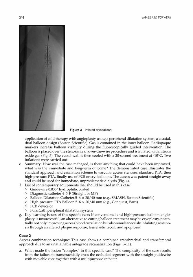
IHBK065-17 IHBK065-Minar April 26, 2010 11:7 Specs: 7x10 tight 7in×10in Char Count=
246 HAAGE AND VORWERK
Figure 3 Inflated cryoballoon.
application of cold therapy with angioplasty using a peripheral dilatation system, a coaxial,dual balloon design (Boston Scientific). Gas is contained in the inner balloon. Radiopaquemarkers increase balloon visibility during the fluoroscopically guided intervention. Theballoon is placed over the stenosis in an over-the-wire procedure and is inflated with nitrousoxide gas (Fig. 3). The vessel wall is then cooled with a 20-second treatment at -10◦C. Twoinflations were carried out.
e. Summary: How was the case managed, is there anything that could have been improved,what was the immediate and long-term outcome? The demonstrated case illustrates thestandard approach and escalation scheme to vascular access stenoses: standard PTA, thenhigh-pressure PTA, finally use of PCB or cryoballoons. The access was patent straight awayand could be used for immediate, unproblematic dialysis (Fig. 4).
f. List of contemporary equipments that should be used in this case:� Guidewire 0.035′′ hydrophilic coated� Diagnostic catheter 4–5-F (Straight or MP)� Balloon Dilatation Catheter 5–6 × 20/40 mm (e.g., SMASH, Boston Scientific)� High-pressure PTA Balloon 5–6 × 20/40 mm (e.g., Conquest, Bard)� PCB device or� PolarCath peripheral dilatation system
g. Key learning issues of this specific case: If conventional and high-pressure balloon angio-plasty is unsuccessful, an alternative to cutting balloon treatment may be cryoplasty, poten-tially not only improving access blood circulation but also simultaneously inhibiting resteno-sis through an altered plaque response, less elastic recoil, and apoptosis.
Case 2Access combination technique: This case shows a combined transbrachial and transfemoralapproach due to an unattainable antegrade recanalization (Figs. 5–11).
a. What made the lesion “complex” in this specific case? The complexity of the case resultsfrom the failure to transbrachially cross the occluded segment with the straight guidewirewith movable core together with a multipurpose catheter.

IHBK065-17 IHBK065-Minar April 26, 2010 11:7 Specs: 7x10 tight 7in×10in Char Count=
DIALYSIS FISTULAS 247
Figure 4 Result after PolarCath treatment.
b. What was the clinical indication for treatment of this specific case? The indication for treat-ment was chronic swelling of the access arm with disabling pain and paresthesia.
c. What were the potential hazards of treating this specific patient and this lesion? Localbleeding at both catheter insertion sites, venous perforation and disruption, early rethrom-bosis, embolism, stent misplacement, immediate or delayed central migration of theendoprosthesis.
d. Which interventional strategies were considered and which interventional tools were usedto handle this lesion? Usually the intervention is performed via an antegrade approachthrough the venous outflow tract in Seldinger technique. Recanalization is attempted with ahydrophilic 0.035-in. guidewire (Terumo, Leuven, Belgium) for vessel stenosis or a straightguidewire with a movable core (Cook, Bjaeverskov, Denmark) for occlusion. Since theobstruction could not be negotiated with the brachial approach (Fig. 5), recanalization wastried transfemorally with a hydrophilic guidewire and a straight 5-F catheter (Fig. 6). Aftersafe retrograde passage of the obstructed segment (Fig. 7), a 6-mm and subsequently 7-mm
Figure 5 DSA shows occlusion of the rightbrachiocephalic vein with pronounced collateralflow.

IHBK065-17 IHBK065-Minar April 26, 2010 11:7 Specs: 7x10 tight 7in×10in Char Count=
248 HAAGE AND VORWERK
Figure 6 Transfemoral occlusion negotiation.
balloon catheter was advanced into the segment via a long sheath and dilated by hand,without pressure monitoring (Figs. 8 and 9). Inflation time each was >30 seconds. Then, aflexible, self-expanding 14/40 mm SMART Control stent (Cordis, Langenfeld, Germany) wasdeployed transbrachially. The stent was dilated with a 10/40 mm and 12/40 mm XXL balloonafter deployment to ensure as close contact to the venous wall as possible (Fig. 10). Restora-tion of flow and vanishing of collateral vessels after placement was achieved (Fig. 11).
e. Summary: How was the case managed, is there anything that could have beenimproved, what was the immediate and long-term outcome? The introduced caseillustrates the standard approach to central venous obstruction and reaction to failure of
Figure 7 After transfemoral occlusion passing.

IHBK065-17 IHBK065-Minar April 26, 2010 11:7 Specs: 7x10 tight 7in×10in Char Count=
DIALYSIS FISTULAS 249
Figure 8 Transfemoral PTA.
this method. If by antegrade approach the lesion cannot be overcome, then the only reason-able alternative is to access transfemorally. In the discussed patient, pain and paresthesiawere reduced to a minimum and the access patent with normal dialysis protocols during anobservation period of 16 months.
f. List of contemporary equipments that should be used in this case:� Guidewire 0.035′′ hydrophilic coated (e.g., Terumo) or straight with movable core� Diagnostic catheter 5-F (Straight or MP)� Guiding catheter 6-F (Straight, e.g., COOK, Denmark)� High-pressure PTA Balloon 6–7 × 40 mm (e.g., POWERFLEX EXTREME, Cordis)� 14/40 mm self-expanding Nitinol stent system (e.g., S.M.A.R.T. CONTROL, Cordis)� Balloon dilatation catheter 10–12 × 40 mm (e.g., Fox, Abbott; XXL, Boston Scientific)
g. Key learning issues of this specific case: In upper torso occlusion, to achieve exact stentplacement a transbrachial approach by antegradely puncturing the outflow vein in Seldinger
Figure 9 Result after transfemoral PTA.

IHBK065-17 IHBK065-Minar April 26, 2010 11:7 Specs: 7x10 tight 7in×10in Char Count=
250 HAAGE AND VORWERK
Figure 10 In-stent PTA immediately after stentplacement.
technique should be used. If the outflow vein is severely kinked or too small and theobstruction cannot be negotiated with a brachial approach, a transfemoral approach is apossible alternative. In this case, primary stent placement is recommended.
Case 3Mechanical thrombectomy and near-anastomosis stent placement: This case shows the ratherexceptional mechanical treatment of partly thrombosed venous aneurysms with additionaloutflow endoprosthesis deployment (Figs. 12–17).
a. What made the lesion “complex” in this specific case? The patient was primarily plannedfor treatment of an outflow stenosis (Fig. 12). After diagnostic angiography and ineffective
Figure 11 Final result shows that unob-structed patency has been re-established.

IHBK065-17 IHBK065-Minar April 26, 2010 11:7 Specs: 7x10 tight 7in×10in Char Count=
DIALYSIS FISTULAS 251
Figure 12 Arterial DSA demonstrates partly thrombosed aneurysm andstenosis in the outflow vein.
PTA due to stenosis recoil, flow enhancement had to be attempted by central thrombusdislocation and additionally flow redirection with stent placement.
b. What was the clinical indication for treatment of this specific case? The initial indication forthe treatment was a malfunctioning AV shunt with declining access flow.
c. What were the potential hazards of treating this specific patient and this lesion? Procedure-related access site bleeding, soaring thrombus formation, complete venous vessel occlusion,dissection or perforation, distal arterial or pulmonary embolism, access infection.
d. Which interventional strategies were considered and which interventional tools were usedto handle this lesion? Typically, the percutaneous intervention is performed via a retrogradeapproach in Seldinger technique for postanastomotic obstructions. Passage is done with ahydrophilic, 0.035-in. guidewire and a diagnostic catheter (Fig. 13), followed by PTA. Inthis setting, angioplasty with 4-mm and 6-mm regular and high-pressure PTA balloons wasineffective and thrombus volume increased (Fig. 14), requiring immediate thrombus mac-eration and dislocation with a rotating 5-F pigtail catheter (Figs. 15 and 16). To circumventthe access abandonment due to complete vessel occlusion, a lineup of four overlappingself-expanding SMART Control stent (Cordis, Langenfeld, Germany) with diameters of 8and 9 mm, respectively, were placed. Ultimately, adequate and unobstructed blood flow wasattained (Fig. 17).
e. Summary: How was the case managed, is there anything that could have been improved,what was the immediate and long-term outcome? The presented case shows the difficul-ties the interventionist may encounter during a supposedly standard PTA. In retrospect, aprimary surgical approach with aneurysm resection would have been a viable alternative.In case of stent placement, however, it became necessary to pave the complete distancefrom the AV anastomosis beyond the venous stenosis. The postinterventional short- andlong-term (15 months restenosis free interval) result was excellent without development ofearly or late complications.
f. List of contemporary equipments that should be used in this case:� Guidewire 0.035′′ hydrophilic coated� Diagnostic catheter 4–5-F (Straight, Aachen 1)� Balloon dilatation catheter 4 + 6 × 20 mm (e.g., SMASH, Boston Scientific)� High-pressure PTA balloon 4 + 6 × 20 mm (e.g., Conquest, Bard)� 5-F pigtail rotating catheter
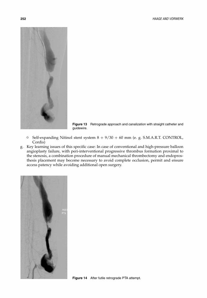
IHBK065-17 IHBK065-Minar April 26, 2010 11:7 Specs: 7x10 tight 7in×10in Char Count=
252 HAAGE AND VORWERK
Figure 13 Retrograde approach and canalization with straight catheter andguidewire.
� Self-expanding Nitinol stent system 8 + 9/30 + 60 mm (e. g. S.M.A.R.T. CONTROL,Cordis)
g. Key learning issues of this specific case: In case of conventional and high-pressure balloonangioplasty failure, with peri-interventional progressive thrombus formation proximal tothe stenosis, a combination procedure of manual mechanical thrombectomy and endopros-thesis placement may become necessary to avoid complete occlusion, permit and ensureaccess patency while avoiding additional open surgery.
Figure 14 After futile retrograde PTA attempt.

IHBK065-17 IHBK065-Minar April 26, 2010 11:7 Specs: 7x10 tight 7in×10in Char Count=
DIALYSIS FISTULAS 253
Figure 15 Manual mechanical thrombus dislocation with rotating pigtailcatheter.
Figure 16 Stent placement covering the PTA-resistant stenosis.

IHBK065-17 IHBK065-Minar April 26, 2010 11:7 Specs: 7x10 tight 7in×10in Char Count=
254 HAAGE AND VORWERK
Figure 17 Unrestricted flow after placement of four overlappingself-expanding stents.
PART 3: KEY ISSUES FOR SUCCESSFUL AND SAFE TREATMENT OF COMPLEXAV FISTULA LESIONS� High-quality treatment of AV fistula dysfunction is a complex undertaking in which the
interventional radiologist, vascular surgeon, and nephrologist should collaborate closely.� The primary percutaneous treatment approach is a very viable alternative to more invasive
procedures. As long as less-invasive equivalent therapeutic alternatives to preserve the AVaccess are feasible, the existing vascular access should be sustained.
� In stenoses and short-segment thromboses, percutaneous transluminal angioplasty is thefirst treatment option.
� In access thrombosis percutaneous thrombectomy, whether pharmacomechanical or purelymechanical, has no major effect on patency. Each type of method for access recanalization issuitable and should be based on the experience of the interventionist with the therapeuticmodality.
� In case of central venous obstruction, treatment should be performed percutaneously byPTA with additional stent implantation, if necessary.
REFERENCES1. U.S. Renal Data System. USRDS 2007 Annual Data Report: Atlas of Chronic Kidney Disease and
End-Stage Renal Disease in the United States. Bethesda, MD: National Institutes of Health, NationalInstitute of Diabetes and Digestive and Kidney Diseases, 2007.
2. Dapunt O, Feurstein M, Rendl KH, et al. Transluminal angioplasty versus conventional operation inthe treatment of haemodialysis fistula stenosis: results from a 5-year study. Br J Surg 1987; 74:1004–1005.
3. McCutcheon B, Weatherford D, Maxwell G, et al. A preliminary investigation of balloon angioplastyversus surgical treatment of thrombosed dialysis access grafts. Am Surg 2003; 69:663–667.
4. Uflacker R, Rajagopalan PR, Selby JB, et al; Investigators of the Clinical Trial Sponsored by MicrovenaCorporation. Thrombosed dialysis access grafts: randomized comparison of the Amplatz thrombec-tomy device and surgical thromboembolectomy. Eur Radiol 2004; 14:2009–2014.
5. Tordoir J, Canaud B, Haage P, et al. EBPG on vascular access. Nephrol Dial Transplant 2007; 22(suppl2):88–117.

IHBK065-17 IHBK065-Minar April 26, 2010 11:7 Specs: 7x10 tight 7in×10in Char Count=
DIALYSIS FISTULAS 255
6. Miyayama S, Matsui O, Taki K, et al. Occluded Brescia-cimino hemodialysis fistulas: endovasculartreatment with both brachial arterial and venous access using the pull-through technique. CardiovascIntervent Radiol 2005; 28:806–812.
7. Beathard GA, Litchfield T; Physician Operators Forum of RMS Lifeline, Inc. Effectiveness and safetyof dialysis vascular access procedures performed by interventional nephrologists. Kidney Int 2004;66:1622–1232.
8. Turmel-Rodrigues L, Pengloan J, Baudin S, et al. Treatment of stenosis and thrombosis inhaemodialysis fistulas and grafts by interventional radiology. Nephrol Dial Transplant 2000; 15:2029–2036.
9. Duijm LE, Liem YS, van der Rijt RH, et al. Inflow stenoses in dysfunctional hemodialysis access fistulaeand grafts. Am J Kidney Dis 2006; 48:98–105.
10. Turmel-Rodrigues L, Pengloan J, Blanchier D, et al. Insufficient dialysis shunts: improved long-termpatency rates with close hemodynamic monitoring, repeated percutaneous balloon angioplasty, andstent placement. Radiology 1993; 187:273–278.
11. Vorwerk D, Adam G, Muller-Leisse C, et al. Hemodialysis fistulas and grafts: use of cutting balloonsto dilate venous stenoses. Radiology 1996; 201:864–867.
12. Vorwerk D. Non-traumatic vascular emergencies: management of occluded hemodialysis shunts andvenous access. Eur Radiol 2002; 12:2644–2650.
13. Haage P, Vorwerk D, Wildberger JE, et al. Percutaneous treatment of thrombosed primary arteriove-nous hemodialysis access fistulae. Kidney Int 2000; 57:1169–1175.
14. Bush RL, Lin PH, Lumsden AB. Management of thrombosed dialysis access: thrombectomy versusthrombolysis. Semin Vasc Surg 2004; 17:32–39.
15. Gray RJ. Percutaneous intervention for permanent hemodialysis access: a review. J Vasc Interv Radiol1997; 8:313–327.
16. Glanz S, Gordon DH, Butt K, et al. The role of percutaneous angioplasty in the management of chronichemodialysis fistulas. Ann Surg 1987; 206:777–781.
17. Schwab SJ, Quarles LD, Middleton JP, et al. Hemodialysis-associated subclavian vein stenosis. KidneyInt 1988; 33:1156–1159.
18. Ingram TL, Reid SH, Tisnado J, et al. Percutaneous transluminal angioplasty of brachiocephalic veinstenoses in patients with dialysis shunts. Radiology 1988; 166:45–47.
19. Haage P, Vorwerk D, Piroth W, et al. Treatment of hemodialysis-related central venous stenosis orocclusion: results of primary wallstent placement and follow-up in 50 patients. Radiology 1999; 212:175–180.
20. Hingorani A, Ascher E, Kallakuri S, et al. Impact of reintervention for failing upper-extremity arteri-ovenous autogenous access for hemodialysis. J Vasc Surg 2001; 34:1004–1009.
21. Liang HL, Pan HB, Chung HM, et al. Restoration of thrombosed Brescia-Cimino dialysis fistulas byusing percutaneous transluminal angioplasty. Radiology 2002; 223:339–344.
22. Mansour M, Kamper L, Altenburg A, et al. Radiological central vein treatment in vascular access. JVasc Access 2008; 9:85–101.
23. Bakken AM, Protack CD, Saad WE, et al. Long-term outcomes of primary angioplasty and primarystenting of central venous stenosis in hemodialysis patients. J Vasc Surg 2007; 45:776–783.
24. Chen CY, LiangG HL, Pan HB, et al. Metallic stenting for treatment of central venous obstruction inhemodialysis patients. J Chin Med Assoc 2003; 66:166–172.
25. Mickley V. Stent or bypass? Treatment results in benign central venous obstruction. Zentralbl Chir2001; 126:445–449.
26. Oderich GS, Treiman GS, Schneider P, et al. Stent placement for treatment of central and peripheralvenous obstruction: a long-term multi-institutional experience. J Vasc Surg 2000; 32:760–769.
27. Mickley V, Gorich J, Rilinger N, et al. Stenting of central venous stenoses in hemodialysis patients:long-term results. Kidney Int 1997; 51:277–280.
28. Vorwerk D, Guenther RW, Mann H, et al. Venous stenoses and occlusions in hemodialysis shunts:follow-up results of stent placement in 65 patients. Radiology 1995; 195;140–146.
29. Quinn SF, Schuman ES, Demlow TA, et al. Percutaneous transluminal angioplasty versus endovascularstent placement in the treatment of venous stenoses in patients undergoing hemodialysis: intermediateresults. J Vasc Interv Radiol 1995; 6:851–855.
30. Shoenfeld R, Hermans H, Novick A, et al. Stenting of proximal venous obstructions to maintainhemodialysis access. J Vasc Surg 1994; 19:532–538.
31. Bhatia DS, Money SR, Ochsner JL, et al. Comparison of surgical bypass and percutaneous balloondilatation with primary stent placement in the treatment of central venous obstruction in the dialysispatient: one-year follow-up. Ann Vasc Surg 1996; 10:452–455.
32. Maya ID, Saddekni S, Allon M. Treatment of refractory central vein stenosis in hemodialysis patientswith stents. Semin Dial 2007; 20:78–82.

IHBK065-17 IHBK065-Minar April 26, 2010 11:7 Specs: 7x10 tight 7in×10in Char Count=
256 HAAGE AND VORWERK
33. Levit RD, Cohen RM, Kwak A, et al. Asymptomatic central venous stenosis in hemodialysis patients.Radiology 2006; 238:1051–1056.
34. Surowiec SM, Fegley AJ, Tanski WJ, et al. Endovascular management of central venous stenosesin the hemodialysis patient: results of percutaneous therapy. Vasc Endovasc Surg 2004; 38:349–354.
35. Kalman PG, Lindsay TF, Clarke K, et al. Management of upper extremity central venous obstructionusing interventional radiology. Ann Vasc Surg 1998; 12;202–206.

IHBK065-18 IHBK065-Minar April 26, 2010 11:26 Specs: 7x10 tight 7in×10in Char Count=
18 Venous Interventions for Iliofemoral DVTAnthony J. Comerota and Marilyn H. GravettJobst Vascular Center, Toledo, Ohio, U.S.A.
PART 1: INTRODUCTIONAcute deep venous thrombosis (DVT) represents a disease spectrum ranging from asymp-tomatic calf vein thrombosis to the painful swollen limb of phlegmasia cerulea dolens resultingfrom extensive multisegment DVT. Although patient presentation and extent of venous throm-bosis vary considerably, national and international guidelines for the treatment of acute DVThad, until recently, demonstrated a “one-size-fits-all” approach by recommending a singletreatment—anticoagulation alone—for all variants of DVT. The latest published guidelines ofthe American College of Chest Physicians (ACCP) Consensus Conference on Antithromboticand Thrombolytic Therapy include recommendations that depart significantly from previousversions (1). In the guideline, authors recognized the link between extensive venous thrombosisand severe long-term postthrombotic morbidity and acknowledged the existing evidence sup-porting a strategy of thrombus removal using treatment techniques that include contemporaryvenous thrombectomy, catheter-directed thrombolysis, and pharmacomechanical thrombolysis.
What Makes a Lesion Complex in Venous Interventions?The departure from anticoagulation alone to adopt a strategy of thrombus removal is whatmakes this approach complex. It is basically the physician’s misunderstanding of the patho-physiology of postthrombotic venous disease that leads to its severe morbidity. Physicians oftenfail to recognize that luminal venous obstruction resulting from persistent residual thrombus isthe major contributor to ambulatory venous hypertension, and hence the clinical morbidity ofchronic venous disease. This obstruction can be efficiently eliminated early during the phase ofacute DVT; however, recanalization of the chronically occluded venous system is not associatedwith the same ease of treatment or the same degree of success.
Key factors contributing to the revision of the ACCP guidelines were studies demonstrat-ing that patients with postthrombotic syndrome suffer significant reduction in their quality oflife (QOL) and that the severity of their initial DVT is predictive of postthrombotic morbidity(2,3). Nowhere are these outcomes more evident than in patients with iliofemoral DVT, a clin-ically relevant subset of patients with acute DVT who suffer the most severe postthromboticsequelae (4–6). If iliofemoral DVT patients are treated with anticoagulation alone, 90% willhave ambulatory venous hypertension resulting in venous insufficiency, 40% will experiencevenous claudication, and up to 15% will develop venous ulceration within five years (5,6). Allof these patients will have reduced QOL (7). The body of evidence demonstrates that a strategyof thrombus removal offers patients with iliofemoral DVT the best long-term outcome and istherefore the preferred management for their disease.
Unfortunately, many physicians fail to discern the difference in pathophysiology betweenprimary and postthrombotic venous insufficiency and consequently underestimate the valueof thrombus removal in preventing postthrombotic morbidity, especially in patients withiliofemoral disease. The pathophysiology of postthrombotic venous insufficiency is ambulatoryvenous hypertension, which is defined as an elevated venous pressure during exercise(8,9). Key anatomical components that contribute to ambulatory venous hypertension arevenous obstruction and venous valvular incompetence. Studies demonstrate that patients withchronic venous obstruction, particularly those with multisegment and/or iliofemoral venousobstruction, have the most severe postthrombotic sequelae (8,10). Although ultrasonographycan assess valvular function by quantifying valve closure times, obstruction adverselyaffects venous hemodynamics long before imaging techniques can detect it. The inability toquantitate obstruction has contributed to widespread underappreciation of its contribution topostthrombotic pathophysiology.

IHBK065-18 IHBK065-Minar April 26, 2010 11:26 Specs: 7x10 tight 7in×10in Char Count=
258 COMEROTA AND GRAVETT
Figure 1 Algorithm illustrating our current treatment protocol for patients with iliofemoral DVT.
Experimental canine studies of acute DVT have shown that successful thrombolysis pre-serves endothelial function and valve competence (11,12). Natural history studies of acute DVThave shown that patients with persistent venous obstruction treated with anticoagulation alonedeveloped distal valve incompetence, even when distal veins were not involved at onset withthrombus (13–15). Moreover, if spontaneous lysis occurred early (clot lysis within 90 days), valvefunction was frequently preserved (15). It is intuitive that successfully removing acute throm-bus eliminates luminal obstruction and, if thrombus is eliminated early (within days/weeks),preserves valve function.
Advances in all methods of eliminating thrombus from the deep venous system haveoccurred in the last 10 to 15 years, and this chapter will review three key strategies: (i) con-temporary venous thrombectomy, (ii) intrathrombus catheter-directed thrombolysis, and (iii)pharmacomechanical thrombolysis.
Clinical Indications for Treatment of Complex Lesions in Venous InterventionsAll patients with iliofemoral DVT should be evaluated and considered for a strategy of thrombusremoval. Assuming the patient is ambulatory, contraindications to either surgical or catheter-based treatment should be carefully assessed. The algorithm shown in Figure 1 illustrates ourcurrent management strategy for patients with iliofemoral DVT and provides a decision matrixfor selecting the appropriate treatment strategy.
Patients who have occlusive thrombus of the common femoral vein have essentially oblit-erated venous drainage from their lower extremity, which leads to severe postthrombotic mor-bidity. Although most individuals diagnosed with acute DVT can be treated as outpatients, thesepatients should be admitted and considered for a treatment strategy that includes thrombusremoval. Another subset of patients likely to suffer severe postthrombotic morbidity are thosewith thrombotic obliteration of their popliteal vein and adjoining proximal tibial veins. Becauseadequate collaterals do not develop, these patients incur severe distal venous hypertension.

IHBK065-18 IHBK065-Minar April 26, 2010 11:26 Specs: 7x10 tight 7in×10in Char Count=
VENOUS INTERVENTIONS FOR ILIOFEMORAL DVT 259
Table 1 Evidence-Based Recommendations for the Treatment of Patients with Iliofemoral DVT (1)
Recommendation Grade
In patients with extensive (iliofemoral) DVT, operative venous thrombectomy may be used toreduce acute symptoms and postthrombotic morbidity
2B
Following venous thrombectomy, the same duration and intensity of anticoagulation shouldbe used as in those who do not undergo thrombectomy
1C
In patients with extensive proximal DVT and low risk for bleeding, catheter-directedthrombolysis (CDT) can be used to reduce acute symptoms and postthrombotic morbidity
2B
Pharmacomechanical thrombolysis should be considered in preference to CDT alone 2CFollowing CDT, underlying venous lesions should be corrected 2CFollowing CDT, the same intensity and duration of anticoagulation should be used for those
who are not treated with CDT1C
Patients with acute DVT should be treated with a 30–40 mm Hg compression stocking toreduce postthrombotic morbidity
1A
The 8th ACCP guidelines on antithrombotic therapy recommend catheter-directed throm-bolysis for patients with acute iliofemoral DVT who are appropriate risk candidates (Table 1).The writing committee emphasized the importance of correcting underlying venous lesionsfollowing successful thrombus removal and noted that, when using catheter-directed throm-bolysis, pharmacomechanical techniques may be useful to shorten treatment time. They furtheremphasized that the same intensity and duration of anticoagulant therapy be given to patientstreated with thrombolysis and venous thrombectomy as those treated with anticoagulationalone, underscoring the value of ongoing therapeutic anticoagulation to reduce the risk ofrethrombosis.
Contemporary venous thrombectomy is recommended as an option for those patientswho have contraindications to thrombolysis or in medical communities where catheter-basedtechniques are not available. There are few contraindications to operative thrombectomy, oldthrombus (≥10 days) that adheres to the vein wall being the most important.
Patients with spontaneous and extensive venous thrombosis often have a clinically obvi-ous etiology, such as trauma, an underlying thrombophilia, or an occult cancer. Earlier in ourexperience we routinely performed a complete thrombophilia evaluation; however, today webelieve it is less important and not necessary for proper treatment of the patient during bothacute and follow-up phases. The implications of a thrombophilia evaluation should be carefullyconsidered, as some patients may face health insurance restrictions, increased premiums, andperhaps employment restrictions. However, young patients who intend to have children maybenefit from such an evaluation, as the results may be relevant for family planning as well asfuture care. Similarly, a family history of venous thromboembolic disease suggests the need fora thrombophilia evaluation.
Both the proximal and distal extent of thrombus should be clearly defined in all patients,including imaging of the vena cava. If a patient has nonocclusive vena caval thrombus, werecommend a vena caval filter for those undergoing catheter-based techniques. If operating,either proximal balloon occlusion or filtration of the cava is warranted.
For patients with normal renal function, we strongly recommend a computed tomographic(CT) scan of the head, chest, abdomen, and pelvis with contrast. In at least 50% of patients withacute iliofemoral DVT, an asymptomatic pulmonary embolism (PE) will be found. This may notrequire immediate therapy but the diagnosis of PE will be appreciated in the subsequent three- tofive-day period, when up to 25% of these patients develop pleuritic chest pain (16). Awarenessof the presence of an asymptomatic PE when the patient presents permits the physician toconfidently treat the patient, knowing that delayed symptoms of pleuritic irritation were neithera complication of the strategy of thrombus removal nor a failure of anticoagulation.
Hazards and ComplicationsMany physicians are understandably concerned about the risk of procedure-related PE. Patientswith nonobstructive thrombus in their vena cava have the highest risk of procedure-related PE(Case #1), so it is vital to determine both proximal and distal extent of thrombus, with particular

IHBK065-18 IHBK065-Minar April 26, 2010 11:26 Specs: 7x10 tight 7in×10in Char Count=
260 COMEROTA AND GRAVETT
attention paid to the amount and level of vena caval involvement. Knowing this will assist indeciding whether or not vena caval filtration or another form of embolic protection should beused. Nonocclusive thrombus in the vena cava is a clear indication for a vena caval filter beforecatheter-directed thrombolysis and proximal caval protection during venous thrombectomy.
Traditional concerns about bleeding risk with thrombolytic agents are reduced by the useof intrathrombus infusion as well as adjunctive pharmacomechanical techniques. Reducing thedose of lytic agent as well as the duration of treatment lessens the systemic response to theinfused plasminogen activator (17).
Interventional StrategiesContemporary venous thrombectomy has the potential of offering patients with acute iliofemoralDVT an opportunity for rapid removal of thrombus and restoration of normal venous drainage,resulting in a significant reduction in postthrombotic morbidity. The long-term benefits ofvenous thrombectomy relate to its ability to restore venous patency, especially of the iliofemoralsegment, and maintain distal valve competence. Both are influenced by initial technical successand avoiding recurrent thrombosis. Initial success in achieving patency is, in turn, influencedby timely intervention and attention to technical detail.
Intrathrombus catheter-directed thrombolysis evolved from earlier attempts to use systemicthrombolysis as treatment for acute DVT. Although systemic thrombolysis removed thrombusmore effectively than anticoagulation alone, results were less than optimal (18), and highrates of bleeding complications occurred. Because plasminogen activators were administeredintravenously, they failed to adequately penetrate thrombus, leading to higher rates of lyticfailure (19).
The essential mechanism of thrombolysis is the activation of fibrin-bound plasmino-gen to form the active enzyme plasmin, which dissolves thrombus (20). During thrombosis,circulating Glu-plasminogen binds to fibrin and is converted to Lys-plasminogen within thethrombus, which has greater affinity for plasminogen activators. The intrathrombus delivery ofplasminogen activators activates Lys-plasminogen to form plasmin. Moreover, intrathrombustechniques protect the plasminogen activators from neutralization by circulating plasminogenactivator inhibitor (PAI-1) and protect the generated plasmin from immediate neutralizationby circulating antiplasmins. Finally, infusing plasminogen activators directly into thrombusreduces both the dose and duration necessary to achieve lytic success, thereby reducing thelikelihood of complications.
Pharmacomechanical thrombolysis, one of the newer strategies for thrombus removal,involves adding pharmacomechanical techniques to catheter-directed thrombolysis. Pharma-comechanical therapies have the potential to significantly shorten treatment times and reducethe amount of lytic agent required to achieve lysis, thereby making treatment both more cost-effective and safer for patients.
Interventional ToolsMechanical techniques used alone or in conjunction with thrombolysis to clear the venous sys-tem more rapidly have demonstrated varying results. Vedantham et al. (21) reviewed the effec-tiveness of several mechanical devices in combination with thrombolysis in 28 patients. Devicesused included the Amplatz (ev3, Inc, Plymouth, MN), AngioJet R© (Possis Medical, Minneapolis,MN), Trerotolo (Arrow International, Reading, PA), and Oasis (Boston Scientific/Medi-tech,Natick, MA) catheter systems. Venographic scoring was performed throughout the procedures.When mechanical thrombectomy was used as the sole treatment, only 26% of thrombus wasremoved, whereas 82% of thrombus was removed when a plasminogen activator solutionwas added (including nonresponding patients with chronic occlusions). Mechanical thrombec-tomy alone was successful when used to eliminate intraprocedural thrombus, which is typi-cally gelatinous and not crosslinked with fibrin. Overall infusion time averaged approximately17 hours and major bleeding complications occurred in 14% of patients.
An eight-year experience using pharmacomechanical thrombolysis in 98 patientswas reported by Lin et al. (22) Forty-six patients received catheter-directed thrombolysiswhile 52 received pharmacomechanical thrombolysis using the AngioJet rheolytic thrombec-tomy catheter. Those undergoing pharmacomechanical treatment had significantly fewer

IHBK065-18 IHBK065-Minar April 26, 2010 11:26 Specs: 7x10 tight 7in×10in Char Count=
VENOUS INTERVENTIONS FOR ILIOFEMORAL DVT 261
Figure 2 The Trellis-8 peripheral infusion sys-tem (Bacchus Vascular, Santa Clara, CA) deliv-ers the lytic agent into the thrombus segmentedbetween two occluding balloons. A dispersionwire inserted into the center of the catheterassumes a spiral configuration, dispenses thelytic agent, and mechanically macerates thethrombus. The resulting liquefied and small par-ticulate thrombus is aspirated to minimize oravoid a systemic lytic effect.
phlebograms, reduced ICU and hospital stays, and fewer blood transfusions. Kasirajan et al. (23)treated a small patient group with rheolytic thrombectomy and demonstrated that mechanicalthrombectomy was less effective when used alone than when combined with a plasminogenactivator solution.
An interesting new pharmacomechanical catheter, the EKOS R© EndoWaveTM system (Both-ell, WA), uses ultrasound-accelerated thrombolysis to remove acute thrombus. Parikh et al. (24)recently reported their initial clinical experience in 52 patients with both upper and lowerextremity acute DVT. Using a variety of lytic agents, they reported that complete lysis (≥90%)was observed in 70% of patients, with overall lysis (complete plus partial) in 91%. Only 4% ofpatients had major complications (puncture site hematomas) and median infusion time was 22hours.
One of the more promising pharmacomechanical techniques is isolated, segmental phar-macomechanical thrombolysis (ISPMT), which is achieved using the Trellis R© catheter (BacchusVascular, Santa Clara, CA). This double-balloon catheter (Fig. 2) is guided into the thrombosedvenous segment, with the proximal balloon positioned at the upper edge of the thrombus. Oncethe balloons are inflated, a plasminogen activator is infused into the thrombosed segment thathas been isolated by the balloons. The intervening catheter assumes a spiral configuration androtates at 1500 rpms for approximately 15 to 20 minutes, followed by aspiration of the lique-fied and fragmented thrombus. If successful, the catheter is repositioned to treat additionalsegments. However, if residual thrombus persists, repeat treatment or adjunctive interventions(rheolytic thrombectomy, ultrasound-accelerated thrombolysis, balloon angioplasty, or stenting)are performed.
Summary of OutcomesThe pooled data shown in Table 2 (25–31) illustrate patency rates achieved with venousthrombectomy. Recent improvements to the technique include the use of the venous thrombec-tomy catheter (large balloon), fluoroscopic guidance, completion intraoperative phlebography,correction of an underlying venous stenosis, construction of an arteriovenous fistula (AVF), andimmediate and prolonged therapeutic anticoagulation. These additions should substantiallyimprove acute and long-term results.
Table 2 Venous Thrombectomy with Arteriovenous Fistula: Long-Term Valve Competence of FemoropoplitealVenous Segment
References No. Follow-up (mo) Femoral-popliteal valve competence
25 31 6 5226 53 10 4227 17 91 8228 37 24 5629 37 55 8030 150 60 8031 27 60 30Total 352 45 mo (mean) 63% (mean)

IHBK065-18 IHBK065-Minar April 26, 2010 11:26 Specs: 7x10 tight 7in×10in Char Count=
262 COMEROTA AND GRAVETT
Table 3 Efficacy and Complications of Catheter-Directed Thrombolysis in Three Series
Bjarnason et al.(33) (n = 77) (%)
Mewissen et al.(34) (n = 287) (%)
Comerota and Kagan(35) (n = 58) (%)
Initial success 79 83 84Iliac 63 64 78Femoral 40 47 —
Primary patency at 1 yrIliac 63 64 78Femoral 40 47 —
Iliac stent: patency at 1 yr+ stent 54 74 89− stent 75 53 71
ComplicationsMajor bleed 5 11 9Intracranial bleeding 0 <1 0Pulmonary embolism 1 1 0Fatal pulmonary embolism 0 0.2 0Death secondary to lysis 0 0.4 0%
(?2%)a
aDeath due to multiorgan system failure 30 days post lysis, thought not related to lytic therapy.
Many investigators have reported good outcomes of catheter-directed thrombolysis foracute DVT; these have been previously discussed in detail (32). Three large reports (422 patients)demonstrated success rates of approximately 80% [Table 3 (33–35)], rates that likely would havebeen higher had the studies treated only patients with acute iliofemoral DVT. As investigatorsdeveloped confidence with the technique, more patients were referred with severe symptomslonger after their acute presentation; the less than optimal results are included as a reflectionof treating older (>1 month) thrombus. Bleeding complications occurred in 5% to 10%, mostcommonly at the venous access site. Intracranial bleeding and fatal PE were rare, occurring inonly four patients in these three series.
Early studies using ISPMT demonstrate reduced treatment times and dose of lytic agents.Martinez et al. (17) reported on 52 consecutive limbs treated for iliofemoral DVT, 27 of whichwere treated with catheter-directed thrombolysis and 25 with ISPMT plus catheter-directedthrombolysis when necessary. Treatment outcomes and thrombus burden were quantified. Most(93%) were treated with recombinant tissue plasminogen activator (rt-PA), and all underwentvenoplasty and stenting to correct underlying stenoses. All were treated with long-term thera-peutic anticoagulation. Of the 27 legs treated with catheter-directed thrombolysis, 16 requiredadjunctive mechanical treatment to clear the thrombus; of the 25 limbs treated with ISPMT,only 7 required additional techniques. Moreover, a larger percentage of thrombus was removedwith ISPMT. Complete lysis (≥ 90%) was achieved in 11% of limbs receiving catheter-directedthrombolysis compared with 28% in ISPMT-treated limbs (P = 0.077). Treatment duration wasdecreased (23.4 vs. 55.4 hours, P < 0.001) and rt-PA dose reduced (33.4 vs. 59.3 mg, P = 0.009)with ISPMT. Bleeding complications occurred in 5% of both groups.
PART 2: TEACHING CASES
Case 1: Contemporary Venous Thrombectomy
What Made the Case Complex?A 24-year-old woman who was 32 weeks pregnant presented to the emergency department inthe evening with a swollen, painful left lower extremity and right-sided pleuritic chest pain. Herleft leg had become progressively more symptomatic during the past three days and she hadbegun to feel lethargic, short of breath, and was experiencing right chest discomfort with deepbreathing. She was a nonsmoker with no family or personal history of venous thromboembolicdisease. Upon physical examination, the patient had a swollen, cyanotic left leg that was tender

IHBK065-18 IHBK065-Minar April 26, 2010 11:26 Specs: 7x10 tight 7in×10in Char Count=
VENOUS INTERVENTIONS FOR ILIOFEMORAL DVT 263
Figure 3 An iliocavagram performed to evaluate the proximalextent of thrombus shows clot extending from the common iliacvein into the vena cava and up to the level of the renal veins.
to palpation, especially palpating the common femoral vein. Her left thigh and calf measured73 cm and 45 cm, compared to 64 cm and 40 cm on the right.
What Was the Clinical Indication for Treatment of This Patient?This patient’s presentation was clinically consistent with iliofemoral DVT associated with a PE.The adventitia of the femoral vein is innervated with sensory nerves; therefore, pain to palpationof the femoral vein as a result of its distension is a frequent physical finding. The femoral veindistends as a result of the associated venous hypertension and thrombosis.
The patient was started on intravenous heparin. A venous duplex scan revealed thrombo-sis of the left external iliac, common femoral, femoral, popliteal, posterior tibial, and proximalgreat saphenous veins. A subsequent iliocavagram was performed to evaluate the proximalextent of thrombus and potential vena caval involvement. It confirmed thrombosis of the leftcommon iliac vein and extensive clot in the vena cava extending to the level of the renalveins (Fig. 3).
What Were the Potential Hazards of Treating This Patient and Anatomy?Patients presenting during off hours to the emergency department who are at high clinical riskof a venous thromboembolic condition should be anticoagulated until a definitive diagnosis ismade. A CT scan of the chest or ventilation/perfusion (V/Q) lung scan was not performed inthis patient because she was pregnant and the clinical probability of a PE was high. Since thispatient is being managed for acute DVT, her treatment would not be altered by the V/Q scanfindings. There is also reluctance to expose the pregnant patient to the excessive radiation of CTscans or a radioisotope.
Which Interventional Strategies Were Considered?Standard ascending phlebography was not necessary, since the clinical presentation and venousduplex established the diagnosis with a high degree of accuracy. Once anticoagulation wasstarted and therapeutic, it was not necessary and actually counterproductive to maintain thepatient at bed rest. An echocardiogram is advisable in all patients who have the diagnosis of PEto evaluate its impact on right ventricular function; however, it was not necessary in this patient

IHBK065-18 IHBK065-Minar April 26, 2010 11:26 Specs: 7x10 tight 7in×10in Char Count=
264 COMEROTA AND GRAVETT
to perform an “off hours” echocardiogram since the patient had no evidence of hemodynamiccompromise and could wait to be properly treated until the next business day.
A retrievable Gunther Tulip filter (William Cook, Europe) was placed in the suprarenalinferior vena cava (IVC) under fluoroscopic guidance. Post procedure, a discussion was heldwith the patient and family to review alternative treatment to eliminate thrombus, includ-ing catheter-directed thrombolysis, pharmacomechanical thrombolysis, and operative venousthrombectomy. The decision was made to proceed with venous thrombectomy.
SummaryThe patient was taken to the operating room for a venous thrombectomy under fluoroscopicguidance with fetal monitoring. A cut-down was performed on the left common femoral andfemoral veins, with exposure of the saphenofemoral junction. A longitudinal venotomy wasperformed at the level of the saphenofemoral junction, followed by protrusion of a large amountof acute thrombus (Fig. 4). The leg was raised and a tight rubber bandage applied with minimalextrusion of the infrainguinal thrombus. Attempts to pass a catheter from the inguinal ligamentdistally into the femoral vein and attempts to pass a guidewire distally were unsuccessful.
A cut-down on the posterior tibial vein in the lower leg was performed. Following aposterior tibial venotomy, a #3 Fogarty catheter was passed upwards through the thrombosedvenous system, exiting the common femoral venotomy. This catheter was used to guide a #4Fogarty catheter distally through the venous valves by placing both catheter tips within a 14-gauge silastic IV catheter sheath after the hub was amputated. Following a mechanical ballooncatheter thrombectomy, the leg was flushed with a heparin saline solution using a bulb syringe,which flushed additional thrombus from the common femoral venotomy. After clamping thefemoral vein, the deep venous system was then filled with 300 cc of a dilute recombinant tissueplasminogen solution (6 mg rt-PA in 300 cc).
The iliofemoral and vena caval thrombectomy was performed under fluoroscopic guid-ance, filling the balloon with contrast to ensure that the suprarenal caval filter was not dislodged.After completing the thrombectomy, an operative iliocavagram was performed to assess the ade-quacy of thrombectomy and to ensure unobstructed venous drainage into the vena cava. Aniliac vein stenosis was observed.
A balloon angioplasty catheter was placed into the lesion and an angioplasty performed.The iliac vein was dilated to 14 mm without evidence of recoil.
An AVF using the proximal saphenous vein anastomosed to the superficial femoral arteryincreases flow velocity through the iliofemoral venous system, reducing the risk of rethrombosis.
(A)
(B)
Figure 4 (A) A venotomy performed at thelevel of the saphenofemoral junction is followedby protrusion of a large amount of acute throm-bus. (B) Thrombus removed from iliofemoralvenous system during thrombectomy.

IHBK065-18 IHBK065-Minar April 26, 2010 11:26 Specs: 7x10 tight 7in×10in Char Count=
VENOUS INTERVENTIONS FOR ILIOFEMORAL DVT 265
Figure 5 Following venous thrombectomy, an AVF was con-structed to increase the flow velocity through the iliofemoralvenous system, reducing the risk of rethrombosis. A smallpiece of PTFE wrapped around the saphenous AVF andlooped with O-Prolene served as a guide should the AVFrequire closure in the future.
A thrombectomy of the proximal great saphenous vein was required in this patient, as is oftenthe case. Since the goal of the AVF is to increase venous blood flow velocity but not venouspressure, the size of the anastomosis is limited to 3.5 to 4 mm, in order to avoid a steal and avoidvenous hypertension. A small piece of polytetrafluoroethylene (PTFE) is wrapped around thesaphenous AVF and looped with a 2-cm piece of O-Prolene, which is left in the subcutaneoustissue (Fig. 5). This will serve as a guide should the AVF require operative closure in the future.However, since the AVF is small, it is considered permanent and closure is not anticipatedand rarely required. Common femoral venous pressure is recorded before and after the AVF isopened. It should show no change. If the AVF increases femoral venous pressure, an iliac veinstenosis should be suspected, or the AVF is too large.
To further reduce the risk of rethrombosis, a heparin infusion catheter (pediatric feedingtube) is placed into the proximal posterior tibial vein and brought out through a separatestab wound adjacent to the lower leg incision. Infusing unfractionated heparin through thiscatheter to achieve a therapeutic PTT ensures a high concentration of heparin in the targetvein, a concentration much higher than would be achieved if the patient was treated withstandard IV anticoagulation through an arm vein. A monofilament suture is looped around theposterior tibial vein cephalad to catheter entry and brought out through the skin and securedwith a sterile button. This is used to occlude the vein after five to six days when the catheter isremoved following full oral anticoagulation with warfarin.
In the case of this pregnant patient, IV anticoagulation through the leg veins was main-tained for four days, after which she was converted to subcutaneous enoxaparin at 1 mg/kgq12 hours. The catheter was removed and the patient discharged. The patient was maintainedon S.Q. enoxaparin 1 mg/kg BID until she delivered a healthy baby six weeks later.
Case 2: Catheter-Directed Thrombolysis
What Made the Case Complex?A 37-year-old gentleman was referred for left lower extremity phlegmasia cerulea dolens eightdays after undergoing an uneventful elective total colectomy for ulcerative colitis.

IHBK065-18 IHBK065-Minar April 26, 2010 11:26 Specs: 7x10 tight 7in×10in Char Count=
266 COMEROTA AND GRAVETT
Figure 6 Contralateral iliocavagrams demon-strated nonocclusive thrombus in the vena cava,illustrating importance of identifying the proximalextent of thrombus.
What Was the Clinical Indication for Treatment of This Patient?An ascending phlebogram showed thrombus in the femoral vein, common femoral vein, andiliofemoral venous system of his left leg. A contralateral iliocavagram illustrated nonocclusivethrombus of his vena cava (Fig. 6)
What Were the Potential Hazards of Treating This Patient and Anatomy?Patients at highest risk of procedure-related PE are those with nonocclusive thrombus in theirvena cava, as was revealed by this patient’s iliocavagrams. It is important to image the proximaland distal extent of thrombus, and particularly the amount and level of vena caval involvement.A vena caval filter (Fig. 7) was placed in this patient to prevent a large PE from occurring duringcatheter-directed thrombolysis.
Figure 7 A Bird’s nest filter was placed in the IVC to prevent alarge PE from occurring during catheter-directed thrombolysis.

IHBK065-18 IHBK065-Minar April 26, 2010 11:26 Specs: 7x10 tight 7in×10in Char Count=
VENOUS INTERVENTIONS FOR ILIOFEMORAL DVT 267
Figure 8 Completion cavagram revealed trapped thrombusinside the IVC filter, preventing a pulmonary embolus.
Which Interventional Tools Were Used to Manage This Case?Catheter-directed thrombolysis was begun by accessing the left popliteal vein via an ultrasound-guided popliteal vein puncture. Long-segment side-hole infusion catheters were placed into theacute thrombus in the femoral vein and iliofemoral venous system.
SummaryLytic therapy was successful; however, it demonstrated that there was compression of the leftcommon iliac vein as it is crossed by the right common iliac artery, representing a typicalMay–Thurner picture. Venoplasty and stenting were performed to restore unobstructed venousdrainage into the vena cava.
A completion cavagram demonstrated that the IVC filter indeed trapped an embolus intransit (Fig. 8).
The patient was subsequently treated with therapeutic anticoagulation using unfraction-ated heparin converted to oral anticoagulation with Coumadin targeting an international nor-malized ratio (INR) of 2.5. The patient was placed in a 30 to 40 mm Hg ankle gradient compres-sion stocking and discharged.
On follow-up, the patient had minimal if any swelling, even when not wearing his com-pression stockings. The lower extremity venous system remained patent with valve function inthe borderline normal range [venous valve closure times of 0.5–0.7 seconds and a venous fillingindex on air plethysmography (APG) of 2.2].
Case 3: Pharmacomechanical Thrombolysis
What Made the Case Complex?A sixty-eight-year-old male was referred with phlegmasia cerulea dolens one day after a majorabdominal operation.
What Was the Clinical Indication for Treatment of This Patient?Venous ultrasound demonstrated extensive venous thrombosis extending from the calf veins tothe vena cava, which was confirmed with ascending phlebography (Fig. 9).

IHBK065-18 IHBK065-Minar April 26, 2010 11:26 Specs: 7x10 tight 7in×10in Char Count=
268 COMEROTA AND GRAVETT
Figure 9 Phlebography illustrated extensive venous thrombosis that extended from the calf to the vena cava.
What Were the Potential Hazards of Treating This Patient and Anatomy?A CT scan showed PE and extensive mediastinal and retroperitoneal lymphadenopathy. CTscans of the abdomen and pelvis also demonstrated retroperitoneal lymphadenopathy consis-tent with lymphoma.
Which Interventional Tools Were Used to Manage This Case?The iliofemoral thrombosis was treated expediently with the Trellis catheter (Fig. 10), usingISPMT with 15-minute runs, aspirating liquified thrombus and residual thrombus after eachrun. The thrombus extending from the posterior tibial vein through the popliteal and into thefemoral vein was treated with ultrasound-accelerated thrombolysis with rt-PA after passing anEKOS catheter from the posterior tibial vein at the ankle into the distal femoral vein. Tissue
Figure 10 Both the Trellis and EKOS infusion catheterswere used to eliminate clot as part of a strategy of throm-bus removal. The Trellis catheter was used on thrombus inthe femoral and iliac veins, and the EKOS catheter-treatedclot in the posterior tibial and popliteal veins.
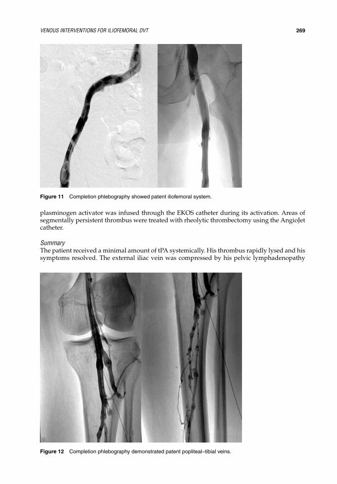
IHBK065-18 IHBK065-Minar April 26, 2010 11:26 Specs: 7x10 tight 7in×10in Char Count=
VENOUS INTERVENTIONS FOR ILIOFEMORAL DVT 269
Figure 11 Completion phlebography showed patent iliofemoral system.
plasminogen activator was infused through the EKOS catheter during its activation. Areas ofsegmentally persistent thrombus were treated with rheolytic thrombectomy using the AngioJetcatheter.
SummaryThe patient received a minimal amount of tPA systemically. His thrombus rapidly lysed and hissymptoms resolved. The external iliac vein was compressed by his pelvic lymphadenopathy
Figure 12 Completion phlebography demonstrated patent popliteal–tibial veins.

IHBK065-18 IHBK065-Minar April 26, 2010 11:26 Specs: 7x10 tight 7in×10in Char Count=
270 COMEROTA AND GRAVETT
and required stenting. Completion phlebography showed a patent venous system (Figs. 11and 12). Posttreatment, noninvasive studies demonstrated a patent deep venous system withcompetent venous valves and, at 16-month follow-up, the patient was asymptomatic and had apatent deep venous system with normally functioning valves.
PART 3: SUMMARY OF KEY ISSUESPatients with iliofemoral DVT are a clinically relevant subset of patients with acute DVT whosuffer the most severe postthrombotic morbidity if treated with anticoagulation alone. Therefore,aggressive therapy designed to eliminate thrombus from the iliofemoral venous system isthe preferred management strategy and offers these patients the best long-term outcomes.Strategies of thrombus removal include contemporary venous thrombectomy, catheter-directedthrombolysis, and pharmacomechanical thrombolysis, all of which demonstrate good patientoutcomes via randomized trials and clinical series.
The 2008 ACCP guidelines on antithrombotic therapy recommended a strategy of throm-bus removal and emphasized the importance of correcting underlying venous lesions followingthrombus removal. They further emphasized that the same intensity and duration of antico-agulant therapy be given to patients treated with thrombolysis and venous thrombectomy asthose treated with anticoagulation alone, underscoring the value of therapeutic anticoagulationto prevent rethrombosis.
REFERENCES1. Kearon C, Kahn SR, Agnelli G, et al. Antithrombotic therapy for venous thromboembolic disease:
ACCP evidence-based clinical practice guidelines (8th ed). Chest 2008; 133(6):454S–545S.2. Kahn SR, Kearon C, Julian JA, et al. Predictors of the post-thrombotic syndrome during long-term
treatment of proximal deep vein thrombosis. J Thromb Haemost 2005; 3(4):718–723.3. Kahn SR, Hirsch A, Shrier I. Effect of postthrombotic syndrome on health-related quality of life after
deep venous thrombosis. Arch Intern Med 2002; 162(10):1144–1148.4. O’Donnell TF, Browse NL, Burnand KG, et al. The socioeconomic effects of an iliofemoral venous
thrombosis. J Surg Res 1977; 22(5):483–488.5. Delis KT, Bountouroglou D, Mansfield AO. Venous claudication in iliofemoral thrombosis: long-term
effects on venous hemodynamics, clinical status, and quality of life. Ann Surg 2004; 239(1):118–126.6. Akesson H, Brudin L, Dahlstrom JA, et al. Venous function assessed during a 5 year period after acute
ilio-femoral venous thrombosis treated with anticoagulation. Eur J Vasc Surg 1990; 4(1):43–48.7. Comerota AJ, Throm RC, Mathias SD, et al. Catheter-directed thrombolysis for iliofemoral deep venous
thrombosis improves health-related quality of life. J Vasc Surg 2000; 32(1):130–137.8. Shull KC, Nicolaides AN, Fernandes e Fernandes J, et al. Significance of popliteal reflux in relation to
ambulatory venous pressure and ulceration. Arch Surg 1979; 114(11):1304–1346.9. Nicolaides AN, Schull K, Fernandes E. Ambulatory venous pressure: new information. In: Nico-
laides AN, Yao JS, eds. Investigation of Vascular Disorders. New York: Churchill Livingstone, 1981:488–494.
10. Johnson BF, Manzo RA, Bergelin RO, et al. Relationship between changes in the deep venous systemand the development of the postthrombotic syndrome after an acute episode of lower limb deep veinthrombosis: a one- to six-year follow-up. J Vasc Surg 1995; 21(2):307–312.
11. Cho JS, Martelli E, Mozes G, et al. Effects of thrombolysis and venous thrombectomy on valvularcompetence, thrombogenicity, venous wall morphology, and function. J Vasc Surg 1998; 28(5):787–799.
12. Rhodes JM, Cho JS, Gloviczki P, et al. Thrombolysis for experimental deep venous thrombosis main-tains valvular competence and vasoreactivity. J Vasc Surg 2000; 31(6):1193–1205.
13. Killewich LA, Bedford GR, Beach KW, et al. Spontaneous lysis of deep venous thrombi: rate andoutcome. J Vasc Surg 1989; 9(1):89–97.
14. Markel A, Manzo RA, Bergelin RO, et al. Valvular reflux after deep vein thrombosis: incidence andtime of occurrence. J Vasc Surg 1992; 15(2):377–382.
15. Meissner MH, Manzo RA, Bergelin RO, et al. Deep venous insufficiency: the relationship betweenlysis and subsequent reflux. J Vasc Surg 1993; 18(4):596–605.
16. Monreal M, Rey-Joly BC, Ruiz MJ, et al. Asymptomatic pulmonary embolism in patients with deepvein thrombosis. Is it useful to take a lung scan to rule out this condition? J Cardiovasc Surg (Torino)1989; 30(1):104–107.
17. Martinez J, Comerota AJ, Kazanjian S, et al. The quantitative benefit of isolated, segmental, pharma-comechanical thrombolysis for iliofemoral DVT. J Vasc Surg 2008; 48(6):1532–1537.

IHBK065-18 IHBK065-Minar April 26, 2010 11:26 Specs: 7x10 tight 7in×10in Char Count=
VENOUS INTERVENTIONS FOR ILIOFEMORAL DVT 271
18. Goldhaber SZ, Buring JE, Lipnick RJ, et al. Pooled analyses of randomized trials of streptokinase andheparin in phlebographically documented acute deep venous thrombosis. Am J Med 1984; 76(3):393–397.
19. Comerota AJ, Aldridge SC, Cohen G, et al. A strategy of aggressive regional therapy for acuteiliofemoral venous thrombosis with contemporary venous thrombectomy or catheter-directed throm-bolysis. J Vasc Surg 1994; 20(2):244–254.
20. Alkjaersig N, Fletcher AP, Sherry S. The mechanism of clot dissolution by plasmin. J Clin Invest 1959;38(7):1086–1095.
21. Vedantham S, Vesely TM, Parti N, et al. Lower extremity venous thrombolysis with adjunctive mechan-ical thrombectomy. J Vasc Interv Radiol 2002; 13(10):1001–1008.
22. Lin PH, Zhou W, Dardik A, et al. Catheter-direct thrombolysis versus pharmacomechanical thrombec-tomy for treatment of symptomatic lower extremity deep venous thrombosis. Am J Surg 2006;192(6):782–788.
23. Kasirajan K, Gray B, Ouriel K. Percutaneous AngioJet thrombectomy in the management of extensivedeep venous thrombosis. J Vasc Interv Radiol 2001; 12(2):179–185.
24. Parikh S, Motarjeme A, McNamara T, et al. Ultrasound-accelerated thrombolysis for the treatment ofdeep vein thrombosis: initial clinical experience. J Vasc Interv Radiol 2008; 19(4):521–528.
25. Plate G, Einarsson E, Ohlin P, et al. Thrombectomy with temporary arteriovenous fistula: the treatmentof choice in acute iliofemoral venous thrombosis. J Vasc Surg 1984; 1(6):867–876.
26. Einarsson E, Albrechtsson U, Eklof B. Thrombectomy and temporary AV-fistula in iliofemoral veinthrombosis. Technical considerations and early results. Int Angiol 1986; 5(2):65–72.
27. Ganger KH, Nachbur BH, Ris HB, et al. Surgical thrombectomy versus conservative treatment for deepvenous thrombosis; functional comparison of long-term results. Eur J Vasc Surg 1989; 3(6):529–538.
28. Neglen P, al-Hassan HK, Endrys J, et al. Iliofemoral venous thrombectomy followed by percutaneousclosure of the temporary arteriovenous fistula. Surgery 1991; 110(3):493–499.
29. Kniemeyer HW, Sandmann W, Schwindt C, et al. Thrombectomy with arteriovenous fistula forembolizing deep venous thrombosis: an alternative therapy for prevention of recurrent pulmonaryembolism. Clin Investig 1993; 72(1):40–45.
30. Juhan C, Alimi Y, Di Mauro P, et al. Surgical venous thrombectomy. Cardiovasc Surg 1999; 7(6):586–590.31. Meissner AJ, Huszcza S. Surgical strategy for management of deep venous thrombosis of the lower
extremities. World J Surg 1996; 20(9):1149–1155.32. Comerota AJ, Gravett MH. Iliofemoral venous thrombosis. J Vasc Surg 2007; 46(5):1065–1076.33. Bjarnason H, Kruse JR, Asinger DA, et al. Iliofemoral deep venous thrombosis: safety and efficacy
outcome during 5 years of catheter-directed thrombolytic therapy. J Vasc Interv Radiol 1997; 8(3):405–418.
34. Mewissen MW, Seabrook GR, Meissner MH, et al. Catheter-directed thrombolysis for lower extremitydeep venous thrombosis: report of a national multicenter registry. Radiology 1999; 211(1):39–49.
35. Comerota AJ, Kagan SA. Catheter-directed thrombolysis for the treatment of acute iliofemoral deepvenous thrombosis. Phlebology 2000; 15:149–155.

IHBK065-19 IHBK065-Minar May 1, 2010 8:19 Specs: 7x10 tight 7in×10in Char Count=
19 The Complex Patient withMultivessel DiseaseN. DiehmSwiss Cardiovascular Center, Clinical and Interventional Angiology, Inselspital,University Hospital Bern, Bern, Switzerland
Barry KatzenBaptist Cardiac and Vascular Institute, Miami, Florida, U.S.A.
Jennifer Franke and Horst Sievert∗
CardioVascular Center Frankfurt Sankt Katharinen, Frankfurt, Germany
PART 1: INTRODUCTIONMultivessel disease is encountered in up to more than two-thirds of patients with critical limbischemia (CLI) (1). Especially in the presence of diabetes mellitus and chronic renal insufficiency,CLI is frequently associated with infrapopliteal lesions and extensive calcifications of the arterialmedia (1–3).
Considering the dismal natural course of CLI (4), there is no doubt that arterial revascu-larization improves prognosis, even in diabetic patients (5,6). Indeed, current recommendationscall for attempts of arterial reconstruction in any patient with CLI if one-year probability of sur-vival and limb salvage can be estimated higher than 25% (7,8). Level-A evidence indicates thatan endovascular-first strategy in patients presenting with severe limb ischemia due to infrain-guinal disease is preferred over a primary surgical approach since outcomes are comparable tothose of bypass surgery (9). Moreover, angioplasty offers advantages such as minimal accesstrauma, low infection rates, and shorter hospital stays (9).
What Makes a Patient with Multivessel Disease “Complex”?Patients with multilevel disease and CLI are oftentimes medically frail and many of thesepatients, for various reasons, cannot tolerate exceedingly endovascular procedures. Thus,besides the technical complexity associated with multilevel revascularization procedures, elab-oration of individually tailored treatment strategies oftentimes requires an interdisciplinaryapproach.
At present it remains unclear, whether a maximum revascularization strategy is necessaryor if direct flow to the foot through one patent artery is sufficient to grant relief from CLIsymptoms. Given the comparatively high rates of restenosis in patients with multilevel diseaseand infrageniculate disease involvement, extensive use of below-the-knee angioplasty might beadvantageous in CLI patients (10,11) (Table 1).
What Are the Clinical Indications for Endovascular Treatment of Complex Lesions inPatients with Multivessel Disease?Given limited data on outcomes of complex multilevel revascularization procedures in claudi-cants, the currently well-accepted indication is acute or chronic CLI.
Hazards and Complications of Endovascular Treatment of Complex Lesions in Patientswith Multivessel DiseaseA maximum revascularization approach might not be a viable option in a subset of CLI patientsfor various reasons (Table 2). Individually tailored treatment strategies and multistep revascu-larization will be required in this challenging patient group.
∗Complex Case 2 Provided by Drs. Franke and Sievert.

IHBK065-19 IHBK065-Minar May 1, 2010 8:19 Specs: 7x10 tight 7in×10in Char Count=
THE COMPLEX PATIENT WITH MULTIVESSEL DISEASE 273
Table 1 Advantages of a Maximum Revascularization Attempt in CLI Patients with Multilevel Disease
� Optimization of flow to the foot by revascularization of at least one straight line� Patients may benefit from two or even three patent crural vessels
Interventional Strategies for Endovascular Treatment of Complex Lesions in Patientswith Multivessel DiseaseInterventional strategies have to be custom-tailored to the very specific needs of each individualpatient. In general, we recommend the following strategies:
� Adequate pre and posthydration in patients with impairment of renal function.� Preprocedural planning using duplex sonography for the aortoiliac and femoropopliteal
tract to limit use of contrast agents.� Antegrade (primary 4-F) puncture in case of femorocrural lesion pattern, retrograde
crossover approach in case of iliac artery obstruction.� In selected cases, pedal puncture of dorsalis pedis artery may be necessary to obtain access
to the anterior tibial artery.� Glycoprotein IIb/IIIa antagonists for multivessel revascularizations of exceeding extent and
duration (12).� Dual platelet therapy for patients undergoing stent placement in the iliac, femoropopliteal
or tibial arteries for 28 days (compassionate use despite lack of evidence).
Interventional Tools for Endovascular Treatment of Complex Lesions in Patients withMultivessel DiseaseBesides standard interventional tools, our clinical practice involves the following devices:
� CTO devices (such as the Outback R© catheter).� Balloon expandable stents for use in the iliac, common femoral, deep femoral, and proximal
superficial arteries.� Self-expanding nitinol stents for use in the femoropopliteal arteries. In general, the use of
nitinol stents in the femoropopliteal segment should become more liberal with increasinglesion lengths and worsening clinical presentation of the patient.
� Long low-profile balloon catheters for extensive obstructions of tibial arteries.� Drug-eluting coronary stents for complex tibial artery interventions (such as recurrence of
CLI in focal lesions or restenosis of bypass anastomoses).
Summary of OutcomesClinical outcomes of CLI patients undergoing complex endovascular multilevel revasculariza-tion should be judged in the light of its dismal natural course if left untreated. In a series byLepantalo and colleagues, unreconstructed CLI was associated with a 54% mortality and a 46%amputation rate at one year (4).
Faglia and colleagues analyzed a prospective series of 993 consecutive diabetic patientswith CLI undergoing primary endovascular treatment. Two-thirds of these patients had mul-tilevel arterial obstructions (11). Technical success was 83.6% in these patients with severelycalcified arteries. During a mean follow-up of 26.2 ± 15.1 months, clinical restenosis rate was11.3% and major amputations were necessary in 1.7% of patients. Five-year primary patencyrate was 88% and mortality rate was 6.7% per year.
According to our own experience with 383 CLI patients undergoing surgical or endovas-cular revascularization, however, repeated revascularization procedures may be necessary togrant clinical midterm success (6).
Table 2 Hazards of a Maximum Revascularization Attempt in CLI Patients with Multilevel Disease
� Contrast amount (renal failure)� Fluid amount (heart failure)� Risk of complications� Age—general condition

IHBK065-19 IHBK065-Minar May 1, 2010 8:19 Specs: 7x10 tight 7in×10in Char Count=
274 DIEHM ET AL.
PART 2: CASE PRESENTATIONS
Complex Case 1: Limb Salvage in a Patient with Critical Limb Ischemia Due to SeverelyCalcified Multilevel Lower Limb Obstructions
Case Report
What Was the Clinical Indication for Treatment of This Patient?A 71-year-old male was admitted for limb-threatening ischemia of the left forefoot. He com-plained about ulceration of the left great toe. His risk factor background contained type II dia-betes mellitus, arterial hypertension, hyperlipidemia, and mild renal insufficiency. The patienthad undergone coronary artery bypass grafting and percutaneous transluminal angioplasty(PTA) of the left renal artery 10 years ago, and was asymptomatic with regard to cardiacsymptoms.
Upon admission, the patients’ lower limb distal pulses were not palpable. Ankle brachialindex (ABI) could not be measured bilaterally due to arterial incompressibility. Oscillographicreading of the left leg was conspicuous of relevant lesions in the femoral and below-the-kneearteries. Duplex ultrasound revealed an occlusion of the left distal superficial femoral artery.The patient was scheduled for a diagnostic angiogram and an endovascular revascularizationin the same session.
What Made the Case “Complex”?The case was considered complex for the presence of multilevel disease, presence of severelycalcified arteries due to medial arterial calcinosis and considering the limited amount of contrastmedium that could be used in this patient with impaired renal function.
Which Interventional Strategies Were Considered?Access was gained in the right common femoral artery using fluoroscopic guidance and asingle-wall 18-gauge needle. A 5-French pigtail catheter was advanced into the abdominal aortaand an abdominal aortic angiogram and peripheral runoff was performed. Digital subtractionangiography of the left leg revealed significant atherosclerotic changes including an occlusion ofthe superficial femoral artery within the distal adductor canal (Fig. 1) and a high-grade, severelycalcified stenosis in the tibiofibular trunk (Fig. 2).
Figure 1 Angiographic image of the distal superficial femoralartery.
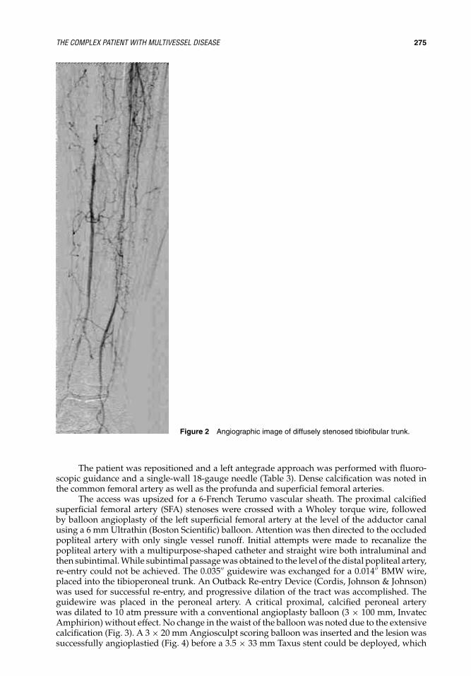
IHBK065-19 IHBK065-Minar May 1, 2010 8:19 Specs: 7x10 tight 7in×10in Char Count=
THE COMPLEX PATIENT WITH MULTIVESSEL DISEASE 275
Figure 2 Angiographic image of diffusely stenosed tibiofibular trunk.
The patient was repositioned and a left antegrade approach was performed with fluoro-scopic guidance and a single-wall 18-gauge needle (Table 3). Dense calcification was noted inthe common femoral artery as well as the profunda and superficial femoral arteries.
The access was upsized for a 6-French Terumo vascular sheath. The proximal calcifiedsuperficial femoral artery (SFA) stenoses were crossed with a Wholey torque wire, followedby balloon angioplasty of the left superficial femoral artery at the level of the adductor canalusing a 6 mm Ultrathin (Boston Scientific) balloon. Attention was then directed to the occludedpopliteal artery with only single vessel runoff. Initial attempts were made to recanalize thepopliteal artery with a multipurpose-shaped catheter and straight wire both intraluminal andthen subintimal. While subintimal passage was obtained to the level of the distal popliteal artery,re-entry could not be achieved. The 0.035′′ guidewire was exchanged for a 0.014′′ BMW wire,placed into the tibioperoneal trunk. An Outback Re-entry Device (Cordis, Johnson & Johnson)was used for successful re-entry, and progressive dilation of the tract was accomplished. Theguidewire was placed in the peroneal artery. A critical proximal, calcified peroneal arterywas dilated to 10 atm pressure with a conventional angioplasty balloon (3 × 100 mm, InvatecAmphirion) without effect. No change in the waist of the balloon was noted due to the extensivecalcification (Fig. 3). A 3 × 20 mm Angiosculpt scoring balloon was inserted and the lesion wassuccessfully angioplastied (Fig. 4) before a 3.5 × 33 mm Taxus stent could be deployed, which

IHBK065-19 IHBK065-Minar May 1, 2010 8:19 Specs: 7x10 tight 7in×10in Char Count=
276 DIEHM ET AL.
Table 3 List of Equipment Used in this Case
� 6-French vascular sheath (Terumo)� 3 × 20 mm Angiosculpt scoring balloon (AngioScore)� 6 mm Ultrathin balloon (Boston Scientific)� 5 FMPA catheter (Cook Medical)� LLT wire (Cook Medical)� 6 × 80 mm LifeStent (Edwards Life Sciences)� 6 × 4 mm balloon (Boston Scientific)� 0.014′′ BMW wire (Abbott Vascular)� 3 × 100 mm balloon (Amphirion, Invatec)� Angiosculpt balloon 3.0 × 20 mm (Angioscore)� 3.5 × 33 mm Taxus Stent (Boston Scientific)
was postdilated to 3.5 mm at 10 atm (Fig. 5). Following treatment of the outflow, a 6 × 80 mmLifeStent (Edwards Life Sciences) was deployed in the popliteal artery and postdilated to 6 mm.
The completion angiogram demonstrated excellent flow across the distal left superficialfemoral artery stent and the stent in the left tibioperoneal trunk. There was inline flow via theperoneal artery to the foot (Fig. 6).
What Were the Potential Hazards of Treating This Patient and Anatomy?Potential treatment hazards for these lesions included dissections of these rigid vessels as wellas embolization with subsequent clinical deterioration.
Summary: How Was the Case Managed, Is there Anything That Could Have Been Improved, What Wasthe Immediate and Long-Term Outcome?Technical and clinical success in complex multivessel endovascular revascularization may bedependent on dedicated interventional devices facilitating lesion success in highly calcified
WB
Figure 3 Unability to fully expand a 3/100 “0.014 low profile balloon(B) due to severe calcification of tibifibular trunk resulting in a waist(W) of the balloon.

IHBK065-19 IHBK065-Minar May 1, 2010 8:19 Specs: 7x10 tight 7in×10in Char Count=
THE COMPLEX PATIENT WITH MULTIVESSEL DISEASE 277
Figure 4 Balloon angioplasty with a 3/20 Angiosculptscoring balloon.
obstructions. Although level-A evidence on the primary use of drug-eluting stents in tibialarteries is currently lacking, use of this technology can be associated with favorable clinicaloutcomes.
Four months postprocedurally, the patients’ ulcers were healed and oscillographic readingsignificantly improved on the left side. Duplex ultrasound showed wide patency of the treatedfemoral and infrapopliteal segments.
Figure 5 Deployment of a 3.5/33 drug eluting stent.

IHBK065-19 IHBK065-Minar May 1, 2010 8:19 Specs: 7x10 tight 7in×10in Char Count=
278 DIEHM ET AL.
Figure 6 Final angiogram showing straight flow after recanalisation of thedistal femoral artery and tibiofibular trunk.
Complex Case 2: Catheter Treatment in Complex Supra-Aortic Disease
Clinical SummaryA 68-year-old woman was referred by her general practitioner with pain in her right shoulderand paresthesia of her right hand. Blood pressure measured 81/56 mm Hg on the right and113/68 mm Hg on the left side. MR-Angio showed an aortic arch type III, an ostial stenosis ofthe left common carotid artery, a 2-cm long occlusion of the brachiocephalic trunk, retrogradeflow in the right common and internal carotid artery as well as bilateral inverse flow of theophthalmic arteries.
First Procedure: Recanalization/Stenting of the Brachiocephalic TrunkAngiographic imaging confirmed the occlusion of the brachiocephalic trunk (Fig. 7). Recanal-ization was performed via right radial access. Femoral access was challenging due to an acute-angled takeoff of the brachiocephalic trunk as showed in Figure 8. An 8 mm Dynamic Biotronicstent was implanted. Figure 9 demonstrates the final angiographic result. After postdilatationof the stent, the patient developed paresthesia of her left hand and rapidly lost consciousness.The intracranial runs revealed embolic occlusions of middle cerebral artery side branches. Byusing microcatheters, urokinase, and tirofiban, the occluded intracerebral arteries were succes-sively reopened [Fig. 10(A) and 10(B)]. The patient regained consciousness during the processof intracranial rescue, and suffered no residual neurological sequelae.
Interventional Tools0.035′′ Emerald Guidewire (Cordis)Radiofocus stiff Guidewire (Terumo)5-French radial sheath (Terumo)4-French radial sheath (Terumo)

IHBK065-19 IHBK065-Minar May 1, 2010 8:19 Specs: 7x10 tight 7in×10in Char Count=
THE COMPLEX PATIENT WITH MULTIVESSEL DISEASE 279
Figure 7 Occlusion of the brachiocephalic trunk.
Figure 8 Type III aortic arch.
Figure 9 Result after stent implantation.

IHBK065-19 IHBK065-Minar May 1, 2010 8:19 Specs: 7x10 tight 7in×10in Char Count=
280 DIEHM ET AL.
(A) (B)
Figure 10 (A) Embolic occlusions of middle cerebral artery side branches. (B) Intracranial rescue.
8/40/130 Sailor plus balloon catheter (Krauth/Invatec)6 mm/40 mm Submarine balloon catheter (Krauth/Invatec)8 mm/25 mm Dynamic stent (Biotronic)0.014′′ Transcend Guidewire (Boston Scientific)0.035 Amplatz extra stiff wire (Cook)Taper 14-Flex Guidewire (Boston Scientific)V-18 Control wire (Boston Scientific)
Second Procedure: High-Grade Restenosis of the Brachiocephalic TrunkTwo months after the initial procedure, the patient was admitted with a suspected restenosisof the brachiocephalic trunk. The angiogram (Fig. 11) revealed severe stent compression atthe ostium. The guide that was initially introduced via femoral access was caught betweenthe proximal stent struts. A second access via the right brachial artery was established toperform retrograde crossing of the stent. The wire was then directed out of femoral access anda long sheath was placed via the femoral access (Fig. 12). During these maneuvers, embolicprotection was performed by intermittent manual compression of the right common carotidartery. Angioplasty was performed with an 8-mm balloon and a second stent (9 mm/19 mm
Figure 11 Restenosis due to stent compression.

IHBK065-19 IHBK065-Minar May 1, 2010 8:19 Specs: 7x10 tight 7in×10in Char Count=
THE COMPLEX PATIENT WITH MULTIVESSEL DISEASE 281
Figure 12 Wire directed out of femoral access.
Onda Stent, Krauth/Invatec) was implanted at the ostium of the brachiocephalic trunk. Afterattending the brachiocephalic trunk, a third stent (8 mm/5 mm Dynamic stent, Biotronic) wasplaced in the right subclavian artery (Fig. 13). The final result indicated no measurable pressuregradient (Fig. 14).
Interventional Tools4-French radial sheath (Terumo)5-French radial sheath (Terumo)0.035′′ Emerald Guidewire (Cordis)V-18 Control wire (Boston Scientific)8 mm/15 mm Dynamic stent (Biotronic)Radiofocus stiff Guidewire (Terumo)Iron Man wire (Terumo)Admiral Xtreme Balloon catheter (Krauth/Invatec)9 mm/19 mm Onda Stent (Krauth/Invatec)
Third Procedure: Stenosis of the Left Subclavian and Common Carotid ArteryA third procedure was scheduled four months later to treat the stenosis of the left subclavianartery and left common carotid artery (Fig. 15). Angioplasty of the left subclavian artery wasperformed via a femoral access. A retrograde approach was attempted with a wire crossing from
Figure 13 Stent implantation in the right subclavianartery.

IHBK065-19 IHBK065-Minar May 1, 2010 8:19 Specs: 7x10 tight 7in×10in Char Count=
282 DIEHM ET AL.
Figure 14 Final result. No measurable pressure gradient.
the right brachial artery to the left femoral artery (Fig. 16). A third access was gained via theright femoral artery. This enabled crossing of the left common carotid artery with two coronarywires. Balloon angioplasty was done with a 4-mm coronary balloon (Fig. 17). The final resultafter balloon angioplasty is shown in Figure 18.
Interventional ToolsV-18 Control wire (Boston Scientific)Iron Man wire (Terumo)0.035′′ Emerald Guidewire (Cordis)Balance Middleweight wire (Abbott Vascular)5-French diagnostic catheter (Cordis)5 mm/20 mm Avion plus Balloon catheter (Krauth/Invatec)8-French femoral Sheath (Terumo)
Key Learning IssuesAs demonstrated in this patient, embolic events can occur during angioplasty of the brachio-cephalic trunk. However, in this challenging scenario, establishing embolic protection is quitedifficult. Intermittent manual compression of the common carotid artery during the most dan-gerous steps of the procedure is an alternative.
Figure 15 Stenosis of the left subclavian artery.

IHBK065-19 IHBK065-Minar May 1, 2010 8:19 Specs: 7x10 tight 7in×10in Char Count=
THE COMPLEX PATIENT WITH MULTIVESSEL DISEASE 283
Figure 16 Wire crossed from the right brachial artery tothe left femoral artery.
Figure 17 Access via the right femoral artery, crossingwith 2 coronary wires, balloon angioplasty.
Figure 18 Final result.

IHBK065-19 IHBK065-Minar May 1, 2010 8:19 Specs: 7x10 tight 7in×10in Char Count=
284 DIEHM ET AL.
Table 4 Equipment Checklist for Complex Interventions in Patients with Multivessel Disease
� CTO devices (such as the Outback R© catheter)� Balloon expandable stents for use in the iliac, common femoral, deep femoral, and proximal superficial
arteries� Self-expanding nitinol stents for use in the femoropopliteal arteries. In general, the use of nitinol stents in
the femoropopliteal segment should become more liberal with increasing lesion lengths and worseningclinical presentation of the patient
� Low-profile (0.018′′ and 0.014′′) wires and long low-profile balloon catheters for obstructions of tibialarteries
� Drug-eluting coronary stents for complex tibial artery interventions (such as recurrence of CLI in focallesions or restenosis of bypass anastomoses)
Table 5 Key Learning Issues
� Patients with CLI frequently present with multilevel disease� Presence of diabetes mellitus and renal insufficiency is associated with severe
arterial calcifications that might preclude technical success of plain balloonangioplasty
� Therefore, the interventionist treating patients with complex multilevel diseaseshould be aware and experienced using an advanced endovasculararmamentarium
� Utilization of a scoring balloon allows for a controlled disruption of fibrocalcificlesions
SUMMARYIn recent years, technical improvements (Table 4) have substantially facilitated endovasculartreatment of CLI. Thus, endovascular treatment is increasingly considered to be the primarytreatment approach in CLI patients (Table 5) (9,13).
The present case highlights that the interventional management of patients with CLIand multilevel disease may require advanced endovascular techniques such as the use of bal-loons with a more aggressive outward luminal expansion or bare metal or drug-eluting stents.Endovascular treatment using a standard PTA balloon might be limited in highly calcifiedarteries and diffuse disease in patients with diabetes mellitus and renal insufficiency (14).
The patient treated in the present case clearly benefited from the use of the scoring balloonsince the use of a conventional balloon did not allow for a recanalization of this heavily calcifiedlesion. Initial results from a prospective study assessing the AngioSculpt scoring balloon in CLIpatients indicate that use of the this device can be applied with a high technical success rate (14).Endovascular technology is unfolding more rapidly than it can be assessed in a scientific setting(15). Thus, in contrast to the femoropopliteal segment (16–21), no level-A data supporting thepreferential use of various devices such as scoring or cutting balloons, bare metal or drug-elutingstents, etc. are currently available in the infrapopliteal segment.
Alternative treatment options in this patient would have been conservative treatmentalone. However, it has recently been shown that diabetics with CLI clearly benefit from endovas-cular as well as from surgical revascularization approaches (6).
Clearly, patency rates were reported to be substantially lower as compared to clinical suc-cess rates (22). However, once ulcer healing has occurred, less perfusion is required to maintainstable skin conditions (tide-over concept) (22). Thus, aggressive invasive follow-up strategiesand repeated endovascular procedures are mandatory in CLI patients with infrapopliteal dis-ease (3,6).
REFERENCES1. Graziani L, Silvestro A, Bertone V, et al. Vascular involvement in diabetic subjects with ischemic
foot ulcer: a new morphologic categorization of disease severity. Eur J Vasc Endovasc Surg 2007;33(4):453–460.

IHBK065-19 IHBK065-Minar May 1, 2010 8:19 Specs: 7x10 tight 7in×10in Char Count=
THE COMPLEX PATIENT WITH MULTIVESSEL DISEASE 285
2. Diehm N, Shang A, Silvestro A, et al. Association of cardiovascular risk factors with pattern of lowerlimb atherosclerosis in 2659 patients undergoing angioplasty. Eur J Vasc Endovasc Surg 2006; 31(1):59–63.
3. Brosi P, Baumgartner I, Silvestro A, et al. Below-the-knee angioplasty in patients with end-stage renaldisease. J Endovasc Ther 2005; 12(6):704–713.
4. Lepantalo M, Matzke S. Outcome of unreconstructed chronic critical leg ischaemia. Eur J Vasc EndovascSurg 1996; 11(2):153–157.
5. Ebskov LB, Schroeder TV, Holstein PE. Epidemiology of leg amputation: the influence of vascularsurgery. Br J Surg 1994; 81(11):1600–1603.
6. Dick F, Diehm N, Galimanis A, et al. Surgical or endovascular revascularization in patients with criticallimb ischemia: influence of diabetes mellitus on clinical outcome. J Vasc Surg 2007; 45(4):751–761.
7. Nasr MK, McCarthy RJ, Hardman J, et al. The increasing role of percutaneous transluminal angioplastyin the primary management of critical limb ischaemia. Eur J Vasc Endovasc Surg 2002; 23(5):398–403.
8. Norgren L, Hiatt WR, Dormandy JA, et al. Inter-society consensus for the management of peripheralarterial disease (TASC II). J Vasc Surg 2007; 45(suppl 1 ):S5–S67.
9. Adam DJ, Beard JD, Cleveland T, et al. Bypass versus angioplasty in severe ischaemia of the leg(BASIL): multicentre, randomised controlled trial. Lancet 2005; 366(9501):1925–1934.
10. Faglia E, Mantero M, Caminiti M, et al. Extensive use of peripheral angioplasty, particularlyinfrapopliteal, in the treatment of ischaemic diabetic foot ulcers: clinical results of a multicentricstudy of 221 consecutive diabetic subjects. J Intern Med 2002; 252(3):225–232.
11. Faglia E, Dalla Paola L, Clerici G, et al. Peripheral angioplasty as the first-choice revascularizationprocedure in diabetic patients with critical limb ischemia: prospective study of 993 consecutive patientshospitalized and followed between 1999 and 2003. Eur J Vasc Endovasc Surg 2005; 29(6):620–627.
12. Keo H, Diehm N, Baumgartner R, et al. Single center experience with provisional abciximab therapyin complex lower limb interventions. Vasa 2008; 37(3):257–264.
13. Kudo T, Chandra FA, Kwun WH, et al. Changing pattern of surgical revascularization for critical limbischemia over 12 years: endovascular vs. open bypass surgery. J Vasc Surg 2006; 44(2):304–313.
14. Bosiers M, Deloose K, Cagiannos C, et al. Use of the angiosculpt scoring balloon for infrapopliteallesions in patients with critical limb ischemia: 1-year outcome. Vascular 2009; 17(1):29–35.
15. Diehm N, Baumgartner I, Jaff M, et al. A call for uniform reporting standards in studies assessingendovascular treatment for chronic ischaemia of lower limb arteries. Eur Heart J 2007; 28(7):798–805.
16. Schillinger M, Sabeti S, Loewe C, et al. Balloon angioplasty versus implantation of nitinol stents in thesuperficial femoral artery. N Engl J Med 2006; 354(18):1879–1888.
17. Schillinger M, Sabeti S, Dick P, et al. Sustained benefit at 2 years of primary femoropopliteal stentingcompared with balloon angioplasty with optional stenting. Circulation 2007; 115(21):2745–2749.
18. Krankenberg H, Schluter M, Steinkamp HJ, et al. Nitinol stent implantation versus percutaneoustransluminal angioplasty in superficial femoral artery lesions up to 10 cm in length: the femoral arterystenting trial (FAST). Circulation 2007; 116(3):285–292.
19. Katzen B. Paper Presented at: International Symposium of Endovascular Therapy; 2008; Miami, FL,USA.
20. Amighi J, Schillinger M, Dick P, et al. De novo superficial femoropopliteal artery lesions: periph-eral cutting balloon angioplasty and restenosis rates—randomized controlled trial. Radiology 2008;247(1):267–272.
21. Diehm N, Silvestro A, Do DD, et al. Endovascular brachytherapy after femoropopliteal balloon angio-plasty fails to show robust clinical benefit over time. J Endovasc Ther 2005; 12(6):723–730.
22. Soder HK, Manninen HI, Jaakkola P, et al. Prospective trial of infrapopliteal artery balloon angioplastyfor critical limb ischemia: angiographic and clinical results. J Vasc Interv Radiol 2000; 11(8):1021–1031.

IHBK065-19 IHBK065-Minar May 1, 2010 8:19 Specs: 7x10 tight 7in×10in Char Count=

IHBK065-Ind IHBK065-Minar May 13, 2010 7:49 Specs: 7x10 tight 7in×10in Char Count=
Index
AAAs. see Abdominal aortic aneurysmsAbciximab, 13, 14, 65Abdominal aortic aneurysms, 131
endovascular treatment for (see Endovascularaortic aneurysm repair)
ABI. see Ankle–brachial indexACCORD-study, 18ACCP. see American College of Chest PhysiciansAcculink stents, 80Acetylcysteine, 19Acetylsalicylic acid. see AspirinACT. see Activated coagulation timeActivated coagulation time, 13, 163, 215Acute mesenteric ischemia, 158Alfimeprase, 13, 14Ambulatory venous hypertension, 257American College of Chest Physicians, 11, 257, 259,
270Amplatz wire, 78, 92, 94AngioJet rheolytic thrombectomy catheter, 260AngioSculpt (AngioScore), 207–208Ankle–brachial index, 22–24, 53, 205, 220, 274Antegrade popliteal access, 41Anterior spinal artery, 103Anticoagulation, in DVT, 257, 259Antiplatelet therapy, 10–12
subclavian intervention and, 90Antispasmodic agents, 16Antithrombotic therapy, 12–15
ACCP guidelines on, 257, 259, 270Aorto-uni-iliac (AUI), 134Arterial PCD
devices, commercially available, 47use of, 44
complications by, 45–46guidelines for, 46
Arteriovenous (AV) fistulae, dysfunction of, 239Aspiration catheter, 199Aspirin, 10–13, 15, 63–65, 160, 172Atherosclerotic thoracic aortic aneurysms, 120, 121,
123–125Atrial fibrillation (AF), 15AV fistula interventions, 239, 243
case studies onaccess combination technique, 246–250cryoplasty, 243–246mechanical thrombectomy and endoprosthesis
placement, 250–254complexity in, 258
complications from, 240devices for, 244indications for, 239, 240strategies for
access route, 240complex stenoses, 241complex thromboses, 241–242peri-interventional imaging, 240preinterventional diagnostic workup,
239–240stent placement, 242–243
success and safety of, 254treatment outcomes after, 244
Balloon angioplasty, 1–4, 91, 104, 207–208, 224Balloon protection, in CAS, 78Basilar artery, angioplasty in, 110–112Basilar artery thrombosis, 63Bechterew’s disease and CAS, 82, 83Below-the-knee (BTK) revascularization, 224Berenstein catheter, 104�-blocking agents, 17–18Bivalirudin, 13Booster laser, 207Brachial access site, 41–42
complications, 43–44in subclavian intervention, 91
Bridge Assurant stent, 94
CAPRIE trial, 10–11Cardiovascular Outcomes for Renal Artery Lesions
(CORAL) trial, 32Carotid artery stenting, 77
case studies onCAS after CEA, 80–82CAS in tandem stenosis, 82–86cerebral artery aneurysms and CAS, 86stent in ICA with no surgery option, 82
CAS vs. CEA, 79–80indications for, 79–80specific training, need of, 77technique of, 77
anchoring technique, 78cerebral protection, devices for, 78guiding catheter technique, 78stent, choice of, 78–79telescoping technique, 78
Carotid endarterectomy, 37, 77, 79–80CAS. see Carotid artery stenting

IHBK065-Ind IHBK065-Minar May 13, 2010 7:49 Specs: 7x10 tight 7in×10in Char Count=
288 INDEX
Catheter treatment, in supra-aortic disease, 278brachiocephalic trunk, recanalization of, 278–279
embolic occlusions of middle cerebral artery,280
intracranial rescue, 280occlusion of brachiocephalic trunk, 279tools used, 278
restenosis of brachiocephalic trunk, 278,280–281
embolic protection by manual compression,280
femoral access approach, 281stent compression at ostium, 280tools used, 281
stenosis of left subclavian and carotid artery, 281access approach, 281–282balloon angioplasty, 282–283tools for, 282
CEA. see Carotid endarterectomyCerebral angiography, 37Cerebral arteries, anatomical features of, 62–63Cerebral protection devices, 78, 86CFA. see Common femoral arteryCHARISMA trial, 11, 66Chronic mesenteric ischemia, 158
endovascular treatment of (see Mesenteric arteryinterventions)
Cilostazol, 12CLI. see Critical limb ischemiaClopidogrel, 10–13, 15, 64, 172CMI. see Chronic mesenteric ischemiaColor duplex ultrasound, 143Common femoral artery, 41, 48, 50, 57, 160, 170,
185, 186Complex lesions
characteristics of, 4–6endovascular treatment of, 1 (see also specific
interventions)clinical indications for, 6hazards in, 6interventional strategies and tools for, 8issues involved in, 8
Computed tomography angiography, 28, 36abdominal aortic intervention, 132carotid imaging, 35mesenteric arteries, 160RAS diagnosis, 143renal arteries, 31–32
Contemporary venous thrombectomy, 260,262–265
Contrast-enhanced MRA, 27, 30, 34Contrast-induced nephropathy, risk factors for,
32Critical limb ischemia, 2, 10, 16, 18, 19, 220. see also
Multivessel disease, endovascular treatmentfor; Tibioperoneal interventions
Cryoplasty, 243–246CTA. see Computed tomography angiography
Deep venous thrombosis, 257. see also IliofemoralDVT, venous interventions for
Diabetesand amputation risk, 220multivessel disease and, 272and PAD, 16, 18
Digital subtraction angiography, 28, 30, 34, 35, 86,239, 274
Double-balloon technique, 91DSA. see Digital subtraction angiographyDual antiplatelet therapy, 11–12Duplex ultrasonography, 24, 26, 31, 36, 64
AV fistulae, obstructions in, 239carotid DUS, 33femoropopliteal lesion, 205Leriche syndrome, 172lower extremities, 24–26mesenteric arteries, 160RAS diagnosis, 29–30
DUS. see Duplex ultrasonographyDVT. see Deep venous thrombosis
EKOS R© EndoWaveTM system, 261Emergency Management of Stroke (EMS) Bridging
Trial, 63Endoleak formation, prevention of, 120, 123, 129Endovascular aortic aneurysm repair, 131
access route, decision on, 132–133case studies
AAA involving visceral arteries, 139–141excessive tortuosity of proximal aortic neck,
148–149total occlusion of iliac vessel, 137–139
complications, avoidance of, 141devices for treating AAAs, 135indications for, 131, 132patient selection, 131potential risks of, 132preoperative CTA, 131selection of device, 131–132treatment strategies, 131
Endovascular therapy. see also specificinterventions
as revascularization strategy, 1–3treatment of complex lesions by, 1
European Safe Implementation of Thrombolysis inStroke-Monitoring Study, 64
EVAR. see Endovascular aortic aneurysm repairExpress LD stent, 99
Femoral neuralgia syndrome, 46Femoropopliteal segment, endovascular treatment
of, 218case studies
highly calcified popliteal occlusion, 209–210mid-SFA occlusion, 209–211safety and success of, 218severely calcified SFA occlusion, 217–219
complexity in, 205complications in, 206indications for, 221strategies and tools for, 205–206
access approach, 205–206

IHBK065-Ind IHBK065-Minar May 13, 2010 7:49 Specs: 7x10 tight 7in×10in Char Count=
INDEX 289
balloon angioplasty, 207–208devices for complex lesions, 207patency rate after recanalization, 207preinterventional diagnostic workup, 205recanalization, devices for, 206–208stenting, 205tools for complex lesions, 205
success rate and outcomes of, 207Filter protection, in CAS, 78Flush occlusion, 91, 209
of subclavian artery, 88, 91Frontrunner CTO catheter, 206–207
Gateway balloon, 68, 71–73Global carotid artery stent registry, 77Glycoprotein (GP) IIb/IIIa receptor inhibitors,
13–14Guiding catheter, 78Guiding catheter technique, for CCA access, 78,
79
Heart Outcomes Prevention Evaluation (HOPE)study, 18
Heart Protection Study, 16Heparin, 42, 44, 65, 93, 107–108, 124, 243
unfractionated, 12–13Hybrid stent, 78–79
I-Cast stent, 96Iliac interventions, 183–185
case studiesdistal embolization, during procedure,
187–189intraprocedural complication, management of,
191–194vascular insufficiency, complex case of,
194–195complexity in, 187complications of, 188devices and techniques for, 186–187
bifurcation stenting, 189primary stenting vs. elective stenting, 187stent grafting, 187–190
endovascular iliac treatment, stratified by TASCclassification, 184
equipment checklist, 191literature overview for, 190strategies for, 185
access techniques, 185–186Iliofemoral DVT, venous interventions for, 257
case studiescatheter-directed thrombolysis, 265–266contemporary venous thrombectomy, 262–264pharmacomechanical thrombolysis, 264–265
catheter-directed thrombolysis, outcomes of, 270complexity in, 272indications for, 258–259management of, 258patency rates with venous thrombectomy, 261strategies for, 260tools for, 260–261
Imaging techniques, 22extracranial carotid artery, 32
anatomic consideration, 36carotid DUS, 33CTA, 34indications for arteriography, 36–37MRA, 34plaque characterization, 35–36
lower extremitiesankle–brachial index, 22–23computed tomography angiography, 28duplex ultrasonography, 24–26exercise stress testing, 24indications for arteriography, 28magnetic resonance angiography, 26–28pulse volume recordings, 23–24
renal artery, 29computerized tomographic angiography,
31–32DUS, 29–30, 31indications for arteriography, 32magnetic resonance angiography, 30
IMH. see Intramural hematomaIntracranial atherosclerosis, 62Intracranial interventions, 73–74
case studies onintra-arterial thrombolysis, 68–69MERCI Retrieval System, 69–71Wingspan stent, 71–73
clinical indications foracute stroke, 63elective stenting, 63–64
complexity in, 62–63complications of, 64–65equipment checklist for, 67–68goal of, 62, 73
challenges to, 73and primary evaluation, 65procedure management in, 65
anesthesia, 65–66postprocedure management, 66premedication, 65
tools for, 66Intramural hematoma, 120–123, 126Intrathrombus catheter-directed thrombolysis, 260Ipsilateral antegrade access, 41Isolated, segmental pharmacomechanical
thrombolysis (ISPMT), 261–262IVUS. see Intravascular ultrasound
Kissing balloon technique, 8, 138, 143, 153, 178, 189
Left internal mammary artery (LIMA), 88, 89Leriche syndrome, 172
case study on, 173–181aortic stent, placement of, 176–177coronary wires, placement of, 173, 174interventional strategies and tools, 183–184kissing-balloon postdilatation of stent, 178, 179left iliac artery, wire passage from, 175nitinol stents in iliac arteries, placement of, 179

IHBK065-Ind IHBK065-Minar May 13, 2010 7:49 Specs: 7x10 tight 7in×10in Char Count=
290 INDEX
Leriche syndrome (Continued)predilatation with ballon, 176–177preprocedural MRA, 173right iliac artery, wire passage of, 174snaring of wires, 176support catheter, intraluminal position of,
175endovascular treatment of, 183
complexity of lesion in, 172complications of, 172strategy for, 172
materials used, 181surgery in, 172symptoms of, 172
Magnetic resonance angiography, 26, 30, 90,172–173, 240
cervical carotid arteries, 343D gadolinium-enhanced MRA, 26–28Leriche syndrome, 172mesenteric arteries, 160in renal interventions, 143
Manual compression, 41, 43, 44, 47, 49, 278,280
MATCH trial, 64, 66Mechanical thrombectomy device, 70–71.
see also MERCI Retrieval System;Penumbra device
MERCI Retrieval System, 63, 67Mesenteric artery interventions, 158–160
case studies onceliac artery occlusion with SMA narrowing,
165–167in-stent restenosis of celiac artery,
166–170lesion in celiac artery, 163–164
complexity in, 158complications, 159equipment checklist, 161indications for, 158–159Ochsner Clinic series, results of, 160–161patient selection, 169–170strategies for, 159–160tools for, 160
MRA. see Magnetic resonance angiographyMulti-track angiocatheter, 97–99Multivessel disease, endovascular treatment for,
272, 284case study on
limb salvage in CLI, 274–284complexity in, 272complications in, 272, 273critical limb ischemia and, 274equipment checklist for, 284indications for, 272outcomes of, 273strategies for, 273tools for, 273
Nephrogenic systemic fibrosis, 28Nitroglycerin, 13, 16, 44, 229
ONTARGET trial, 18Oral anticoagulation (OAC), 12
OAC therapy, 15
PAD. see Peripheral artery diseasePARTNERS program, 17PAU. see Penetrating atherosclerotic ulcersPenetrating atherosclerotic ulcers, 120–122, 123, 125Penumbra device, 67
in acute stroke, 63Percutaneous closure device (PCD), 44Percutaneous transluminal angioplasty (PTA), 49,
159Peripheral artery disease, 1, 10
pharmacological treatment, 10, 16antiplatelet therapy, 10–12antithrombotic therapy, 12–15OAC therapy for AF patients, 15patients with renal impairment, therapy for, 19vasodilators, application of, 16
risk factor modification, 16antihypertensive treatment, 17–18diabetes, control of, 18statins, use of, 16–17
Peripheral cutting balloon (PCB), 241, 260Pharmacomechanical thrombolysis, 260–264Plasminogen activator inhibitor (PAI-1), 260Plasminogen activators, 260Posterior inferior cerebellar artery (PICA), 103, 108,
114, 115, 117PROACT II study, 63Pro-CAS (prospective registry of CAS), 77PROMPT trial, 14Prostanoids, 13, 16Pulmonary embolism (PE), 251, 259
Ramipril, 18REACH (Reduction of Atherothrombosis for
Continued Health) Registry, 10, 16Recombinant tissue plasminogen activator (rt-PA),
13, 14, 262RELAX trial, 14Renal artery disease (RAS), imaging for, 29–32Renal artery interventions, 143, 156
case studies onkissing balloon technique, use of, 152–154laser-assisted recanalization procedure,
154–156telescoping technique, use of, 148–152
complexity in, 148complications of, 144
avoidance of, 159equipment checklist, 148, 151indications for, 144outcomes of, 145–147strategies for, 143–144tools for, 144–145
Restenosis, 146–147Restenosis rates, for ostial stent angioplasty, 151Retrograde femoral access, 41
complications, 43

IHBK065-Ind IHBK065-Minar May 13, 2010 7:49 Specs: 7x10 tight 7in×10in Char Count=
INDEX 291
Retrograde tibial access, 41Revascularization, 1. see also specific endovascular
therapy approachopen vascular surgery vs. endovascular therapy,
1–3
SFA. see Superficial femoral arterySilverHawk atherectomy device, 207SMA. see Superior mesenteric arterySpectranetics Multi-track catheter, 98Statins, 16–17Stroke, 15, 16, 62, 77, 79, 84, 103–105. see also
Intracranial interventionsSubclavian artery, 88Subclavian intervention, 88, 91–92
case studiescomplex subclavian lesion, with cardiac
disease, 95–97left carotid–subclavian bypass stenosis, 97–100left subclavian artery total occlusion, 93–95
clinical indications for, 88, 89complexity of, 88equipment checklist, 92potential complications of, 88–90strategies for, 90
imaging modalities, 90IVUS, use of, 90local anesthesia with mild sedation, 91oral antiplatelet therapy, 90percutaneous brachial approach, 90, 91, 94right subclavian treatment, 91stent selection and placement, 91
tools for, 91, 92Subintimal angioplasty, 185Supera stent, 202Superficial femoral artery, 1, 2, 4–8, 41, 43, 44, 47,
172, 194Superior mesenteric artery, 139, 158, 165, 173, 174,
182
Telmisartan, 18TEVAR. see Thoracic endovascular aortic repairThoracic endovascular aortic repair, 120, 128–130
and atherosclerotic thoracic aortic aneurysms,120
case study on, 124–125complications in, 121indications for, 131, 143outcomes of TEVAR therapy, 123
complicated type B dissections and, 120equipment checklist, 123–125imaging for, 122interventional strategy, 122interventional tools for, 122intramural hematoma, 121
case study on, 126–128complications in, 122indications for, 121outcomes of therapy, 123
penetrating atherosclerotic ulcers, 120case study on, 125–126
complications in, 122indications for, 120outcomes of therapy, 123
and quality control, 123Thrombolytic agents, 14–15Thrombus removal. see Iliofemoral DVT, venous
interventions forTibioperoneal interventions, 220
case studiesanterior tibial artery recanalization, 231–234occlusion of AT and dorsalis pedis artery,
233–234, 237retrograde recanalization of plantar artery,
227–234complications in, 214–222, 229devices for, 230indications for, 214, 229preinterventional diagnostic workup, 222strategies for, 222–220tools for, 222
Ticlopidine, 12, 63, 65, 66Tirofiban, 14, 278TransAtlantic Inter-Society Consensus (TASC)
working group, 1, 183aortoiliac lesion classification and evolution, 185femoropopliteal lesions, therapeutic approach
on, 205, 208lesion classification categories, 184recommendations for management of PAD, 183
Transpopliteal retrograde access, 41complications, 43
Treadmill testing, 24, 172Trellis-8 catheter, 261Type B dissections, 120–123
U.K. Prospective Diabetes Study, 18Unfractionated heparin (UFH), 12Urokinase, 13, 14, 68, 69, 221, 241, 242U.S. Food and Drug Administration (FDA), 28
Vascular access and vessel closureaccess route (see specific access sites)case studies on
balloon rupture with difficulties in retrieval,56–60
calf claudication, 48–53stenosis by dislocated anchor of closure
device, 53–56clinical indications for, 44interventional strategies, 47–48vascular complications, 42, 43–46
risk factors for, 44Vena caval filter, 259Venous interventions. see Iliofemoral DVT, venous
interventions forVertebral angioplasty. see Vertebral interventionVertebral artery, segments in, 102Vertebral basilar insufficiency (VBI), 102Vertebral interventions, 104–105
case studies onstenosis in V4 segment, 110–112

IHBK065-Ind IHBK065-Minar May 13, 2010 7:49 Specs: 7x10 tight 7in×10in Char Count=
292 INDEX
Vertebral interventions (Continued)symptomatic lesions treatment by team
approach, 112–118VBI symptoms and two vertebral arteries
treatment, 106–110CAVATAS study on, 105Cochrane Review and, 105complexity of, 102complications of, 103equipment checklist
embolic protection devices, 106interventional guide catheters,
105
interventional guidewires, 105PTA balloons, 105stents, 106
indications for, 102–103strategies for, 103–104tools for, 104
Vertebrobasilar ischemia, 91
Warfarin Antiplatelet Vascular Evaluation (WAVE)trial, 12
Wingspan stent system, 66, 71–73
Zenith stent graft, 131–132

cp IHBK065-Minar April 24, 2010 13:7 Specs: 7x10 tight 7in×10in Char Count=
Figure 3.3A Normal triphasic Doppler waveform performed during Duplex ultrasonography. However, note theincorrect Doppler angle, demonstrating lack of parallel placement compared to the direction of arterial blood flow.
Figure 3.6 Renal artery duplex ultrasonography, demonstrating severe right renal artery stenosis. Note thedramatic elevation in peak systolic (972 cm/sec) and end-diastolic velocities (308 cm/sec).
Figure 3.7 Duplex ultrasonography of right renal artery stent, demonstrating peak systolic velocity 390 cm/sec,suggestive of >60% in-stent restenosis.

cp IHBK065-Minar April 24, 2010 13:7 Specs: 7x10 tight 7in×10in Char Count=
Figure 3.10 Carotid duplex ultrasonography, demonstrating critical (90–99%) left internal carotid artery stenosis.Note the peak systolic velocity elevation (710 cm/sec) and end-diastolic velocity (198 cm/sec).
Figure 4.14 Duplex sonography one day postprocedurally showing a tight stenoses at the origin of the superficialfemoral artery.

Complex Cases in Peripheral Vascular Interventions
Edited byMartin Schillinger
Erich Minar
Co
mp
lex C
ase
s in Perip
hera
l Va
scula
r Interve
ntions
Sch
illing
er •
Min
ar
www.informahealthcare.com
Telephone House, 69-77 Paul Street, London EC2A 4LQ, UK
52 Vanderbilt Avenue, New York, NY 10017, USA
About the book
This new title provides a systematic review of complex cases in peripheral vascular interventions (PVI), a minimal invasive endovascular approach to treating peripheral arteries. This increasingly evolving area in interventional therapies offers reduced risk for complications compared to conventional surgery and takes advantage of advanced techniques to treat more complex lesions in severely diseased patients.
This book edited by two highly respected professors from the University of Vienna, Medical School describes typical and atypical complex cases for all peripheral vessel areas, and outlines methods to successfully and safely handle these interventions. The text is divided into two sections. The first reviews general aspects on managing complex patients in PVI, and the second discusses complex cases in specific vessel areas, examining the interventions from puncture to closure, head to toe. Each of the chapters in this section is further divided into three parts and provides:
• An introduction to each vessel, specific information on thecomplexities of this lesion and the hazards of treatment, and a checklist for readers of recommended equipment when treating this specific vessel area.
• Selected teaching cases, whereby specific vessel areas aredescribed step-by-step.
• A summary of the key issues for the successful and safetreatment of complex lesions in this vessel area.
Complex Cases in Peripheral Vascular Interventions offers readers the opportunity to tap into the knowledge of those working on complex cases in this cutting edge field and receive both theoretical information and practical tips on how to approach specific vascular lesions. This book is a highly recommended read for beginners in endovascular interventions who want to get an impression on the advanced scope of treatment options as well as for experienced interventionists who want to extend their knowledge on difficult cases.
About the editors
MArTIN SCHILLINgEr, MD is a Professor and Senior Physician at the Department of Internal Medicine at general Hospital Vienna, Austria. Professor Schillinger gained his Medical Degree with honours in 1998 from the University of Vienna and in 2002 was appointed Associate Professor at the University. He has held his position at general Hospital Vienna since July 2008 where he also specialises in cardiology and angiology. Professor Schillinger has been lead investigator of numerous clinical trials and collaborated on many multi-center trials. He is on the advisory board of Boston Scientific, Abbott Vascular and Amaranth, among others, and editorial board of numerous societies and journals. Professor Schillinger has published and presented his work extensively.
ErICH MINAr, MD is a Professor of Internal Medicine at the Medical University of Vienna. He graduated from the Medical School at the University of Vienna in 1977 and following a position as an assistant in Internal Medicine at the University Clinic Vienna, was appointed as Assistant Professor at the Department of Angiology in 1984, and Professor of Internal Medicine in 1993. Since 1998 he has served as Deputy Head of the Department of Angiology at the University. Professor Minar has published more than 350 papers in peer-reviewed journals. He is co-editor of the European Journal of Vascular Diseases and editor of several books, including Complications in Peripheral Vascular Interventions (2007).
Complex Cases in Peripheral Vascular Interventions