management of pregnancy in patients with complex congenital heart disease · circulation....
TRANSCRIPT

Circulation. 2017;135:00-00. DOI: 10.1161/CIR.0000000000000458 TBD, 2017 e1
CLINICAL STATEMENTS
AND GUIDELINES
ABSTRACT: Today, most female children born with congenital heart disease will reach childbearing age. For many women with complex congenital heart disease, carrying a pregnancy carries a moderate to high risk for both the mother and her fetus. Many such women, however, do not have access to adult congenital heart disease tertiary centers with experienced reproductive programs. Therefore, it is important that all practitioners who will be managing these women have current information not only on preconception counseling and diagnostic evaluation to determine maternal and fetal risk but also on how to manage them once they are pregnant and when to refer them to a regional center with expertise in pregnancy management.
Expanded diagnostic, medical, and surgical management options have improved the long-term survival of patients with congenital heart disease (CHD). Thus, most women born with CHD will reach reproductive age. The ability to bear
children is a major point of care for this growing population. As a result, pregnancy counseling and management are among the major noncardiac issues facing pediat-ric and congenital cardiac providers.
For the majority of patients, the ability to conceive and carry a pregnancy to term will present little problem. However, for those with complex CHD, pregnancy may be associated with an increased risk compared with women with milder forms of CHD, regardless of whether they are clinically stable at the time of conception. This docu-ment provides an overview of the management of the patient with complex CHD who becomes pregnant.
DEfINING ThE PoPULATIoNSimple CHD lesions include mild pulmonary valve stenosis, a small, uncomplicated atrial septal defect or ventricular septal defect, patent ductus arteriosus, and suc-cessfully repaired atrial septal defect, ventricular septal defect, patent ductus ar-teriosus, and anomalous pulmonary venous connection without important residua. Complex CHD, on the other hand, includes any complex anatomical or physiological lesion as defined by the Bethesda conference.1 Some patients with simple CHD lesions would, however, be considered high pregnancy risk because of the pres-ence of comorbid conditions. An example is the patient with an atrial septal defect and pulmonary hypertension or atrial fibrillation; this patient would require a higher level of care during pregnancy and in the peripartum period. Thus, a designation of simple versus complex CHD is not adequate when referring to patients with CHD considering pregnancy.
Mary M. Canobbio, RN, MN, FAHA, Chair
Carole A. Warnes, MD, FRCP, Co-Chair
Jamil Aboulhosn, MDHeidi M. Connolly, MDAmber Khanna, MDBrian J. Koos, MD, DPhilSeema Mital, MD, FAHA,
FRCPCCarl Rose, MDCandice Silversides, MD,
FRCPCKaren Stout, MD, FAHAOn behalf of the Ameri-
can Heart Association Council on Cardiovas-cular and Stroke Nurs-ing; Council on Clinical Cardiology; Council on Cardiovascular Disease in the Young; Council on Functional Genomics and Translational Biology; and Council on Quality of Care and Outcomes Research
Management of Pregnancy in Patients With Complex Congenital heart DiseaseA Scientific Statement for healthcare Professionals from the American heart Association
© 2017 American Heart Association, Inc.
Key Words: AHA Scientific Statements ◼ heart defects, congenital ◼ heart diseases ◼ pregnancy
AhA SCIENTIfIC STATEMENT
by guest on February 5, 2017http://circ.ahajournals.org/
Dow
nloaded from
Canobbio et al
TBD, 2017 Circulation. 2017;135:00-00. DOI: 10.1161/CIR.0000000000000458e2
The modified World Health Organization (WHO) classifi-cation categorized patients into 4 pregnancy risk classes (classes I–IV) as determined by their medical condition (Table 1).2 Patients in class I have no detectable increased risk of maternal mortality and either no or a mild increase in morbidity; thus, they are not included in this document. Women in class II might have a small increase in mater-nal mortality or a moderate increase in morbidity with pregnancy, and those in class III might have a significant increase in maternal mortality or severe morbidity. Those in class IV, however, may carry an extremely high risk of maternal mortality or severe morbidity such that preg-nancy is ill advised. These patients should be counseled to avoid pregnancy. If pregnancy is confirmed in a woman in WHO class IV, then termination is advised.
High-risk patients are further identified in observation-al data to include patients with prosthetic valves or those with more than moderate atrioventricular valve regurgi-tation or New York Heart Association (NYHA) class II or higher heart failure before pregnancy.4–7
PhySIoLoGICAL ADAPTATIoN of PrEGNANCyMaternal organ systems undergo significant physiologi-cal alterations during pregnancy. The following sections highlight the physiological changes that have particular relevance to the management of gravidas with CHD.
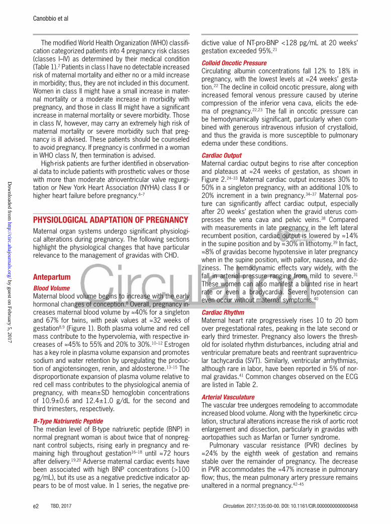
AntepartumBlood VolumeMaternal blood volume begins to increase with the early hormonal changes of conception.8 Overall, pregnancy in-creases maternal blood volume by ≈40% for a singleton and 67% for twins, with peak values at ≈32 weeks of gestation8,9 (Figure 1). Both plasma volume and red cell mass contribute to the hypervolemia, with respective in-creases of ≈45% to 55% and 20% to 30%.10–12 Estrogen has a key role in plasma volume expansion and promotes sodium and water retention by upregulating the produc-tion of angiotensinogen, renin, and aldosterone.13–15 The disproportionate expansion of plasma volume relative to red cell mass contributes to the physiological anemia of pregnancy, with mean±SD hemoglobin concentrations of 10.9±0.6 and 12.4±1.0 g/dL for the second and third trimesters, respectively.
B-Type Natriuretic PeptideThe median level of B-type natriuretic peptide (BNP) in normal pregnant woman is about twice that of nonpreg-nant control subjects, rising early in pregnancy and re-maining high throughout gestation16–18 until ≈72 hours after delivery.19,20 Adverse maternal cardiac events have been associated with high BNP concentrations (>100 pg/mL), but its use as a negative predictive indicator ap-pears to be of most value. In 1 series, the negative pre-
dictive value of NT-pro-BNP <128 pg/mL at 20 weeks’ gestation exceeded 95%.21
Colloid Oncotic PressureCirculating albumin concentrations fall 12% to 18% in pregnancy, with the lowest levels at ≈24 weeks’ gesta-tion.22 The decline in colloid oncotic pressure, along with increased femoral venous pressure caused by uterine compression of the inferior vena cava, elicits the ede-ma of pregnancy.22,23 The fall in oncotic pressure can be hemodynamically significant, particularly when com-bined with generous intravenous infusion of crystalloid, and thus the gravida is more susceptible to pulmonary edema under these conditions.
Cardiac OutputMaternal cardiac output begins to rise after conception and plateaus at ≈24 weeks of gestation, as shown in Figure 2.24–33 Maternal cardiac output increases 30% to 50% in a singleton pregnancy, with an additional 10% to 20% increment in a twin pregnancy.34–37 Maternal pos-ture can significantly affect cardiac output, especially after 20 weeks’ gestation when the gravid uterus com-presses the vena cava and pelvic veins.38 Compared with measurements in late pregnancy in the left lateral recumbent position, cardiac output is lowered by ≈14% in the supine position and by ≈30% in lithotomy.39 In fact, ≈8% of gravidas become hypotensive in later pregnancy when in the supine position, with pallor, nausea, and diz-ziness. The hemodynamic effects vary widely, with the fall in arterial pressure ranging from mild to severe.31 These women can also manifest a blunted rise in heart rate or even a bradycardia. Severe hypotension can even occur without maternal symptoms.40
Cardiac RhythmMaternal heart rate progressively rises 10 to 20 bpm over pregestational rates, peaking in the late second or early third trimester. Pregnancy also lowers the thresh-old for isolated rhythm disturbances, including atrial and ventricular premature beats and reentrant supraventricu-lar tachycardia (SVT). Similarly, ventricular arrhythmias, although rare in labor, have been reported in 5% of nor-mal gravidas.41 Common changes observed on the ECG are listed in Table 2.
Arterial VasculatureThe vascular tree undergoes remodeling to accommodate increased blood volume. Along with the hyperkinetic circu-lation, structural alterations increase the risk of aortic root enlargement and dissection, particularly in gravidas with aortopathies such as Marfan or Turner syndrome.
Pulmonary vascular resistance (PVR) declines by ≈24% by the eighth week of gestation and remains stable over the remainder of pregnancy. The decrease in PVR accommodates the ≈47% increase in pulmonary flow; thus, the mean pulmonary artery pressure remains unaltered in a normal pregnancy.42–45
by guest on February 5, 2017http://circ.ahajournals.org/
Dow
nloaded from

Pregnancy in Patients With Complex CHD
Circulation. 2017;135:00-00. DOI: 10.1161/CIR.0000000000000458 TBD, 2017 e3
CLINICAL STATEMENTS
AND GUIDELINES
A primary fall in systemic vascular resistance (SVR), which reduces preload and afterload, may trigger many of the changes in the hemodynamics and volume homeosta-sis that accompany early pregnancy. Within 8 weeks after the last menstrual period, SVR has fallen by ≈30%, reach-ing a nadir at ≈24 weeks’ gestation (Figure 2).23,25,27,43,46–50 After 24 weeks’ gestation, SVR begins to increase again, approaching preconception levels by term.
The fall in SVR is associated with 10 to 15 mm Hg reduction in diastolic pressure at 20 to 24 weeks’ gesta-
tion (measured in the lateral recumbent position), which is followed by a rise toward nonpregnant measurements (Figure 2).27,51
Respiratory ChangesTidal volume increases 40% with a proportional rise in minute ventilation.52–54 This physiological hyperventila-tion is greater than the increase in oxygen consumption and accounts for the breathlessness that begins in early pregnancy.55–58
Table 1. Modified Who Classification of Maternal Cardiovascular risk
Who Pregnancy risk Category risk Description Maternal risk factors
I No detectable increase in maternal mortality and no/mild increase in morbidity risk
Uncomplicated small/mild pulmonary stenosis, PDA, mitral valve prolapse
Successfully repaired simple lesions (ASD, VSD, PDA, anomalous pulmonary venous drainage)
Atrial or ventricular ectopic beats, isolated
II Small increase in maternal mortality and moderate increase in morbidity risk
If otherwise well and uncomplicated:
Unoperated ASD, VSD
Repaired TOF
Most arrhythmias
II–III Moderate increase in maternal mortality morbidity risk
Mild LV impairment
Hypertrophic cardiomyopathy
Native or tissue valvular disease (not considered risk category I or IV)
Marfan syndrome without aortic dilation
Aortic dilation <45 mm in bicuspid aortic valve aortopathy
Repaired coarctation
III Significantly increased maternal mortality or severe morbidity risk. Expert counseling required. In the event of pregnancy, intensive specialist cardiac and obstetric monitoring needed throughout pregnancy, childbirth, and the puerperium.
Mechanical valve
Systemic RV
Fontan circulation
Cyanotic heart disease (unrepaired)
Other complex CHD
Aortic dilation 40–45 mm in Marfan syndrome
Aortic dilation 45–50 mm in bicuspid aortic valve aortopathy
IV Extremely high maternal mortality or severe morbidity risk. Pregnancy is contraindicated. In the event of pregnancy, termination should be discussed. If pregnancy continues, care should follow class III recommendations.
Pulmonary arterial hypertension (of any cause)
Severe systemic ventricular dysfunction (LV ejection fraction <30%, NYHA class III-IV)
Previous peripartum cardiomyopathy with any residual impairment of LV function
Severe mitral stenosis, severe symptomatic aortic stenosis
Aortic dilation >45 mm in Marfan syndrome
Aortic dilation >50 mm in bicuspid aortic valve aortopathy
Native severe coarctation
AS indicates aortic stenosis; ASD, atrial septal defect; CHD, congenital heart disease; LV, left ventricular; NYHA, New York Heart Association; PDA, patent ductus arteriosus; RV, right ventricle; TOF, tetralogy of Fallot; VSD, ventricular septal defect; and WHO, World Health Organization.
Modified from Thorne et al2 with permission from the BMJ Publishing Group Ltd. Copyright © 2006, BMJ Publishing Group Ltd and the British Cardiovascular Society. Modified from Balci et al3 with permission from the BMJ Publishing Group Ltd. Copyright © 2014, BMJ Publishing Group Ltd and the British Cardiovascular Society.
by guest on February 5, 2017http://circ.ahajournals.org/
Dow
nloaded from

Canobbio et al
TBD, 2017 Circulation. 2017;135:00-00. DOI: 10.1161/CIR.0000000000000458e4
IntrapartumLabor results in important hemodynamic changes, in-cluding elevations in heart rate, central venous pressure, and cardiac output.59–61 A change from the supine to a lateral recumbent position between contractions (basal conditions) increases maternal cardiac output by ≈22% and decreases heart rate by 6%.60 Basal systemic arte-rial pressure rises with the progression of the first stage of labor, with further increases during uterine contrac-tions.33,59,61 Uterine contractions augment maternal car-diac output as a result of enhanced sympathetic activity (via anxiety, pain) and expulsion of uterine blood into the central venous circulation.58–60 During a contraction, the uterus expels up to 400 mL blood into the central venous circulation, leading to a rise in central venous pressure, right atrial pressure,62 cardiac output, and arterial pres-sure. The labor-induced augmentation of cardiac output is attenuated by effective epidural anesthesia.
Maternal position modulates these hemodynamic re-sponses. In the supine position, contractions are associ-ated with an ≈15% rise in cardiac output; in the lateral position, cardiac output rises only ≈8% to 11%.32,35,63 In the supine position, compression of the inferior vena cava by the gravid uterus reduces venous return and cardiac output, whereas virtual occlusion of the distal aorta and its branches by the uterus results in a greater increase in arterial pressure.63.64 Lateral recumbency is not associated with impeded venous return or elevations in systemic arterial pressure. Overall, maternal cardiac output increases 10% to 30% during the first stage of labor and up to 50% in the second stage.32,46,58–60,65,66
The labor-associated rise in arterial pressure can be attenuated by effective pain control (eg, epidural) and lat-eral tilting of the patient.67,68 Because epidural anesthe-sia via venodilatation can reduce return of blood to the heart, cautious incremental dosing is advised, especially in gravidas whose maternal cardiac output is sensitive to falls in preload.
The pelvic descent of the fetus in the second stage of labor elicits pressure and an urge to bear down against a closed glottis in a gravida without effective epidural anesthesia. When performed in the absence of contrac-
tions or between contractions, the Valsalva maneuver typically elicits a moderate transient fall in cardiac out-put resulting from decreased venous return.58 Minimiz-ing maternal expulsive effort by passive delivery and facilitating the second stage by assisted delivery (for-ceps or vacuum extraction) to avoid the Valsalva maneu-ver and thereby minimize hemodynamic perturbations should be considered in those with critical obstructive lesions (eg, aortic stenosis [AS]), fragile aortas (bicus-pid aortic valve with aortopathy, coarctation), and pul-monary hypertension.68
PostpartumFurther challenges to maternal cardiac reserve occur at delivery, that result from increased preload via vena ca-val decompression and extrusion of blood from the con-tracted uterus into the inferior vena cava. Augmented
50Plasma
40
30
20
10
0
WholeBlood
Blo
od V
olum
es
% c
ontro
l
Erythrocytes
10 20 30 40
figure 1. Pregnancy changes in blood volume. Reprinted with permission from Longo et al.13 Copyright © 1983, American Physiological Society.
20
10
00 10 20 30 40
0
-20
-40
0
0
20
40
60
10 20 30 40
020
10
0
-20
-40
10 20 30 40
0 10 20 30 40
SystolicDiastolic
figure 2. Pregnancy changes in maternal systemic hemodynamics.Reproduced with permission from Robson et al.24 Copyright © 1989, American Physiological Society.
by guest on February 5, 2017http://circ.ahajournals.org/
Dow
nloaded from

Pregnancy in Patients With Complex CHD
Circulation. 2017;135:00-00. DOI: 10.1161/CIR.0000000000000458 TBD, 2017 e5
CLINICAL STATEMENTS
AND GUIDELINES
venous return increases maternal cardiac output by 60% to 80% after vaginal delivery.58–62,65 This abrupt rise in cardiac output dissipates to prelabor values within ≈1 hour postpartum and falls further over the following 24 weeks. Mean arterial pressure can be elevated for 1 to 2 days postpartum60 before declining over the ensuing 2 weeks. PVR rises to preconception values within 6 months postpartum.42
Maternal blood loss in the first postpartum hour aver-ages 600 mL for vaginal and 1000 mL for cesarean delivery.69 Excessive blood loss is associated with ma-ternal tachycardia and decreased stroke volume.70,71
ASSESSMENT AND EvALUATIoNPreconception Counseling and Diagnostic EvaluationOptimal preconception diagnostic cardiovascular evalu-ation should accurately assess an individual patient’s pregnancy risk and direct appropriate therapies to re-duce risk. At present, risk algorithms are imperfect for assessing individual risk but provide a framework from which to evaluate patients. The most reliable appears to be the modified WHO classification of maternal car-diovascular risk (see Estimating Maternal and Fetal Risk and Table 1).3
Preconception CounselingAll women of reproductive age with CHD should be coun-seled about pregnancy early in their medical care by a provider who is knowledgeable in the care of the CHD patient. Ideally, age-appropriate counseling should be initially offered at or soon after sexual maturity in the patient’s teen years. Counseling should include a discus-sion of the anticipated impact of pregnancy on maternal heart disease and the importance of pregnancy plan-ning, including effective contraception options.
For the woman with complex CHD, preconception planning is imperative. The woman and her partner should have a clear understanding of the potential risk of pregnancy for her and her offspring. For the woman contemplating an immediate pregnancy, a more focused conversation is required about pregnancy risk and possi-ble need to undergo additional diagnostic cardiovascular evaluation to determine maternal risk, to optimize ma-
ternal status, and to treat or repair any residual defects or other problems before conception as needed. The discussion should also include the possibility that preg-nancy may contribute to a decline in maternal cardiac status that may not recover to baseline after pregnancy. It is also important to identify any potential impediments such as insurance, transportation, and geographic dis-tance from the tertiary center. Finally, it is important to stress that women with complex CHD must be cared for by an obstetrician and cardiologist experienced in the management of adult CHD.68,72
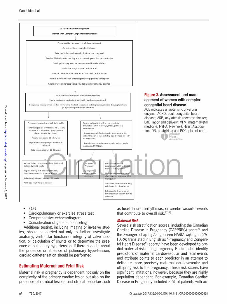
A brief overview of the anticipated delivery plan (vagi-nal versus cesarean delivery, anesthesia) should also be discussed (Figure 3). A preconception visit with a mater-nal-fetal medicine obstetrician can be helpful, particularly for women at higher risk for complications.
Genetic counseling may be particularly valuable to women for whom there is a significant risk of recurrence in offspring. The risk for recurrence of CHD varies widely from 3% to 50%, depending on the type of maternal heart disease73,74 (see Estimating Maternal and Fetal Risk).
Diagnostic Evaluation The preconception evaluation of a patient with complex CHD begins with a thorough review of medical records that include information on the primary defect, surgical history, including both palliative and reparative proce-dures, and the presence of comorbidities and any residua or sequelae associated with the specific cardiac lesions or surgery. Similarly, reports from prior testing such as echocardiograms, magnetic resonance imaging (MRI), and exercise tests should be available for comparison. The initial diagnostic evaluation includes measurement of arterial oxygen saturation, ECG, and echocardiogram.
Because maternal functional capacity is an important predictor of a woman’s ability to tolerate pregnancy, as-sessment of exercise capacity may be helpful. For most patients with complex disease, objective exercise test-ing by a cardiopulmonary exercise test or an exercise stress test may be in order to obtain an objective as-sessment of functional capacity and to facilitate the iden-tification of exercise-induced arrhythmias, particularly because many patients underreport or underrecognize the degree of their limitation.75,76
Suggestions for Clinical PracticeThe initial diagnostic evaluation should include the following:
• Detailed history, including any current cardiovascu-lar symptoms and family history
• Review of medications for benefits and risks, with appropriate adjustment or changes of drugs known to be teratogenic
• Arterial oxygen saturation• Baseline laboratory studies, including complete
blood count, electrolytes, and thyroid and liver function tests
Table 2. Normal Electrocardiographic Changes Associated With Pregnancy
Left axis shift is seen, with the greatest shift in the third trimester caused by elevation of the diaphragm.
Shortening of the PR, QRS, and QT intervals may accompany the increase in resting heart rate.
Nonspecific ST abnormalities, including segment depression or flattened and inverted T waves in lead III, occur frequently.
by guest on February 5, 2017http://circ.ahajournals.org/
Dow
nloaded from
Canobbio et al
TBD, 2017 Circulation. 2017;135:00-00. DOI: 10.1161/CIR.0000000000000458e6
• ECG• Cardiopulmonary or exercise stress test• Comprehensive echocardiogram• Consideration of genetic counselingAdditional testing, including imaging or invasive stud-
ies, should be carried out only to further investigate anatomy, ventricular function or integrity of valve func-tion, or calculation of shunts or to determine the pres-ence of pulmonary hypertension. If there is doubt about the presence or absence of pulmonary hypertension, cardiac catheterization should be performed.
Estimating Maternal and fetal riskMaternal risk in pregnancy is dependent not only on the complexity of the primary cardiac lesion but also on the presence of residual lesions and clinical sequelae such
as heart failure, arrhythmias, or cerebrovascular events that contribute to overall risk.77–79
Maternal RiskSeveral risk stratification scores, including the Canadian Cardiac Disease in Pregnancy (CARPREG) score74 and the Zwangerschap bij Aangeboren HARtAfwijkingen (ZA-HARA; translated in English as “Pregnancy and Congeni-tal Heart Disease”) score,4 have been developed to pre-dict maternal risk during pregnancy. Both models identify predictors of maternal cardiovascular and fetal events and attribute points to each predictor in an attempt to delineate more precisely maternal cardiovascular and offspring risk to the pregnancy. These risk scores have significant limitations, however, because they are highly population dependent. For example, Canadian Cardiac Disease in Pregnancy included 22% of patients with ac-
figure 3. Assessment and man-agement of women with complex congenital heart disease. ACE indicates angiotensin-converting enzyme; ACHD, adult congenital heart disease; ARB, angiotensin receptor blocker; L&D, labor and delivery; MFM, maternal-fetal medicine; NYHA, New York Heart Associa-tion; OB, obstetrics; and POC, plan of care.
by guest on February 5, 2017http://circ.ahajournals.org/
Dow
nloaded from
Pregnancy in Patients With Complex CHD
Circulation. 2017;135:00-00. DOI: 10.1161/CIR.0000000000000458 TBD, 2017 e7
CLINICAL STATEMENTS
AND GUIDELINES
quired heart disease, and 4% of the population were included because of arrhythmias. Therefore, in an effort to prevent high-risk patients from becoming pregnant, including those with severe pulmonary hypertension and severely dilated aortas and those who are not repre-sented in these studies, a prepregnancy counseling ses-sion should be organized in an adult CHD center.
More recently, a prospective validation study re-ported that the modified WHO classification of maternal cardiovascular risk was the most reliable predictor of maternal cardiovascular complications, the most com-mon of which are arrhythmias and heart failure.3 The recommendation to each patient, however, should be individualized. If a reparable lesion or clinical problem is identified at the time of prepregnancy counseling, a recommendation for directed therapies to address the problem is made.
For some patients, the evaluation may suggest a pregnancy risk that is unacceptable to the patient. Ap-propriate contraception is vital for those women who are counseled against pregnancy and for those women who choose not to pursue pregnancy.
Fetal RiskMaternal CHD is major determinant of risk to the fetus and the neonate, resulting in the following:
• Higher frequency of spontaneous abortions, rang-ing between 15% and 25%, and intrauterine fetal demise in selected defects.79,80
• Higher frequency of recurrence of CHD, underscor-ing the need to offer fetal echocardiography to all pregnant women with CHD at 18 to 22 weeks.
• Higher preterm birth rate (10%–12%), especially in those with complex CHD (22%–65%).
• Higher frequency of neonatal events, for example, small for gestational age, respiratory distress syn-drome, interventricular hemorrhage, and neona-tal death (27.8%). Specific maternal risk factors such as subaortic ventricular outflow obstruction, maternal cyanosis, and reduced cardiac output have been reported as predictors of adverse peri-natal events.79,80
• Higher perinatal mortality that may be >4-fold higher than in the general population (<1%) and is common with premature delivery or recurrence of CHD. Perinatal mortality is highest in patients with Eisenmenger syndrome (27.7%).4,78,79
Genetic CounselingIt is important to provide genetic counseling to women with CHD on the potential for recurrence risk in the off-spring, ideally before conception.
Genetic counseling should include the assessment of genetic risk, a discussion of the genetic screening tests available and the implications of genetic test results, and pretest and posttest counseling. Positive consequences include a more accurate estimation of transmission risk
to offspring and greater awareness and better manage-ment of comorbid conditions that can influence fetal and maternal outcomes. A detailed 3-generation family history, including history of consanguinity and history of miscarriages, should be obtained in all pregnancies.81,82
Maternal and paternal evaluation should be performed for clinical features suggestive of a syndromic pheno-type. The absence of typical phenotypic features does not preclude the presence of a genetic or chromosomal anomaly; therefore, genetic testing may still be offered if the index of suspicion for a genetic pathogenesis is high. Most genetic conditions associated with CHD are autoso-mal dominant conditions, which have a transmission risk of 50%, including the Marfan, Holt-Oram, Noonan, Ala-gille, CHARGE (coloboma, heart defect, atresia choanae, retarded growth and development, genital abnormality, and ear abnormality), 22q11.2 microdeletion, and Wil-liams syndromes. For CHD that arises de novo, the risk of CHD recurrence in offspring is between 3% and 5%.83 The risk of recurrence is higher with heterotaxy, atrio-ventricular septal defect, and obstructive lesions of the left ventricular outflow tract.84 Besides genetic factors, it is important to assess for environmental risk factors such as obesity, diabetes mellitus, hypertension, infec-tions, alcohol, smoking, and teratogenic medications that can negatively affect fetal growth and well-being and increase the risk of fetal birth defects.85
Because a number of conotruncal abnormalities may be associated with chromosomal abnormalities such as 22q11.2 deletion syndrome, genetic testing should be offered not only to women with strong family history of CHD but also to women with cardiac lesions known to be associated with genetic disorders. These include interrupted aortic arch, truncus arteriosus, tetralogy of Fallot, pulmonary atresia, ventricular septal defect with aortic arch anomaly, isolated aortic arch anomaly, and discontinuous branch pulmonary arteries.
Two consensus statements from the American Heart Association (AHA) and the Canadian Cardiovascular So-ciety provide excellent guidance for genetic testing in CHD.85,86
Suggestions for Clinical Practice• A 3-generation family history, including any con-
sanguinity, should be completed.• Genetic evaluation should be made available to all
women with CHD to determine recurrence, par-ticularly in those patients with a family history of CHD and those with possible autosomal dominant lesions (eg, 22q 11 deletion).
PrEGNANCy MANAGEMENTIdeally, the cardiovascular evaluation should occur be-fore conception. For the patient who presents for the first time with an unplanned pregnancy, however, the
by guest on February 5, 2017http://circ.ahajournals.org/
Dow
nloaded from

Canobbio et al
TBD, 2017 Circulation. 2017;135:00-00. DOI: 10.1161/CIR.0000000000000458e8
diagnostic workup must be individualized with consider-ation of the potential risk to both mother and fetus.
Diagnostic Testing in PregnancyElectrocardiogramA standard 12-lead ECG is a simple method for evaluat-ing abnormal heart rhythm, chamber enlargement, evi-dence of ischemia, and medication effects. Normal elec-trocardiographic changes associated with pregnancy are listed in Table 2. Holter monitors and extended event recorders may be helpful in the evaluation of a patient with palpitations, presyncope, or syncope.
EchocardiographyTransthoracic echocardiography remains a mainstay of cardiac evaluation in the CHD population and can provide information on cardiac structure, anatomic abnormali-ties, chamber and great vessel dimensions, ventricular function, valvular function, and hemodynamics.
The use of agitated saline during pregnancy has not been systematically studied but is generally considered safe.87 Intravenous contrast agents such as perflutren lipid microspheres (Definity) or perflutren protein type A (Optison) have not been studied in pregnant women. Ei-ther may be used if a clear maternal benefit is defined.
An echocardiogram taken before the pregnancy is useful in establishing the baseline status and may be repeated safely throughout pregnancy.88 In those with limited echocardiographic windows, transesophageal echocardiography is relatively safe during pregnancy but with attention to airway protection to avoid the risk of vomiting and aspiration.68,89
Exercise Stress TestingExercise stress testing and measurement of maximal oxygen consumption (V⋅ o2max) during exercise can be useful in evaluating cardiopulmonary reserve, functional status, or potential exercise-induced arrhythmias during pregnancy, although they are rarely necessary.90–93 A submaximal stress protocol (80% of the maximal pre-dicted heart rate) is recommended.68,91 A stress echo-cardiogram is indicated to determine the risk of myo-cardial ischemia in patients with suspected or confirmed underlying coronary artery disease or in patients with mildly reduced ventricular function.
ImagingChest RadiographyChest radiography is generally not advocated during preg-nancy unless specifically indicated. Even the normal heart may appear to be increased in size during pregnancy.94
Magnetic Resonance ImagingMRI is particularly useful for evaluating extracardiac vas-cular structures, including the aorta and left ventricular
myocardium. The majority of studies evaluating MRI safety during pregnancy report that maternal risk asso-ciated with the use of MRI is the same as for nonpreg-nant patients.95–97 Safety concerns arise, however, with respect to the fetus. Although no ill effects to the fetus have been reported, concerns include the potential for fetal acoustic damage because MRI coils produce a loud tapping noise.98,99 Limited data on organogenesis are available, but MRI is probably safe after the first trimes-ter.68 The American College of Radiology recommends that gadolinium be avoided and used only when other imaging (transthoracic and transesophageal echocar-diography) is insufficient for diagnosis and that the risks and benefits be discussed with the pregnant patient and referring physician.96,97 If required, it is preferable to wait until the second or third trimester when organogenesis is completed.97,100
Computed TomographyThe risk of radiation exposure from computed tomog-raphy (CT) procedures varies, depending on gestational age and dose of radiation. CT is recommended only if other imaging is insufficient for the diagnosis.87.101 If pul-monary embolism is suspected and CT is thought nec-essary for definitive diagnosis, CT of the chest may be performed at low dose of fetal exposure with a thorough discussion of risks and benefits.68,101 V/Q scans carry similar risk. The threshold for fetal risk appears to be increased at doses >100 mGy.101,102
Cardiac CatheterizationConcerns exist over the short- and long-term conse-quences of fetal radiation exposure during diagnostic and interventional cardiac catheterization. However, in clinical instances when hemodynamic compromise or an emergent intervention requires either right-sided heart or full cardiac catheterization, radiation exposure can be minimized by ensuring shortened fluoroscopy time and ensuring that the radiation dose remains at lower levels.102
The radiation exposure to the fetus arises predomi-nantly from scattered radiation within the patient. Ex-ternal lead shielding of the maternal pelvis is of limited value although generally still used. The radiation dose absorbed by the fetus without shielding is only 3% higher than that with external shielding for all periods of gestation.87,97
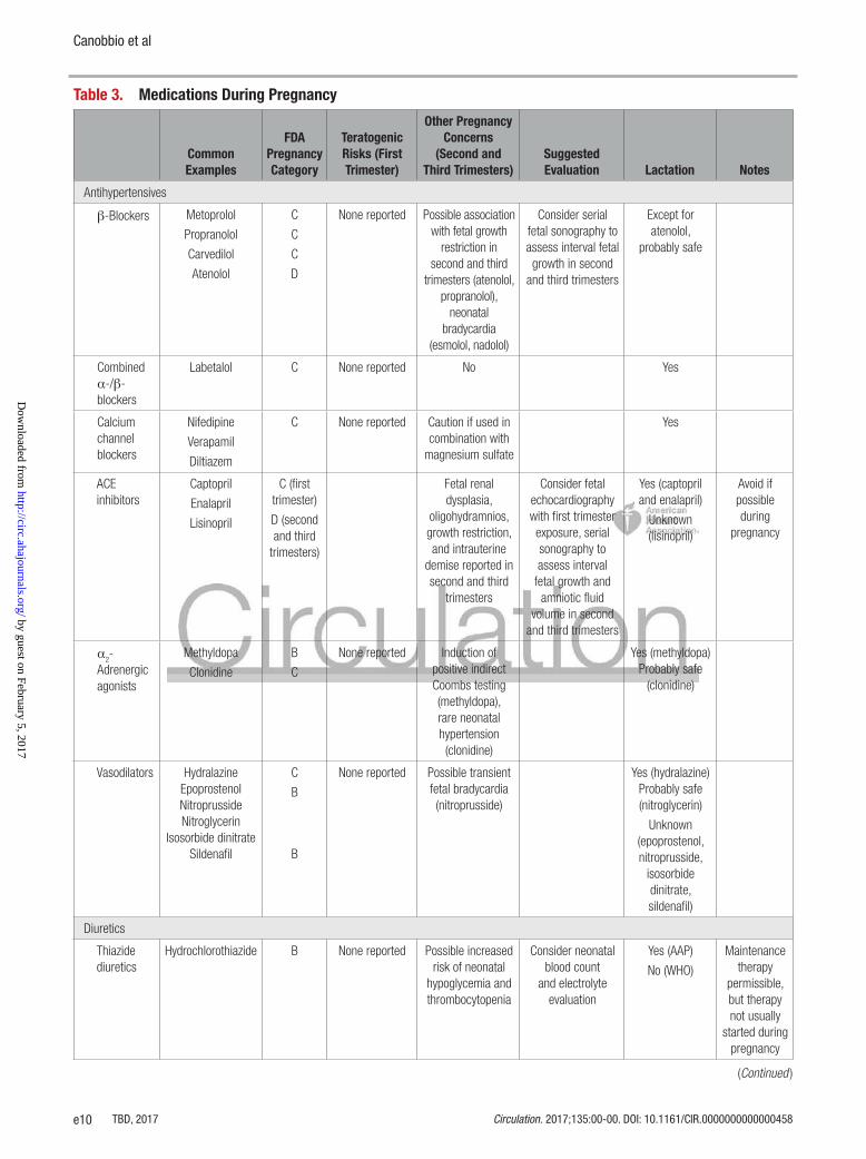
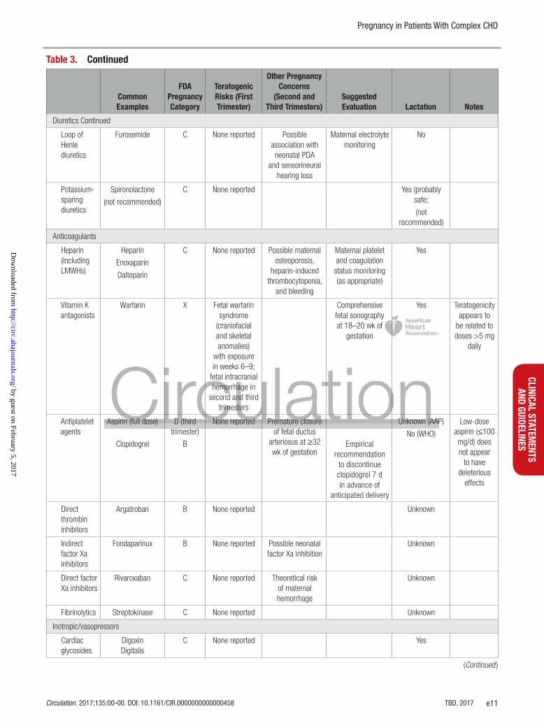
Medications in PregnancyThe relative safety of any medication during pregnancy involves the comparative determination of maternal benefit versus potential fetal risk. Teratogenicity occurs primarily during postmenstrual weeks 4 to 12, with ex-posures during the first 2 weeks after conception consid-ered to result in either loss of pregnancy or no significant effect. Table 3 presents an overview of common medica-
by guest on February 5, 2017http://circ.ahajournals.org/
Dow
nloaded from

Pregnancy in Patients With Complex CHD
Circulation. 2017;135:00-00. DOI: 10.1161/CIR.0000000000000458 TBD, 2017 e9
CLINICAL STATEMENTS
AND GUIDELINES
tions encountered in the adult CHD population, highlight-ing specific concerns during pregnancy. The individual Micromedex REPROTOX drug summaries (Micromedex 2.0) provide a more comprehensive discussion.103
Currently, the US Food and Drug Administration clas-sifies drugs into risk categories based on safety during pregnancy and breastfeeding.1 Table 4 shows the risk category for some commonly used drugs in patients with heart disease.
Angiotensin-converting enzyme (ACE) inhibitors and angiotensin receptor blockers are pregnancy category D drugs because of the risk of reduced fetal renal function and increased fetal and neonatal morbidity and death, es-pecially with use during the second and third trimesters. Cooper et al104 reported an additional risk of malforma-tion among infants exposed to ACE inhibitors during the first trimester. Ideally, women taking an ACE inhibitor or angiotensin receptor blocker who desire to become preg-nant should be advised to stop taking these drugs before conception to minimize the risk of fetal abnormality. Dur-ing this time, repeated clinical evaluation with echocar-diography should be carried out to reassess ventricular function and exercise capacity. Stability in ejection frac-tion suggests that patients are less likely to be intolerant of the volume load that pregnancy imposes.
β-Blockers have a favorable safety profile, and no teratogenic risks have been reported with their use.105 They have been associated with an increase in small-for-gestational-age infants and neonatal bradycardia and hypoglycemia but in practical terms are considered safe to use. Atenolol has been associated with low birth weight and therefore is not the β-blocker of choice dur-ing pregnancy.106
The routine use of diuretics is discouraged because of the potential reduction in maternal plasma volume early in gestation that is potentially harmful to the fetus.107 If in-dicated, they should be used judiciously. Thiazides have been associated with maternal and fetal thrombocytope-nia. Although listed as a category C agent, spironolac-tone is not recommended for use during pregnancy or lactation because of the reported antiandrogenic effects and feminization of male fetuses. Amiodarone has been associated with a 9% incidence of fetal hypothyroidism and a 21% incidence of intrauterine growth retardation and should be reserved for cases of refractory ventricu-lar arrhythmias.108
Drugs listed as category X include the following:• Anticoagulants. Vitamin K antagonists cross the
placenta and are potentially fetotoxic. They are commonly used in women with mechanical valves, and the risks and benefits associated with their use should be discussed with the patient before preg-nancy is contemplated (see Mechanical Valves and Anticoagulation).
• Endothelin receptor antagonists (bosentan, ambrisen-tan, and macitentan) (Micromedex and REPROTOX).
• Statins. Although inadvertent exposure to statins during early pregnancy appears unlikely to increase the risk of adverse pregnancy outcome, theoretical considerations concerning the role of cholesterol in embryo development and the lack of demonstrated benefit of treating hyperlipidemia during gestation are reasons for not recommend-ing statins during pregnancy.109,110 They remain pregnancy category X drugs in the United States.
fetal Screening During PregnancyFormal fetal echocardiography at 18 to 22 weeks’ ges-tation is recommended for all patients with CHD and partners of male patients with CHD68; earlier echocardio-graphic screening may be recommended in patients with high rates of familial CHD recurrence and more severe cardiac defects. Referral to a maternal-fetal medicine specialist, pediatric cardiologist, neonatologist, and ge-neticist is indicated if fetal CHD is discovered to discuss prognosis and perinatal management.
Antepartum CareExisting adult CHD and pregnancy guidelines recom-mend that patients with complex CHD should be man-aged and delivered at a regional or tertiary center where a multidisciplinary team with knowledge and experience in adult CHD is available.68,73,86 This team includes a cardiologist, a high-risk obstetrician, an anesthesiolo-gist, and a neonatologist. Additional providers, includ-ing a geneticist, an advanced practice nurse, a social worker, and an ethicist, should be identified to assist in the coordination of care as required. Early coordination and ongoing communication between members of the multidisciplinary team are crucial to optimizing maternal outcomes. Prenatal care is individualized on the basis of the patient’s maternal risk, accounting for the com-plexity of the CHD, the patient’s functional capacity, and the presence of any existing or potential clinical issues (ie, rhythm disturbances). Practical factors such as cur-rent social situation, insurance coverage, and proximity to the adult CHD center and delivery hospital should be considerations in clinical scheduling.
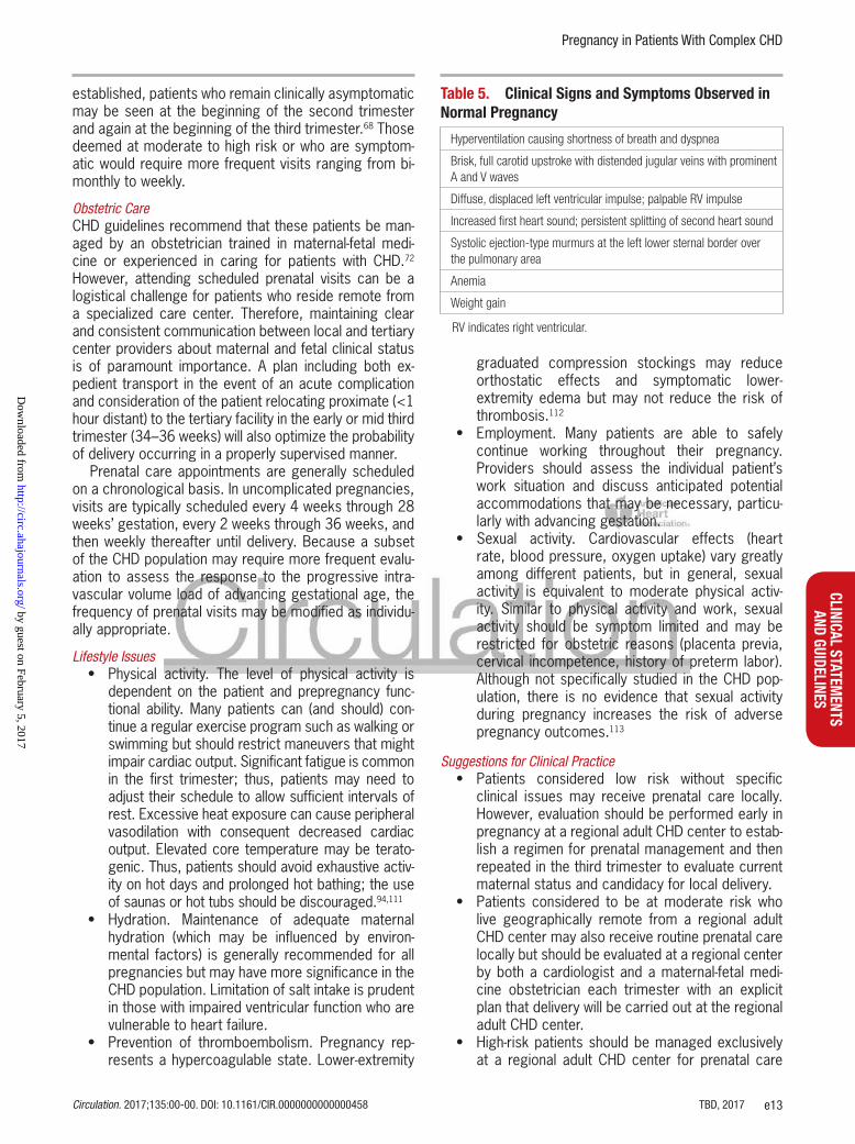
First Trimester (0–14 Weeks)Cardiology CareThe initial visit should include a review of the patient’s car-diac history and preconception diagnostic evaluations to elucidate any new symptoms such as palpitation, short-ness of breath, or edema. The physician should perform a comprehensive cardiovascular examination, keeping in mind the normal physiological changes apparent at 12 weeks of pregnancy (Table 5). Particular attention should be given to the development of arrhythmias, new mur-murs, or clinical evidence of heart failure.
by guest on February 5, 2017http://circ.ahajournals.org/
Dow
nloaded from
Canobbio et al
TBD, 2017 Circulation. 2017;135:00-00. DOI: 10.1161/CIR.0000000000000458e10
Table 3. Medications During Pregnancy
Common Examples
fDA Pregnancy Category
Teratogenic risks (first Trimester)
other Pregnancy Concerns
(Second and Third Trimesters)
Suggested Evaluation Lactation Notes
Antihypertensives
β-Blockers Metoprolol
Propranolol
Carvedilol
Atenolol
C
C
C
D
None reported Possible association with fetal growth
restriction in second and third
trimesters (atenolol, propranolol),
neonatal bradycardia
(esmolol, nadolol)
Consider serial fetal sonography to assess interval fetal growth in second
and third trimesters
Except for atenolol,
probably safe
Combined α-/β-blockers
Labetalol C None reported No Yes
Calcium channel blockers
Nifedipine
Verapamil
Diltiazem
C None reported Caution if used in combination with
magnesium sulfate
Yes
ACE inhibitors
Captopril
Enalapril
Lisinopril
C (first trimester)
D (second and third
trimesters)
Fetal renal dysplasia,
oligohydramnios, growth restriction, and intrauterine
demise reported in second and third
trimesters
Consider fetal echocardiography with first trimester exposure, serial sonography to assess interval
fetal growth and amniotic fluid
volume in second and third trimesters
Yes (captopril and enalapril)
Unknown (lisinopril)
Avoid if possible during
pregnancy
α2-
Adrenergic agonists
Methyldopa
Clonidine
B
C
None reported Induction of positive indirect Coombs testing (methyldopa), rare neonatal hypertension (clonidine)
Yes (methyldopa) Probably safe
(clonidine)
Vasodilators Hydralazine Epoprostenol Nitroprusside Nitroglycerin
Isosorbide dinitrate Sildenafil
C
B
B
None reported Possible transient fetal bradycardia (nitroprusside)
Yes (hydralazine) Probably safe (nitroglycerin)
Unknown (epoprostenol, nitroprusside,
isosorbide dinitrate, sildenafil)
Diuretics
Thiazide diuretics
Hydrochlorothiazide B None reported Possible increased risk of neonatal
hypoglycemia and thrombocytopenia
Consider neonatal blood count
and electrolyte evaluation
Yes (AAP)
No (WHO)
Maintenance therapy
permissible, but therapy not usually
started during pregnancy
(Continued )
by guest on February 5, 2017http://circ.ahajournals.org/
Dow
nloaded from
Pregnancy in Patients With Complex CHD
Circulation. 2017;135:00-00. DOI: 10.1161/CIR.0000000000000458 TBD, 2017 e11
CLINICAL STATEMENTS
AND GUIDELINES
Diuretics Continued
Loop of Henle diuretics
Furosemide C None reported Possible association with neonatal PDA
and sensorineural hearing loss
Maternal electrolyte monitoring
No
Potassium-sparing diuretics
Spironolactone
(not recommended)
C None reported Yes (probably safe;
(not recommended)
Anticoagulants
Heparin (including LMWHs)
Heparin
Enoxaparin
Dalteparin
C None reported Possible maternal osteoporosis,
heparin-induced thrombocytopenia,
and bleeding
Maternal platelet and coagulation
status monitoring (as appropriate)
Yes
Vitamin K antagonists
Warfarin X Fetal warfarin syndrome
(craniofacial and skeletal anomalies)
with exposure in weeks 6–9;
fetal intracranial hemorrhage in
second and third trimesters
Comprehensive fetal sonography at 18–20 wk of
gestation
Yes Teratogenicity appears to
be related to doses >5 mg
daily
Antiplatelet agents
Aspirin (full dose)
Clopidogrel
D (third trimester)
B
None reported Premature closure of fetal ductus
arteriosus at ≥32 wk of gestation
Empirical recommendation
to discontinue clopidogrel 7 d in advance of
anticipated delivery
Unknown (AAP)
No (WHO)
Low-dose aspirin (≤100 mg/d) does not appear
to have deleterious
effects
Direct thrombin inhibitors
Argatroban B None reported Unknown
Indirect factor Xa inhibitors
Fondaparinux B None reported Possible neonatal factor Xa inhibition
Unknown
Direct factor Xa inhibitors
Rivaroxaban C None reported Theoretical risk of maternal hemorrhage
Unknown
Fibrinolytics Streptokinase C None reported Unknown
Inotropic/vasopressors
Cardiac glycosides
DigoxinDigitalis
C None reported Yes
Table 3. Continued
Common Examples
fDA Pregnancy Category
Teratogenic risks (first Trimester)
other Pregnancy Concerns
(Second and Third Trimesters)
Suggested Evaluation Lactation Notes
(Continued )
by guest on February 5, 2017http://circ.ahajournals.org/
Dow
nloaded from

Canobbio et al
TBD, 2017 Circulation. 2017;135:00-00. DOI: 10.1161/CIR.0000000000000458e12
The current medication regimen should also be re-viewed for appropriate indications, potential risks, and any need for dose adjustments or discontinuation. For the patient who presents for the first time early in preg-nancy for cardiac care, the initial visit should include a
thorough assessment as described previously for a pa-tient contemplating pregnancy.
The frequency of cardiology visits is largely individual-ized by estimated risk and the development of symp-toms or complications. Once antepartum care has been
Table 4. fDA Drug Classifications
Rating A Controlled studies in women fail to demonstrate a risk to the fetus in the first trimester; no evidence of a risk.
Rating B Either animal-reproduction studies have not demonstrated a fetal risk but there are no controlled studies in pregnant women or animal-reproduction studies have shown adverse effect (other than a decrease in fertility) that was not confirmed in controlled studies in women in the first trimester (and there is no evidence of a risk in later trimesters).
Rating C Either studies in animals have revealed adverse effects on the fetus (teratogenic, embryocidal, or other) and there are no controlled studies in women or studies in women and animals are not available. Drugs should be given only if the potential benefit justifies the potential risk to the fetus.
Rating D There is positive evidence of human fetal risk, but the benefits from use in pregnant women may be acceptable despite the risk (eg, if the drug is needed in a life-threatening situation or for a serious disease for which safer drugs cannot be used or are ineffective).
Rating X Studies in animals or human beings have demonstrated fetal abnormalities, there is evidence of fetal risk based on human experience, or both, and the risk of the use of the drug in pregnant women clearly outweighs any possible benefit. The drug is contraindicated in women who are or may become pregnant.
FDA indicates US Food and Drug Administration.Data are from REPROTOX.103
Inotropic/vasopressors Continued
Adrenergic agents
Epinephrine
Norepinephrine
Phenylephrine
Dopamine
Isoproterenol
C Possible association with
gastroschisis or hemifacial microsomia
(phenylephrine)
Probably safe (dopamine)
Unknown (epinephrine,
norepinephrine, phenylephrine, isoproterenol)
Antiarrhythmics
β-Blockers Metoprolol
Propranolol
Carvedilol
Atenolol
C
C
C
D
None reported Association with fetal growth restriction in second and third trimesters (atenolol,
propranolol), neonatal bradycardia (esmolol, nadolol)
Consider serial fetal sonography to assess interval fetal growth in second
and third trimesters
Except for atenolol,
probably safe
Class 1A Quinidine Procainamide
C
C
None reported Yes
Class 1C Flecainide C None reported Yes
Class III Sotalol
Amiodarone
B
D
None reported
Thyroid dysfunction
No
No WHO)/ (AAP Yes)
Purine nucleosides
Adenosine C None reported Unknown
AAP indicates American Academy of Pediatrics; ACE, angiotensin-converting enzyme; FDA, US Food and Drug Administration; LMWH, low-molecular-weight heparin; PDA, patent ductus arteriosus; and WHO, World Health Organization.
Table 3. Continued
Common Examples
fDA Pregnancy Category
Teratogenic risks (first Trimester)
other Pregnancy Concerns
(Second and Third Trimesters)
Suggested Evaluation Lactation Notes
by guest on February 5, 2017http://circ.ahajournals.org/
Dow
nloaded from
Pregnancy in Patients With Complex CHD
Circulation. 2017;135:00-00. DOI: 10.1161/CIR.0000000000000458 TBD, 2017 e13
CLINICAL STATEMENTS
AND GUIDELINES
established, patients who remain clinically asymptomatic may be seen at the beginning of the second trimester and again at the beginning of the third trimester.68 Those deemed at moderate to high risk or who are symptom-atic would require more frequent visits ranging from bi-monthly to weekly.
Obstetric CareCHD guidelines recommend that these patients be man-aged by an obstetrician trained in maternal-fetal medi-cine or experienced in caring for patients with CHD.72 However, attending scheduled prenatal visits can be a logistical challenge for patients who reside remote from a specialized care center. Therefore, maintaining clear and consistent communication between local and tertiary center providers about maternal and fetal clinical status is of paramount importance. A plan including both ex-pedient transport in the event of an acute complication and consideration of the patient relocating proximate (<1 hour distant) to the tertiary facility in the early or mid third trimester (34–36 weeks) will also optimize the probability of delivery occurring in a properly supervised manner.
Prenatal care appointments are generally scheduled on a chronological basis. In uncomplicated pregnancies, visits are typically scheduled every 4 weeks through 28 weeks’ gestation, every 2 weeks through 36 weeks, and then weekly thereafter until delivery. Because a subset of the CHD population may require more frequent evalu-ation to assess the response to the progressive intra-vascular volume load of advancing gestational age, the frequency of prenatal visits may be modified as individu-ally appropriate.
Lifestyle Issues• Physical activity. The level of physical activity is
dependent on the patient and prepregnancy func-tional ability. Many patients can (and should) con-tinue a regular exercise program such as walking or swimming but should restrict maneuvers that might impair cardiac output. Significant fatigue is common in the first trimester; thus, patients may need to adjust their schedule to allow sufficient intervals of rest. Excessive heat exposure can cause peripheral vasodilation with consequent decreased cardiac output. Elevated core temperature may be terato-genic. Thus, patients should avoid exhaustive activ-ity on hot days and prolonged hot bathing; the use of saunas or hot tubs should be discouraged.94,111
• Hydration. Maintenance of adequate maternal hydration (which may be influenced by environ-mental factors) is generally recommended for all pregnancies but may have more significance in the CHD population. Limitation of salt intake is prudent in those with impaired ventricular function who are vulnerable to heart failure.
• Prevention of thromboembolism. Pregnancy rep-resents a hypercoagulable state. Lower-extremity
graduated compression stockings may reduce orthostatic effects and symptomatic lower-extremity edema but may not reduce the risk of thrombosis.112
• Employment. Many patients are able to safely continue working throughout their pregnancy. Providers should assess the individual patient’s work situation and discuss anticipated potential accommodations that may be necessary, particu-larly with advancing gestation.
• Sexual activity. Cardiovascular effects (heart rate, blood pressure, oxygen uptake) vary greatly among different patients, but in general, sexual activity is equivalent to moderate physical activ-ity. Similar to physical activity and work, sexual activity should be symptom limited and may be restricted for obstetric reasons (placenta previa, cervical incompetence, history of preterm labor). Although not specifically studied in the CHD pop-ulation, there is no evidence that sexual activity during pregnancy increases the risk of adverse pregnancy outcomes.113
Suggestions for Clinical Practice• Patients considered low risk without specific
clinical issues may receive prenatal care locally. However, evaluation should be performed early in pregnancy at a regional adult CHD center to estab-lish a regimen for prenatal management and then repeated in the third trimester to evaluate current maternal status and candidacy for local delivery.
• Patients considered to be at moderate risk who live geographically remote from a regional adult CHD center may also receive routine prenatal care locally but should be evaluated at a regional center by both a cardiologist and a maternal-fetal medi-cine obstetrician each trimester with an explicit plan that delivery will be carried out at the regional adult CHD center.
• High-risk patients should be managed exclusively at a regional adult CHD center for prenatal care
Table 5. Clinical Signs and Symptoms observed in Normal Pregnancy
Hyperventilation causing shortness of breath and dyspnea
Brisk, full carotid upstroke with distended jugular veins with prominent A and V waves
Diffuse, displaced left ventricular impulse; palpable RV impulse
Increased first heart sound; persistent splitting of second heart sound
Systolic ejection-type murmurs at the left lower sternal border over the pulmonary area
Anemia
Weight gain
RV indicates right ventricular.
by guest on February 5, 2017http://circ.ahajournals.org/
Dow
nloaded from

Canobbio et al
TBD, 2017 Circulation. 2017;135:00-00. DOI: 10.1161/CIR.0000000000000458e14
and delivery. If geographical and financial con-cerns preclude this option, coordinated shared care between a regional center and a local obste-trician and cardiologist who are willing to communi-cate directly with the regional adult CHD should be explored once fetal viability has been established.
• For patients considered exceedingly high risk for maternal morbidity or mortality such as those with elevated pulmonary pressures or important aortic root enlargement, therapeutic termination of the pregnancy should be offered (see Termination of Pregnancy).
Second Trimester (14–28 Weeks)The second trimester is associated with the greatest magnitude of hemodynamic changes (Figures 1 and 2). The frequency of evaluation must be individualized. A repeat echocardiogram may be indicated to evaluate the hemodynamic effects of the pregnancy on cardiac and valvular function. Comprehensive fetal echocar-diography is usually performed at 18 to 22 weeks’ ges-tation and may be repeated if any fetal anomaly has been detected.
A clear and coordinated plan for labor, delivery, and postpartum care should be developed by the end of the second trimester and distributed to all members of the multidisciplinary team, including labor and delivery staff, in the event of spontaneous or indicated preterm delivery. For patients deemed at particularly high risk (eg, those who have pulmonary hypertension or severe AS), an initial multidisciplinary planning meeting includ-ing all providers potentially involved in their care should be organized once fetal viability has been established, usually after week 23 to 24. A specific delivery plan is outlined with contingencies for early hospital admis-sion and requirement for urgent delivery. If cardiac sup-port systems such as ventricular assist devices are re-quired or if cardiac surgery is being considered during pregnancy or concomitant with delivery, members of the cardiothoracic team should also be included. The involvement of social services and possibly an institu-tional ethics team should be also considered in high-risk cases.
Third Trimester (28–42 Weeks)The frequency of cardiac evaluation in late pregnancy must be individualized. Once the peak hemodynamic load of pregnancy is reached, normal symptoms of preg-nancy (edema, dyspnea on exertion) may worsen, so patients must be monitored closely to distinguish such symptoms and signs of normal pregnancy from those that may reflect hemodynamic compromise. Continued participation in physical activity, work, and sexual activ-ity may prove difficult as the pregnancy progresses; all activities should be limited by symptomatology or ob-stetric concerns. Planning and contingencies for delivery should be finalized in the third trimester.
Obstetric ComplicationsBecause these patients remain at risk for the spectrum of obstetric complications independently of their cardiac condition, specific obstetric concerns in this population include the following:
• Spontaneous abortion. The rate of spontaneous abortion varies between 12% and 15%, depending on the primary cardiac anomaly.79,114
• Fetal growth restriction. Small-for-gestational-age neonates (birth weight <10%) have been reported in 4% to 8% of pregnancies, with intrauterine demise reported in 3%.79
• Preterm delivery. Approximately 17% to 21% of all patients with CHD ultimately deliver preterm as a result of either spontaneous preterm labor/pre-term premature rupture of membranes (59%) or indicated delivery (41%).74,79 The use of tocolytic agents is generally considered safe, but caution is warranted with the use of terbutaline, particularly in patients with a history of arrhythmias.
• Hypertension. Increases in SVR such as with gestational hypertension/preeclampsia may be poorly tolerated in patients with marginal cardiac output.
Termination of PregnancyFor the patient with complex CHD in whom continua-tion of pregnancy presents a substantial risk of mater-nal morbidity or mortality, a forthright discussion of the advisability of therapeutic termination is imperative. However, because termination incurs an increased ma-ternal risk with advancing gestational age regardless of method, expediency in patient decisiveness is impor-tant. The therapeutic benefit of termination for patients presenting at ≥20 weeks of gestation is controversial because many of the physiological cardiopulmonary ad-aptations are established by this time and consequently would not necessarily be mitigated by interruption of the pregnancy.
• First trimester. Surgical dilation and suction curet-tage represents the most common method of pregnancy termination through 12 weeks’ gesta-tion. Complication rates are low for skilled practi-tioners, and selection of anesthetic technique may be dictated by maternal status. The procedure should be carried out in a hospital setting where careful monitoring is accessible. Medical abor-tion regimens using combined antiprogesterone (mifepristone) and prostaglandin E1 (misoprostol) agents are similar in efficacy to suction curettage if administered within the first 7 weeks of preg-nancy; however, because the process and subse-quent hemorrhage typically occur in a relatively unpredictable manner in an unmonitored outpa-tient setting, this may not be an appropriate option for hemodynamically fragile patients.
by guest on February 5, 2017http://circ.ahajournals.org/
Dow
nloaded from

Pregnancy in Patients With Complex CHD
Circulation. 2017;135:00-00. DOI: 10.1161/CIR.0000000000000458 TBD, 2017 e15
CLINICAL STATEMENTS
AND GUIDELINES
• Second trimester. Midtrimester medical termi-nation can be accomplished with transvaginal misoprostol to induce labor on an inpatient basis. Disadvantages of this approach include prolonged duration (>24 hours), intrapartum discomfort, and potential requirement for uterine curettage if placental retention occurs. Surgical dilation and evacuation is more frequently performed, offering the principal advantage of effecting termination under controlled circumstances in a surgical suite. Although no studies have specifically evaluated adult patients with CHD, in the general population, dilation and evacuation by experienced providers appears to have a lower complication rate than labor induction at gestational ages between 13 and 24 weeks.115
Intrapartum CareClinically stable patients with complex CHD should antici-pate a normal labor and delivery. The risks for gravidas with functionally significant CHD can be minimized by proper planning and management of labor, delivery, and the puerperium. In these high-risk cases, elective induc-tion of labor under controlled conditions is recommend-ed.116 Careful consideration should be given to maternal benefit versus neonatal risk when elective delivery before 39 weeks is contemplated because induction of labor may be less successful at earlier gestational ages.116
From a practical point of view, inductions should be initiated so that delivery will likely occur during regular hours when the adult CHD teams are readily available. Labor should be conducted in a right or left lateral tilt position to maximize maternal hemodynamic stability; this posture reduces compression of the inferior vena by the gravid uterus, which maintains cardiac preload. Vaginal delivery with adequate relief of pain with paren-tal narcotic analgesia or epidural anesthesia is generally preferred. Cesarean delivery is typically reserved for ob-stetric indications.
The uterine contractions of the second stage of la-bor (which extends from the time of complete cervical dilatation until delivery) are normally augmented by ma-ternal Valsalva maneuvers. Historically, Valsalva maneu-vers have been discouraged for patients with significant cardiac disease because of the associated increase in maternal O2 consumption and reduction in cardiac return and cardiac output. Management should be individual-ized; with some cardiac disorders, a passive second stage of labor may be more appropriate (the fetus de-scends through the birth canal exclusively via uterine contractions), particularly in women whose venous return or myocardial contractility is significantly compromised. Regional epidural anesthesia can suppress the Valsalva reflex arising from fetal pelvic descent, which can be re-instated by reducing the intensity of anesthesia. Forceps-
or vacuum-assisted operative delivery may be performed to facilitate delivery from a low or outlet station.
Neuraxial anesthetic techniques should be used cau-tiously if cardiac output is sensitive to a reduction in pre-load; a narcotic combined spinal-epidural may be opti-mal. General anesthesia may be used, but the inhalation agents must be carefully selected and administered by an anesthesiologist.
Intrapartum Management• Intravenous crystalloid should be administered
when necessary for hydration with close monitor-ing of fluid balance. All patients with right-to-left shunts should have filtered vascular lines to pre-vent paradoxical air embolization.117 Patients with complex CHD should have continuous pulse oxim-etry for monitoring changes in systemic arterial oxygen saturation (Sao2). In those with right-to-left shunts, Sao2 provides a continuous estimate of the extent of the right-to-left shunt.118 Maintenance of systemic pressures in these gravidas is critical to maintain the balance of systemic and pulmonary blood flow. A decrease in SVR or an increase in PVR leads to increased right-to-left shunting, result-ing in an increase in hypoxemia and an increase in the risk of maternal and fetal death.117
• Continuous ECG monitoring (eg, via telemetry) is indicated for patients with history of arrhythmias before or during pregnancy or for gravidas with reduced ventricular function who have become symptomatic during gestation.
• Invasive hemodynamic monitoring is rarely required for the vast majority of patients with complex CHD; exceptions include patients with clinical evidence of congestive heart failure and volume overload, in whom a central venous catheter may be useful to guide intravenous fluid administration. An arterial line can be helpful in monitoring fluid shifts and blood loss. If implemented, hemodynamic monitor-ing should continue for 24 hours after delivery.
• Neither transvaginal nor cesarean delivery is consid-ered to inherently impart a high risk of bacteremia. Consequently, the AHA119 does not recommend antibiotic prophylaxis for delivery. Because patients with CHD can suffer life-threatening consequences (eg, patients with Eisenmenger syndrome, cyanotic patients7) should they develop endocarditis, it is not unreasonable to administer antibiotics to high-risk patients. When indicated, antibiotics for endocardi-tis prophylaxis should be given at least 30 minutes before anticipated delivery.
Postpartum CareImmediately after delivery, the intravascular volume is augmented by an “autotransfusion” of 500 mL blood
by guest on February 5, 2017http://circ.ahajournals.org/
Dow
nloaded from
Canobbio et al
TBD, 2017 Circulation. 2017;135:00-00. DOI: 10.1161/CIR.0000000000000458e16
from the involuting uterus. In uncomplicated pregnan-cies, stroke volume and cardiac output increase imme-diately by 71% and 60% to 80%, respectively. These changes begin to reverse shortly after delivery and con-tinue to decrease over the next 24 hours, resolving over the next 6 to 8 weeks.59–61,120,121 The extravascular fluid accumulation that occurs during gestation will typically resolve over a similar time interval.
Because many of these hemodynamic changes occur concurrently, cardiopulmonary complications may ensue immediately after delivery, depending on the maternal hemodynamic compensatory ability. Consequently, rec-ommendations for postpartum monitoring are largely dependent on the patient’s underlying congenital cardiac abnormality, predisposition to arrhythmias, presence or absence of heart failure signs or symptoms, and clini-cal course during pregnancy and delivery.122 Telemetry cardiac monitoring should continue for at least 24 hours for those patients with symptoms or signs of significant antepartum or intrapartum arrhythmias. For the patient considered at highest risk or who has demonstrated signs of decompensation during the pregnancy or deliv-ery period, management in an intensive care unit/critical care unit setting for the first 24 to 48 hours after deliv-ery for hemodynamic monitoring should be considered.
If a patient is delivered in a local or community-based institution with limited resources or staff unfamiliar with managing complex CHD, it may be prudent to care for these patients in an intensive care unit/critical care unit setting, along with appropriate obstetric nursing surveil-lance, for the first 24 to 48 hours postpartum. Standard postpartum units are less likely to have cardiac moni-toring capability or clinical experience in recognizing and managing early cardiopulmonary decompensation. However, patients who have remained clinically stable throughout pregnancy and delivery may be transferred directly to postpartum units with instructions to monitor for cardiopulmonary symptoms. Early ambulation and continued use of support stockings may reduce the risk of thromboembolism.117
The hemodynamic effects of pregnancy typically re-solve primarily during the initial 6 to 12 weeks postpar-tum but may persist for up to 6 months.123 Thus, dis-charge planning should include appropriate return visits for routine postpartum and cardiac evaluation and de-tailed patient instructions for signs and symptoms merit-ing medical evaluation.
ContraceptionContraception is an important topic to readdress pe-riodically throughout the reproductive life of a woman with complex CHD. The optimal method is governed by medical candidacy, potential side effects, compliance, and risk of unplanned conception. The ideal time to discuss contraceptive methods is before conception, but it is equally important to have a discussion on con-
traceptive choices with the pregnant patient as part of her postpartum management. Typical methods include the following:
• Combination estrogen-progesterone oral con-traceptive pills. Advantages include predictable menstrual cyclicity, high efficacy, and reduction in incidences of gynecologic malignancy. In gen-eral, estrogen-containing oral contraceptives have historically been avoided during lactation because of concern about a pharmacological reduction in milk supply, although existing data are conflict-ing.124 Other contraindications in the CHD popula-tion include a history of previous thrombosis (not requiring maintenance anticoagulation), cyanosis, or elevated hepatic functions. Because of the rela-tively thrombophilic state immediately postpartum, combination oral contraceptive pills are usually ini-tiated 21 to 30 days after delivery.
• Progesterone-only contraception. This category includes progesterone-only contraceptive pills, depot medroxyprogesterone acetate injections, and subdermal etonogestrel implants. Because progesterone is not considered to increase the risk of thrombosis, any of these methods may be initiated during the postpartum inpatient hos-pitalization. Long-acting (36-month) subdermal etonogestrel implants (Implanon, Nexplanon) are also widely available. For patients with a history of venous thromboembolism or those with cyanotic conditions, progesterone-only contraception may represent an optimal selection.
• Intrauterine device. Although historically the intra-uterine device was avoided in patients with CHD because of concern about maternal bacteremia and anemia from menorrhagia, the newer levo-norgestrel-releasing intrauterine devices (Mirena, Skyla) may strike an optimal compromise between risk of unplanned pregnancy and effective con-traception. Insertion is usually delayed until ≥4 weeks postpartum to minimize the incidence of spontaneous expulsion, although insertion imme-diately postpartum after placental delivery or at the time of cesarean section has been described. Neither type of intrauterine device appears to affect lactation.125
• Sterilization. If subsequent fertility is not desired, sterilization via tubal ligation or occlusion proce-dures may offer the lowest risk of future unin-tended conception. If cesarean delivery is planned or required, concurrent bilateral tubal ligation may be performed expediently, with a low cumulative 10-year risk of failure (1:133).126 Office-based hys-teroscopic fallopian tubal occlusion with a metallic insert (Essure) offers an efficacious alternative to an abdominal surgical approach, although interval contraception is required until subsequent imaging
by guest on February 5, 2017http://circ.ahajournals.org/
Dow
nloaded from
Pregnancy in Patients With Complex CHD
Circulation. 2017;135:00-00. DOI: 10.1161/CIR.0000000000000458 TBD, 2017 e17
CLINICAL STATEMENTS
AND GUIDELINES
is performed to confirm complete occlusion 3 months after the procedure. Recent reports have raised concern that patients with metal allergies or a history of pelvic inflammatory disease or other autoimmune conditions maybe suboptimal candi-dates for this procedure.127
Although partner vasectomy may be an option for couples in a monogamous relationship, this procedure does not directly address the risk of pregnancy in an individual CHD patient. Furthermore, because in some cases the male partner may outlive the female partner with CHD, the decision for vasectomy should factor in the possibility that later he may wish to have a family.
CArDIAC CoMPLICATIoNSArrhythmiasArrhythmia Risk During PregnancyPregnancy is associated with physiological changes that can cause shortening of the PR, QRS, and QT inter-vals as a result of an increase in heart rate or a leftward shift of the axis (small q wave, inverted T wave in lead III) resulting from an elevation of the diaphragm causing rotation of the heart. Ectopic beats are common and usually benign.41 However, pregnancy can also lower the threshold for ventricular arrhythmias and reentrant SVT; therefore, clinically significant arrhythmias are not uncommon in pregnant women with CHD, particularly in those with history of arrhythmias. These are often related to the extra volume load, enhanced adrenergic receptor excitability, and presence of surgical scar. In a single-center study of 73 women with 83 pregnancies, 44% of women showed recurrence of tachyarrhyth-mias during pregnancy or early postpartum.128 Recur-rence rates were 50% to 52% in those with a history of SVT or paroxysmal atrial fibrillation/atrial flutter and 27% in those with a history of ventricular tachycardia. Adverse fetal events occurred in 20% of pregnancies with a 3- to 4-fold higher risk in those who developed antepartum arrhythmias. Thus, the presence of a pre-existing arrhythmia in women with complex CHD must be considered at the time of prepregnancy counseling because the recurrence risk may be high. Appropriate antiarrhythmic medications should be continued during pregnancy, with consideration given to increasing the dose as the volume load of pregnancy increases and drug levels decline.
Arrhythmia Management During PregnancyIn general, the principles of arrhythmia management in pregnant women are similar to those in nonpregnant women, but the underlying hemodynamic conditions spe-cific to each congenital anomaly must be considered. In patients with repaired complex CHD who have residual ventricular dysfunction or other abnormalities, tachyar-rhythmias may be associated with hemodynamic com-
promise and significant maternal and fetal morbidity (eg, single-ventricle physiology after the Fontan operation). The same is true for those with tenuous hemodynam-ics (eg, those with Eisenmenger syndrome). It should be kept in mind that untreated maternal arrhythmias lead to poor placental and fetal perfusion, which can result in fetal compromise and even preterm delivery. Addition-ally, during labor and the postpartum period, cardiac ar-rhythmias are more prevalent; therefore, women with a history of arrhythmias before conception or who have had documented arrhythmias during pregnancy should be placed on continuous cardiac monitoring (direct or telemonitoring) throughout labor, delivery, and the post-partum period.
Many antiarrhythmic drugs cross the placenta and may adversely affect fetal development; the risk-bene-fit ratio must be considered in every case. In general, drugs are avoided in the first trimester unless neces-sary, but if they are required for arrhythmia control, sev-eral options are available. Table 4 gives the US Food and Drug Administration classification of the safety of anti-arrhythmic drugs during pregnancy. Additionally, direct current cardioversion is safe and may be used to treat hemodynamically unstable arrhythmias. Radiofrequency catheter ablation therapy, however, is discouraged and should be considered only in drug-refractory, poorly tol-erated arrhythmias (see Cardioversion).
Paroxysmal SVTDigoxin or β-blockers are often used to treat SVT or paroxysmal atrial fibrillation during pregnancy. Adenos-ine or β-blockers are used for acute exacerbation. Ad-enosine does not cross the placenta, does not have adverse fetal effects, and will terminate most SVTs in-volving the atrioventricular node as part of a reentry circuit. Although β-blockers can cross the placenta and may result in fetal bradycardia, hypoglycemia, prema-ture birth, and low birth weight, they are generally well tolerated. The exception is atenolol, which should be avoided during pregnancy because of an increased as-sociation with fetal growth retardation.129 Calcium chan-nel blockers are negatively inotropic, leading to slowing of atrioventricular conduction and reduced heart rate. Used for arrhythmias and maternal hypertension and as tocolytic agents to prevent premature labor and preeclampsia, they pose no teratogenic risk to the fe-tus.105,130,131
Antiarrhythmic drugs such as quinidine, procain-amide, and digoxin may be used in patients with con-cealed accessory pathway. Digoxin is the safest; most other drugs are in category C. Hemodynamically un-stable SVT should be treated with direct current car-dioversion.
Atrial Flutter or FibrillationRhythm control is the first step for treatment of atrial tachycardia or flutter. Selective β-blocking agents (eg,
by guest on February 5, 2017http://circ.ahajournals.org/
Dow
nloaded from

Canobbio et al
TBD, 2017 Circulation. 2017;135:00-00. DOI: 10.1161/CIR.0000000000000458e18
metoprolol) are recommended drugs of first choice and are considered safe in pregnancy.68 Sotalol is the only oral antiarrhythmic drug that is considered category B during pregnancy and can be used for atrial flutter, atrial fibrillation, or ventricular tachycardia. Calcium channel blockers can be used for long-term management if other atrioventricular node–blocking agents fail. Drugs such as diltiazem, verapamil, and ibutilide are considered relatively safe (category C) during pregnancy. Because of their potential proarrhythmic effect, class 1C agents such as propafenone and flecainide (category C) should be used when necessary with monitoring for proarrhyth-mia at initiation. Amiodarone should not be used unless absolutely necessary because of the risk of fetal toxicity. Protection against thromboembolism is recommended in women considered high risk such as those with atrial flutter. The choice of antithrombotic therapy (heparin, antiplatelet therapy) depends on the relative risk and the stage of pregnancy.68
Ventricular TachyarrhythmiasVentricular arrhythmias may arise or be exacerbated during pregnancy. Patients receiving β-blocker therapy for arrhythmias should continue treatment throughout pregnancy, delivery, and postpartum unless specifically contraindicated because β-blockers are considered safe for mother and fetus.132 Short-term treatment of ventric-ular tachycardia includes intravenous lidocaine, which is considered safe during pregnancy (category B) but should not be used during delivery. Intravenous mexili-tene and amiodarone can be used in emergency situa-tions. Cardioversion may be necessary for those with hemodynamic compromise.
Heart BlockAtrioventricular block during pregnancy is often pro-gressive and should be closely monitored for need for pacing. If permanent pacing becomes necessary during pregnancy, the procedure should be performed after the eighth week of gestation, and echocardiography-guided insertion is preferred to avoid fetal radiation exposure (see Imaging).68
In patients paced before pregnancy, pacing is well tolerated.133 In patients with sick sinus syndrome, the lower rate limit of the pacemaker should be increased to parallel the physiological increase that occurs in first trimester.
Suggestions for Clinical Practice• Cardiac monitoring (direct vs telemonitoring) is
recommended during active labor and delivery for women with history of preconception arrhythmias or those who have developed arrhythmias during pregnancy.
• Pacemaker function, including battery life, should be checked before pregnancy.
Mechanical valves and AnticoagulationPregnancy in women with mechanical heart valves is as-sociated with significant maternal, fetal, and neonatal risks. Maternal risks include valve thrombosis, throm-boembolic complications, heart failure, arrhythmias, endocarditis, and hemorrhage. Fetal and neonatal risks include fetal loss, still births, preterm births, growth re-striction, and prematurity.134–148 The rates of complica-tions vary in relation to the woman’s underlying cardiac condition, the function and location of the mechanical valve, and the type of anticoagulation used during preg-nancy. Because of the high-risk nature of these pregnan-cies, all women should be seen for preconception coun-seling by a cardiologist with expertise in pregnancy and valve disease.
The most common life-threatening complication in pregnant women with mechanical valves is valve throm-bosis. The prothrombotic state of pregnancy and the challenge of maintaining therapeutic anticoagulation contribute to thromboembolic complications in this group of women. The risk of thromboembolic complica-tions is higher in women with older mechanical valves (ie, ball and cage valves), in women with valves in the mitral position (compared with the aortic position), when there is >1 implanted mechanical valve, when the valve does not function normally, and when heparin is used for anticoagulation compared with warfarin. Patient compli-ance is a major factor affecting outcome regardless of the types of anticoagulants used.
To prevent serious thromboembolic complications, therapeutic anticoagulation is required in all pregnant women with mechanical valves. Anticoagulation options include vitamin K antagonists (eg, warfarin), low-molec-ular-weight heparin (LMWH), and unfractionated heparin (UFH). New direct oral anticoagulants are not used in patients with mechanical valves. The risks and benefits of various anticoagulation options should be discussed with women before pregnancy or, if not previously ad-dressed, at the time of the first antenatal visit. All forms of anticoagulation increase the risk of spontaneous abor-tion, retroplacental bleeding, stillbirth, and fetal death. Warfarin is the safest anticoagulant for the mother, asso-ciated with the lowest rates of maternal thromboembolic complications (2.9%–3.9% of pregnancies) and maternal mortality (1.1%–1.3%).149,150 However, warfarin crosses the placenta and can cause an embryopathy if exposure occurs during embryogenesis (6–12 weeks’ gestation). Warfarin embryopathy is less common when lower doses of warfarin (≤5 mg/d) are adequate to achieve therapeutic anticoagulation.151 LMWH does not cross the placenta and is therefore not associated with an embry-opathy. However, LMWH is associated with higher rates of thromboembolic complications in pregnant women with mechanical valves. Most thromboembolic complica-tions often occur when LMWH levels are subtherapeutic,
by guest on February 5, 2017http://circ.ahajournals.org/
Dow
nloaded from

Pregnancy in Patients With Complex CHD
Circulation. 2017;135:00-00. DOI: 10.1161/CIR.0000000000000458 TBD, 2017 e19
CLINICAL STATEMENTS
AND GUIDELINES
and therefore, close monitoring of peak anti-Xa levels is necessary. In studies of pregnant women with mechani-cal heart valves on LMWH who also had anti-Xa moni-toring during pregnancy, thromboembolic complications occurred in 4% to 16% of pregnancies.143–148 Rates of thromboembolic complications with UFH range from 13% to 33%.149,150 Long-standing use of UFH is associ-ated with osteoporosis and thrombocytopenia.
ManagementThe management of the pregnant patient on anticoagula-tion is challenging, with no method being totally devoid of maternal or fetal risk. Detailed preconception counsel-ing should be provided to the patient and partner by a team with expertise in CHD and pregnancy with valvular disease. The importance of compliance with scheduled clinical visits and laboratory appointments to maintain dose adjustments should be emphasized. For women who live far from the adult CHD center, arrangements need to be explored to ensure compliance with the pre-scribed protocol.
Guidelines on the management of anticoagulation in pregnant women with mechanical valves are available from the European Society of Cardiology68 and the AHA/American College of Cardiology (ACC).152 The AHA/ACC guidelines for the management of patients with valvular heart disease suggest that women who are therapeutic on ≤5 mg/d warfarin should be maintained on warfarin throughout pregnancy until before delivery. Women re-quiring >5 mg/d warfarin can use LMWH with meticu-lous monitoring (administered at least twice per day) or continuous intravenous UFH during the first trimester with an activated partial thromboplastin time more than twice normal. Because of the difficulty in attaining sta-ble therapeutic anticoagulation with subcutaneous UFH, it is not recommended in the “2014 AHA/ACC Guide-line for the Management of Patients with Valvular Heart Disease.”152 Current guidelines recommend that during the second trimester, all women should be transitioned to warfarin. Women with mechanical valves require close follow-up throughout pregnancy and postpartum. Follow-up, as frequently as weekly, may be required, de-pending on the clinical status and anticoagulation moni-toring requirements. Joint care with an anticoagulation clinic can optimize outcomes.147 Women taking warfarin should be maintained on the dose necessary to achieve an international normalized ratio between 2.5 and 3. 5. For those who elect to use LMWH, weight-based dosing is inadequate, and meticulous anticoagulation monitor-ing must be used, aiming for a target anti-Xa level be-tween 0.8 and 1.2 U/mL measured 4 to 6 hours after administration. The measurement of trough levels may also be helpful, although data are scant.153–155 In addi-tion to anticoagulants, all pregnant women with mechan-ical valves should receive low-dose aspirin (75–100 mg once a day).
A coordinated plan for delivery must be developed, documented, and circulated to the obstetric and adult CHD team as the patient approaches her scheduled ad-mission date to allow the transition from warfarin to UFH. It is necessary to discontinue warfarin well before deliv-ery (>1 week) because maternal warfarin use close to delivery results in persisting warfarin effect in the fetus and increases the risk for fetal intracranial hemorrhage during vaginal delivery. Intravenous UFH should be sub-stituted when warfarin is withdrawn and discontinued just before labor. Because of the risk of preterm labor that is known to occur in some women with complex CHD, transfer to UFH or LMWH should be considered at ≈32 to 34 weeks. The timing of induction is individualized but should factor in the individual’s risk of a premature de-livery. If the patient is on LMWH, it should be withdrawn at least 36 hours before delivery and changed to intra-venous UFH.68 Once active labor begins, heparin is with-held so that epidural anesthesia may be administered. Regional anesthesia should be performed only after clot-ting studies (activated partial thromboplastin time) have been restored to normal to avoid spinal hemorrhage. A cesarean delivery is required if the mother has not stopped her oral anticoagulant at least 1 week before delivery. Postpartum bleeding is increased in women receiving anticoagulation. UFH should be restarted as soon after delivery as deemed safe by the obstetric team, typically 6 to 8 hours after delivery. The timing of the reintroduction of warfarin varies among centers, but warfarin commonly is restarted within 48 hours of deliv-ery unless there is a hemorrhagic complication.
If valve thrombosis is suspected or if a woman presents with a thromboembolic event, transesophageal echocar-diography is the most direct and usually quickest way to assess valve function and thrombus burden. If the trans-esophageal echocardiography does not provide a defini-tive answer, fluoroscopy can be used after a discussion with the patient and in accordance with protocols to mini-mize radiation exposure to the fetus. Pregnant women with thromboembolic complications such as valve thrombosis should be managed at a tertiary care center by a multidis-ciplinary team that includes cardiac surgeons. Treatment options for a woman with documented valve thrombosis should be individualized and should take into account her clinical status, the severity of valve dysfunction, the throm-bus burden, the maternal cardiac status, and the gesta-tional age of the fetus. Treatment options include con-tinuous intravenous UFH, thrombolytic therapy, and valve replacement. Surgery during pregnancy continues to carry a significant fetal risk, with fetal mortality of up to 33% in earlier reports (see Cardiovascular Surgery).156–158
Suggestions for Clinical Practice• All women with mechanical valves should undergo
prepregnancy counseling by a cardiologist with
by guest on February 5, 2017http://circ.ahajournals.org/
Dow
nloaded from

Canobbio et al
TBD, 2017 Circulation. 2017;135:00-00. DOI: 10.1161/CIR.0000000000000458e20
expertise in valvular heart disease and pregnancy in accordance with the AHA/ACC guidelines. The risks to both mother and baby of embarking on a pregnancy should be reviewed in detail, as well as the risks and benefits of each anticoagulation strategy.
• Before pregnancy, a comprehensive clinical evalu-ation should be undertaken, including transtho-racic echocardiography to assess valve function.
• A detailed plan of care stressing the importance of compliance with scheduled clinical visits and laboratory appointments to maintain therapeutic anticoagulation must be provided to the woman who desires pregnancy and her partner.
• To prevent serious thromboembolic complications, therapeutic anticoagulation with frequent monitor-ing is required in all pregnant women with mechani-cal valves.
• Patients should be managed throughout pregnancy and delivery at a tertiary care center by a multidis-ciplinary team with expertise in the management of such patients.
• Continuation of low-dose aspirin (75–100 mg/d) is recommended in the second and third trimesters in accordance with AHA/ACC guidelines.
• Discontinuation of warfarin with substitution of LMWH or UFH to achieve therapeutic anticoagu-lation is recommended at least 1 week before planned delivery. LMWH must be discontinued at least 36 hours before epidural anesthesia, at which time UFH may be substituted.
heart failureFor women with complex CHD, a number of potential factors may increase the risk of developing heart fail-ure during pregnancy. Tachyarrhythmias such as atrial fibrillation or intra-atrial reentrant tachycardia can occur during pregnancy and can precipitate heart failure. The discontinuation of cardiac medications such as ACE in-hibitors before or during pregnancy may also increase the risk. Obstetric complications such as gestational hypertension, preeclampsia, and anemia can precipitate heart failure in the high-risk patient.
A history of heart failure before pregnancy4,74 and re-duced subaortic ventricular systolic function (ejection fraction <40%) identify women at risk for cardiac com-plications and mortality. In the ROPAC study (Registry on Pregnancy and Cardiac Disease), cardiomyopathy, NYHA functional class III or higher, WHO risk classifica-tion of 3 or greater, prepregnancy heart failure, and pulmonary hypertension were predictors of develop-ing heart failure during pregnancy.159 Women with left-sided obstructive lesions, D-transposition of the great arteries (D-TGA) with Mustard or Senning operation, congenitally corrected transposition of the great ar-
teries (CC-TGA), Eisenmenger syndrome, or significant subaortic ventricular dysfunction are at risk of devel-oping pulmonary edema.5–7,78,114,157,160–165 Women with repaired right ventricular (RV) outflow tract obstruction (eg, tetralogy of Fallot or pulmonary atresia) are at risk of developing right-sided heart failure.78,166,167 Pregnan-cy is contraindicated in women with severe systemic ventricular dysfunction (left ventricular ejection frac-tion <30%, NYHA class III or IV).1 Women should also be informed of the potential for deterioration (both reversible and irreversible) with systemic ventricular dysfunction during pregnancy.163,168
For pregnant women with chronic heart failure, medi-cations should be optimized before delivery. In general, women who develop heart failure should be treated simi-larly to nonpregnant patients, avoiding fetotoxic medica-tions (ie, ACE inhibitors, angiotensin receptor blockers, aldosterone antagonists), and β-blockers should be con-tinued. Hydralazine and oral isosorbide dinitrate can be used as afterload-reducing agents as alternatives to ACE inhibitors and angiotensin receptor blockers.
Acute heart failure typically occurs in the late second or third trimester or early postpartum and may be pre-cipitated by eclampsia or preeclampsia.159 It is impor-tant to differentiate the normal signs and symptoms of pregnancy from those of heart failure. Although avoid-ance of radiation is optimal, if the diagnosis of pulmo-nary edema is in question, a chest radiograph should be performed to facilitate a diagnosis of pulmonary edema. Serum BNP can be helpful for diagnosis.18 Potential precipitants such as hypertension or anemia should be treated. Women with pulmonary edema or right-sided heart failure should be treated with loop diuretics such as furosemide, but these should be used judiciously, beginning with a low dose, because they can decrease placental flow. Digoxin is safe in pregnancy. Vasodilators such as nitroprusside and nesiritide have potential fetal toxicity. Although safety data are not available, inotropic drugs such as dopamine or dobutamine have been used in women with heart failure who are not responding to conventional medical therapy.
ManagementWomen with ventricular dysfunction require close follow-up throughout pregnancy and delivery at a regional center by an adult CHD multidisciplinary team with a preliminary delivery plan in place in the event of early de-compensation necessitating early delivery. Women with refractory heart failure should be managed at a center with ventricular assist device and heart transplantation capabilities.
During the antenatal period, heart failure therapy is tailored to the individual patient and is based on informa-tion from the patient on changes in symptoms such as sudden weight gain and peripheral edema in excess of expected changes during pregnancy. The patient should
by guest on February 5, 2017http://circ.ahajournals.org/
Dow
nloaded from

Pregnancy in Patients With Complex CHD
Circulation. 2017;135:00-00. DOI: 10.1161/CIR.0000000000000458 TBD, 2017 e21
CLINICAL STATEMENTS
AND GUIDELINES
be instructed on exercise allowances and limitations and the need to rest/sleep in the lateral decubitus position.
Women who are clinically symptomatic may require weekly visits; women who remain clinically stable may be seen less frequently. Repeated transthoracic echo-cardiograms facilitate the assessment of ventricular function, valvular regurgitation, and RV systolic pressure when necessary.
Induction of labor is determined by the clinical course but should be delayed beyond week 37 unless there is a threat to maternal or fetal morbidity and mortality. Early delivery may be required in pregnant women with refrac-tory heart failure, and this may necessitate a cesarean section delivery. Careful pain management with early epi-dural analgesia and a facilitated second stage of labor may help to prevent complications.168a Fetal lung matu-rity must be addressed, and corticosteroids should be given when necessary. Prostaglandin E analogs, used during induction, can decrease SVR and contribute to reflex tachycardia; these changes can be detrimental to women with heart failure, and careful monitoring is required. Oxygen saturation monitoring should be used at the time of labor and delivery in women with heart fail-ure. Arterial blood pressure monitoring and Swan-Ganz catheterization may be used in women with acute heart failure or significant subaortic ventricular dysfunction to optimize hemodynamic monitoring and fluid status. To avoid fetal compression of the inferior vena cava, wom-en should labor in the left lateral decubitus position. The amount of intravenous fluid administered at the time of labor and delivery should be minimized to prevent vol-ume overload. Reassessment of volume status after de-livery is important.
The risk of heart failure continues after delivery, and frequent clinical evaluations should continue in the post-partum period. ACE inhibitors may be restarted post-partum; both captopril and enalapril have been used in breastfeeding women.169 Most β-blockers can be used during breastfeeding, although atenolol is not recom-mended.
Suggestions for Clinical Practice• Preconception counseling is important for women
at risk of developing heart failure during pregnancy.• Pregnancy is contraindicated in women with severe
systemic ventricular dysfunction (left ventricular ejection fraction <30%, NYHA class III or IV).
• For pregnant women at high risk for heart fail-ure, frequent surveillance during pregnancy is necessary.
• Women who develop heart failure during preg-nancy should be treated similarly to nonpregnant patients, avoiding fetotoxic medications.
• Women should be followed up at a center with experience in pregnancy and heart disease.
• Before delivery, a detailed delivery plan should be circulated to all members of the healthcare team.
Cyanosis (Excluding Patients With Eisenmenger Syndrome)Cyanosis in a patient with unrepaired or palliated com-plex CHD indicates the presence of a right-to-left shunt at the atrial, ventricular, or ductal level. As pregnancy advances, hypoxemia may worsen because of the fall in SVR, which increases the right-to-left shunt.7,68,117 Be-cause of the inherent clotting abnormalities associated with cyanosis, patients are at increased risk of both thrombosis and hemorrhage. The prothrombotic state of pregnancy enhances the risk of thrombosis, in addition to the other risks of heart failure, arrhythmias, hemopty-sis, and endocarditis.7,68,117
Maternal cyanosis also carries substantial risk to the fetus with a higher chance of fetal loss in the first trimes-ter, prematurity, and small-for-gestational-age infants. The risk is higher with greater maternal hypoxia, and if maternal oxygen saturation is ≤85%, only 12% of infants are born alive.7,79,169a
ManagementLittle is known about the optimal management of women with cyanosis during pregnancy, but because cyanosis poses serious risks for the mother and the fetus, preg-nancy should be discouraged until the defect is repaired. If pregnancy is unplanned, termination should be consid-ered. If a decision to proceed with pregnancy is made, patients with cyanosis require very close monitoring at a regional center with an experienced adult CHD team (see Pregnancy Management). Hemoglobin and hematocrit should also be monitored, with careful iron supplementa-tion to avoid relative anemia.68 Phlebotomy is not recom-mended because of the risk of iron deficiency and lack of benefit in nonpregnant patients.169b Oxygen saturations should be measured at each clinic visit. Supplemental oxygen is unlikely to improve saturations dramatically in women with CHD, but it may slightly increase oxygen delivery to the placenta, and its initiation should be in-dividualized for the patient. Given the increased risk of prematurity and small-for-gestational-age neonates, fetal growth should be closely monitored.
Anticoagulation remains a complex issue. Heparin, either UFH or LMWH, may be useful, particularly for women who are on bed rest. Compression stockings or sequential compression devices should be considered in women who are hospitalized or on bed rest.
The timing and mode of delivery should be individu-alized. Delivery should be in a center with expertise in high-risk obstetrics and preferably with adult CHD. Oxy-gen would generally be used during labor and delivery. Because of the right-to-left shunting, air filters should be used on all intravenous lines. Diuretics should be used
by guest on February 5, 2017http://circ.ahajournals.org/
Dow
nloaded from
Canobbio et al
TBD, 2017 Circulation. 2017;135:00-00. DOI: 10.1161/CIR.0000000000000458e22
cautiously to avoid hemoconcentration. Vaginal delivery is preferred, with cesarean section typically reserved for obstetric indications. If a cesarean section is indicated, anesthesia should be managed by a cardiac anesthesi-ologist.170
Repair of the hysterotomy should be done inside the abdomen without exteriorization of the uterus to prevent venous air embolus. For vaginal delivery, an assisted second stage may be required. Patients should be moni-tored by telemetry with continuous pulse oximetry. There should be close monitoring for postpartum hemorrhage with aggressive management. Volume status should be carefully monitored during labor and postpartum. Pa-tients will be at risk of both volume depletion and volume overload.
Because the critical period for major cardiovascular events is the first 24 to 48 hours after delivery, close observation in the intensive care unit is usually indicated. Early ambulation with support stockings can reduce risk of thromboembolism. Because of the continued risk of future pregnancies, contraception should be discussed with appropriate arrangements before discharge stress-ing the importance of avoiding an unplanned pregnan-cy. Cardiac follow-up commonly should be within 1 to 2 weeks of discharge. The patient should be instructed on any worrisome signs and symptoms to report before scheduled follow-up appointments.
Suggestions for Clinical Practice• Prepregnancy counseling should be provided by
a cardiologist with expertise in the management of such patients. Because of the increased risk associated with cyanosis to the mother and high risk of fetal loss, pregnancy termination should be considered.
• Thromboembolic prophylaxis should be used for patients on prolonged bed rest for cardiac or obstetrical reasons, including compression stock-ings, sequential compression devices, air filters on intravenous lines, and use of low-dose heparin, but each case should be individualized.
• Patients with right-to-left shunts should have fil-tered vascular lines to prevent paradoxical air embolization.
• Delivery of patients with cyanotic CHD should be managed by a multidisciplinary team in a tertiary care center.
Interventional TherapiesTranscatheter Diagnostic Procedures and InterventionsTranscatheter diagnostic and interventional procedures may be performed during pregnancy when necessary and when no alternative is available (see Imaging).87 Minimizing fetal radiation exposure can be accomplished
by shortening fluoroscopic time, reducing the x-ray tube voltage, reducing the tube current, reducing the imag-ing frames per second to the lowest setting (usually 7.5 frames per second), using single-plane fluoroscopy, and avoiding cineangiography. Internal jugular or subclavian/radial approaches are preferable, avoiding femoral ac-cess if possible. In addition, the use of complementary imaging modalities such as transthoracic echocardiog-raphy, intracardiac echocardiography, transesophageal echocardiography, and 3-dimensional MRI and CT over-lay techniques may significantly reduce radiation expo-sure.
Invasive diagnostic catheterization is seldom neces-sary during pregnancy in women with CHD. The rare patients who may require it include those in whom pul-monary artery pressure and arterial resistance cannot be estimated with noninvasive methods. Transcatheter interventional procedures are rarely performed during pregnancy, and if a procedure is considered, the patient should be referred to an adult CHD center with interven-tional expertise in the treatment of such conditions.171 RV obstructive lesions are generally well tolerated, and an intervention to relieve pulmonary stenosis is rarely necessary during pregnancy in the absence of RV failure or symptoms of RV pressure overload.167 Pulmonary bal-loon valvuloplasty may be performed successfully during pregnancy with expected improvement in the degree of stenosis.172 Transcatheter occlusion of various shunts can be performed with transcatheter techniques but is rarely indicated during pregnancy. Transcatheter closure of atrium-level shunts could be considered in pregnant women with cyanosis because of right-to-left shunting at the atrial level and poor fetal growth. However, this should not be done in the presence of elevated pulmo-nary arterial resistance or right heart failure. In the con-text of paradoxical embolism and stroke, closure of an atrial septal defect or patent foramen ovale is not usu-ally performed in pregnancy. Severe aortic valve steno-sis may be associated with maternal and fetal cardiac complications, including premature labor and heart fail-ure (see AS and Left Ventricular Outflow Tract Obstruc-tion).161,173 Transcatheter balloon aortic valvuloplasty may be performed successfully during pregnancy, but only in those with a mobile, pliable valve and no more than mild aortic regurgitation.174
Cardiovascular Surgery During PregnancyCardiothoracic surgery should be avoided during preg-nancy unless absolutely necessary. When other treat-ment options fail, an experienced multidisciplinary team usually can safely perform and facilitate cardiovascular surgery on the pregnant patient. Emergent surgery con-fers a higher risk of maternal and fetal complications than elective surgery. Fetal prematurity and death are associated with urgent, high-risk surgery, maternal co-morbidity, and early gestational age.158,176
by guest on February 5, 2017http://circ.ahajournals.org/
Dow
nloaded from

Pregnancy in Patients With Complex CHD
Circulation. 2017;135:00-00. DOI: 10.1161/CIR.0000000000000458 TBD, 2017 e23
CLINICAL STATEMENTS
AND GUIDELINES
Patients with CHD often have had 1 or more prior operations when they become pregnant. These patients are more likely to require urgent or emergent operations during pregnancy than patients without CHD. Maternal mortality associated with cardiopulmonary bypass (CPB) during pregnancy was once reported to be 3% to 15%.171 In the current era, the maternal mortality rate is similar to that of CPB in nonpregnant women.156,177
Fetal mortality rates of 16% to 33% have been re-ported in early series when CPB is required during pregnancy.156 Maternal mean arterial blood pressure decreases during CPB, and there is a reduction in pulsatile uterine blood flow. These factors can cause uteroplacental hypoperfusion, which can precipitate uterine contractions.
Fetal loss is associated with urgent, high-risk cardiac surgery, maternal comorbidities, and operations per-formed at an early gestational age. Data suggest that fetal mortality can be decreased when cardiac surgery is delayed and the fetus is allowed to mature.176,178
Additional measures used to reduce fetal risks include avoiding hypothermia,179 minimizing intraoperative blood loss, using normothermic CPB, minimizing CPB times, maintaining a high CPB flow rate (>2.5 L·min−1·m−2), and maintaining a mean arterial pressure >70 mm Hg while the patient is on CPB.180 In addition, maintaining uterine displacement to avoid aortocaval compression appears to provide fetal protection by avoiding impairment of uteroplacental blood flow. Uterine displacement is per-formed for any parturient at a gestation of >20 weeks and can be accomplished by placing the patient in the left lateral recumbent position during CPB.
Cardioplegia may increase serum potassium levels, particularly in cases with prolonged periods of cardiople-gic arrest. Maternal serum potassium concentration is closely monitored and modified as needed. In addition, optimizing maternal oxygen saturation and avoiding ma-ternal hypoglycemia are important for preventing fetal bradycardia.181,182
Delaying maternal cardiac surgery when possible min-imizes the risks associated with prematurity and fetal demise. Fetal heart rate monitoring during CPB is gen-erally recommended at a gestational age >24 weeks. CPB flow rates, mean arterial pressure, and maternal temperature are optimized to maintain fetal heart rates between 110 and 160 bpm. Prolonged fetal bradycardia (<80 bpm) that is unresponsive to therapeutic strategies is an indication for cesarean delivery if the fetus is at a viable gestational age.
Limited data document the safety of anesthetic agents used for cardiac operations during pregnancy. Adverse maternal and fetal outcomes from cardiac surgery during pregnancy are generally related to the underlying cardiac status of the mother and the urgency and duration of CPB. Sympathomimetic agents such as ephedrine and phenylephrine can be used to maintain perfusion pres-
sure, although increasing CPB flow rates is preferable and will result in increased placental perfusion.183
One of the most critical and challenging decisions in the management of the parturient with cardiac disease is determining the optimal timing of cardiac surgical in-tervention. Maternal risk may decrease with early inter-vention, but early intervention increases the risk of fetal compromise.
Delaying cardiac surgery until after delivery may re-sult in excess maternal morbidity and mortality. If the fetus is of advanced gestational age and the planned maternal surgery is anticipated to be complicated or pro-longed in length or anticoagulation will be needed, deliv-ery before CPB should be considered. Cesarean delivery has been successfully performed immediately before or even while the mother was on CPB.184
other Cardiac TherapiesCardiopulmonary ResuscitationCardiac arrest occurs in 1 in 30 000 pregnancies,185 with CHD being the third most common cause of arrest in cardiac patients. Management is in accord with current resuscitation guidelines with the same protocols as for nonpregnant patients185,186 with modifications that ac-count for the impact of the gravid uterus on maternal car-diopulmonary function. At a gestation age of 20 weeks and beyond, the pregnant uterus can press against the inferior vena cava and the aorta, impeding venous return and cardiac output. Uterine obstruction of venous return can produce prearrest hypotension or shock and in the critically ill patient may precipitate arrest.186 Positioning the patient in the left lateral tilt results in improved ma-ternal hemodynamics.186
In accordance with the 2015 guidelines, a “bundled emergency code call” (eg, maternal code blue) to all necessary responders may save time and avoid confu-sion.185 This team should include obstetrics, obstetrical anesthesiology, cardiology, and neonatology.
Defibrillation can be performed according to the rec-ommended advanced cardiac life support guidelines be-cause pregnancy does not change thoracic impedance or defibrillation thresholds. Although there is a small risk of fetal arrhythmias, defibrillation is considered safe at all stages of pregnancy.185 Additionally, if fetal monitor-ing is being used, it should be removed during defibrilla-tion. Intravenous access should be above the umbilicus (diaphragm) because medications and volume given via femoral access may be limited by aortocaval compres-sion by the uterus.
Cardiopulmonary resuscitation may require a more cephalad hand positioning than usual if the uterus is above the umbilicus. Manually pushing the uterus left-ward ≈1.5 cm from midline will alleviate the aortocaval compression more effectively than left lateral position-ing while retaining the advantages of supine positioning
by guest on February 5, 2017http://circ.ahajournals.org/
Dow
nloaded from
Canobbio et al
TBD, 2017 Circulation. 2017;135:00-00. DOI: 10.1161/CIR.0000000000000458e24
for cardiopulmonary resuscitation.187 If resuscitation is ongoing for 4 minutes and the uterus is sufficiently large to cause aortocaval compression, a cesarean section should be performed. The cesarean delivery early in re-suscitation may be lifesaving for the mother because it alleviates aortocaval compression.187
CardioversionCardioversion can be performed safely with standard protocols.188,189 Because the energy transmission to the fetus is minimal, the risk of inducing fetal arrhythmia is very small and should not preclude effective treatment of maternal arrhythmias requiring cardioversion.
AblationAlthough ablation is seldom required in pregnancy, it may be indicated in patients who develop tachyarrhyth-mias that are poorly tolerated and refractory to medical therapy. There are case reports of ablations performed in pregnancy, with a general strategy to minimize fluo-roscopy during the procedure.189,189a,190 Fetal exposure can be minimized, particularly with current mapping technology and the use of short bursts of fluoroscopy, as previously described (see Pregnancy Management). Only providers with expertise in minimizing fluoroscopy should perform ablations in pregnant women.68
Implantable Defibrillators and PacemakersImplantable cardiac defibrillators (ICDs) are well tolerat-ed in pregnant women.190,191 Similarly, pacemakers pres-ent no problem in pregnancy. However, the pacemaker should be interrogated, ideally before pregnancy, to evaluate lead function, battery life, presence of arrhyth-mias, and determination of pacemaker dependency. For patients with pacemakers for sick sinus syndrome, the lower rate limit of the pacemaker should be increased to parallel the physiological heart rate increase that nor-mally occurs during pregnancy.105,133
Pacemaker or ICD implantation can be performed with little to no radiation. For ICD implantation, one should consider the hemodynamic impact of testing the device on maternal and fetal stability. As with ablation, providers experienced in techniques that minimize ra-diation should perform these procedures. There is no evidence suggesting that an ICD should be turned off during delivery. Recent studies show that no arrhythmias or ICD discharges were precipitated during delivery.191 However, if electrocautery is used for cesarean section, the ICD should be deactivated with a portable defibrilla-tor on standby.
SPECIfIC LESIoNSPulmonary Arterial hypertensionPulmonary hypertension, considered one of the least well-tolerated conditions in the pregnant patient, is de-
fined as an increase in mean pulmonary arterial pressure ≥25 mm Hg at rest.192,193 In patients with CHD, pulmo-nary arterial hypertension (PAH) most commonly occurs as a result of long-term left-to-right shunting, leading to increased pulmonary flow that eventually causes high PVR, resulting in reversed or bidirectional shunts, which is referred to as Eisenmenger syndrome. It should be noted, however, that PAH may also develop in patients who have undergone surgical or transcatheter repair of their defects.
Eisenmenger syndrome is associated with decreased oxygen saturation in the systemic circulation, cyanosis, and erythrocytosis.194–197 Although maternal mortality has declined in recent years, it remains unacceptably high, reportedly between 30% and 50%, and thus, preg-nancy is contraindicated.4 The greatest risk of death is in the postpartum period but may extend beyond 6 weeks after delivery.4,164,193 The hemodynamic factors occurring in pregnancy that contribute to poor clinical outcomes in PAH include the following:
• Pulmonary vascular disease prevents the fall in PVR normally associated with pregnancy, leading to a further rise in pulmonary artery pressure. The obligatory increase in cardiac output cannot be achieved, thereby resulting in right-sided heart failure.
• Plasma volume increases by 50% by the third tri-mester with resultant volume overload of a poorly compliant RV that is unable to adequately augment its output.
• In Eisenmenger syndrome, the decrease in SVR of pregnancy augments right-to-left shunting, wors-ens hypoxia and cyanosis, and thus may increase the pulmonary arterial resistance.
• During labor and delivery, the risk of cardiovascu-lar collapse increases as a result of hemodynamic perturbations, including volume shifts caused by blood loss and uterine contractions, withdrawal of sympathetic tone, or RV failure.
• The prothrombotic changes of pregnancy may contribute to in situ pulmonary thrombosis in the context of abnormal pulmonary vasculature and preexisting pulmonary thrombus.
Death may be attributed to arrhythmias, RV failure, or hemorrhage, which decreases blood volume and in-creases left-to-right shunting as a result of a decrease in SVR, which increases cyanosis and tissue hypoxia. Fetal loss is equally high, with rates of spontaneous abortion of 40% to 50%.164,193–197 The fetus is typically exposed to a hypoxic environment and is at risk for intrauterine growth restriction and preterm delivery.
The prognosis in patients with PAH, whether they are cyanotic or not, is driven largely by RV function. Those with preserved RV systolic function and normal central venous pressure are well compensated; those with fail-ing RVs, as manifested by increased central venous
by guest on February 5, 2017http://circ.ahajournals.org/
Dow
nloaded from
Pregnancy in Patients With Complex CHD
Circulation. 2017;135:00-00. DOI: 10.1161/CIR.0000000000000458 TBD, 2017 e25
CLINICAL STATEMENTS
AND GUIDELINES
pressure and decreased cardiac output, are decompen-sated and have a far worse prognosis. The degree of pulmonary arterial resistance elevation plays a major role, but so does the chronicity of pulmonary hyperten-sion. Those with large central congenital defects (eg, ventricular septal defect or patent ductus arteriosus) have a high pressure and high flow pulmonary arterial circuit until the pulmonary arterial resistance inevitably rises, eventually resulting in the Eisenmenger syndrome. Patients with congenital heart disease with pulmonary hypertension and cyanosis are invariably limited by cya-nosis and a variety of other multisystem derangements, but their RV function may be preserved. On the other hand, RV function in women who develop PAH later in life (eg, those with atrium-level shunts or idiopathic pul-monary hypertension) is often reduced, as is the cardiac output.
Women with PAH should be strongly counseled against pregnancy at the time the diagnosis of PAH is made with appropriate contraceptive provided. If they do become pregnant, the safer option is termination of pregnancy, but termination in these patients, particularly beyond 12 weeks, is also considered high risk and man-aged accordingly (see Termination of Pregnancy).
In the current era of pulmonary vasodilators and pul-monary vascular remodeling agents, the outcomes of pregnancy in Eisenmenger syndrome have improved somewhat, and the use of such agents should be consid-ered early in pregnancy.4,5,68 Although endothelin recep-tor antagonists are teratogenic and prohibited in preg-nancy, phosphodiesterase-5 inhibitors are used and may be effective. Both nebulized iloprost and intravenous iloprost have been used safely.198 More intensive paren-teral or subcutaneous infusion with prostacyclin can also be used during pregnancy to decrease the likelihood of RV failure. These targeted pulmonary vascular therapies are aimed at reducing pulmonary arterial resistance to stabilize the RV function and can be used in conjunction with the cautious use of diuretics. However, the outcome for many of these women is still often fatal, and publica-tion bias means that the poor outcomes are often not reported. The choice of therapy and the stage of preg-nancy at which it should be introduced remain uncertain.
ManagementFor the woman who chooses to proceed with pregnancy, a detailed plan of care must be organized once fetal vi-ability is established at about 24 to 28 weeks of gesta-tion. The pregnancy and delivery should be carried out in a tertiary center with a multidisciplinary team that includes a pulmonary hypertension specialist. Manage-ment is focused on the optimization of RV preload and systolic function and reduction in PVR. Therefore, the pa-tient should be followed up monthly or more frequently as needed because deterioration may occur as early as the second trimester. The arterial saturation should be moni-
tored, and if it drops below 85%, admission is warranted for fetal monitoring. Baseline laboratory studies should include a complete blood count, platelets, electrolytes, and coagulation panel. In the patient with Eisenmenger syndrome, iron deficiency may occur. Iron supplementa-tion should be given in small doses, and the erythrocyte response should be checked periodically to ensure that excessive erythrocytosis does not occur.94,195 Echocar-diography should be obtained regularly to monitor de-creases in RV function and tricuspid regurgitation. If the patient exhibits sign of failure with significantly elevated jugular venous pressure, the addition of diuretics such as furosemide is indicated. Although important in con-trolling RV volume overload, diuretics should be used judiciously to avoid hemoconcentration and intravascu-lar volume depletion. Spironolactone is contraindicated because of antiandrogenic effects.
If followed up on an outpatient basis, the patient should be cautioned to minimize cardiac demands through reduced activity levels, enhanced bed rest with legs elevated, limited salt intake, and resting in a lateral position to avoid caval vein compression and to maintain venous return. The addition of anticoagulation is contro-versial but should be considered if the patient has a pre-pregnancy indication such as atrial fibrillation or history of thrombotic event or when admitted for bed rest.5,199
Hospital admission for bed rest and monitoring is usually recommended by the third trimester.5,68,193,195 Cardiac monitoring, pulse oximetry, peripheral vein ac-cess for fluids with bubble filters to avoid the risk of air emboli, and compression stockings to avoid venous stasis should be standard care for all in-patients. Oxygen therapy is indicated in the event of arterial desaturation or maternal decompensation. Low-dose subcutaneous heparin may be used, but there is greater concern for hemorrhagic complications in the cyanotic patients who have an inherent bleeding diathesis with concomitant thrombocytopenia.164
More invasive monitoring such as with an arterial line and central venous catheter for monitoring right atrial pressure should be considered for the patient who be-gins to show signs of decompensation, in preparation for delivery, and for managing fluid volume, as well as for careful monitoring during the postdelivery period. Place-ment of a pulmonary artery catheter should be avoided because the benefits of the information obtained are out-weighed by the risks.195,196
The timing and mode of delivery, although conten-tious, will depend on the clinical status of the mother and fetal maturity. If vaginal delivery is selected, then induction of labor is recommended. Careful regional an-esthesia may be used with an assisted second stage of labor because bearing down must be avoided. For many patients, delivery by cesarean is elected in an attempt to abbreviate the duration of hemodynamic perturba-tions or because of maternal decompensation.4,5,162,195
by guest on February 5, 2017http://circ.ahajournals.org/
Dow
nloaded from

Canobbio et al
TBD, 2017 Circulation. 2017;135:00-00. DOI: 10.1161/CIR.0000000000000458e26
Regional anesthesia has been used, but if epidural anes-thesia is selected, then small incremental doses must be used, and single-dose spinal anesthesia must be avoided because of the risks of hypotension.
Large-volume boluses and excessive blood loss can destabilize the hemodynamics of the patient during the peripartum period and should be avoided. Volume over-load should be treated with diuretics. Inhaled nitric ox-ide can decrease pulmonary arterial resistance further and improve pulmonary blood flow and oxygenation and is indicated for women with cardiac decompensation. Inhaled supplemental oxygen is often used in conjunc-tion with nitric oxide to improve oxygenation. Continu-ous ECG, pulse oximetry, and invasive arterial pressure monitoring are important during delivery. Oxytocic drugs may have harmful effects and should be avoided.
Given the reported high risk of mortality in postpar-tum period, these patients should recover in an intensive care unit with the capability of monitoring cardiac pres-sure, arterial pressure, oxygen saturation, and central venous pressure. Intravenous anticoagulation such as UFH is used in the postpartum period to reduce the risk of thromboembolic complications. However, the benefit of these agents must be weighed against the risk of postpartum bleeding, which is higher in a cyanotic pa-tient because of the inherent bleeding diathesis.
Hospital discharge should be delayed 7 to 10 days after delivery or longer to continue monitoring for com-plications such as RV failure and to adjust the dose of pulmonary vasodilators.
AS and Left ventricular outflow Tract obstructionCongenital bicuspid aortic valve is the most common cause of AS in women of childbearing age. Patients with severe AS may first become symptomatic during pregnancy. Echocardiography is the diagnostic test of choice for patients with suspected AS; it confirms the diagnosis, determines the severity, and identifies associ-ated conditions. Prepregnancy exercise testing is rec-ommended in asymptomatic patients with AS to confirm symptom status and to identify high-risk features such as reduced exercise capacity, abnormal blood pressure response, ischemic electrocardiographic changes, and arrhythmias during exercise.152
Bicuspid aortic valve is often associated with aortic dilatation. All women with a bicuspid aortic valve should undergo imaging of the aorta before pregnancy to as-sess aortic dimensions and to rule out coarctation. As-cending aortic replacement should be considered before pregnancy when the aortic diameter is >50 mm.
Maternal cardiac morbidity during pregnancy is relat-ed to AS severity and symptoms. The increased cardiac output that occurs during pregnancy adds a volume load to a pressure-loaded left ventricle, which, in addition to the fall in afterload, can lead to an increase in gradi-
ent.200 Pregnancy is well tolerated in asymptomatic pa-tients with mild (mean gradient <25 mm Hg) or moderate (mean gradient, 25–40 mm Hg) AS. Patients with severe AS (mean gradient >40 mm Hg, atrioventricular area <1.0 cm2) may tolerate pregnancy and remain asymp-tomatic, but careful individualized assessment is need-ed173,201,202 because complications occur with increased frequency. The most common complications include heart failure and arrhythmias; angina is less common.161
Pregnancy is not advised for symptomatic patients with severe AS or asymptomatic patients with severe AS and impaired left ventricular function, abnormal exercise test with the development of arrhythmia, evidence of myocardial ischemia, failure to augment blood pressure, or ventricular dysfunction, as well as severe left ventricu-lar hypertrophy. These patients should be referred for valvuloplasty or valve replacement before pregnancy. Recent progression of AS severity warrants additional surveillance before declaring whether it is safe to pro-ceed with pregnancy.68,152
ManagementDuring pregnancy, the patient with moderate or severe AS should receive regular follow-up by an experienced multidisciplinary team. These patients may become more symptomatic as pregnancy progresses as the volume load increases and the fall in afterload exaggerates the aortic gradient. They should be evaluated frequently to assess for early signs and symptoms of heart failure, angina, or syn-cope/presyncope. The frequency of evaluations is individu-alized. High-risk patients may need to be hospitalized and seen daily; others, weekly or monthly. Echocardiography is recommended frequently to reassess AS severity and ventricular function. An ECG should be reviewed initially for signs of left ventricular strain or ischemia and thereafter if the patient develops chest pain or tachycardia. Congestive symptoms should be promptly assessed, and surgical or percutaneous intervention should be considered.
Percutaneous valvuloplasty can be undertaken dur-ing pregnancy in severely symptomatic patients not re-sponding to medical therapy when AS is associated with a noncalcified valve and there is minimal regurgitation.203 For patients with life-threatening symptoms and AS not amenable to percutaneous therapy, valve replacement may be required. This can be performed during preg-nancy or timed with caesarean delivery.158
Before delivery, a detailed delivery plan should be developed and circulated to all members of the multidis-ciplinary team. A planned induction of labor is suggest-ed; the timing depends on the patient’s cardiovascular status. In the stable patient, labor is generally induced at or after 39 weeks’ gestation in an effort to prevent spontaneous onset of labor during off-hours when an experienced multidisciplinary team is not available. The timing of induction may vary, depending on maternal he-modynamics and symptom status.
by guest on February 5, 2017http://circ.ahajournals.org/
Dow
nloaded from
Pregnancy in Patients With Complex CHD
Circulation. 2017;135:00-00. DOI: 10.1161/CIR.0000000000000458 TBD, 2017 e27
CLINICAL STATEMENTS
AND GUIDELINES
In nonsevere AS, vaginal delivery is generally favored. If the aorta is dilated or if the patient is clinically stable, the delivery is facilitated by the use of forceps or vacuum delivery. Regional anesthesia and analgesia are used, and the aim is to avoid a decrease in peripheral vascular resistance during delivery. In a symptomatic patient with severe AS, caesarean delivery with endotracheal intuba-tion and general anesthesia may be considered.
Limited data are available on the preferred mode of delivery in patients with bicuspid aortic valve and dilated aorta. If the aorta has increased substantially in size dur-ing pregnancy or is >4.5 cm, it is reasonable to consider a caesarean delivery.68
Monitoring during labor and delivery is dictated by the severity of the AS and functional status of the pa-tient. In the absence of symptoms, electrocardiographic monitoring may be used during active labor and delivery. Placement of an arterial line and a pulmonary artery cath-eter can be considered in the patient with left ventricular dysfunction in an effort to maintain ventricular pressures by maintaining cardiac filling without excessive preload, which could result in pulmonary edema.204
Antibiotic prophylaxis for endocarditis at the time of vaginal delivery is not routinely recommended. However, some experts continue to administer antibiotics because the risk of developing endocarditis has such serious con-sequences.
Patients with severe AS also have increased potential for obstetric complications. Intrauterine growth retarda-tion, preterm birth, and low birth weight have been re-ported to occur in up to 25% of the offspring of mothers with moderate and severe AS.161
Although less common, AS in pregnant women may be attributable to subvalvular and supravalvular AS. These lesions are managed during pregnancy with principles similar to those for patients with valvular AS, although percutaneous intervention is not an option in either of these disorders.
After delivery, the patient who is clinically stable may recover similarly to the general obstetric patient but with telemonitoring for the first 24 hours. The hemody-namic changes of pregnancy may take up to 6 months to normalize; the patient is generally seen by the car-diologist within the first few months after delivery to reassess clinical status. Long-term follow-up is critical for women with moderate to severe AS to reassess the hemodynamics and to provide counseling about future pregnancy.173
D-Transposition of the Great Arteries (D-TGA): Congenitally Corrected Transposition of the Great Arteries (CC-TGA or L-TGA)Transposition of the great arteries most commonly oc-curs as 1 of 2 abnormalities, either D-TGA or L-transpo-sition of the great arteries.
L-transposition of the great arteries is often referred to as CC-TGA because, in addition to transposed great vessels, ventricular inversion is present, which results in a physiologically normal circulation, although with the RV as the systemic pump. Patients with CC-TGA may have had prior surgery or may be unoperated. Surgery is most common in those with concomitant abnormalities such as ventricular septal defect or pulmonary stenosis. Adults with CC-TGA are at risk for complete heart block and sys-temic atrioventricular valve regurgitation (the anatomic tricuspid valve) and heart failure.
D-TGA must be repaired, however, because the paral-lel circulation that results from the aorta arising from the RV and pulmonary artery from the left ventricle is not compatible with long-term survival. However, the type of repair depends on the era in which the child was born and concomitant cardiac abnormalities.
Atrial RepairThe atrial repair of D-TGA, also known as an atrial switch (Mustard or Senning repair), involves the redirection of the systemic and pulmonary venous return at the atrial level so that deoxygenated blood returns to the left ven-tricle and then to the pulmonary artery, and oxygenated blood returns to the RV and then the aorta. With an atrial switch repair, the circulation is physiologically normal, but again, the RV serves as the systemic ventricle. Adults with an atrial switch repair are at significant risk for sinus node dysfunction and atrial arrhythmias result-ing from atrial surgical scarring related to the Mustard or Senning repair and sudden death resulting from ventricu-lar arrhythmias.205 Baffle obstruction or leak can occur; heart failure caused by systemic ventricular dysfunction is a significant concern; and pulmonary hypertension is seen in a minority of patients.206,207
Pregnancy in a patient with a systemic RV, whether D-TGA with an atrial switch or CC-TGA, may be poorly toler-ated. The increased blood volume, heart rate, stroke vol-ume, and cardiac output that occur with pregnancy may unmask heart failure, worsen tricuspid regurgitation, or exacerbate arrhythmias.32,162,205 In a multicenter study of 70 pregnancies, cardiac complications occurred in 36% of pregnancies, including arrhythmias, heart failure, and hemoptysis, with these complications developing in the third trimester.162
Some data suggest that pregnancy accelerates the decline in systemic RV function and progression of sys-temic atrioventricular valve regurgitation. This may re-sult in an irreversible decrement in either systolic func-tion, functional capacity, or both.163,208,209 Other studies suggest, however, that RV function is not changed by pregnancy.210 Because data are derived from small case series, the data are controversial, and as with many concerns about pregnancy and CHD, it is difficult to de-termine whether the decline in systemic RV function is simply a function of time or truly is made worse by preg-
by guest on February 5, 2017http://circ.ahajournals.org/
Dow
nloaded from
Canobbio et al
TBD, 2017 Circulation. 2017;135:00-00. DOI: 10.1161/CIR.0000000000000458e28
nancy.168 Regardless of cause, heart failure can compli-cate both the pregnancy and the years after pregnancy. Thus, patients who want to become pregnant should be counseled accordingly.
Preconception evaluation of the patient with D-TGA or CC-TGA should be directed toward ventricular and valvu-lar function and the presence of arrhythmias and should include echocardiogram or MRI, cardiopulmonary exer-cise testing, and Holter monitoring.
More invasive hemodynamic assessment is indi-cated only if history, examination, imaging, or exercise testing suggests a significant hemodynamic lesion that warrants invasive investigation or treatment such as heart failure, residual shunt, pulmonary hypertension, coronary ischemia, or obstruction of Mustard/Senning pathways.
Patients with moderate to severe ventricular dysfunc-tion (NYHA class III or IV) should be advised against be-coming pregnant.
ManagementBecause of the potential for deterioration during preg-nancy, once pregnancy is confirmed, a careful history and clinical examination are needed with a baseline ECG and echocardiogram. In patients with moderate or se-vere ventricular dysfunction, baseline BNP may be useful as an indicator of ventricular overload in patients who begin to develop symptoms during pregnancy. For those with a history of arrhythmias, an ECG or interval ambula-tory ECG monitoring should be done if the patient com-plains of palpitations.
If clinically stable, the patient may be seen by a car-diologist every 4 to 6 weeks in the second trimester and every 2 to 4 weeks in the third. Women who be-come symptomatic may require visits as often as every 1 to 2 weeks. Patients who remain asymptomatic may continue routine activities but should be cautioned to restrict active exercise as they progress with pregnancy and to rest at regular intervals; they should also monitor the amount of dietary sodium and report persistent pe-ripheral edema, especially if associated with increased dyspnea.
Before delivery, a detailed delivery plan should be de-veloped and disseminated to team members. If a patient has become symptomatic, induction of labor should be considered after week 39 or earlier as indicated by clini-cal presentation. Depending on the clinical status at the time of admission, pulse oximetry and cardiac monitor-ing during active labor and delivery may be all that is required.
Women with good functional status and no significant ventricular or valvular dysfunction or arrhythmias may do well. If, however, a patient is admitted with heart failure or arrhythmias, labor may be poorly tolerated, and she will require more acute management (see Arrhythmias; Heart Failure).
Vaginal delivery with a facilitated second stage of labor and regional anesthesia is recommended unless cesarean delivery is warranted for obstetric reasons. Regional anesthesia is recommended to minimize hemo-dynamic perturbations of labor.
The increase in maternal blood volume that occurs in the first 24 to 48 hours after delivery may result in clini-cal heart failure, in some cases even if heart failure had not previously been present. However, if the patient is clinically stable at the time of delivery, she may be trans-ferred to postpartum care with cardiac monitoring for 24 hours, with the nursing staff instructed to observe for symptoms of volume overload or arrhythmias. Patients must continue to be monitored because symptoms of decreased ventricular function may not occur until 3 to 5 days after delivery and have been reported to occur as late as 4 to 6 months after delivery.168
Arterial SwitchThe more modern repair of D-TGA is the arterial switch repair, which moves the great arteries and coronary ar-teries so that the aorta arises from the left ventricle and the pulmonary artery from the RV. This repair affords both physiologically and anatomically normal circulation. The arterial switch repair was not commonly performed until the late 1980s and 1990s, so neither the long-term sequelae of the repair nor the experience with preg-nancy and the arterial switch repair is robust. However, patients with an arterial switch thus far have done well, with neoaortic dilation, aortic valve regurgitation, supra-valvular and branch pulmonary stenosis, and coronary abnormalities developing in a minority of patients.211–213 Women with an arterial switch repair are anticipated to do well in pregnancy, provided that there are no hemo-dynamically important residua such as valve dysfunction, supravalvular or branch pulmonary stenosis, supravalvu-lar AS, coronary ischemia, or ventricular dysfunction. Thus far, there are scant data on pregnancy in women with an arterial switch repair, and complications in 1 se-ries were related to associated abnormalities that would have been identified as high risk in any circumstance (mechanical valve replacement and left ventricular dys-function).214
Evaluation and management during pregnancy will be dictated by symptoms or residual hemodynamic ab-normalities. In the asymptomatic woman with an arterial switch and no hemodynamic abnormalities, evaluation and management may be similar to that in lower-risk car-diac patients. In women with pulmonary stenosis in the main or branch pulmonary arteries, supravalvular AS at the anastomotic site, neoaortic dilation, or aortic regur-gitation or coronary ischemia, more frequent and more intensive evaluation may be necessary. Neoaortic dilation may require an early epidural and good pain management and may necessitate a facilitated second stage of delivery or even a cesarean section, depending on the severity.
by guest on February 5, 2017http://circ.ahajournals.org/
Dow
nloaded from

Pregnancy in Patients With Complex CHD
Circulation. 2017;135:00-00. DOI: 10.1161/CIR.0000000000000458 TBD, 2017 e29
CLINICAL STATEMENTS
AND GUIDELINES
Single-ventricle Physiology/fontan PalliationSingle-ventricle physiology refers to the presence of a dominant systemic ventricle and typically a second hypo-plastic “ventricle”; less commonly, patients present with a true single ventricle. There are a host of morphological and anatomical variants of single-ventricle physiology (ie, tricuspid atresia, double-inlet left ventricle, hypoplastic left ventricle), but all have the presence of only 1 func-tional pump that must be used to perfuse the systemic arterial bed. Surgical palliations consist of a variety of systemic arterial to pulmonary arterial and systemic ve-nous to pulmonary arterial connections.215,216
The Fontan operation is considered definitive repair in that the deoxygenated systemic venous flow is directed to the pulmonary arterial circulation without the pres-ence of an intervening pump, resulting in alleviation of cyanosis but also in a host of long-term sequelae related to chronically elevated systemic venous pressure, dimin-ished cardiac output, and cardiac reserve.217
Clinically, patients tend to do well for many years after the procedure, but a number of clinical problems and complications occur over time, including arrhythmias, reduced functional capacity resulting from impaired ven-tricular function, protein-losing enteropathy, thromboem-bolism, and portal hypertension.218–220
These data present a challenge in the setting of preg-nancy given the obligatory increase in heart rate, cardiac output, and plasma volume, especially during the sec-ond and third trimesters.
Additionally, during delivery, there are invariably in-creases in central venous pressure and decreases in cardiac output with straining, the vasoplegic effects of anesthetic agents may be magnified,6 and the fluid shifts noted in the postpartum period may result in volume overload and necessitate diuresis. Arrhythmias are an-other area of concern in patients with Fontan circulation, especially in those with the older right atrial to pulmonary artery connections. Data emerging on the risk of preg-nancy in the setting of Fontan physiology indicate that in selected, clinically stable women, those with good ven-tricular function with no history of atrial arrhythmias or thromboembolic events, pregnancy and delivery may be well tolerated but are not without risk,114,221–224 and early delivery is frequently necessary because of maternal de-compensation. Atrial arrhythmias, particularly in those with history of prepregnancy arrhythmias, thromboem-bolic events, and heart failure, are the primary cardiac events to be reported.114,224 Obstetric complications have also been reported, including a high rate of fetal loss (miscarriage or fetal demise), premature rupture of membranes, and preterm labor leading to small-for-ges-tational-age neonates and low birth weight.224 Therefore, it is important that these patients undergo a full precon-ception diagnostic evaluation to determine ventricular function, the presence of intracardiac thrombosis, the
risk of developing arrhythmias, oxygen saturation, and pulmonary pressures.
ManagementThese patients must be under the direct management of an experienced adult CHD multidisciplinary team. Be-cause of the potential for deterioration during pregnancy, once pregnancy is confirmed, a full cardiac medication and obstetric history and a clinical examination are re-quired. A repeat echocardiogram should be considered at the start of each trimester to determine ventricular function. For those with a history of arrhythmias or if the patient complains of persistent palpitations, ambulatory electrocardiographic monitoring may be indicated. Base-line arterial oxygen saturation and liver function studies should be obtained. Oxygen saturations should be ob-tained at each clinic visit.
For women who were on anticoagulation before preg-nancy, the need to continue anticoagulation throughout pregnancy is controversial, but for those who have a his-tory of a thromboembolic event or atrial arrhythmias, the use of LMWH would seem to be justified (see Mechani-cal Valves and Anticoagulation). For those on low-dose aspirin, there are no data showing increased obstetric bleeding or adverse fetal effects, but the degree of ben-efit is unclear.
Regular cardiology follow-up visits should be sched-uled and increased if the patient becomes symptomatic. Patients who remain asymptomatic may continue routine activities but should be cautioned to restrict active exer-cise as they progress with pregnancy, to rest at regular intervals, and to monitor the amount of dietary sodium. They should be instructed to report persistent palpita-tions and the presence of early or persistent peripheral edema, especially if associated with increased dyspnea.
Before delivery, a detailed delivery plan should be developed and circulated to all members of the mul-tidisciplinary team, including labor and delivery. Be-cause of the risk of preterm labor, the delivery plan should be drafted and distributed at the beginning of the third trimester if not earlier. If the patient has be-come symptomatic, induction of labor should be con-sidered after week 38 or earlier as indicated by clinical presentation.
Depending on the clinical status at time of admission, cardiac monitoring, pulse oximetry, peripheral vein ac-cess for fluids, and compression stockings to avoid ve-nous stasis should be standard care for all in-patients. Intravenous air filters should be used if shunts have been detected to avoid the risk of air emboli. The patient should labor in the left lateral position.
Oxygen therapy is indicated only in the event of arterial desaturation or in the event of maternal decompensation. More invasive monitoring such as arterial line and central venous catheter should be considered only for the patient who begins to show signs of decompensation.
by guest on February 5, 2017http://circ.ahajournals.org/
Dow
nloaded from
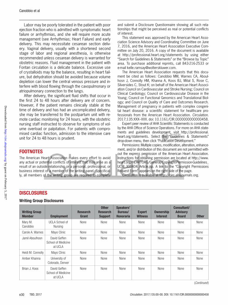
Canobbio et al
TBD, 2017 Circulation. 2017;135:00-00. DOI: 10.1161/CIR.0000000000000458e30
Labor may be poorly tolerated in the patient with poor ejection fraction who is admitted with symptomatic heart failure or arrhythmias, and she will require more acute management (see Arrhythmias; Heart Failure) and early delivery. This may necessitate cesarean section deliv-ery. Vaginal delivery, usually with a shortened second stage of labor and regional anesthesia, is otherwise recommended unless cesarean delivery is warranted for obstetric reasons. Fluid management in the patient with Fontan circulation is a delicate balance. Excessive use of crystalloids may tip the balance, resulting in heart fail-ure, but dehydration should be avoided because volume depletion can lower the central venous pressure and in-terfere with blood flowing through the cavopulmonary or atriopulmonary connection to the lungs.
After delivery, the significant fluid shifts that occur in the first 24 to 48 hours after delivery are of concern. However, if the patient remains clinically stable at the time of delivery and has had an uncomplicated delivery, she may be transferred to the postpartum unit with re-mote cardiac monitoring for 24 hours, with the obstetric nursing staff instructed to observe for symptoms of vol-ume overload or palpitation. For patients with compro-mised cardiac function, admission to the intensive care unit for 24 to 48 hours is prudent.
fooTNoTESThe American Heart Association makes every effort to avoid any actual or potential conflicts of interest that may arise as a result of an outside relationship or a personal, professional, or business interest of a member of the writing panel. Specifical-ly, all members of the writing group are required to complete
and submit a Disclosure Questionnaire showing all such rela-tionships that might be perceived as real or potential conflicts of interest.
This statement was approved by the American Heart Asso-ciation Science Advisory and Coordinating Committee on June 7, 2016, and the American Heart Association Executive Com-mittee on July 20, 2016. A copy of the document is available at http://professional.heart.org/statements by using either “Search for Guidelines & Statements” or the “Browse by Topic” area. To purchase additional reprints, call 843-216-2533 or e-mail [email protected].
The American Heart Association requests that this docu-ment be cited as follows: Canobbio MM, Warnes CA, Aboul-hosn J, Connolly HM, Khanna A, Koos BJ, Mital S, Rose C, Silversides C, Stout K; on behalf of the American Heart Associ-ation Council on Cardiovascular and Stroke Nursing; Council on Clinical Cardiology; Council on Cardiovascular Disease in the Young; Council on Functional Genomics and Translational Biol-ogy; and Council on Quality of Care and Outcomes Research. Management of pregnancy in patients with complex congeni-tal heart disease: a scientific statement for healthcare pro-fessionals from the American Heart Association. Circulation. 2017;135:XXX–XXX. doi: 10.1161/CIR.0000000000000458.
Expert peer review of AHA Scientific Statements is conducted by the AHA Office of Science Operations. For more on AHA state-ments and guidelines development, visit http://professional.heart.org/statements. Select the “Guidelines & Statements” drop-down menu, then click “Publication Development.”
Permissions: Multiple copies, modification, alteration, enhance-ment, and/or distribution of this document are not permitted with-out the express permission of the American Heart Association. Instructions for obtaining permission are located at http://www.heart.org/HEARTORG/General/Copyright-Permission-Guidelines_UCM_300404_Article.jsp. A link to the “Copyright Permissions Request Form” appears on the right side of the page.
Circulation is available at http://circ.ahajournals.org.
DISCLoSUrESWriting Group Disclosures
Writing Group Member Employment
research Grant
other research Support
Speakers’ Bureau/
honorariaExpert
Witnessownership
Interest
Consultant/Advisory
Board other
Mary M. Canobbio
UCLA School of Nursing
None None None None None None None
Carole A. Warnes Mayo Clinic None None None None None None None
Jamil Aboulhosn David Geffen School of Medicine
at UCLA
None None None None None None None
Heidi M. Connolly Mayo Clinic None None None None None None None
Amber Khanna University of Colorado, Denver
None None None None None None None
Brian J. Koos David Geffen School of Medicine
at UCLA
None None None None None None None
(Continued )
by guest on February 5, 2017http://circ.ahajournals.org/
Dow
nloaded from
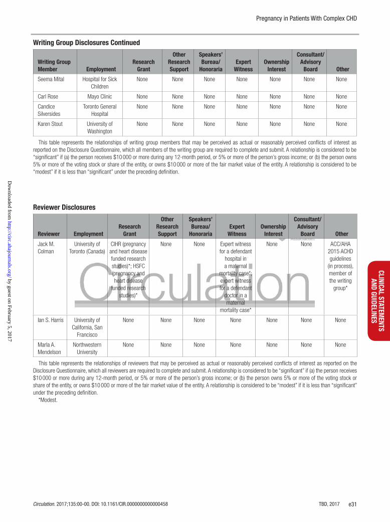
Pregnancy in Patients With Complex CHD
Circulation. 2017;135:00-00. DOI: 10.1161/CIR.0000000000000458 TBD, 2017 e31
CLINICAL STATEMENTS
AND GUIDELINES
reviewer Disclosures
reviewer Employmentresearch
Grant
other research Support
Speakers’ Bureau/
honorariaExpert
Witnessownership
Interest
Consultant/Advisory
Board other
Jack M. Colman
University of Toronto (Canada)
CIHR (pregnancy and heart disease funded research studies)*; HSFC (pregnancy and heart disease
funded research studies)*
None None Expert witness for a defendant
hospital in a maternal
mortality case*; expert witness for a defendant
doctor in a maternal
mortality case*
None None ACC/AHA 2015 ACHD guidelines
(in process), member of the writing
group*
Ian S. Harris University of California, San
Francisco
None None None None None None None
Marla A. Mendelson
Northwestern University
None None None None None None None
This table represents the relationships of reviewers that may be perceived as actual or reasonably perceived conflicts of interest as reported on the Disclosure Questionnaire, which all reviewers are required to complete and submit. A relationship is considered to be “significant” if (a) the person receives $10 000 or more during any 12-month period, or 5% or more of the person’s gross income; or (b) the person owns 5% or more of the voting stock or share of the entity, or owns $10 000 or more of the fair market value of the entity. A relationship is considered to be “modest” if it is less than “significant” under the preceding definition.
*Modest.
Seema Mital Hospital for Sick Children
None None None None None None None
Carl Rose Mayo Clinic None None None None None None None
Candice Silversides
Toronto General Hospital
None None None None None None None
Karen Stout University of Washington
None None None None None None None
This table represents the relationships of writing group members that may be perceived as actual or reasonably perceived conflicts of interest as reported on the Disclosure Questionnaire, which all members of the writing group are required to complete and submit. A relationship is considered to be “significant” if (a) the person receives $10 000 or more during any 12-month period, or 5% or more of the person’s gross income; or (b) the person owns 5% or more of the voting stock or share of the entity, or owns $10 000 or more of the fair market value of the entity. A relationship is considered to be “modest” if it is less than “significant” under the preceding definition.
Writing Group Disclosures Continued
Writing Group Member Employment
research Grant
other research Support
Speakers’ Bureau/
honorariaExpert
Witnessownership
Interest
Consultant/Advisory
Board other
by guest on February 5, 2017http://circ.ahajournals.org/
Dow
nloaded from
Canobbio et al
TBD, 2017 Circulation. 2017;135:00-00. DOI: 10.1161/CIR.0000000000000458e32
rEfErENCES 1. Warnes CA, Liberthson R, Danielson GK, Dore A, Harris L, Hoff-
man JI, Somerville J, Williams RG, Webb GD. Task force 1: the changing profile of congenital heart disease in adult life. J Am Coll Cardiol. 2001;37:1170–1175.
2. Thorne S, MacGregor A, Nelson-Piercy C. Risks of contraception and pregnancy in heart disease. Heart. 2006;92:1520–1525. doi: 10.1136/hrt.2006.095240.
3. Balci A, Sollie-Szarynska KM, van der Bijl AG, Ruys TP, Mulder BJ, Roos-Hesselink JW, van Dijk AP, Wajon EM, Vliegen HW, Dren-then W, Hillege HL, Aarnoudse JG, van Veldhuisen DJ, Pieper PG; ZAHARA-II Investigators. Prospective validation and assessment of cardiovascular and offspring risk models for pregnant women with congenital heart disease. Heart. 2014;100:1373–1381. doi: 10.1136/heartjnl-2014-305597.
4. Drenthen W, Boersma E, Balci A, Moons P, Roos-Hesselink JW, Mulder BJ, Vliegen HW, van Dijk AP, Voors AA, Yap SC, van Veld-huisen DJ, Pieper PG; ZAHARA Investigators. Predictors of preg-nancy complications in women with congenital heart disease. Eur Heart J. 2010;31:2124–2132. doi: 10.1093/eurheartj/ehq200.
5. Bédard E, Dimopoulos K, Gatzoulis MA. Has there been any prog-ress made on pregnancy outcomes among women with pulmo-nary arterial hypertension? Eur Heart J. 2009;30:256–265. doi: 10.1093/eurheartj/ehn597.
6. Weiss BM, Zemp L, Seifert B, Hess OM. Outcome of pulmonary vascular disease in pregnancy: a systematic overview from 1978 through 1996. J Am Coll Cardiol. 1998:31:1650–1657.
7. Presbitero P, Somerville J, Stone S, Aruta E, Spiegelhalter D, Ra-bajoli F. Pregnancy in cyanotic congenital heart disease: outcome of mother and fetus. Circulation. 1994;89:2673–2676.
8. Hytten FE, Paintin DB. Increase in plasma volume during normal pregnancy. J Obstet Gynaecol Br Emp. 1963;70:402–407.
9. Rovinsky JJ, Jaffin H. Cardiovascular hemodynamics in pregnancy, I: blood and plasma volumes in multiple pregnancy. Am J Obstet Gynecol. 1965;93:1–15.
10. Caton WL, Roby CC, Reid DE, Caswell R, Maletskos CJ, Fluharty RG, Gibson JG 2nd. The circulating red cell volume and body hematocrit in normal pregnancy and the puerperium by direct measurement using radioactive red cells. Am J Obstet Gynecol. 1951;61:1207–1217.
11. Dahlstrom H, Ihrman K. A clinical and physiological study of preg-nancy in a material from northern Sweden, IV: observations on the blood volume during and after pregnancy. Acta Soc Med Ups. 1960;65:295–304.
12. Pritchard JA. Changes in the blood volume during pregnancy and delivery. Anesthesiology. 1965;26:393–399.
13. Longo LD. Maternal blood volume and cardiac output during pregnancy: a hypothesis of endocrinologic control. Am J Physiol. 1983:245(pt 1):R720–R729.
14. Yusef YR, Thomas W, Harvey BJ. Estrogen increases ENaC activ-ity via PKCδ signaling in renal cortical collecting duct cells. Physiol Rep. 2014:2:e12020. doi: 10.14814/phy2.12020.
15. Lumbers ER, Pringle KG. Roles of the circulating renin-angiotensin-aldosterone system in human pregnancy. Am J Physiol Regul In-tegr Comp Physiol. 2014:306:R91–R101.
16. Chisholm M. A controlled clinical trial of prophylactic folic acid and iron in pregnancy. J Obstet Gynaecol Br Commonw. 1966:73:191–196.
17. Hameed AB, Chan K, Ghamsary M, Elkayam U. Longitudinal changes in the B-type natriuretic peptide levels in normal preg-nancy and postpartum. Clin Cardiol. 2009:32:E60–E62. doi: 10.1002/clc.20391.
18. Tanous D, Siu SC, Mason J, Greutmann M, Wald RM, Parker JD, Sermer M, Colman JM, Silversides CK. B-type natriuretic pep-tide in pregnant women with heart disease. J Am Coll Cardiol. 2010;56:1247–1253. doi: 10.1016/j.jacc.2010.02.076.
19. Resnik JL, Hong C, Resnik R, Kazanegra R, Beede J, Bhalla V, Maisel A. Evaluation of B-type natriuretic peptide (BNP) lev-els in normal and preeclamptic women. Am J Obstet Gynecol. 2005;193:450–454. doi: 10.1016/j.ajog.2004.12.006.
20. Yoshimura T, Yoshimura M, Yasue H, Ito M, Okamura H, Mu-koyama M, Nakao K. Plasma concentration of atrial natri-uretic peptide and brain natriuretic peptide during normal human pregnancy and the postpartum period. J Endocrinol. 1994;140:393–397.
21. Kampman MA, Balci A, van Veldhuisen DJ, van Dijk AP, Roos-Hes-selink JW, Sollie-Szarynska KM, Ludwig-Ruitenberg M, van Melle JP, Mulder BJ, Pieper PG; ZAHARA II Investigators. N-terminal pro-B-type natriuretic peptide predicts cardiovascular complications in pregnant women with congenital heart disease. Eur Heart J. 2014;35:708–715. doi: 10.1093/eurheartj/eht526.
22. Robertson EG, Cheyne GA. Plasma biochemistry in relation to oedema of pregnancy. J Obstet Gynaecol Br Commonw. 1972;79:769–776.
23. Brown MA, Zammit VC, Mitar DM. Extracellular fluid volumes in pregnancy-induced hypertension. J Hypertens. 1992;10:61–68.
24. Robson SC, Hunter S, Boys RJ, Dunlop W. Serial study of factors influencing changes in cardiac output during human pregnancy. Am J Physiol. 1989:256(pt 2):H1060–H1065.
25. Duvekot JJ, Cheriex EC, Pieters FA, Menheere PP, Peeters LH. Early pregnancy changes in hemodynamics and volume homeostasis are consecutive adjustments triggered by a pri-mary fall in systemic vascular tone. Am J Obstet Gynecol. 1993;169:1382–1392.
26. Chapman AB, Abraham WT, Zamudio S, Coffin C, Merouani A, Young D, Johnson A, Osorio F, Goldberg C, Moore LG, Dahms T, Schrier RW. Temporal relationships between hormonal and hemodynamic changes in early human pregnancy [published correction appears in Kidney Int. 1999;55:1185]. Kidney Int. 1998;54:2056–2063. doi: 10.1046/j.1523-1755.1998.00217.x.
27. Ueland K, Metcalfe J. Circulatory changes in pregnancy. Clin Ob-stet Gynecol. 1975;18:41–50.
28. Clark SL, Cotton DB, Pivarnik JM, Lee W, Hankins GD, Benedetti TJ, Phelan JP. Position change and central hemodynamic profile dur-ing normal third-trimester pregnancy and post partum [published correction appears in Am J Obstet Gynecol. 1991;165:241]. Am J Obstet Gynecol. 1991;164:883–887.
29. McLennan FM, Haites NE, Rawles JM. Stroke and minute distance in pregnancy: a longitudinal study using Doppler ultrasound. Br J Obstet Gynaecol. 1987;94:499–506.
30. Armstrong S, Fernando R, Columb M, Jones T. Cardiac index in term pregnant women in the sitting, lateral, and supine positions: an observational, crossover study. Anesth Analg. 2011;113:318–322. doi: 10.1213/ANE.0b013e31821f125c.
31. McRoberts WA Jr. Postural shock in pregnancy. Am J Obstet Gy-necol. 1951;62:627–632.
32. Quilligan EJ, Tyler C. Postural effects on the cardiovascular status in pregnancy: a comparison of the lateral and supine postures. Am J Obstet Gynecol. 1959;78:465–471.
33. Ueland K, Hansen JM. Maternal cardiovascular dynamics, II: posture and uterine contractions. Am J Obstet Gynecol. 1969:103:1–7.
34. Kametas NA, McAuliffe F, Krampl E, Chambers J, Nicolaides KH. Maternal cardiac function in twin pregnancy. Obstet Gynecol. 2003;102:806–815.
35. Kuleva M, Youssef A, Maroni E, Contro E, Pilu G, Rizzo N, Pelusi G, Ghi T. Maternal cardiac function in normal twin pregnancy: a lon-gitudinal study. Ultrasound Obstet Gynecol. 2011;38:575–580. doi: 10.1002/uog.8936.
36. Robson SC, Hunter S, Boys RJ, Dunlop W. Hemodynamic changes during twin pregnancy: a Doppler and M-mode echocardiographic study. Am J Obstet Gynecol. 1989;161:1273–1278.
37. Veille JC, Morton MJ, Burry KJ. Maternal cardiovascular adaptations to twin pregnancy. Am J Obstet Gynecol. 1985;153:261–263.
by guest on February 5, 2017http://circ.ahajournals.org/
Dow
nloaded from
Pregnancy in Patients With Complex CHD
Circulation. 2017;135:00-00. DOI: 10.1161/CIR.0000000000000458 TBD, 2017 e33
CLINICAL STATEMENTS
AND GUIDELINES
38. McLennan CE. Antecubital and femoral venous pressure in normal and toxemic pregnancy. Am J Obstet Gynecol. 1943:45:568–591.
39. Vorys N, Ullery JC, Hanusek GE. The cardiac output chang-es in various positions in pregnancy. Am J Obstet Gynecol. 1961;82:1312–1321.
40. Kinsella SM, Lohmann G. Supine hypotensive syndrome. Obstet Gynecol. 1994;83(pt 1):774–788.
41. Knotts RJ, Garan H. Cardiac arrhythmias in pregnancy. Semin Peri-natol. 2014;38:285–288. doi: 10.1053/j.semperi.2014.04.017.
42. Robson SC, Hunter S, Boys RJ, Dunlop W. Serial changes in pulmonary haemodynamics during human pregnancy: a non-invasive study using Doppler echocardiography. Clin Sci (Lond). 1991;80:113–117.
43. Bader RA, Bader ME, Rose DF, Braunwald E. Hemodynamics at rest and during exercise in normal pregnancy as studies by cardiac catheterization. J Clin Invest. 1955;34:1524–1536. doi: 10.1172/JCI103205.
44. Clark SL, Cotton DB, Lee W, Bishop C, Hill T, Southwick J, Pivarnik J, Spillman T, DeVore GR, Phelan J, Hankins GDV, Bemedetto TJ, Tolley D. Central hemodynamic assessment of normal term preg-nancy. Am J Obstet Gynecol. 1989;161(pt 1):1439–1442.
45. Werko L. Pregnancy and heart disease. Acta Obstet Gynecol Scand. 1954;33:162–210.
46. Walters WA, MacGregor WG, Hills M. Cardiac output at rest during pregnancy and the puerperium. Clin Sci. 1966;30:1–11.
47. Clapp JF 3rd, Capeless E. Cardiovascular function before, dur-ing, and after the first and subsequent pregnancies. Am J Cardiol. 1997;80:1469–1473.
48. Capeless EL, Clapp JF. Cardiovascular changes in early phase of pregnancy. Am J Obstet Gynecol. 1989;161(pt 1):1449–1453.
49. Easterling TR, Benedetti TJ, Schmucker BC, Millard SP. Maternal hemodynamics in normal and preeclamptic pregnancies: a longi-tudinal study. Obstet Gynecol. 1990;76:1061–1069.
50. Veille JC, Morton MJ, Burry K, Nemeth M, Speroff L. Estradiol and hemodynamics during ovulation induction. J Clin Endocrinol Metab. 1986;63:721–724. doi: 10.1210/jcem-63-3-721.
51. Moutquin JM, Rainville C, Giroux L, Raynauld P, Amyot G, Bi-lodeau R, Pelland N. A prospective study of blood pressure in pregnancy: prediction of preeclampsia. Am J Obstet Gynecol. 1985;151:191–196.
52. Awe RJ, Nicotra MB, Newsom TD, Viles R. Arterial oxygenation and alveolar-arterial gradients in term pregnancy. Obstet Gynecol. 1979;53:182–186.
53. Rees GB, Broughton Pipkin F, Symonds EM, Patrick JM. A longi-tudinal study of respiratory changes in normal human pregnancy with cross-sectional data on subjects with pregnancy-induced hy-pertension. Am J Obstet Gynecol. 1990;162:826–830.
54. DeSwiet M. Pulmonary disorders. In: Creasy RK, Resnik R, ed. Maternal-Fetal Medicine. 4th ed. Philadelphia, PA: WB Saunders; 1999:921–934.
55. Lehmann V. Dyspnea in pregnancy. J Perinat Med. 1975;3:154–160. 56. Milne JA, Howie AD, Pack AI. Dyspnoea during normal pregnancy.
Br J Obstet Gynaecol. 1978;85:260–263. 57. Hirnle L, Lysenko L, Gerber H, Lesnik P, Baranowska A, Rach-
walik M, Leszczyszyn J, Strozecki L. Respiratory function in pregnant women. Adv Exp Med Biol. 2013;788:153–160. doi: 10.1007/978-94-007-6627-3_23.
58. Hendricks CH, Quilligan EJ. Cardiac output during labor. Am J Ob-stet Gynecol. 1956;71:953–972.
59. Robson SC, Dunlop W, Boys RJ, Hunter S. Cardiac output during labour. Br Med J (Clin Res Ed). 1987;295:1169–1172.
60. Ueland K, Hansen JM. Maternal cardiovascular dynamics, 3: labor and delivery under local and caudal analgesia. Am J Obstet Gyne-col. 1969;103:8–18.
61. Adams JQ, Alexander AM Jr. Alterations in cardiovascular physiol-ogy during labor. Obstet Gynecol. 1958;12:542–549.
62. Palmer AJ, Walker AH. The maternal circulation in normal preg-nancy. J Obstet Gynaecol Br Emp. 1949;56:537–547.
63. Lee W, Rokey R, Miller J, Cotton DB. Maternal hemodynamic ef-fects of uterine contractions by M-mode and pulsed-Doppler echo-cardiography. Am J Obstet Gynecol. 1989;161:974–977.
64. Bieniarz J, Yoshida T, Romero-Salinas G, Curuchet E, Caldeyro-Bar-cia R, Crottogini JJ. Aortocaval compression by the uterus in late human pregnancy. IV. Circulatory homeostasis by preferential per-fusion of the placenta. Am J Obstet Gynecol. 1969;103:19–31.
65. Hendricks CH. The hemodynamics of a uterine contraction. Am J Obstet Gynecol. 1958;76:969–982.
66. Atkins AF, Watt JM, Milan P, Davies P, Crawford JS. A longitudinal study of cardiovascular dynamic changes throughout pregnancy. Eur J Obstet Gynecol Reprod Biol. 1981;12:215–224.
67. Metcalfe J, Ueland K. Maternal cardiovascular adjustments to pregnancy. Prog Cardiovasc Dis. 1974;16:363–374.
68. Regitz-Zagrosek V, Blomstrom Lundqvist C, Borghi C, Cifkova R, Ferreira R, Foidart J- M, Gibbs JSR, Gohlke-Baerwolf C, Gorenek B, Iung B, Kirby M, Maas AH, Morais J, Nihoyannopoulos P, Pieper PG, Presbitero P, Roos-Hesselink JW, Schaufelberger M, Seeland U, Torracca L; ESC Committee for Practice Guidelines. ESC guidelines on the management of cardiovascular diseases during pregnancy: the Task Force on the Management of Cardiovascular Diseases During Pregnancy of the European Society of Cardiology (ESC). Eur Heart J. 2011:32:3147–3197.
69. Ueland K. Maternal cardiovascular dynamics. VII. Intrapartum blood volume changes. Am J Obstet Gynecol. 1976;126:671–677.
70. James CF, Banner T, Caton D. Cardiac output in women undergo-ing cesarean section with epidural or general anesthesia. Am J Obstet Gynecol. 1989;160(pt 1):1178–1184.
71. Robson SC, Boys RJ, Hunter S, Dunlop W. Maternal hemodynam-ics after normal delivery and delivery complicated by postpartum hemorrhage. Obstet Gynecol. 1989;74:234–239.
72. Warnes CA, Williams RG, Bashore TM, Child JS, Connolly HM, Dea-rani JA, del Nido P, Fasules JW, Graham TP Jr, Hijazi ZM, Hunt SA, King ME, Landzberg MJ, Miner PD, Radford MJ, Walsh EP, Webb GD. ACC/AHA 2008 guidelines for the management of adults with congenital heart disease: a report of the American College of Cardiology/American Heart Association, Task Force on Practice Guidelines (Writing Committee to Develop Guidelines on the Management of Adults With Congenital Heart Disease). Circu-lation. 2008;118:e714–e833. doi: 10.1161/CIRCULATIONAHA. 108.190690.
73. Whittemore R, Hobbins JC, Engle MA. Pregnancy and its outcome in women with and without surgical treatment of congenital heart disease. Am J Cardiol. 1982;50:641–651.
74. Siu SC, Sermer M, Colman JM, Alvarez AN, Mercier LA, Morton BC, Kells CM, Bergin ML, Kiess MC, Marcotte F, Taylor DA, Gor-don EP, Spears JC, Tam JW, Amankwah KS, Smallhorn JF, Farine D, Sorensen S; Cardiac Disease in Pregnancy (CARPREG) Investi-gators. Prospective multicenter study of pregnancy outcomes in women with heart disease. Circulation. 2001;104:515–521.
75. Diller GP, Dimopoulos K, Okonko D, Li W, Babu-Narayan SV, Bro-berg CS, Johansson B, Bouzas B, Mullen MJ, Poole-Wilson PA, Francis DP, Gatzoulis MA. Exercise intolerance in adult congenital heart disease: comparative severity, correlates, and prognos-tic implication. Circulation. 2005;112:828–835. doi: 10.1161/ CIRCULATIONAHA.104.529800.
76. Lui GK, Silversides CK, Khairy P, Fernandes SM, Valente AM, Nickolaus MJ, Earing MG, Aboulhosn JA, Rosenbaum MS, Cook S, Kay JD, Jin Z, Gersony DR; Alliance for Adult Research in Con-genital Cardiology (AARCC). Heart rate response during exercise and pregnancy outcome in women with congenital heart disease. Circulation. 2011;123:242–248. doi: 10.1161/CIRCULATIONAHA. 110.953380.
77. Opotowsky AR, Siddiqi OK, D’Souza B, Webb GD, Fernandes SM, Landzberg MJ. Maternal cardiovascular events during
by guest on February 5, 2017http://circ.ahajournals.org/
Dow
nloaded from
Canobbio et al
TBD, 2017 Circulation. 2017;135:00-00. DOI: 10.1161/CIR.0000000000000458e34
childbirth among women with congenital heart disease. Heart. 2012;98:145–151. doi: 10.1136/heartjnl-2011-300828.
78. Drenthen W, Pieper PG, Roos-Hesselink JW, van Lottum WA, Voors AA, Mulder BJ, van Dijk AP, Vliegen HW, Yap SC, Moons P, Ebels T, van Veldhuisen DJ; ZAHARA Investigators. Outcome of pregnancy in women with congenital heart disease: a literature review. J Am Coll Cardiol. 2007;49:2303–2311. doi: 10.1016/j.jacc.2007.03.027.
79. Khairy P, Ouyang DW, Fernandes SM, Lee-Parritz A, Economy KE, Landzberg MJ. Pregnancy outcomes in women with congenital heart disease. Circulation; 2006:113:517–524. doi: 10.1161/CIRCULATIONAHA.105.589655.
80. Gelson E, Curry R, Gatzoulis MA, Swan L, Lupton M, Steer P, John-son M. Effect of maternal heart disease on fetal growth [published correction appears in Obstet Gynecol. 2011;113(pt1):364]. Obstet Gynecol. 2011;117:886–891. doi: 10.1097/AOG.0b013e31820cab69.
81. Burchill L, Greenway S, Silversides CK, Mital S. Genetic counsel-ing in the adult with congenital heart disease: what is the role? Curr Cardiol Rep. 2011;13:347–355. doi: 10.1007/s11886-011-0188-z.
82. Pierpont ME, Basson CT, Benson DW Jr, Gelb BD, Giglia TM, Gold-muntz E, McGee G, Sable CA, Srivastava D, Webb CL. Genetic basis for congenital heart defects: current knowledge: a scien-tific statement from the American Heart Association Congenital Cardiac Defects Committee, Council on Cardiovascular Disease in the Young. Circulation. 2007;115:3015–3038. doi: 10.1161/CIRCULATIONAHA.106.183056.
83. Øyen N, Poulsen G, Boyd HA, Wohlfahrt J, Jensen PK, Melbye M. Recurrence of congenital heart defects in families. Circula-tion. 2009;120:295–301. doi: 10.1161/CIRCULATIONAHA. 109.857987.
84. Gill HK, Splitt M, Sharland GK, Simpson JM. Patterns of recur-rence of congenital heart disease: an analysis of 6,640 consecu-tive pregnancies evaluated by detailed fetal echocardiography. J Am Coll Cardiol. 2003;42:923–929.
85. Jenkins KJ, Correa A, Feinstein JA, Botto L, Britt AE, Daniels SR, Elixson M, Warnes CA, Webb CL. Noninherited risk factors and congenital cardiovascular defects: current knowledge: a scientific statement from the American Heart Association Council on Car-diovascular Disease in the Young. Circulation. 2007;115:2995–3014. doi: 10.1161/CIRCULATIONAHA.106.183216.
86. Silversides CK, Marelli A, Beauchesne L, Dore A, Kiess M, Sale-hian O, Bradley T, Colman J, Connelly M, Harris L, Khairy P, Mital S, Niwa K, Oechslin E, Poirier N, Schwerzmann M, Taylor D, Vonder Muhll I, Baumgartner H, Benson L, Celermajer D, Greutmann M, Horlick E, Landzberg M, Meijboom F, Mulder B, Warnes C, Webb G, Therrien J. Canadian cardiovascular soci-ety 2009 consensus conference on the management of adults with congenital heart disease: executive summary. Can J Car-diol; 2010:26:143–150.
87. Colletti PM, Lee KH, Elkayam U. Cardiovascular imaging of the pregnant patient. AJR Am J Roentgenol. 2013;200:515–521. doi: 10.2214/AJR.12.9864.
88. Naqvi TZ, Elkayam U. Serial echocardiographic assessment of the human heart in normal pregnancy. Circ Cardiovasc Imaging. 2012;5:283–285. doi: 10.1161/CIRCIMAGING.112.974808.
89. Campos O. Doppler echocardiography during pregnancy: physiological and abnormal findings. Echocardiography. 1996;13:135–146.
90. MacPhail A, Davies GA, Victory R, Wolfe LA. Maximal exer-cise testing in late gestation: fetal responses. Obstet Gynecol. 2000;96:565–570.
91. Moore DH, Jarrett JC 2nd, Bendick PJ. Exercise-induced chang-es in uterine artery blood flow, as measured by Doppler ultra-sound, in pregnant subjects. Am J Perinatol. 1988;5:94–97. doi: 10.1055/s-2007-999663.
92. van Doorn MB, Lotgering FK, Struijk PC, Pool J, Wallenburg HC. Maternal and fetal cardiovascular responses to strenuous bicycle exercise. Am J Obstet Gynecol. 1992;166:854–859.
93. Piran S, Veldtman G, Siu S, Webb GD, Liu PP. Heart failure and ventricular dysfunction in patients with single or systemic right ventricles. Circulation. 2002;105:1189–1194.
94. Perloff JK, Koos B. Pregnancy and congenital heart disease: the mother and the fetus: medical considerations. In: Perloff JK, Child JS, eds. Congenital Heart Disease in Adults. Philadelphia, PA: WB Saunders; 1998:144–164.
95. Patenaude Y, Pugash D, Lim K, Morin L; Diagnostic Imaging Committee; Lim K, Bly S, Butt K, Cargill Y, Davies G, Denis N, Hazlitt G, Morin L, Naud K, Ouellet A, Salem S; Society of Obstetricians and Gynaecologists of Canada. The use of magnet-ic resonance imaging in the obstetric patient. J Obstet Gynaecol Can. 2014;36:349–363.
96. Kanal E, Barkovich AJ, Bell C, Borgstede JP, Bradley WG Jr, Froelich JW, Gilk T, Gimbel JR, Gosbee J, Kuhni-Kaminski E, Lester JW Jr, Nyenhuis J, Parag Y, Schaefer DJ, Sebek-Scoumis EA, Weinreb J, Zaremba LA, Wilcox P, Lucey L, Sass N; ACR Blue Ribbon Panel on MR Safety. ACR guidance document for safe MR practices: 2007. AJR Am J Roentgenol. 2007;188:1447–1474. doi: 10.2214/AJR.06.1616.
97. American College of Radiology (ACR); Society for Pediatric Radiology (SPR). Practice guideline for the safe and optimal performance of fetal magnetic resonance imaging (MRI). 2010 (Resolution 13). http://www.acr.org/~/media/ CB384A65345F40 2083639E6756CE513F.pdf. Accessed December 9, 2013.
98. Glover P, Hykin J, Gowland P, Wright J, Johnson I, Mansfield P. An assessment of the intrauterine sound intensity level dur-ing obstetric echo-planar magnetic resonance imaging. Br J Radiol. 1995;68:1090–1094. doi: 10.1259/0007-1285-68- 814-1090.
99. Baker PN, Johnson IR, Harvey PR, Gowland PA, Mansfield P. A three-year follow-up of children imaged in utero with echo- planar magnetic resonance. Am J Obstet Gynecol. 1994;170(pt 1): 32–33.
100. Shellock FG, Spinazzi A. MRI safety update 2008, part 2: screen-ing patients for MRI. AJR Am J Roentgenol. 2008;191:1140–1149. doi: 10.2214/AJR.08.1038.2.
101. Chen MM, Coakley FV, Kaimal A, Laros RK Jr. Guidelines for com-puted tomography and magnetic resonance imaging use during pregnancy and lactation. Obstet Gynecol. 2008;112(pt 1):333–340. doi: 10.1097/AOG.0b013e318180a505.
102. Damilakis J, Perisinakis K, Voloudaki A, Gourtsoyiannis N. Estimation of fetal radiation dose from computed tomogra-phy scanning in late pregnancy: depth-dose data from routine examinations [published correction appears in Invest Radiol. 2000;35:706]. Invest Radiol. 2000;35:527–533.
103. REPROTOX database. www.reprotox.org. Accessed September 1, 2014.
104. Cooper WO, Hernandez-Diaz S, Arbogast PG, Dudley JA, Dyer S, Gideon PS, Hall K, Ray WA. Major congenital malformations after first-trimester exposure to ACE inhibitors. N Engl J Med. 2006;354:2443–2451. doi: 10.1056/NEJMoa055202.
105. Lee JCR, Wetzel G, Shannon K. Maternal arrhythmia manage-ment during pregnancy in patients with structural heart disease. Prog Pediatr Cardiol. 2004;19:71–82.
106. Lip GY, Beevers M, Churchill D, Shaffer LM, Beevers DG. Effect of atenolol on birth weight. Am J Cardiol. 1997;79:1436–1438.
107. Lindheimer MD, Katz AI. Pregnancy and the kidney. J Reprod Med. 1973;11:14–18.
108. McKenna WJ, Rowland E, Krikler DM. Amiodarone: the ex-perience of the past decade. Br Med J (Clin Res Ed). 1983;287:1654–1656.
109. Lecarpentier E, Morel O, Fournier T, Elefant E, Chavatte-Palmer P, Tsatsaris V. Statins and pregnancy: between supposed
by guest on February 5, 2017http://circ.ahajournals.org/
Dow
nloaded from

Pregnancy in Patients With Complex CHD
Circulation. 2017;135:00-00. DOI: 10.1161/CIR.0000000000000458 TBD, 2017 e35
CLINICAL STATEMENTS
AND GUIDELINES
risks and theoretical benefits. Drugs. 2012;72:773–788. doi: 10.2165/11632010-000000000-00000.
110. Costantine MM, Cleary K; Eunice Kennedy Shriver National Institute of Child Health and Human Development Obstetric–Fetal Pharmacology Research Units Network. Pravastatin for the pre-vention of preeclampsia in high-risk pregnant women. Obstet Gynecol. 2013;121(pt 1):349–353. doi: http://10.1097/AOG. 0b013e31827d8ad5.
111. Canobbio MM, Chugh R. Congenital heart disease and preg-nancy. In: Wzszynski DF, Correa-Villasenor, Graham TP, eds. Congenital Heart Defects: From Origin to Treatment. Oxford, UK: Oxford Press; 2011:301–328.
112. Kahn SR, Lim W, Dunn AS, Cushman M, Dentali F, Akl EA, Cook DJ, Balekian AA, Klein RC, Le H, Schulman S, Murad MH; American College of Chest Physicians. Prevention of VTE in nonsurgical patients: Antithrombotic Therapy and Prevention of Thrombosis, 9th ed: American College of Chest Physicians Evidence-Based Clinical Practice Guidelines. Chest. 2012;141(suppl):e195S–e226S. doi: 10.1378/chest.11-2296.
113. Sayle AE, Savitz DA, Thorp JM, Hertz-Picciotta I, Wilcox AJ. Sexual activity during late pregnancy and risk of preterm delivery. Obstet Gynecol. 2011:283–289.
114. Canobbio MM, Mair DD, van der Velde M, Koos BJ. Pregnancy outcomes after the Fontan repair. J Am Coll Cardiol. 1996;28:763–767.
115. Bryant AG, Grimes DA, Garrett JM, Stuart GS. Second-trimester abortion for fetal anomalies or fetal death: labor induction compared with dilation and evacuation. Obstet Gynecol. 2011;117:788–792. doi: 10.1097/AOG.0b013e31820c3d26.
116. American College of Obstetricians and Gynecologists. ACOG committee opinion no. 560: medically indicated late-preterm and early-term deliveries. Obstet Gynecol. 2013;121:908–1010. doi: 10.1097/01.AOG.0000428648.75548.00.
117. Koos B. Management of uncorrected, palliated, and repaired cyanotic congenital heart disease in pregnancy. Prog Pediatr Cardiol. 2004:19: 25–45. doi: http://dx.doi.org/10.1016/j.ppedcard.2003.09.004.
118. Cole PJ, Cross MH, Dresner M. Incremental spinal anaesthesia for elective caesarean section in a patient with Eisenmenger’s syndrome. Br J Anaesth. 2001;86:723–726.
119. Wilson W, Taubert KA, Gewitz M, Lockhart PB, Baddour LM, Levison M, Bolger A, Cabell CH, Takahashi M, Baltimore RS, Newburger JW, Strom BL, Tani LY, Gerber M, Bonow RO, Pallasch T, Shulman ST, Rowley AH, Burns JC, Ferrieri P, Gardner T, Goff D, Durack DT. Prevention of infective endocarditis: guidelines from the American Heart Association: a guideline from the American Heart Association Rheumatic Fever, Endocarditis, and Kawasaki Disease Committee, Council on Cardiovascular Disease in the Young, and the Council on Clinical Cardiology, Council on Cardiovascular Surgery and Anesthesia, and the Quality of Care and Outcomes Research Interdisciplinary Working Group [published correction appears in Circulation. 2007;116:e376–e377]. Circulation. 2007;116:1736–1754. doi: 10.1161/CIRCULATIONAHA.106.183095.
120. Kjeldsen J. Hemodynamic investigations during labour and deliv-ery. Acta Obstet Gynecol Scand Suppl. 1979;89:1–252.
121. Robson SC, Dunlop W, Moore M, Hunter S. Combined Doppler and echocardiographic measurement of cardiac output: theory and application in pregnancy. Br J Obstet Gynaecol. 1987;94:1014–1027.
122. Zeeman GG, Wendel GD Jr, Cunningham FG. A blueprint for ob-stetric critical care. Am J Obstet Gynecol. 2003;188:532–536.
123. Capeless EL, Clapp JF. When do cardiovascular parameters return to their preconception values? Am J Obstet Gynecol. 1991;165(pt 1):883–886.
124. Truitt ST, Fraser AV, Grimes DA, Gallo MF, Schulz KF. Combined hormonal versus nonhormonal versus progestin-only contracep-tion in lactation. Cochrane Database Syst Rev. 2003:CD003988.
125. American College Obstetrics Gynecology. ACOG Practice Bulletin No. 121: long-acting reversible contraceptives: implants and intrauterine devices. Obstet Gynecol. 2011:118:184–196. doi: 10.1097/AOG.0b013e318227f05e.
126. Peterson HB, Xia Z, Hughes JM, Wilcox LS, Tylor LR, Trussell J. The risk of pregnancy after tubal sterilization: findings from the U.S. Collaborative Review of Sterilization. Am J Obstet Gynecol. 1996;174:1161–1168.
127. US Food and Drug Administration. 2015 Meeting materials of the Obstetrics and Gynecology Devices Panel. 2015. http://www.fda.gov/AdvisoryCommittees/CommitteesMeetingMaterials/Medica lDev ices/Medica lDev icesAdv isoryCommit tee/ObstetricsandGynecologyDevices/ucm463457.htm. Accessed February 22, 2016.
128. Silversides CK, Harris L, Haberer K, Sermer M, Colman JM, Siu SC. Recurrence rates of arrhythmias during pregnancy in women with previous tachyarrhythmia and impact on fetal and neonatal outcomes. Am J Cardiol. 2006;97:1206–1212. doi: 10.1016/j.amjcard.2005.11.041.
129. Lydakis C, Lip GY, Beevers M, Beevers DG. Atenolol and fe-tal growth in pregnancies complicated by hypertension. Am J Hypertens. 1999;12:541–547.
130. Bryerly WG, Hartman A. Foster DE, Tannenbaum AK. Verapamil in the treatment of paroxysmal supraventricular tachycardia. Ann Emerg Med. 1991;20:552–554.
131. Holbrook RH Jr, Gibson RN, Voss EM. Tocolytic and cardio-vascular effects of the calcium antagonist diltiazem in the near-term pregnant rabbit. Am J Obstet Gynecol. 1988;159: 591–595.
132. Zipes DP, Camm AJ, Borggrefe M, Buxton AE, Chaitman B, Fromer M, Gregoratos G, Klein G, Moss AJ, Myerburg RJ, Priori SG, Quinones MA, Roden DM, Silka MJ, Tracy C. ACC/AHA/ESC 2006 guidelines for management of patients with ventricular arrhythmias and the prevention of sudden car-diac death: a report of the American College of Cardiology/American Heart Association Task Force and the European Society of Cardiology Committee for Practice Guidelines (Writing Committee to Develop Guidelines for Management of Patients With Ventricular Arrhythmias and the Prevention of Sudden Cardiac Death). Circulation. 2006:114:e385–e484. doi: 10.1161/CIRCULATIONAHA.106.178233.
133. Thaman R, Curtis S, Faganello G, Szantho GV, Turner MS, Trinder J, Sellers S, Stuart GA. Cardiac outcome of pregnancy in women with a pacemaker and women with untreated atrioventricular conduction block. Europace. 2011;13:859–863. doi: 10.1093/europace/eur018.
134. Sbarouni E, Oakley CM. Outcome of pregnancy in women with valve prostheses. Br Heart J. 1994;71:196–201.
135. Meschengieser SS, Fondevila CG, Santarelli MT, Lazzari MA. Anticoagulation in pregnant women with mechanical heart valve prostheses. Heart. 1999;82:23–26.
136. Sadler L, McCowan L, White H, Stewart A, Bracken M, North R. Pregnancy outcomes and cardiac complications in women with mechanical, bioprosthetic and homograft valves. BJOG. 2000;107:245–253.
137. Cotrufo M, De Feo M, De Santo LS, Romano G, Della Corte A, Renzulli A, Gallo C. Risk of warfarin during pregnancy with me-chanical valve prostheses. Obstet Gynecol. 2002;99:35–40.
138. Nassar AH, Hobeika EM, Abd Essamad HM, Taher A, Khalil AM, Usta IM. Pregnancy outcome in women with prosthetic heart valves. Am J Obstet Gynecol. 2004;191:1009–1013. doi: 10.1016/j.ajog.2004.05.064.
139. Suri V, Keepanasseril A, Aggarwal N, Chopra S, Bagga R, Sikka P, Vijayvergiya R. Mechanical valve prosthesis and antico-agulation regimens in pregnancy: a tertiary centre experience. Eur J Obstet Gynecol Reprod Biol. 2011;159:320–323. doi: 10.1016/j.ejogrb.2011.09.011.
by guest on February 5, 2017http://circ.ahajournals.org/
Dow
nloaded from

Canobbio et al
TBD, 2017 Circulation. 2017;135:00-00. DOI: 10.1161/CIR.0000000000000458e36
140. Basude S, Hein C, Curtis SL, Clark A, Trinder J. Low-molecular-weight heparin or warfarin for anticoagulation in pregnant women with mechanical heart valves: what are the risks? A retrospec-tive observational study. BJOG. 2012;119:1008–1013. doi: 10.1111/j.1471-0528.2012.03359.x.
141. De Santo LS, Romano G, Della Corte A, D’Oria V, Nappi G, Giordano S, Cotrufo M, De Feo M. Mechanical aortic valve re-placement in young women planning on pregnancy: maternal and fetal outcomes under low oral anticoagulation, a pilot observa-tional study on a comprehensive pre-operative counseling pro-tocol. J Am Coll Cardiol. 2012;59:1110–1115. doi: 10.1016/j.jacc.2011.10.899.
142. Bian C, Wei Q, Liu X. Influence of heart-valve replacement of warfarin anticoagulant therapy on perinatal outcomes. Arch Gynecol Obstet. 2012;285:347–351. doi: 10.1007/s00404-011-1962-2.
143. Oran B, Lee-Parritz A, Ansell J. Low molecular weight heparin for the prophylaxis of thromboembolism in women with prosthetic mechanical heart valves during pregnancy. Thromb Haemost. 2004;92:747–751. doi: 10.1160/TH04-06-0337.
144. Abildgaard U, Sandset PM, Hammerstrøm J, Gjestvang FT, Tveit A. Management of pregnant women with mechanical heart valve prosthesis: thromboprophylaxis with low molecular weight heparin. Thromb Res. 2009;124:262–267. doi: 10.1016/j.thromres.2008.12.005.
145. Quinn J, Von Klemperer K, Brooks R, Peebles D, Walker F, Cohen H. Use of high intensity adjusted dose low molecular weight hepa-rin in women with mechanical heart valves during pregnancy: a single-center experience. Haematologica. 2009;94:1608–1612. doi: 10.3324/haematol.2008.002840.
146. McLintock C, McCowan LM, North RA. Maternal complications and pregnancy outcome in women with mechanical prosthetic heart valves treated with enoxaparin. BJOG. 2009;116:1585–1592. doi: 10.1111/j.1471-0528.2009.02299.x.
147. Yinon Y, Siu SC, Warshafsky C, Maxwell C, McLeod A, Colman JM, Sermer M, Silversides CK. Use of low molecular weight heparin in pregnant women with mechanical heart valves. Am J Cardiol. 2009;104:1259–1263. doi: 10.1016/j.amjcard.2009.06.040.
148. Nelson-Piercy C, Greer IA. Anticoagulation with tinzaparin for women with mechanical valves in pregnancy: a retrospective case series. Thromb Res. 2013;131:185–186. doi: 10.1016/j.thromres.2012.11.022.
149. Chan WS, Anand S, Ginsberg JS. Anticoagulation of pregnant women with mechanical heart valves: a systematic review of the literature. Arch Intern Med. 2000;160:191–196.
150. Hassouna A, Allam H. Anticoagulation of pregnant women with mechanical heart valve prosthesis: a systematic review of the literature (2000–2009). J Coagul Disord. 2010:2:81–88.
151. Vitale N, De Feo M, De Santo LS, Pollice A, Tedesco N, Cotrufo M. Dose-dependent fetal complications of warfarin in preg-nant women with mechanical heart valves. J Am Coll Cardiol. 1999;33:1637–1641.
152. Nishimura RA, Otto CM, Bonow RO, Carabello BA, Erwin JP 3rd, Guyton RA, O’Gara PT, Ruiz CE, Skubas NJ, Sorajja P, Sundt TM 3rd, Thomas JD; ACC/AHA Task Force Members. 2014 AHA/ACC guideline for the management of patients with valvular heart disease: executive summary: a report of the American College of Cardiology/American Heart Association Task Force on Practice Guidelines [published correction appears in Circulation. 2014;129:e650]. Circulation. 2014;129:2440–2492. doi: 10.1161/CIR.0000000000000029.
153. Berresheim M, Wilkie J, Nerenberg KA, Ibrahim Q, Bungard TJ. A case series of LMWH use in pregnancy: should trough anti-Xa levels guide dosing? Thromb Res. 2014;134:1234–1240. doi: 10.1016/j.thromres.2014.09.033.
154. Bates SM, Greer IA, Middeldorp S, Veenstra DL, Prabulos AM, Vandvik PO; American College of Chest Physicians.
VTE, thrombophilia, antithrombotic therapy, and pregnancy: Antithrombotic Therapy and Prevention of Thrombosis, 9th ed: American College of Chest Physicians Evidence-Based Clinical Practice Guidelines. Chest. 2012;141(suppl):e691S–e736S. doi: 10.1378/chest.11-2300.
155. Goland S, Schwartzenberg S, Fan J, Kozak N, Khatri N, Elkayam U. Monitoring of anti-Xa in pregnant patients with mechanical prosthetic valves receiving low-molecular-weight heparin: peak or trough levels? J Cardiovasc Pharmacol Ther. 2014;19:451–456. doi: 10.1177/1074248414524302.
156. Weiss BM, von Segesser LK, Alon E, Seifert B, Turina MI. Outcome of cardiovascular surgery and pregnancy: a systematic review of the period 1984–1996. Am J Obstet Gynecol. 1998:179(pt 1):1643–1653.
157. Arnoni RT, Arnoni AS, Bonini RC, de Almeida AF, Neto CA, Dinkhuysen JJ, Issa M, Chaccur P, Paulista PP. Risk factors asso-ciated with cardiac surgery during pregnancy. Ann Thorac Surg. 2003;76:1605–1608.
158. John AS, Gurley F, Schaff HV, Warnes CA, Phillips SD, Arendt KW, Abel MD, Rose CH, Connolly HM. Cardiopulmonary bypass during pregnancy. Ann Thorac Surg. 2011;91:1191–1196. doi: 10.1016/j.athoracsur.2010.11.037.
159. Ruys TP, Roos-Hesselink JW, Hall R, Subirana-Domènech MT, Grando-Ting J, Estensen M, Crepaz R, Fesslova V, Gurvitz M, De Backer J, Johnson MR, Pieper PG. Heart failure in preg-nant women with cardiac disease: data from the ROPAC. Heart. 2014;100:231–238. doi: 10.1136/heartjnl-2013- 304888.
160. Hameed A, Karaalp IS, Tummala PP, Wani OR, Canetti M, Akhter MW, Goodwin I, Zapadinsky N, Elkayam U. The effect of valvular heart disease on maternal and fetal outcome of pregnancy. J Am Coll Cardiol. 2001;37:893–899.
161. Yap SC, Drenthen W, Pieper PG, Moons P, Mulder BJ, Mostert B, Vliegen HW, van Dijk AP, Meijboom FJ, Steegers EA, Roos-Hesselink JW; ZAHARA Investigators. Risk of com-plications during pregnancy in women with congenital aortic stenosis. Int J Cardiol. 2008;126:240–246. doi: 10.1016/j.ijcard.2007.03.134.
162. Canobbio MM, Morris CD, Graham TP, Landzberg MJ. Pregnancy outcomes after atrial repair for transposition of the great ar-teries. Am J Cardiol. 2006;98:668–672. doi: 10.1016/j.amjcard.2006.03.050.
163. Guédès A, Mercier LA, Leduc L, Bérubé L, Marcotte F, Dore A. Impact of pregnancy on the systemic right ventricle af-ter a Mustard operation for transposition of the great arter-ies. J Am Coll Cardiol. 2004;44:433–437. doi: 10.1016/j.jacc.2004.04.037.
164. Avila WS, Grinberg M, Snitcowsky R, Faccioli R, Da Luz PL, Bellotti G, Pileggi F. Maternal and fetal outcome in pregnant women with Eisenmenger’s syndrome. Eur Heart J. 1995;16:460–464.
165. Connolly HM, Grogan M, Warnes CA. Pregnancy among women with congenitally corrected transposition of great arteries. J Am Coll Cardiol. 1999;33:1692–1695.
166. Veldtman GR, Connolly HM, Grogan M, Ammash NM, Warnes CA. Outcomes of pregnancy in women with tetralogy of Fallot. J Am Coll Cardiol. 2004;44:174–180. doi: 10.1016/j.jacc.2003.11.067.
167. Greutmann M, Von Klemperer K, Brooks R, Peebles D, O’Brien P, Walker F. Pregnancy outcome in women with congenital heart dis-ease and residual haemodynamic lesions of the right ventricular outflow tract. Eur Heart J. 2010;31:1764–1770. doi: 10.1093/eurheartj/ehq157.
168. Bowater SE, Selman TJ, Hudsmith LE, Clift PF, Thompson PJ, Thorne SA. Long-term outcome following pregnancy in wom-en with a systemic right ventricle: is the deterioration due to pregnancy or a consequence of time? Congenit Heart Dis. 2013;8:302–307. doi: 10.1111/chd.12001.
by guest on February 5, 2017http://circ.ahajournals.org/
Dow
nloaded from

Pregnancy in Patients With Complex CHD
Circulation. 2017;135:00-00. DOI: 10.1161/CIR.0000000000000458 TBD, 2017 e37
CLINICAL STATEMENTS
AND GUIDELINES
168a. Robertson JE, Silversides CK, Mah ML, Kulikowski J, Maxwell C, Wald RM, Colman JM, Siu SC, Sermer M. A contemporary approach to the obstetric management of women with heart disease. J Obstet Gynaecol Can. 2012;34:812–819.
169. Beardmore KS, Morris JM, Gallery ED. Excretion of antihyper-tensive medication into human breast milk: a systematic re-view. Hypertens Pregnancy. 2002;21:85–95. doi: 10.1081/PRG-120002912.
169a. Siu SC, Colman JM, Sorensen S, Smallhorn JF, Farine D, Amankwah KS, Spears JC, Sermer M. Adverse neonatal and cardiac outcomes are more common in pregnant women with cardiac disease. Circulation. 2002:105:2179–2184.
169b. Ammash N, Warnes CA. Cerebrovascular events in adult pa-tients with cyanotic congenital heart disease. J Am Coll Cardiol. 1996;28:768–772.
170. Chambers CE, Clark SL. Cardiac surgery during pregnancy. Clin Obstet Gynecol. 1994;37:316–323.
171. Aboulhosn J, Levi DS, Child JS. Common congenital heart disorders in adults: percutaneous therapeutic procedures. Curr Probl Cardiol. 2011:36:263–284. doi: 10.1016/j.cpcardiol.2011.04.003.
172. Oylumlu M, Aykent K, Soydinc HE, Oylumlu M, Ertas F, Ozer HO, Sari I. Pulmonary balloon valvuloplasty during pregnancy. Case Rep Cardiol. 2012;2012:353168. doi: 10.1155/2012/353168.
173. Silversides CK, Colman JM, Sermer M, Farine D, Siu SC. Early and intermediate-term outcomes of pregnancy with congenital aortic stenosis. Am J Cardiol. 2003;91:1386–1389.
174. Avila WS, Hajjar LA, Souza Tda R, Gomex Junior MP, Grinberg M, Zugaib M. Aortic valvuloplasty with balloon catheter in maternal-fetal emergency in adolescence. Arq Bras Cardiol. 2009:93: e76–e79, e89-e92.
175. Deleted in proof. 176. Pomini F, Mercogliano D, Cavalletti C, Caruso A, Pomini P.
Cardiopulmonary bypass in pregnancy. Ann Thorac Surg. 1996:61:259–268.
177. Martin SR, Foley MR. Intensive care in obstetrics: an evidence based review. Am J Obstet Gynecol. 2006:195:673–689.
178. Hawkins JA, Paape KL, Adkins TP, Shaddy RE, Gay WA Jr. Extracorporeal circulation in the fetal lamb: effects of hy-pothermia and perfusion rate. J Cardiovasc Surg (Torino). 1991;32:295–300.
179. Chandrasekhar S, Cook CR, Collard CD. Cardiac surgery in the parturient. Anesth Analg. 2009;108:777–785. doi: 10.1213/ane.0b013e31819367aa.
180. Parry AJ, Westaby S. Cardiopulmonary bypass during pregnancy. Ann Thorac Surg. 1996;61:1865–1869. doi: 10.1016/0003-4975(96)00150-6.
181. Kole SD, Jain SM, Walia A, Sharma M. Cardiopulmonary bypass in pregnancy. Ann Thorac Surg. 1997;63:915–916.
182. Arendt K, Abel M. The pregnant patient and cardiopulmonary bypass. In: Cohen N.H. ed. Medically Challenging Patients Undergoing Cardiothoracic Surgery: A Society of Cardiovascular Anesthersiologists Monograph. Philadelphia, PA: Lippincott Williams & Wilkins; 2009:215–244.
183. Martin MC, Pernoll ML, Boruszak AN, Jones JW, LoCicero J 3rd. Cesarean section while on cardiac bypass: report of a case. Obstet Gynecol. 1981;57(suppl):41S–45S.
184. The confidential enquiry into maternal and child health (CEMACH): saving mothers’ lives: reviewing maternal deaths to make motherhood safer 2003–2005. In: Lewis G, ed. The Seventh Confidential Enquiry Into Maternal Deaths in the United Kingdom. London, UK; RCOG Press; 2007.
185. Jeejeebhoy FM, Zelop CM, Lipman S, Carvalho B, Joglar J, Mhyre JM, Katz VL, Lapinsky SE, Einav S, Warnes CA, Page RL, Griffin RE, Jain A, Dainty KN, Arafeh J, Windrim R, Koren G, Callaway CW; American Heart Association Emergency Cardiovascular Care Committee, Council on Cardiopulmonary, Critical Care,
Perioperative and Resuscitation, Council on Cardiovascular Diseases in the Young, and Council on Clinical Cardiology. Cardiac arrest in pregnancy: a scientific statement from the American Heart Association. Circulation. 2015;132:1747–1773. doi: 10.1161/CIR.0000000000000300.
186. Kundra P, Khanna S, Habeebullah S, Ravishankar M. Manual dis-placement of the uterus during caesarean section. Anaesthesia. 2007;62:460–465. doi: 10.1111/j.1365-2044.2007.05025.x.
187. Dijkman A, Huisman CM, Smit M, Schutte JM, Zwart JJ, van Roosmalen JJ, Oepkes D. Cardiac arrest in pregnancy: in-creasing use of perimortem caesarean section due to emergency skills training? BJOG. 2010;117:282–287. doi: 10.1111/j.1471-0528.2009.02461.x.
188. European Heart Rhythm Association; European Association for Cardio-Thoracic Surgery, Camm AJ, Kirchhof P, Lip GY, Schotten U, Savelieva I, Ernst S, Van Gelder IC, Al-Attar N, Hindricks G, Prendergast B, Heidbuchel H, Alfieri O, Angelini A, Atar D, Colonna P, De Caterina R, De Sutter J, Goette A, Gorenek B, Heldal M, Hohloser SH, Kolh P, Le Heuzey JY, Ponikowski P, Rutten FH. Guidelines for the management of atrial fibrillation: the Task Force for the Management of Atrial Fibrillation of the European Society of Cardiology [published correction appears in Eur Heart J. 2011;32:1172]. Eur Heart J. 2010;31:2369–2429.
189. Tromp CH, Nanne AC, Pernet PJ, Tukkie R, Bolte AC. Electrical cardioversion during pregnancy: safe or not? Neth Heart J. 2011;19:134–136.
189a. Driver K, Chisholm CA, Darby AE, Malhotra R, Dimarco JP, Ferguson JD. Catheter ablation of arrhythmia during pregnancy. J Cardiovasc Electrophysiol. 2015;26:698–702.
190. Miyoshi T, Kamiya CA, Katsuragi S, Ueda H, Kobayashi Y, Horiuchi C, Yamanaka K, Neki R, Yoshimatsu J, Ikeda T, Yamada Y, Okamura H, Noda T, Shimizu W. Safety and efficacy of im-plantable cardioverter-defibrillator during pregnancy and after delivery. Circ J. 2013;77:1166–1170.
191. Natale A, Davidson T, Geiger MJ, Newby K. Implantable car-dioverter-defibrillators and pregnancy: a safe combination? Circulation. 1997;96:2808–2812.
192. Bao Z, Zhang J, Yang D, Xu X. Analysis of high risk factors for patient death and its clinical characteristics on pregnancy associated with pulmonary arterial hypertension [in Chinese]. Zhonghua Fu Chan Ke Za Zhi. 2014;49:495–500.
193. Pieper PG, Lameijer H, Hoendermis ES. Pregnancy and pul-monary hypertension. Best Pract Res Clin Obstet Gynaecol. 2014;28:579–591. doi: 10.1016/j.bpobgyn.2014.03.003.
194. Daliento L, Somerville J, Presbitero P, Menti L, Brach-Prever S, Rizzoli G, Stone S. Eisenmenger syndrome: factors relating to deterioration and death. Eur Heart J. 1998;19:1845–1855.
195. Warnes CA. Pregnancy and pulmonary hypertension. Int J Cardiol. 2004;97(suppl 1):11–13. doi: 10.1016/j.ijcard.2004.08.004.
196. Zhang XJ, Wang H, Zhang LH, Lin D. Severe idiopathic pulmo-nary arterial hypertension in pregnancy: a review of 10 cases. Zhonghua Fu Chan Ke Za Zhi, 2014;49:419–423.
197. Gleicher N, Midwall J, Hochberger D, Jaffin H. Eisenmenger’s syndrome and pregnancy. Obstet Gynecol Surv. 1979;34:721–741.
198. Elliot CA, Stewart P, Webster VJ, Mills GH, Hutchinson SP, Howarth ES, Bu’lock FA, Lawson RA, Armstrong IJ, Kiely DG. The use of iloprost in early pregnancy in patients with pulmo-nary arterial hypertension. Eur Respir J. 2005;26:168–173. doi: 10.1183/09031936.05.00128504.
199. Curry RA, Fletcher C, Gelson E, Gatzoulis MA, Woolnough M, Richards N, Swan L, Steer PJ, Johnson MR. Pulmonary hypertension and pregnancy: a review of 12 pregnancies in nine women. BJOG. 2012;119:752–761. doi: 10.1111/j. 1471-0528.2012.03295.x.
by guest on February 5, 2017http://circ.ahajournals.org/
Dow
nloaded from

Canobbio et al
TBD, 2017 Circulation. 2017;135:00-00. DOI: 10.1161/CIR.0000000000000458e38
200. Easterling TR, Chadwick HS, Otto CM, Benedetti TJ. Aortic steno-sis in pregnancy. Obstet Gynecol. 1988;72:113–118.
201. Lao TT, Sermer M, MaGee L, Farine D, Colman JM. Congenital aortic stenosis and pregnancy: a reappraisal. Am J Obstet Gynecol. 1993;169:540–545.
202. Leśniak-Sobelga A, Tracz W, KostKiewicz M, Podolec P, Pasowicz M. Clinical and echocardiographic assessment of pregnant women with valvular heart diseases–maternal and fetal outcome. Int J Cardiol. 2004;94:15–23. doi: 10.1016/j.ijcard.2003.03.017.
203. Bhargava B, Agarwal R, Yadav R, Bahl VK, Manchanda SC. Percutaneous balloon aortic valvuloplasty during pregnancy: use of the Inoue balloon and the physiologic antegrade approach. Cathet Cardiovasc Diagn. 1998;45:422–425.
204. Mendelson MA. Pregnancy in patients with obstructive lesions: aortic stenosis, coarctation of the aorta and mitral stenosis. Prog Pediatr Cardiol. 2004;19:61–70.
205. Gelatt M, Hamilton RM, McCrindle BW, Connelly M, Davis A, Harris L, Gow RM, Williams WG, Trusler GA, Freedom RM. Arrhythmia and mortality after the Mustard procedure: a 30-year single-center experience. J Am Coll Cardiol. 1997;29:194–201.
206. Moons P, Gewillig M, Sluysmans T, Verhaaren H, Viart P, Massin M, Suys B, Budts W, Pasquet A, De Wolf D, Vliers A. Long term out-come up to 30 years after the Mustard or Senning operation: a na-tionwide multicentre study in Belgium. Heart. 2004;90:307–313.
207. Dobson R, Danton M, Nicola W, Hamish W. The natural and un-natural history of the systemic right ventricle in adult survivors [published correction appears in J Thorac Cardiovasc Surg. 2015;149:950]. J Thorac Cardiovasc Surg. 2015;145:1493–1501. doi: 10.1016/j.jtcvs.2013.02.030.
208. Trigas V, Nagdyman N, Pildner von Steinburg S, Oechslin E, Vogt M, Berger F, Schneider KT, Ewert P, Hess J, Kaemmerer H. Pregnancy-related obstetric and cardiologic problems in women after atrial switch operation for transposition of the great arter-ies. Circ J. 2014;78:443–449.
209. Metz TD, Jackson GM, Yetman AT. Pregnancy outcomes in women who have undergone an atrial switch repair for con-genital d-transposition of the great arteries. Am J Obstet Gynecol. 2011;205:273.e1–273.e5. doi: 10.1016/j.ajog.2011. 06.042.
210. Kowalik E, Klisiewicz A, Biernacka EK, Hoffman P. Pregnancy and long-term cardiovascular outcomes in women with congeni-tally corrected transposition of the great arteries. Int J Gynaecol Obstet. 2014;125:154–157. doi: 10.1016/j.ijgo.2013.10.022.
211. Lo Rito M, Fittipaldi M, Haththotuwa R, Jones TJ, Khan N, Clift P, Brawn WJ, Barron DJ. Long-term fate of the aortic valve af-ter an arterial switch operation. J Thorac Cardiovasc Surg. 2015;149:1089–1094. doi: 10.1016/j.jtcvs.2014.11.075.
212. Fricke TA, d’Udekem Y, Richardson M, Thuys C, Dronavalli M, Ramsay JM, Wheaton G, Grigg LE, Brizard CP, Konstantinov IE.
Outcomes of the arterial switch operation for transposition of the great arteries: 25 years of experience. Ann Thorac Surg. 2012;94:139–145. doi: 10.1016/j.athoracsur.2012.03.019.
213. Warnes CA. Transposition of the great arteries. Circulation. 2006;114:2699–2709. doi: 10.1161/CIRCULATIONAHA.105. 592352.
214. Tobler D, Williams WG, Jegatheeswaran A, Van Arsdell GS, McCrindle BW, Greutmann M, Oechslin EN, Silversides CK. Cardiac outcomes in young adult survivors of the arterial switch operation for transposition of the great arteries. J Am Coll Cardiol. 2010;56:58–64. doi: 10.1016/j.jacc.2010.03.031.
215. Aboulhosn JA, Child JS. A blue baby grows up. Congenit Heart Dis. 2009;4:378–383. doi: 10.1111/j.1747-0803.2009.00276.x.
216. Aboulhosn J, Child JS. The adult with a Fontan operation. Curr Cardiol Rep. 2007;9:331–335.
217. Gewillig M. The Fontan circulation. Heart. 2005;91:839–846. 218. Khairy P, Fernandes SM. Mayer JE Jr, Triedman JK, Walsh EP,
Lock JE, Landzberg MJ. Long-term survival, modes of death, and predictors of mortality in patients with Fontan surgery. Circulation. 2008;117:85–92. doi: 10.1161/CIRCULATIONAHA. 107.738559.
219. Elder RW, McCabe NM, Veledar E, Kogon BE, Jokhadar M, Rodriguez FH 3rd, McConnell ME, Book WM. Risk factors for ma-jor adverse events late after Fontan palliation. Congenit Heart Dis. 2015;10:159–168. doi: 10.1111/chd.12212.
220. Pundi KN, Johnson JN, Dearani JA, Pundi KN, Li Z, Hinck CA, Dahl SH, Cannon BC, O’Leary PW, Driscoll DJ, Cetta F. 40-Year follow-up after the Fontan operation: long-term outcomes of 1,052 pa-tients. J Am Coll Cardiol. 2015;66:1700–1710. doi: 10.1016/j.jacc.2015.07.065.
221. Drenthen W, Pieper PJ, Roos-Hesselink JW, van Lottum WA, Voors AA, Mulder BJ, van Dijk AP, Vliegen HW, Sollie KM, Moons P, Ebels T, van Veldhuisen DJ; ZAHARA Investigators. Pregnancy and delivery in women after Fontan palliation. Heart. 2006;92:1290–1294.
222. Nitsche JF, Phillips SD, Rose CH, Brost BC, Watson WJ. Pregnancy and delivery in patients with Fontan circula-tion: a case report and review of obstetric management. Obstet Gynecol Surv. 2009;64:607–614. doi: 10.1097/OGX. 0b013e3181b279d6.
223. Gouton M, Nizard J, Patel M, Sassolas F, Jimenez M, Radojevic J, Mathiron A, Amedro P, Barre E, Labombarda F, Vaksmann G, Chantepie A, Le Gloan L, Ladouceur M. Maternal and fetal out-comes of pregnancy with Fontan circulation: a multicentric obser-vational study. Int J Cardiol. 2015;187:84–89. doi: 10.1016/j.ijcard.2015.03.344.
224. Canobbio MM, Cetta F, Silversides C, Warnes C, Aboulhosn J, Colman J. Pregnancy after Fontan operation: early and late outcomes. J Am Coll Cardiol. 2013;61. doi: 10.1016/S0735-1097(13)60427-1.
by guest on February 5, 2017http://circ.ahajournals.org/
Dow
nloaded from

and Translational Biology; and Council on Quality of Care and Outcomes ResearchCardiology; Council on Cardiovascular Disease in the Young; Council on Functional Genomics American Heart Association Council on Cardiovascular and Stroke Nursing; Council on Clinical
Brian J. Koos, Seema Mital, Carl Rose, Candice Silversides, Karen Stout and On behalf of the Mary M. Canobbio, Carole A. Warnes, Jamil Aboulhosn, Heidi M. Connolly, Amber Khanna,Scientific Statement for Healthcare Professionals From the American Heart Association
Management of Pregnancy in Patients With Complex Congenital Heart Disease: A
Print ISSN: 0009-7322. Online ISSN: 1524-4539 Copyright © 2017 American Heart Association, Inc. All rights reserved.
is published by the American Heart Association, 7272 Greenville Avenue, Dallas, TX 75231Circulation published online January 12, 2017;Circulation.
http://circ.ahajournals.org/content/early/2017/01/12/CIR.0000000000000458World Wide Web at:
The online version of this article, along with updated information and services, is located on the
http://circ.ahajournals.org//subscriptions/
is online at: Circulation Information about subscribing to Subscriptions:
http://www.lww.com/reprints Information about reprints can be found online at: Reprints:
document. Permissions and Rights Question and Answer this process is available in the
click Request Permissions in the middle column of the Web page under Services. Further information aboutOffice. Once the online version of the published article for which permission is being requested is located,
can be obtained via RightsLink, a service of the Copyright Clearance Center, not the EditorialCirculationin Requests for permissions to reproduce figures, tables, or portions of articles originally publishedPermissions:
by guest on February 5, 2017http://circ.ahajournals.org/
Dow
nloaded from