management of spinal deformities in spinal muscular atrophy · management of spinal deformities in...
TRANSCRIPT

Management of Spinal Deformities
in Spinal Muscular Atrophy
Samuel R Rosenfeld, MD
Brian Snyder, MD, PhD

Disclosures
• Samuel Rosenfeld
― Consultant, MediCrea
Spine
― I have no potential
conflicts with this
presentation.
• Brian Snyder
― I have no potential
conflicts with this
presentation.

Spinal Deformities and the Lungs
• Chest wall deformities and scoliosis contribute to restrictive pulmonary disease.
• Pulmonary complications cause morbidity and mortality.
• Weak intercostal muscles and unopposed diaphragmatic function may result in the bell-shaped chest (parasol rib deformity).
• Symptoms include poor management of airway secretions, hypoventilation during sleep, poor chest wall development, recurrent pulmonary infections, skin pressure areas, and back and buttock pain.

Scoliosis
• Scoliosis occurs in greater than 50% of
patients with SMA 1 and 2.
• Non-ambulatory patients are at greater risk for
scoliosis.
• Pelvic obliquity and kyphosis are often
associated with this spinal deformity.
• Because of the progression of the scoliosis and
pulmonary compromise, early intervention is
important.

Goals of Treatment
• Improve sitting balance/tolerance
• Decrease likelihood of decubiti, aspiration
• Relief of pain in hips and back
• Decrease need for assistance
• Eliminate use upper extremities for support
• Facilitate positioning/transfers
• Improve pulmonary function or pulmonary
growth

Nonsurgical Management
• Careful observation for mild deformity.
• Orthotic management ( avoid further
constriction of thorax leading to impaired
pulmonary function )
• Wheelchair seating systems to maintain sitting
posture and accommodate pelvic obliquity.
• Orthoses may slow scoliosis progression;
however, discontinue if there is progressive
spinal deformity.

Surgery in Skeletal Immaturity (<10
years of age)
• Growing rod constructs without arthrodesis
• Distraction-based Systems:
― Vertical Expandable Prosthetic Titanium Rib (VEPTR)
― MAGEC Rods
• Guided Growth Systems:
― Luque Trolley
― Shilla
• Complications: infection, anchor displacement, laminar fracture, implant prominence, rod failure, premature arthrodesis, multiple surgical procedures


MAGEC Rod

MAGEC Rod

Surgery – Age 9 Years

2 Years Post Op

9 Years Post Op

Surgery – Age 4 Years

6 Years Post Op

13 Years Post Op

Surgery – Age 7 Years

Post Op

7 Months Post Op

6 Years Post Op

Surgery in Skeletal Maturity (>10
years of age)
• Posterior spinal arthrodesis, osteotomies to
correct deformity, with segmental spinal
instrumentation, pelvic fixation, and autologous
/ allograft bone graft.
• Complications: pseudoarthrosis, infection,
functional deterioration, blood loss /
transfusion, implant failure, thromboembolic
phenomenon

Halo Traction
• Large rigid curves where spinal balance cannot be safely obtained via Anterior + Posterior procedure
• Halo-pelvic, Halo-femoral, Halo-gravity ― Keep head/trunk elevated, sit up
• Traction applied before or between staged anterior and posterior procedure
• Must be able to tolerate traction ― Normal Cervical spine – no instability
― Monitor neuro status every shift
― cranial n (esp Abducens), cervical chain
lEfficacy of Perioperative halo-gravity traction in the tratment of severe scoliosis in children. Sink, et al. JPO 2001
lPerioperative halo-gravity traction in the treatment of severe scoliosis and kyphosis. Lenke, et al. SPINE 2005


Safe Surgery
• Pre-op pulmonary / cardiology evaluation
• Total intravenous anesthetic technique
• Potassium supplementation
• Replace blood loss
• Cell-Saver
• Aminocaproic acid / Tranexamic acid
• Thromboembolic prophylaxis
• Steroid prep
• Malignant Hyperthermia Precautions

Spinal Cord Monitoring
• Somatosensory evoked
potentials
• Motor evoked potentials
• EMG

Autologous Blood Transfusion
• Pre donation
• Cell-saver
• Constavac reinfusion

Pulmonary Management /
Intervention
• Volume recruitment
• Ventilators
• Tracheostomy
• Mechanical insufflator / exsufflator
• Mucus mobilization devices
• Pneumococcal, influenza immunizations

Cardiac Management
• Evaluation: ECG, ECHO, Holter
• Intervention: angiotensin-converting-enzyme
inhibitor (ACE inhibitor) i.e. enalapril
• Beta-blockers (carvedilol)

Gastroenterology / Nutrition
• Swallowing evaluation
• Diet control
• Supplementation
• Gastrostomy
• Pharmacologic
• Constipation management
• GERD management

Dietary Supplements
• Calcium citrate (better absorbed than Calcium
carbonate)
― Age 5 to 10 up to 600 mg./day
― Age 11 to adult more than 1300 mg./day (in
divided dosage)
• Vitamin D3 (better absorbed than D2)
― Age 5 to 10 at least 800 I.U./day
― Age 11 to adult over 5000 I.U./day

Wheelchair Indications
• Prevent muscle fatigue
• Appropriate seating system
• Part-time use for long distance mobility;
encourage short distance ambulation and
transfers

Wheelchair Specifications
• Rigid seat and back
• Jay or Roho seating systems
• Appropriate trunk support, head control
• Tilt-in-space >> reclining
• Power assist modifications / controls
• Accommodate ventilatory support and growth
adjustments

Musculoskeletal Issues in SMA:
Appendicular Skeleton
Brian Snyder MD/PhD Department of Orthopaedic Surgery
Boston Children's Hospital, Harvard Medical School

Contractures • Loss of joint motion due to structural changes in muscles,
ligaments, and tendons
• Contractures can occur at any joint
• Caused by weakness, decreased range of motion and
prolonged positioning of extremity

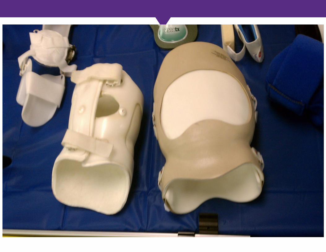
Prevention of contractures
• Range of motion
• Splints and orthotics
• Equipment that help positioning
• Focus on all joints - emphasize joints that are tight ― Incorporate stretching/passive ROM into daily routine
− Diaper changes
− Bath time
− Before bed

Tendon Lengthening • Surgical intervention indicated for fixed contractures that impair
seating, mobility, function
• Contractures rarely occur in isolation
• Muscles that cross 2 joints most frequently affected
• WARNING: Weakens an already WEAK MUSCLE

Adduction contracture
• Limits ability to spread legs interferes with toileting,
positioning in wheel chair,
standing,
walking (scissoring)
• Lengthen Adductor and
Gracilis muscles

Hip flexion contracture
• Contributes to anterior pelvic tilt
• Fixed hip flexion contracture important to consider when fusing the spine to the pelvis
Ambulatory SMA 3 Fractionally lengthen psoas tendon @ pelvic brim to preserve hip
flexion strength Non-ambulatory SMA 2
Detach iliopsoas tendon from lesser trochanter

Clinical characteristics
Clinical exam: Popliteal angle (>55°) Straight leg raise (<60°)
Limits ability to stand upright,
Affects pelvic position when fusing spine to pelvis
Hamstring Contracture

Fractional lengthening Hamstrings
•Supine position •Single posterior incision above popliteal fossa
Tenotomy of Semitendinosus, Gracilis
Fractionally lengthen Aponeurosis of Semimembranosus, Biceps Femoris

Gastoc-Soleus muscle + Achilles tendon contracture
Toe walking during gait training/standing
• Younger patient without fixed contracture:
Serial cast / AFO
• Fixed contracture or severe foot deformity
Recession of gastrocnemius
Avoid Achilles lengthening
1
2
3

Osteopenia (low bone density) Increases Risk for Fragility Fractures
• Common in children who don’t walk
• Fractures caused by minimal trauma
• Common (reports up to 40%)
― 40% femur, 26% tibia,14% humerus, 9% clavicle
• Risk factors Stiff joints
Decreased bone mineral density due to lack of weight bearing
Poor balance leading to falls

Factors Affecting Fracture Risk
Structural Geometry
Loading Condition
Material
Applied Load Load Capacity Strength
+ +
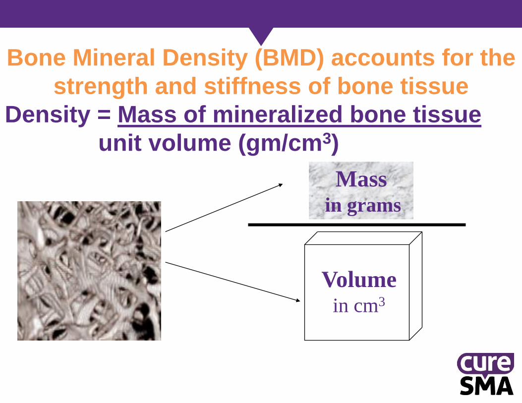
Density = Mass of mineralized bone tissue
unit volume (gm/cm3)
Mass in grams
Volume in cm3
Bone Mineral Density (BMD) accounts for the
strength and stiffness of bone tissue

Bone profoundly weakened when
apparent on X-ray (osteopenia)
• Bone density
reduced 30-50%
before evident on
radiographs
Corresponds to
40-75% reduction
bone strength

Geometric Properties of Bone Are
As Important As Bone Density
Moment of Inertia
A structure is more
rigid when its mass
is distributed away
from the bending
axis

Ability of long bones to support or withstand
load severely limited
• Material – Bone tissue
Severe osteopenia
• Geometry – Bone shape
Narrowed cross-section diameter
Thinned cortices
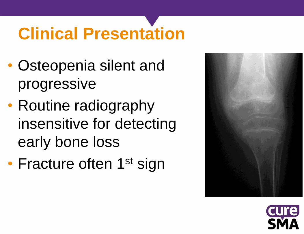
Clinical Presentation
• Osteopenia silent and
progressive
• Routine radiography
insensitive for detecting
early bone loss
• Fracture often 1st sign

Evaluation of bone density
• Uses detection system to
measure transmission of
X-rays through body
• Determines intensity that
is transmitted
• Low radiation: 1-3 mrem
X-Ray source
Table Subject
Low & High
Energy Detectors Dual Energy X-ray Absorptiometry (DXA)

Limitation: DXA Does Not Give True Density
• Bone Mineral Content
divided by projected area
of analysis: aBMD gm/cm2
aBMD = BMC Area
Area region
of analysis
• Fails to account for
out of plane
dimension
Affected by the SIZE
of the bone
(diminutive in SMA)
• Results reported as a
Z-score = number
Standard Deviations
different from age
matched able bodied
children, use height
matched instead

DXA Sites Evaluated
• Osteopenia appears initially @ sites comprised of trabecular (spongy) bone
• Trabecular bone metabolically more active than cortical bone
• Hip and spine contain greater proportion of trabecular bone
• However hip and spine may be abnormal in SMA

Limitations of DXA in Children with SMA
• Diminutive bone size – smaller than age matched peers
Compare to height matched control rather than age
• Unreliable @ hip and lumbar spine:
Hip – effected by flexion contractures, subluxation, previous
surgery, hardware
Spine – effected by pelvic contracture, scoliosis
• BMD lumbar spine unreliable predictor bone status @
proximal femur (Henderson Skel Radiol 26:544, 1997)

Lateral Femoral DXA to Assess BMD non-ambulatory Children
• Distal Femur most common site for
pathologic fracture
• Distal Femoral Lateral aBMD
predicts osteopenia & fragility
fracture better than lumbar aBMD
Region 1: distal femur metaphysis =
trabecular bone
Region 2: meta-diaphysis
Region 3: diaphysis = cortical bone
[Harcke Pediatr Radiol 28:241 (1998)]

Treatment reduced bone density
(osteopenia)
• Passive weight bearing
― Standing frame (>1.5hr/day x 5 day)
― may improve bone density
• Maximize calcium and vitamin D
supplementation
― 2000 IU vitD3, 1500mg Ca divided over 3
meals
• If >2 non-traumatic fractures
― Bisphosphonates: Alendronate (oral),
Pamidronate (IV)

Treatment of Fractures • Treat long bone fractures w/ surgical stabilization to
maintain wt bearing potential and to facilitate
transfers
Avoid prolonged cast immobilization
― Early mobilization

Hip
• Instability
• Dysplasia
• Dislocation

Who is at risk for hip instability
• Risk for Hip & Spine deformity inversely correlated with physical function capability ― Highest for SMA 1 and 2
• Hip subluxation / dislocation common non-walking children age 5-8 yrs
• Unilateral or bilateral
• Associated with Pelvic Obliquity

Pathophysiology
• Hips normal at birth
• Global symmetric weakness
Core and hip girdle muscles
• Ligamentous laxity
• Absence of weight bearing
Attenuates mechanical stimulus for growth and remodeling hip joint Coxa Valgus + persistent femoral anteversion
• Associated with Pelvic Obliquity and Scoliosis (unclear which comes 1st)

Adductors > Abductors
Hip Flexors > Extensors
Ref # 9

Coxa Valgus: ↑ Neck Shaft Angle

Excessive Femoral Anteversion

Bone Morphology
Femoral Version
Ref # 1,2,3

Acetabular Dysplasia: Deficient postero-lateral coverage of hip

Posterolateral Migration of hip
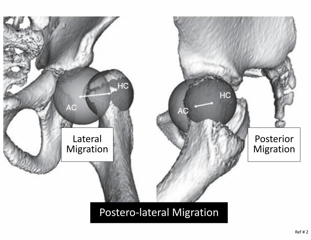
Ref # 2
Lateral Migration
Posterior Migration
Postero-lateral Migration

Center of Rotation Δ

Summary: Anatomical Changes
Coxa valgus reduces efficiency (moment arm) hip
abduction by gluteus medius (swing phase of gait)
Upward and lateral migration femoral head out of
acetabulum
Maturation of acetabulum retarded by pressure from subluxed femoral head on lateral pelvic apophysis ― Greatest when hip > 50% subluxed
Posterior and superior acetabular deficiency

Physical Exam
• Young children – hip very unstable, pops in/out of joint as hip is abducted – adducted during diaper changes, toilet/hygeine
• Older children – dislocated hip becomes “stiff” ― Symmetric loss of hip abduction flexion contracture
― Asymmetric abduction = windblown hip deformity, pelvic obliquity, scoliosis
• Physical exam alone unreliable to dx hip subluxation

“Windblown” Hips
• Adduction one hip, Abduction other
• pelvic obliquity risk unilateral hip dislocation ― not seen in children with level pelvis
• Hip dislocates on side of elevated hemiplevis
• Relationship hip instability pelvic obliquity scoliosis ― Hip subluxation occurs before scoliosis
― pelvic obliquity Scoliosis
― Scoliosis less frequent in children without hip instability
― Result of worsening muscle weakness?

Monitoring
• Supine AP hips yearly -
screen for dysplasia
• Standing AP (if
possible) – wt bearing
provocative test for hip
subluxation
• Evaluate whether hip
reduces in Abduction
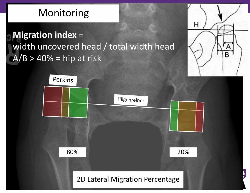
Monitoring
2D Lateral Migration Percentage
80% 20%
Migration index = width uncovered head / total width head A/B > 40% = hip at risk
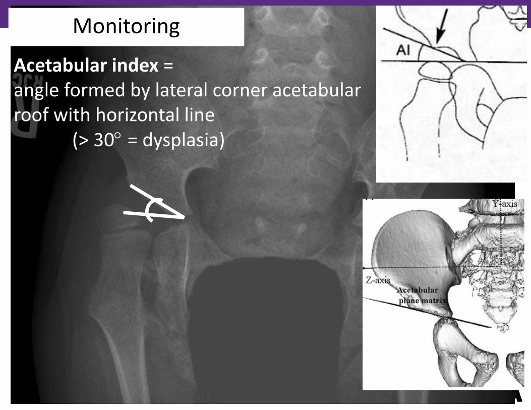
Monitoring
Acetabular index = angle formed by lateral corner acetabular roof with horizontal line (> 30 = dysplasia)

Additional Imaging Studies: CT with 3D
Reconstruction
• Assess deformity of femoral head
• Assess location of acetabular deficiency
• Images through distal femur to calculate
femoral anteversion
― (May be adversely affected by coxa valgus)

Treatment of subluxated hips
• Few natural history studies untreated hips (Sporer & Smith JPO 2003)
― >50% with hip instability
― Few had pain at long term follow-up
― Few had functional limitations with seating, positioning, hygiene
― None had skin breakdown, ulcers
― BUT Pelvic Obliquity & Scoliosis in 91% non-ambulatory children
• Recurrence of coxa valgus and hip subluxation reported after surgical
treatment in young children
Controversial

My Opinion
• No controversy for children who walk –
Reconstruct hip if unstable
• Non-walking children:
― Prevent hips from dislocating for comfort,
maintenance pelvic alignment, improved sitting
balance
― May help prevent pelvic obliquity scoliosis
― Less complicated surgery children <10yrs
― Primary disease persists – recurrent coxa valgus &
subluxation may occur – revision may be required

Non-surgical treatment
• PT – maintains hip motion, promotes wt bearing (standers) ― No evidence that prevents
hip subluxation
― However Standing ~ 2hr/day X5 day/wk helps maintain bone mass – may prevent fragility fractures
• Abduction bracing ― No evidence that prevents
hip dislocation long term

Indications for Surgical Reconstruction
• Child > 4 y/o, Migration > 40% ― <4 y/o, 96% will loose correction
neck-shaft angle and rotation
• Child > 8y/o, Acetabulum shallow (acetabular index > 25°) ― Must address both sides of hip
joint – acetabulum, femur
• Comprehensive approach: ― Soft tissue release +
Reconstruct both sides of hip joint – femur + acetabulum

Hip must reduce and be contained in abduction
Varus Shortening Derotation Intertrochanteric
Osteotomy (VDRO)
• Do both hips (even if only one hip subluxed)
• Unilateral VDRO may result in pelvic obliquity
• Adduction osteotomy must have sufficient
(>45°) abduction adductor tenotomy first

Varus Derotation Osteotomy (VDRO) • Lateral approach proximal femur,
• Set neck shaft angle: 100° non-walkers, 120° walkers
• Shorten femur, derotate femur (set anteversion ~15°)
• Rigid Hip Plate for fixation to allow early Range of Motion
Remove 1-2 cm from Shaft lengthens Hams, Rectus Femorus
Recess iliopsoas tendon Medialize shaft To maintain Mechanical axis

Hip uncovered laterally after VDRO
Indications for Acetabular Osteotomy ― Shallow hip socket (Acetabular Index > 25°)
― Hips that do not reduce concentrically after VDRO
• Type of acetabular osteotomy depends on patient age (open vs. closed tri-radiate cartilage), ambulatory status, extent degenerative changes femoral head & acetabulum

Reduce radius of curvature postero-lateral deficiency to contain hip •Incomplete osteotomy above sourcil
•Hinges on tri-radiate
Pemberton-Dega Acetabuloplasty
Interposed autograft stable

Non-walking adults
Walking adults
Chronic Dislocated Hips in Young Adults
• Leave out if painless
• IF Pain & deformity interfere w/ sitting & hygiene: Procedures to relieve
pain / improve ROM
Perform bilaterally if contralateral hip located to avoid pelvic obliquity (?)

Peri-operative Management
• Epidural anesthesia 3 days post-op
• Diazepam if muscle spasms
• A-frame long leg casts for 3 weeks ― No SPICA casts
― Be vigilant for skin breakdown, pressure ulcers
• Sitting, Active assisted ROM post-op day 2
• Chest PT, incentive spirometry for pulmonary toilet
• DVT prophylaxis older children (LMW heparin)
• Aggressive PT x 6 mos: strengthen hip girdle, trunk, quads, gait training (SMA 3)

Complications
• Depends on co-morbidities
― Feeding/constipation Pulmonary
• Implant fails osteopenic bone
― Use high angle blade plate
• Decubitus ulcers from cast
• Fragility Fractures
― No SPICA casts