spinal deformities dr. abdulmonem alsiddiky, md, ssc-orth. consultant ped. ortho., ped. spine &...
TRANSCRIPT

SPINAL DEFORMITIES
Dr. ABDULMONEM ALSIDDIKY , MD , SSC-Orth.
Consultant ped. Ortho., ped. Spine& spinal deformities
KKUH Riyadh , Saudi Arabia
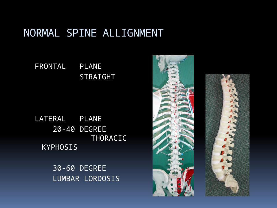
NORMAL SPINE ALLIGNMENT
FRONTAL PLANE STRAIGHT
LATERAL PLANE 20-40 DEGREE
THORACIC KYPHOSIS
30-60 DEGREE LUMBAR LORDOSIS

SCOLIOSIS
Def. Lat. deviation
of the spine from midline with rotation

Rotation in scoliosis

SCOLIOSIS
Types : Congenital (structural abn. In vertebrae
or ribs ) Neuromuscular (eg. cp,mmc,sma…) Idiopathic (most common ) Others
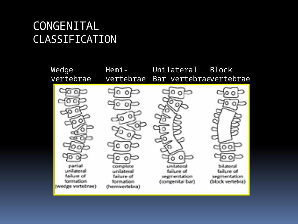
CONGENITALCLASSIFICATION
Wedgevertebrae
Hemi-vertebrae
UnilateralBar vertebrae
Block vertebrae


IDIOPATHIC SCOLIOSIS
Spinal deformity in a spine which was normal
Causes
? Properioception disorders Brain stem Melatonin hormones UNKNOWN

IDIOPATHIC SCOLIOSIS
TYPES Infantile (0-4 yrs ) Juvenile (4-9 yrs ) Adolescent (> 10 yrs ) [most common]

IDIOPATHIC SCOLIOSIS
Incidence More in female Rt thoracic curve is the most
common ? Family Hx More in twins

IDIOPATHIC SCOLIOSIS
C/O : Loss of self image Family observation Pain Early fatigue Cardio-pulmonary dysfunction ( if curve
> 90 )

IDIOPATHIC SCOLIOSIS
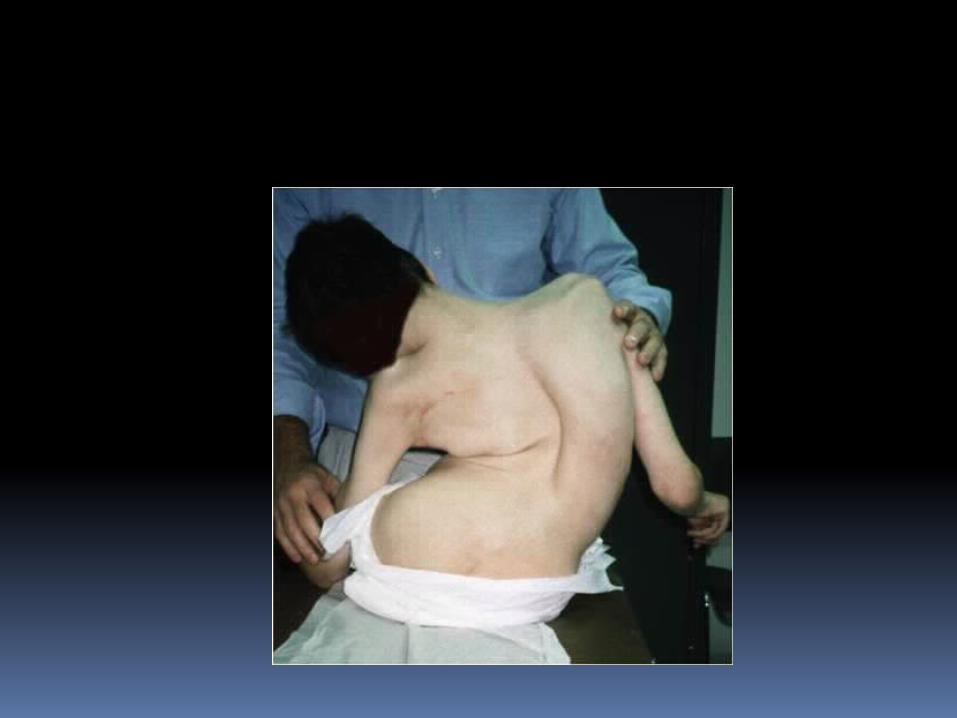
O/E : Shoulder level inequality Waist line asymmetry Spinal deformity Rib hump Adam foreword flexion test Full neurological exam

clinically

IDIOPATHIC SCOLIOSIS
Radiological exam : X-rays :
AP – LAT standing long film AP supine AP Pelvis LAT L-S spine
IDIOPATHIC SCOLIOSIS
MRI :If abnormal curve suspected ( any curve
other than rt. thoracic curve in young female )
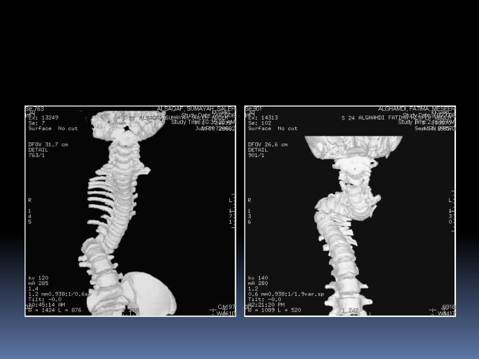
Ct scan :If congenital scoliosis suspected

X-ray
71ْ
53ْ

Cobb and Lippmann
Determine end vertebrae Those most
tilted from horizontal
Line along upper end plate prox. & lower endplate distally
Measure formed angle
Transitionalvertebrae

Treatment
Based on :
1. Maturity of the pt.
Menarche Rissor’s stage
2. Magnitude of deformity
3. Curve progression

Risser’s stage eg.

Treatment
Options
1. Observation2. Bracing3. Surgery
? Physical therapy & exercise

Treatment ( protocol ) Mature pt.
< 50 ْ observation progression ~ 1 ْ / year > 50 ْ surgery
Immature pt. 0-25 ْ Observation every 4-6
months clinically &
radiologically 25-40 ْ Bracing > 40 ْ Surgery

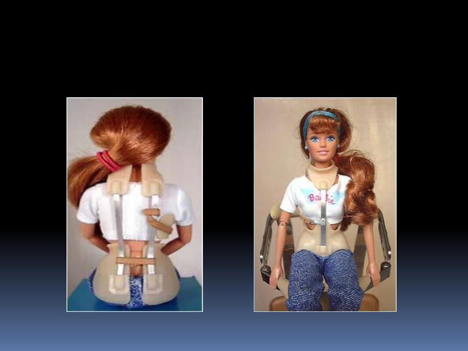
Braces eg.

Treatment
Braces : Did not correct the deformity Might stop the progression of the curve (or slow it down) Effect is dose related (more worn better
effect) Best 23 hours / day If curve apex above T7
Milwaukee brace If curve apex bellow T8
Boston brace

Treatment
Surgery : Anterior spinal fusion
severe curve young pt. < 10 years
Post spinal fusion & instrumentation The gold standard treatment for most of
cases Both
For selected cases
Treatment
Complications of surgery
Neurological deficit Bleeding Infection Pseudoarthrosis Crank shaft phenomena

Examples


please be careful when you deal
with any case of Spinal deformity
specially
scoliosis
