manual 15 endpoint ascertainment procedures · pdf filemanual 15 endpoint ascertainment...
TRANSCRIPT

MOP 15: HCHS/SOL, Endpoint Ascertainment Procedures 9/12/2011 ver. 1.0
Manual 15 Endpoint Ascertainment Procedures
September 12, 2011 - Version 1.0
Prepared by the HCHS/SOL Endpoints Subcommittee
Study website - http://www.cscc.unc.edu/hchs/
MOP 15: HCHS/SOL, Endpoint Ascertainment Procedures 9/12/2011 ver. 1.0 TOC Page 1 of 1
Endpoint Ascertainment Procedures TABLE OF CONTENTS
1.0 ENDPOINT ASCERTAINMENT PROCEDURES ................................................................................................................ 1 1.1 OVERVIEW. ............................................................................................................................................................... 1 1.2 SPECIFIC DISEASE ENDPOINTS. ........................................................................................................................... 1 1.3 ASCERTAINING POTENTIAL EVENTS. ................................................................................................................ 1 1.4 COLLECTION AND ABSTRACTION OF MEDICAL INFORMATION. ................................................................ 2 1.5 DEATH INVESTIGATIONS. ..................................................................................................................................... 4 1.6 REVIEW AND VALIDATION. .................................................................................................................................. 4 1.7 CLINICAL SITE PROCEDURES FOR IDENTIFYING AND PROCESSING EVENTS FOR ENDPOINT
CLASSIFICATION ..................................................................................................................................................... 4 1.8 MEDICAL RECORDS PROCESSING ....................................................................................................................... 7
2.0 ENDPOINT SURVEILLANCE FOR HOSPITALIZED ACUTE MYOCARDIAL INFARCTION ...................................... 9
2.1 INTRODUCTION ....................................................................................................................................................... 9 2.2 EVENT IDENTIFICATION ....................................................................................................................................... 9 2.3 SCREENING CODES ................................................................................................................................................. 9 2.4 DIAGNOSTIC CRITERIA .......................................................................................................................................... 9
3.0 ENDPOINT SURVEILLANCE FOR HOSPITALIZED HEART FAILURE ....................................................................... 14 3.1 INTRODUCTION ..................................................................................................................................................... 14 3.2 EVENT IDENTIFICATION ...................................................................................................................................... 14 3.3 DIAGNOSTIC CRITERIA FOR ACUTE DECOMPENSATED HEART FAILURE ............................................. 14
4.0 ENDPOINT SURVEILLANCE FOR STROKE AND TRANSIENT ISCHEMIC ATTACKS ............................................ 16 4.1 EVENT IDENTIFICATION ..................................................................................................................................... 16 4.2 DIAGNOSTIC CRITERIA ........................................................................................................................................ 16 4.3 DEFINITIONS OF TIA AND STROKE ................................................................................................................... 16 4.4. DEFINITIONS OF STROKE SUBTYPES ............................................................................................................... 17 4.5 ISCHEMIC STROKE CLASSIFICATION .............................................................................................................. 18 4.6 ISCHEMIC STROKE CLASSIFICATION .............................................................................................................. 20
5.0 ENDPOINT SURVEILLANCE FOR EXACERBATIONS DUE TO CHRONIC OBSTRUCTIVE PULMONARY DISEASE OR ASTHMA .................................................................................................................................................... 20 5.1 INTRODUCTION ..................................................................................................................................................... 20 5.2 EVENT IDENTIFICATION ...................................................................................................................................... 20 5.3 DIAGNOSTIC CRITERIA FOR PULMONARY EVENTS ..................................................................................... 21
6.0 ENDPOINT SURVEILLANCE OF FATAL EVENTS ......................................................................................................... 25 6.1 EVENT IDENTIFICATION ..................................................................................................................................... 25 6.2 DIAGNOSTIC CRITERIA ....................................................................................................................................... 25 6.3 FATAL CORONARY HEART DISEASE EVENTS ............................................................................................... 25 6.4 FATAL PULMONARY EVENTS ............................................................................................................................ 27
7.0 EVENT CLASSIFICATION COMMITTEE ......................................................................................................................... 27 7.1 INTRODUCTION ..................................................................................................................................................... 27 7.2 REVIEW PROCESS .................................................................................................................................................. 27 7.3 DISAGREEMENT RESOLUTION .......................................................................................................................... 27 7.4 CONFIDENTIALITY ............................................................................................................................................... 29
REFERENCES .............................................................................................................................................................................. 30 APPENDIX A. MINNESOTA CODE ECG CRITERIA .............................................................................................................. 32 APPENDIX B. GLOSSARY OF KEY EVENT DATA COLLECTIONS TERMS .................................................................... 35 APPENDIX C. DATA COLLECTION FORMS REQUIRED FOR ASSESSING ENDPOINTS: ............................................. 36 APPENDIX D ABSTRACTION FORMS .................................................................................................................................... 55 APPENDIX E REVIEWER FORMS .......................................................................................................................................... 85 APPENDIX F - EVENT SUMMARY FORMS ........................................................................................................................... 90
MOP 15: HCHS/SOL, Endpoint Ascertainment Procedures 9/12/2011 ver. 1.0 Page 1 of 39
1.0 Endpoint Ascertainment Procedures 1.1 Overview. The mission of the Endpoints Ascertainment and Classification Committee is to design and implement a system for event ascertainment, including review and validation of a variety of endpoints to facilitate the classification of incident events occurring among the Hispanic Community Health Study / Study of Latinos (HCHS/SOL) cohort participants. The specific objectives of the endpoints procedure manual are to clearly describe how the HCHS/SOL will:
identify acute myocardial infarction, stroke, heart failure, asthma and chronic obstructive pulmonary disease (COPD) events that have required hospitalization following the initial examination;
identify acute exacerbations of asthma or COPD requiring emergency department (ED) care; and review and evaluate clinical information collected from medical records from hospitals and
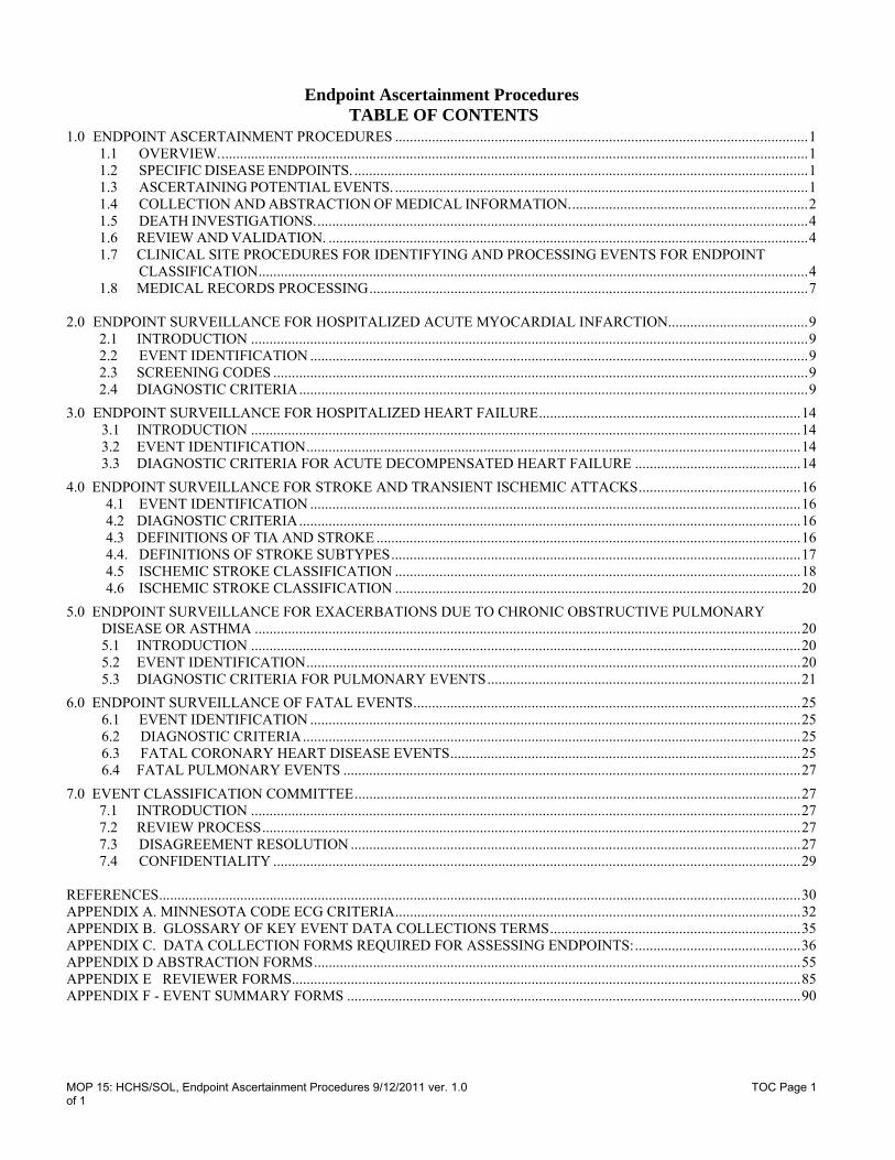
emergency departments to classify each event type. The identification and classification of health events in the HCHS/SOL outlined in this manual follows standard principles of population-based cohort surveillance. These principles include ascertaining potential events, gathering medical information about these events, and reviewing collected data to validate the types of events of interest. The aim of surveillance of the HCHS/SOL cohort is to identify all hospitalizations and emergency department visits for each cohort participant (regardless of reason) and validate the diagnosis of all potential coronary, stroke and pulmonary disease events that occur between baseline exam and the subsequent follow up. We will also investigate deaths to validate cause of death from cardiovascular and pulmonary disease. The general approach to defining endpoints of interest, ascertainment of potential cases, gathering medical information, and review/validation of events is outlined below. Details of each of these steps are provided in the proceeding chapters of this manual. 1.2 Specific Disease Endpoints. The specific cardiovascular and respiratory disease endpoints of primary consideration in HCHS/SOL are: 1) hospitalizations for myocardial infarction, stroke, heart failure, COPD, and asthma, and 2) emergency department visits for COPD and asthma exacerbations. The cause of death of any cohort participant will also be established and is a primary endpoint. Only cardiovascular or pulmonary disease related deaths will be validated. 1.3 Ascertaining Potential Events. Event surveillance of the cohort uses information obtained from the annual phone follow-up interview. When the annual follow-up interview indicates that the participant has either died, been admitted to a hospital, or been seen in an emergency department, mechanisms to obtain the appropriate medical records or death certificate are initiated. Fatal events are also ascertained from review of vital statistics lists and obituaries for the state in which the community is located. The HCHS/SOL records the occurrence of all hospitalizations and all emergency department visits and captures the discharge diagnosis and procedure codes (ICD-9 codes) but only conducts detailed investigations for the selected kinds of medical events noted above. Detailed investigation of recalled hospitalized and emergency department events will be triggered initially by the reported reason for the event but verified by the presence of certain discharge diagnoses or procedure codes (see Figure 1 below). Presence of certain presenting symptoms will trigger investigation of emergency department or emergency medical services (EMS) records. Death investigation will be triggered by certain underlying cause of death codes on the death certificate; and investigation out out of hospital deaths will include interviews with next of kin and mailing questionnaires to appropriate physicians, medical examiners or coroners. See Chapter 7 for Endpoint Surveillance of Fatal Events.
MOP 15: HCHS/SOL, Endpoint Ascertainment Procedures 9/12/2011 ver. 1.0 Page 2 of 39
Figure 1. Summary of event investigation based on initial reason for hospitalization or emergency department visit as reported by cohort participant
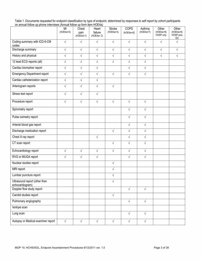
1.4 Collection and Abstraction of Medical Information. A detailed abstraction form specific to the type of event will be used by trained staff at the coordinating center to collect relevant data from medical records of eligible events. Copies of discharge summaries, history and physical, electrocardiograms, echocardiography reports, neuro-imaging reports, consult reports and other pertinent documents will be obtained by field center staff and sent to the coordinating center for abstraction. The type of records from the medical chart to be sought, copied, and sent to the coordinating center is summarized in Table 1. Abstractors follow detailed question by question instructions (QXQ) for the standardized abstraction of medical record information to a database. Abstractors are trained and certified. A brief summary (1-2 pages) of information abstracted from these materials (the Event Summary Form or ESF) will be provided to the Event Classification Committee (ECC) for their review when classifying the event. In addition, copies of selected portions of the materials from the medical record will be provided to the Events Classification Committee members for their use in determining the final event classification of each event.
Follow-up interview form (AFE) contact year 1
Was a hospitalization or ED visit reported? (AFE item 3)
Yes
No
Hospital, ED, or both ? (AFE item 4)
No investigation
What was main reason for event ? (AFE 4a)
Hospitalization (only) ED visit (only) Hospitalization and
Hospitalization (only) ED visit (only) Hospitalization and
Target endpoint (AFE 4a = 0 – 7)
Other (AFE 4a=8)
* Key words specified in AFE 4a = 8 include: pneumonia, bronchitis, cough, wheezing, cold
Obtain medical records materials as specified on Table X.
Target endpoint (AFE 4a = 0 – 7)
Other (AFE 4a=8)
Yes
Obtain discharge summary, coding summary (including ICD-9 codes for diagnoses and procedures), history and physical.
Obtain medical records materials as specified on Table X.
Obtain discharge summary, coding summary (including ICD-9 codes for diagnoses and procedures), history and physical, emergency department report.
Obtain full emergency department report, coding summary (including ICD-9 codes for diagnoses and procedures)
No
Pulmonary target endpoint (AFE 4a = (2, 6 or 7) or (8
with key words* ))
AFE 4a = 0 – 7: MI, angina, heart failure, stroke, TIA, PAD, VTE, COPD, asthma AFE 8 = other (specify)
Select a random sample
MOP 15: HCHS/SOL, Endpoint Ascertainment Procedures 9/12/2011 ver. 1.0 Page 3 of 39
Table 1. Documents requested for endpoint classification by type of endpoint, determined by responses to self report by cohort participants on annual follow up phone interviews (Annual follow up form item HOE4a)
MI (HOE4a=0)
Chest pain
(HOE4a=1)
Heart failure
(HOE4a= 2)
Stroke (HOE4a=3)
COPD (HOE4a=6)
Asthma (HOE4a=7)
Other (HOE4a=8) HOSP only
Other (HOE4a=8) HOSP plus
ED
Coding summary with ICD-9-CM codes
Discharge summary
History and physical
12 lead ECG reports (all)
Cardiac biomarker report
Emergency Department report
Cardiac catheterization report
Arteriogram reports
Stress test report
Procedure report
Spirometry report
Pulse oximetry report
Arterial blood gas report
Discharge medication report
Chest X-ray report
CT scan report
Echocardiology report
RVG or MUGA report
Nuclear studies report
MRI report
Lumbar puncture report
Ultrasound report (other than echocardiogram)
Doppler flow study report
Carotid studies report
Pulmonary angiography
Isotope scan
Lung scan
Autopsy or Medical examiner report
MOP 15: HCHS/SOL, Endpoint Ascertainment Procedures 9/12/2011 ver. 1.0 Page 4 of 39
1.5 Death Investigations. A death certificate form will be completed for all eligible fatal events. For in-hospital deaths, data collected on the death certificate form and the hospital abstraction form will be combined for use by the ECC for review and event classification. Deaths occurring outside the regular acute care hospital are categorized as out of hospital deaths. This includes persons dead on arrival at acute care hospitals, and those dying in outpatient departments or emergency rooms, or admitted without vital signs. For out of hospital deaths meeting underlying cause of death code criteria, information is sought from the decedent’s family and physician within 6 months after death. The former is contacted by telephone and the latter by mailed questionnaire. Often the informant is the spouse or other family member of the decedent. Information provided by the informant and the physician is combined for use by the ECC for review and event classification. 1.6 Review and Validation. Diagnostic information obtained through abstraction of the medical record combined with documents copied from the medical records are de-identified and prepared for review by members of the ECC. Cause of death ICD-10 codes obtained through abstraction of the death certificate are combined with supplemental information from informants (in the case of out of hospital deaths) and prepared for review by members of the ECC. ECC members complete an event classification form which indicates their judgment as to the diagnosis of the events. ECC reviewers are trained and certified to follow standardized rules and case laws when determining the final event classification of each case. 1.7 Clinical Site Procedures for Identifying and Processing Events for Endpoint Classification Annual Follow-Up Form (AFU) (See Manual 16 and study web site for example and QxQ) During the completion of the annual follow-up interview (contact year 1), field center staff will ask the participant whether they had been admitted to a hospital or seen in an emergency department (ED), at any time since their last SOL center visit (HOEA item 3). If no events are reported (HOEA item 1 = ‘no’ or ‘unsure’) there will be no events to be investigated. If participants respond ‘Yes’ to HOEA item 1 then they are asked to identify the type of event in HOEA item 4. HOEA item 4 identifies whether the event was a visit to the ED or an admission to the hospital, or both. This information on event type appears on the Event Tracking Report and can be useful in field center efforts to obtain medical records. Item 4a on the HOEA form asks: What was the main reason for going to the (insert emergency room or hospital) that day? The discharge summary and associated summary of all discharge diagnosis and procedure codes are important parts of the record that will always be obtained for all hospitalized or emergency department events. Events reported during the annual follow up interview that involve an emergency department visit without subsequent hospital admission (a stand-alone ED visit) are selected for further investigation as shown Figure 1 above. Emergency department visits without a subsequent hospitalization are investigated if they are suspected to be for COPD or asthma based on the self-reported reason for the visit (from item 4a of the HOEA form). Event Identification (ID) Numbers When an event eligible for investigation is reported during an Annual Follow Up interview and the data is entered into the data management system, a unique event ID number is assigned to each reported event by the endpoints management system. This event ID is derived from the cohort ID number and the reported hospitalization by computer algorithm. If more than one eligible event is reported during an annual follow interview for an individual, then the data management system creates new event IDs for each reported event. For example, for a given cohort ID number there may be several event IDs. Information about the
MOP 15: HCHS/SOL, Endpoint Ascertainment Procedures 9/12/2011 ver. 1.0 Page 5 of 39
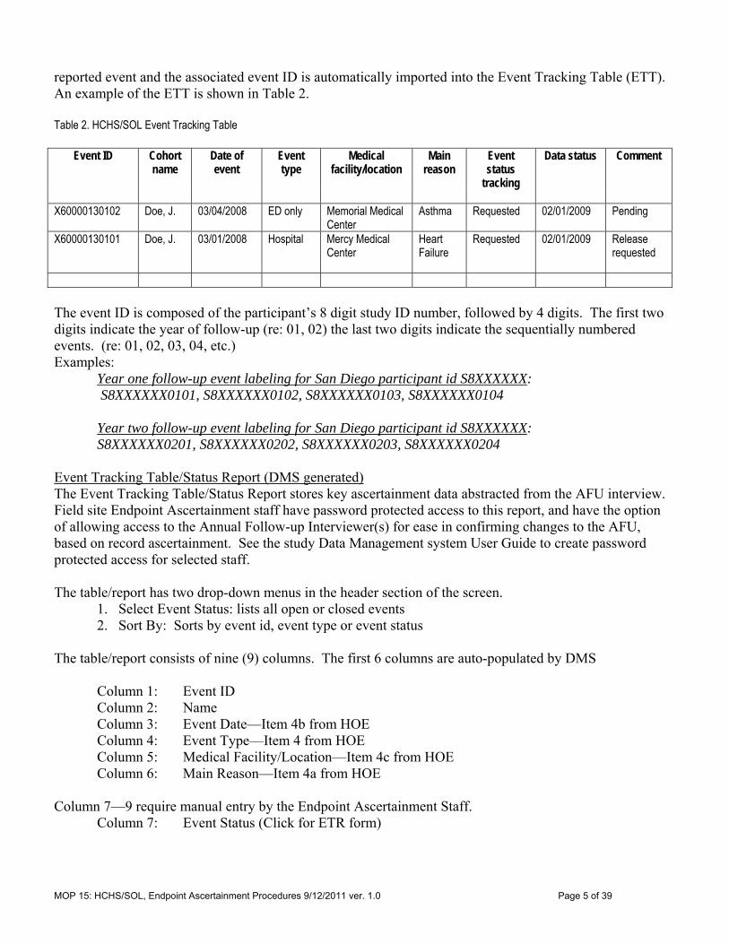
reported event and the associated event ID is automatically imported into the Event Tracking Table (ETT). An example of the ETT is shown in Table 2. Table 2. HCHS/SOL Event Tracking Table
Event ID Cohort name
Date of event
Event type
Medical facility/location
Main reason
Event status
tracking
Data status Comment
X60000130102 Doe, J. 03/04/2008 ED only Memorial Medical
Center Asthma Requested 02/01/2009 Pending
X60000130101 Doe, J. 03/01/2008 Hospital Mercy Medical Center
Heart Failure
Requested 02/01/2009 Release requested
The event ID is composed of the participant’s 8 digit study ID number, followed by 4 digits. The first two digits indicate the year of follow-up (re: 01, 02) the last two digits indicate the sequentially numbered events. (re: 01, 02, 03, 04, etc.) Examples:
Year one follow-up event labeling for San Diego participant id S8XXXXXX: S8XXXXXX0101, S8XXXXXX0102, S8XXXXXX0103, S8XXXXXX0104 Year two follow-up event labeling for San Diego participant id S8XXXXXX: S8XXXXXX0201, S8XXXXXX0202, S8XXXXXX0203, S8XXXXXX0204
Event Tracking Table/Status Report (DMS generated) The Event Tracking Table/Status Report stores key ascertainment data abstracted from the AFU interview. Field site Endpoint Ascertainment staff have password protected access to this report, and have the option of allowing access to the Annual Follow-up Interviewer(s) for ease in confirming changes to the AFU, based on record ascertainment. See the study Data Management system User Guide to create password protected access for selected staff. The table/report has two drop-down menus in the header section of the screen.
1. Select Event Status: lists all open or closed events 2. Sort By: Sorts by event id, event type or event status
The table/report consists of nine (9) columns. The first 6 columns are auto-populated by DMS Column 1: Event ID Column 2: Name Column 3: Event Date—Item 4b from HOE Column 4: Event Type—Item 4 from HOE Column 5: Medical Facility/Location—Item 4c from HOE Column 6: Main Reason—Item 4a from HOE Column 7—9 require manual entry by the Endpoint Ascertainment Staff. Column 7: Event Status (Click for ETR form)

MOP 15: HCHS/SOL, Endpoint Ascertainment Procedures 9/12/2011 ver. 1.0 Page 6 of 39
Event Tracking Report Form (ETR) (See appendix XX for example and QxQ). This record will be used to track the site efforts on obtaining, processing and shipping the medical records for an event under investigation. Once an event has been listed on the Event Tracking Status Report in DMS, the staff will create an ETR by clicking on “No ETR” under column 7. In order for the ETR form to auto-save in the system you must completely enter the form. (Event ID and Event Date are pre-populated). This form should be updated as record acquisition work progresses through “pending records request” to “shipping records to the Coordinating Center”. Study reports developed will use the ETR to give the field centers and steering committee feedback on the progress of end points investigation activities at each site. All “closed events” (with terminating/final codes: 4, 5 or 9) will be auto-stored in the Closed Event Table, which is accessed by the “Select Event Status” menu on the Event Tracking Table/Status Report. Column 8: Status Date--Auto populates once the ETR form is entered. Column 9: Click for VER Form Verification Form (VERA) (See appendix C4 for example) When medical records of interest are received for an event, enter the ICD-9 codes appearing on the discharge summary page into the verification form (VERA) by clicking on the equal sign (=) in the last column for the event. Once the ICD-9 codes have been entered, the DMS will list the medical record materials expected for this event. For each listing, code whether the document of interest was:
1=received, 2=pending, 3=not available Successful verification of the discharge codes will produce a face sheet for the materials being transferred to the coordinating center. Print the VER form and face sheet to send with the documents. Please note, if the face sheet is not pre-populated with the Subject ID, Date of Event and Event ID—contact the DCC and the staff will assist you.
Updating Events There may be instances when the participant has reported an incorrect or incomplete event date, which is discovered when the medical records are obtained. The AFU or Data Management (DM) staff can correct the appropriate HOEA by line number for the event. It is very important to select and update the precise HOE record of interest by going to the existing entry for the case, and not creating a new entry (which is default for entering HOE forms). To do this, right click on the “page” icon just to the left of the participant’s HOS form on the menu on the left side of the DMS screen. Use the “jump to” sub-menu to select the appropriate line. The program that creates the endpoints workflow table incorporates changes to items on the related HOE form overnight. Corrected information should appear once the CHANGE transaction has cleared the processing system.
MOP 15: HCHS/SOL, Endpoint Ascertainment Procedures 9/12/2011 ver. 1.0 Page 7 of 39
Events Not Reported During Annual Follow-up Interviews Additional events, not self-reported at annual follow-up may be identified when medical records are obtained for a self-reported event. These newly discovered events must be assigned event IDs and must be distinguished from the self-reported events on the HOEA form for the contact year in which it took place. To correctly capture this information: 1) “jump to” the last HOEA line in the sequence, 2) tab to question 4f, and change that from a “No” to a “Yes” which will bring up a blank HOE form to complete, 3) enter the information for the newly discovered event, 4) Create a note log for HOE item 4b, Date of Event, that says: “Detected after AFU interview”, and 5) Save the changes. If the newly discovered event is eligible for investigation, it will appear in the work panel as a new entry after the system updates the tracking table overnight. Numbering Events If a participant has an ED or hospitalized event where he/she was transferred directly to another facility, this is considered one (1) event only. If a participant has discrete admission and discharge dates that are not continuous (with intervening days not in the hospital), these are considered two (2) events. 1.8 Medical Records Processing Medical Record Release Forms and Cover Sheet (See appendix XX for example) In order to obtain medical records for a specific event, a current and signed medical record release form will need to be sent to each medical facility identified (as indicated on the Event Tracking Status Report) for each event. Medical record release forms are valid for 90 days from the time the patient signs and dates the form. Keeping in mind that a patient may have more than one event or may have been seen in more than one institution; it may be helpful to have the participant sign several release forms at one time, or to sign one and leave the date blank. Alternatively, some institutions may release medical records with a copy of the participant’s signed HIPAA consent form. The medical records release form must include the specific dates of the event and the name of the healthcare provider requesting the records. A cover sheet with auto-populated demographics and participant’s self-reported reason for event from the AFU will accompany each request for medical records. When records are received, the field center (FC) determines if the records are sufficient to ascertain the event. The first step is to directly compare what was received to what was requested. ICD Codes (International Classification of Diseases) Obtaining discharge diagnosis and procedure ICD codes for all events is critical for the standardized ascertainmnent of potential events. Originally constructed to provide comparable international data on causes of death, today it is used in many countries for coding hospital discharge diagnoses for billing purposes. For both hospitalizations and ED visits, an ICD code is assigned for each diagnosis. Usually there is one primary discharge diagnosis/ICD Code for each hospitalization/ED visit, but there may be several secondary diagnoses listed as well. The secondary diagnoses may include old and new diagnoses. Usually an ICD summary page is included in medical records for any event and this is often on the face page. If this is not received for a specific event, the FC will need to follow-up with the medical records department to obtain it before the event can be sent to abstraction and processed further. classification . It

MOP 15: HCHS/SOL, Endpoint Ascertainment Procedures 9/12/2011 ver. 1.0 Page 8 of 39
is necessary to obtain and send the ICD summary page to the Data Coordinating Center (DCC) as part of the medical records package for each identified event. If the ICD-9 summary is not available after exhausting efforts at the medical records department, an attempt to obtain it through the hospital’s billing department should then be pursued. If attempts to obtain the ICD-9 summary page are unsuccessful, code the preferred terms used in the discharge summary using the online reference provided below: ICD9 CODES ONLINE: http://icd9cm.chrisendres.com/index.php If the field center is coding diagnoses without the summary page from medical records, the coder should indicate such on the VER form, Item 2, by entering “3“ (unavailable). De-Identifying and Labeling Medical Records In order to comply with the de-identification rules for research conducted under HIPAA, field centers are to mask or de-identify the following items on the medical records. Each page of medical records received must be checked and the following de-identified:
Participant name and/or initials Hospital name and street address Institutional letterheads and/or logos Names of everyone (keep degrees or titles) Telephone numbers Medical record numbers Health plan ID numbers Account numbers Social security number Electronic mail addresses Web addresses or URLs, IP addresses
Prior to de-identifying, each page of the medical record must be labeled with the event ID and the date of the event. This may be done by placing a pre-printed label on each page, or writing the information on each page. De-identification may be done using a regular point black Sharpie or similar marker. Shipping Materials to the Data Coordinating Center Bundle materials for each participant event separately. The face sheet printed from the VER form must appear as the first page for each event packet. More than one event may be shipped together as long as each event has been accurately labeled. All medical records must be shipped by a service providing a tracking mechanism, such as Federal Express. Medical records may be shipped using 2nd or 3rd day delivery option. Next day delivery is not necessary and is more expensive. Remember to maintain the tracking number of the package(s) until notification of receipt by the Data Coordinating Center has been emailed to you. Please mail medical record packet(s) to: Monica Miles (phone: (919)-962-3095) HCHS/SOL Event Receiving Collaborative Studies Coordinating Center

MOP 15: HCHS/SOL, Endpoint Ascertainment Procedures 9/12/2011 ver. 1.0 Page 9 of 39
137 East Franklin St., Suite 203 Chapel Hill, NC 27514 In the event that a record received at the CSCC has not been de-identified, the FC will be notified of this error, the record at the CSCC will be appropriately destroyed and a de-identified copy will need to be sent from the FC. 2.0 Endpoint Surveillance for Hospitalized Acute Myocardial Infarction 2.1 Introduction The aim of surveillance of the HCHS/SOL cohort in regard to acute myocardial infarction is to identify all hospitalizations for each participant and validate the diagnosis of all potential coronary events. The criteria outlined below were created to be comparable to that used by MESA. Ascertainment and validation of all out-of-hospital fatal events that are potentially cardiac-related are also completed (see Endpoint surveillance of fatal events, Section 7 of this manual). 2.2 Event Identification If a participant reports any hospitalization, field center staff requests the discharge summary, discharge diagnoses an associated ICD-9-CM codes, and any related test results and progress notes from the hospital (see Table 1). A recent signed consent is required by most hospitals in order to release records (see earlier in this section for more information about consents). Once the record is received, field center staff matches the reported hospitalization to the actual record and, if discrepancies are found, re-contacts the participant to resolve these differences. If the event involved a transfer to another hospital or other health-care facility, field center staff obtain all pertinent records from all institutions. Transfers are considered together as one potential investigation. Endpoints staff at the Coordinating Center reviews the ICD-9-CM codes and, if necessary, the discharge summary, to complete the Events Eligibility form, which determines whether the hospitalization is eligible for further detailed record abstraction. (Please see Table 3.1 for a list of eligible ICD codes.) Pertinent parts of the hospital medical record will be copied and sent to the coordinating center for central abstraction. Components of the chart are scanned at the coordinating center and stored for use by the Endpoints Review committee. 2.3 Screening Codes Events with CPT procedure code 35 or ICD-9 discharge diagnosis codes 250, 390–459, 745–747, 794.3, 798-799 are eligible for detailed abstraction by coordinating center staff for myocardial infarction. Events without these target codes, yet upon review the discharge summary by coordinating center abstractors contain evidence of eligible conditions an acute myocardial infarction event are also eligible for detailed abstraction.. 2.4 Diagnostic Criteria 2.4.1 Myocardial Infarction. Myocardial infarction is defined as the death of part of the myocardium due to an occlusion of a coronary artery from any cause, including spasm, embolus, thrombus or rupture of a plaque. The algorithm for classifying MI includes history of chest pain,, evidence from cardiac biomarkers, and ECGs, The criteria to be used in HCHS/SOL was designed to be comparable with that used in the MESA Study. Additional event classification elements (e.g. anatomical location of MI) not available in MESA were also incorporated into the HCHS/SOL MI validation process.
MOP 15: HCHS/SOL, Endpoint Ascertainment Procedures 9/12/2011 ver. 1.0 Page 10 of 39
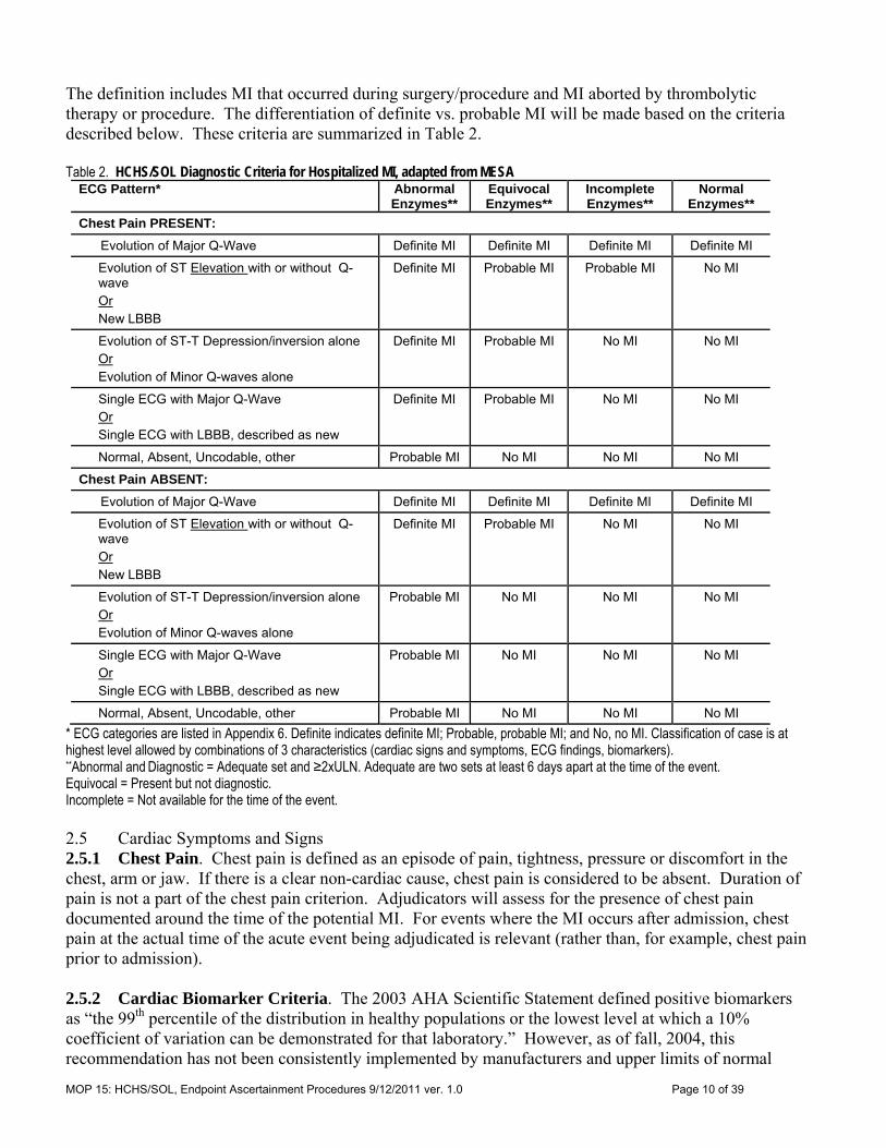
The definition includes MI that occurred during surgery/procedure and MI aborted by thrombolytic therapy or procedure. The differentiation of definite vs. probable MI will be made based on the criteria described below. These criteria are summarized in Table 2. Table 2. HCHS/SOL Diagnostic Criteria for Hospitalized MI, adapted from MESA
ECG Pattern* Abnormal Enzymes**
Equivocal Enzymes**
Incomplete Enzymes**
Normal Enzymes**
Chest Pain PRESENT:
Evolution of Major Q-Wave Definite MI Definite MI Definite MI Definite MI
Evolution of ST Elevation with or without Q-wave Or New LBBB
Definite MI Probable MI Probable MI No MI
Evolution of ST-T Depression/inversion alone Or Evolution of Minor Q-waves alone
Definite MI Probable MI No MI No MI
Single ECG with Major Q-Wave Or Single ECG with LBBB, described as new
Definite MI Probable MI No MI No MI
Normal, Absent, Uncodable, other Probable MI No MI No MI No MI
Chest Pain ABSENT:
Evolution of Major Q-Wave Definite MI Definite MI Definite MI Definite MI
Evolution of ST Elevation with or without Q-wave Or New LBBB
Definite MI Probable MI No MI No MI
Evolution of ST-T Depression/inversion alone Or Evolution of Minor Q-waves alone
Probable MI No MI No MI No MI
Single ECG with Major Q-Wave Or Single ECG with LBBB, described as new
Probable MI No MI No MI No MI
Normal, Absent, Uncodable, other Probable MI No MI No MI No MI
* ECG categories are listed in Appendix 6. Definite indicates definite MI; Probable, probable MI; and No, no MI. Classification of case is at highest level allowed by combinations of 3 characteristics (cardiac signs and symptoms, ECG findings, biomarkers). **Abnormal and Diagnostic = Adequate set and ≥2xULN. Adequate are two sets at least 6 days apart at the time of the event. Equivocal = Present but not diagnostic. Incomplete = Not available for the time of the event. 2.5 Cardiac Symptoms and Signs 2.5.1 Chest Pain. Chest pain is defined as an episode of pain, tightness, pressure or discomfort in the chest, arm or jaw. If there is a clear non-cardiac cause, chest pain is considered to be absent. Duration of pain is not a part of the chest pain criterion. Adjudicators will assess for the presence of chest pain documented around the time of the potential MI. For events where the MI occurs after admission, chest pain at the actual time of the acute event being adjudicated is relevant (rather than, for example, chest pain prior to admission). 2.5.2 Cardiac Biomarker Criteria. The 2003 AHA Scientific Statement defined positive biomarkers as “the 99th percentile of the distribution in healthy populations or the lowest level at which a 10% coefficient of variation can be demonstrated for that laboratory.” However, as of fall, 2004, this recommendation has not been consistently implemented by manufacturers and upper limits of normal

MOP 15: HCHS/SOL, Endpoint Ascertainment Procedures 9/12/2011 ver. 1.0 Page 11 of 39
range from the 95th percentile to the 99th percentile, with coefficients of variation difficult to ascertain at these levels. Many manufacturers continue to include an “indeterminate” range, often from the upper limit of normal to some higher value. This indeterminate range is not recommended to be of interest for determination of MI in epidemiologic studies according to the 2003 Position Statement, and will not be of interest to HCHS/SOL. Because of the continued inconsistency of reporting of the 99th percentile and the 10% coefficient of variation, HCHS/SOL will follow the practice of the MESA summarized in Table 3. In the event that the actual laboratory values are not included in the medical record then biomarker results reported in physician notes are acceptable, as long as actual values are reported. Reports of biomarkers being “positive” or “negative” will not be sufficient. Use Table 1 to classify cardiac biomarkers. To summarize Table 1, equivocal biomarkers are between “above normal” and twice the Upper Limit of Normal (ULN), whereas “abnormal” biomarkers are greater than twice the upper limit of normal. When there has been muscle trauma, liver trauma, or hemolysis then positive enzymes are downgraded to equivocal. These criteria apply as long as the patient has not had Coronary Artery Bypass Surgery (CABG) or Percutaneous Transluminal Coronary Angioplasty (PTCA) in the previous 24 hours. In that case, see foot notes to classify. Table 3. Algorithm to classify cardiac enzymes as abnormal, equivocal, or normal
Enzyme Value If: a) no known muscle trauma or hemolysis, and 2) no PTCA or CABG in previous 48 hours*
If Muscle trauma or liver trauma or hemolysis
exists then: CK-MB = “present”, where only present or absent provided Abnormal Equivocal CK-MB ≥ 2X ULN (upper limit of normal) Abnormal Equivocal CK-MB** ≥ 10% Total CK, if no ULN is given Abnormal Equivocal Total CK ≥ 2X ULN and LDH ≥ 2X ULN
Abnormal Equivocal
LDH1: LDH2>1 Abnormal Equivocal LDH1 ≥ 2X ULN if LDH2 is missing Abnormal Equivocal Total CK ≥ 2X ULN or LDH ≥ 2X ULN
Equivocal Normal
Normal < Total CK < 2X ULN and Normal < LDH < 2X ULN
Equivocal Normal
5% Total CK < CK-MB** < 9 % Total CK or CK-MB “weakly present”
Equivocal Equivocal
Normal < CK-MB < 2X ULN Equivocal Equivocal Normal < LDH1 < 2X ULN Equivocal Equivocal Data present, but insufficient for criteria Incomplete Incomplete Normal< Troponins < 2X ULN Equivocal Equivocal Troponins > 2X ULN Abnormal Abnormal Troponins < ULN Normal Normal CK-MB < ULN Normal Normal All other results Normal Normal *If PTCA then abnormal in first 48 hours if Troponins or LDH1 or CK or CK-MB>3X ULN; equivocal if 1-3X ULN. If CABG then abnormal in first 48 hours if troponins or LDH1 or CK-MB>5X ULN; equivocal if 1-5X ULN. **CK and CK-MB must be in same units for this criterion
MOP 15: HCHS/SOL, Endpoint Ascertainment Procedures 9/12/2011 ver. 1.0 Page 12 of 39
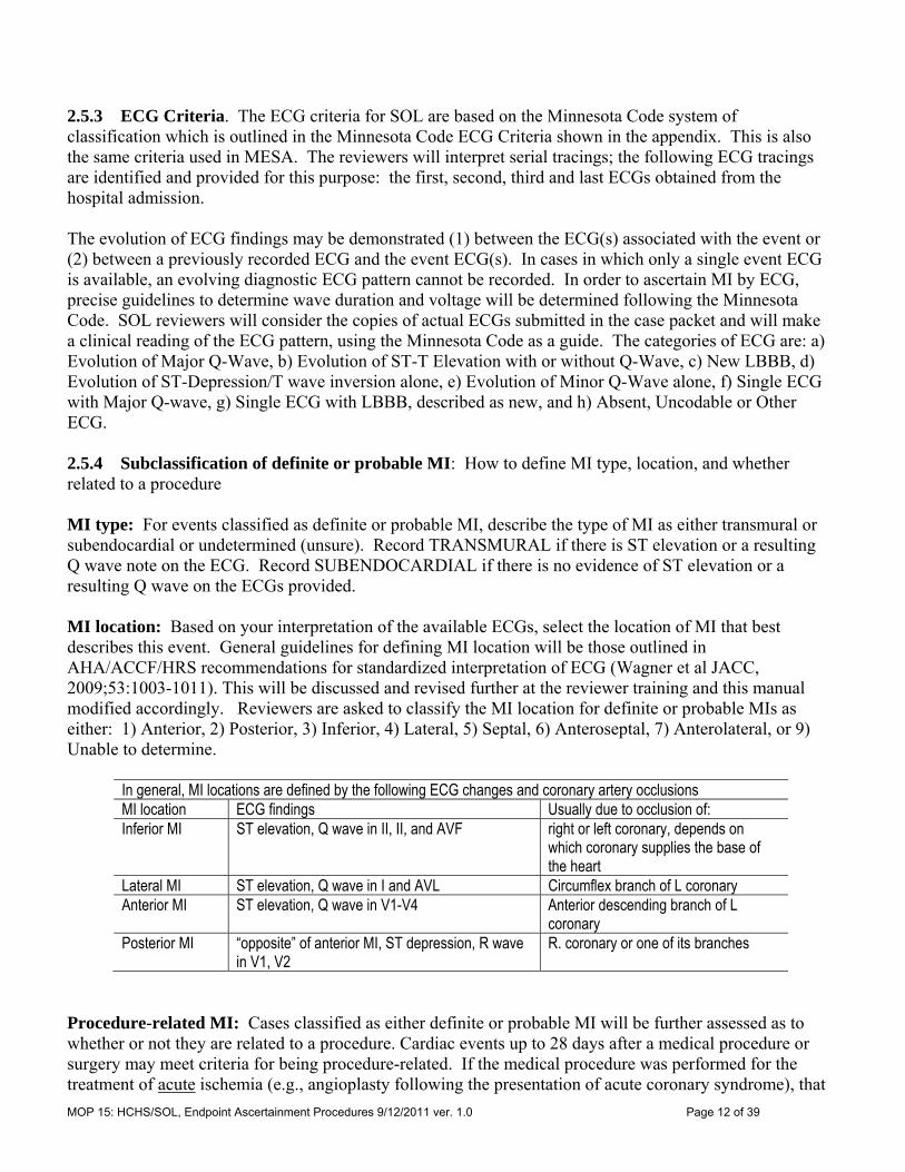
2.5.3 ECG Criteria. The ECG criteria for SOL are based on the Minnesota Code system of classification which is outlined in the Minnesota Code ECG Criteria shown in the appendix. This is also the same criteria used in MESA. The reviewers will interpret serial tracings; the following ECG tracings are identified and provided for this purpose: the first, second, third and last ECGs obtained from the hospital admission. The evolution of ECG findings may be demonstrated (1) between the ECG(s) associated with the event or (2) between a previously recorded ECG and the event ECG(s). In cases in which only a single event ECG is available, an evolving diagnostic ECG pattern cannot be recorded. In order to ascertain MI by ECG, precise guidelines to determine wave duration and voltage will be determined following the Minnesota Code. SOL reviewers will consider the copies of actual ECGs submitted in the case packet and will make a clinical reading of the ECG pattern, using the Minnesota Code as a guide. The categories of ECG are: a) Evolution of Major Q-Wave, b) Evolution of ST-T Elevation with or without Q-Wave, c) New LBBB, d) Evolution of ST-Depression/T wave inversion alone, e) Evolution of Minor Q-Wave alone, f) Single ECG with Major Q-wave, g) Single ECG with LBBB, described as new, and h) Absent, Uncodable or Other ECG. 2.5.4 Subclassification of definite or probable MI: How to define MI type, location, and whether related to a procedure MI type: For events classified as definite or probable MI, describe the type of MI as either transmural or subendocardial or undetermined (unsure). Record TRANSMURAL if there is ST elevation or a resulting Q wave note on the ECG. Record SUBENDOCARDIAL if there is no evidence of ST elevation or a resulting Q wave on the ECGs provided. MI location: Based on your interpretation of the available ECGs, select the location of MI that best describes this event. General guidelines for defining MI location will be those outlined in AHA/ACCF/HRS recommendations for standardized interpretation of ECG (Wagner et al JACC, 2009;53:1003-1011). This will be discussed and revised further at the reviewer training and this manual modified accordingly. Reviewers are asked to classify the MI location for definite or probable MIs as either: 1) Anterior, 2) Posterior, 3) Inferior, 4) Lateral, 5) Septal, 6) Anteroseptal, 7) Anterolateral, or 9) Unable to determine.
In general, MI locations are defined by the following ECG changes and coronary artery occlusions MI location ECG findings Usually due to occlusion of: Inferior MI ST elevation, Q wave in II, II, and AVF right or left coronary, depends on
which coronary supplies the base of the heart
Lateral MI ST elevation, Q wave in I and AVL Circumflex branch of L coronary Anterior MI ST elevation, Q wave in V1-V4 Anterior descending branch of L
coronary Posterior MI “opposite” of anterior MI, ST depression, R wave
in V1, V2 R. coronary or one of its branches
Procedure-related MI: Cases classified as either definite or probable MI will be further assessed as to whether or not they are related to a procedure. Cardiac events up to 28 days after a medical procedure or surgery may meet criteria for being procedure-related. If the medical procedure was performed for the treatment of acute ischemia (e.g., angioplasty following the presentation of acute coronary syndrome), that
MOP 15: HCHS/SOL, Endpoint Ascertainment Procedures 9/12/2011 ver. 1.0 Page 13 of 39
event should not be considered procedure-related (Luepker, et al, 2003). The procedure-related MI category is intended to identify MIs that occurred only after the procedure, and were not already in evolution prior to the procedure. In determining whether the MI was procedure related, answer YES (and choose whether it was a cardiovascular or non-cardiovascular procedure) if you think it is unlikely this MI would have occurred had the procedure not been performed. Answer YES, CARDIOVASCULAR PROCEDURE if the MI occurred within 28 days of a cardiovascular procedure or surgery AND in your judgment the MI was a complication (or related to) the procedure. Cardiovascular procedures include: CABG, valve replacement, AICD or pacemaker placement, PTCI, etc. Answer YES, NON-CARDIOVASCULAR PROCEDURE if the MI occurred within 28 days of a non-cardiovascular procedure or surgery AND in your judgment the MI was a complication (or related to) the procedure. Non-cardiovascular procedures include all procedures or surgeries that are not cardiovascular. Answer UNKNOWN/UNSURE if you are not certain as to whether the MI was procedure related or not. 2.5.5 Angina For events reviewed that are classified as “no MI” or “unclassifiable, then as a secondary endpoint reviewers are asked to state whether angina was present. Angina is a symptomatic event generally involving ischemic chest, left arm, or jaw pain, though the symptoms may be "atypical." Atypical anginal symptoms can include shortness of breath, exertional dyspnea, epigastric discomfort, and back pain, in addition to pain that is isolated to the arm or the jaw. SOL endpoint reviewers categorize angina events as “definite,” “probable,” and “no Angina” based on their clinical judgment in light of the following criteria from the MESA study in answering this question:
a. Physician diagnosis of angina and receiving medical treatment for angina (e.g., nitrates, beta-blockers, or calcium-channel blockers)
b. CABG surgery or other revascularization procedure
c. 70% or greater obstruction of any coronary artery per angiography
d. Horizontal or down-sloping ST-segment depression or abnormal ST elevation of >1 mm on exercise or pharmacological stress testing with pain
e. Scintigraphic or echocardiographic stress test positive for ischemia
f. Resting ECG shows horizontal or down-sloping ST depression or abnormal ST elevation >1 mm with pain that is not present on ECG without pain
Given the difficultly in the diagnosis of angina yet the need to standardize its classification as much as possible, SOL endpoint reviewers are instructed to follow the guidelines a-d below when recording their answer.
a. Clear and thorough documentation of symptoms is needed to identify an event as “definite angina.” Even if a test such as an ETT lists “angina” or “chest pain” as its indication, angina should not be ruled as being present unless there is additional, explicit information from the physician regarding symptoms. Likewise, a test showing positive ischemia or the performance of a further procedure (e.g., catheterization) is not enough to rule for angina if other SOL criteria are not met.
b. Only classify an event as angina if it is distinct from an MI.
c. Reviewers should not classify angina as part of pain symptoms of an MI.
d. Angina will require clinical symptoms. If there is only a physician diagnosis/treatment, then the diagnosis cannot be ‘definite.’ If there is more than just a physician diagnosis, then the reviewer can assign ‘definite” instead of ‘probable.’
MOP 15: HCHS/SOL, Endpoint Ascertainment Procedures 9/12/2011 ver. 1.0 Page 14 of 39
2.5.6 Revascularization procedure interrupting an MI. Revascularization procedures occurring during the course of hospitalization in general will be documented by the abstractors. However, SOL endpoint reviewers are asked to record whether in their judgement an intervention performed early in the clinical presentation of a potential MI may have prevented an MI. In cases where revascularization was performed without clinical symptoms, SOL endpoint reviewers will record NO to this item. Reviewers should record YES, if on presentation with chest pain or other MI symptoms, the patient is immediately recieved a revascularization procedure. 3.0 Endpoint Surveillance for Hospitalized Heart Failure 3.1 Introduction All cases of hospitalized heart failure among HCHS/SOL participants will be identified through the annual follow up call. All eligible hospitalization will be investigated and processed through the HCHS/SOL Event Classification Committee. Heart failure events resulting in outpatient diagnosis and treatment without hospitalization will not be identified and reviewed by HCHS/SOL event reviewers. See nonfatal outpatient event surveillance for details. 3.2 Event Identification Events to be investigated for hospitalized heart failure include those with the following target ICD-9 discharge diagnosis codes: 402, 404, 415, 416, 425, 428, 518.4, and 786. Specified components of the medical record from eligible events will be copied and sent to the coordinating center for processing. Data from the medical record of hospitalizations with these discharge diagnosis codes will be abstracted using the HCHS/SOL heart failure abstraction form.Materials from the medical record to be copied and provided to the HCHS/SOL event reviewers include: the first three (3) chest X-ray reports, echocardiography reports, cardiology consult report, discharge summary, and cardiac catheterization report. 3.3 Diagnostic Criteria for Acute Decompensated Heart Failure The HCHS/SOL criteria for heart failure were adapted from the MESA and the Atherosclerosis Risk in Communities study. HCHS/SOL physician reviewers will determine if the event has a heart failaure diagnosis from the provider and whether the patient was treated for heart failure. The reviewer will also determine if there is sufficient evidence to indicate the patient has history of heart failure and whether there was X-ray pulmonary edema or congestion. Evaluation of these items follow MESA guidelines and allow for comparability of heart failure diagnosis between MESA and HCHS/SOL. (See Heart Failure Diagnosis (HFD) Form) In addition HCHS/SOL physician reviewers categorize acute decompensated HF (ADHF) events as “definite,” “probable,” “no ADHF”, and “unknown” in a manner adapted from the ARIC classification scheme for heart failure.. (See Heart Failure Diagnosis (HFD) Form). Specifically the HCHS/SOL reviewers are asked to evaluate the evidence for the following items:
a. Heart failure diagnosed by physician, and treatment provided for heart failure,
b. Acute decompensated heart failure,
c. Pulmonary edema/congestion by chest X-ray,
d. Cardiac imaging study results, (Each of these questions is asked separately and if present then specify, as to whether the finding was by history, or by current imaging.)
1. Dilated ventricle or 2. Poor left ventricular function (e.g., low ejection fraction or wall motion abnormalities),

MOP 15: HCHS/SOL, Endpoint Ascertainment Procedures 9/12/2011 ver. 1.0 Page 15 of 39
3. Poor right ventricular function, or 4. Left ventricular diastolic dysfunction. 5. If available, the quantitative ejection fraction is specifically provided within a range of
choices of > 50, 40-49, 30-39, 20-29, <20 or unknown.
This approach has the advantage of easily permitting a range of analyses based on definitions of heart failure that include "soft" criteria or various types of "hard" criteria. In general, the reviewer should examine the original report of a procedure rather than accept references to results of the diagnostic or therapeutic procedures in discharge summaries. If an original full report is not available, a convincing reference to the procedure results in the discharge summary is acceptable.
In additon, HCHS/SOL reviewers evaluate the evidence or against history of heart failure, severity of heart failure and presence of right-sided heart failure, comorbid conditions, and asymptomatic ventricular dysfunction. These are described in more detail below.
3.3.1 Prior history of heart failure. Reviewers are asked to classify the event to whether participant had a prior history of HF or not. Prior history is relevant as the evaluation of a patient with an established history of HF may be more limited as compared to a patient with a new diagnosis of heart failure. This also allows the HCHS/SOL to classify those with chronic stable heart failure if the answer is YES to prior history of HF and NO for the classification of ADHF. 3.3.2 Severity of the heart failure exacerbation. Reviewers classify the severity of the HF exacerbation for those classified as probable or definite ADHF. Classify the event as SEVERE if treatment with mechanical ventilation, non-rebreather mask, CPAP, hemofiltration, intraortic balloon pump, or thoracentesis was required for management of HF exacerbation. Classify event as MODERATE, if it is clear the event was neither SEVERE nor MILD. Classify event as MILD if this exacerbation could have been managed in the outpatient setting had the patient been an outpatient. In these cases, the primary reason for being hospitalized will likely be something other than HF. If it is unclear as to the severity of the event then classify as UNKNOWN. 3.3.3 Predominantly right-sided heart failure. The most common presentation for ADHF is left sided HF which is dyspnea and pulmonary edema, however symptoms of pure right sided HF do not include dyspnea. In the future when considering criteria for the diagnosis of HF, it may be necessary to differentiate left and right sided HF. Answer YES if the patient had right-sided heart failure symptoms only of lower extremity edema and possible ascites and a normal LV ejection fraction. Answer NO/NR if the patient had both right-sided and left-sided signs/symptoms, or if it is clearly not right sided HF, or it is unclear whether the patient had predominantly right sided failure. 3.3.4 Co-morbid and potentially precipitating factors to ADHF. Acute decompensated HF may be precipitated by multiple conditions. In addition, many of the listed comorbid condition may be caused by the exacerbation. As a result, it can be difficult to tell whether the condition precipitated the event or not. Here we ask reviewers to consider the timeline of occurrence of the condition with the HF exacerbation and based on their judgment state as to whether the condition may have precipitated the HF exacerbation. As stated in the question by question instructions for the HFD form, reviewers indicate all co-morbid conditions that were active during this hospitalization AND may have precipitated the event. The temporal association of between the event and the HF exacerbation should make sense. For example, if the patient came in for surgery and had an exacerbation of HF immediately following surgery due to fluid overload then check YES to fluid overload. If the same patient then had a complicated coarse and developed a PE many days later then check NO/NR for PE, if it temporal association with the HF
MOP 15: HCHS/SOL, Endpoint Ascertainment Procedures 9/12/2011 ver. 1.0 Page 16 of 39
exacerbation is not correct. If a patient presents to the hospital with heart failure AND one of these diagnosis that is active then check YES. For example, if presents with both HF and atrial fibrillation the check YES for atrial fibrillation. Reviewers answer No/Not recorded or YES to all of the following: 1) Myocardial infarction, 2) Atrial fibrillation or atrial flutter, 3) Other arrhythmia, 4) Fluid overload or volume overload- this refers only to either iatrogenic fluid overload OR to excessive drinking of fluids, or renal failure due to missed or inadequate hemodialysis, 5) Medication noncompliance – Medication noncompliance would include refusal of medications for patients in the hospital, but more commonly this would be outpatient noncompliance. Noncompliance includes those patients who did not get their medications due to lack of funds, 6) Pulmonary embolus, 7) Renal insufficiency or failure – use your judgement as to whether renal failure was of a severity that it may have contributed to the HF. This would likely be those who are approaching dialysis and are less responsive to diuretics, 8) Cardiovascular procedure/surgery, 9) Non-cardiovascular procedure/surgery, 10) Pulmonary disease, and 11) Uncontrolled Hypertension – systolic blood pressure >180 at the time of the event. 3.3.5 Asymptomatic left ventricular dysfunction. The focus of the primary outcome in HCHS/SOL is classification of acute decompensated heart failure (adapted from ARIC criteria) and the MESA definition of physician diagnosed and treated heart failure. In addition, for those events that are not classified as “definite” or “probable” ADHF then we ask reviewers to identify those with asymptomatic left ventricular dysfunction, defined here as documented ejection fraction < 50%, but no HF symptoms previously or during this admission.
4.0 Endpoint Surveillance for Stroke and Transient Ischemic Attacks 4.1 Event Identification A hospitalization is eligible for stroke evaluation and classification by the HCHS/SOL reviewers if if it has an ICD-9 procedure code 38-39 or discharge diagnosis codes for cerebrovascular (ICD-9-CM 430-438). If a hospitalization meet procedure or discharge code criteria for stroke or transient ischemic attack (TIA,) the medical records obtained by field center staff and sent to the coordinating center for detailed abstraction using the HCHS/SOL stroke abstraction form. These data will be summarized and made available to the stroke group of the Event Classification Committee. Copies of spcific portions of the medial record are also provided to the HCHS/Sol reviewers. The reviewers will follow the criteria below in making a final event classification. 4.2 Diagnostic Criteria The classification criteria for stroke and TIA for HCHS/SOL were adapted from MESA. 4.3 Definitions of TIA and Stroke 4.3.1 Transient Ischemic Attack (TIA) Transient ischemic attack is a temporary stroke-like event that lasts for only a short time and is caused by temporarily blocked blood vessels to part of the brain. For the purposes of HCHS/SOL classification of TIA, a TIA includes one or more episodes of [acute] focal neurologic deficit, lasting more than 30 seconds, with complete resolution of focal neurologic deficit within 24 hours. It must have no clinically relevant lesion on brain imaging (or brain imaging not done). In order for an event to be classified as TIA it must NOT have any of the following features: clonic jerking, conjugate eye deviation, prolonged focal seizure with spread, scintillating scotoma, headache with nausea and vomiting, or immediately-preceding head trauma. A clinically relevant lesion on brain imaging includes finding judged to be consistent with signs and symptoms regardless of timing of brain imaging (i.e., less or greater than 24 hours), regardless of stroke
MOP 15: HCHS/SOL, Endpoint Ascertainment Procedures 9/12/2011 ver. 1.0 Page 17 of 39
type (i.e., with or without blood), and regardless of imaging technique (i.e., cranial competed tomography [CT scan] or cranial magnetic resonance imaging [MRI]). 4.3.2 Stroke A stroke is defined as loss of muscle function, vision, sensation or speech resulting from brain cell damage caused by an insufficient supply of blood to part of the brain. Synonyms for stroke include apoplexy, cerebrovascular accident, or cerebral vascular accident. For the purpose of HCHS/SOL classification of stroke, a stroke involves rapid onset of neurologic deficit, headache, or meningismus and neurologic deficits not secondary to brain trauma (closed head injury), tumor, infection (e.g., encephalitis or meningitis), or other non-vascular cause. Clinical evidence or suspicion of embolic stroke secondary to SBE is counted as stroke. Classification of stroke requies either clinically relevant lesion on brain imaging, or duration of symptoms greater than 24 hours, or death within 24 hours of symptoms. 4.4. Definitions of stroke subtypes 4.4.1 Subarachnoid hemorrhage (SAH) A subarachnoid hemorrhage has a clinical presentation of sudden onset of headache, meningismus, loss of consciousness, or coma. Focal neurologic deficit may also be present. Classification of SAH also requires consistent imaging findings with blood mainly in the subarachnoid space (basal cistern or convexity) or isolated intraventricular hemorrhage, or cerebral fluid bloody or xanthochromic on direct non-traumatic examination. Surgical or autopsy evidence of subarachnoid hemorrhage is sufficient for a classification of SAH. 4.4.2. Intraparenchymal hemorrhage (IPH) A intraparenchymal hemorrhage has a clinical presentation of focal neurologic deficit, coma as a possible accompanying condition. Classification of IPH requires consistent imaging findings with mainly intraparenchymal, dense hemorrhage, or if there is no imaging available, cerebral spinal fluid bloody or xanthochromic on direct non-traumatic examination or surgical or autopsy evidence of intraparenchymal hemorrhage is suffiecent for a classification of IPH. 4.4.3. Other hemorrhage (OH) If there is insufficient data to classify subarachnoid or intraparencymal hemorrhage, and imaging shows blood in the parenchyma, subarachnoid space, or both then a classification of other hemorrhage is made. Cerebrospinal fluid bloody or xanthochromic on direct non-traumatic examination, or surgical or autopsy evidence of blood in parenchyma, subarachnoid space, or both in the absence of a classification of SAH or IPH is sufficient for a classification of other hemorrhage. 4.4.4. Brain infarction (ischemic stroke) (INF) If a case does not meet criteria for SAH, IPH, or OH and there is a clinical presentation of focal neurologic deficit (with or without coma present) and there is consistent imaging findings without clinically relevant lesion or with clinically relevant mainly non-hemorrhagic lesion or hemorrhagic lesion indicating a hemorrhagic infarction a classification of INF can be made. Surgical or autopsy evidence of brain infarction is also sufficient for a classification of INF. 4.4.5. Other stroke types (OS) If a case does not meet criteria for SAH, IPH, OH, or INF and there is a clinical presentation of focal neurologic deficit it may be classified as OS. Examples include venous thrombosis with bleed and arterial dissection. 4.4.6. Unknown stroke type If a case does not meet criteria for SAH, IPH, OH, INF, or OS and there is a clinical presentation of focal neurologic deficit it may be classified as UNK. Examples includes evidence of symptoms but no work-up was done. A classification of NO stroke is made if the case meets non of the criteria above.
MOP 15: HCHS/SOL, Endpoint Ascertainment Procedures 9/12/2011 ver. 1.0 Page 18 of 39
4.5 Ischemic Stroke Classification Ischemic Stroke Classification (modified TOAST criteria utilizing findings of Neuroimaging, especially MRI and MRA) Hospitalizations classified by HCHS/SOL reviewers as ischemic strokes (INF) are further classified by reviewers using the following sub-classification criteria. 4.5.1. Cardioembolic stroke Symptoms consistent with brain infarction (see general definition of brain infarction); and acute or subacute appearing, cortical or cerebellar infarcts and brain stem or subcortical hemispheric infarcts greater than 1.5 cm in diameter on neuroimaging (CT or MRI), either in one or multiple vascular territories; and
evidence suggestive for high-risk or medium-risk cardiac source for emboli (clinical and laboratory evidence); and no supportive evidence by vascular imaging for a stenosis of greater than 50% of the appropriate extracranial artery proximal to the infarct (diagnosed either by ultrasound studies, CTA, MRA, or conventional DSA); and no laboratory abnormality suggestive for non-cardiac, non-lacunar, or non-atherothromboembolic cause (for example nonatherosclerotic vasculopathies, hypercoagulable states, or hematologic disorders). 4.5.2 Extracranial large-artery atherosclerosis Symptoms consistent with brain infarction (see general definition of brain infarction); and acute or subacute appearing, cortical or cerebellar infarcts and brain stem or subcortical hemispheric infarcts greater than 1.5 cm in diameter on neuroimaging (CT or MRI); and supportive evidence by vascular imaging of a stenosis of greater than 50% of the appropriate extracranial artery proximal to the infarct (diagnosed either by ultrasound studies, CTA, MRA, or conventional DSA); and no evidence suggestive for high-risk or medium-risk cardiac source for emboli (clinical and laboratory evidence); and no laboratory abnormality suggestive for non-cardiac, non-lacunar, or non-atherothromboembolic cause (for example nonatherosclerotic vasculopathies, hypercoagulable states, or hematologic disorders).
4.5.3. Intracranial large-artery atherosclerosis Symptoms consistent with brain infarction (see general definition of brain infarction); and acute or subacute appearing, cortical or cerebellar infarcts and brain stem or subcortical hemispheric infarcts greater than 1.5 cm in diameter on neuroimaging (CT or MRI); and supportive evidence by vascular imaging of a stenosis of greater than 50% of the appropriate intracranial artery proximal to the infarct (diagnosed either by ultrasound studies, CTA, MRA, or conventional DSA); and no evidence suggestive for high-risk or medium-risk cardiac source for emboli (clinical and laboratory evidence); and no laboratory abnormality suggestive for non-cardiac, non-lacunar, or non-atherothromboembolic cause (for example nonatherosclerotic vasculopathies, hypercoagulable states, or hematologic disorders). 4.5.4. Lacunar Symptoms consistent with brain infarction (see general definition of brain infarction); and symptoms consistent with a lacunar stroke syndrome (i.e. pure motor, pure sensory, mixed sensorymotor syndrome without cortical signs, ataxic hemiparesis, dysarthria-clumsy hand syndrome); acute or subacute appearing infarct <1.5 cm in diameter in a subcortical structure or brainstem in the territory of a small penetrating artery on neuroimaging (CT or MRI): Infarct should be smaller than 1.5 cm (or described as ‘small’ or ‘lacunar’). Typical subcortical locations include corona radiata, internal capsule, external capsule, basal ganglia, thalamus. Lesions may also be located in the pons or medulla. Reviewers may score this response if ANY one of multiple new lacunar infarcts is appropriate for symptoms. Reviewers may also be scored if there is a clinically appropriate lesion of undetermined age. Do not score if the patient is described as having ‘Binswanger’s disease (encephalopathy)’ unless there is a discrete new lesion appropriate for symptoms; and no evidence suggestive for high-risk or medium-risk cardiac source for emboli (clinical and laboratory evidence); and no evidence for intra- or extracranial large artery
MOP 15: HCHS/SOL, Endpoint Ascertainment Procedures 9/12/2011 ver. 1.0 Page 19 of 39
stenosis in the artery proximal to the infarct (by CTA, MRA, or conventional angiography); and no laboratory abnormality suggestive for non-cardiac, non-lacunar, or non-atherothromboembolic cause (for example nonatherosclerotic vasculopathies, hypercoagulable states, or hematologic disorders). 4.5.5. Other determined etiology Symptoms consistent with brain infarction (see general definition of brain infarction); and acute or subacute appearing infarct of any size in diameter either in cortical or subcortical structure or brainstem on neuroimaging (CT or MRI); and no evidence suggestive for high-risk or medium-risk cardiac source for emboli (clinical and laboratory evidence); and no evidence for intra- or extracranial large artery stenosis in the artery proximal to the infarct (by CTA, MRA, or conventional angiography); and laboratory abnormality suggestive for non-cardiac, non-lacunar, or non-atherothromboembolic cause (for example nonatherosclerotic vasculopathies, hypercoagulable states, or hematologic disorders). 4.5.6. Undetermined etiology, complete Symptoms consistent with brain infarction (see general definition of brain infarction) ; and acute or subacute appearing, cortical or cerebellar infarcts and brain stem or subcortical hemispheric infarcts greater than 1.5 cm in diameter on neuroimaging (CT or MRI); and no supportive evidence by vascular imaging of a stenosis of greater than 50% of the appropriate intra- or extracranial artery proximal to the infarct (diagnosed either by ultrasound studies, CTA, MRA, or conventional DSA); and no evidence suggestive for high-risk or medium-risk cardiac source for emboli (clinical and laboratory evidence); and no laboratory abnormality suggestive for non-cardiac, non-lacunar, or non-atherothromboembolic cause (for nonatherosclerotic vasculopathies, hypercoagulable states, or hematologic disorders). 4.5.7. Undetermined etiology, incomplete evaluation Symptoms consistent with brain infarction (see general definition of brain infarction); and incomplete cardiac evaluation (EKG or transthoracic echocardiography not performed); or incomplete evaluation of extra- or intracranial arteries (carotid ultrasound, CTA or MRA not performed). 4.5.8. Multiple possible etiologies Symptoms consistent with brain infarction (see general definition of brain infarction); and a combination of at least 2 or more of the following:
o Cortical or cerebellar lesions and brain stem or subcortical hemispheric infarcts greater than 1.5 cm in diameter on neuroimaging (CT or MRI)
o Cortical or cerebellar lesions and brain stem or subcortical hemispheric infarcts less or
equal than 1.5 cm in diameter on neuroimaging (CT or MRI)
o supportive evidence by vascular imaging of a stenosis of greater than 50% of the appropriate extracranial artery proximal to the infarct (diagnosed either by ultrasound studies, CTA, MRA, or conventional DSA).
o supportive evidence by vascular imaging of a stenosis of greater than 50% of the appropriate intracranial artery proximal to the infarct (diagnosed either by ultrasound studies, CTA, MRA, or conventional DSA).
o evidence suggestive for high-risk or medium-risk cardiac source for emboli (clinical and
laboratory evidence)
MOP 15: HCHS/SOL, Endpoint Ascertainment Procedures 9/12/2011 ver. 1.0 Page 20 of 39
o no laboratory abnormality suggestive for non-cardiac, non-lacunar, or non-atherothromboembolic cause (for example nonatherosclerotic vasculopathies, hypercoagulable states, or hematologic disorders).
4.6 Ischemic Stroke Classification Use of Probable and Possible Brain Infarct Subtype Classification (modified TOAST criteria) A "possible" diagnosis is made when the clinical findings and neuroimaging data suggest a specific subtype but other studies are not done. A “probable” diagnosis is made when the clinical findings and diagnostic work-up are complete in order to allow classification to one of the major brain infarct subtypes. The diagnostic work-up must include clinical syndrome description, neuroimaging, intra- and extracranial vascular imaging, EKG, at least transthoracic echocardiography, and basic laboratory studies. 5.0 Endpoint Surveillance for Exacerbations due to Chronic Obstructive Pulmonary Disease or Asthma 5.1 Introduction All cases of exacerbation due to COPD or asthma resulting in either a hospitalization or emergency room visit among HCHS/SOL participants will be identified through the annual follow up call. All eligible events will be investigated and processed through the HCHS/SOL Event Classification Committee. Self-reported COPD and asthma exacerbations resulting in outpatient diagnosis and treatment without hospitalization or emergency department visits will also be identified through the annual follow up call. However, the outpatient records will not be obtained for verification. COPD and asthma are the most common chronic lung diseases in adults. Patients with asthma have intermittent airway obstruction while those with COPD have irreversible airway obstruction (90% due to smoking). The distinction between asthma and COPD in adults has become blurred during the past five years, as the term COPD has been advertised to primary care physicians and the general public. Industry-sponsored COPD guidelines cause many adults with asthma (even never-smokers) to be falsely labeled as COPD. Asthma can begin at any age (even after age 75). Only half of asthma in adults is associated with or triggered by allergies (extrinsic asthma). There is some overlap between COPD and asthma. By definition, GOLD criteria classify a non-smoking asthmatic that develops irreversible changes as COPD, although this type of case would be clarified clinically as COPD secondary to asthma. About half of adults with asthma are smokers. Both asthma and COPD are under-diagnosed and over-diagnosed, so self-reports and physician diagnoses are unreliable. Misclassification rates are high because objective test results are often not obtained or are incorrectly interpreted. In particular, pulmonary function tests are not generally obtained at the time of a respiratory exacerbation. About half of those with asthma or COPD in general population samples have not been diagnosed. On the other hand, some patients who have been prescribed inhalers for asthma or COPD have neither asthma nor COPD. 5.2 Event Identification Hospitalization and emergency room events to be investigated for exacerbation due to COPD should have one of the following ICD-9 codes: 490, 491 (chronic obstructive bronchitis), 492 (emphysema), 494 (bronchiectasis), 496 (chronic airway obstruction not otherwise classified), 415, and 416.9. The codes for asthma start with 493. ICD Code 518 (Respiratory Failure, secondary to COPD) will also be investigated.
MOP 15: HCHS/SOL, Endpoint Ascertainment Procedures 9/12/2011 ver. 1.0 Page 21 of 39
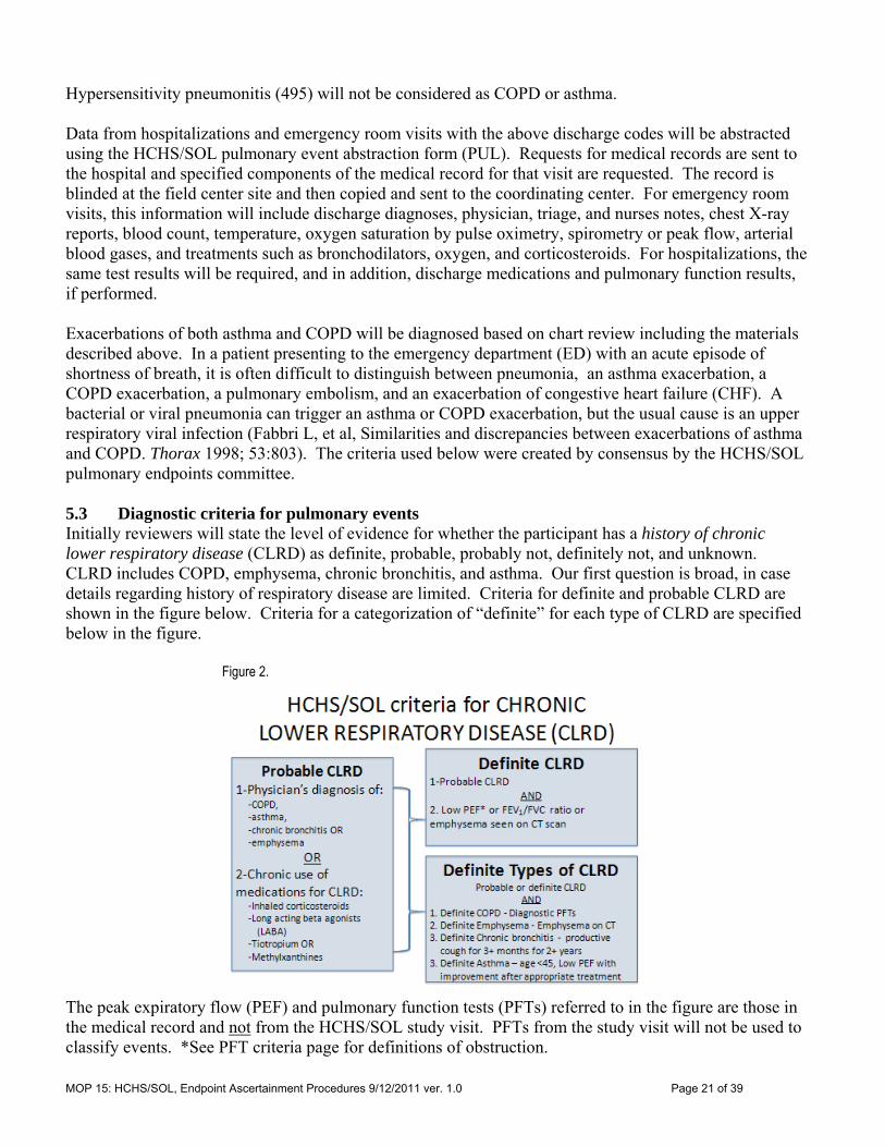
Hypersensitivity pneumonitis (495) will not be considered as COPD or asthma. Data from hospitalizations and emergency room visits with the above discharge codes will be abstracted using the HCHS/SOL pulmonary event abstraction form (PUL). Requests for medical records are sent to the hospital and specified components of the medical record for that visit are requested. The record is blinded at the field center site and then copied and sent to the coordinating center. For emergency room visits, this information will include discharge diagnoses, physician, triage, and nurses notes, chest X-ray reports, blood count, temperature, oxygen saturation by pulse oximetry, spirometry or peak flow, arterial blood gases, and treatments such as bronchodilators, oxygen, and corticosteroids. For hospitalizations, the same test results will be required, and in addition, discharge medications and pulmonary function results, if performed. Exacerbations of both asthma and COPD will be diagnosed based on chart review including the materials described above. In a patient presenting to the emergency department (ED) with an acute episode of shortness of breath, it is often difficult to distinguish between pneumonia, an asthma exacerbation, a COPD exacerbation, a pulmonary embolism, and an exacerbation of congestive heart failure (CHF). A bacterial or viral pneumonia can trigger an asthma or COPD exacerbation, but the usual cause is an upper respiratory viral infection (Fabbri L, et al, Similarities and discrepancies between exacerbations of asthma and COPD. Thorax 1998; 53:803). The criteria used below were created by consensus by the HCHS/SOL pulmonary endpoints committee. 5.3 Diagnostic criteria for pulmonary events Initially reviewers will state the level of evidence for whether the participant has a history of chronic lower respiratory disease (CLRD) as definite, probable, probably not, definitely not, and unknown. CLRD includes COPD, emphysema, chronic bronchitis, and asthma. Our first question is broad, in case details regarding history of respiratory disease are limited. Criteria for definite and probable CLRD are shown in the figure below. Criteria for a categorization of “definite” for each type of CLRD are specified below in the figure.
Figure 2.
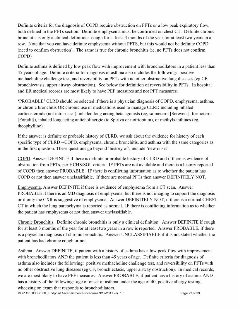
The peak expiratory flow (PEF) and pulmonary function tests (PFTs) referred to in the figure are those in the medical record and not from the HCHS/SOL study visit. PFTs from the study visit will not be used to classify events. *See PFT criteria page for definitions of obstruction.
MOP 15: HCHS/SOL, Endpoint Ascertainment Procedures 9/12/2011 ver. 1.0 Page 22 of 39
Definite criteria for the diagnosis of COPD require obstruction on PFTs or a low peak expiratory flow, both defined in the PFTs section. Definite emphysema must be confirmed on chest CT. Definite chronic bronchitis is only a clinical definition: cough for at least 3 months of the year for at least two years in a row. Note that you can have definite emphysema without PFTS, but this would not be definite COPD (need to confirm obstruction). The same is true for chronic bronchitis (ie, no PFTs does not confirm COPD)
Definite asthma is defined by low peak flow with improvement with bronchodilators in a patient less than 45 years of age. Definite criteria for diagnosis of asthma also includes the following: positive methacholine challenge test, and reversibility on PFTs with no other obstructive lung diseases (eg CF, bronchiectasis, upper airway obstruction). See below for definition of reversibility in PFTs. In hospital and ER medical records are most likely to have PEF measures and not PFT measures.
‘PROBABLE’ CLRD should be selected if there is a physician diagnosis of COPD, emphysema, asthma, or chronic bronchitis OR chronic use of medications used to manage CLRD including inhaled corticosteroids (not intra-nasal), inhaled long acting beta agonists (eg, salmeterol [Serevent], formoterol [Foradil]), inhaled long acting anticholinergic (ie Spiriva or tiotriopium), or methylxanthines (eg, theophylline).
If the answer is definite or probable history of CLRD, we ask about the evidence for history of each specific type of CLRD --COPD, emphysema, chronic bronchitis, and asthma with the same categories as in the first question. These questions go beyond ‘history of’, include ‘new onset’.
COPD. Answer DEFINITE if there is definite or probable history of CLRD and if there is evidence of obstruction from PFTs, per HCHS/SOL criteria. IF PFTs are not available and there is a history reported of COPD then answer PROBABLE. IF there is conflicting information as to whether the patient has COPD or not then answer unclassifiable. If there are normal PFTs then answer DEFINITELY NOT.
Emphysema. Answer DEFINITE if there is evidence of emphysema from a CT scan. Answer PROBABLE if there is an MD diagnosis of emphysema, but there is not imaging to support the diagnosis or if only the CXR is suggestive of emphysema. Answer DEFINITELY NOT, if there is a normal CHEST CT in which the lung parenchyma is reported as normal. IF there is conflicting information as to whether the patient has emphysema or not then answer unclassifiable.
Chronic Bronchitis. Definite chronic bronchitis is only a clinical definition. Answer DEFINITE if cough for at least 3 months of the year for at least two years in a row is reported. Answer PROBABLE, if there is a physician diagnosis of chronic bronchitis. Answer UNCLASSIFIABLE if it is not stated whether the patient has had chronic cough or not.
Asthma. Answer DEFINITE, if patient with a history of asthma has a low peak flow with improvement with bronchodilators AND the patient is less than 45 years of age. Definite criteria for diagnosis of asthma also includes the following: positive methacholine challenge test, and reversibility on PFTs with no other obstructive lung diseases (eg CF, bronchiectasis, upper airway obstruction). In medical records, we are most likely to have PEF measures. Answer PROBABLE, if patient has a history of asthma AND has a history of the following: age of onset of asthma under the age of 40, positive allergy testing, wheezing on exam that responds to bronchodilators.

MOP 15: HCHS/SOL, Endpoint Ascertainment Procedures 9/12/2011 ver. 1.0 Page 23 of 39
If there is some other lung disease present, then specify the type. Next reviewers are asked about an exacerbation of underlying chronic lower respiratory disease (CLRD). Answer PROBABLE, if the patient has new onset or worsening of respiratory symptoms which must include the following symptoms: dyspnea, cough, change in sputum color, change in sputum volume, or wheeze (either reported or on exam). If the primary reason the patient is in the ER or hospital is for one of these symptoms then assume symptoms are worse from baseline. Answer 3a. Answer DEFINITE, if the patient has criteria for a probable CLRD exacerbation AND a low peak flow or low FEV1/FVC, AND absence of alternative diagnosis likely to cause CLRD symptoms and/or low peak flow or low FEV1/FVC. Answer DEFINITELY NOT, if the patient does not have symptoms of CLRD exacerbation and/or has a clear alternative cause of symptoms. Answer PROBABLY NOT, if there is not strong enough evidence to answer DEFINITELY NOT, but you feel it is not likely that the patient has an exacerbation of CLRD. Answer UNCLASSIFIABLE if there is inadequate information to determine if there is an exacerbation of CLRD. Skip to question 4.
Figure 3.
If definite or probable exacerbation, then specify which type of lung disease is likely the cause: COPD predominant, asthma predominant, either asthma or COPD or unclassifiable. Answer ‘asthma predominant’ if it is clear from the medical record that asthma is the cause of the exacerbation. Answer ‘COPD predominant’ if it is clear from the medical record that COPD is the cause of the exacerbation. Answer ‘either asthma or COPD’ if the physicians are not sure if the patient has asthma or COPD and/or if there is conflicting evidence as to which diagnosis that is the cause of the exacerbation. Answer UNCLASSIFIABLE if there is not adequate information to determine whether COPD or asthma was the cause of the exacerbation.
Pneumonia causes respiratory symptoms that are similar to a COPD exacerbation, it is somewhat controversial as to whether person with COPD and pneumonia also has a COPD exacerbation, therefore
MOP 15: HCHS/SOL, Endpoint Ascertainment Procedures 9/12/2011 ver. 1.0 Page 24 of 39
reviewers need to state whether the patient had pneumonia, defined as a new infiltrate on Chest Xray or on chest CT scan. The same 5 categories from definite to unknown are available as options.
Interpretation of PFTS and Peak expiratory flow from medical records
In order to classify pulmonary events, one must have criteria for interpreting pulmonary function tests and peak expiratory flow values that are found in the medical record. Although as mentioned, it is mostly PEF values that will be found in a medical record for a potential CLRD exacerbation. The criteria described and summarized below.
“Low PEF” is defined as PEF < 70% predicted. PEF predicted values will be based upon HCHS/SOL predicted values (currently pending) using age at time of clinical PEF measurement, gender, and measured height at HCHS/SOL exam. These 3 variables are included in the event summary form available with all medical records sent to reviewers.
Low PEF “w/ improvement” is defined as an increase in PEF of ≥30% or, in absence of repeat PEF, clear improvement in clinical status.
“Low FEV1/FVC ratio” is defined as pre-bronchodilator FEV1/FVC < LLN (lower limit of normal). The LLN for FEV1/FVC is based upon HCHS/SOL reference equations, determined on each individual patient, (currently pending) using age at time of clinical PFT measurement, gender, and measured height at HCHS/SOL exam.
“COPD” from PFT results are based ideally on post-bronchodilator PFTS; however, if not available then pre-bronchodilator PFTs are used.
The definition for COPD from post-bronchodilator PFTs is: 1) FEV1/FVC ratio <0.70 or <LLN.
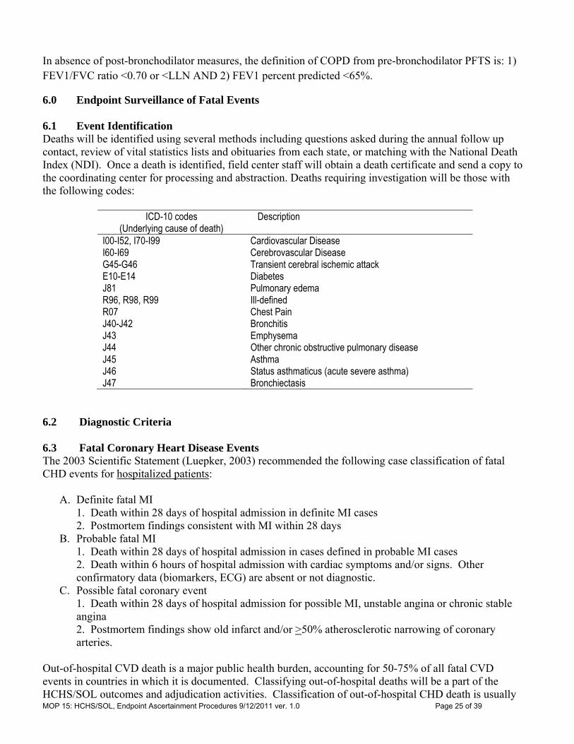
MOP 15: HCHS/SOL, Endpoint Ascertainment Procedures 9/12/2011 ver. 1.0 Page 25 of 39
In absence of post-bronchodilator measures, the definition of COPD from pre-bronchodilator PFTS is: 1) FEV1/FVC ratio <0.70 or <LLN AND 2) FEV1 percent predicted <65%.
6.0 Endpoint Surveillance of Fatal Events 6.1 Event Identification Deaths will be identified using several methods including questions asked during the annual follow up contact, review of vital statistics lists and obituaries from each state, or matching with the National Death Index (NDI). Once a death is identified, field center staff will obtain a death certificate and send a copy to the coordinating center for processing and abstraction. Deaths requiring investigation will be those with the following codes:
ICD-10 codes (Underlying cause of death)
Description
I00-I52, I70-I99 Cardiovascular Disease I60-I69 Cerebrovascular Disease G45-G46 Transient cerebral ischemic attack E10-E14 Diabetes J81 Pulmonary edema R96, R98, R99 Ill-defined R07 Chest Pain J40-J42 Bronchitis J43 Emphysema J44 Other chronic obstructive pulmonary disease J45 Asthma J46 Status asthmaticus (acute severe asthma) J47 Bronchiectasis
6.2 Diagnostic Criteria 6.3 Fatal Coronary Heart Disease Events The 2003 Scientific Statement (Luepker, 2003) recommended the following case classification of fatal CHD events for hospitalized patients:
A. Definite fatal MI 1. Death within 28 days of hospital admission in definite MI cases 2. Postmortem findings consistent with MI within 28 days B. Probable fatal MI 1. Death within 28 days of hospital admission in cases defined in probable MI cases 2. Death within 6 hours of hospital admission with cardiac symptoms and/or signs. Other
confirmatory data (biomarkers, ECG) are absent or not diagnostic. C. Possible fatal coronary event 1. Death within 28 days of hospital admission for possible MI, unstable angina or chronic stable
angina 2. Postmortem findings show old infarct and/or >50% atherosclerotic narrowing of coronary
arteries. Out-of-hospital CVD death is a major public health burden, accounting for 50-75% of all fatal CVD events in countries in which it is documented. Classifying out-of-hospital deaths will be a part of the HCHS/SOL outcomes and adjudication activities. Classification of out-of-hospital CHD death is usually
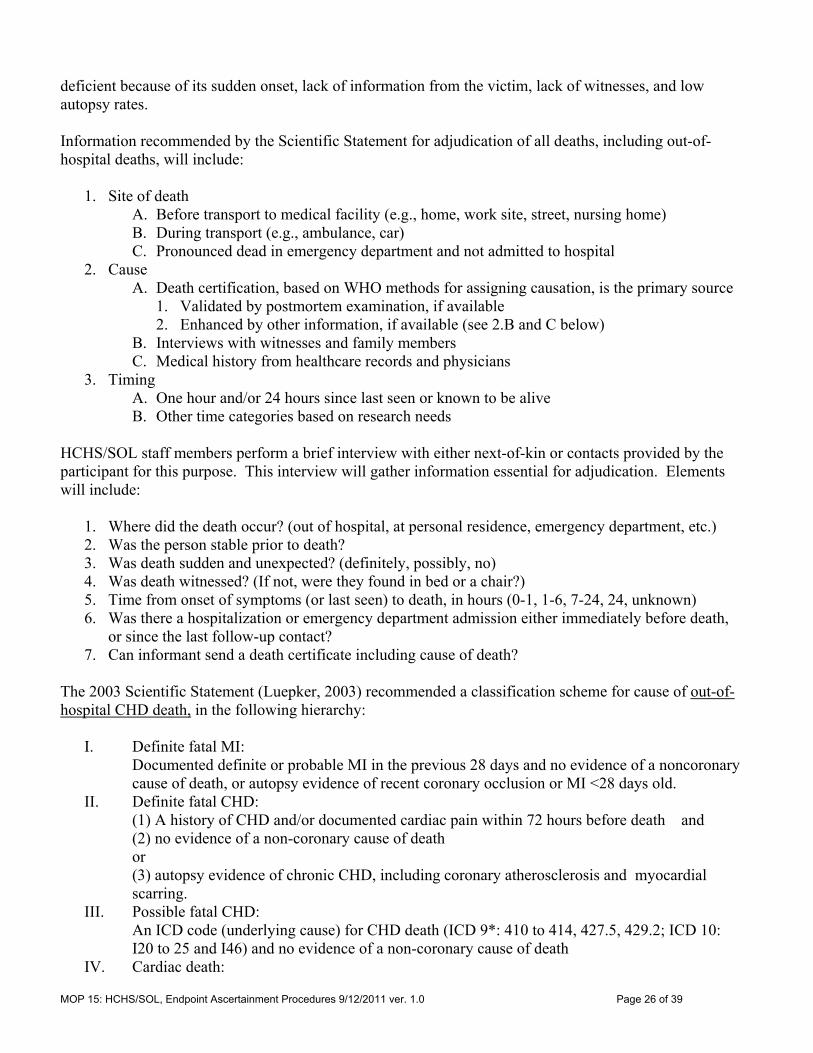
MOP 15: HCHS/SOL, Endpoint Ascertainment Procedures 9/12/2011 ver. 1.0 Page 26 of 39
deficient because of its sudden onset, lack of information from the victim, lack of witnesses, and low autopsy rates. Information recommended by the Scientific Statement for adjudication of all deaths, including out-of-hospital deaths, will include:
1. Site of death A. Before transport to medical facility (e.g., home, work site, street, nursing home) B. During transport (e.g., ambulance, car) C. Pronounced dead in emergency department and not admitted to hospital
2. Cause A. Death certification, based on WHO methods for assigning causation, is the primary source
1. Validated by postmortem examination, if available 2. Enhanced by other information, if available (see 2.B and C below)
B. Interviews with witnesses and family members C. Medical history from healthcare records and physicians
3. Timing A. One hour and/or 24 hours since last seen or known to be alive B. Other time categories based on research needs
HCHS/SOL staff members perform a brief interview with either next-of-kin or contacts provided by the participant for this purpose. This interview will gather information essential for adjudication. Elements will include:
1. Where did the death occur? (out of hospital, at personal residence, emergency department, etc.) 2. Was the person stable prior to death? 3. Was death sudden and unexpected? (definitely, possibly, no) 4. Was death witnessed? (If not, were they found in bed or a chair?) 5. Time from onset of symptoms (or last seen) to death, in hours (0-1, 1-6, 7-24, 24, unknown) 6. Was there a hospitalization or emergency department admission either immediately before death,
or since the last follow-up contact? 7. Can informant send a death certificate including cause of death?
The 2003 Scientific Statement (Luepker, 2003) recommended a classification scheme for cause of out-of-hospital CHD death, in the following hierarchy:
I. Definite fatal MI:
Documented definite or probable MI in the previous 28 days and no evidence of a noncoronary cause of death, or autopsy evidence of recent coronary occlusion or MI <28 days old.
II. Definite fatal CHD: (1) A history of CHD and/or documented cardiac pain within 72 hours before death and (2) no evidence of a non-coronary cause of death or (3) autopsy evidence of chronic CHD, including coronary atherosclerosis and myocardial scarring.
III. Possible fatal CHD: An ICD code (underlying cause) for CHD death (ICD 9*: 410 to 414, 427.5, 429.2; ICD 10: I20 to 25 and I46) and no evidence of a non-coronary cause of death
IV. Cardiac death:
MOP 15: HCHS/SOL, Endpoint Ascertainment Procedures 9/12/2011 ver. 1.0 Page 27 of 39
When death certificates are the only source of information: ICD 9: 390 to 398, 402, 404 to 429; ICD 10: I00 to I09, I11, I13, I20 to I25, I27, I30 to I52
V. Non-CHD death: Evidence of a noncoronary cause of death
VI. Unclassifiable: Insufficient information to determine whether the death was a CHD death (at any certainty level) or a noncardiac death: ICD 9 code 799
6.4 Fatal Pulmonary Events The hospital records will be obtained when a respiratory death (J42, J44, J45, J46) occurred during a hospitalization, or when an out-of-hospital death occurred within 30 days following a hospitalization. Ascertainment of cause-specific mortality in patients with COPD is difficult, but should be attempted by a central committee, not site investigators (McGarvey LP, et al. Ascertainment of cause-specific mortality in COPD: operations of the TORCH Clinical Endpoint Committee. Thorax 2007; 63:411). Death certificates will be obtained in all cases. If a death certificate cannot be obtained, it will be stated in the documentation. If medical records are inadequate and a death certificate cannot be obtained, a cause of death will be adjudicated based on the best available evidence of record. If a probable cause cannot be adjudicated, it will be classified as “unknown”. The primary cause of death should be attributed to the disorder that causes the patient to present for medical treatment and should be distinguished from terminal events that are the immediate cause of death. For example, if a patient is admitted to the hospital with a COPD exacerbation and during the exacerbation subsequently develops complications such as pneumonia, respiratory failure, renal failure, sepsis or myocardial infarction, the primary cause of death should be attributed to COPD. 7.0 Event Classification Committee 7.1 Introduction The Event Classification Committee (ECC) is organized into three working groups consisting of a cardiac group, a stroke group, and a pulmonary group. See Figure 1. The cardiac group is responsible for reviewing hospitalized myocardial infarction, heart failure, hospitalized cardiac death, and out of hospital cardiac death. The stroke group is responsible for hospitalized stroke and TIA reviews. The pulmonary group is responsible for reviewing hospitalized COPD and asthma as well as emergency department only events for exacerbations of COPD and asthma. A chair or lead reviewer is designated for each group. 7.2 Review Process The Coordinating Center assembles and disseminates all review packets to the appropriate Physician Reviewers through an online system accessible through the HCHS/SOL website. The reviewers have 2 weeks, from when notified that a set of cases is ready for review, to complete a set of reviews. A reviewer’s classification of an event applies only to the specific hospitalization or emergency department visit or outpatient event under review. The reviewer should not be concerned if there is a history of prior events or records. Each event should be judged separately. Each case is reviewed independently by 2 reviewers. Disagreements will be resolved by the chair of the respective review group. 7.3 Disagreement Resolution
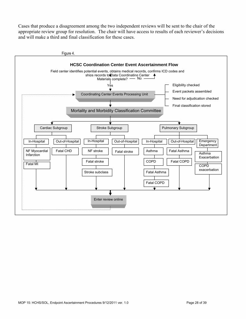
MOP 15: HCHS/SOL, Endpoint Ascertainment Procedures 9/12/2011 ver. 1.0 Page 28 of 39
Cases that produce a disagreement among the two independent reviews will be sent to the chair of the appropriate review group for resolution. The chair will have access to results of each reviewer’s decisions and will make a third and final classification for these cases.
Figure 4.
HCSC Coordination Center Event Ascertainment Flow
Field center identifies potential events, obtains medical records, confirms ICD codes and ships records to Data Coordinating Center
Coordinating Center Events Processing Unit
Eligibility checked
Materials complete?
Event packets assembled
Mortality and Morbidity Classification Committee
Cardiac Subgroup Stroke Subgroup Pulmonary Subgroup
Yes
No
In-Hospital Out-of-Hospital Emergency Department
NF Myocardial Infarction
Stroke subclass
Asthma Asthma Exacerbation
COPD COPD exacerbation
In-Hospital Out-of-Hospital Out-of-Hospital In-Hospital
Fatal CHD
Fatal MI
NF stroke
Fatal stroke
Fatal stroke Fatal Asthma
Fatal Asthma
Enter review online
Fatal COPD
Fatal COPD
Need for adjudication checked
Final classification stored
MOP 15: HCHS/SOL, Endpoint Ascertainment Procedures 9/12/2011 ver. 1.0 Page 29 of 39
7.4 Confidentiality Several procedures are in place to protect the security of the personal identifying information obtained from medical records and used in the event ascertainment process. Personal identifying information (name, SSN, date of birth, etc) from cohort members is used for the purpose of linkage to the National Death Index. This information is needed to determine vital status of cohort participants who are lost to follow up. All personal identifiers of cohort participants’, treating physicians, hospital name and location, and other identifying information are blacked out on any paper copy of medical records sent to the coordinating center and doubled checked prior to blinding for distribution to the ECC. Any copies of these documents remaining at the coordinating center are stored in locked secure rooms. ECC members are instructed in the proper confidential destruction of any medical record information they are provided. Study personnel involved in processing the medical record information, from abstraction to handling of these data have been trained on the protection of human subjects in research.
MOP 15: HCHS/SOL, Endpoint Ascertainment Procedures 9/12/2011 ver. 1.0 Page 30 of 39
References Luepker RV, Apple FS, Christenson RH, Crow RS, Fortmann SP, Goff D, Goldberg RJ, Hand MM, Jaffe AS, Julian DG, Levy D, Manolio T, Mendis S, Mensah G, Pajak A, Prineas RJ, et al. Case definitions for acute coronary heart disease in epidemiology and clinical research studies. A statement from the AHA Council on Epidemiology and Prevention; AHA Statistics Committee, World Heart Federation Council on Epidemiology and Prevention; the European Society of Cardiology Working Group on Epidemiology and Prevention; Centers for Disease Control and Prevention; and the National Heart, Lung and Blood Institute. Circulation 2003;108:2543-2549.
MOP 15: HCHS/SOL, Endpoint Ascertainment Procedures 9/12/2011 ver. 1.0 Page 31 of 39
APPENDIX A. Minnesota Code ECG Criteria APPENDIX B. GLOSSARY of Key Event Data Collections Terms APPENDIX C. Data Collection Forms required for assessing Endpoints: HCHS/SOL Event Tracking Form (ETR) & QxQ HCHS/SOL Informant Interview Form (IIE) & QxQ HCHS/SOL Death Certificate Form (DTH) & QxQ APPENDIX D. Abstraction Forms Myocardial Infarction Abstraction Form (MIF) Heart Failure Abstraction Form (HTF) Pulmonary Abstraction Form (PUL) APPENDIX E. Reviewer Forms for classification and adjudication Myocardial Infarction Form (MIF) Stroke and TIA Form Heart Failure Exacerbations Form (HFT) COPD and Asthma Form (PUL) APPENDIX F. Event Summary Forms (ESF) Myocardial Infarction Abstraction Form (MIF) Heart Failure Abstraction Form (HTF) Pulmonary Abstraction Form (PUL)
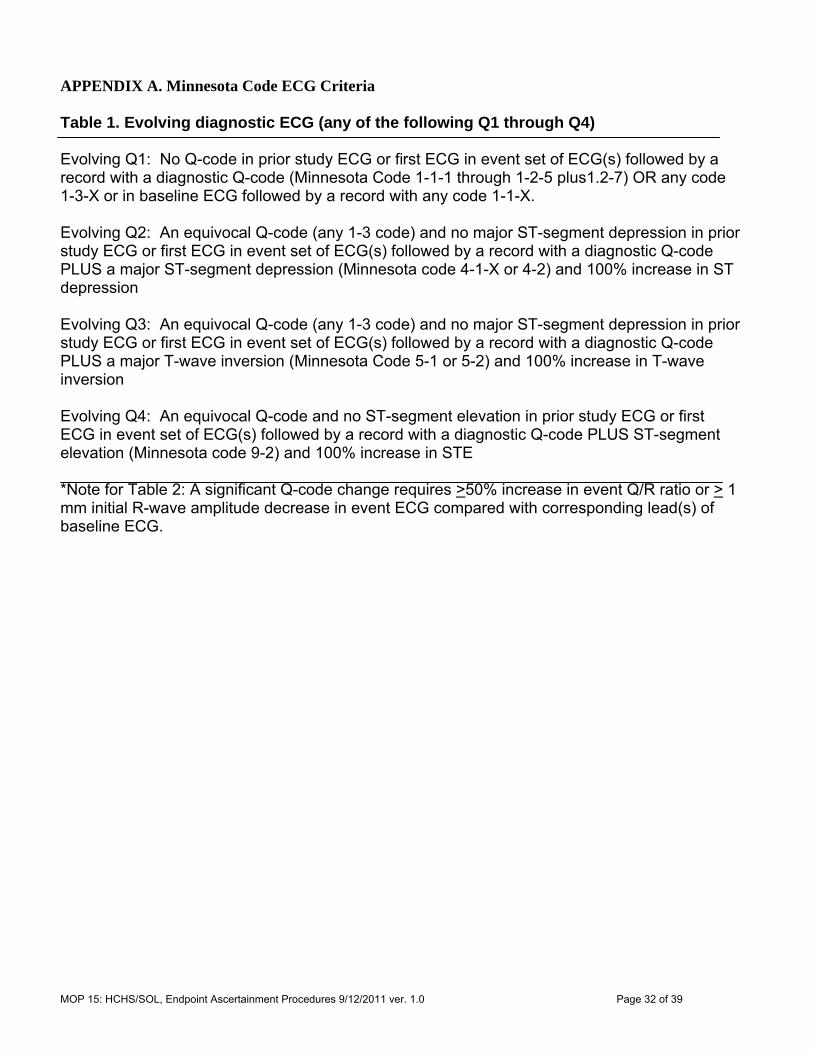
MOP 15: HCHS/SOL, Endpoint Ascertainment Procedures 9/12/2011 ver. 1.0 Page 32 of 39
APPENDIX A. Minnesota Code ECG Criteria Table 1. Evolving diagnostic ECG (any of the following Q1 through Q4) Evolving Q1: No Q-code in prior study ECG or first ECG in event set of ECG(s) followed by a record with a diagnostic Q-code (Minnesota Code 1-1-1 through 1-2-5 plus1.2-7) OR any code 1-3-X or in baseline ECG followed by a record with any code 1-1-X. Evolving Q2: An equivocal Q-code (any 1-3 code) and no major ST-segment depression in prior study ECG or first ECG in event set of ECG(s) followed by a record with a diagnostic Q-code PLUS a major ST-segment depression (Minnesota code 4-1-X or 4-2) and 100% increase in ST depression Evolving Q3: An equivocal Q-code (any 1-3 code) and no major ST-segment depression in prior study ECG or first ECG in event set of ECG(s) followed by a record with a diagnostic Q-code PLUS a major T-wave inversion (Minnesota Code 5-1 or 5-2) and 100% increase in T-wave inversion Evolving Q4: An equivocal Q-code and no ST-segment elevation in prior study ECG or first ECG in event set of ECG(s) followed by a record with a diagnostic Q-code PLUS ST-segment elevation (Minnesota code 9-2) and 100% increase in STE *Note for Table 2: A significant Q-code change requires >50% increase in event Q/R ratio or > 1 mm initial R-wave amplitude decrease in event ECG compared with corresponding lead(s) of baseline ECG.
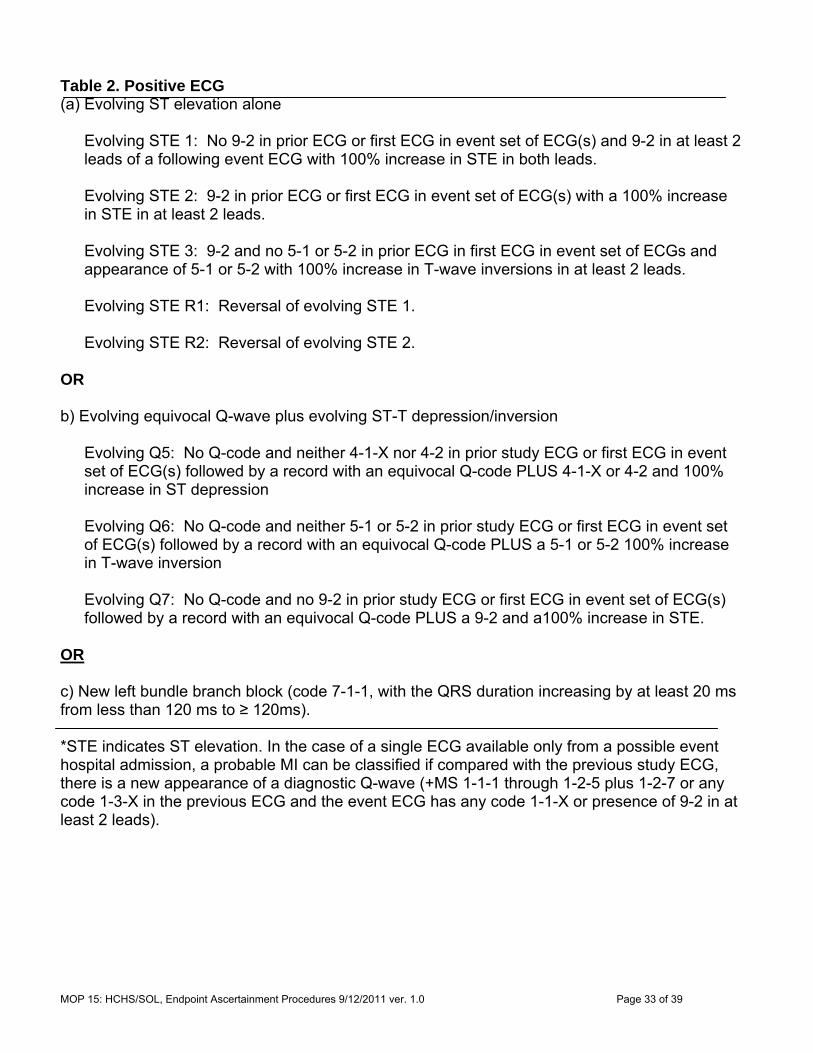
MOP 15: HCHS/SOL, Endpoint Ascertainment Procedures 9/12/2011 ver. 1.0 Page 33 of 39
Table 2. Positive ECG (a) Evolving ST elevation alone
Evolving STE 1: No 9-2 in prior ECG or first ECG in event set of ECG(s) and 9-2 in at least 2 leads of a following event ECG with 100% increase in STE in both leads. Evolving STE 2: 9-2 in prior ECG or first ECG in event set of ECG(s) with a 100% increase in STE in at least 2 leads. Evolving STE 3: 9-2 and no 5-1 or 5-2 in prior ECG in first ECG in event set of ECGs and appearance of 5-1 or 5-2 with 100% increase in T-wave inversions in at least 2 leads. Evolving STE R1: Reversal of evolving STE 1. Evolving STE R2: Reversal of evolving STE 2.
OR b) Evolving equivocal Q-wave plus evolving ST-T depression/inversion
Evolving Q5: No Q-code and neither 4-1-X nor 4-2 in prior study ECG or first ECG in event set of ECG(s) followed by a record with an equivocal Q-code PLUS 4-1-X or 4-2 and 100% increase in ST depression Evolving Q6: No Q-code and neither 5-1 or 5-2 in prior study ECG or first ECG in event set of ECG(s) followed by a record with an equivocal Q-code PLUS a 5-1 or 5-2 100% increase in T-wave inversion Evolving Q7: No Q-code and no 9-2 in prior study ECG or first ECG in event set of ECG(s) followed by a record with an equivocal Q-code PLUS a 9-2 and a100% increase in STE.
OR c) New left bundle branch block (code 7-1-1, with the QRS duration increasing by at least 20 ms from less than 120 ms to ≥ 120ms). *STE indicates ST elevation. In the case of a single ECG available only from a possible event hospital admission, a probable MI can be classified if compared with the previous study ECG, there is a new appearance of a diagnostic Q-wave (+MS 1-1-1 through 1-2-5 plus 1-2-7 or any code 1-3-X in the previous ECG and the event ECG has any code 1-1-X or presence of 9-2 in at least 2 leads).
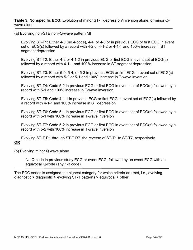
MOP 15: HCHS/SOL, Endpoint Ascertainment Procedures 9/12/2011 ver. 1.0 Page 34 of 39
Table 3. Nonspecific ECG: Evolution of minor ST-T depression/inversion alone, or minor Q-wave alone (a) Evolving non-STE non–Q-wave pattern MI
Evolving ST-T1: Either 4-0 (no 4-code), 4-4, or 4-3 or in previous ECG or first ECG in event set of ECG(s) followed by a record with 4-2 or 4-1-2 or 4-1-1 and 100% increase in ST segment depression Evolving ST-T2: Either 4-2 or 4-1-2 in previous ECG or first ECG in event set of ECG(s) followed by a record with 4-1-1 and 100% increase in ST segment depression Evolving ST-T3: Either 5-0, 5-4, or 5-3 in previous ECG or first ECG in event set of ECG(s) followed by a record with 5-2 or 5-1 and 100% increase in T-wave inversion Evolving ST-T4: Code 5-2 in previous ECG or first ECG in event set of ECG(s) followed by a record with 5-1 and 100% increase in T-wave inversion Evolving ST-T5: Code 4-1-1 in previous ECG or first ECG in event set of ECG(s) followed by a record with 4-1-1 and 100% increase in ST depression Evolving ST-T6: Code 5-1 in previous ECG or first ECG in event set of ECG(s) followed by a record with 5-1 with 100% increase in T-wave inversion Evolving ST-T7: Code 5-2 in previous ECG or first ECG in event set of ECG(s) followed by a record with 5-2 with 100% increase in T-wave inversion Evolving ST-T R1 through ST-T R7_the reverse of ST-T1 to ST-T7, respectively
OR (b) Evolving minor Q wave alone
No Q code in previous study ECG or event ECG, followed by an event ECG with an equivocal Q-code (any 1-3 code)
The ECG series is assigned the highest category for which criteria are met, i.e., evolving diagnostic > diagnostic > evolving ST-T patterns > equivocal > other.

MOP 15: HCHS/SOL, Endpoint Ascertainment Procedures 9/12/2011 ver. 1.0 Page 35 of 39
APPENDIX B. GLOSSARY of Key Event Data Collections Terms This section has intentionally been left blank.
MOP 15: HCHS/SOL, Endpoint Ascertainment Procedures 9/12/2011 ver. 1.0 Page 36 of 39
APPENDIX C. Data Collection Forms required for assessing Endpoints: HCHS/SOL Event Tracking Form (ETR) HCHS/SOL Informant Interview Form (IIE) HCHS/SOL Physician Inverview Form(PQE) HCHS/SOL Death Certificate Form (DTH) Note that current copies of all study data collection forms and QxQs can also be found on the study web site page: http://www.cscc.unc.edu/hchs/utilities/docfilter.php?study=hchs&filter_type=forms
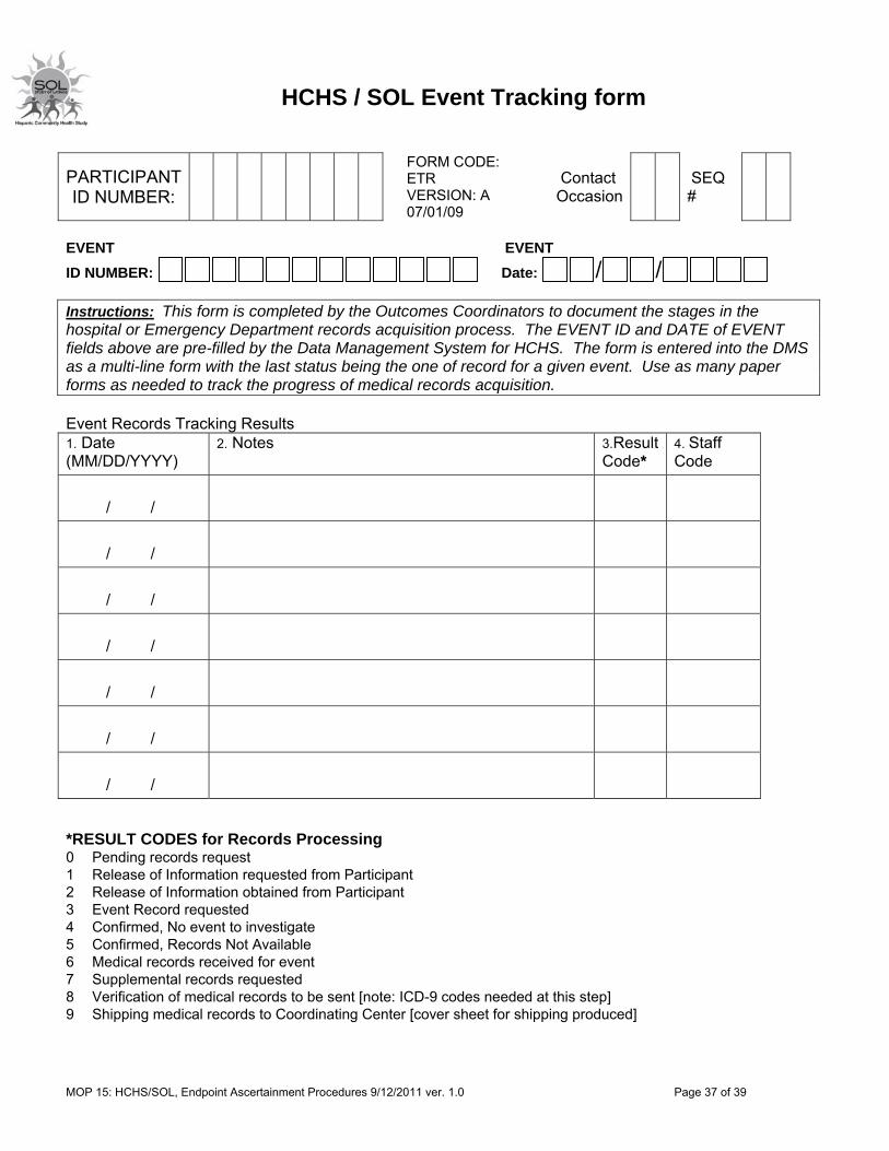
MOP 15: HCHS/SOL, Endpoint Ascertainment Procedures 9/12/2011 ver. 1.0 Page 37 of 39
HCHS / SOL Event Tracking form
EVENT EVENT
ID NUMBER: Date: / /
Instructions: This form is completed by the Outcomes Coordinators to document the stages in the hospital or Emergency Department records acquisition process. The EVENT ID and DATE of EVENT fields above are pre-filled by the Data Management System for HCHS. The form is entered into the DMS as a multi-line form with the last status being the one of record for a given event. Use as many paper forms as needed to track the progress of medical records acquisition. Event Records Tracking Results 1. Date (MM/DD/YYYY)
2. Notes 3.Result Code*
4. Staff Code
/ /
/ /
/ /
/ /
/ /
/ /
/ /
*RESULT CODES for Records Processing 0 Pending records request 1 Release of Information requested from Participant 2 Release of Information obtained from Participant 3 Event Record requested 4 Confirmed, No event to investigate 5 Confirmed, Records Not Available 6 Medical records received for event 7 Supplemental records requested 8 Verification of medical records to be sent [note: ICD-9 codes needed at this step] 9 Shipping medical records to Coordinating Center [cover sheet for shipping produced]
PARTICIPANT ID NUMBER:
FORM CODE: ETR VERSION: A 07/01/09
Contact Occasion
SEQ #
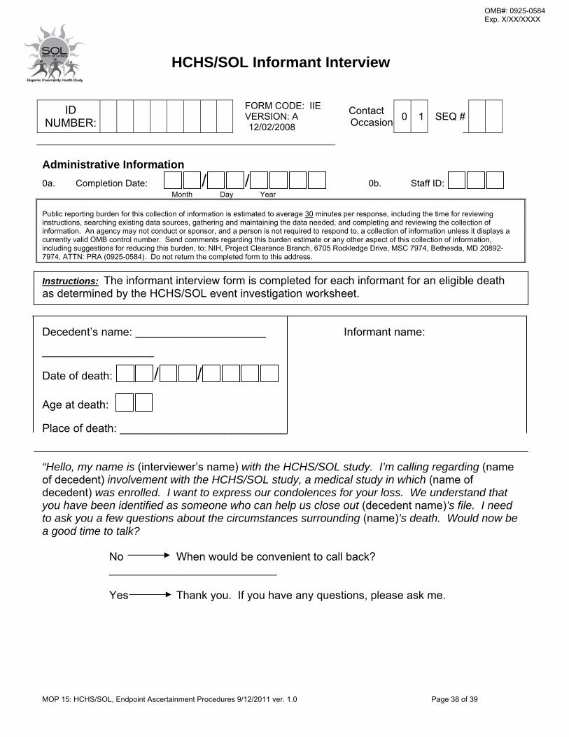
MOP 15: HCHS/SOL, Endpoint Ascertainment Procedures 9/12/2011 ver. 1.0 Page 38 of 39
HCHS/SOL Informant Interview
Administrative Information
0a. Completion Date: / / 0b. Staff ID:
Month Day Year
Public reporting burden for this collection of information is estimated to average 30 minutes per response, including the time for reviewing instructions, searching existing data sources, gathering and maintaining the data needed, and completing and reviewing the collection of information. An agency may not conduct or sponsor, and a person is not required to respond to, a collection of information unless it displays a currently valid OMB control number. Send comments regarding this burden estimate or any other aspect of this collection of information, including suggestions for reducing this burden, to: NIH, Project Clearance Branch, 6705 Rockledge Drive, MSC 7974, Bethesda, MD 20892-7974, ATTN: PRA (0925-0584). Do not return the completed form to this address.
Instructions: The informant interview form is completed for each informant for an eligible death as determined by the HCHS/SOL event investigation worksheet. Decedent’s name: _____________________ Informant name:
__________________
Date of death: / /
Age at death:
Place of death: ___________________________ “Hello, my name is (interviewer’s name) with the HCHS/SOL study. I’m calling regarding (name of decedent) involvement with the HCHS/SOL study, a medical study in which (name of decedent) was enrolled. I want to express our condolences for your loss. We understand that you have been identified as someone who can help us close out (decedent name)’s file. I need to ask you a few questions about the circumstances surrounding (name)’s death. Would now be a good time to talk?
No When would be convenient to call back? ___________________________ Yes Thank you. If you have any questions, please ask me.
ID NUMBER:
FORM CODE: IIE VERSION: A 12/02/2008
Contact Occasion
0 1 SEQ #
OMB#: 0925-0584 Exp. X/XX/XXXX
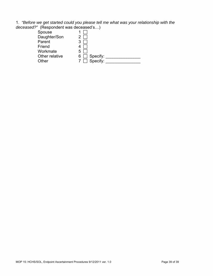
MOP 15: HCHS/SOL, Endpoint Ascertainment Procedures 9/12/2011 ver. 1.0 Page 39 of 39
1. “Before we get started could you please tell me what was your relationship with the deceased?” (Respondent was deceased’s…)
Spouse 1 Daughter/Son 2 Parent 3 Friend 4 Workmate 5 Other relative 6 Specify: _______________ Other 7 Specify: _______________
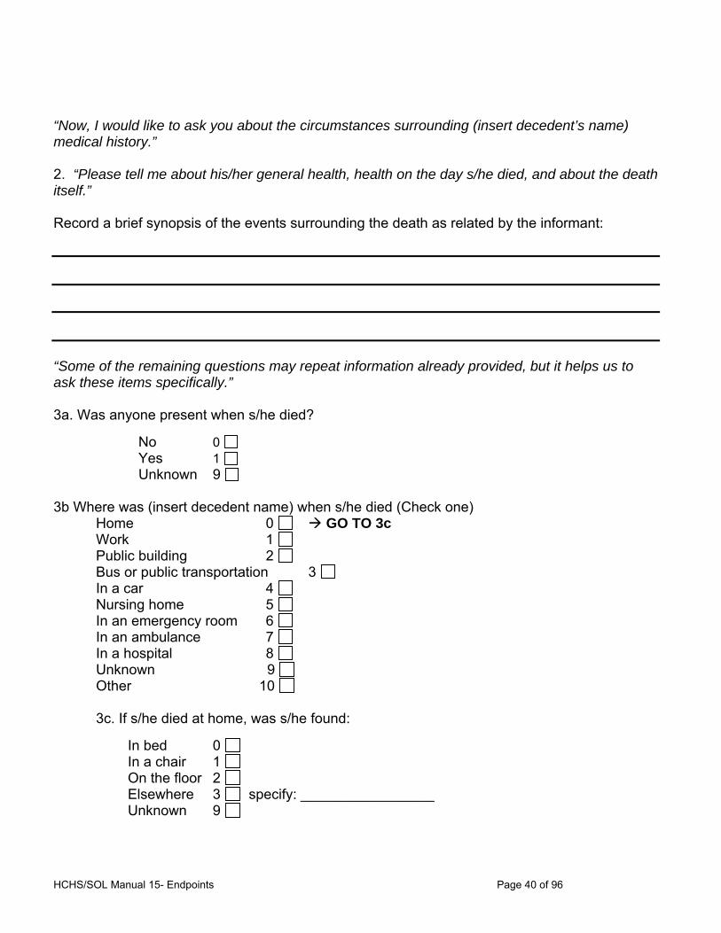
HCHS/SOL Manual 15- Endpoints Page 40 of 96
“Now, I would like to ask you about the circumstances surrounding (insert decedent’s name) medical history.” 2. “Please tell me about his/her general health, health on the day s/he died, and about the death itself.” Record a brief synopsis of the events surrounding the death as related by the informant:
“Some of the remaining questions may repeat information already provided, but it helps us to ask these items specifically.” 3a. Was anyone present when s/he died?
No 0 Yes 1 Unknown 9
3b Where was (insert decedent name) when s/he died (Check one)
Home 0 GO TO 3c Work 1 Public building 2 Bus or public transportation 3 In a car 4 Nursing home 5 In an emergency room 6 In an ambulance 7 In a hospital 8 Unknown 9 Other 10
3c. If s/he died at home, was s/he found:
In bed 0 In a chair 1 On the floor 2 Elsewhere 3 specify: _________________ Unknown 9

HCHS/SOL Manual 15- Endpoints Page 41 of 96
4. Was anyone close enough to hear (insert decedent’s name) if s/he had called out?
No 0 Yes 1 Unknown 9
5. How long was it between the time (insert decedent’s name) was last known to be alive and the time s/he was found dead?
Less than 5 minutes 1 5 minutes to 1 hour 2 1 to 24 hours 3 Longer than 24 hours 4 Unknown 9
6. Please tell me who was present. (check all that apply)
Self 1 (skip to question 8) Health care person(s) 2 Other person(s) 3
7. When was the last time you saw (insert decedent’s name) prior to his/her death?
Less than 5 minutes 1 5 minutes to 1 hour 2 1 to 24 hours 3 Longer than 24 hours 4 Unknown 9
HISTORY The next few questions concern (insert decedent’s name) medical history. 8. Was s/he restricted to home, able to leave home only with assistance or great effort, or was his/her activity unrestricted?
Restricted to home 1 Able to leave home only with assistance or great effort 2 Unrestricted 3
9. Was s/he hospitalized within the four weeks prior to death?
No 0 Skip to question 13 Yes 1 Unknown 9 Skip to question 13
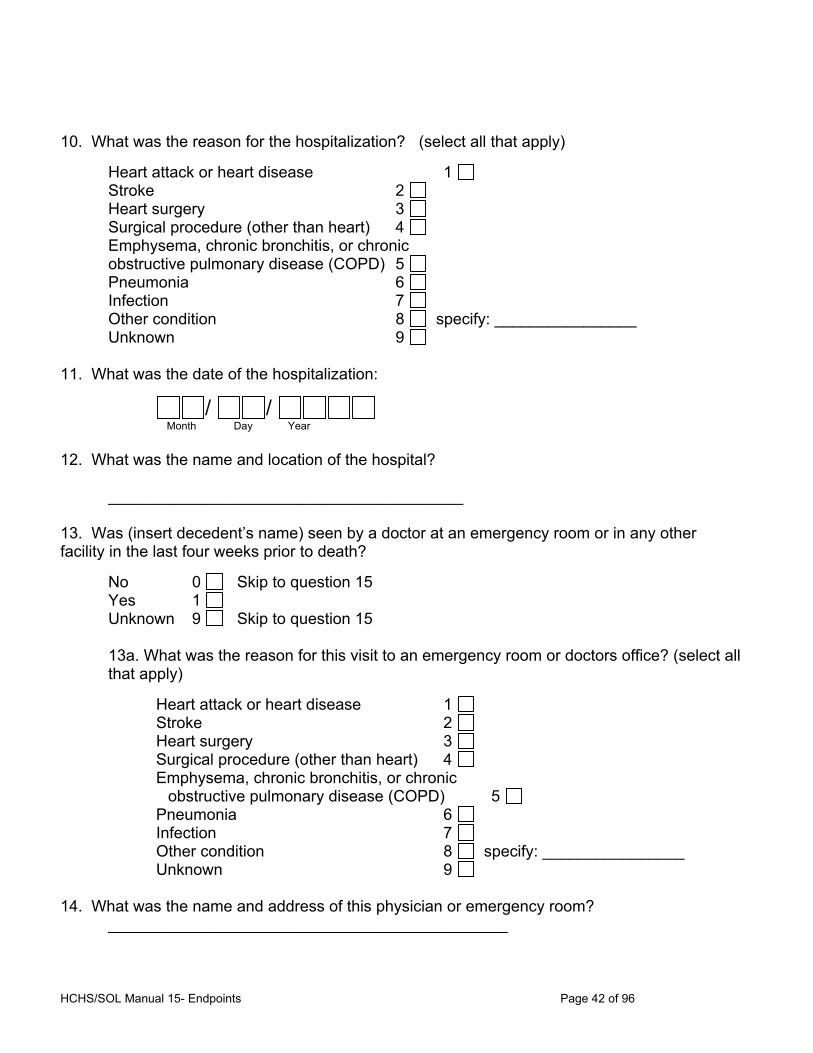
HCHS/SOL Manual 15- Endpoints Page 42 of 96
10. What was the reason for the hospitalization? (select all that apply)
Heart attack or heart disease 1 Stroke 2 Heart surgery 3 Surgical procedure (other than heart) 4 Emphysema, chronic bronchitis, or chronic obstructive pulmonary disease (COPD) 5 Pneumonia 6 Infection 7 Other condition 8 specify: ________________ Unknown 9
11. What was the date of the hospitalization:
/ / Month Day Year
12. What was the name and location of the hospital? ________________________________________ 13. Was (insert decedent’s name) seen by a doctor at an emergency room or in any other facility in the last four weeks prior to death?
No 0 Skip to question 15 Yes 1 Unknown 9 Skip to question 15
13a. What was the reason for this visit to an emergency room or doctors office? (select all that apply)
Heart attack or heart disease 1 Stroke 2 Heart surgery 3 Surgical procedure (other than heart) 4 Emphysema, chronic bronchitis, or chronic obstructive pulmonary disease (COPD) 5 Pneumonia 6 Infection 7 Other condition 8 specify: ________________ Unknown 9
14. What was the name and address of this physician or emergency room? _____________________________________________
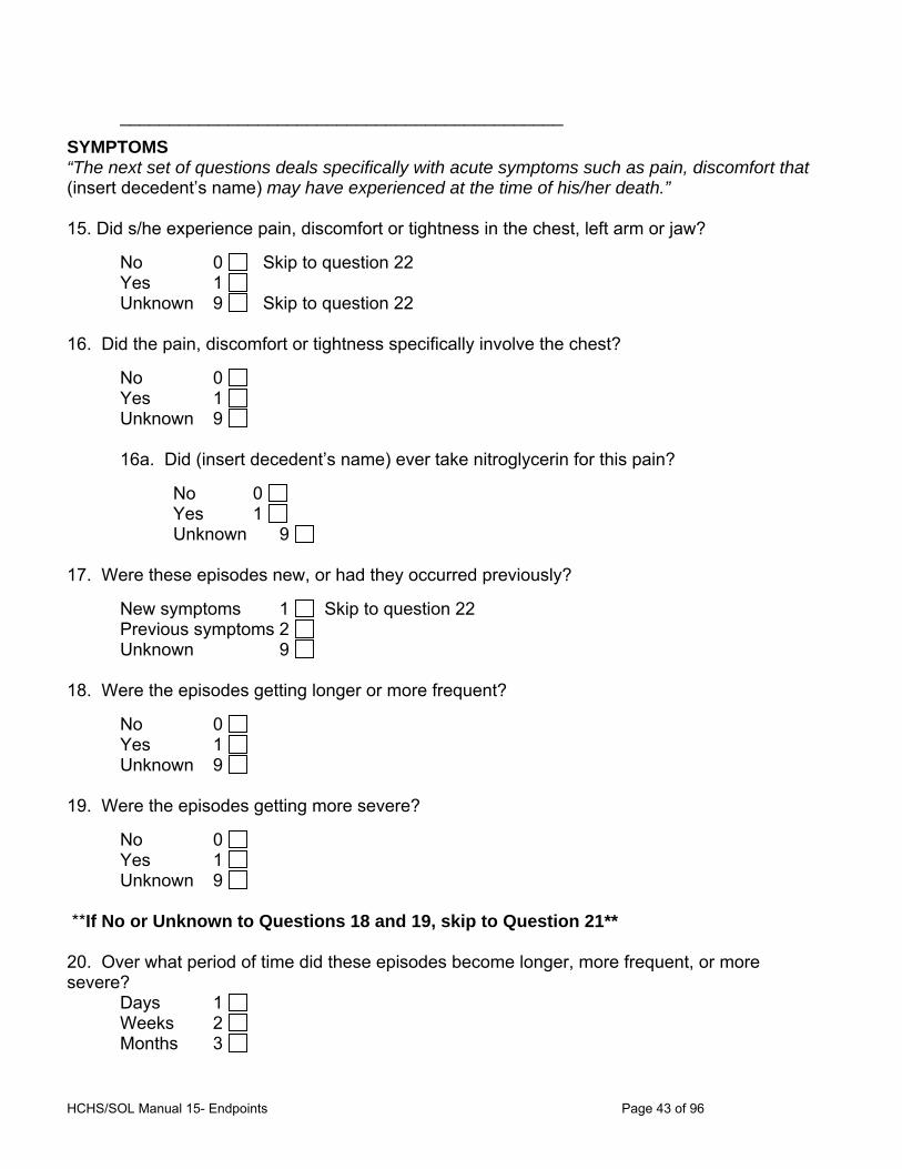
HCHS/SOL Manual 15- Endpoints Page 43 of 96
_____________________________________________
SYMPTOMS “The next set of questions deals specifically with acute symptoms such as pain, discomfort that (insert decedent’s name) may have experienced at the time of his/her death.” 15. Did s/he experience pain, discomfort or tightness in the chest, left arm or jaw?
No 0 Skip to question 22 Yes 1 Unknown 9 Skip to question 22
16. Did the pain, discomfort or tightness specifically involve the chest?
No 0 Yes 1 Unknown 9
16a. Did (insert decedent’s name) ever take nitroglycerin for this pain?
No 0 Yes 1 Unknown 9
17. Were these episodes new, or had they occurred previously?
New symptoms 1 Skip to question 22 Previous symptoms 2 Unknown 9
18. Were the episodes getting longer or more frequent?
No 0 Yes 1 Unknown 9
19. Were the episodes getting more severe?
No 0 Yes 1 Unknown 9
**If No or Unknown to Questions 18 and 19, skip to Question 21** 20. Over what period of time did these episodes become longer, more frequent, or more severe?
Days 1 Weeks 2 Months 3
HCHS/SOL Manual 15- Endpoints Page 44 of 96
Unknown 9 21. Did s/he experience shortness of breath?
No 0 Skip to item 22 Yes 1 Unknown 9 Skip to item 22
21a. Did s/he have shortness of breath while at rest?
No 0 Yes 1 Unknown 9
22. “I apologize if this question sounds hard or if it makes you uncomfortable. Please be assured we respect your feelings about this unfortunate event. How long was it from (insert decedent’s name) last episode of symptoms to the time that s/he stopped breathing on his/her own?”
Less than 5 minutes 1 Less than 1 hour 2 Less than 24 hours 3 Greater than 24 hours 4 Unknown 9
EMERGENCY MEDICAL CARE “The next few questions are concerned with emergency medical care (insert decedent’s name) may have received prior to or at the time of death. You may have already given this information in an answer to an earlier question. Since it is important to obtain information specifically on emergency medical care, I hope you don’t mind if these questions seem repetitive.” 23. Was a physician, ambulance or other emergency medical team called?
No 0 Skip to question 24 Yes 1 Unknown 9 Skip to question 24
23a. How long was it from the time the last episode of symptoms started to the time that medical assistance was called for?
5 minutes or less 1 10 minutes or less 2 1 hour or less 3 6 hours or less 4 24 hours or less 5 More than 24 hours 6
HCHS/SOL Manual 15- Endpoints Page 45 of 96
Unknown 9
23b. How long was if from the time medical care was called to the time when it arrived?
5 minutes or less 1 10 minutes or less 2 1 hour or less 3 6 hours or less 4 24 hours or less 5 More than 24 hours 6 Unknown 9
24. Were resuscitation measures, such as CPR attempted?
No 0 Skip to question 25 Yes 1 Unknown 9 Skip to question 25
24a. Who started the CPR or resuscitation?
Bystander 1 Physician 2 Ambulance personnel 3 Fireman or Police 4 The informant 5 Other 6 Unknown 9
25. Was (insert decedent’s name) taken to the hospital, emergency room or any other emergency care facility?
No 0 Yes 1 Unknown 9
26. Is there anyone else we could contact who might be able to provide additional information about the circumstances surrounding (insert decedent’s name) death or his/her usual state of health?
No 0 Skip to Closing Script Yes 1 Unknown 9 Skip to Closing Script
HCHS/SOL Manual 15- Endpoints Page 46 of 96
27. How is s/he related to the deceased?
Spouse 1 Daughter/Son 2 Parent 3 Friend 4 Workmate 5 Other relative 6 Specify: _______________ Other 7 Specify: _______________
28. What is the name and address of this person?
___________________________________________
___________________________________________
CLOSING SCRIPT “Thank you very much for your assistance in this study. Do you have any questions? Thanks again for your help.” RELIABILITY (To be completed after the interview) 29. On the basis of these questions, give your rating of reliability of the interview.
Poor 1 Fair 2 Good 3

HCHS/SOL Manual 15- Endpoints Page 47 of 96
HCHS/SOL Physician Questionnaire
Administrative Information
0a. Completion Date: / / 0b. Staff ID:
month may year Instructions: Please complete the following questions to the best of your ability by filling in the appropriate bubbles or writing the answer in the blank provided. Please return completed forms in the self addressed stamped envelope provided to the local HCHS/SOL field center. Public reporting burden for this collection of information is estimated to average 30 minutes per response, including the time for reviewing instructions, searching existing data sources, gathering and maintaining the data needed, and completing and reviewing the collection of information. An agency may not conduct or sponsor, and a person is not required to respond to, a collection of information unless it displays a currently valid OMB control number. Send comments regarding this burden estimate or any other aspect of this collection of information, including suggestions for reducing this burden, to: NIH, Project Clearance Branch, 6705 Rockledge Drive, MSC 7974, Bethesda, MD 20892-7974, ATTN: PRA (0925-0584). Do not return the completed form to this address.
DETAILS OF DEATH 1. Are you familiar with the events surrounding the decedent's death?
No 0 Yes 1
2. Did you witness the death?
No 0 Yes 1
If informant answered “Yes” to one or both of Items 1 and 2, please skip to Item 4. 3. If you answered "No" to both Questions, are you aware of another physician who could provide information regarding the death?
No 0 Please sign and date the bottom of this form Yes 1
3a. Provide contact information. Please then sign and date the bottom of this form.
Name of physician: ___________________________________ Address: _______________________________________
_______________________________________
_______________________________________
ID NUMBER:
FORM CODE: PQE VERSION: A 12/02/2008
Contact Occasion
0 1 SEQ #
OMB#: 0925-0584 Exp. X/XX/XXXX
HCHS/SOL Manual 15- Endpoints Page 1 of 105
CIRCUMSTANCES SURROUNDING DEATH
4. What do you believe to be the underlying cause of death?
Acute Myocardial Infarction 1 Other Ischemic Heart Disease 2 Cerebrovascular Disease 3 Other Cardiovascular Disease 4 Emphysema, chronic bronchitis or chronic
obstructive pulmonary disease (COPD) 5 Pneumonia 6 Asthma 7 Other Lung Disease 8 specify: ______________ Non Cardio - Pulmonary Disease 9 specify: ______________
5. Please specify the time between the onset of the acute episode of symptoms and death. (We are defining death as the point where spontaneous breathing ceased and the patient never recovered.) Please check the appropriate time period.
Less than 5 minutes 1 5 minutes to 1 hour 2 1 hour to 24 hours 3 More than 24 hours 4 1 day to 3 days 5 More than 3 days 6 Unknown 9
6. Was there an acute episode of pain in the chest, left arm or jaw during the last 72 hours prior to death?
No 0 Yes 1 Unknown 9
7. Was there an acute episode of shortness of breath during the 72 hours prior to death?
No 0 Yes 1 Unknown 9
8. Was there an acute episode of wheezing during the 72 hours prior to death?
No 0 Yes 1 Unknown 9
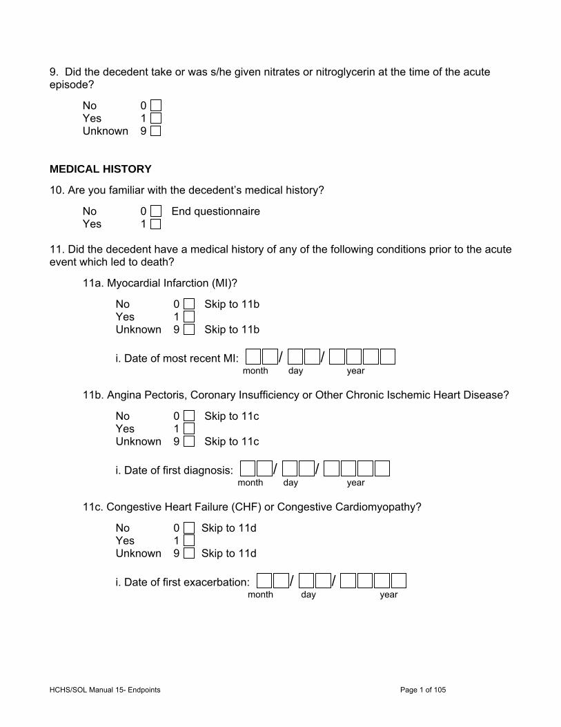
HCHS/SOL Manual 15- Endpoints Page 1 of 105
9. Did the decedent take or was s/he given nitrates or nitroglycerin at the time of the acute episode?
No 0 Yes 1 Unknown 9
MEDICAL HISTORY
10. Are you familiar with the decedent’s medical history?
No 0 End questionnaire Yes 1
11. Did the decedent have a medical history of any of the following conditions prior to the acute event which led to death?
11a. Myocardial Infarction (MI)?
No 0 Skip to 11b Yes 1 Unknown 9 Skip to 11b
i. Date of most recent MI: / / month day year
11b. Angina Pectoris, Coronary Insufficiency or Other Chronic Ischemic Heart Disease?
No 0 Skip to 11c Yes 1 Unknown 9 Skip to 11c
i. Date of first diagnosis: / / month day year 11c. Congestive Heart Failure (CHF) or Congestive Cardiomyopathy?
No 0 Skip to 11d Yes 1 Unknown 9 Skip to 11d
i. Date of first exacerbation: / / month day year
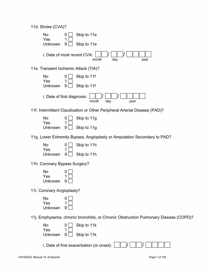
HCHS/SOL Manual 15- Endpoints Page 1 of 105
11d. Stroke (CVA)?
No 0 Skip to 11e Yes 1 Unknown 9 Skip to 11e
i. Date of most recent CVA: / / month day year 11e. Transient Ischemic Attack (TIA)?
No 0 Skip to 11f Yes 1 Unknown 9 Skip to 11f
i. Date of first diagnosis: / / month day year 11f. Intermittent Claudication or Other Peripheral Arterial Disease (PAD)?
No 0 Skip to 11g Yes 1 Unknown 9 Skip to 11g
11g. Lower Extremity Bypass, Angioplasty or Amputation Secondary to PAD?
No 0 Skip to 11h Yes 1 Unknown 9 Skip to 11h
11h. Coronary Bypass Surgery?
No 0 Yes 1 Unknown 9
11i. Coronary Angioplasty?
No 0 Yes 1 Unknown 9
11j. Emphysema, chronic bronchitis, or Chronic Obstruction Pulmonary Disease (COPD)?
No 0 Skip to 11k Yes 1 Unknown 9 Skip to 11k
i. Date of first exacerbation (or onset): / /
HCHS/SOL Manual 15- Endpoints Page 1 of 105
month day year 11k. Asthma?
No 0 Yes 1 Unknown 9
i. Approximate age asthma first started:
12. If you saw the participant within one month of death, please fill out the following for the most recent visit:
12a. Date of visit: / / month day year 12b. Chief Complaint:____________________________________
____________________________________
12c. Primary Diagnosis:____________________________________
____________________________________
12d. Changes in Medical Management:____________________________________
_____________________________________
_____________________________________
Form completed by: _______________________________ Date: ___________________
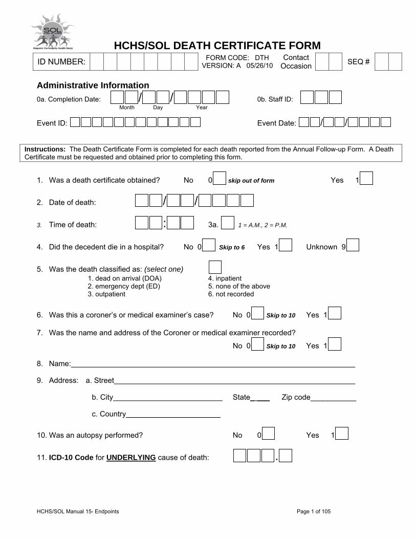
HCHS/SOL Manual 15- Endpoints Page 1 of 105
HCHS/SOL DEATH CERTIFICATE FORM
Administrative Information
0a. Completion Date: / / 0b. Staff ID:
Month Day Year
Event ID: Event Date: / /
Instructions: The Death Certificate Form is completed for each death reported from the Annual Follow-up Form. A Death Certificate must be requested and obtained prior to completing this form.
1. Was a death certificate obtained? No 0 skip out of form Yes 1
2. Date of death: / /
3. Time of death: : 3a. 1 = A.M., 2 = P.M.
4. Did the decedent die in a hospital? No 0 Skip to 6 Yes 1 Unknown 9
5. Was the death classified as: (select one) 1. dead on arrival (DOA) 4. inpatient 2. emergency dept (ED) 5. none of the above 3. outpatient 6. not recorded
6. Was this a coroner’s or medical examiner’s case? No 0 Skip to 10 Yes 1
7. Was the name and address of the Coroner or medical examiner recorded?
No 0 Skip to 10 Yes 1
8. Name:
9. Address: a. Street
b. City State_ ___ Zip code___________
c. Country
10. Was an autopsy performed? No 0 Yes 1
11. ICD-10 Code for UNDERLYING cause of death: .
ID NUMBER: FORM CODE: DTH VERSION: A 05/26/10
Contact Occasion
SEQ #
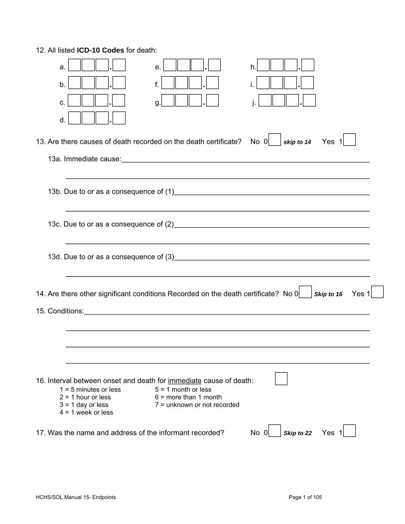
HCHS/SOL Manual 15- Endpoints Page 1 of 105
12. All listed ICD-10 Codes for death:
a. . e. . h. .
b. . f. . i. .
c. . g. . j. .
d. .
13. Are there causes of death recorded on the death certificate? No 0 skip to 14 Yes 1
13a. Immediate cause:
13b. Due to or as a consequence of (1)
13c. Due to or as a consequence of (2)
13d. Due to or as a consequence of (3)
14. Are there other significant conditions Recorded on the death certificate? No 0 Skip to 16 Yes 1
15. Conditions:
16. Interval between onset and death for immediate cause of death: 1 = 5 minutes or less 5 = 1 month or less 2 = 1 hour or less 6 = more than 1 month 3 = 1 day or less 7 = unknown or not recorded 4 = 1 week or less
17. Was the name and address of the informant recorded? No 0 Skip to 22 Yes 1
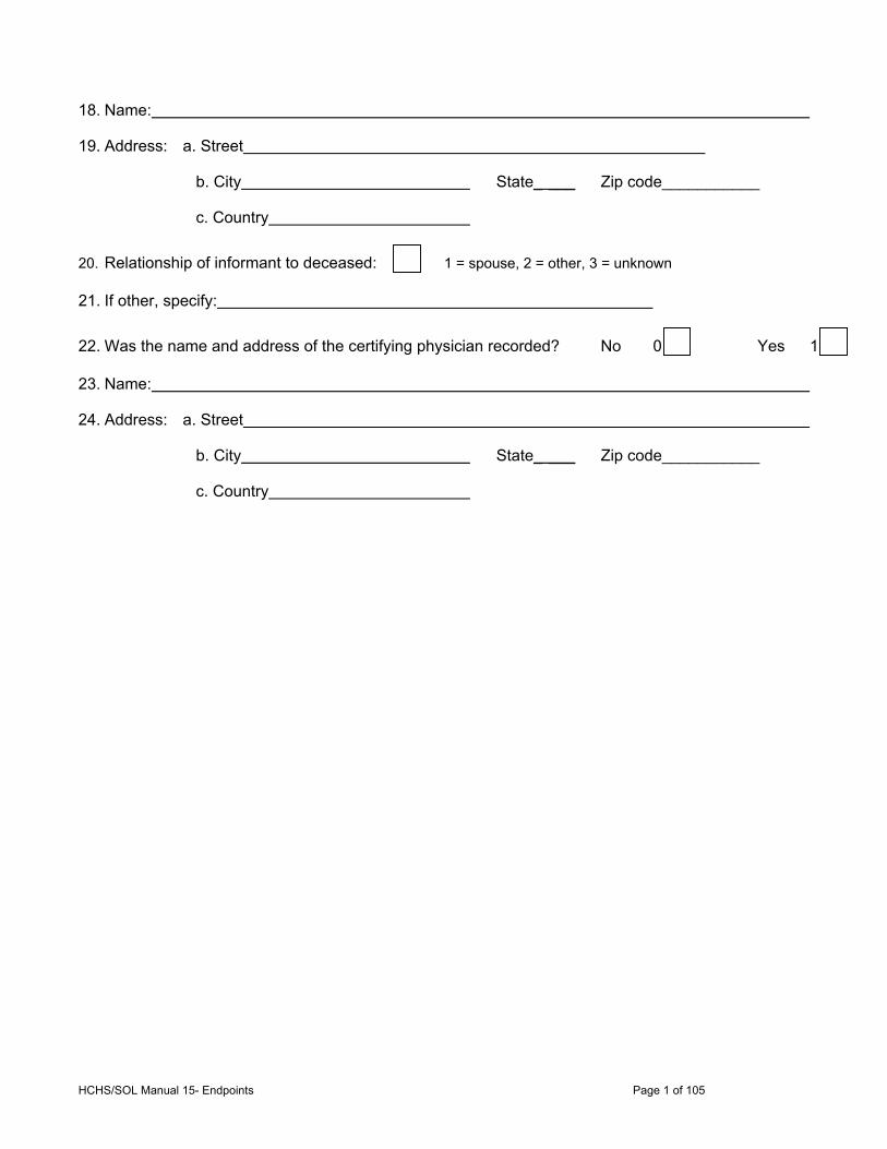
HCHS/SOL Manual 15- Endpoints Page 1 of 105
18. Name:
19. Address: a. Street
b. City State_ ___ Zip code___________
c. Country
20. Relationship of informant to deceased: 1 = spouse, 2 = other, 3 = unknown
21. If other, specify:
22. Was the name and address of the certifying physician recorded? No 0 Yes 1
23. Name:
24. Address: a. Street
b. City State_ ___ Zip code___________
c. Country

HCHS/SOL Manual 15- Endpoints Page 1 of 105
APPENDIX D Abstraction Forms
Myocardial Infarction Abstraction Form (MIF) Heart Failure Abstraction Form (HTF) Pulmonary Abstraction Form (PUL)

HCHS/SOL Manual 15- Endpoints Page 1 of 105
HCHS/SOL MYOCARDIAL INFARCTION ABSTRACTION FORM (MIF)
ADMINISTRATIVE INFORMATION
0A. Completion Date: / / 0B. Staff ID: Month/Day/Year
Event ID: Event Date: / /
Instructions: Answers are derived from the medical records received. Do not complete this form until all records are received (or classified as unobtainable) as indicated on the Verification of ICD Discharge Codes Form
A. GENERAL INFORMATION
1. Was the event (choose one): 1= In hospital only 2= Emergency Dept. visit only(ED) 3= Both ED and in hospital
2. Date of arrival: (mm/dd/yyyy) / /
a. Time of arrival : 1= A.M., 2 = P.M.
b. Date of admission / /
3. Date of discharge: (mm/dd/yyyy) / /
a. Time of discharge : 1= A.M., 2 = P.M.
4. What was the primary admitting diagnosis code? .
5. What was the primary discharge diagnosis code? .
No/NR Yes 6. Did an emergency medical service unit transport the patient to this hospital? 0 1
7. Was the patient transferred to this hospital from another hospital? 0 1
8. Was the patient’s code status ever “no-code” or “DNR” (do not resuscitate)? 0 1
9. Was the patient alive at discharge? If Yes, go to Item 10 0 1
9.a. Was the patient dead on arrival? No 0 Yes 1
9.b. Did the patient die in the Emergency Department? No 0 Yes 1
9.c. Was an autopsy performed? No 0 Yes 1
ID NUMBER: FORM CODE: MIF VERSION: A 08/04/11
Contact Occasion
SEQ #
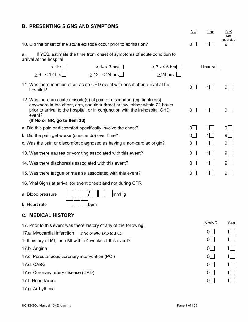
HCHS/SOL Manual 15- Endpoints Page 1 of 105
B. PRESENTING SIGNS AND SYMPTOMS No Yes NR Not
recorded
10. Did the onset of the acute episode occur prior to admission? 0 1 9
a. If YES, estimate the time from onset of symptoms of acute condition to arrival at the hospital
< 1hr > 1- < 3 hrs > 3 - < 6 hrs Unsure
> 6 - < 12 hrs > 12 - < 24 hrs > 24 hrs.
11. Was there mention of an acute CHD event with onset after arrival at the hospital?
0 1 9
12. Was there an acute episode(s) of pain or discomfort (eg: tightness) anywhere in the chest, arm, shoulder throat or jaw, either within 72 hours prior to arrival to the hospital, or in conjunction with the in-hospital CHD event? (If No or NR, go to Item 13)
0 1 9
a. Did this pain or discomfort specifically involve the chest? 0 1 9
b. Did the pain get worse (crescendo) over time? 0 1 9
c. Was the pain or discomfort diagnosed as having a non-cardiac origin? 0 1 9
13. Was there nausea or vomiting associated with this event? 0 1 9
14. Was there diaphoresis associated with this event? 0 1 9
15. Was there fatigue or malaise associated with this event? 0 1 9
16. Vital Signs at arrival (or event onset) and not during CPR
a. Blood pressure / mmHg
b. Heart rate bpm
C. MEDICAL HISTORY
17. Prior to this event was there history of any of the following: No/NR Yes
17.a. Myocardial infarction If No or NR, skip to 17.b. 0 1
1. If history of MI, then MI within 4 weeks of this event? 0 1
17.b. Angina 0 1
17.c. Percutaneous coronary intervention (PCI) 0 1
17.d. CABG 0 1
17.e. Coronary artery disease (CAD) 0 1
17.f. Heart failure 0 1
17.g. Arrhythmia
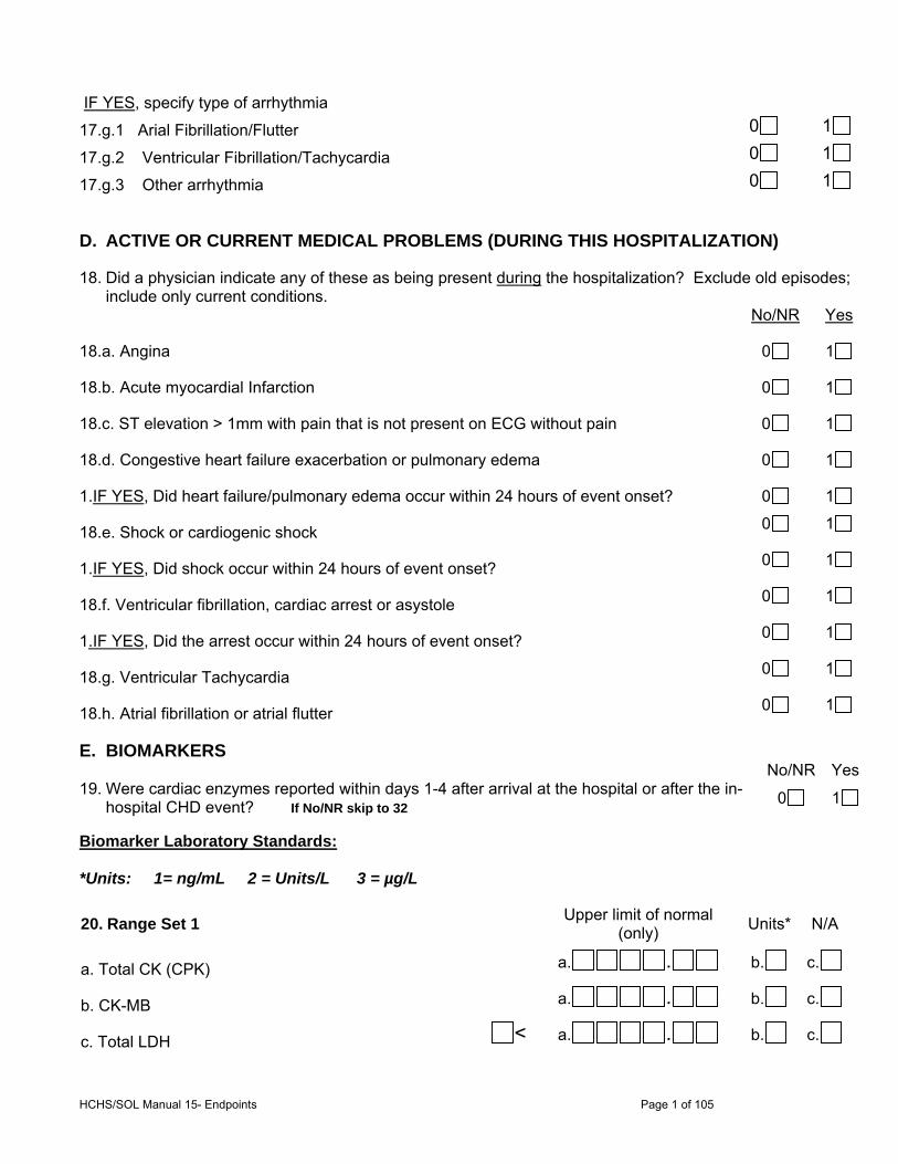
HCHS/SOL Manual 15- Endpoints Page 1 of 105
IF YES, specify type of arrhythmia
17.g.1 Arial Fibrillation/Flutter 0 1
17.g.2 Ventricular Fibrillation/Tachycardia 0 1
17.g.3 Other arrhythmia 0 1
D. ACTIVE OR CURRENT MEDICAL PROBLEMS (DURING THIS HOSPITALIZATION)
18. Did a physician indicate any of these as being present during the hospitalization? Exclude old episodes; include only current conditions.
No/NR Yes
18.a. Angina 0 1
18.b. Acute myocardial Infarction 0 1
18.c. ST elevation > 1mm with pain that is not present on ECG without pain 0 1
18.d. Congestive heart failure exacerbation or pulmonary edema 0 1
1.IF YES, Did heart failure/pulmonary edema occur within 24 hours of event onset? 0 1
18.e. Shock or cardiogenic shock 0 1
1.IF YES, Did shock occur within 24 hours of event onset? 0 1
18.f. Ventricular fibrillation, cardiac arrest or asystole 0 1
1.IF YES, Did the arrest occur within 24 hours of event onset? 0 1
18.g. Ventricular Tachycardia 0 1
18.h. Atrial fibrillation or atrial flutter 0 1
E. BIOMARKERS No/NR Yes19. Were cardiac enzymes reported within days 1-4 after arrival at the hospital or after the in-
hospital CHD event? If No/NR skip to 32 0 1
Biomarker Laboratory Standards: *Units: 1= ng/mL 2 = Units/L 3 = µg/L
20. Range Set 1 Upper limit of normal
(only) Units* N/A
a. Total CK (CPK) a. . b. c.
b. CK-MB a. . b. c.
c. Total LDH < a. . b. c.
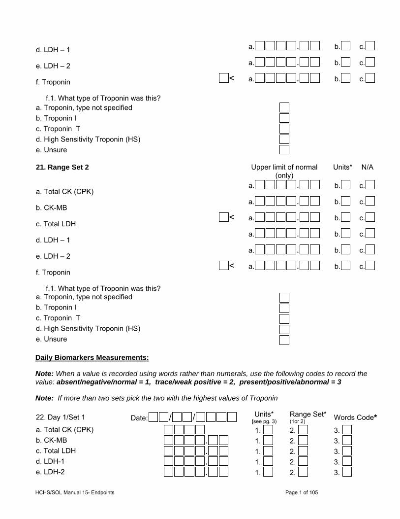
HCHS/SOL Manual 15- Endpoints Page 1 of 105
d. LDH – 1 a. . b. c.
e. LDH – 2 a. . b. c.
f. Troponin < a. . b. c.
f.1. What type of Troponin was this?
a. Troponin, type not specified
b. Troponin I c. Troponin T d. High Sensitivity Troponin (HS)
e. Unsure
21. Range Set 2 Upper limit of normal (only)
Units* N/A
a. Total CK (CPK) a. . b. c.
b. CK-MB a. . b. c.
c. Total LDH < a. . b. c.
d. LDH – 1 a. . b. c.
e. LDH – 2 a. . b. c.
f. Troponin < a. . b. c.
f.1. What type of Troponin was this?
a. Troponin, type not specified
b. Troponin I c. Troponin T d. High Sensitivity Troponin (HS)
e. Unsure
Daily Biomarkers Measurements:
Note: When a value is recorded using words rather than numerals, use the following codes to record the value: absent/negative/normal = 1, trace/weak positive = 2, present/positive/abnormal = 3
Note: If more than two sets pick the two with the highest values of Troponin
22. Day 1/Set 1 Date: / / Units* (see pg. 3)
Range Set* (1or 2) Words Code*
a. Total CK (CPK) 1. 2. 3. b. CK-MB . 1. 2. 3. c. Total LDH . 1. 2. 3. d. LDH-1 . 1. 2. 3. e. LDH-2 . 1. 2. 3.
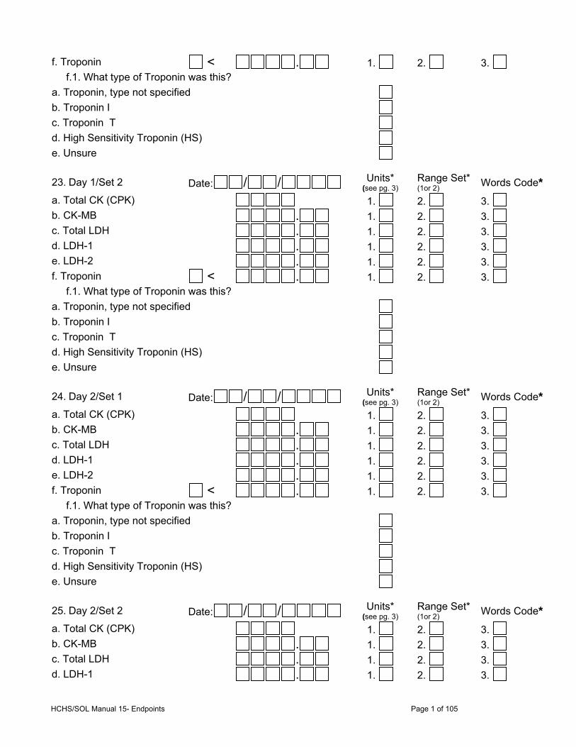
HCHS/SOL Manual 15- Endpoints Page 1 of 105
f. Troponin < . 1. 2. 3. f.1. What type of Troponin was this?
a. Troponin, type not specified
b. Troponin I c. Troponin T d. High Sensitivity Troponin (HS)
e. Unsure
23. Day 1/Set 2 Date: / / Units* (see pg. 3)
Range Set* (1or 2) Words Code*
a. Total CK (CPK) 1. 2. 3. b. CK-MB . 1. 2. 3. c. Total LDH . 1. 2. 3. d. LDH-1 . 1. 2. 3. e. LDH-2 . 1. 2. 3. f. Troponin < . 1. 2. 3. f.1. What type of Troponin was this?
a. Troponin, type not specified
b. Troponin I c. Troponin T d. High Sensitivity Troponin (HS)
e. Unsure
24. Day 2/Set 1 Date: / / Units* (see pg. 3)
Range Set* (1or 2) Words Code*
a. Total CK (CPK) 1. 2. 3. b. CK-MB . 1. 2. 3. c. Total LDH . 1. 2. 3. d. LDH-1 . 1. 2. 3. e. LDH-2 . 1. 2. 3. f. Troponin < . 1. 2. 3. f.1. What type of Troponin was this?
a. Troponin, type not specified
b. Troponin I c. Troponin T d. High Sensitivity Troponin (HS)
e. Unsure
25. Day 2/Set 2 Date: / / Units* (see pg. 3)
Range Set* (1or 2) Words Code*
a. Total CK (CPK) 1. 2. 3. b. CK-MB . 1. 2. 3. c. Total LDH . 1. 2. 3. d. LDH-1 . 1. 2. 3.
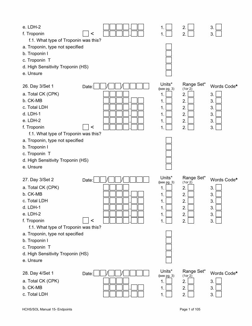
HCHS/SOL Manual 15- Endpoints Page 1 of 105
e. LDH-2 . 1. 2. 3. f. Troponin < . 1. 2. 3. f.1. What type of Troponin was this?
a. Troponin, type not specified
b. Troponin I c. Troponin T d. High Sensitivity Troponin (HS)
e. Unsure
26. Day 3/Set 1 Date: / / Units* (see pg. 3)
Range Set* (1or 2) Words Code*
a. Total CK (CPK) 1. 2. 3. b. CK-MB . 1. 2. 3. c. Total LDH . 1. 2. 3. d. LDH-1 . 1. 2. 3. e. LDH-2 . 1. 2. 3. f. Troponin < . 1. 2. 3. f.1. What type of Troponin was this?
a. Troponin, type not specified
b. Troponin I c. Troponin T d. High Sensitivity Troponin (HS)
e. Unsure
27. Day 3/Set 2 Date: / / Units* (see pg. 3)
Range Set* (1or 2) Words Code*
a. Total CK (CPK) 1. 2. 3. b. CK-MB . 1. 2. 3. c. Total LDH . 1. 2. 3. d. LDH-1 . 1. 2. 3. e. LDH-2 . 1. 2. 3. f. Troponin < . 1. 2. 3. f.1. What type of Troponin was this?
a. Troponin, type not specified
b. Troponin I c. Troponin T d. High Sensitivity Troponin (HS)
e. Unsure
28. Day 4/Set 1 Date: / / Units* (see pg. 3)
Range Set* (1or 2) Words Code*
a. Total CK (CPK) 1. 2. 3. b. CK-MB . 1. 2. 3. c. Total LDH . 1. 2. 3.
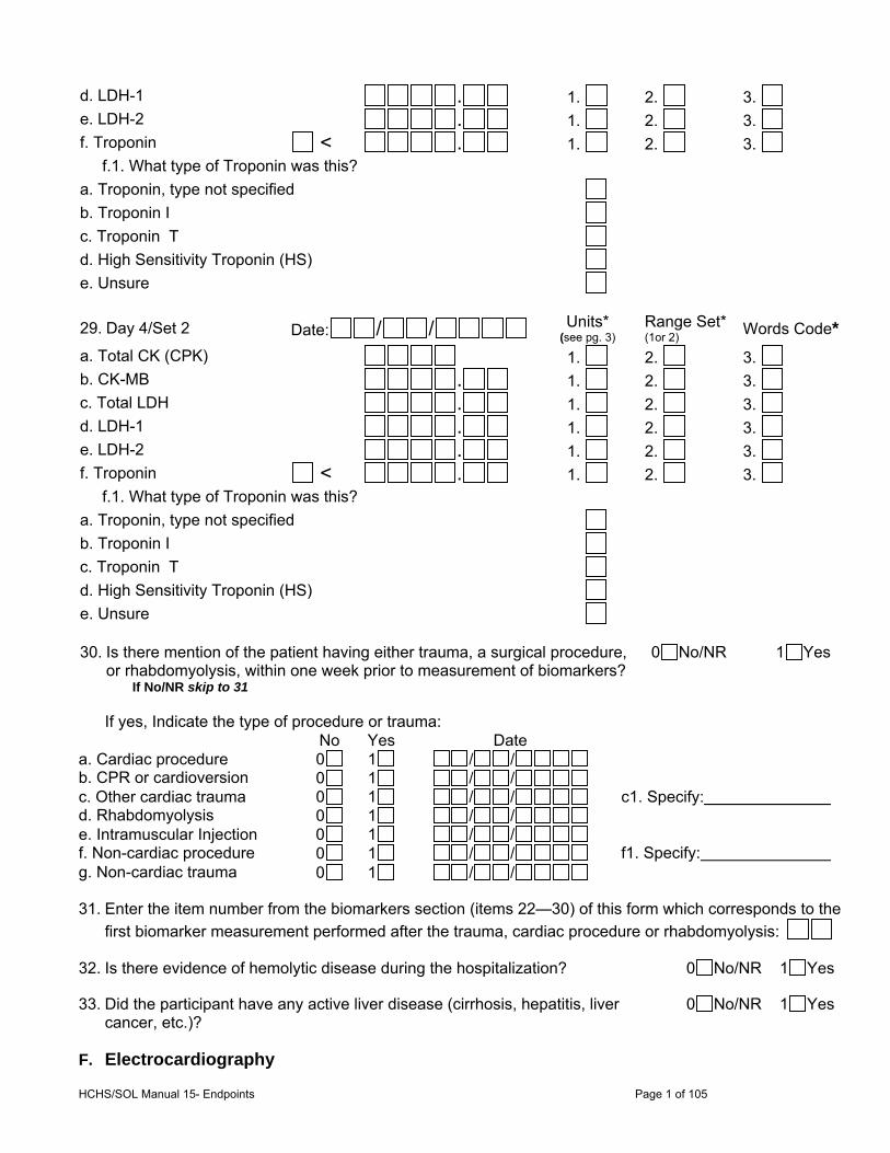
HCHS/SOL Manual 15- Endpoints Page 1 of 105
d. LDH-1 . 1. 2. 3. e. LDH-2 . 1. 2. 3. f. Troponin < . 1. 2. 3. f.1. What type of Troponin was this?
a. Troponin, type not specified
b. Troponin I c. Troponin T d. High Sensitivity Troponin (HS)
e. Unsure
29. Day 4/Set 2 Date: / / Units* (see pg. 3)
Range Set* (1or 2) Words Code*
a. Total CK (CPK) 1. 2. 3. b. CK-MB . 1. 2. 3. c. Total LDH . 1. 2. 3. d. LDH-1 . 1. 2. 3. e. LDH-2 . 1. 2. 3. f. Troponin < . 1. 2. 3. f.1. What type of Troponin was this?
a. Troponin, type not specified
b. Troponin I c. Troponin T d. High Sensitivity Troponin (HS)
e. Unsure
30. Is there mention of the patient having either trauma, a surgical procedure, or rhabdomyolysis, within one week prior to measurement of biomarkers? If No/NR skip to 31
0 No/NR 1 Yes
If yes, Indicate the type of procedure or trauma: No Yes Date a. Cardiac procedure 0 1 / / b. CPR or cardioversion 0 1 / / c. Other cardiac trauma 0 1 / / c1. Specify: d. Rhabdomyolysis 0 1 / / e. Intramuscular Injection 0 1 / / f. Non-cardiac procedure 0 1 / / f1. Specify: g. Non-cardiac trauma 0 1 / /
31. Enter the item number from the biomarkers section (items 22—30) of this form which corresponds to the
first biomarker measurement performed after the trauma, cardiac procedure or rhabdomyolysis:
32. Is there evidence of hemolytic disease during the hospitalization? 0 No/NR 1 Yes
33. Did the participant have any active liver disease (cirrhosis, hepatitis, liver cancer, etc.)?
0 No/NR 1 Yes
F. Electrocardiography
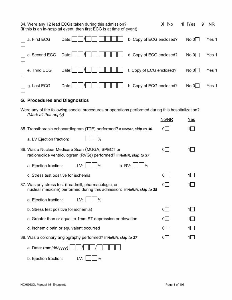
HCHS/SOL Manual 15- Endpoints Page 1 of 105
34. Were any 12 lead ECGs taken during this admission? 0 No 1 Yes 9 NR (If this is an in-hospital event, then first ECG is at time of event)
a. First ECG Date: / b. Copy of ECG enclosed? No 0 Yes 1
c. Second ECG Date: / d. Copy of ECG enclosed? No 0 Yes 1
e. Third ECG Date: / f. Copy of ECG enclosed? No 0 Yes 1
g. Last ECG Date: / h. Copy of ECG enclosed? No 0 Yes 1
G. Procedures and Diagnostics
Were any of the following special procedures or operations performed during this hospitalization? (Mark all that apply) No/NR Yes
35. Transthoracic echocardiogram (TTE) performed? If No/NR, skip to 36 0 1
a. LV Ejection fraction: %
36. Was a Nuclear Medicare Scan (MUGA, SPECT or 0 1 radionuclide ventriculogram (RVG)) performed? If No/NR, skip to 37
a. Ejection fraction: LV: % b. RV: %
c. Stress test positive for ischemia 0 1
37. Was any stress test (treadmill, pharmacologic, or 0 1 nuclear medicine) performed during this admission: If No/NR, skip to 38
a. Ejection fraction: LV: %
b. Stress test positive for ischemia) 0 1
c. Greater than or equal to 1mm ST depression or elevation 0 1
d. Ischemic pain or equivalent occurred 0 1
38. Was a coronary angiography performed? If No/NR, skip to 37 0 1
a. Date: (mm/dd/yyyy) / /
b. Ejection fraction: LV: %
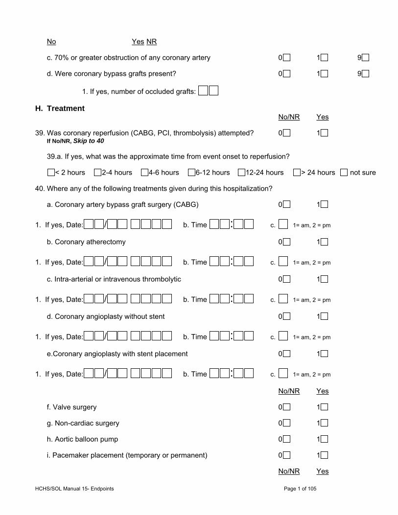
HCHS/SOL Manual 15- Endpoints Page 1 of 105
No Yes NR
c. 70% or greater obstruction of any coronary artery 0 1 9
d. Were coronary bypass grafts present? 0 1 9
1. If yes, number of occluded grafts:
H. Treatment No/NR Yes
39. Was coronary reperfusion (CABG, PCI, thrombolysis) attempted? 0 1 If No/NR, Skip to 40
39.a. If yes, what was the approximate time from event onset to reperfusion?
< 2 hours 2-4 hours 4-6 hours 6-12 hours 12-24 hours > 24 hours not sure
40. Where any of the following treatments given during this hospitalization?
a. Coronary artery bypass graft surgery (CABG) 0 1
1. If yes, Date: / b. Time : c. 1= am, 2 = pm
b. Coronary atherectomy 0 1
1. If yes, Date: / b. Time : c. 1= am, 2 = pm
c. Intra-arterial or intravenous thrombolytic 0 1
1. If yes, Date: / b. Time : c. 1= am, 2 = pm
d. Coronary angioplasty without stent 0 1
1. If yes, Date: / b. Time : c. 1= am, 2 = pm
e.Coronary angioplasty with stent placement 0 1
1. If yes, Date: / b. Time : c. 1= am, 2 = pm
No/NR Yes
f. Valve surgery 0 1
g. Non-cardiac surgery 0 1
h. Aortic balloon pump 0 1
i. Pacemaker placement (temporary or permanent) 0 1
No/NR Yes
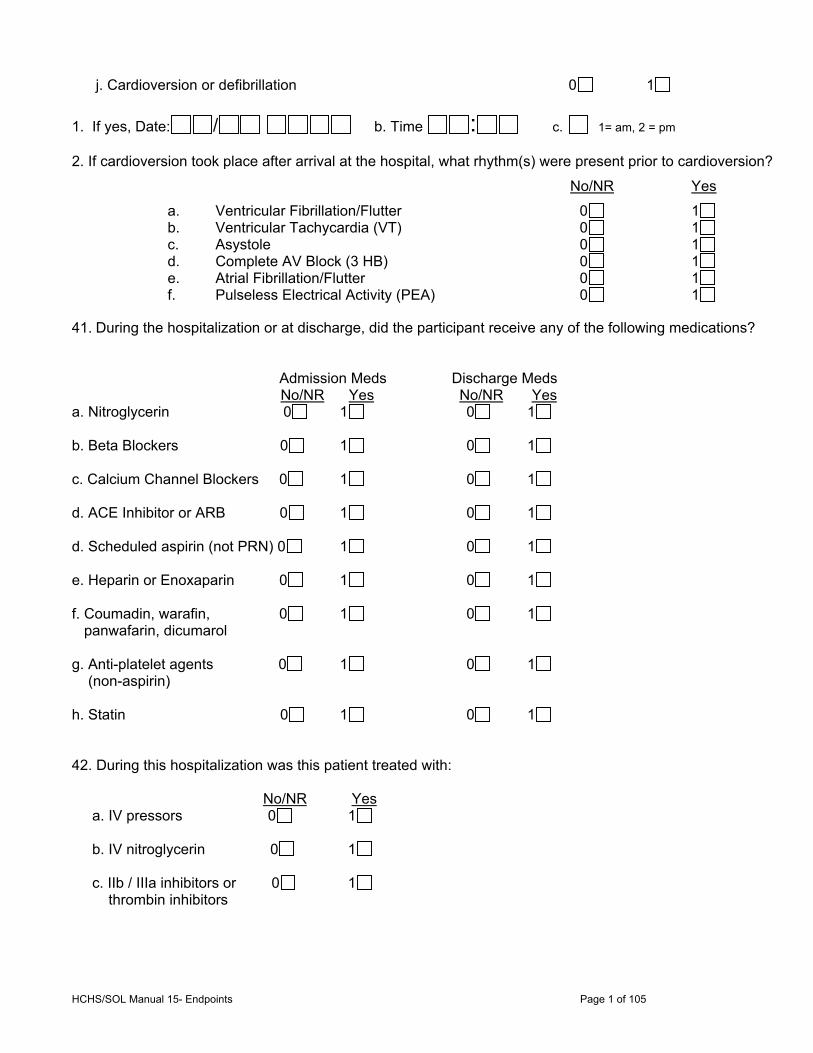
HCHS/SOL Manual 15- Endpoints Page 1 of 105
j. Cardioversion or defibrillation 0 1
1. If yes, Date: / b. Time : c. 1= am, 2 = pm
2. If cardioversion took place after arrival at the hospital, what rhythm(s) were present prior to cardioversion?
No/NR Yes
a. Ventricular Fibrillation/Flutter 0 1 b. Ventricular Tachycardia (VT) 0 1 c. Asystole 0 1 d. Complete AV Block (3 HB) 0 1 e. Atrial Fibrillation/Flutter 0 1 f. Pulseless Electrical Activity (PEA) 0 1
41. During the hospitalization or at discharge, did the participant receive any of the following medications? Admission Meds Discharge Meds No/NR Yes No/NR Yes a. Nitroglycerin 0 1 0 1 b. Beta Blockers 0 1 0 1 c. Calcium Channel Blockers 0 1 0 1 d. ACE Inhibitor or ARB 0 1 0 1 d. Scheduled aspirin (not PRN) 0 1 0 1 e. Heparin or Enoxaparin 0 1 0 1 f. Coumadin, warafin, 0 1 0 1 panwafarin, dicumarol g. Anti-platelet agents 0 1 0 1 (non-aspirin) h. Statin 0 1 0 1 42. During this hospitalization was this patient treated with: No/NR Yes a. IV pressors 0 1 b. IV nitroglycerin 0 1 c. IIb / IIIa inhibitors or 0 1 thrombin inhibitors
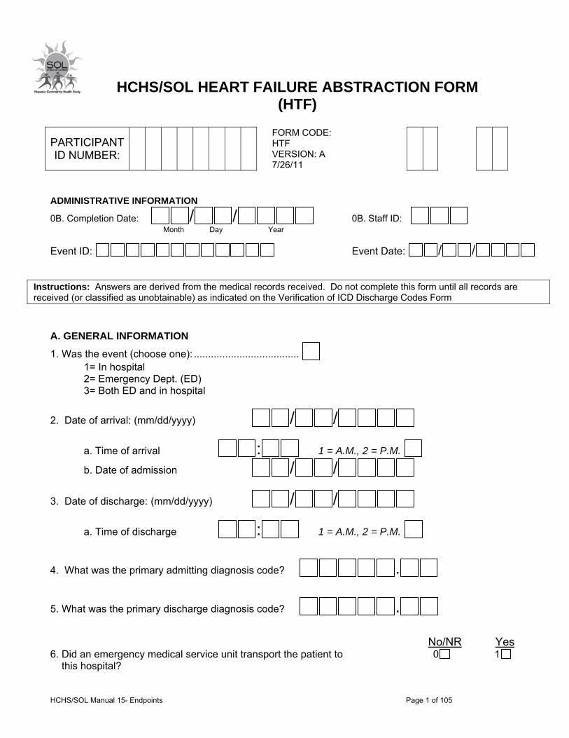
HCHS/SOL Manual 15- Endpoints Page 1 of 105
HCHS/SOL HEART FAILURE ABSTRACTION FORM (HTF)
ADMINISTRATIVE INFORMATION
0B. Completion Date: / / 0B. Staff ID:
Month Day Year
Event ID: Event Date: / /
Instructions: Answers are derived from the medical records received. Do not complete this form until all records are received (or classified as unobtainable) as indicated on the Verification of ICD Discharge Codes Form
A. GENERAL INFORMATION
1. Was the event (choose one): ..................................... 1= In hospital 2= Emergency Dept. (ED) 3= Both ED and in hospital
2. Date of arrival: (mm/dd/yyyy) / /
a. Time of arrival : 1 = A.M., 2 = P.M. b. Date of admission / /
3. Date of discharge: (mm/dd/yyyy) / /
a. Time of discharge : 1 = A.M., 2 = P.M.
4. What was the primary admitting diagnosis code? . 5. What was the primary discharge diagnosis code? . No/NR Yes 6. Did an emergency medical service unit transport the patient to 0 1 this hospital?
PARTICIPANT ID NUMBER:
FORM CODE: HTF VERSION: A 7/26/11

HCHS/SOL Manual 15- Endpoints Page 1 of 105
7. Was the patient transferred to this hospital from another hospital? 0 1 8. Was the patient’s code status ever “no-code” or “DNR” (do not resuscitate)? 0 1 9. Was the patient alive at discharge? 0 1 B. SIGNS AND SYMPTOMS I. Signs and Symptoms 10. Did the patient have any of the following signs or symptoms at the time of event ? No Yes NR Not recorded
a. An increase or new onset of paroxysmal nocturnal dyspnea (PND) 0 1 9 b. An increase or new onset of orthopnea? 0 1 9 c. An increase or new onset of shortness of breath? 0 1 9 d. An increase or new onset edema? 0 1 9 e. Hypoxia 0 1 9
f. Dyspnea (at rest) or tachypnea (RR>22) 0 1 9 g. Dyspnea (walking or on exertion) 0 1 9
II. Evidence in Physicians’ Notes of Reason for Event 11. Was there evidence in the doctor’s notes that the reason for this 0 1 9 event was an exacerbation of heart failure? 12. Did the patient have new onset or progressive signs/symptoms of heart failure prior to presentation in ED or hospital? 0 1 9 13. Did the physician’s note or discharge summary indicate the presence of any of the following specific types of heart failure? (check all that apply) No Yes
a. Diastolic heart failure 0 1
b. Systolic heart failure 0 1
c. Right-sided heart failure 0 1
d. Ischemic cardiomyopathy 0 1
e. Idiopathic/dilated cardiomyopathy 0 1
f. Myocarditis 0 1
HCHS/SOL Manual 15- Endpoints Page 1 of 105
g. Peripartum cardiomyopathy 0 1
h. Other specific cardiomyopathy/heart failure 0 1 1. If other cardiomyopathy, specify type ______________________________________________
III. Prior cardiac testing 14. Was cardiac imaging performed prior to this hospitalization?
No/NR 0 Yes 1 skip to 15
a. Lowest LV ejection fraction recorded: %
b. Qualitative description of ejection fraction: Normal…………….………… N Mildly reduced……………….M Decreased moderately……...D Severely reduced……………S None of the above…………..O Unsure-Not available..………U
c. Time (months) since recording of lowest ejection fraction: . months d. Type of Imaging from which ejection fraction was obtained:
1. ECHO 2. MUGA 3. Catheterization with ventriculography 4. CT 5. MRI 6. Other 7. Unknown
C. MEDICAL HISTORY No/NR Yes 15. Prior to this event was there a history of any of the following: a. Diagnosis of heart failure 0 1 b. Prior hospitalization for heart failure 0 1
c. Treatment for heart failure 0 1
d. Valvular heart disease 0 1
e. Rheumatic heart disease (RHD) 0 1
f. Congenital heart disease 0 1
g. Coronary heart disease (ever) 0 1
HCHS/SOL Manual 15- Endpoints Page 1 of 105
h. Coronary heart disease (within year) 0 1
i. Angina 0 1
j. Myocardial infarction 0 1
No/NR Yes
k. Atrial fibrillation/atrial flutter 0 1
l. Heart block or other bradycardia 0 1
m. Ventricular fibrillation or tachycardia 0 1
n. Hypertension 0 1
o. Diabetes 0 1
p. Chronic Obstructive Pulmonary Disease (COPD) 0 1
q. Cor pulmonale 0 1
r. Pulmonary hypertension 0 1 D. SURGICAL HISTORY 16. Past cardiac procedures No/NR Yes
a. CABG 0 1 b. Percutaneous coronary intervention (PCI) 0 1 c. Valve surgery 0 1 d. Pacemaker 0 1 e. Automatic Internal Cardiac Defibrillator (AICD) 0 1 f. Ablation for arrhythmia 0 1 g. Cardiac transplant 0 1 h. Ventricular Assist Device (VAD) 0 1 E. HOSPITAL COURSE 17. Current or Active Problems No/NR Yes

HCHS/SOL Manual 15- Endpoints Page 1 of 105
a. Myocardial Infarction 0 1
b. Shock or Cardiogenic Shock 0 1
c. Ventricular Fibrillation, Cardiac Arrest or Asystole 0 1
d. Ventricular Tachycardia 0 1
e. Atrial Fibrillation/Atrial Flutter 0 1
No/NR Yes
f. COPD exacerbation 0 1
g. Cardiac Surgery – CABG or valvular surgery 0 1
h. Non-cardiac surgery 0 1
i. Pulmonary Embolus 0 1
j. Pneumonia 0 1
F. PHYSICAL EXAM 18. Vital Signs at Admission (or at onset of event)
a. First available weight or BMI 1= weight in lbs 2= weight in Kg
3= BMI 19. Did the patient have any of the following signs? No Yes NR
Not recorded a. Jugular venous distension (JVD) 0 1 9 b. Heart/Lung sounds: 0 1 9
1. crackles or rales 0 1 9
2. wheezing 0 1 9
3. rhonchi 0 1 9
4. S3 gallop 0 1 9
c. Lower extremity edema-unilateral 0 1 9 d. Lower extremity edema-bilateral 0 1 9

HCHS/SOL Manual 15- Endpoints Page 1 of 105
G. DIAGNOSTIC TESTS 20. Was a chest X-ray performed during this event? No/NR 0 skip to 21 Yes 1
21. Did the patient have any of the following signs on chest x-ray at any time during this event? NoNR Yes
a. Pulmonary edema or CHF 0 1
b. Cardiomegaly or Cardiothoracic ratio > 0.5 0 1 No/NR Yes c. Pulmonary vascular congestion or Interstitial edema 0 1 d. Bilateral or unilateral pleural effusion 0 1
22. Was a chest/lung CT scan or CT angiogram (CTA) performed during this hospitalization? 0 1 23. Did the patient have any of the following signs on CT scan at any time during this hospitalization?
a. Pulmonary edema or pulmonary vascular congestion 0 1
b. Cardiomegaly 0 1 c. Bilateral or unilateral pleural effusion 0 1 d. Enlarged superior or inferior vena cava 0 1 e. Enlarged Pulmonary arteries 0 1
24. Was a transthoracic echocardiogram (TTE) performed? 0 1 Skip to 27 First transthoracic echocardiogram performed after onset of event:
a. Date (mm/dd/yyyy) / / b. Left Ventricular Ejection Fraction: % c. Record the following if present on transthoracic echocardiogram: None Mild Mod Severe NR 1. Left ventricular hypertrophy (LVH) 0 3 4 5 9
HCHS/SOL Manual 15- Endpoints Page 1 of 105
2. Impaired LV systolic function 0 3 4 5 9 3. Impaired RV systolic function 0 3 4 5 9 4. Pulmonary hypertension 0 3 4 5 9 5. Valvular heart disease 0 3 4 5 9 No/NR Yes
6. Diastolic dysfunction 0 1 7. Stress test positive for ischemia 0 1 8. Regional wall motion abnormalities 0 1 No/NR Yes 9. Dilated left ventricle 0 1 10. Dilated right ventricle 0 1
25. Was a transesophageal echocardiogram (TEE) performed? 0 1 skip to 26 First transesophageal echocardiogram performed after onset of event:
a. Date (mm/dd/yyyy) / / b. Ejection fraction: b.1. LV % b.2. RV % c. Record the following if present on transesophageal echocardiogram:
None Mild Moderate Severe NR
1. Impaired LV systolic function 0 3 4 5 9
2. Impaired RV systolic function 0 3 4 5 9 No/NR Yes
3. Regional wall motion abnormalities 0 1 4. Dilated left ventricle 0 1 5. Dilated right ventricle 0 1 6. Valvular heart disease 0 1
No/NR Yes

HCHS/SOL Manual 15- Endpoints Page 1 of 105
26. Was coronary angiography performed? 0 1 skip to 27
a. Date: (mm/dd/yyyy) / / b. LV Ejection fraction: % No/NR Yes
c. 70% or greater obstruction of any coronary artery 0 1 No/NR Yes 27. Was a cardiac multiple-gated acquisition scan (MUGA) or RVG performed? 0 1 skip to 28
a. Ejection fraction: LV: % b. RV: %
No/NR Yes c. Dilated ventricle or impaired ventricular function 0 1
28. Was a cardiac Magnetic Resonance Imaging (MRI) performed? 0 1 skip to 29
a. Ejection fraction: LV: % b. RV: % 29. Did any imaging/diagnostic test show:
a. Ejection fraction: LV: % No/NR Yes b. Stress test positive for ischemia? 0 1 c. Dilated ventricle or impaired ventricular function 0 1 d. Left ventricular diastolic dysfunction 0 1 e. Ventricular Septal Defect (VSD) 0 1 f. Atrial Septal Defect (ASD) 0 1 g. Patent Ductus Arteriosus (PDA) 0 1 h. Artificial heart valve 0 1 i. Hypertrophic Obstructive Cardiomyopathy (HOCM) 0 1 j. Valvular Heart Disease 0 1

HCHS/SOL Manual 15- Endpoints Page 1 of 105
H. LABORATORY TESTS a.Worst* b.Last c. Upper Limit Normal
30. BNP (pg/mL) . . .
31. ProBNP (pg/mL) . . .
32. Troponin < a. . c. < .
a. If troponin value available, then what type of Troponin was this? 1. Troponin, type not specified
2. Troponin I 3. Troponin T 4. High Sensitivity Troponin (HS)
5. Unsure a.Worst*
33. Sodium (mEq/L)
34. Serum creatinine (mg/dL) .
35. BUN (mg/dL) 36. Hemoglobin (g/dL) .
37. Hematocrit (%) .
*Worst = highest value with exception of hemoglobin, hematocrit and sodium. For these the worst is the
lowest value I. TREATMENTS 38. Were any of the following treatments given during this visit? No/NR Yes
a. Cardioversion or Defibrillation 0 1
b. Aortic balloon pump 0 1
c. Percutaneous coronary intervention (PCI) 0 1
d. CPAP or BIPAP 0 1 e. Mechanical Ventilation 0 1
f. Thoracentesis (therapeutic or diagnostic) 0 1
HCHS/SOL Manual 15- Endpoints Page 1 of 105
g. Ventricular Assist Device (VAD) 0 1 h. Heart transplant 0 1 J. MEDICATIONS Prior to hospitalization At discharge No/NR Yes No/NR Yes 39. ACE inhibitors 0 1 0 1 40. Angiotensin II receptor Blockers 0 1 0 1 41. Beta blockers 0 1 0 1 42. Digitalis 0 1 0 1 43. Diuretics 0 1 0 1 No/NR Yes 44. Aldosterone blocker 0 1 0 1 45. Lipid lowering agents 0 1 0 1 46. Nitrates 0 1 0 1 47. Hydralazine 0 1 0 1 48. IV drugs during this hospitalization? No/NR Yes a. IV inotropes 0 1 b. IV diuretics 0 1
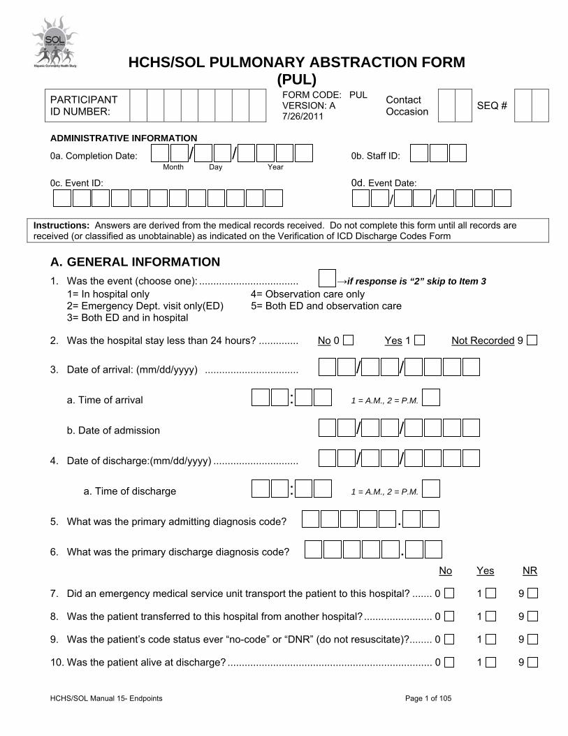
HCHS/SOL Manual 15- Endpoints Page 1 of 105
HCHS/SOL PULMONARY ABSTRACTION FORM (PUL)
ADMINISTRATIVE INFORMATION
0a. Completion Date: / / 0b. Staff ID:
Month Day Year
0c. Event ID: 0d. Event Date:
/ /
Instructions: Answers are derived from the medical records received. Do not complete this form until all records are received (or classified as unobtainable) as indicated on the Verification of ICD Discharge Codes Form
A. GENERAL INFORMATION
1. Was the event (choose one): ................................... →if response is “2” skip to Item 3 1= In hospital only 4= Observation care only 2= Emergency Dept. visit only(ED) 5= Both ED and observation care 3= Both ED and in hospital
2. Was the hospital stay less than 24 hours? .............. No 0 Yes 1 Not Recorded 9
3. Date of arrival: (mm/dd/yyyy) ................................. / /
a. Time of arrival : 1 = A.M., 2 = P.M.
b. Date of admission / /
4. Date of discharge:(mm/dd/yyyy) .............................. / /
a. Time of discharge : 1 = A.M., 2 = P.M.
5. What was the primary admitting diagnosis code? .
6. What was the primary discharge diagnosis code? .
No Yes NR
7. Did an emergency medical service unit transport the patient to this hospital? ....... 0 1 9
8. Was the patient transferred to this hospital from another hospital? ........................ 0 1 9
9. Was the patient’s code status ever “no-code” or “DNR” (do not resuscitate)?........ 0 1 9
10. Was the patient alive at discharge? ........................................................................ 0 1 9
PARTICIPANT ID NUMBER:
FORM CODE: PUL VERSION: A 7/26/2011
Contact Occasion
SEQ #
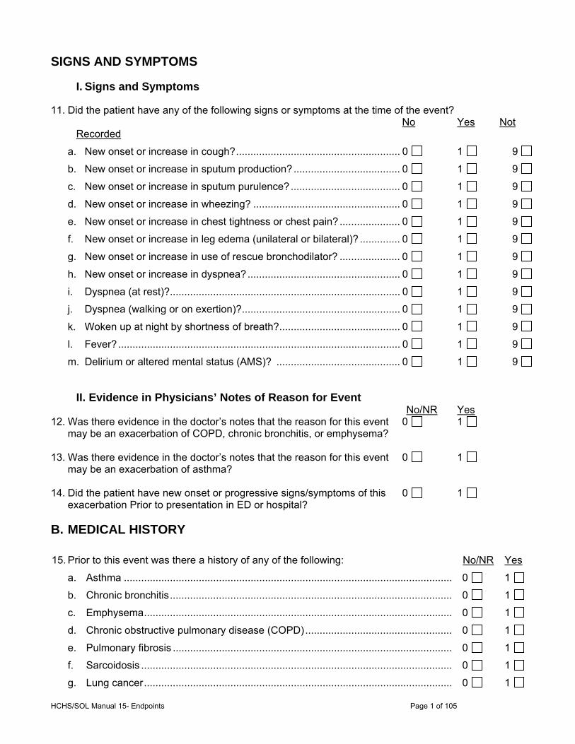
HCHS/SOL Manual 15- Endpoints Page 1 of 105
SIGNS AND SYMPTOMS
I. Signs and Symptoms
11. Did the patient have any of the following signs or symptoms at the time of the event? No Yes Not
Recorded
a. New onset or increase in cough? ......................................................... 0 1 9
b. New onset or increase in sputum production? ..................................... 0 1 9
c. New onset or increase in sputum purulence? ...................................... 0 1 9
d. New onset or increase in wheezing? ................................................... 0 1 9
e. New onset or increase in chest tightness or chest pain? ..................... 0 1 9
f. New onset or increase in leg edema (unilateral or bilateral)? .............. 0 1 9
g. New onset or increase in use of rescue bronchodilator? ..................... 0 1 9
h. New onset or increase in dyspnea? ..................................................... 0 1 9
i. Dyspnea (at rest)? ................................................................................ 0 1 9
j. Dyspnea (walking or on exertion)? ....................................................... 0 1 9
k. Woken up at night by shortness of breath? .......................................... 0 1 9
l. Fever? .................................................................................................. 0 1 9
m. Delirium or altered mental status (AMS)? ........................................... 0 1 9
II. Evidence in Physicians’ Notes of Reason for Event No/NR Yes
12. Was there evidence in the doctor’s notes that the reason for this event 0 1 may be an exacerbation of COPD, chronic bronchitis, or emphysema?
13. Was there evidence in the doctor’s notes that the reason for this event 0 1
may be an exacerbation of asthma? 14. Did the patient have new onset or progressive signs/symptoms of this 0 1
exacerbation Prior to presentation in ED or hospital?
B. MEDICAL HISTORY
15. Prior to this event was there a history of any of the following: No/NR Yes
a. Asthma .................................................................................................................. 0 1
b. Chronic bronchitis .................................................................................................. 0 1
c. Emphysema ........................................................................................................... 0 1
d. Chronic obstructive pulmonary disease (COPD) ................................................... 0 1
e. Pulmonary fibrosis ................................................................................................. 0 1
f. Sarcoidosis ............................................................................................................ 0 1
g. Lung cancer ........................................................................................................... 0 1
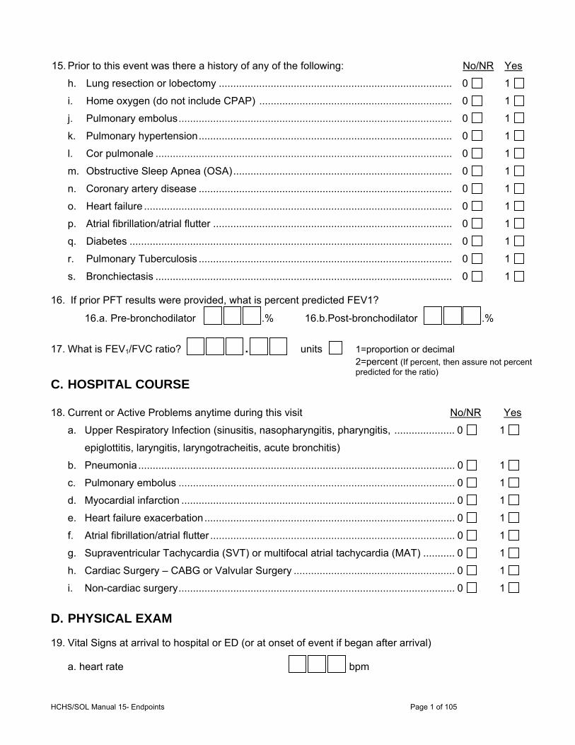
HCHS/SOL Manual 15- Endpoints Page 1 of 105
15. Prior to this event was there a history of any of the following: No/NR Yes
h. Lung resection or lobectomy ................................................................................. 0 1
i. Home oxygen (do not include CPAP) ................................................................... 0 1
j. Pulmonary embolus ............................................................................................... 0 1
k. Pulmonary hypertension ........................................................................................ 0 1
l. Cor pulmonale ....................................................................................................... 0 1
m. Obstructive Sleep Apnea (OSA) ............................................................................ 0 1
n. Coronary artery disease ........................................................................................ 0 1
o. Heart failure ........................................................................................................... 0 1
p. Atrial fibrillation/atrial flutter ................................................................................... 0 1
q. Diabetes ................................................................................................................ 0 1
r. Pulmonary Tuberculosis ........................................................................................ 0 1
s. Bronchiectasis ....................................................................................................... 0 1 16. If prior PFT results were provided, what is percent predicted FEV1?
16.a. Pre-bronchodilator .% 16.b.Post-bronchodilator .%
17. What is FEV1/FVC ratio? . units 1=proportion or decimal 2=percent (If percent, then assure not percent predicted for the ratio)
C. HOSPITAL COURSE
18. Current or Active Problems anytime during this visit No/NR Yes
a. Upper Respiratory Infection (sinusitis, nasopharyngitis, pharyngitis, ..................... 0 1
epiglottitis, laryngitis, laryngotracheitis, acute bronchitis)
b. Pneumonia .............................................................................................................. 0 1
c. Pulmonary embolus ................................................................................................ 0 1
d. Myocardial infarction ............................................................................................... 0 1
e. Heart failure exacerbation ....................................................................................... 0 1
f. Atrial fibrillation/atrial flutter ..................................................................................... 0 1
g. Supraventricular Tachycardia (SVT) or multifocal atrial tachycardia (MAT) ........... 0 1
h. Cardiac Surgery – CABG or Valvular Surgery ........................................................ 0 1
i. Non-cardiac surgery ................................................................................................ 0 1
D. PHYSICAL EXAM
19. Vital Signs at arrival to hospital or ED (or at onset of event if began after arrival)
a. heart rate bpm
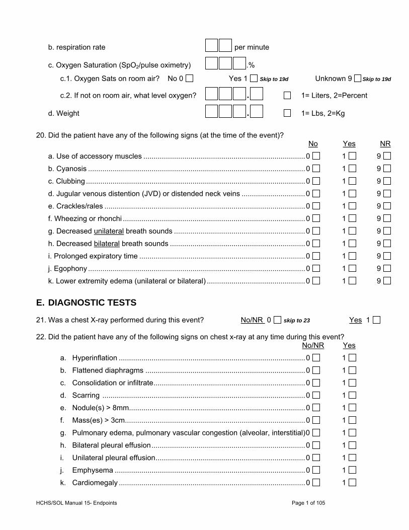
HCHS/SOL Manual 15- Endpoints Page 1 of 105
b. respiration rate per minute
c. Oxygen Saturation (SpO2/pulse oximetry) .%
c.1. Oxygen Sats on room air? No 0 Yes 1 Skip to 19d Unknown 9 Skip to 19d
c.2. If not on room air, what level oxygen? . 1= Liters, 2=Percent
d. Weight . 1= Lbs, 2=Kg
20. Did the patient have any of the following signs (at the time of the event)?
No Yes NR
a. Use of accessory muscles ............................................................................... 0 1 9
b. Cyanosis .......................................................................................................... 0 1 9
c. Clubbing ........................................................................................................... 0 1 9
d. Jugular venous distention (JVD) or distended neck veins ............................... 0 1 9
e. Crackles/rales .................................................................................................. 0 1 9
f. Wheezing or rhonchi ......................................................................................... 0 1 9
g. Decreased unilateral breath sounds ................................................................ 0 1 9
h. Decreased bilateral breath sounds .................................................................. 0 1 9
i. Prolonged expiratory time ................................................................................. 0 1 9
j. Egophony .......................................................................................................... 0 1 9
k. Lower extremity edema (unilateral or bilateral) ................................................ 0 1 9
E. DIAGNOSTIC TESTS
21. Was a chest X-ray performed during this event? No/NR 0 skip to 23 Yes 1 22. Did the patient have any of the following signs on chest x-ray at any time during this event?
No/NR Yes
a. Hyperinflation ........................................................................................... 0 1
b. Flattened diaphragms .............................................................................. 0 1
c. Consolidation or infiltrate .......................................................................... 0 1
d. Scarring ................................................................................................... 0 1
e. Nodule(s) > 8mm ...................................................................................... 0 1
f. Mass(es) > 3cm ........................................................................................ 0 1
g. Pulmonary edema, pulmonary vascular congestion (alveolar, interstitial) 0 1
h. Bilateral pleural effusion ........................................................................... 0 1
i. Unilateral pleural effusion ......................................................................... 0 1
j. Emphysema ............................................................................................. 0 1
k. Cardiomegaly ........................................................................................... 0 1

HCHS/SOL Manual 15- Endpoints Page 1 of 105
23. Was a chest/lung CT scan or CT angiogram (CTA) performed during this event?
No/NR 0 skip to 25 Yes 1
24. Did the patient have any of the following signs on CT scan at any time during this event?
No/NR Yes
a. Emphysema ................................................................................................... 0 1
b. Nodule(s) > 8mm ........................................................................................... 0 1
c. Mass(es) > 3cm ............................................................................................. 0 1
d. Lymphadenopathy .......................................................................................... 0 1
e. Ground glass changes ................................................................................... 0 1
f. Pneumonia ..................................................................................................... 0 1
g. Fibrosis or honeycombing .............................................................................. 0 1
h. Filling defect—vascular (PE) .......................................................................... 0 1
i. Filling defect—mucus plug ............................................................................. 0 1
j. Cysts or blebs ................................................................................................ 0 1
k. Atelectasis ...................................................................................................... 0 1
l. Calcifications .................................................................................................. 0 1
m. Pulmonary embolus ....................................................................................... 0 1
n. Enlarged pulmonary artery ............................................................................. 0 1
o. Bronchiectasis ................................................................................................ 0 1
p. Pulmonary edema or pulmonary vascular congestion ................................... 0 1
q. Cardiomegaly ................................................................................................. 0 1
r. Bilateral pleural effusion ................................................................................. 0 1
s. Unilateral pleural effusion ............................................................................... 0 1
t. Airway wall thickening .................................................................................... 0 1 25. Was spirometry (lung function testing) performed during this hospitalization?
No/NR 0 Skip to 26 Yes 1
a1. FEV1 . L a2. FEV1 Percent Predicted %
b1. FVC . L b2. FVC Percent Predicted %
c1. FEV1/FVC ratio . units 1=proportion or decimal
2=percent (If percent, then assure not percent predicted for the ratio)
HCHS/SOL Manual 15- Endpoints Page 1 of 105
26. Was post-bronchodilator spirometry measured? No/NR 0 Skip to 27 Yes 1
a1. FEV1 . L a2. FEV1 Percent Predicted %
b1. FVC . L b2. FVC Percent Predicted %
c1. FEV1/FVC ratio . units 1=proportion or decimal
2=percent (If percent, then assure not percent predicted for the ratio)
27. Was peak expiratory flow rate (PEFR or PEF) obtained at the time of event?
No/NR 0 Skip to 28 Yes 1
a. Date of first PEF(R) taken at time of event: (mm/dd/yyyy) / /
b. First PEF recording
c. Worst or lowest PEF recording (anytime during hospitalization) 28. Was peak expiratory flow rate (PERF or PEF) obtained at discharge?
No/NR 0 Skip to 29 Yes 1
a. Date of last PEF(R) taken at discharge: (mm/dd/yyyy) / /
b. Last PEF recording
29. Was a ventilation perfusion scan (VQ Scan) done? No/NR 0 Skip to 30 Yes 1
a. Ventilation perfusion scan results (record number of answer) 1. High probability 2. Intermediate probability 3. Low probability 4. No evidence of Pulmonary Embolus 5. Indeterminate 30. Was an echocardiogram (TTE or TEE) performed? No/NR 0 Skip to 31 Yes 1
If more than one ECHO performed, then use the worst value for each question
a. Ejection fraction: % b. RVSP (right ventricular systolic pressure) . mmHg
Record the following if present on echocardiogram:
None Present Mild Mod Severe NR
c. Right Ventricular Hypertrophy ........................ 0 1 2 3 4 9
d. Impaired RV systolic function ......................... 0 1 2 3 4 9
e. Pulmonary hypertension ................................ 0 1 2 3 4 9
f. Tricuspid Regurgitation ................................... 0 1 2 3 4 9
g. Diastolic dysfunction ...................................... 0 1 9
HCHS/SOL Manual 15- Endpoints Page 1 of 105
BIOCHEMICAL TESTS
31. White Blood Cell Count .
a. First (at event)
32. Hemoglobin (g/dL) .
33. Hematocrit (%) .
34. Sodium (mEq/L)
35. Serum creatinine (mg/dL) .
36. BUN (mg/dL) .
37. Bicarbonate (total CO2) .
a.First (at event) b.Upper limit normal
38. BNP (pg/mL) a. . b. .
39. ProBNP (pg/mL) a. . b. .
40. Were Arterial Blood Gases (ABGs) obtained? No/NR 0 Skip to 41 Yes 1
a.First blood gas (at time of event) b. Last blood gas
pH 1. .
1. .
PaCO2 2. . mmHg
2. . mmHg
PaO2 3. . mmHg
3. . mmHg
O2 Saturation 4. . %
4. . %
c. Blood gas on room air? No 0 Yes 1 Skip to 41 Not Recorded 9 Skip to 41
c.1. If not on room air, what level oxygen? . 1= Liters, 2=Percent
41. Was a sputum culture done? No/NR 0 Skip to 42 Yes 1
a. Culture Results Neg 0 Skip to 42 Pos 1 Not Recorded 9 Skip to 42
HCHS/SOL Manual 15- Endpoints Page 1 of 105
b. If yes, were any of the following reported in the sputum culture? No Yes
1. Haemophilus Influenzae ................................................................. 0 1
2. Moraxella Catarrhalis ..................................................................... 0 1
3. Streptococcus pneumoniae ............................................................ 0 1
4. Methicillin-resistant Staphylococcus Aureus (MRSA) .................... 0 1
5. Staphylococcus aureus (not MRSA) .............................................. 0 1
6. Mycoplasma pneumoniae .............................................................. 0 1
7. Pseudomonas Aureginosa ............................................................. 0 1
8. Chlamydophila (or Chlamydia) pneumoniae .................................. 0 1
9. Oropharyngeal flora ........................................................................ 0 1
10. Other__________________________________________________________
42. Was a blood culture done? No/NR 0 Skip to 43 Yes 1
a. Culture Results Neg 0 Skip to 43 Pos 1 Not Recorded 9 Skip to 43 b. If yes, were any of the following reported in the blood culture? No Yes
1. Haemophilus Influenzae ................................................................. 0 1
2. Moraxella Catarrhalis ..................................................................... 0 1
3. Streptococcus pneumoniae ............................................................ 0 1
4. Methicillin-resistant Staphylococcus Aureus (MRSA) .................... 0 1
5. Staphylococcus aureus (not MRSA) .............................................. 0 1
6. Other________________________________________________________________
43. Influenza swab Neg 0 Pos 1 Not Recorded 9
F. TREATMENTS / MEDICATIONS No/NR Yes
44. CPAP or BiPap .................................................................................................... 0 1
45. Mechanical Ventilation ......................................................................................... 0 1
46. Inhaled short-acting beta-agonists (ie,albuterol, xopenex) .................................. 0 1
47. Inhaled short-acting anticholinergics (ie, atrovent, ipratropium) .......................... 0 1
48. Nebulized Bronchodilators ................................................................................... 0 1
49. Magnesium injections in ED ................................................................................. 0 1
50. Oxygen (continuous or prn) ................................................................................. 0 1
51. IV Antibiotics ........................................................................................................ 0 1
52. Systemic Corticosteroid (IV or PO) ...................................................................... 0 1
53. IV Lasix or Furosemide ........................................................................................ 0 1
HCHS/SOL Manual 15- Endpoints Page 1 of 105
At onset of
time of event At discharge
No/NR Yes No/NR Yes
54. Antibiotics-oral ...........................................................a. 0 1
............ b. 0 1
55. Systemic corticosteroid (ie prednisone) .....................a. 0 1
............ b. 0 1
56. Inhaled short acting beta-agonists (ie albuterol) ........a. 0 1
............ b. 0 1
57. Inhaled long-acting beta-agonist (ie, serevent) ..........a. 0 1
............ b. 0 1
58. Inhaled short-acting anticholinergics (ie, atrovent) ....a. 0 1
............ b. 0 1
59. Inhaled long-acting anticholinergics ...........................a. 0 1
............ b. 0 1
60. Inhaled corticosteroids ...............................................a. 0 1
............ b. 0 1
61. Nebulized bronchodilators .........................................a. 0 1
............ b. 0 1
62. Leukotriene antagonist ...............................................a. 0 1
............ b. 0 1
63. Home oxygen .............................................................a. 0 1
............ b. 0 1

HCHS/SOL Manual 15- Endpoints Page 1 of 105
APPENDIX E Reviewer Forms Myocardial Infarction diagnosis form (MID) Heart Failure diagnosis form (HFD) Pulmonary diagnosis form (PLD)
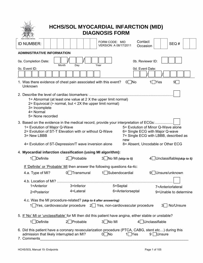
HCHS/SOL Manual 15- Endpoints Page 1 of 105
HCHS/SOL MYOCARDIAL INFARCTION (MID) DIAGNOSIS FORM
ADMINISTRATIVE INFORMATION
0a. Completion Date: / / 0b. Reviewer ID:
Month Day Year 0c. Event ID: 0d. Event Date:
/ / 1. Was there evidence of chest pain associated with this event? 0 No 1 Yes 9
Unknown
2. Describe the level of cardiac biomarkers: ........................................................................... 1= Abnormal (at least one value at 2 X the upper limit normal) 2= Equivocal (> normal, but < 2X the upper limit normal) 3= Incomplete 4= Normal 5= None recorded
3. Based on the evidence in the medical record, provide your interpretation of ECGs: ................ 1= Evolution of Major Q-Wave 5= Evolution of Minor Q-Wave alone 2= Evolution of ST-T Elevation with or without Q-Wave 6= Single ECG with Major Q-wave 3= New LBBB 7= Single ECG with LBBB, described as
new 4= Evolution of ST-Depression/T wave inversion alone 8= Absent, Uncodable or Other ECG
4. Myocardial infarction classification (using MI algorithm):
1 Definite 2 Probable 3 No MI (skip to 5) 4 Unclassifiable(skip to 5)
If ‘Definite’ or ‘Probable’ MI then answer the following questions 4a-4c:
4.a. Type of MI? 0 Transmural 1 Subendocardial 9 Unsure/unknown
4.b. Location of MI? .................................................................................................................. 1=Anterior 3=Inferior 5=Septal 7=Anteriorlateral
2=Posterior 4=Lateral 6=Anteriorseptal 9=Unable to determine
4.c. Was the MI procedure-related? (skip to 6 after answering)
1 Yes, cardiovascular procedure 2 Yes, non-cardiovascular procedure 3 No/Unsure
5. If ‘No’ MI or ‘unclassifiable’ for MI then did this patient have angina, either stable or unstable?
1 Definite 2 Probable 3 No MI 4 Unclassifiable
6. Did this patient have a coronary revascularization procedure (PTCA, CABG, stent etc…) during this admission that likely interrupted an MI? 0 No 1 Yes 9 Unsure
7. Comments_________________________________________________________________________
ID NUMBER: FORM CODE: MID VERSION: A 08/17/2011
Contact Occasion SEQ #
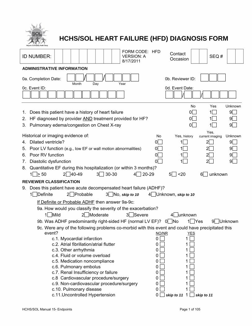
HCHS/SOL Manual 15- Endpoints Page 1 of 105
8.
HCHS/SOL HEART FAILURE (HFD) DIAGNOSIS FORM
ADMINISTRATIVE INFORMATION
0a. Completion Date: / / 0b. Reviewer ID:
Month Day Year 0c. Event ID: 0d. Event Date:
/ /
No Yes Unknown
1. Does this patient have a history of heart failure 0 1 9
2. HF diagnosed by provider AND treatment provided for HF? 0 1 9
3. Pulmonary edema/congestion on Chest X-ray 0 1 9
Historical or imaging evidence of: No Yes, history Yes,
current imaging Unknown
4. Dilated ventricle? 0 1 2 9
5. Poor LV function (e.g., low EF or wall motion abnormalities) 0 1 2 9
6. Poor RV function 0 1 2 9
7. Diastolic dysfunction 0 1 2 9
8. Quantitative EF during this hospitalization (or within 3 months)?
1 > 50 2 40-49 3 30-30 4 20-29 5 <20 6 unknown
REVIEWER CLASSIFICATION
9. Does this patient have acute decompensated heart failure (ADHF)?
1 Definite 2 Probable 3 No, skip to 10 4 Unknown, skip to 10
If Definite or Probable ADHF then answer 9a-9c:
9a. How would you classify the severity of the exacerbation?
1 Mild 2 Moderate 3 Severe 4 unknown
9b. Was ADHF predominantly right-sided HF (normal LV EF)? 0 No 1 Yes 9 Unknown
9c. Were any of the following problems co-morbid with this event and could have precipitated this event? NO/NR YES
c.1. Myocardial infarction 0 1 c.2. Atrial fibrillation/atrial flutter 0 1 c.3. Other arrhythmia 0 1 c.4. Fluid or volume overload 0 1 c.5. Medication noncompliance 0 1 c.6. Pulmonary embolus 0 1 c.7. Renal Insufficiency or failure 0 1 c.8 Cardiovascular procedure/surgery 0 1 c.9. Non-cardiovascular procedure/surgery 0 1 c.10. Pulmonary disease 0 1 c.11.Uncontrolled Hypertension 0 skip to 11 1 skip to 11
ID NUMBER: FORM CODE: HFD VERSION: A 8/17/2011
Contact Occasion SEQ #

HCHS/SOL Manual 15- Endpoints Page 1 of 105
10. If not definite or probable ADHF, then does this patient have Asymptomatic LV dysfunction (EF< 50%)?
0 No/NR 1 Yes 9 Unknown
11. Reviewer comments: ___________________________________________________________
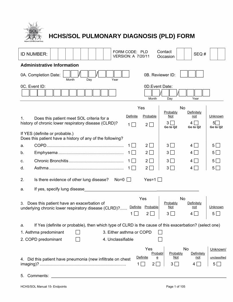
HCHS/SOL Manual 15- Endpoints Page 1 of 105
HCHS/SOL PULMONARY DIAGNOSIS (PLD) FORM
Administrative Information
0A. Completion Date: / / 0B. Reviewer ID:
Month Day Year
0C. Event ID: 0D.Event Date:
/ / Month Day Year
Yes No
1. Does this patient meet SOL criteria for a history of chronic lower respiratory disease (CLRD)?
Definite Probable Probably
Not Definitely
not Unknown
1 2 3 Go to Q2
4 Go to Q2
5 Go to Q2
If YES (definite or probable.) Does this patient have a history of any of the following?
a. COPD .................................................................. 1 2 3 4 5
b. Emphysema ........................................................ 1 2 3 4 5
c. Chronic Bronchitis ............................................... 1 2 3 4 5
d. Asthma ................................................................ 1 2 3 4 5
2. Is there evidence of other lung disease? No=0 Yes=1
a. If yes, specify lung disease_______________________________
3. Does this patient have an exacerbation of underlying chronic lower respiratory disease (CLRD)? ......
Yes No
Definite ProbableProbably
Not Definitely
not Unknown
1 2 3 4 5
a. If Yes (definite or probable), then which type of CLRD is the cause of this exacerbation? (select one)
1. Asthma predominant 3. Either asthma or COPD
2. COPD predominant 4. Unclassifiable
Yes No Unknown/
4. Did this patient have pneumonia (new infiltrate on chest imaging)? ..........................................................................
DefiniteProbabl
e Probably
Not Definitely
not unclassified
1 2 3 4 5
5. Comments:
ID NUMBER: FORM CODE: PLD VERSION: A 7/20/11
Contact Occasion SEQ #

HCHS/SOL Manual 15- Endpoints Page 1 of 105
Appendix F - Event Summary Forms Myocardial Infarction Event Summary Heart Failure Event Summary Pulmonary Event Summary
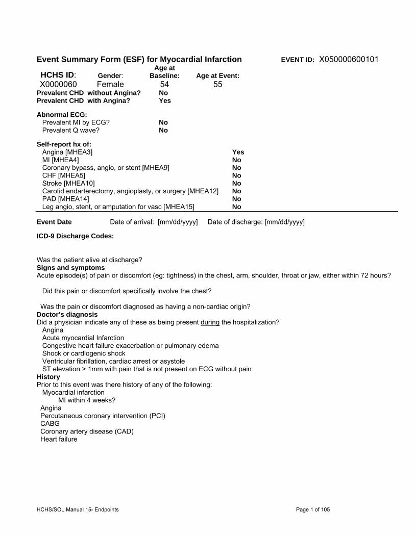
HCHS/SOL Manual 15- Endpoints Page 1 of 105
Event Summary Form (ESF) for Myocardial Infarction EVENT ID: X050000600101
HCHS ID: Gender: Age at
Baseline: Age at Event:X0000060 Female 54 55
Prevalent CHD without Angina? No Prevalent CHD with Angina? Yes Abnormal ECG: Prevalent MI by ECG? No Prevalent Q wave? No Self-report hx of: Angina [MHEA3] Yes MI [MHEA4] No Coronary bypass, angio, or stent [MHEA9] No CHF [MHEA5] No Stroke [MHEA10] No Carotid endarterectomy, angioplasty, or surgery [MHEA12] No PAD [MHEA14] No Leg angio, stent, or amputation for vasc [MHEA15] No Event Date Date of arrival: [mm/dd/yyyy] Date of discharge: [mm/dd/yyyy]
ICD-9 Discharge Codes:
Was the patient alive at discharge? Signs and symptoms Acute episode(s) of pain or discomfort (eg: tightness) in the chest, arm, shoulder, throat or jaw, either within 72 hours? Did this pain or discomfort specifically involve the chest? Was the pain or discomfort diagnosed as having a non-cardiac origin? Doctor’s diagnosis Did a physician indicate any of these as being present during the hospitalization? Angina Acute myocardial Infarction Congestive heart failure exacerbation or pulmonary edema Shock or cardiogenic shock Ventricular fibrillation, cardiac arrest or asystole ST elevation > 1mm with pain that is not present on ECG without pain History Prior to this event was there history of any of the following: Myocardial infarction MI within 4 weeks? Angina Percutaneous coronary intervention (PCI) CABG Coronary artery disease (CAD) Heart failure
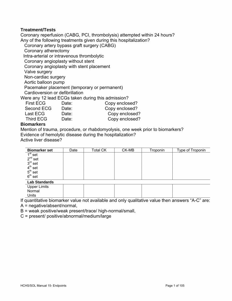
HCHS/SOL Manual 15- Endpoints Page 1 of 105
Treatment/Tests Coronary reperfusion (CABG, PCI, thrombolysis) attempted within 24 hours? Any of the following treatments given during this hospitalization? Coronary artery bypass graft surgery (CABG) Coronary atherectomy
Intra-arterial or intravenous thrombolytic Coronary angioplasty without stent Coronary angioplasty with stent placement Valve surgery Non-cardiac surgery Aortic balloon pump Pacemaker placement (temporary or permanent) Cardioversion or defibrillation Were any 12 lead ECGs taken during this admission? First ECG Date: Copy enclosed? Second ECG Date: Copy enclosed? Last ECG Date: Copy enclosed? Third ECG Date: Copy enclosed? Biomarkers Mention of trauma, procedure, or rhabdomyolysis, one week prior to biomarkers? Evidence of hemolytic disease during the hospitalization? Active liver disease?
Biomarker set Date Total CK CK-MB Troponin Type of Troponin 1st set 2nd set 3rd set 4th set 5th set 6th set
Lab Standards Upper Limits Normal
Units If quantitative biomarker value not available and only qualitative value then answers “A-C” are: A = negative/absent/normal, B = weak positive/weak present/trace/ high-normal/small, C = present/ positive/abnormal/medium/large

HCHS/SOL Manual 15- Endpoints Page 1 of 105
Event Summary Form (ESF) for Heart Failure EVENT ID: X00000080101
HCHS ID: Gender: Age at
Baseline: Age at Event:X0000008 Male 57 58
Self-report hx of: CHF [MHEA5] No Angina [MHEA3] No MI [MHEA4] No Coronary bypass, angioplasty, or stent [MHEA9] No Prevalent CHD without Angina? Yes Prevalent CHD with Angina? Yes Event Date Date of arrival: [mm/dd/yyyy] Date of discharge: [mm/dd/yyyy] ICD-9 Discharge Codes: Was the patient alive at discharge? Signs and symptoms An increase or new onset of paroxysmal nocturnal dyspnea (PND) An increase or new onset of orthopnea An increase or new onset of shortness of breath An increase or new onset edema Hypoxia Dyspnea (at rest) Dyspnea (walking or on exertion) New onset or progressive signs/symptoms of HF prior to presentation? History Prior to this event was there a history of any of the following: Diagnosis of heart failure Prior hospitalization for heart failure Treatment for heart failure Physician Diagnosis Evidence that event was exacerbation of HF? Ejection Fraction Prior to this hospitalization: During this hospitalization: TTE TEE Coronary angiography Radionuclide ventriculogram (RVG)
Chest X-ray Did the patient have any of the following signs on chest x-ray at any time during this event? Pulmonary edema or pulmonary vascular congestion Cardiomegaly or Cardiothoracic ratio > 0.5 Bilateral pleural effusion Unilateral pleural effusion
BNP Levels (include worst, upper limit normal) BNP (pg/mL)
ProBNP (pg/mL) Treatment with IV diuretic
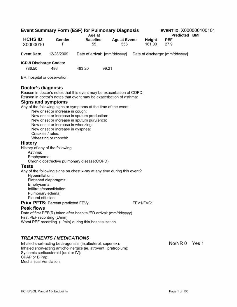
HCHS/SOL Manual 15- Endpoints Page 1 of 105
Event Summary Form (ESF) for Pulmonary Diagnosis EVENT ID: X000000100101
HCHS ID: Gender: Age at
Baseline: Age at Event: Height Predicted BMI PEF
X0000010 F 55 556 161.00 27.9 Event Date 12/28/2009 Date of arrival: [mm/dd/yyyy] Date of discharge: [mm/dd/yyyy] ICD-9 Discharge Codes:
786.50 486 493.20 99.21 ER, hospital or observation: Doctor’s diagnosis Reason in doctor’s notes that this event may be exacerbation of COPD: Reason in doctor’s notes that event may be exacerbation of asthma: Signs and symptoms Any of the following signs or symptoms at the time of the event:
New onset or increase in cough: New onset or increase in sputum production: New onset or increase in sputum purulence: New onset or increase in wheezing: New onset or increase in dyspnea: Crackles / rales: Wheezing or rhonchi:
History History of any of the following: Asthma: Emphysema: Chronic obstructive pulmonary disease(COPD): Tests Any of the following signs on chest x-ray at any time during this event? Hyperinflation: Flattened diaphragms: Emphysema: Infiltrate/consolidation: Pulmonary edema: Pleural effusion: Prior PFTS: Percent predicted FEV1: FEV1/FVC: Peak flows Date of first PEF(R) taken after hospital/ED arrival: (mm/dd/yyyy) First PEF recording (L/min) Worst PEF recording (L/min) during this hospitalization
TREATMENTS / MEDICATIONS Inhaled short-acting beta-agonists (ie,albuterol, xopenex): No/NR 0 Yes 1 Inhaled short-acting anticholinergics (ie, atrovent, ipratropium): Systemic corticosteroid (oral or IV): CPAP or BiPap: Mechanical Ventilation: