masterclass 6 july 2009 - university of nottingham
TRANSCRIPT

1
Masterclass
6 July 2009
Things You Can’t See

2
General Introduction
Welcome to the Masterclass – Things You Can’t See. This
information pack includes introductions to the people who will be working with you today, and summaries of the activities you will
participate in. It also contains important information about how to
keep safe during today’s activities.
Table of Contents
People involved 3
Safety and Responsibility 4
What is DNA? What does it look like? 8
Watching the Spread of Disease 9
An Introduction to Animal Parasites 10
Activated Porous Carbon for Separation and Gas storage 13
Further Information and Links 14
Timetable
Time Activity
11.00-11.15 Arrival/Housekeeping/Agenda
11.15-11.35 Mini lecture by Dr Aziz Aboobaker
11.40-12.40 Lab Session 1
12.45-1.15 Lunch and info on courses
1.20-1.40 Mini lecture by Prof John Brookfield
1.45-2.45 Lab Session 2
2.50-3.00 Q & A Session

3
People involved Prof John Brookfield studied for a BA in Zoology at the University of Oxford before going on to work at University College Swansea, the
National Institute of Environmental Health Sciences in North Carolina, and the University of Leicester. He then moved to the University of Nottingham where he is Professor of Evolutionary Genetics. His
research looks at genome evolution with a focus on the evolution of DNA sequences which control development, and on the evolution of mobile
repetitive DNA sequences. Prof Brookfield is also Vice-President (External Affairs) for the Genetics Society and Appointed Fellow of the
Institute of Biology.
Dr Aziz Aboobaker studied for a BA in Natural Sciences at the University of Cambridge before going on to complete a PhD. He has
also worked as a scientist in Edinburgh, Berkeley (California), and Nottingham. He is an evolutionary developmental biologist studying the
genetic basis for the diversity of animal body plans, given that so many of the genes that control what animals look like seem to be the same.
He uses flat worms in his research to look at the role of micro RNAs during the evolution of developmental processes.
Dr Jess Tyson is a Senior Research Fellow in the Institute of Genetics. She works in the field of Human Genetics, in particular her work looks at
DNA variation and its consequence. To prepare for this, she completed A levels in Biology, Chemistry, Maths and General Studies, did a degree in
Biological and Biochemical Sciences and completed a PhD on the
Genetics of Deafness.
Emma Langford completed Art and Design AS and Geography,
Chemistry and Biology A-levels before studying a degree in Microbiology (BSc Hons.) at the University of Nottingham. This led her to study
environmental microbiology at PhD level with an aim to reducing heavy metal pollution around a disused mine.
Eric Masika is a PhD student in Chemistry working on synthesis of
zeolite materials to use as a hard template for the highly sought after high surface area and morphologically controlled carbon materials. This course was preceded by a BSc in Chemistry at JKUAT- Kenya and an
MSc Nanoscience at the University of Nottingham.
Job de Roij is a PhD student in evolutionary biology. During his undergraduate degree in Zoology at the University of Nottingham he
developed an interest in the study of parasites and how they interact with their hosts (the organisms they infect). After taking a gap year to
go travelling, he returned to Nottingham to start a PhD looking at how parasites influence the evolution of different populations of three-spined
stickleback fish.
4
Safety and Responsibility
We want to make sure your visit to the University of Nottingham is as
safe and interesting as possible. Please read the notes below and, while on campus today, please ensure you follow all instructions from your
teachers and the researchers leading the activities.
Please be aware of where you and your belongings are at all times. In
every building you visit, you will be told where the nearest emergency exit is. If you hear an alarm, please make your way quickly and calmly
to the exit.
No bags or coats should be taken into laboratories. In laboratories, please do not touch any equipment unless you are specifically invited to
do so.
If at any point in the day you are injured, you must inform the researcher you are working with or another member of staff
immediately. Certified First Aiders will be available to help you.
Thank you very much. With your cooperation we can have a safe and productive day. A full risk assessment has been completed for today’s
activities. If you would like to see a copy, please ask.
Laboratory Safety
The laboratories are equipped and run so that with appropriate care,
you can work without risking the health and safety of yourself and others. Accidents are unexpected or undesirable events, but they are
avoidable with due care and attention.
You have a duty of care to work in a way that will not harm the health and safety of yourself and/or others.
All experimental procedures are assessed for risks, any necessary safety
measures put in place, and documentary evidence kept. All containers in the laboratory are labelled with their relevant hazards and any
equipment that may present a hazard or needs instruction before use will carry clear notices.
Ensure that you read any methods before and during the practical class, listen to instructions from staff and watch carefully any demonstrations
of experimental procedures.
REMEMBER: Any mishap with a chemical (or apparatus) MUST be reported to a member of staff IMMEDIATELY so that they can deal with
the problem and remove any hazards correctly.

5
Safety is an integral part of good laboratory practice. Even LOW HAZARD chemicals may be hazardous if misused. Risks from hazardous
chemicals are minimised by handling them correctly.
HAZARDOUS chemicals require careful handling at all times because of one or more of the following characteristics:
(a) flammability (b) explosive nature
(c) toxic, hazardous, irritant, etc: with effects on or through the skin.
with effects on or through the respiratory tract. with effects on or through the eye.
with effects following ingestion. (d) reactive with water.
(e) reactive with air. (f) detrimental effect on the environment – especially to
aquatic life. Containers of hazardous chemicals carry a pictogram indicating
the type of danger.
Toxic Highly Flammable Explosive Biological
Hazard
Corrosive Harmful, Irritant Oxidising
Ensure that you know how to deal with any spillage BEFORE you embark on an experiment.
Do not ignore the warning signs displayed in the School. They are there
for your protection. The British and European standard safety signs are used:
Prohibitory Signs: e.g. “No Smoking”, are circular with a red border and crossbar over a black symbol on a white
background.
Warning Signs: e.g. “Caution, risk of ionising radiation”, are
triangular with a black border on a yellow background.
(Yellow with black border/text)
(Orange with black text)

6
Mandatory Signs: e.g. “Eye Protection”, are circular on a blue background with symbols in white; used when there is
an obligation to wear safety equipment.
Emergency Signs: e.g. “Emergency Shower”, square or rectangular,
on a green background with symbols in white.
Fire doors must be kept closed at all times. Know where the fire-fighting, first-aid equipment, eye-wash and emergency showers are
situated.
Ensure that you know the location of fire exits.
In the event of a FIRE ALARM or an EMERGENCY EVACUATION of the laboratory, turn off all your equipment as you leave the laboratory and follow staff to the fire assembly point.
Working Safely in the Masterclass
Before commencing any operation in the laboratory give due care and
attention to how the procedure can be carried out without risk to yourself or fellow workers. If you are in doubt, consult a member of
staff before you start.
Risks from hazardous chemicals are minimised by handling them
correctly.
Check that the container has the correct chemical you require. Read the hazard information on the label before opening.
• Pour liquids carefully to avoid spillage and splashing.
• Containers should be opened with caution and away from the face. • Take care not to ingest or breathe in vapours or powders.
• Some containers should only be opened and the chemical used in a fume hood, especially for those likely to produce fumes or vapours.
• Handle any chemicals in the fume hood that the schedule advises. • Wear nitrile gloves when handling hazardous chemicals.
• Open wounds, e.g. cuts on hands, should be covered/protected. • Turn back over-long cuffs on clothes and laboratory coats to reduce
the chances of knocking over equipment or contaminating cuffs / sleeves.
• Do not put pens / fingers etc in mouths and do not eat or drink anything, including sweets or gum.
• Do not rub eyes / face with contaminated hands /nitrile gloves.

7
• Risks are minimised by working tidily, cleanly and avoiding spillages. • All spillages must be wiped up immediately.
• Dispose of all products and surplus chemicals correctly. • IF IN DOUBT – ASK
• Wash hands at the end of your laboratory session.
Spillages / accidents / FIRST AID • Spillages / splashes on the skin should be rinsed off immediately with
plenty of cold running water, then washed with soap and warm water.
• Any splashes in the eye(s) should be washed out immediately with an eye wash bottle and / or with plenty of cold running water.
• All cuts and scrapes on hands should be rinsed with cold running water.
• Any burns should be held under cold running water for at least 10 minutes.
Seek advice from a member of staff if you have an accident or if you feel unwell. A trained first aider will give advice, ensure any injuries are
treated and that relevant documentation is completed.
Safe use of equipment
Glassware
• Do not use any glassware that has chips or cracks – hand it to a member of staff for disposal.
• Take care when rinsing out glassware for reuse or to send for
washing – if there are any chipped edges or cracks – hand to a member of staff – do not rinse out.
• In the event of breakages, do not pick up the pieces of glass. Ask a member of staff to sweep up the pieces with a brush and dustpan.
Sharps The glass Pasteur pipettes and microscope slides should be handled with
care and disposed of in the 1.5% Trigene containers provided.

8
What is DNA? What does it look like? Jess Tyson
DNA is amazing. We have loads of the stuff. Inside almost every
cell in our body is a nucleus and inside each nucleus is about 2m of
DNA. Because the cell is very small, and because organisms have
many DNA molecules per cell, each DNA molecule must be tightly
packaged. This packaged form of the DNA is called a chromosome. DNA spends a lot of time in its chromosome form. But during cell
division, DNA unwinds so it can be copied and the copies transferred
to new cells. DNA also unwinds so that its instructions can be used to
make proteins and for other biological processes.
Before we can work on DNA in the
laboratory we need to be able to
extract it from whichever source,
(be it blood, buccal cells or tissue) that we have been given. DNA is
extracted from human cells for a
variety of reasons. With a pure
sample of DNA you can test a
newborn for a genetic disease for
example, or analyze forensic
evidence, or study a gene involved
in cancer.
Today, we will be extracting DNA,
but not from humans! Instead we will be using spinach and
strawberries as examples!
DNA extraction is typically the first step in a longer laboratory
process. Its an important part because DNA needs to be purified
away from proteins and other cellular contaminants. Once we’ve
extracted the DNA we will look at techniques to study DNA which are
used everyday in the research lab.

9
Watching the Spread of Disease Emma Langford
Swine ‘flu H1N1 is a very high profile health issue today. With the
number of cases increasing daily it has recently been announced as a
pandemic (globally distributed human-human transmitted disease).
Another pandemic the world is suffering from today is HIV, initially
transmitted to humans from simians.
But a key part of coping with any disease – be it on a global scale or
an outbreak of food poisoning from a local food outlet – is tracking
the spread of the disease and finding the initial point of infection. In this activity we will be looking at how quickly a disease can spread
through poor hygiene, and how the initial point of infection can be
identified on a small scale. We will do this using fluorescent dyes to represent micro-organisms and ‘infecting’ one of you, then tracking
the spread of the ‘infection’ through the group. We will also use
microscopy and culturing techniques to look at how the micro-
organisms (in this case bacteria not viruses) can be identified to confirm a disease.
At the end of the activity we will discuss some of the factors that
make real life epidemiology (studying the spread of disease) more
challenging than our simulation.
For more information on the number of swine ‘flu cases across the
globe look at
http://gamapserver.who.int/h1n1/atlas.html?select=ZZZ&filter=filter
4,confirmed.
For more information about swine ‘flu and why it was declared a
pandemic, look at http://www.who.int/mediacentre/news/statements/2009/h1n1_pand
emic_phase6_20090611/en/index.html.

10
An Introduction to Animal Parasites Job de Roij
Parasites are all around us. In fact, many biologists believe that they
make up over half of the total number of species on the planet. Every
animal species has at least one parasite species that infects it.
Parasites are organisms that live in or on another organism (the
host), obtaining from it part or all of its nutrients and causing some
degree of real damage to its host. Parasites include a range of
organisms, from microscopic viruses, fungi and bacteria to macroscopic worms and insects. The aim of today’s activity is to
introduce you to a range of parasites, by dissecting a three-spined
stickleback fish.
Three-spined stickleback fish are common in many areas of the
Northern Hemisphere such as North America and Europe. You often
find them in lakes, streams and rivers. However, they are originally a marine species. The main reason why they are interesting to study is
because since the last ice age, when sea levels changed dramatically,
they colonised many freshwater areas. As a result they have evolved
very rapidly into different body sizes and shapes, and are a great
example of natural selection in action. Below is a diagram that
captures just how diverse three-spined sticklebacks are:
A three-spined stickleback
Because they have evolved so much in terms of body size and body
shape, we think they may also have evolved differences in other
traits such as growth, how they reproduce and how well they resist
certain parasites. Sticklebacks are an important host for a number of
parasites, but the ones you will be coming across today are:
11
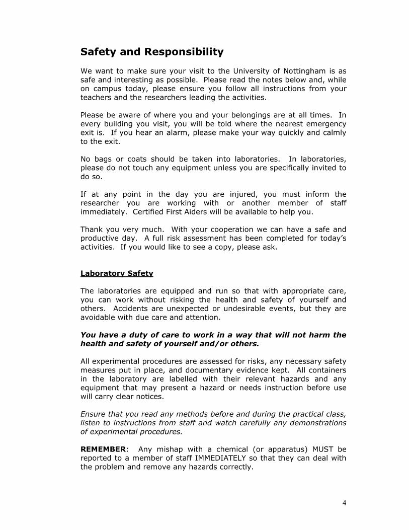
Schistocephalus solidus
- A tapeworm that lives in the body cavity
- The worms can grow to be extremely large - heavy infections
may make up 50% of the total body weight of the fish! Can
you imagine something similar in human beings?
- Fortunately it doesn’t exist, but it’s easy to picture why being
infected with this parasite has serious consequences for the
fitness of the fish
- It has been shown to slow down growth of the fish as well as
making it virtually impossible for female fish to develop eggs
and reproduce
- They also change the behaviour of sticklebacks, making them
less able to escape predator attacks. This is very handy for the
parasite, as it needs to be eaten by stickleback-eating birds to
complete its life cycle
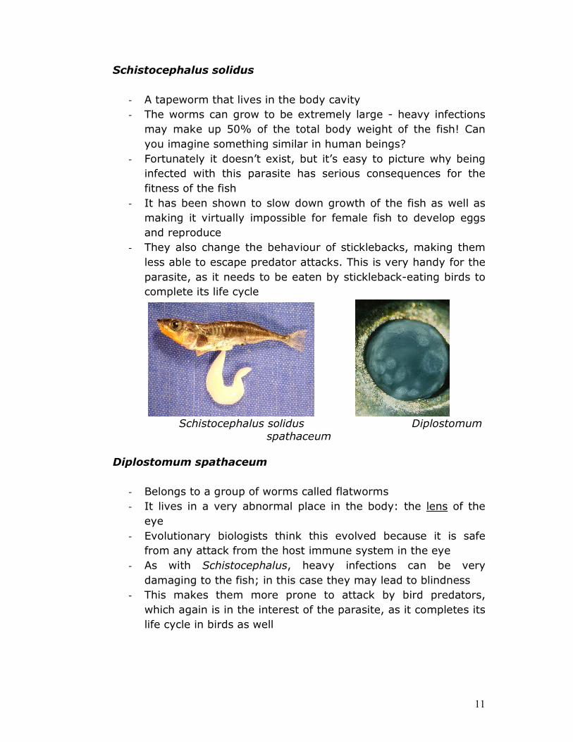
Schistocephalus solidus Diplostomum
spathaceum
Diplostomum spathaceum
- Belongs to a group of worms called flatworms
- It lives in a very abnormal place in the body: the lens of the
eye
- Evolutionary biologists think this evolved because it is safe
from any attack from the host immune system in the eye
- As with Schistocephalus, heavy infections can be very
damaging to the fish; in this case they may lead to blindness
- This makes them more prone to attack by bird predators,
which again is in the interest of the parasite, as it completes its
life cycle in birds as well

12
Gyrodactylus spp.
- Belong to a class of flatworms called Monogenea
- They are ectoparasites (‘ecto’ = outside) that live on the gills,
skin and fins of fish
- Infections with certain Gyrodactylus species can lead to very
high mortality
- Biologists are interested in studying gyrodactylid parasites
because they have a very unusual way of reproducing: each
worm is born with a developed embryo inside it, which itself
contains a developing embryo – for this reason they are named
the ‘Russian Doll’ killers!
Gyrodactylus salaris
There are also some microscopes slides with samples of human
parasites. Don’t worry: these parasites are dead and not infective.
They have been preserved in special chemical solution. These will
give you an idea of just how diverse parasitic worms can be.

13
Activated Porous Carbon for Separation and Gas storage
Eric Masika
Porous carbon materials with well ordered pores systems represent an exciting, intellectually challenging and rapidly expanding area of
research with many applications. They are of high performance in catalysis, separation, as energy storage materials and components of
electrodes. The growing list of application is attributed to the unique properties of the highly sought after materials such as high surface
area, large pore volume, chemical inertness and mechanical stability. It is desirable to control the morphology of the solid-templated porous
carbon materials, because the morphology (including particle size and shape) since it is a vital factor in the use of porous carbon on the
various applications. Though, it is not possible to create carbon frameworks on its own necessitating the use of scaffolds. In this case,
the use of zeolite materials with generous structural composition and morphologically tuneable has provided a timely solution making it an
area of intensive research in the recent past.
The practical use of porous carbon materials demonstrated in this work is separation in which students will be exposed to how carbon materials
carbon are used as Molecular Sieves. The diagram below illustrates simple filtration process using carbon as a sieve.
Based on the unique properties of these materials, demonstration using
potassium permanganate is used to show how molecules can be adsorbed. Then students will then use food colours to investigate what happens when activated carbon is used as a sieve. In addition, will
investigate whether it is possible to retrieve the dyes from carbon materials which make the process complete in the case of hydrogen
storage as reversibility is crucial for hydrogen economy to scale up.
WE
ARE
BIG
we are small

14
Further Information and Links
Thank you for coming to the Masterclass! We hope you enjoyed the
day!
For further information on anything you have seen today or details
about studying at the University of Nottingham, please email
[email protected], or go to the project
website at www.nottingham.ac.uk/sop.
For additional information about any of the departments involved in
the Science Outreach Project or today’s event, check out the
following webpages:
School of Biology: www.nottingham.ac.uk/biology
School of Biosciences: www.nottingham.ac.uk/biosciences
School of Chemistry: www.nottingham.ac.uk/chemistry