materials and methods - fred hutchresearch.fhcrc.org/content/dam/stripe/shou/files/2004...materials...
TRANSCRIPT

Materials and Methods
Strain construction, materials, and Net1 mutagenesis
All strains used are in the W303 background (can1-100, leu2-3, his3-11, trp1-1,
ura3-1, ade2-1) except where noted in the strain table (Supplementary Table 1). A strain
expressing a stable form of Clb2 lacking both the KEN and destruction boxes
(Clb2C2DK100)HA3 was used in over-expression experiments with Clb2 (1).
Net1 mutant constructs were created as previously described (2). Briefly, a wild
type NET1-myc9 epitope tagged construct was cloned into a modified pRS304 vector
containing 300bp upstream of the ATG translation start site using NcoI and EagI. Site-
directed mutagenesis of Serine/Threonine to Alanine was carried out using QuikChange
Site-Directed mutagenesis kit from Stratagene (La Jolla, CA). The indicated
Serine/Threonine were mutated to Alanine in Net1-13m
(166,169,212,231,252,259,356,362,384,385,497,611,676), Net1-6m
(166,169,212,231,252,259), Net1-3-Cdk (166,212,252), and Net1-6Cdk (62*,
166,212,252,297,304) where * indicates that residue 62 was mutated to ensure complete
elimination of all Cdk consensus sites even though it was not determined to be
phosphorylated in vivo. Mutagenesis was confirmed using restriction digests followed by
DNA sequencing. All constructs were targeted by linearization with BstXI to the trp1
locus in a NET1/net1::his5+ heterozygous diploid. The strains were sporulated and
tetrads were dissected to obtain a haploid isolate of the integrant over net1::his5+. Copy
number of integrants was estimated by normalizing extract protein from transformed and
wild type cells and blotting for Net1 levels. Proper localization of all net1 phosphosite

mutants was confirmed by indirect immuno-fluorescence against the myc epitope (data
not shown).
Production and purification of antibodies made against phosphorylated peptides
was performed by Abgent (San Diego, CA). A pair of peptides – phosphorylated or not
at the indicated, underlined residue – was synthesized to generate and purify each of the
following antibodies against Net1: anti-phosphopeptide A corresponding to aa 159-173
(RSKLNNGSPQSVQPQC); anti-phosphopeptide B corresponding to aa 205-219
(NGSMRVWTPLARQIYC); and anti-phosphopeptide C corresponding to aa 245-259
(PPPTQPQSPPIRISSC). All peptides contained a Cysteine at the C-terminus, and anti-
phosphopeptide B was modified by replacing Serine with Tryptophan at the –1 position
relative to the underlined Threonine to optimize the phosphoepitope presentation.
Antibodies specifically reactive against the phosphopeptides were positively selected on a
resin derivatized with the phosphopeptide immunogen and negatively selected by passage
through a resin derivatized with the unphosphorylated version of the peptide. Anti-
phosphopeptide B (α-PP-B) was used in all experiments described since it generated the
strongest signal against Phospho-Net1 (Fig. 2A).
Cell Growth and Synchronization Procedures
Cells were grown in yeast extract-peptone (YP) or in yeast minimal (YM) media
containing 2% glucose (YPD,YMD), 2% raffinose (YPR,YMR) or 2% galactose
(YPG,YMG) as carbon source. Where appropriate, minimal media were supplemented
with leucine, histidine, tryptophan, uracil, and adenine to complement auxotrophies.
Synchronization of cells in G1 phase was achieved with α-factor added at 10 µg/ml for

BAR1 cells and 0.1 µg/ml for bar1∆ cells for at least 3 hrs at 25°C. Cells were judged to
be arrested when greater than 90% of cells displayed the elongated "shmoo" phenotype.
Cells were released from α factor by filtration through a 0.2µm filter followed by a wash
with 150 ml of YP, then resuspended in the desired volume at a density of 1 O.D.600/ml.
For elutriation, cells were grown overnight in YP containing 2% raffinose and 2%
galactose and harvested at log phase. Elutriation was performed as described (3-5) for
the collection of small, unbudded G1 cells; contamination with budded cells was
measured to be no more than 2%. For galactose induction experiments, cells were grown
overnight in either YMR or YPR until an O.D.600 of 1.0 was reached, then induced with
2% galactose followed by time point collection.
Cell Extract Preparation and Western Blotting
Cells were grown to an O.D.600 of 1.0, and for every time point 2 ml of culture
was collected and TCA added to a final concentration of 20%. Cells were collected by
centrifugation and washed with 2 ml of Tris-HCl (pH 7.5). SDS loading buffer [70 µl of
100mM Tris-HCl (pH 7.5), 20% glycerol, 4% SDS, 2M Urea, 200mM DTT] was added,
tubes were boiled for 3 minutes, and 100µl of acid-washed glass beads (500µm) were
added to each tube followed by boiling for an additional 2 minutes. Tubes were vortexed
for 45 sec using a Bio 101 multi-bead vortexer at setting 5.5. Tubes were boiled again
for 2 minutes and 5µl of sample was fractionated on a 10% SDS-PAGE gel followed by
transfer to a nitrocellulose membrane. Western blot analysis was performed with the
following primary antibodies at the indicated dilutions: All anti-phospho Net1 antibodies
(α-PP-A, α-PP-B, α-PP-C) at 2µg/ml; anti-Clb2 (1:3000), anti-RPA190 (1:5000), anti-

Cdc28 (1:5000), anti-Clb3 (1:2000), anti-myc (9E10) (1:5000), anti-His (1:250), anti-HA
(1:5000), and anti-Cdc14 (1:1000).
Immunoprecipitation and Clb2–Cdk release/kinase assay.
To prepare extracts for immunoprecipitation, 10 O.D.600 units of a log phase cell
culture was harvested and washed with 2 ml of Tris-HCl (pH 7.5). Cells were re-
suspended in 500µl lysis buffer [25mM HEPES/KOH (pH7.5), 150mM NaCl, 1mM
DTT, 0.2% Triton, 1mM EDTA, 1mM PMSF, 1mM Benzamidine, 1x Protease Inhibitor
Cocktail (Aprotinin, Chymostatin, Leupeptin, and PepstatinA all at 5µg/ml in 90%
DMSO)], transferred to a flat-bottom 2 ml tube and supplemented with 100µl of acid-
washed glass beads (500µm). Samples were vortexed using a Bio 101 multi-beads
vortexer at setting 5.5 (speed) and 45sec (time). Tubes were then centrifuged for 5
minutes at 14,000 rpm and the supernatant was collected. Clarified extract (400µl) was
incubated with 60µl of 9E10-coupled protein A beads for 1 hour on a rotator at 4°C.
Beads were collected and washed ten times in wash buffer [25mM HEPES/KOH (pH7.5),
150mM NaCl, 1mM DTT, 0.2% Triton], and divided to approximately 15µl beads per
reaction condition. For protein kinase assays, 3µl of either Clb2–Cdk or Clb5–Cdk was
used with either 1µg of myc9-Net1 (purified from insect cells infected with a
recombinant baculovirus) (6), or 5µg of Histone H1. For assays that monitored release of
Cdc14 from bead-bound Net1-myc9, varying concentrations of in vitro assembled
Clb2–Cdk in 30µl kinase buffer [25mM Tris-HCl (pH 7.5), 10mM MgCl2, 1mM ATP,
1mM DTT, 0.1mg/ml BSA, 50mM NaCl] were mixed with 15µl 9E10 beads coated with
RENT complex. Reactions were allowed to proceed for 30 minutes on a rotator at 25°C.

Supernatant and beads were processed for Western blot analysis as previously described
(2). For in vitro assays with bacterially expressed constructs, approximately 116ng of
Net1 per 20µl of Ni+2-NTA beads, 10ng Cdc14, and 5µl of roughly 30ng/µl stock of
Clb2–Cdk was used per reaction.
Immuno-fluorescence and Cdc14 release quantification
Immuno-fluorescence was performed as previously described (2, 7). The analysis
of Cdc14 localization for (fig. S1) was performed in haploid cells carrying the
transposon-mutagenized net1 allele. Rabbit anti-Cdc14 (1/3000) and rat anti-tubulin
monoclonal antibody YL1/34 (1/1000) were used at the indicated dilutions. Images of
synchronized cells at 70-110 minutes following release from α factor were collected on a
Zeiss Axioskop or Axiovert 200M microscope using a Hamamatsu CCD digital camera.
Spindle length measurements were performed using Zeiss Axiovision software.

1
Supplementary fig. 1. Cell cycle-regulated binding site for Cdc14 resides in the N-terminal
half of Net1
The numbers adjacent to each construct indicate where a transposon insertion occurred in the
NET1 locus (8, 9) with the exception of the aa 1-621 HA-fragment of Net1 in which endogenous
NET1 was replaced by the truncated allele (RJD1783). The localization of Cdc14 and the length
of microtubule spindles in asynchronous cell populations were determined by indirect immuno-
fluorescence using anti-Cdc14 and anti-tubulin antibodies, respectively. Cell cycle position was
estimated from the length of the microtubule spindle. The hatched bars indicate fragments of
Net1 with proper nucleolar localization as reported in the TRIPLES database (9). ‘N+C’ refers
to both nuclear and cytoplasmic staining.
Supplementary fig. 2. Net1 phosphosite mutants are defective in Cdc14 release in early
anaphase.
(A) Mutant cdc15-2 cells carrying either net1-13m (RJD2611) or net1-6m (RJD2612) alleles
were synchronized with | factor at 25°C and released in Yeast-Peptone 2% glucose at 37°C.
Cells were collected for analysis by indirect immuno-fluorescence at 10 to 15 min time intervals.
Staining was performed with DAPI, anti-Cdc14, and anti-tubulin antibodies for determination of
nuclear position, Cdc14 localization, and spindle length, respectively. Cell outlines are indicated.
(B) Mutant cdc15-2 cells carrying the net1-6m allele were synchronized with | factor at 25°C
and released in Yeast-Peptone 2% glucose at 37°C. Cells collected at 70 to 110 min after |
factor release were double-labeled with anti-Cdc14 and anti-tubulin antibodies. Release of
Cdc14 from the nucleolus was determined to be either complete (black boxes) or partial (white

2
boxes; see legend to Fig. 1A) and was plotted against spindle length. Over 350 cells were
counted for each panel. The wild type NET1 control is shown in (Fig. 1A).
(C) Mutant cdc15-2 cells carrying net1-3Ax (an allele in which the 3 non-Cdk sites S169, S231,
and S259 from net1-6m were mutated to Alanine) were synchronized with | factor at 25°C and
released in Yeast-Peptone 2% glucose at 37°C. Cells collected at 70 to 110 min after | factor
release were double-labeled with anti-Cdc14 and anti-tubulin antibodies. Release of Cdc14 from
the nucleolus was determined to be either complete (black boxes) or partial (white boxes; see
legend to Fig. 1A) and was plotted against spindle length. Over 350 cells were counted for each
panel. The wild type NET1 control is shown in (Fig. 1A).
Supplementary fig. 3. Net1 phosphosite mutants are defective in Cdc14 release in early
anaphase.
(A) Defective release of Cdc14 from the nucleolus in early anaphase in Net1 mutants. Mutant
cdc15-2 cells carrying either NET1 (WT) (RJD2617), net1-3Cdk (3Cdk; RJD2613), or net1-6Cdk
(6Cdk; RJD2614) alleles were synchronized with | factor at 25°C and transferred to Yeast-
Peptone media containing 2% glucose at 37°C. Cells were collected at 10 to 15 min time
intervals and stained with DAPI, or antibodies to Cdc14 and tubulin to determine nuclear
position, Cdc14 localization, and microtubule spindle length, respectively. Cell outlines are
indicated.
(B) Enhanced temperature-sensitive growth phenotype of dbf2-2 and cdc15-2 combined with
net1-3Cdk and net1-6Cdk, respectively. Starting with 3000 cells, serially diluted with 3 volumes
of Yeast-Peptone media containing 2% glucose. Strains of dbf2-2 (RJD2625), dbf2-2 carrying a
net1-3Cdk allele (RJD2626), cdc15-2 (RJD2610), and cdc15-2 carrying a net1-6Cdk allele

3
(RJD2614) were spotted on YPD plates from right to left, and incubated at the indicated
temperature for 2-3 days. Two independent isolates of each strain were used. The first two
isolates in the 33.5°C panel were compiled from different sections of the same plate.
Supplementary fig. 4. Cdk sites are conserved in Net1 orthologs from different yeast
species.
Sequence alignment of S. cerevisiae Net1 and its orthologs from other budding yeasts reveals
that Cdk phosphorylation sites mapped in vivo are highly conserved. Percent identities refers to
exact matches in amino acid alignment for both sequences being compared divided by the total
sequence length of S. cerevisiae Net1. Percent positives refers to matches where an amino acid
difference exists between the two aligned sequences but both amino acids belong to the same
family (acidic, basic, uncharged polar, nonpolar) divided by the total sequence length of S.
cerevisiae Net1. Percent gaps refers to the number of spaces introduced into an alignment to
compensate for insertions and deletions in one sequence relative to another divided by the total
sequence length of S. cerevisiae Net1. Each parameter is shown for each species compared to
Net1 from Saccharomyces cerevisiae.
Supplementary fig. 5. Loading controls and immuno-fluorescence analysis of clb2 and
clb1 clb2 GAL1p-CLB2 cells.
(A) Phospho-specific antibodies to Net1. Phospho-specific antibodies were raised against three
peptides containing phosphorylated Serine166 (Anti-PP-A), Threonine 212 (Anti-PP-B), or
Serine 252 (Anti-PP-C). Crude extracts from cdc14-1 strains carrying either a wild type (+;
RJD2615) or a mutant (3Cdk; RJD2616) NET1-myc9 allele in which the three sites were

4
converted to alanine were fractionated by SDS-PAGE and immunoblotted with the different
antibodies. Anti-myc (9E10) detection of Net1-myc9 was used as a loading control.
(B) Clb2–Cdk phosphorylates Net1 on physiological sites. Same as in (Fig.2B) except
immunoblotted with antibodies to PP-C or myc.
(C) Loading controls using (anti-Cdc28) and Clb2 amounts (anti-Clb2) for (Fig. 2C).
(D) Loading controls using (anti-Cdc28) for (Fig. 2D).
(E) Samples of clb2 cells from (Fig. 2C) were subjected to indirect immuno-fluorescence with
anti-Cdc14 and anti-tubulin antibodies as previously described for the indicated time points to
monitor Cdc14 localization and spindle length, respectively.
(F) Samples of clb1 clb2 GAL1p-CLB2 cells from (Fig. 2D) were subjected to indirect
immuno-fluorescence with anti-Cdc14 and anti-tubulin antibodies as previously described for the
indicated time points.
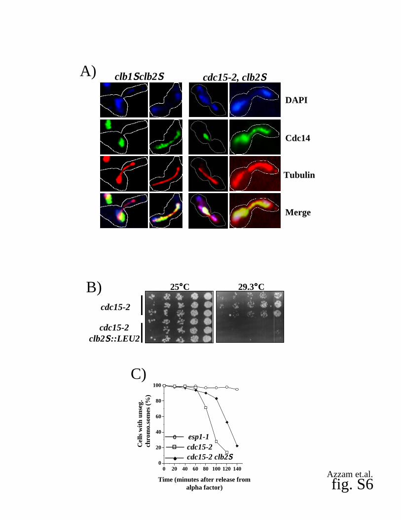
Supplementary fig. 6. Mutant clb2S cells display FEAR defects.
(A) Impaired release of Cdc14 from the nucleolus in early anaphase in clb1 clb2 and cdc15-2
clb2 mutants. Samples of clb1 clb2 GAL1p-CLB2 (RJD2624) and cdc15-2 clb2 cells (RJD
2622) were collected after elutriation/GAL-shutoff (RJD2624) or | factor block/release (RJD
2622) and stained with DAPI, anti-Cdc14, and anti-tubulin antibodies. Two panels are shown
for each mutant cell line, representing early anaphase (1st panel) and late anaphase (2nd panel).
Cell outlines are indicated.
(B) Mutant clb2 exacerbates the temperature-sensitive growth phenotype of MEN mutants.
Starting with 3000 cells, three-fold serial dilutions of cdc15-2 (RJD602) and cdc15-2 clb2 cells

5
(RJD2622) were spotted on Yeast-Peptone plates containing 2% glucose as described in the
legend of (fig. S3B). Three independent isolates of each strain were used.
(C) Mutant cdc15-2 clb2 cells arrest in late anaphase. Mutant cdc15-2 cells (open squares;
RJD2630), cdc15-2 clb2 cells (closed diamonds; RJD2631), and esp1-1 cells (open circles;
RJD2629) containing a tetO112 array at the URA3 locus and expressing 3tetR-GFP from the
HIS3 locus were synchronized in G1 phase with | factor and released at 37°C. Samples were
taken at the indicated time points to determine the percentage of cells with unsegregated (one
GFP dot) or segregated (two GFP dots) chromosomes.
Supplementary fig. 7. Cdc28 loading controls and cell cycle analysis of Cdc14 release and
spindle length for (Fig. 4, A and B).
(A) Cdc28 levels (anti-Cdc28) were used as a loading controls for the indicated time points and
strains for (Fig. 4A).
(B) Cdc28 levels (anti-Cdc28) were used as a loading controls for the indicated time points for
(Fig. 4B). Roman numerals refer to the following strain genotypes: (i) wild type (RJD2617), (ii)
cdc15-2 (RJD2610), (iii) cdc15-2 spo12 (RJD2620), and (iv) cdc15-2 slk19 (RJD2621).
(C) Cdc15 along with either Spo12 or Slk19 is required for proper release of Cdc14 from the
nucleolus during early anaphase. The same aliquots collected for (Fig. 4B) were also analyzed by
indirect immuno-fluorescence with anti-Cdc14 and anti-tubulin antibodies as previously described.
(D) Levels of Clb2 (anti-Clb2) and loading control (anti-Cdc28) for (Fig. 4C)
(E) Levels of Cdc5( DB) (anti-HA) and amount of Clb2 (anti-Clb2) for (Fig. 4D).
Supplementary Table 1. Net1 in vivo phosphorylation sites.

6
Supplementary Table 2. Strains used in this study.

fig. S1
Cdc14 Localization
nucleolus N+Cnucleolusnucleolusnucleolusnucleolus
nucleolus
N+CN+CN+CN+C
N+CN+C
N+C
N+C
N+C
N+C
N+C
G1 Anaphase
11791104
835
754738
621
259197
189
Net1
Azzam et.al.

DAPI
Cdc14
Tubulin
Merge
fig. S2
net1-13m
net1-6m
A) B)
DAPI Cdc14 Tubulin Merge
0
20
40
60
80
100
0-2 2-4 4-6 6-8 8-10 >10
Spindle length (µµµµm)
Cdc
14 r
elea
sed
(%)
net1-6m
Azzam et.al.
Spindle length (µµµµm)
Cdc
14 r
elea
sed
(%)
net1-3Ax
0
20
40
60
80
100
0-2 2-4 4-6 6-8 8-10 >10
C)

DAPI
Cdc14
Tubulin
Merge
WT 3Cdk 6CdkA)
dbf2-225°°°°C 33.5°°°°CB)
cdc15-230.7°°°°C25°°°°C
3Cdk dbf2-2
6Cdkcdc15-2 fig. S3
Azzam et.al.

fig. S4
S. cerevisiae T62P S166P T212P S252P S297P T304P %identities %positives %GapsS. bayanus + + + + + + 5 6 6 3 4
S. mikatae + + + + + + 6 0 6 6 2
S. paradoxus + + + + + + 6 6 6 9 1
S. castellii - - + + + + 2 9 4 2 2 0
S. kudriavzevii + + + + + + 6 0 6 5 2
Candida glabrata + - + + + + 3 6 5 0 1 2
Kluyveromyces lactis - - + + + + 3 1 4 4 1 9
Azzam et.al.

fig. S5 Azzam et.al.
Anti-Cdc28
Anti-Clb2
alph
a10 20 30 40 50 60 70 80 90 100
110
120
130C)
WT
(min)
Anti-Cdc28
Anti-Clb2clb2SSSS
Anti-Cdc28 Anti-Cdc28
120
135
150
165
0 180
195
210
225
240
255
270
285
300
Wild type
(min) 180
210
225
240
0 255
270
285
300
315
330
345
360
375
clb1SSSSclb2SSSS GAL1p-CLB2
(min)
D)
0
20
40
60
80
100
0 20 40 60 80 100 120 140
Time (minutes)
Cel
ls (
%)
clb2SSSS
E) % Cdc14 released% Anaphase Spindles% Metaphase Spindles
Wild type clb1SSSSclb2SSSS
0
20
40
60
80
100
0 50 100 150 200 250 300 350
Time (minutes after elutriation)
Cel
ls (
%)
% Cdc14 released%Anaphase Spindles
0
20
40
60
80
100
0 50 100 150 200 250 300 350 400
Time (minutes after elutriation)
% Cdc14 released%Anaphase Spindles
Cel
ls (
%)
F)
3Cdk +cdc14-1
Anti-PP-B
Anti-myc
Anti-PP-A
Anti-PP-C
A)
Anti-myc
Anti-PP-C
B)+ - ATP
C)
0
20
40
60
80
100
0 20 40 60 80 100 120 140
Time (minutes after release fromalpha factor)
Cel
ls w
ith
uns
eg.
chro
mo.
som
es(%
)
esp1-1
cdc15-2 clb2SSSScdc15-2
cdc15-2
cdc15-2clb2SSSS::LEU2
B) 25°°°°C 29.3°°°°C
fig. S6Azzam et.al.
cdc15-2, clb2SSSSclb1SSSSclb2SSSSA)
DAPI
Cdc14
Tubulin
Merge

fig. S7Azzam et.al.
WT
slk19SSSS
spo12SSSS
Anti-Cdc28
Anti-Cdc28
i
ii
iii
iv
A) B)|||| 15 30 45 60 70 80 90 100 110 120 135 150 180 (min)
|||| 10 20 30 40 50 60 70 80 90 100 110 120 130(min)
0
20
40
60
80
100
% C
ells
0 20 40 60 80 100 120 140 160 180
Time (minutes)
0
20
40
60
80
100%
Cel
ls
0 20 40 60 80 100 120 140 160 180
Time (minutes)
0
20
40
60
80
100
% C
ells
0 20 40 60 80 100 120 140 160 180
Time (minutes)
0
20
40
60
80
100
% C
ells
0 20 40 60 80 100 120 140 160 180
Time (minutes)
C)% Cdc14 Released
% Anaphase Spindles
(i) WT
(ii) cdc15-2
(iii) cdc15-2 spo12SSSS
(iv) cdc15-2 slk19SSSS
Cel
ls (
%)
Cel
ls (
%)
Cel
ls (
%)
Cel
ls (
%)
D)
Anti-Cdc28
Anti-Clb2 + 5 14 5, 14 + 5 14 5, 14
25°°°°C 37°°°°C
E)
Anti-Cdc28
Anti-HA
0 2 4 0 2 4GAL
induction (hr)
Wild type Cdc5(SSSSDB)

Table S1. Net1 in vivo phosphorylation sites.
Peptide Recovered a Net1 Sequence b
Mutated in
(13m) c
Mutated in
(6m) c
Mutated in
(3Cdk) c
Mutated in
(6Cdk) c,f
160-184 +1P R>SKLNNGSPQSVQPQQQIPSSSGVLR>Id
Y, Y Y,Y Y, N Y, N
210-216 +1P R>VSTPLAR>Q Y Y Y Y
226-241 +1P K>IVSNNSDDEDEDIGER>S Y Y N N
242-256 +1P R>SFLPPPTQPQSPPIR>I Y Y Y Y
257-265 +1P 257-266 +1P R>ISSGIDAGKK>I Y Y N N
292-307 +2Pe
R>LLSGTPIMSTMTPNR>V N, N N, N N, N Y, Y
352-359 +1P R>KPPVTTPR>I Y N N N
360-366 +1P R>ITSGM(ox)LK>I Y N N N
381-393 +1P 381-393 +2P K>EGPSSPASILPAK>A Y,Y N, N N, N N, N
495-505 +1P R>KSSLETIVEK>K Y N N N
606-618 +1P R>IVPQDSDSSSFPK>S Y N N N
670-678 +1P R>NILPQRTPR>S Y N N N
742-749 +1P K>DISLHSLK>G N N N N
1013-1023 +1P R>VVVNTPREPVR>S N N N N
1028-1038 +1P K>IEAPSPSVNKK>I N N N N
1079-1092 +1P K>VPRSLSSLSDLVSR>G N N N Na ‘+xP’ refers to the number of phosphate groups per peptide as determined by mass spectrometry.b Larger-sized font indicates mapped phosphorylation sites. Carets (>) mark sites of trypsin cleavage that yielded the peptides shown. ‘ox’ refers to oxidizedMethionine.c ‘Y’ indicates that the codon for a given site was mutated to encode Alanine, whereas ‘N’ indicates that it was not mutated.d Initial analysis was unable to determine which Serine was phosphorylated in the fragment spanning amino acids 160-184, but subsequent analysis with a phospho-specific antibody implicated Serine166. Phosphorylation status of Serine169 is unknown (underlined).e Yielded a small amount of a species at +80 Da from the doubly-charged form at the same retention time in the HPLC analysis, but we could not detect anymonophosphate in the precursor scans and it was later determined that this peptide was most likely phosphorylated on both Threonines as indicated.f In the (6Cdk) mutant, Threonine 62 was also mutated to Alanine to completely eliminate all Cdk consensus sites from the first 341aa N-terminal fragment of Net1.
Azzam et.al.

Azzam et.al.Table S2. Strains.
Strain name Strain Genotype Strain comments Used in FiguresRJD 2603 net1::mTn3/URA3 clone V13E1 with insertion point at aa(1104)* S1RJD 2604 net1::mTn3/URA3 clone V39B3 with insertion point at aa(835)* S1RJD 2605 net1::mTn3/URA3 clone V130G6 with insertion point at aa(754)* S1RJD 2606 net1::mTn3/URA3 clone V30E10 with insertion point at aa(738)* S1RJD 2607 net1::mTn3/URA3 clone V148D2 with insertion point at aa(259)* S1RJD 2608 net1::mTn3/URA3 clone V66G4 with insertion point at aa(197)* S1RJD 2609 net1::mTn3/URA3 clone V109B1 with insertion point at aa(189)* S1RJD 1783 net1::net1(1-621)-HA::KanMX6 * * S1
RJD 2610 net1S::his5, NET1-TEV-myc9::TRP1, cdc15-2 MATa4B, 1A, S3A,S3B, S8B
RJD 2611 net1S::his5, net1-13m-TEV-myc9::TRP1, cdc15-2, MATa 1A, S2ARJD 2612 net1S::his5, net1-6m-TEV-myc9::TRP1, cdc15-2, MATa S2A, S2BRJD 2613 net1S::his5, net1-3Cdk-TEV-myc9::TRP1, cdc15-2, MATa 1A, S3A, S3BRJD 2614 net1S::his5, net1-6Cdk-TEV-myc9::TRP1, cdc15-2, MATa 1A, S3A, S3BRJD 2615 net1S::his5, NET1-TEV-myc9::TRP1, cdc14-1, MATa S5ARJD 2616 net1S::his5, net1-3Cdk-TEV-myc9::TRP1, cdc14-1, MATa S5A
RJD 2617 net1S::his5, NET1-TEV-myc9::TRP1, MATa1B, 2B, 2C, 2D,4A, S5B,C,E,F
RJD 2618 net1S::his5, NET1-TEV-Myc9::TRP1, slk19S::KanMX6, MATa 4ARJD 2619 net1S::his5, NET1-TEV-Myc9::TRP1, spo12S::URA3, MATa 1B, 4ARJD 2620 net1S::his5, NET1-TEV-myc9::TRP1, cdc15-2, spo12S::URA3, MATa 4BRJD 2621 net1S::his5, NET1-TEV-myc9::TRP1, cdc15-2, slk19S::KanMX6, MATa 4BRJD 2622 clb2S::LEU2, cdc15-2, MATa S7A, S7BRJD 2623 net1S::his5, NET1-TEV-myc9::TRP1, clb2S::LEU2, MATa 2C, S5ERJD 2624 net1S::his5 , NET1-TEV-myc9::TRP1, clb1S::URA3, clb2S::LEU2, GAL1p-CLB2::URA3, MATa 2D, S7A, S5D,FRJD 2625 net1S::his5, NET1-TEV-myc9::TRP1, dbf2-2, MATa S3BRJD 2626 net1S::his5, net1-3Cdk-myc9::TRP1, dbf2-2,MATa S3BRJD 2627 net1S::his5, Net1-WT-myc9::TRP1, dbf2-2, YCH111 [CLB2 (C2Dk100)HA3, URA3], MATa S3BRJD 1349 NET1-TEV-myc9::his5+, bar1::hisG, MATalpha 3A, 3BRJD 1417 NET1-TEV-myc9::his5+, bar1::hisG, cdc5-1, MATa 4CRJD 1408 NET1-TEV-myc9::his5+, bar1::LEU2, cdc14-1, MATalpha 4CRJD 2628 NET1-TEV-myc9::his5+, cdc5-1, cdc14-1, bar1::hisG?, pep4::TRP1?, MATa 4CRJD 2629 tetO112 ::URA3x3, 3tetR-GFP::HIS3, esp1-1, MATa S7CRJD 2633 tetO112 ::URA3x3, 3tetR-GFP::HIS3, cdc15-2, MATa S7CRJD 2631 tetO112 ::URA3x3, 3tetR-GFP::HIS3, cdc15-2, clb2S::LEU2, MATa S7CRJD 602 cdc15-2, MATa S7BRJD 2632 net1S::his5, NET1-TEV-myc9::TRP1, cdc28-as1, MATa 2F, S5DRJD 2862 net1S::his5, net1-6Cdk-TEV-myc9::TRP1, MATa 1BRJD 2863 myc9-NET1::LEU2, bar1::hisG, GAL1p-CDC5(SDB)-HA::URA3, MAT a 4DRJD 2864 myc9-NET1::LEU2, pep4::TRP1, MAT a 4D*Y800 background (leu2-98, cry1(R), ade2-10,his3-200, ura3-52, lys2-801, can1(R), trp1-1, cyh2(R))**BJ5459 background (ura3-52, trp1, lys2-801, leu2D1, his3D200, pep4::HIS3, prb1D1.6R can1)

Supplemental References
1. C. Hendrickson, M. A. Meyn, 3rd, L. Morabito, S. L. Holloway, Curr Biol 11,1781-7 (Nov 13, 2001).
2. W. Shou et al., BMC Mol Biol 3, 3 (Apr 17, 2002).3. A. Amon, Methods Enzymol 351, 457-67 (2002).4. L. H. Johnston, A. L. Johnson, Methods Enzymol 283, 342-50 (1997).5. G. M. Walker, Methods Cell Sci 21, 87-93 (1999).6. W. Shou et al., Mol Cell 8, 45-55 (Jul, 2001).7. W. Shou et al., Cell 97, 233-44. (1999).8. N. Burns et al., Genes Dev 8, 1087-105. (1994).9. P. Ross-Macdonald, A. Sheehan, C. Friddle, G. S. Roeder, M. Snyder, Methods
Enzymol 303, 512-32 (1999).