materials and methods -...
TRANSCRIPT
Chapter IV
MATERIALS AND METHODS

43
MATERIALS AND METHODS
4.1 Materials
4.1.1 Plant Material and drug.
Acalypha indica Linn (Kuppamani) of the family Euphorbiaceae, the
plant material selected for the present study, was collected from the suburbs
of Thiruvananthapuram and Kanyakumari districts and got identified by
comparing with the herbarium specimen kept in the Ayurveda Research
institute at Poojapura, Thiruvananthapuram.
Acalypha indica is an annual herb, up to about 75 em height. Leaves
are 3-8 ern long, ovate, thin, usually 3-nerved; margins of the leaves
toothed; leaf stalks longer than leaves. Flowers are in axilary erect spikes;
female flowers supported by conspicuous wedge-shaped bracts; male
flowers are minute, borne towards the top of the spike.225,253 Fruits are
small, hairy, concealed in the bracts.
After preliminary screening and pilot study to detect the most
effective extract, specific quantity of alcohol extract obtained by successive
solvent extraction of the dried leaves was suspended in 1% carboxy methyl
cellulose (CMC) and used as the test drug for the programmed studies.
The standard drug, Silymarin, used in this study was obtained from
Maneesh pharmaceuticals Pvt. Ltd, Mumbai.

44
4.1.2 Reagents and chemicals
The reagents and chemicals used in this study and their sources are
given below.
Reagents/ Chemicals Distributor/ Manufacturer
Nitroblue Tetrazolium (NBT) Sisco Research Lab Pvt. Ltd. Mumbai
Reduced glutathione (GSH) Do
Glutathione reductase (GR) Do
Nicotinamide adenine Do
dinucleotide - phosphate reduced
tetrasodium salt
5,5-dithiobis(2-nitro benzoic acid) Do
(DTNB)
2-deoxy- D- ribose Sigma chemicals
Calf thymus DNA Do
yeast RNA Do
Thiobarbituric acid E.Merck. India Ltd.,
Mumbai
Carbon tetrachloride (CCI4) Do
Paracetamol LP. Kerala State Drugs and
Pharmaceuticals, Alleppy
All other chemicals used were of analytical grade.
45
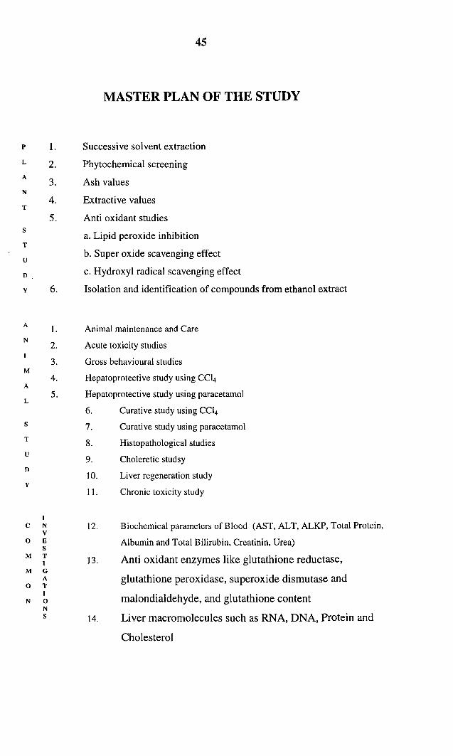
MASTER PLAN OF THE STUDY
p 1. Successive solvent extraction
L 2. Phytochemical screeningA
3. Ash valuesN
4. Extractive valuesT
5. Anti oxidant studiess
a. Lipid peroxide inhibitionT
ub. Super oxide scavenging effect
D c. Hydroxyl radical scavenging effect
y 6. Isolation and identification of compounds from ethanol extract
A I. Animal maintenance and CareN
2. Acute toxicity studies
3. Gross behavioural studiesM
4. Hepatoprotective study using CCl4A
5. Hepatoprotective study using paracetamolL
6. Curative study using CCl4
s 7. Curative study using paracetamolT 8. Histopathological studiesu
9. Choleretic studsyD
10. Liver regeneration studyy
11. Chronic toxicity study
IC N 12. Biochemical parameters of Blood (AST, ALT, ALKP, Total Protein,
v0 E Albumin and Total Bilirubin, Creatinin, Urea)
sM T
13. Anti oxidant enzymes like glutathione reductase,IM G
A glutathione peroxidase, superoxide dismutase and0 T
Imalondialdehyde, and glutathione contentN 0
Ns 14. Liver macromolecules such as RNA, DNA, Protein and
Cholesterol

46
4.2 Extraction and Preliminary Phytochemical Screening254.256
The plant may be considered a biosynthetic laboratory, not only for
the chemical compounds like carbohydrates, proteins and lipids that are
utilized as food by man, but also for a multitude of compounds like
glycosides, alkaloids, volatile oils, tannins etc. that exert a physiologic
effect. The compounds that are responsible for therapeutic effect are
usually the secondary metabolites. A systematic study of a crude drug
embraces thorough consideration of both primary and secondary
metabolites derived as a result of plant metabolism. The plant material may
be subjected to preliminary phytochemical screening for the detection of
various plant constituents on the following lines.
(A) Successive solvent extraction.
1. Thousand grams of the air-dried powdered plant material was
extracted successively with the following solvents in a Soxhlet
extractor.
(a) Petroleum ether (60-80°)
(b) Benzene
(c) Chloroform
(d) Acetone
(e) Ethanol (95%)

47
Each time before extracting with the next solvent, the powdered
material was dried in an air-oven below so'c.
2. Finally, the marc was macerated with chloroform water for 24 hours
to obtain the aqueous extract.
3. Each extract was concentrated by distilling off the solvent and then
evaporated to dryness on a water bath.
4. The extract obtained with each solvent was weighed and their
percentage calculated with reference to the air-dried material. The
colour and consistency was also noted.
Qualitative chemical examination
The extracts obtained as above were then subjected to qualitative
tests for the identification of various plant constituents.
1. Detection of alkaloids
A small portion of the solvent free chloroform, alcoholic and
water extracts were separately stirred with a few milliliters of dilute
hydrochloric acid and filtered. The filtrates were tested carefully with
various alkaloidal reagents.
a) Mayer's test: To 2 ml each of the acidified extracts added 0.5 ml of
Mayer's reagent and noted for the presence of a cream precipitate.
b) Dragendorff's test: The acidified extracts (2ml) were treated with a
few drops of Dragendorff's reagent and observed for the presence of
an orange brown precipitate.
48
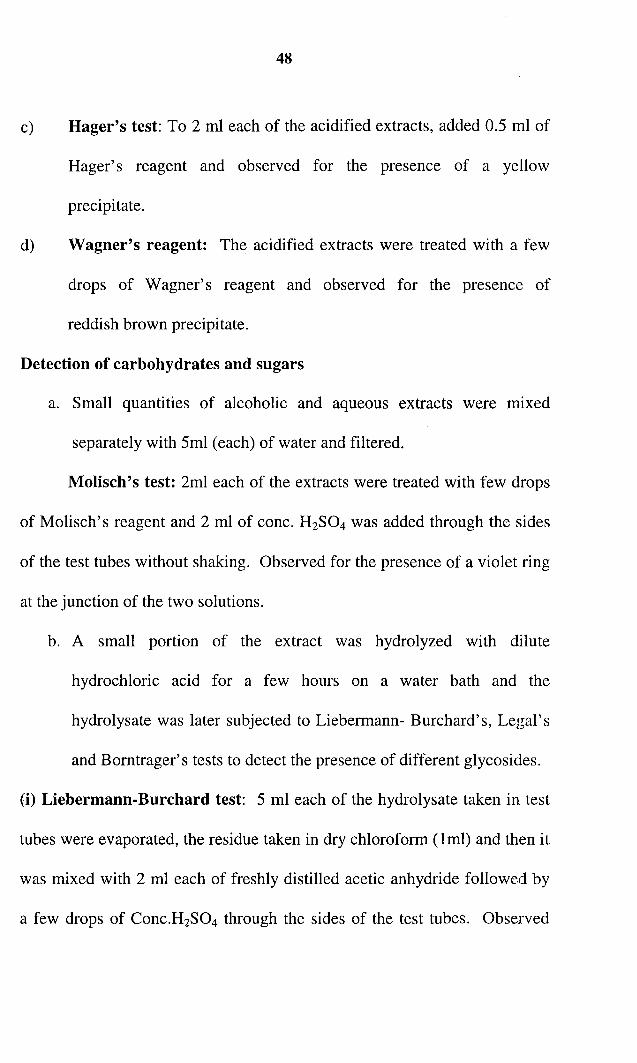
c) Hager's test: To 2 ml each of the acidified extracts, added 0.5 ml of
Hager's reagent and observed for the presence of a yellow
precipitate.
d) Wagner's reagent: The acidified extracts were treated with a few
drops of Wagner's reagent and observed for the presence of
reddish brown precipitate.
Detection of carbohydrates and sugars
a. Small quantities of alcoholic and aqueous extracts were mixed
separately with 5ml (each) of water and filtered.
Molisch's test: 2ml each of the extracts were treated with few drops
of Molisch's reagent and 2 ml of cone. H2S04 was added through the sides
of the test tubes without shaking. Observed for the presence of a violet ring
at the junction of the two solutions.
b. A small portion of the extract was hydrolyzed with dilute
hydrochloric acid for a few hours on a water bath and the
hydrolysate was later subjected to Liebermann- Burchard's, Legal's
and Borntrager's tests to detect the presence of different glycosides.
(i) Liebermann-Burchard test: 5 ml each of the hydrolysate taken in test
tubes were evaporated, the residue taken in dry chloroform (lml) and then it
was mixed with 2 ml each of freshly distilled acetic anhydride followed by
a few drops of Conc.H2S04 through the sides of the test tubes. Observed

49
the development of a deep red colour in the lower portion and green colour
in the upper portion which changes to blue and violet.
(ii) Legal's test: The residue left after evaporation was dissolved in a few
ml of hydrolysate in 2 ml of pyridine. Added 2 ml each of sodium
nitroprusside solution to each test tube and then made alkaline with sodium
hydroxide solution; observed for pink to red colour.
(iii) Borntrager's test: A little of the residue obtained from the
hydrolysate was dissolved III water and shaken with equal volume of
chloroform. The chloroform layer was separated and added dilute ammonia
solution and shaken well. Noted whether any pink colour was present in the
ammoniac layer.
c. A small portion of the extract was dissolved in water and treated
with Fehling's, Barfoed's and Benedict's reagents for the presence
of sugars.
3. Detection of phytosterols:
Refluxed petroleum ether, acetone and alcoholic extracts separately
with solution of alcoholic potassium hydroxide till the completion of
saponification. Diluted the mixtures and extracted with solvent ether.
Evaporated the ethereal extract and subjected the residue to Liebermann's
and Liebermann-Burchard's test.
50
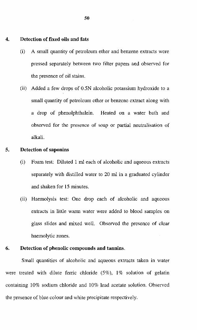
4. Detection of fixed oils and fats
(i) A small quantity of petroleum ether and benzene extracts were
pressed separately between two filter papers and observed for
the presence of oil stains.
(ii) Added a few drops of 0.5N alcoholic potassium hydroxide to a
small quantity of petroleum ether or benzene extract along with
a drop of phenolphthalein. Heated on a water bath and
observed for the presence of soap or partial neutralisation of
alkali.
5. Detection of saponins
(i) Foam test: Diluted 1 ml each of alcoholic and aqueous extracts
separately with distilled water to 20 ml in a graduated cylinder
and shaken for 15 minutes.
(ii) Haemolysis test: One drop each of alcoholic and aqueous
extracts in little warm water were added to blood samples on
glass slides and mixed well. Observed the presence of clear
haemolytic zones.
6. Detection of phenolic compounds and tannins.
Small quantities of alcoholic and aqueous extracts taken in water
were treated with dilute ferric chloride (5%), 1% solution of gelatin
containing 10% sodium chloride and 10% lead acetate solution. Observed
the presence of blue colour and white precipitate respectively.

51
7. Detection of proteins and free amino acids
Small quantities of alcoholic and aqueous extracts were subjected to
ninhydrin test and noted the presence of violet colouration.
8. Detection of gums and mucilage
10 ml of aqueous extract was added slowly to 25 ml of absolute
alcohol (with constant stirring) and filtered. The precipitate was then
observed for its swelling properties and for the presence of carbohydrates.
4.3 Ash values257
(i) Ash content: The residue remaining after incineration is the ash,
content of the drug, which simply represents inorganic salts,
naturally occurring in drug or adhering to it or deliberately added
to it, as a form of adulteration. Ash value is a criterion to judge
the identity or purity of crude drugs. Total ash usually consists of
carbonates, phosphates, silicates and silica. Acid insoluble ash,
which is a part of total ash insoluble in dilute Hel, is also
recommended for certain drugs. Adhering dirt and sand may be
determined by acid insoluble ash content.
4.3.1 Total ash value 258, 259
Procedure
2.5g of the ground drug (leaves) was accurately weighed and taken
In a tarred silica crucible previously ignited and weighed; scattered the

52
ground drug in a fine even layer in the bottom of the dish. Incinerated by
gradually increasing the heat until free from carbon. It was then cooled and
weighed to constant weight. Calculated the percentage of ash with
reference to the air-dried drug. The experiment was repeated thrice and the
average calculated.
4.3.2 Acid insoluble ash value.
Procedure
Boiled the ash obtained after total ash value determination for 5
minutes with 25 ml of dil. Hydrochloric acid. The insoluble matter was
collected in an ashless filter paper, washed with hot water, ignited and
weighed to constant weight. Calculated the percentage of acid insoluble ash
with reference to the air- dried drug. The procedure was repeated thrice and
the average calculated.
4.3.3 Sulphated ash
Procedure
About 2-3 g of drug was accurately weighed, moistened with
sulphuric acid and ignited gently. Again moistened and re-ignited, cooled
and weighed. Calculated the percentage of sulphated ash with reference to
the air dried drug.

53
4.3.4 Water-soluble ash
Procedure
Boiled the total ash with 25 ml of water for 5 minutes; insoluble
matter was collected in an ashless filter paper; wet with hot water, and
ignited to constant weight at a low temperature. The weight of insoluble
matter was subtracted from the weight of the ash. The percentage of water
soluble ash was calculated with reference to the air-dried drug.
4.4 Extractive values 260
The extracts obtained by exhausting crude drugs are indicative of
approximate measures of their chemical constituents. Taking into
consideration the diversity in chemical nature and properties of contents of
drugs, various solvents are used for determination of extractives. The
solvent used for extraction is in a position to dissolve appreciable quantities
of substances desired.
4.4.1 Alcohol soluble extractive
Alcohol being an ideal solvent for extraction of various chemicals
like tannins, resins etc. this method is frequently employed to determine the
approximate resin content of drugs. Generally 95% ethyl alcohol is used for
determination of alcohol soluble extractive; dilute alcohol may also be used,
depending upon solubility of the constituents of crude drugs.

54
Procedure
Macerated Sg of dried coarse powder of the drug with 100ml of
alcohol 9S% in a closed flask for 24 hours, shaking frequently during 6
hours and allowed to stand for 18 hours. It was then filtered immediately
taking precautions against loss of alcohol. Twenty-five milliliter of the
filtrate was evaporated to dryness in a tarred flat-bottomed shallow dish.
Dried at 10SoC and weighed. Calculated the percentage of alcohol soluble
extractive with reference to the shade-dried drug.
4.4.2 Water-soluble extractive
This method is applied to drugs, which contain water-soluble active
constituents, such as tannins, plant sugars, mucilage, glycosides etc.
Procedure
Added Sg of coarse powder of the drug to SOml of water at 800e in a
stopper flask. Shaken well and allowed to stand for 10 minutes. Cooled to
lSoC and added 2g of kieselguhr; filtered and transferred Sml of the filtrate
to a tarred evaporating dish; evaporated the solvent on a water bath and
weighed the residue. Calculated the percentage of water-soluble extractive
with reference to the shade-dried drug. The values were tabulated.
4.5 Atomic Absorption Spectroscopy'?'
This technique is based on the fact that when atoms, IOns or IOn
complexes of an element are atomized at ground state in a flame, they
absorb light at the characteristic wavelength of that element. If the

55
absorption process takes place in the flame under reproducible conditions,
the absorbance is proportional to the number of absorbing atoms.
Procedure
19 of air-dried leaves of the plant was accurately weighed and
ashed at 800DC thoroughly. The ash was dissolved in concentrated
hydrochloric acid and made up to l Ouml in a standard flask. For the
analysis of zinc, since it is volatile, the sample was accurately weighed and
dissolved in hydrochloric acid - perchloric acid mixture, boiled for 5
minutes, filtered and made up to 100m!.
The instrument was standardized usmg known concentrations of
solutions and a graph of absorbance vs. concentration drawn by the
instrument itself (instrument used was VARIAN AUSTRALIA). The
sample solutions were fed into the instrument and the concentration of each
element detected using the calibration graph.
4.6 Anti oxidant studies (IN VITRO)
As pilot studies with extracts of successive solvent extraction did not
show significant results except for ethanol extract, further programmed
studies were concentrated on ethanol extract only.
4.6.1 Superoxide Scavenging Activity.
Superoxide scavenging activity of the ethanol extract was
deiermined by the method of McCord and Fridovich262 which depends on

56
the light induced superoxide generation by riboflavin and the corresponding
reduction of nitroblue tetrazolium(NBT).
The reaction mixture contained EDTA (6Ilm) containing 31lg NaCN,
riboflavin (2J.!m), NBT (50llm), various concentrations of the ethanol
extract and phosphate buffer (67mM, pH 7.8) in a final volume of 3ml. The
tubes containing the reaction mixture were uniformly illuminated with an
incandescent lamp for 15 minutes and the absorbance was measured at
530nm before and after the illumination. The percentage inhibition of super
oxide generation was evaluated by comparing the absorbance values of the
control and experimental tubes.
4.6.2 Hydroxyl radical scavenging activity263
Hydroxyl radical scavenging activity was measured by studying the
competition between deoxyribose and the plant extract for hydroxyl radical
generated from the Fe3+- ascorbate - EDTA - H20 2 system (Fenton
reaction). The hydroxyl radical attacks deoxyribose that eventually results
in the formation of thiobarbituric acid reactive substances (TBARS).
The reaction mixture containing deoxyribose (2.8mM),ferric chloride
(O.lmM), ~DTA (O.lmM), H20 2 (lmM), ascorbate (O.lmM), KH2P0 4
KOH buffer (20mM; pH 7.4) and various concentrations of the extracts in a
final volume of 1ml was incubated for 1 hour at 37°C. Deoxyribose
degradation was measured as thiobarbituric acid reactive substance

57
(TBARS) by the method of Okhawa et ae64 and percentage of inhibition
was calculated from the control where no test extracts were added.
4.6.3 Effect on Lipid peroxidation
Effect on the inhibition of lipid peroxidation was determined by the
thiobarbituric acid method. Different concentrations of the plant extract
were incubated at 37°C with rat liver homogenate (25%) (O.lml) containing
30mM KCI, Tris-HCI buffer (O.04M; pH7), ascorbic acid (O.06mM) and
ferrous iron (O.16mM) (total volume was O.5m!) for 1 hour. At the end of 1
hour, thiobarbituric acid reactive substance (TBARS) was measured by the
method of Okhawa et ae64 and percentage of inhibition calculated from the
control where no test extract was added.
4.7 Phytochemical study
10 gm of ethanol extract obtained after successive solvent extraction
was dissolved in 50ml of ethanol. Then it was partitioned with ethyl acetate
and the ethyl acetate fraction was concentrated and dried under vacuum.
The ethanol fraction was also concentrated and dried.
The ethyl acetate fraction was subjected to TLC analysis usmg
hexane- ethyl acetate (1:1) solvent system and 3 spots were obtained. 10%
H2S04 in methanol was used as spray reagent. Later the ethyl acetate
extract was subjected to column chromatography using silica gel as the
adsorbent. Gradient elution using hexane and ethyl acetate, in different

58
proportions, was carried out. The fractions eluted using hexane : ethyl
acetate in the ratio 9:1 gave identical spots with same Rf in TLC analysis
(Rf = 0.92) and they were pooled and concentrated, crystallized and
recrystallized from 9: I hexane - ethyl acetate system. It was designated
as Compound A.
The fractions collected with the same solvents in the ratio (8:2) gave
another compound, crystallized in the same way, designated as compound
B (Rf = 0.86). Further elution was carried out with increasing concentration
of ethyl acetate. Finally at 50:50 ratio of hexane: ethyl acetate system, the
third compound was obtained as in the previous cases. This was designated
as compound C. (Rf = 0.49).
The ethanol fraction was subjected to TLC using n-butanol: acetic
acid: water (40:10:50) system. Mainly 3 spots were observed with the
spray reagent 10% H2S0 4 - methanol. During column chromatography
after gradient elution with chloroform-methanol, the first compound was
obtained at 50:50 ratio. This was designated as compound D.(Rf = 0.62)
Later the proportion of methanol was increased. Finally chloroform
methanol at 10:90 ratio gave another compound which was recrystallised
and later designated as compound E.

59
4.8 ANIMAL STUDIES
Studies related to hepatoprotection
4.8.1 Animal maintenance and care:
Wistar strain of rats (Rattus norvigicus) and Swiss albino mice (Mus
musculus), were the animals used for the study. They were bred in colony
and brought up in our own animal house. Animals were housed in well
ventilated polypropylene cages and fed with a standard pellet diet (Gold
Mohur laboratory animal feed) and water ad libitum. They were kept in
standard environmental conditions. (Temperature- 25-28oC and 12 hours
light/dark cycle) Rats of both sexes weighing 150-250g and mice of average
20-30g were used for animal experiments. (All ethical formalities were
cleared for the conduct of animal experiments using albino rats and mice.)
4.8.2 Acute Toxicity Studies265, 266
Albino rats of either sex weighing 150 - 250g and Albino mice
weighing 20-30 g of either sex were used for carrying out acute toxicity
studies. Before the actual study, a pilot study was carried out in small group
of animals (2 each) giving them widely spaced doses to select the Jose
ranges for the actual acute toxicity studies. Finally the assay was done using
5-6 dose levels with larger number of animals in each group (10 animals in
one group). All animals were fasted overnight before the acute toxicity
studies. After oral administration of the drug, the animals were observed

60
continuously for 2 hours and then intermittently for another 4 hours. After
24 hours, the deaths if any, were noted to calculate the LDso.
4.8.3 Study of Gross behavioral changes
Along with the toxicity tests, the animals were observed for
behavioral, neurological and anatomical profiles and the changes observed
in animals were recorded.
4.8.4 Determination of the most effective extract and its dose
As the pilot study, with all the extracts, showed maximum effect for
the ethanol extractf", a dose response assay was carried out using ]15 th,
1I1Oth and 1I20th of the maximum dose given to the animals to determine the
minimum dose producing maximum hepatoprotective effect. This was used
for the subsequent programmed studies.
4.8.5 Induction of Hepatotoxicity
Hepatic injury was induced using Carbon tetrachloride (CC14) and
paracetamol. In the former case CCl4 was mixed with olive oil in the ratio
1:1 and given intraperitoneally at a dose of 0.5ml/kg body wt. for 5 days or
as needed. In the latter case an overdose of paracetamol (3g/kg body wt.) or
accordingly, was given orally, suspended in 1% carboxy methylcellulose
(CMC).

61
4.8.6 Grouping of animals and different treatment
Animals were grouped and given different treatments as follows.
Male Wistar albino rats weighing IOO-120g were used for
hepatoprotective studies. The animals were divided into four groups of six
each.
(a) Hepatoprotective activity of A.indica using Carbon tetrachloride
Group I
(control)
Group II
Group III
(CCI4 +
A. indica ext.)
Group IV
(CCI4+Silymarin)
Animals did not receive any treatment;
but only 1% CMC (Iml/lOOg)
Animals received CCl4 (O.5mllkg body wt.
intraperitoneallyand 1% CMC (lmVIOOg), orally.
Animals received CCl4 (O.5mllkg body wt.)
and extract (IOOmg / kg body wt) after
10 min, orally
Animals received CCl4 (O.5mllkg body wt.) and
Silymarin (1OOmg / kg body wt) after 10 minutes,
orally.
This procedure was repeated for 5 consecutive days and on the 6th
day animals were sacrificed. The blood was collected for determination of
biochemical parameters and liver for histopathological studies.
62
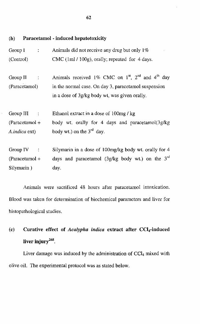
(b) Paracetamol - induced hepatotoxicity
Group I
(Control)
Group II
(Paracetamol)
Group III
(Paracetamol +
A.indica ext)
Group IV
(Paracetamol +
Silymarin)
Animals did not receive any drug but only 1%
CMC (lml / IOOg), orally; repeated for 4 days.
Animals received 1% CMC on i", 2nd and 4th day
in the normal case. On day 3, paracetamol suspension
in a dose of 3g/kg body wt, was given orally.
Ethanol extract in a dose of IOOmg / kg
body wt. orally for 4 days and paracetamol(3g/kg
body wt.) on the 3rd day.
Silymarin in a dose of IOOmg/kg body wt. orally for 4
days and paracetamol (3g/kg body wt.) on the 3rd
day.
Animals were sacrificed 48 hours after paracetamol intoxication.
Blood was taken for determination of biochemical parameters and liver for
histopathological studies.
(c) Curative effect of Acalypha indica extract after CCl4-induced
liver injury268.
Liver damage was induced by the administration of CCl4 mixed with
olive oil. The experimental protocol was as stated below.

63
Experimental protocol
Group Pretreatment Duration Day of Inferencein days withdrawal of
bloodI - - 0 Control value to
compare with the testII CC14 0-5 6111 To induce liver
damageIII CC14 0-5 11th To assess the
prophylactic effectIV A. indica + 1-5 To assess curative
CC14 6-10 11th effectV CC14 + 1-5 6111
"A. indica 1-5
VI CC14 + 1-5 11th"
A. indica 1-5VII CC14 + 1-10 11th
"A. indica 6-10
A.indica extract(lOOmglkg body wt) was administered orally and
CC14 (O.5mllkg body wt), intraperitoneally. A few animals from each group
were sacrificed on the day of withdrawal of blood from that group. Liver
was isolated, weighed and preserved in 10% formalin for histopathological
studies. With the blood all the liver function tests were done and with the
liver all the enzyme studies.
(d) Curative effect of Acalypha indica extract on paracetamol
induced liver injury122.
Liver damage was induced by the administration of paracetamol
(lg/Kg) orally, once daily. The experimental protocol was as stated below

64
Experimental protocolN =6
DurationDay of
Group Pretreatmentin days withdrawal Inference
of bloodI I%CMC - Control values to
0compare with the test
II Paracetamol 2 3HJ To induce liver damageIII Paracetamol 2 s" To assess the
spontaneous recoveryIV Paracetamol+ 1-5 s" To assess the
A. indica 6-7 preventive effectV Paracetamol + 1-5
3rd To assess the curativeA. indica 1-5 effect
VI Paracetamol + 1-56th To assess the curative
A. indica 1-5 effect ..VII Paracetamol + 1-7
8th To assess the curativeA. indica 3-7 effect
As in the previous case the drug was administered orally in a
dose of 100mg/kg body wt. The dose of paracetamol was 1g/kg body wt. A
few animals from each group were sacrificed on the day of withdrawal of
blood from that group: Liver was isolated, weighed and preserved in 10%
formalin for histopathological studies.
4.8.7 Effect of Acalypha leaf extract on Bile flow rate
(Choleretic Activity) 163,269
Male rats of 200-250 g weight were selected. They were divided
into two groups of 6 each. These rats were anesthetized with i.p injection of
Sodium pentobarbitone. The common bile duct was surgically exposed by
middle line laparotomy and cannulated with polyethylene tubing (No. 48).
A heating lamp maintained body temperatures of rats. Bile collected for

65
first 10 minutes was discarded and then collected in graduated tubes. Bile
was collected for 1 hour and then 1 ml of 1% CMC was administered
intraduodenally to control rats (group I). Group II rats received ethanol
extract of Acalypha leaf suspension (100 mg/kg body weight)
intraduodenally. Then the bile was collected for 4 hours. The volume of
bile was noted. An increase in the bile flow in the treated animals
compared to control was taken as the criterion for choleretic activity.
4.8.8 Effect of Acalypha leaf extract on liver regeneration205, 206.
Albino rats (male) of average weight (100 -llOg) were randomly
divided into four groups. Group I was normal sham operated, GroupII,
hepatectomised -untreated, Group III, hepatectomised - test drug treated,
while the group IV consisted of hepatectomised - silymarin treated animals.
Ethanol extract of A. indica and Silymarin, in 1% CMC suspension were
administered orally to rats for 7 consecutive days at a dose of 100mg./kg
body weight. On day 8, partial hepatectomy (about 70% removal of the
liver) of rats of group II, III and IV were done under light ether anaesthesia
according to the method of Higgins and Anderson/". Rats in sham
operated group were laparotomised in the same way except that the partial
hepatectomy was not done.
On selected time intervals post surgery (from 12 to 120 hrs) animals
were sacrificed by decapitation after fasting overnight. The liver was

66
promptly excised, washed thoroughly and subjected to DNA, RNA, protein
and cholesterol estimations.
4.8.9 Chronic toxicity studies271
The alcoholic extract of A. indica was subjected to chronic toxicity
studies.
Albino rats of either sex weighing 150-200 g were selected. Three
dose levels of drugs were administered in the study viz, 200 mg, 400 mg
and 800mg/kg body weight.
A control group with 1% CMC (Iml/l00g) was also used to rule out
the effect of the vehicle if any.
Animals were divided into 4 groups of 12 each. Each group was
again divided into subgroups (a) and (b) of 6 males and 6 females.
Grouping of animals and dose of drug given are summarised below.
Groups of animals and dose of the drug in chronic toxicity studies
Grouping ofTreatment given (p.o) Purpose
animals
A a b 200 mg of extract/kg Chronic effect of low dose
body weight.
B a b 400 mg of ext/kg body Chronic effect of middle
weight. dose
C a b 800 mg of ext/kg body Chronic effect of high
weight. dose
D a b Control animals. 1% Control to compare with
CMC (lml/lOOg) other groups.

67
All animals were given measured amount of food and water daily.
Period of study was 3 months. During the study, body weight and food
intake were monitored and hematological parameters were determined at
regular intervals of every 14 days. At the end of three months, all animals
were sacrificed and autopsy performed. Vital organs (liver, kidney and
spleen) were removed, weighed and examined for gross changes, as well as
for any histological changes. Blood was also collected during sacrifice and
estimations of alkaline phosphatase, AST, ALT, blood urea, creatinine and
serum bilirubin were also done and compared with those of control animals.
4.9 Blood Chemistry
(Determination of various constituents and enzymes)
4.9.1 Collection of blood for various estimations
At the end of each experiment (or even in between, as specified)
blood was collected by retro-orbital bleeding. The blood was then allowed
to clot, and centrifuged at 3000 rpm for 10 minutes. Clear serum obtained
as supernatant was transferred to clean dry screw cap bottles. This was
used for various estimations.
4.9.2 Alkaline Phosphataset"
Reaction conditions
Temperature
pH

68
Final concentration of reagents.
PNPP
MAP
Mg2+
16 n mol/l
1.0 rnol/l
1.0 m mol/l
Principle
The method of Bowers and McComb273 is widely used today
because of its simplicity and sensitivity. In this, p-nitrophenyl phosphate
(PNPP) is the substrate and 2-methyl-2-amino-1-propranol (MAP), the
transphosphorylating buffer. The colourless substrate is hydrolysed to
yellow coloured P-nitro-phenol (PNP) which has an absorption maximum
at 405 nm. This method measures the rate of release of P-nitrophenoxide
in trans phosphorylating buffer.
Procedure
Pipetted out 1ml of substrate and 0.02ml of serum into test tubes.
Mixed well and absorbance was read at 30, 60, 90, and 120 seconds, at
405nm. The changes in absorbance were noted and calculated the results.
lUlL = fj, Almin x F (F=2713)
4.9.3 Alanine Aminotransferase (ALT)
Principle
In the method of Berg and Horder273 NADH is the reaction product
that is quantitated. Lactate dehydrogenase (LDH) and its co-factors are
69
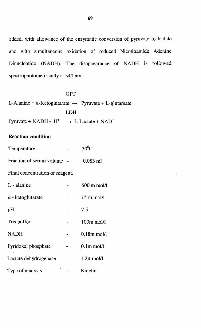
added, with allowance of the enzymatic conversion of pyruvate to lactate
and with simultaneous oxidation of reduced Nicotinamide Adenine
Dinucleotide (NADH). The disappearance of NADH is followed
spectrophotometirically at 340 nm.
GPT
L-Alanine + a-Ketoglutarate ---+ Pyruvate + L-glutamate
LDH
Pyruvate + NADH + H+ ---+ L-Lactate + NAD+
Reaction condition
Temperature 30De
Fraction of serum volume - 0.083 ml
Final concentration of reagent.
L - alanine 500 mmolll
a - ketoglutarate 15 mmolll
pH 7.5
Tris buffer 100m molll
NADH 0.18m molll
Pyridoxal phosphate O.lm mol/l
Lactate dehydrogenase 1.21J molll
Type of analysis Kinetic

70
Procedure
Working reagent (1.0 ml) (Span Diagnostic kit) and sample (0.1 ml)
were mixed, incubated at 37°C for 1 minute. Started the stopwatch and read
the change in absorbance at 1, 2, 3 and 4 minutes.
Wave length, 340 nm
Blank Distilled water.
Determined the i1E/min for every reading and found the mean value
U/L = 1768 x (i1E/min.)
4.9.4 Aspartate Aminotransferase (AST)
Principle
In the method of Bergmeyer et ae74 first described by Karman,
oxaloacetate is the product quantified. Malate dehydrogenase (MDH) is
used to convert the oxaloacetate to malate. The decrease in absorbance at
340 nm caused by NADH (reduced nicotinamide adenine dinucleotide)
consumption is recorded spectrophotometrically.
GOTL - aspartate + a - ketoglutarate ~ Oxalo acetate + L-glutamate
MDHOxalo acetate + NADH + H + ~ Malate + NAD+
Reaction conditions:
Reaction temperature
Sample volume
71
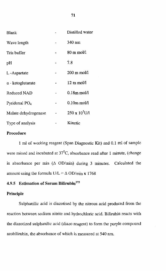
Blank Distilled water
Wave length 340nm
Tris buffer 80mmoVI
pH 7.8
L -Aspartate 200mmoVI
a - ketoglutarate 12 m mol/l
ReducedNAD 0.18mmoVI
Pyridoxal P04 0.10mmoVl
Malate dehydrogenase 250 x 103U/I
Type of analysis Kinetic
Procedure
1 ml of working reagent (Span Diagnostic Kit) and 0.1 ml of sample
were mixed and incubated at 37°C, absorbance read after 1 minute, (change
in absorbance per min (~ OD/min) during 3 minutes. Calculated the
amount using the formula U/L = ~ OD/min x 1768
4.9.5 Estimation of Serum Bilirubirr'"
Principle
Sulphanilic acid is diazotised by the nitrous acid produced from the
reaction between sodium nitrite and hydrochloric acid. Bilirubin reacts with
the diazotized sulphanilic acid (diazo reagent) to form the purple compound
azobilirubin, the absorbance of which is measured at 540 nm.
72
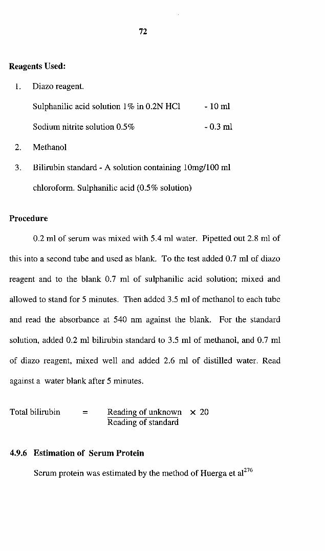
Reagents Used:
1. Diazo reagent.
Sulphanilic acid solution 1% in 0.2N Hel
Sodium nitrite solution 0.5%
2. Methanol
- 10 ml
- 0.3 ml
3. Bilirubin standard - A solution containing 10mg/l00 ml
chloroform. Sulphanilic acid (0.5% solution)
Procedure
0.2 ml of serum was mixed with 5.4 ml water. Pipetted out 2.8 ml of
this into a second tube and used as blank. To the test added 0.7 ml of diazo
reagent and to the blank 0.7 ml of sulphanilic acid solution; mixed and
allowed to stand for 5 minutes. Then added 3.5 ml of methanol to each tube
and read the absorbance at 540 nm against the blank. For the standard
solution, added 0.2 ml bilirubin standard to 3.5 ml of methanol, and 0.7 ml
of diazo reagent, mixed well and added 2.6 ml of distilled water. Read
against a water blank after 5 minutes.
Total bilirubin = Reading of unknown X 20Reading of standard
4.9.6 Estimation of Serum Protein
Serum protein was estimated by the method of Huerga et at276
73
Reagents
1. Stock biuret reagent (45.0g sod.pot.tartarate dissolved in 400ml
of 200mM/l NaOH and added 15gm CUS04 . 2H20 Added 5gm
Potassium iodide and made up to llitre with 200m Mil NaOH.
2. Working biuret reagent (200ml stock reagent diluted to llitre
with 200m MIL NaOH containg 5gm pot.iodide llitre
3. Tartarate iodide solution (9gm sod.pot.tartarate in llitre of 200m
Mil NaOH containing 5gm KIf litre
4. Protein standard (80gm/litre)
Five test tubes marked as test (containing O.lml serum +5ml working
biuret reagent), serum blank (containing O.lml serum +5.0ml tartarate
iodide solution), standard containing O.lml standard +5ml working biuret
reagent), standard blank (containing O.lml standard +5ml tartarate iodide
solution) and reagent blank (O.lml water +5ml working biuret reagent),
were incubated at 37°C for Irnt. After cooling to room temperature
absorbance were measured at 555nm against reagent blank.
Procedure: 5 ml of Biuret reagent was taken to which 0.1 ml of serum was
added. Similarly 0.1 ml protein standard solution was added to 5 ml of
Biuret reagent I. 5 ml Biuret standard I was the blank. All the tubes were
kept at room temperature for 5 minutes. Then the absorbance of the
solutions-standard and test-were measured at 550 nm

74
4.9.7 Estimation of Serum Albumin 277
Serum albumin was estimated by the method of Drupt. Three test
tubes marked as blank (containing 4.5ml bromocresol green reagent),
standard (containing 4.5ml bromocresol green reagent +30 III (4gm%)
albumin standard) and test (containing 4.5ml bromocresol green reagent
+30 III serum were allowed to stand at room temperature for 1min.
Measured the absorbance of test and standard against blank at 600nm.
4.9.8 Urea Estimation 278
Diacetyl monoxime method
Principle
Serum proteins are precipitated with trichloro acetic acid and clear
supernatant is obtained after centrifugation. A portion of this supernatant is
heated with diacetyl monoxime in the presence of acid, oxidizing agent and
thiosemicarbazide and a red complex is formed with urea. The absorbance
of this complex is measured at 520 nm.
Reagents
1. Trichloro aectic acid solution, 10% (w/v). Dissolved 109 of trichloro
acetic acid in water and made up to 100 ml. This solution is stable
indefinitely at 20-25°C.
2. Ferric chloride solution: Dissolved 5.0g of ferric chloride in 100 ml
of water and added 1.0 ml of cone. Sulphuric acid. This solution
75
should be stored in a dark coloured bottle where it is stable for
several months at 20-25°C.
3. Acid Reagent: To 100 ml of water added 8.0 ml of cone. Sulphuric
acid, 1 ml of ortho phosphoric acid and 1 ml of iron III chloride
solution. This is stable at 20-250C indefinitely.
4. Diacetyl monoxime solution: Dissolved 25 g of diacetyl monoxime
in water and made up to 1 litre. This solution is stable for several
months at 20-25°C.
5. Thiosemicarbazide Solution: Dissolved 2.5 g of thiosemicarbazide
in water and made up to 1 litre. This solution is stable for several
months at 20-25°C.
6. Colour reagent: To 30 ml of acid reagent, added 20 ml of water, 1.0
ml of diacetylmonoxime solution and 0.25 ml of thiosemicarbazide
Solution. This should be prepared freshly each day.
7. Stock urea standard 10m mol/l: Dissolved 1.0 g of benzoic acid in
about 800 ml of warm water, allowed to cool, then transferred to a 1
litre volumetric flask and added 0.601 g of urea. When dissolution
was completed, made up to volume with water. This solution is
stable for several months at 20-25°C.
8. Working urea standards: Into 100 ml volumetric flasks, pipetted out
2.5, 5.0, 7.5, 10.0, 15.0, and 20.0 ml of the stock urea standard and
76
made up to volume with benzoic acid solution (1.00 g/l.). The
working standards are equivalent to 2.5, 5.0, 7.5, 10.0, 15.0, 20.0 m
molll under the assay conditions. These standards are stable for
several months at 20-25°C.
Method
1. Pipetted into centrifuge tubes 0.8 ml of water, 0.2ml of sample and
mixed. Then added 1.0 ml of trichloro acetic acid solution, mixed
again and centrifuged.
2. To 0.2 ml of supernatant added 3 ml of colour reagent. For the
reagent blank 0.2 ml of water was used and for the standards 0.2 ml
of each working standard solution used.
3. After mixing, the tubes were closed and incubated at 100°C for 20
minutes.
4. Cooled the tubes in water for 5 minutes. Then read the absorbance of
the solutions at 520 nm, setting the spectrophotometer to zero with
the reagent blank.
4.9.9 Estimation of Creatinine
Principle
Creatinine is determined by quantitating the red pigment, alkaline
creatinine picrate (Jaffe reaction), as described by Giorgio279 (1974).
77
132 mg creatinine /lOml of 01 N NaOH.
Reagents
I.Picric acid
2. Tungstic acid
Polyvinyl alcohol
Sodium. tungstate dihydrate
Sulphuric acid
3.Sodium hydroxide
4. Standard-
0.36 molll.
1gil
11.1gil
O.lmlll
54 gil
Procedure: 0.5 ml of sample was deproteinised with 4 ml of tungstic acid,
centrifuged and 3 ml supernatant was mixed thoroughly with 1 ml of picric
acid followed by 0.5 ml sodium hydroxide. Absorbance was recorded after
15 minutes at 500 nm.
4.10 Estimation of Malondialdehyde (In Vivo)
Preparation of tissue homogenate
Weighed quantity of tissue was homogenised in O.IM Tris-HCl
buffer, pH 7.5, and allowed to stand for 5 minutes. The supernatant was
used for the determination of lipid peroxide level.
Thiobarbituric acid reactive substance (TBARS)
TBARS were estimated by the assay method of Nichans and
Samuelson.v"

78
Reagents
TCA-TBA-HCI reagent
0.25NHCl.
15%(w/v) TCA, 0.375%(w/v) TBA III
Procedure: 1ml of the tissue homogenate was combined with 2ml of TCA-
TBA-HCI reagent and mixed thoroughly. The solution was heated for 15
minutes in a boiling water bath. After cooling, the flocculent precipitate was
removed by centrifugation at 1000 rpm for 10minutes. The absorbance of
the sample was read at 535nm against a blank that contained no tissue
homogenate. The extinction co-efficient of malondialdehyde is 1.56 x 10⁵
M -1 -Iem .
4.11 Activity of Scavenging Enzymes
4.11.1 Assay of Superoxide dismutase (SOD) [EC 1.15.1.1]
SOD activity was determined by the method of Kakkar et a1. 28 1 The
tissue was homogenized in 0.25M sucrose and differentially centrifuged at
10,000 rpm under cold conditions to get the cytosol fraction. The initial
purification was done by precipitating the protein from the supernatant with
90% ammonium sulphate and after dialysis against 0.0025M Tris-HCI
buffer, pH 7.4. The supernatant was used as the enzyme source.
The assay mixture contained 1.2m1 Sod.pyrophosphate buffer
(0.052M, pH8.3), O.lml 1861lM phenazine methosulphate (PMS), 0.3ml
300llm nitroblue tetrazolium (NBT) 0.2ml 780llM NADH, appropriately

79
diluted enzyme preparation and water in a total volume of 3ml. Reaction
was started by the addition of NADH. After incubation at 30°C for 90
seconds reaction was stopped by the addition of 1ml glacial acetic acid. The
reaction mixture was stirred vigorously and shaken with 4ml of n-butanol.
Mixture was then allowed to stand for ten minutes. Centrifuged and butanol
layer was taken out. Colour intensity of the chromogen in the butanol
fraction was measured at 560nm against butanol. A system devoid of
enzyme served as control.
One unit of enzyme activity is defined as the enzyme concentration
required to inhibit chromogen production (OD of 560 nm ) 50% in one
minute under the assay conditions. The specific activity is expressed in
units/ mg protein. Unit is defined as the velocity constant per second.
4.11.2 Assay of Catalase [EC 1.11.1.6]
( HzOz: Hydrogen peroxide oxido reductase)
The catalase activity was assayed by the method of Maehly and
Chancez8z, The tissue was homogenized with 0.91M-phosphate buffer (pH
7.0) at 1- 4°C and centrifuged at 5000 rpm. The estimation was done
spectrophotometrically following the decrease in absorbance at 240nm.
The reaction mixture contained 0.91 M phosphate buffer pH 7.0, 2mM
HzOz (diluted O.lml HzOz to 100ml using buffer) and 50~1 enzyme extract.

80
Specific activity is expressed in terms of units/ mg protein. Unit is defined
as the velocity constant per second.
4.11.3 Assay of Glutathione Peroxidase (EC.1.11.1.9)
(Glutathione: hydrogen peroxide, oxido reductase)
The activity of glutathione peroxidase was determined by the method
of Lawrence and Burk283 as modified by Agerguard and Jense284. Tissue
homogenate (10%) was prepared in 0.25M sucrose, centrifuged at 10,000
rpm for 30 minutes and the supernatant fraction was used for the assay.
Activity was determined in phosphate buffer (50mM pH7.0) containing
EDTA (1.5mM), sodium azide (1.0mM), reduced glutathione (1.0mM),
NADPH (O.lmM) and glutathione reductase (1.0flM/ml). Absorbance was
measured at 340nm at 20 seconds interval. Enzyme activity is defined as
flM ofNADPH oxidized/min /mg protein using 0.25mM H20 2 as substrate.
4.11.4 Assay of Glutathione reductase (EC.1.6.4.2)
(Reduced NAD (P): oxidized glutathione oxido reductase)
Glutathione reductase activity was determined by the method
described by Bergrneyer'f". Tissue homogenate (10%) was prepared in 0.25
M sucrose, centrifuged at 10,000 rpm for 30 minutes and supernatant
fraction was used for the enzyme assay. The assay system contained 1ml
phosphate buffer (0.12M, PH 7.2) O.lml EDTA, O.lml sodium azide
(10mM/I), O.lml oxidized glutathione (6.3mM) and 0.1 ml enzyme source.
81
It was kept for 3 minutes. Then 0.1 ml NADPH (9.6 mM/I) was added. The
absorbance at 340 nm was measured at an interval of 15 seconds for 2
minutes. The activity is expressed as JlM NADPH oxidized per minute I mg
protein.
4.12 Estimation of Antioxidants
4.12.1 Estimation of Glutathione Content
Glutathione content was estimated by the method of Benke et a1286•
20% tissue homogenate prepared in 5% TeA containing O.OOIM
EDTA, was centrifuged at 2000 rpm for 5 minutes; 0.2ml aliquot of each
supernatant fraction was transferred to another tube containing 4.75ml of
O.IM sodium phosphate buffer (pH 8) and to it 0.05ml of O.Olm 5,5
dithiobis-2-nitrobenzoic acid (DTNB) was added. The absorbance was read
at 412nm within 4 minutes.
4.13 Estimation of Liver Protein
The method of Lowry et ae87 was used for the assay.
Principle: The method relies on the formation of a protein- copper
complex and the reduction of the phosphomolybdate - phosphotungstate
reagent (Folin-ciocalteu phenol reagent) by the tyrosine and tryptophan
residues of protein to form coloured product.

82
Reagents
Solution A containing I ml CUS04 (1%), 1 ml sodium potassium
tartarate 2% and 98ml of Na2C03 (2% in O.IN NaOH); solution B
containing Folin-ciocalteu phenol reagent diluted (l: 1) with distilled water.
Procedure
O.lml of tissue homogenate (2.5%) was diluted to 1.2 ml with
distilled water and mixed with 6 ml of solution A. The mixture was
incubated at room temperature for 10 minutes. To this solution 0.3ml
solution B was added. Mixed well and kept at room temperature for 30
minutes. The absorbance was read at 660nm.
4.14 Estimation of Total Cholesterol
Extraction of Liver
Liver was homogenized and extracted with chloroform: Methanol
(2:1). It was filtered and the residue was washed with chloroform: methanol
(2:1) at least 3 times. The filtrate was combined and washed with 0.7% KCI
solution (20% of the total volume of the extract) The aqueous upper phase
was removed and the lower layer was washed 3 times with 5ml of
chloroform : methanol : KCI solution. (3:48:47v/v). The washed lower
layer of chloroform was evaporated to dryness and the residue re dissolved
in a known volume of chloroform.
83
Procedure
Cholesterol was estimated by Carr and Drekter288 method. An aliquot
was taken from the above solution and evaporated to dryness. It was
dissolved in 0.45ml of glacial acetic acid. An aliquot from standard (50mg
cholesterol/50ml glacial acetic acid) and a blank containing glacial acetic
acid was also taken. 2 ml of acetic anhydride was added followed by 0.05ml
water to enhance heat of formation. It was kept for five minutes. One drop
of dehydrating reagent (colour reagent: Acetic acid (1:1 v/v» was added
directly, mixed by gentle rotation, and kept in a water bath at 20De for 10
minutes. Added 0.5ml colour reagent (Acetic acid: H2S04 1:1 v/v) directly
and kept at room temperature exactly for 20 minutes. Absorbance was read
at 620nm.
4.15. Estimation of DNA289
2gm of tissue (liver) was homogenized with 10 ml of ice cold lO9'o
trichloro acetic acid. It was centrifuged at 3000 rpm for 10 minutes.
Supernatant was discarded and the precipitate was suspended in 5 ml ice
cold 10% TCA. Again centrifuged and discarded the supernatant.
Suspended the residue in 5ml of ethanol-ether mixture and once again
centrifuged for ten minutes. The precipitate was taken in 0.5 N NaOH.
Mixed well and kept for 18hours at 37°e and centrifuged. To the
precipitate was added 1 ml of perchloric acid and heated on a boiling water

84
bath for 1 hour, cooled and centrifuged. The supernatant was made up to a
known volume.
The deoxyribose in DNA in presence of acid forms hydroxylevulinic
aldehyde which reacts with diphenylamine to give a blue colour.
Pipetted out different volumes of the solution in test tubes and added
5 ml of diphenylamine reagent. Mixed well and heated on a boiling water
bath for 10 minute. After cooling the absorbance was measured at 595nm.
Calculated the amount of DNA present in the tissue.
4.16 Estimation of RNA289
The supernatant obtained after incubation with 0.5N NaOH(in the
extraction) was mixed with 10% TCA and centrifuged for l0 minutes. The
supernatant obtained was made up to a known volume.
Acid hydrolysis of RNA releases ribose and this in presence of
strong acid, dehydrates to furfural. Orcinol reacts with furfural in presence
of ferric chloride to give a green colour.
Set up different tubes with measured volume of the supernatant and
added 3.0ml of orcinol reagent to each tube and mixed well. Heated on a
boiling water bath for 15 minutes and cooled. The absorbance was read at
665nm. Estimated the amount of RNA in the known volume of solution.
85
4. 17 Histopathological Examinatiorr't"
Tissue specimens were fixed in 10% buffered formalin immediately
after sacrifice of the animals. Specimens were dehydrated by passing
through ascending grades of alcohol, cleared in xylene impregnated and
embedded in paraffin. Thin sections were cut (3 - 5~M) and were stained
using hematoxylin and eosin and studied microscopically. Liver of animals
in all groups was studied besides kidney and spleen after chronic study.
4.18 Statistical Analysis 291
Data obtained were described as mean ± SEM. One way Analysis of
Variance (ANOVA) was carried out to estimate the variation in the means
between groups. Multiple Analysis (MANOVA) of Variance and their F
ratio were also calculated using the SPSS window version 6.