materials science and engineering c - indian institute of...
TRANSCRIPT

Materials Science and Engineering C 68 (2016) 663–674
Contents lists available at ScienceDirect
Materials Science and Engineering C
j ourna l homepage: www.e lsev ie r .com/ locate /msec
Elucidation of differential mineralisation on native and regeneratedsilk matrices
Swati Midha a, Rohit Tripathi a, Hua Geng b, Peter D. Lee b, Sourabh Ghosh a,⁎a Department of Textile Technology, Indian Institute of Technology, New Delhi, Indiab School of Materials, University of Manchester, Manchester, M13 9PL, UK
⁎ Corresponding author.E-mail addresses: [email protected] (S. Midha), tr
(R. Tripathi), [email protected] (H. [email protected] (P.D. Lee), sghosh08@textile.
http://dx.doi.org/10.1016/j.msec.2016.06.0410928-4931/© 2016 Elsevier B.V. All rights reserved.
a b s t r a c t
a r t i c l e i n f oArticle history:Received 6 April 2016Received in revised form 16 May 2016Accepted 12 June 2016Available online 14 June 2016
Bonemineralisation is a well-orchestrated procedure triggered by a protein-based template inducing the nucle-ation of hydroxyapatite (HA) nanocrystals on the matrix. In an attempt to fabricate superior nanocompositesfrom silkfibroin, textile braided structuresmade of natively spunfibres of Bombyxmori silkwormwere comparedagainst regenerated fibroin (lyophilized and films) underpinning the influence of intrinsic properties of fibroinmatrices on HA nucleation. We found that native braids could bind Ca2+ ions through electrostatic attraction,which initiated the nucleation and deposition of HA, as evidenced by discrete shift in amide peaks via ATR-FTIR. This phenomenon also suggests the involvement of amide linkages in promoting HA nucleation on fibroin.Moreover, CaCl2-SBF immersion of native braids resulted in preferential growth of HA along the c-axis, formingneedle-like nanocrystals and possessing Ca/P ratio comparable to commercial HA. Though regenerated lyophi-lized matrix also witnessed prominent peak shift in amide linkages, HA growth was restricted to (211) planeonly, albeit at a significantly lower intensity than braids. Regenerated films, on the other hand, provided no crys-tallographic evidence of HA deposition within 7 days of SBF immersion. The present work sheds light on the pri-mary fibroin structure of B. mori which probably plays a crucial role in regulating template-inducedbiomineralisation on the matrix. We also found that intrinsic material properties such as surface roughness, ge-ometry, specific surface area, tortuosity and secondary conformation exert influence inmodulating the extent ofmineralisation. Thus ourwork generates useful insights andwarrants future studies to further investigate the po-tential of bone mimetic, silk/mineral nanocomposite matrices for orthopaedic applications.
© 2016 Elsevier B.V. All rights reserved.
Keywords:Silk fibroinMineralisationHydroxyapatiteTextile braidSimulated body fluid
1. Introduction
Bone is a complex nanocomposite consisting of organic–inorganicphases of collagen-hydroxyapatite (HA) crystals, where HA[Ca10(PO4)6(OH)2] measures 20–30 nm in width, 100 nm in lengthwith 3–6 nm thickness [1], precisely embedded between longitudinallyarranged collagen fibres such that HA is synthesized on the c-axis planealong the longitudinal axis of collagen [2]. In addition, HA exhibits inti-mate chemical bonding between the organic-inorganic elements withan overall poor crystallinity [2]. Growing interest in the developmentof biomimetic synthetic HA-based composites is primarily due to; (i)developing synthetic “analogs” of HA to be able to precisely replicatethe morphology and chemistry of bone tissue [3], (ii) HA coating onconstructs for improving osteoconductivity and (iii) evaluating the bio-activity of novel materials by studying the nucleation and growth of“bone-like” apatite upon immersion in supersaturated fluids such as
[email protected]),iitd.ac.in (S. Ghosh).
simulated body fluid (SBF) [4]. However, there are still gaps in under-standing the nature of such depositions; surface features andcrystallisation phase especially with respect to the morphology andchemistry of the base material.
Silk fibroin, another fibrous protein that closely mimics collagentype I of bone has gained popularity in tissue engineering due to inter-esting intrinsic properties such as resorbability [5], toughness [6], min-imal immunogenicity [7,8], cytocompatibility with abundant polar,hydrophilic groups [9], strong chemical bonding with HA [10–13] andversatility in processing depending upon the target application [14].Silk fibroin template fabricated in the form of nanofibres [12], films[15], porous 3Dmatrices [16] and composites [17] exposed to supersat-urated ions, SBF [4,10] or fetal bovine serum [11], have been tested be-fore for HA deposition. Silk fibroin plays a crucial role in regulating thesynthesis and growth of HA nanocrystals [18], as HA combines with fi-broin through chemical interactions which have been identified byFTIR analyses whereby the strong chemical bonding between the twocaused peak shifts in the amide bonds of fibroin, commonly referredto as the ‘blue shift’ [2]. However, majority of these studies have largelyignored the role of template chemistry of native fibroin in directing thenucleation and growth of HA. The primary reason could be that most of

664 S. Midha et al. / Materials Science and Engineering C 68 (2016) 663–674
the studies used regenerated silk fibroinmaterials formed by the subse-quent dissolution and re-assembly of native silkworm cocoons whichtends to disrupt the original protein conformation [19]. Moreover, therole of differentmorphologies and other physical characteristics as a po-tential cause of large variations in the resultant mineralisation attainedhave not been addressed. In this study, we hypothesize that native silkfibroinwill experience superiormineralisation in the presence of super-saturated solution, credited to the intact organization of the primaryamino acid composition and structure. To test this hypothesis and un-derstand the underlying chemical interactions governing the processof HA nucleation on fibroin matrix, we examined the change in the in-trinsic properties of fibroin and thoroughly characterized HAcrystallisation on the material surface initiated by incubating silk struc-tures in 1.5× SBF.
Furthermore, there are theories which attribute the mineralisationpotential of fibroin to the amorphous anionic bridges present betweenthe β-sheets present within the structure [20]. In this study, Marelli etal. compared the hydrophobic chains in fibroin against the hydrophilic,electronegative chains and found apatite formation only in the hydro-philic fragments. They concluded that the differences in themineralisation potential of silkfibroinwere due to its constituent anion-ic chains which typically resembled the non-collagenous, anionic frag-ments of collagen type I. In another study, Kino et al. preparedregenerated silk fibroin films with varied calcium chloride content andreported deposition of HA post-immersion in 1.5× SBF, but only infilms possessing N3 wt% of calcium chloride relative to silk fibroin[15]. The reason was attributed to the presence of Ca2+ ions at a partic-ular concentration which reportedly induced more β-sheets in the fi-broin structure [21] and such modified silk fibroin films demonstratedimproved HA coating [15]. However, quantitative analysis of this vary-ing β-sheet content was not done. Also, the reason as to whymineralisation could not be observed on methanol treated pure fibroinfilms (without calcium chloride) which contained at least some amountof β-sheet content remained unexplained. Nevertheless, most studieshave reported an additional requirement of either pre-exposure of fi-broin or its association with calcium- and phosphate-based solutionsto promote nucleation and growth of HA nanocrystals [12,13,16,22].Whether it is the processing parameters during dissolution and recon-stitution that disrupt the native structure of the fibroin chain; firstlyby dissociating the specific amino acid chains and secondly by hamper-ing their tendency to re-assemble the necessary β-sheet conformation,or if it is the inherent inert nature of fibroin chain thatmodulates the ex-tent of mineralisation is still under wraps. These studies furtherhighlighted the fact that our current understanding of the primarystructure of fibroin is insufficient to elucidate the mineralisation capac-ity of silkfibroin in order to use it as a potential bone graft substitute [9].
Apart from the secondary conformation, there are other contributingfactors that determine apatite formation on biomaterials including mo-lecular size, surface chemistry, topography, surface charge, stiffness, tar-get site and rate of in situ degradation which needs to be taken intoaccount. For instance, hydrophobic crystalline fractions of silk fibroinwith a 40 kDa MW are weak templates for mineralisation, whereas 2–10 kDa MW hydrophilic chains are strong templates [20]. Moreover,the specific surface area of the biomaterial plays a crucial role in deter-mining the amount of apatite formation, as a larger hydrated layer of or-ganic template with strong tendency for ion exchange facilitates higherprecipitation of nanocrystalline HA [3]. Therefore, in the present study,we aim to address the following three specific questions with respectto in vitro mineralisation potential of silk fibroin; (i) the role of fibroinas an osteogenic substrate (in regenerated versus native form) in regu-lating nanocrystalline apatite synthesis, (ii) whether change in 3Dmor-phology (lyophilized versus braid) influences HA deposition on fibroinand (iii) whether the modified 3D morphology and resultant variationin the intrinsic material properties modulate the extent ofmineralisation. To the best of our knowledge, this is the first study toprovide an indepth analysis of the difference in mineralisation in silk
fibroinmatrices between the native and regenerated fibres.We hypoth-esized that by inducing morphological changes in the fibroin structure,we could modulate the material properties which will eventually en-hance control over the nucleation and growth of the HA phase. Inorder to characterize the mineralisation quotient, we used a combina-tion of several sophisticated analytical techniques combining high endimaging and compositional analysis of the fabricated matrices to gener-ate correlation between the material properties and the resultantmineralisation. We believe that understanding the process ofmineralisation on the hierarchical assembly of fibroin will providesome innovative ideas for fabricating functional silk-based materialswith customized morphology and chemistry best suited to promotebiomineralisation in situ.
2. Materials and methods
2.1. Isolation of silk fibroin solution
12 tex 2-ply Bombyx mori silk yarns were procured from StarlingMills Pvt. Ltd., Malda district, West Bengal, India. Silk solution was pre-pared directly from the fibres according to a protocol used routinely [23,24]. Briefly, 5 g of B. mori fibres were weighed and cut into fine pieces,followed by boiling in 0.02 M Na2CO3 for 30 min to remove sericin.The fibres were then thoroughly rinsed in deionized (DI) water to iso-late fibroin protein. Once completely dried, fibres were solubilized in9.3 M LiBr solution at 60 °C for 4 h. Fibroin-LiBr solution was dialysedusing Slide-A-Lyzer cassette (Thermo, molecular weight cut off 3500)yielding 6.1 wt% solution of silk fibroin, which was stored at 4 °C untilsubsequent usage.
2.2. Preparation of regenerated silk fibroin matrices
Aqueous silk fibroin solution of the same concentration (i.e. 6.1wt%)was used for the fabrication of both lyophilized matrices and 2D planarfilms.
2.2.1. Silk filmsTo generate silk solution in the form of planar films, 2 mL of silk fi-
broin solution was pipetted over 10 × 10 mm2 teflon plates and driedat RT overnight. Post-drying, the films were immersed in sufficient vol-ume of 80% ethanol for 2 h at RT in order to induce β-sheetcrystallisation.
2.2.2. LyophilizedA randomly porous three-dimensional (3D) morphology was ob-
tained by lyophilisingfibroin solution. Approximately 10mLof the solu-tion was poured into a 30 cm2 glass petri dish and frozen at−20 °C in arefrigerator for 24 h. The resulting silk solution was then lyophilized togenerate a 3Dporousmatrix,whichwas subsequently immersed in eth-anol for 2 h to induce β-sheet crystallisation.
2.2.3. Silk braidsFibres of B. mori silk were fabricated into 3D braids using a 17 spin-
dles flat braiding machine type NG1/16-120 (August HerzogMaschinenfabrik GmbH & Co. KG, Germany). Briefly, yarn bundles (17multi-filament yarns per yarn bundle) were produced from multi-fila-ment yarns comprising of 30 filaments/yarn of silk. The resulting struc-ture was made such that yarns were oriented at 32° angle to the longaxis of the braid using a production rate of 1 m/min to produce braidedstructures. The 3D braids were manually cut into smaller structures ap-proximately 4 × 2 × 1 mm3 dimensions.
2.3. SBF immersion
Post-fabrication, silkfibroin 3Dmatrices andfilmswere immersed in1.5× SBF solution for 7 days. SBF (1.5×) solution was prepared as

665S. Midha et al. / Materials Science and Engineering C 68 (2016) 663–674
described elsewhere under constant stirring at 37 °C and buffered atpH 7.4 using 75 mM Tris buffer [25]. Alternatively, an equal number ofmatrices (films, lyophilized and braids) were immersed in 2 kmol/m3
of CaCl2 and incubated for 24 h at 37 °C [26]. Then the matrices werevery gently washed with deionized water and air dried prior to immer-sion in 1.5× SBF. The temperature of the solutionwasmaintained at 37 °C until the matrices were immersed. The samples were harvested after7 days for analytical purposes.
2.4. Analytical techniques
2.4.1. Micro-CTTo quantify 3Dmorphology, silk fibroinmatrices were characterized
using X-ray micro-CT (Nano-focus, Phoenix X-ray GE, Measurement &Control, Wunstorf, Germany). The conditions used were an acceleratingvoltage of 55 kV, 120 μA current and 1000 projections over 360°. Three2000ms projectionswere averaged per angle to reduce noise. All mate-rials were scanned at low (20 μmvoxels) and high (3.5 μm) resolutions.The resulting 3D volume images were then processed using an edge-preserving filter to remove noise and artefacts, followed by segmenta-tion to allow 3D quantification. Three quantitative assessments wereperformed, as described below.
The specific surface area (SSA)was calculated bydividing the surfacearea of matrix (SAmatrix) by its volume (Vmatrix) [27]:
SSA ¼ SAmatrix
Vmatrix
Tortuosity was calculated by dividing the line length between twopoints and the distance between the loading platens. [28]
Tortuosity ¼ LlengthLdistance
Porosity was calculated by the volume of non-matrix materials bythe total volume in the field of view [29]:
Porosity ¼ 100 � 1−Vbinary� �
where VB is the volume of binarized object (matrix material phase) involume of interest (VOI).
2.4.2. Attenuated total reflectance-fourier transform infrared spectroscopy(ATR-FTIR)
ATR-FTIR spectra of silk fibroin matrices including films, lyophilizedand braids (n= 3 per group) before and after treatment were obtainedusing an Alpha-P spectroscope (Bruker, USA). A total of 72 scans weretaken in absorbance mode in the spectral range of 4000 to 500 cm−1
for each measurement at data acquisition rate of 4 cm−1 per point.Spectral analysis was performed for relative comparison. Deconvolutionwas done on the spectra using Fourier self-deconvolution (FSD).
2.4.3. Atomic force microscope (AFM)Surface roughness of silk fibroin matrices (films, lyophilized and
braids) treated with SBF with or without CaCl2 pre-treatment as wellas untreated controls (n = 3 per group) were imaged using Digital In-struments Nanoscope in contact mode. Three randomly chosen areasof 10 μm × 10 μm dimensions were imaged and the mean of their re-spective surface roughness (Rq) values was calculated. In the case ofbraids, roughness of a single fibre was measured as the presence ofgrooves in braided structures does not permit probe detection of thesurface.
2.4.4. X-ray diffraction (XRD)X-ray diffraction was performed in a Seimens type F X-ray diffrac-
tometer using Ni-filtered Cu KR radiation (30 kV, 20 mA, λ =
0.154 nm) as the source. For scanning, the materials were loaded ontothe aluminum frames and scanned from 10 to 50° (2θ) at 2.0°/min.
Particle mean crystallite size (D) was calculated from the XRD datausing Scherrer equation [30];
D ¼ Κλ=βCos θ
whereλ is thewavelength of CuKα i.e. 1.54nm,β refers to the fullwidthat half-maximum of HA (211, 002 planes) and θ is the diffraction angle.The intensity ratio of the preferred crystalline phase of HA was alsocalculated.
2.4.5. Scanning electron microscope (SEM)Ultrastructural analysis of the matrices (films, lyophilized and
braids; n = 3 per group) either treated or untreated controls, were im-aged for identification of apatite deposition. For SEM analysis, fixedsamples were thoroughly rinsed in DI water followed by dehydrationthrough graded alcohol series. Air dried sampleswere then sputter coat-ed with gold (up to 15–20 nm thickness) using an EMITECH K550X(UK) sputter coater set at 25mA for 240 s. The coated samples were im-aged at variedmagnifications using a JEOL 5610LV (JEOL; Japan) SEM atan accelerating voltage of 5 kV.
2.4.6. Energy dispersive X-ray emission (EDX)To confirm the elemental composition of surface deposition on the
matrices (n = 3 per group), EDX analysis was performed on carboncoated samples. Imagingwas performed using Zeiss EVO 50 high defini-tion SEM and the respective Ca/P ratios were computed from the EDXspectrum.
2.4.7. Transmission electron microscopyFor ultrastructural analysis, silk matrices (n = 3 per group) were
processed as described elsewhere [31]. Briefly, the matrices were ultra-sonically dispersed in ethanol followed by deposition onto carbon-coat-ed grids in dilute suspensions and scanned at 200 kV (JEOL 2000 FX-II)with images captured on Kodak DEF-5 film. For characterization of thecrystalline deposition, high resolution phase contrast imaging of thesamples was performed. From the captured images, interplanar dis-tance and aspect ratio of the depositedHAwas determined using ImageJsoftware.
2.5. Statistical analysis
Statistical analysis was performed using Student's t-test and resultswith p ≤ 0.05were considered to be statistically significant. The data ob-tained from measurements of surface roughness, β-sheet content andaspect ratio were expressed as mean value ± standard error mean.
3. Results
3.1. Physical characterization
The matrices were characterized by different techniques to gain in-sight into theirmorphology, surface chemistry, specific surface area, po-rosity, topography and secondary conformation with potentialrelevance in the extent of mineralisation induced. SEM micrographsfrom a single representative image of film, lyophilized and braid (insetsdisplaying digital camera images) is shown in Fig. 1A. 2D planar filmsdemonstrated a smooth surface morphology with no pores on the sur-face, whereas the lyophilized and braided structures appeared relativelyrougher due to the presence of pores, random porosity andinterconnectivity.
By applyingmicro-CT imaging algorithms, 3D reconstructions of thestructures (lyophilized and braids) were obtained (Fig. 1B). Qualitative-ly these images highlight the criss-cross architecture of the braidedstructures, including (inset; Fig. 1B) a high resolution of the open

Fig. 1. Series of images showing differentmaterialmorphologies used in the study;films, lyophilized andbraids. (A) Toppanel: SEM images, insets display lowmagnification digital imagesof each matrix type; (B) Centre: 3D micro-CT images.; (C) Graph showing specific surface area, porosity and tortuosity computed from micro-CT data.
666 S. Midha et al. / Materials Science and Engineering C 68 (2016) 663–674
porosity in the internal structure, conducive to nutrient perfusion. Forthe lyophilized materials, the pore walls can be seen to form parallel la-mella rather than sphere shapes, with the lamella aligned in several di-rections within a complex 3D structure. The specific surface area washighest for braids (0.72), decreasing slightly for lyophilised (0.66),whilst both were significantly greater than the films (0.04) (Fig. 1C).The tortuosity of each material was also calculated from the 3D imagesusing the algorithm [28], ameasurewhich is directly proportional to po-rosity and inversely proportional to the diffusion coefficient of the sam-ples (Fig. 1C). The results display a trend similar to specific surface area,with maximal diffusion coefficient for braids and nominal for films.
AFM was used to evaluate the surface roughness of the three sur-faces. Results were in accordance with SEM micrographs and depicteda substantial increase (Fig. 2, p b 0.05) in the surface roughness (Rqvalue) of samples from 2D planar films to 3D porous structures (lyoph-ilized and braids). Braided structures, with 77± 5.93 nmRq value, pos-sessed highest surface roughness amongst all the samples (Fig. 2B).
To characterize themacromolecular conformation of silk fibroinma-trices, ATR-FTIRwas performed (Fig. 3). The infrared spectral absorptionfor amide I (1700–1600 cm−1), amide II (1600–1500 cm−1) and amideIII (1300–1200 cm−1) bands were assigned to C_O stretching, N\\Hbending and O–C–N bending, respectively [32]. Silk braids, lyophilizedand films (Fig. 3A) showed characteristic peaks for amides I, II and IIIas shown in Table 1 below.
The infrared spectra of the amide I region were deconvoluted(Fig. 3C–E) using Fourier self-deconvolution (FSD) to determine the sec-ondary conformation of each respectivematerial (Fig. 3B). In braids, thepercentage of β-sheet content was 61.5% with 24.6% β-turns (Fig. 3E).Significantly lower β-sheet content for lyophilized structures was ob-served which corresponded to 53.4% of β-sheet with about 31% of β-turns (Fig. 3D), as observed previously [33]. The fraction of β-sheet con-tent within 2D fibroin filmswas theminimum (p b 0.05), comprising of17.9% β-sheet (weak) and 58.4% random coils andα-helix (Fig. 3C). The
β-sheets were obtained as a result of ethanol treatment, leading to theformation of water-insoluble structure.
XRD pattern of both lyophilized matrices and braids exhibited char-acteristic peaks at 20.9° and 24.6° indicating silk II conformation [13].Broadening of the peaks was observed in all the cases indicating poorcrystallinity. In addition, on the basis of selective area diffraction(SAED) pattern (Fig. 4A), the polycrystalline nature of the 2D planarfilms was distinctly evident. However, clear diffractograms of lyophi-lized and braids could not be obtained due to the bulk nature of thesamples.
3.2. Characterization of mineralised matrix
3.2.1. SEM analysisTo determine the morphology of deposited particles on the treated
materials post-immersion in SBF, SEMwas performed (Fig. 5). The pres-ence of certain low attenuation particles on the surface of all the threesamples (braids, lyophilized,films) indicated thepresence of surfacede-position which spread in a disorderly fashion, but convincingly ap-peared more aggregated in 3D fibroin materials. An increase in theamount of deposition was noticed when structures were pre-treatedwith CaCl2 prior to SBF immersion (Fig. 5B,E,H), which was further val-idated using XRD (Fig. 6). Overall, more deposition was observed on 3Dfibroin matrices compared to 2D planar films, based on visual examina-tion. Three distinctive patterns of HA formation were visible; smallnanorods (50–100 nm), oval-shaped spheres (800–1200 nm) andlarge agglomerates, perhaps formed by the subsequent fusion of nano-rods. However, we were not able to reproduce the order in which thesequence of events occurred as only a single time point was considered.Higher magnification images showed that the small nanorods andspheres weremostly limited to the surface of 2D films (with or withoutpre-treatment with CaCl2) (Fig. 5A–C), whereas 3D fibroin structures

Fig. 2. (A) AFM analysis for roughness value (Rq) of untreated matrices; (B) Quantitative analysis of Rq values. (*) indicates significance, p b 0.05.
Fig. 3. (A) ATR-FTIR spectra of untreatedmatrices; braids, lyophilized andfilms; (B) Comparative analysis of variedβ-sheet content; (C–E) deconvolutedATR-FTIR spectra of amide I region(1600–1700 cm−1) of (C) films, (D) lyophilized and (E) braids. (*) indicates significance of data, p b 0.05 for significance.
667S. Midha et al. / Materials Science and Engineering C 68 (2016) 663–674

Table 1FTIR analysis of materials.
Matrix type
Amides (cm−1)
I II III
Film 1619 1513 1229Lyophilised 1621 1513 1230Braid 1621 1513 1229
668 S. Midha et al. / Materials Science and Engineering C 68 (2016) 663–674
depicted large aggregates and agglomerates spread across the materialarchitecture (Fig. 5D–I).
3.2.2. XRDFig. 6 shows the XRD patterns of silk fibroin matrices post-immer-
sion in CaCl2-SBF for 7 days. No major peak validating HA nucleationon the matrix surface was evident when samples were treated only inSBF solution (Supplementary Fig. S1). The peaks became more intenseupon pre-treatmentwith CaCl2, indicating enhanced nucleation and de-position of HA (Fig. 6) in these groups. The broadhalo at 2θ of 20.2°witha shoulder peak located at 24.6°, attributed to the β-sheet crystallinestructure (silk II) of silk fibroin, which was detected in all the samples[13]. Apart from the peaks corresponding to silk fibroin protein, noother peak was detected in the case of 2D planar silk films in both thetreatment conditions, suggesting no traces of apatite deposition onfilms. Hence the XRD data suggests that there is high probability thatthe deposited low attenuation particles observed on 2D films in SEM(Fig. 5A–C) represented amorphous deposition which may havecrystallised with time, if left in SBF for longer.
In CaCl2 pre-treated materials, a prominent peak was noticed forbraided structures at 31.8°, which corresponded to the characteristic(211) plane of HA according to the standard HAP data (JCPDS09-0432) (Fig. 6). Interestingly, an additional peak at 25.8° correspondingto the (002) plane of HA was conspicuous only in braids. The X-ray in-tensity ratio for (002) to (211) plane of HA on CaCl2-SBF treated braidswas computed from the XRD diffractograms. The value for intensityratio ‘I’, where I is [I(002)/I(211)] was N1; significantly higher thanthe standard intensity ratio [0.4 (JCPDS No. 09-0432)] [34], thusconfirming that the HA crystallographic lattice was extended alongthe c-axis, only in the case of braided structures.
In CaCl2 pre-treated lyophilized matrices, HA deposition was con-firmed displaying the characteristic (211) plane of HA at 31.18° withcrystal size of 19.56 nm (Table 2), however, the peak exhibited signifi-cantly lower intensity compared to braids. Since SBF-treated sampleswere demonstrating minimal peak intensities (Supplementary Fig.S1), therefore, only CaCl2 pre-treated samples were selected for furthercharacterization.
Fig. 4. (A) SAED of polycrystalline silk film; (B) XRD patterns of b
3.2.3. AFM analysisAFMwas used to evaluate the surface roughness of the three HA de-
posited surfaces (Fig. 7). Results were in accordance with SEM micro-graphs and depicted an increasing trend in Rq values from 2D films to3D silk fibroin structures (Fig. 7B, p b 0.05). The surface roughness onbraided structures displaying aggregated particles was 162 ±20.31 nm which significantly decreased to 77.3 ± 10.09 nm in lyophi-lized structures (pb 0.05). Treatedfilms displayingfine, non-aggregatedparticles on the surface corresponded to a minimum Rq value of 27 ±5.41 nm.
3.2.4. ATR-FTIRATR-FTIR absorbance spectra of treated silk matrices were studied
(Fig. 8A) in order to identify the interaction between silk fibroin andHA. The characteristic peaks for HA observed at 474 cm−1, 562 cm−1
and 608 cm−1 in both the 3D samples (lyophilized and braids), albeitmuch sharper in braids, were assigned to the O–P–O bending vibrationmodes of HA (Fig. 8B). Positions 1038 and 962 cm−1 depicted intensebands in braids due to the P_O stretching of HA, however, absence ofthis peak at 962 cm−1 was noticed in lyophilized matrices. No evidentexpression was noticed in 2D planar films. In addition, characteristicband at 873 cm−1 noticed in 3D silk matrices only confirmed that thephosphate siteswere partially replaced by carbonate confirming the na-ture of the deposition as carbonated HA (Fig. 8B). Though we used car-bonate-free solution in our study [22], the appearance of carbonate peakin apatite is usually an outcome of atmospheric CO2. However, the pres-ence of this carbonatespecific band could not be detected in 2D planarfilms. The band at 632 cm−1 indicating the presence of hydroxyl(OH−) ions in apatitic environment were not conspicuous in bothbraids and lyophilized structures (Fig. 8B). Nevertheless, no concreteconclusions can be made based on the absence of OH− bands as thesebands are usually difficult to detect by FTIR spectroscopy in poorlycrystallised apatites; reason being that the lower crystallinity of apatiteleads to enlarged vibration signals, subsequently masking the weakOH− bands [4]. Moreover, the apatites formed are nonstoichiometricin nature comprising of calcium and hydroxide vacancies, and thereforewhatever little amount of OH− ions are present are often difficult to de-tect [4].
Further to this, slight shift in the positioning of amide peaks, com-monly known as the ‘blue shift’ [2], was evident in the CaCl2 pre-treatedlyophilized matrices (Fig. 8a–c) and braids (Fig. 8d–f) validating thestrong chemical interaction between silk fibroin and HA. The amide IIIpeak shifted from 1229 cm−1 to 1259 cm−1 in the case of braids(Fig. 8d) whereas for lyophilized, the peaks now corresponded to1236 cm−1 from1230 cm−1 (Fig. 8a);with braids showing amore dras-tic shift. Moreover, in the amide II region, a shift for 6 cm−1 (from 1513to 1519 cm−1) was observed for lyophilized and 11 cm−1 for braids
raids, lyophilized and films depicted with black dotted lines.

Fig. 5. Series of SEM images of pre-treated silkmatrices examined after 7 days of SBF immersion. Top panel: SBF-treatedmatrices; Centre:matrices pre-treatedwith CaCl2; Bottom: highermagnification ofmineral-like deposition onmatrix surface. From top to bottom;micrographs denote different treatment conditionswithin the same group. From left to right; comparisonof differential HAdeposition between thedifferentmorphologies.Material is darker shade of gray and deposition is light gray. Note the formation ofmineral-like deposition is higher on 3Dmorphologies compared to 2D films.
669S. Midha et al. / Materials Science and Engineering C 68 (2016) 663–674
(Fig. 8b&e respectively). An interesting observation was the presence ofthe ‘red shift’ noticed in the braids (Fig. 8f)whichmay be ascribed to theinteraction between the C_O group of silk fibroin with the Ca2+ ions[17]. Overall, the discrete shift in the respective amide peaks was indic-ative of the formation of new bonds established between Ca2+, C\\Oand N\\H groups and indicated strong chemical interaction betweensilk fibroin and HA.
3.2.5. EDXFig. 9 shows the EDX pattern of pre-treated silk fibroinmatrices post
immersion in SBF. The Ca/P atomic ratio for lyophilized matrix was sig-nificantly lesser than 1.67 (Table 3); standard atomic ratio of pure HA[35]. This indicates that the deposition observed on the surface of
Fig. 6. XRD patterns of different silk matrices post CaCl2-SBF immersion.
lyophilized structures was Ca2+-deficit. On the other hand, the calculat-ed atomic ratio for braids was similar to commercial HA (Sigma, Cat no.677418) (Table 3) suggesting that the calcium phosphate deposition ontheir surface was mostly crystalline HA. 2D planar films did not exhibitany traces of Ca or P within the detected range.
3.2.6. TEMThe (nano)structured crystals formed after 7 days of SBF immersion
were further characterized using TEM (Fig. 10). No obvious HA deposi-tion was visible on films, however, the presence of some spherical-likestructures was seen which may be due to the amorphous nature of de-positions (Fig. 10A), as also confirmed by XRD (Fig. 6). Braided struc-tures demonstrated the presence of aggregated particles depositedalong the entire length of the fibres (Fig. 10C). However, the width ofthe fibre noticed in Fig. 10C is very small compared to the typical fibrediameter of B. mori (20–30 μm in native form and 7–8 μm post-degumming). Reason may be attributed to the extensive mechanicalstress and friction caused by the braiding processwhich can causefibril-lation in addition to the ultrasonication-induced fragmentation duringsample processing. This might justify the smaller width of the braidedfibre observed in Fig. 10C. Higher magnification of crystals displayedan obvious parallel bundle of structures in both the 3Dmatrices (lyoph-ilized; Fig. 10B and braids; Fig. 10D&E)which appeared uniformneedle-like with approximately 80 nm in length and 20 nmwide. As computedfrom the capturedmicrographs of braids, the needle-like structures hadan aspect ratio of 8.03 ± 1.01 with predominant lattice spacing of
Table 2Characterization of XRD data.
Matrixtype Pre-treatment
HAplane
Interplanar distance(d) (Å)
FWHM(°)
Crystal size(D)
Lyophilised CaCl2 211 2.86 4.40 19.56Braid CaCl2 211 2.80 2.77 31.06Braid CaCl2 002 3.54 0.60 54.8
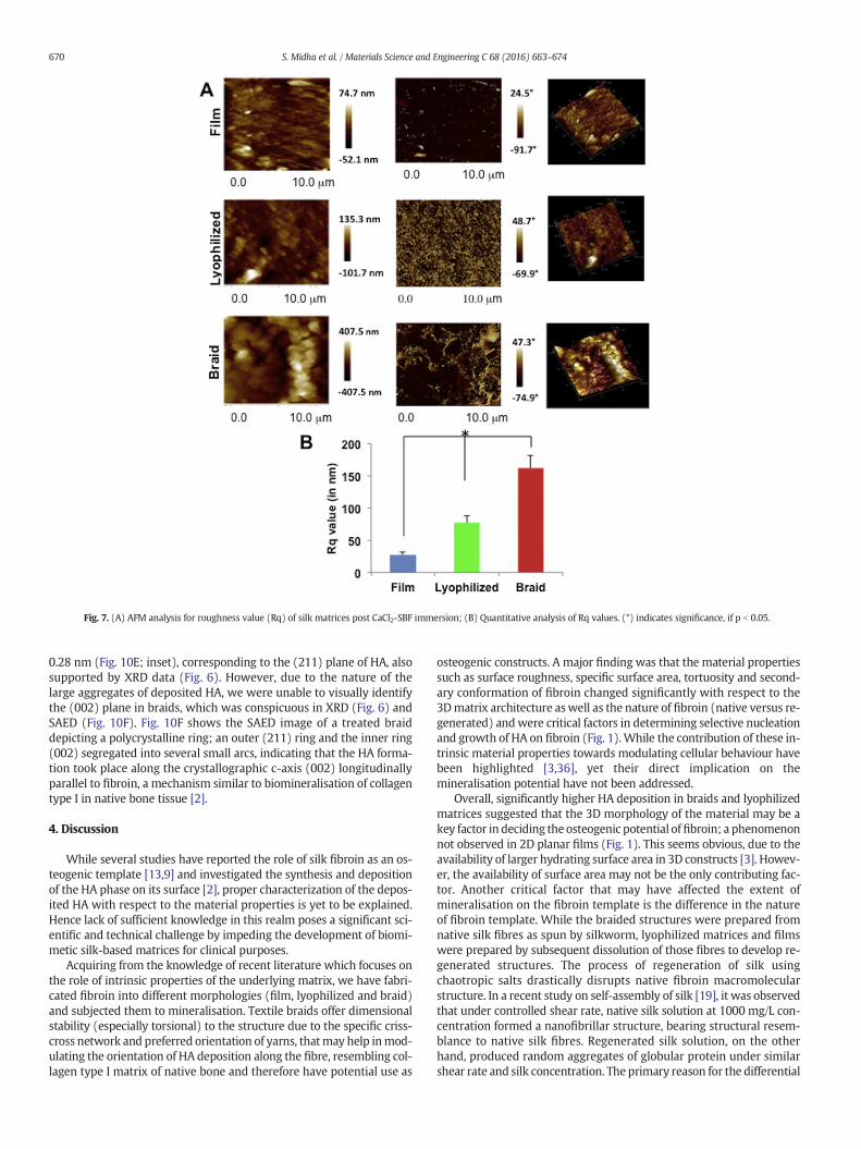
Fig. 7. (A) AFM analysis for roughness value (Rq) of silk matrices post CaCl2-SBF immersion; (B) Quantitative analysis of Rq values. (*) indicates significance, if p b 0.05.
670 S. Midha et al. / Materials Science and Engineering C 68 (2016) 663–674
0.28 nm (Fig. 10E; inset), corresponding to the (211) plane of HA, alsosupported by XRD data (Fig. 6). However, due to the nature of thelarge aggregates of deposited HA, we were unable to visually identifythe (002) plane in braids, which was conspicuous in XRD (Fig. 6) andSAED (Fig. 10F). Fig. 10F shows the SAED image of a treated braiddepicting a polycrystalline ring; an outer (211) ring and the inner ring(002) segregated into several small arcs, indicating that the HA forma-tion took place along the crystallographic c-axis (002) longitudinallyparallel to fibroin, a mechanism similar to biomineralisation of collagentype I in native bone tissue [2].
4. Discussion
While several studies have reported the role of silk fibroin as an os-teogenic template [13,9] and investigated the synthesis and depositionof the HA phase on its surface [2], proper characterization of the depos-ited HA with respect to the material properties is yet to be explained.Hence lack of sufficient knowledge in this realm poses a significant sci-entific and technical challenge by impeding the development of biomi-metic silk-based matrices for clinical purposes.
Acquiring from the knowledge of recent literature which focuses onthe role of intrinsic properties of the underlying matrix, we have fabri-cated fibroin into different morphologies (film, lyophilized and braid)and subjected them to mineralisation. Textile braids offer dimensionalstability (especially torsional) to the structure due to the specific criss-cross network and preferred orientation of yarns, thatmay help inmod-ulating the orientation of HA deposition along the fibre, resembling col-lagen type I matrix of native bone and therefore have potential use as
osteogenic constructs. A major finding was that the material propertiessuch as surface roughness, specific surface area, tortuosity and second-ary conformation of fibroin changed significantly with respect to the3Dmatrix architecture aswell as the nature of fibroin (native versus re-generated) andwere critical factors in determining selective nucleationand growth of HA on fibroin (Fig. 1).While the contribution of these in-trinsic material properties towards modulating cellular behaviour havebeen highlighted [3,36], yet their direct implication on themineralisation potential have not been addressed.
Overall, significantly higher HA deposition in braids and lyophilizedmatrices suggested that the 3D morphology of the material may be akey factor in deciding the osteogenic potential offibroin; a phenomenonnot observed in 2D planar films (Fig. 1). This seems obvious, due to theavailability of larger hydrating surface area in 3D constructs [3]. Howev-er, the availability of surface area may not be the only contributing fac-tor. Another critical factor that may have affected the extent ofmineralisation on the fibroin template is the difference in the natureof fibroin template. While the braided structures were prepared fromnative silk fibres as spun by silkworm, lyophilized matrices and filmswere prepared by subsequent dissolution of those fibres to develop re-generated structures. The process of regeneration of silk usingchaotropic salts drastically disrupts native fibroin macromolecularstructure. In a recent study on self-assembly of silk [19], it was observedthat under controlled shear rate, native silk solution at 1000 mg/L con-centration formed a nanofibrillar structure, bearing structural resem-blance to native silk fibres. Regenerated silk solution, on the otherhand, produced random aggregates of globular protein under similarshear rate and silk concentration. The primary reason for the differential

Fig. 8. (A) ATR-FTIR spectra treated silk matrices post CaCl2-SBF immersion; (B) Spectra showing characteristic HA peaks in selected region (400–1100 cm−1); (a–f) Peak shift in amidegroups of lyophilized (a–c) and braids (d–f).
671S. Midha et al. / Materials Science and Engineering C 68 (2016) 663–674
assembly of the two solutions was attributed to the subsequent frag-mentation of the constituted macromolecules or lack of terminal do-main dimerisation due to treatment with chaotropic salts in the caseof regenerated silk fibroin [19]. This implies that the intact native struc-ture of the silk fibre in braids offers specific organization of the amino
Fig. 9. EDX micrographs of treated silk matrices post CaCl2-SBF immersion; lyophilized,braid and commercial HA.
acid domains for nucleation of HA, which could not be exactlyreproduced in the regenerated matrices due to random aggregated as-sembly and branching of fibroin network post-dissolution. Hence thismay exert influence on the differential mineralisation observed in the3D matrices (lyophilized versus braids), in spite of having comparablephysical characteristics (Fig. 1).
In vitro nucleation and growth of HA nanocrystals onto organic tem-plates is tightly regulated by the presence of electronegative amino acidgroups on the material surface, where the crystal formation originates[37]. Exposure of these different amino acid sites is mainly dependenton the varied surface morphology/architecture of the matrix, whichcan immensely affect biomineralisation on fibroin. Since the specificsurface area (Fig. 1) as well as β-sheet content (Fig. 3) was maximal inthe case of braided structures in addition to the intact native proteinconformation, more Asp groups would be exposed to the surface,hence resulting in distinctive carbonated apatite formationwith prefer-ential (002) orientation along the c-axis plane (Figs. 6 & 10); character-istic of native bone tissue [2]. Moreover, the relatively decreasedtortuosity index of the braided structures in comparison to lyophilizedmatrices (Fig. 1) ensured effective permeability and higher contactwith supersaturated solution which would subsequently precipitate
Table 3Summary of constitutional Ca and P in treated materials.
Matrix typeCa(wt%)
P(wt%) Ca/P ratio
Lyophilized 0.446 0.571 0.78Braid 27.458 15.686 1.75HA commercial 25.338 15.430 1.64

Fig. 10. TEMmicrographs of treated silk matrices post CaCl2-SBF immersion. (A) Ultrasonicated silk fibroin films showing amorphous-like deposition (inset); (B) Lyophilizedmatrix withtypical needle-like HAnanocrystals; (C–E) Braids showing single fibre (arrow)with characteristic HA deposition along its length; (D) Highermagnification of boxed region in (C) showingneedle-like HA morphology; (E) Higher magnification lattice image showing interplanar spacing (inset), (F) SAED of treated braid showing polycrystalline HA deposition.
672 S. Midha et al. / Materials Science and Engineering C 68 (2016) 663–674
Ca2+ and P along the structure. Researchers have also acknowledgedthe existence of another mineralisation mechanism explicitlyestablished in zebra fish bone [38], as well as in human enamel [39],where the nucleation was initiated not through the ions in solutionbut via an intermediary amorphous calcium precursor. However, lackof sufficient knowledge in this realm makes the generality of this path-way debatable [40,41].
SEM analysis confirmed the presence of certain low attenuation de-positions on the material surface with a more aggregated organisationon 3D matrices whose Ca/P ratio was close to commercial HA [35](Fig. 9), while the films depicted more individually scattered nano-par-ticles (Fig. 5). According to FTIR analysis, there was a discrete shift ob-served in amide peaks when the untreated matrices (braids andlyophilized) were compared to CaCl2-SBF treated groups confirmingstrong chemical interactions betweenHA-silk fibroin (Fig. 8). This resultalso highlighted an important aspect of the mechanism underlyingmineralisation indicating the involvement of amide groups in the originof nucleation of HA nanocrystals; as observed previously by others [2].The presence of impurities such as carbonate have frequently been de-tected along with HA and is therefore used as a criterion for HA charac-terization [42], as detected in braids and lyophilized structures. The TEMimages demonstrated that the fibroin could direct the assembly and nu-cleation of HA nanocrystals along the template (Fig. 10). The typicalneedle-like morphology of HA crystals was well represented on 3Dmorphologies only, with aspect ratio measurements of nanocrystalsclose to that of synthesized HA [43]. The SAED images indicated a poly-crystalline diffractogram for braided structures [34] (Fig. 10) with pref-erential orientation extending along the c-axis, as also confirmed fromthe values of intensity ratio (Fig. 6). This preferential orientation of thedeposited crystals along the c-axis of the template is a typical featureof calcium phosphate noted in human bone and dentin where the c-axes of HA crystals align parallel to longitudinal axis of type I collagenfibrils [44]. This was further confirmed from the Scherrer Equation forXRD suggesting that HA crystal formed on braids was located on thetemplate surface (Fig. 6) with high probability of (002) crystal facematching well with spatial configuration of the fibroin template. Fur-ther, broadening of the (002) and (211) peaks corresponding to HA
suggested poor crystallisation in the case of braids and lyophilized sim-ilar to organic-inorganic interactions in bone physiology [2].
In native bone tissue, the structural integrity of bone is a tightly reg-ulated process controlled by coordinated mechanism of collagen syn-thesis by osteoblasts and their subsequent mineralisation by poorlycrystalline HA [45]. The crystal morphology of needle-like nanocrystalsobtained on fibroin in our studywas similar to the dimensions found oncollagen-HA, as reported by previous studies [46]. Also the immensestructural resemblance between the two fibrous proteins; silk fibroinand collagen, raised several questions on their relatable mechanism inregulating the HA crystal growth. In a comparative study conducted tocompare the mineralisation potential of single-templates; collagen-HAand fibroin-HA and bi-template; collagen/fibroin-HA, it was revealedthat crystallite growth orientation along thepreferential c-axis of HAoc-curred in both the templates, similar to native bone while FTIR con-firmed the chemical interaction between HA with fibroin or collagenin single- and bi-templates suggesting their similar osteogenic potential[46]. A cell-independent mechanism described crystal growth of HA oncollagen, whereby non-collagenous proteins present within the gapzones of collagen template direct mineral nucleation from ions in phys-iological solution [45]; a similar mechanism has also been experimen-tally proven for fibroin by Marelli et al. [20] Due to the lack ofconcrete evidence and sufficient literature, aspects of apatite formationon fibroin are much less understood, however, the assistance of β-sheetsecondary conformation in the fibroin mineralisation is relatively wellcharacterized [41]. This phenomenon was also evident in our studywhereby the significantly higher β-sheet content found in the case of3D structures probably played a crucial role in directing the nucleationof HA nanocrystals along the fibres. However, the differentialmineralisation amongst the two 3D fibroin constructs (possessing com-parable β-sheet content) could only be justified by taking into accountthe nature of the fibroin template used (native versus regenerated) asexplained earlier.
Template-induced nucleation and growth of nanocrystals, similar tothe organic template of native bone (collagen type I), is a vital pathwayfor determining the structural relevance of fabricated materials. Severalnucleation mechanisms to regulate crystal growth on such organic

673S. Midha et al. / Materials Science and Engineering C 68 (2016) 663–674
templates have been proposed [2,36,41]. Nevertheless, the most com-monly accepted theory of crystal formation stems from the inorganicions in supersaturated solutions (such as SBF), which get adsorbedonto the templates in a precisely arranged crystallographic order,under controlled pHmicroenvironment. The process of matrix-mediat-edmineralisation in the living systems especially focusing on the role ofspecific amino acid sequences and secondary conformation of proteinsinfluencing the nucleation aspect of calcium phosphate crystals hasbeen reviewed [42]. In accordance with previous literature, we hypoth-esize that the nucleation and growth of the HA crystals was assisted bythe fibroin template where the extent of mineralisation was dictated bythe specific interactions between the crystal plane of HA and the specific3D arrangement of the fibroin template. In this study, we used SBF solu-tion at pH 7.4 (greater than pI of fibroin; pH 4.5), which facilitated theprecipitation of local Ca2+ ions from SBF solution into the β-sheet struc-ture of fibroin matrices (containing hydrophilic groups such as –NH–and –COO– chains of acidic macromolecules, especially aspartic acid)[37]. Once the Ca2+ ions were bound to the fibroin template, the elec-trostatic interactions attracted (PO4)3− and OH− groups to further as-similate more Ca2+ from the solution for promoting nucleation of HAalong the β-sheet assembly of fibroin chain; as stated by the doublelayer theory of calcium phosphate precipitation [47]. Hence the three-dimensionality of the underlying matrix (that aligns and probably dis-tributes the anionic groups in specific 3D arrangement) and tortuosity(that dictates the contact of SBF solution to the organic template; Fig.1) are critical factors that may affect the subsequent nucleation process.In addition, the larger the surface area (Fig. 1), the more probability ofion exchange will occur between the organic template and the ions insolution to facilitate enhancedHAdeposition. Another factor that affectsHA nucleation on the fibroin surface is the interfacial structure. HA willbe extended along a particular direction only when the interplanar dis-tance of HA fits well with the spacing between the amino acid (which isAsp in the case of fibroin) [2], considering that the formation of fibroin–Ca2+was facilitated by anionic functional groups. Since the distance be-tween Aspmolecule i.e. 0.15 nm is close to the distance between Ca2+–Ca2+ in (002) phase (0.13–0.165 nm), HA extended along the c-axisplane, only in the case of braided structures.
Once the nucleation process is initiated, the growth of HAwith timeon fibroin chains may well be justified by nucleation–aggregation–ag-glomeration theory [48,49]. Briefly, the deposited nanocrystals are ag-gregated by molecular attractions [50] which cause subsequentdecrease in the surface free energy. Meanwhile, further crystal growthresulted in the formation of agglomerated structures which further ledto increased particle sizes by promoting further aggregation of the ag-glomerated particles (Fig. 5). Although, the concentration of fibroin issufficient to further increase the crystal size, simultaneous depletionin the Ca2+ ionic concentration limits the resultant crystal size. Fromthe apparent broadening of (002) and (211) peaks and the calculationof Scherrer Equation we can infer a smaller crystal size for 3D fibroinstructures (Table 2). The biological relevance of this small particle sizeis that it may aids in solubility of bone by resorbing osteoclasts, aswell asmaintains themechanical properties, likely preventing the prop-agation of cracks [3]. Hence, we can conclude that HA-fibroin cross-linked together to form the inorganic-organic interactions resemblingnatural bone, though only when the fibroin template was nativelyspun in braided morphology. Hence this study further warrants the in-vestigation of cellular responses with respect to the two matrices (na-tive/regenerated) to gain insights into the underlying molecularmechanisms (for eg. Notch signaling, Wnt signaling etc.) [51] primarilyregulating the extent of osteogenesis on the silk matrices used.
However, one limitation of our study is that for a more direct com-parison between native versus regenerated silk fibroin, it would havebeen ideal to compare identical morphologies. But it is not possible tofabricate braided structures post-dissolution of cocoons. Taken togeth-er, braided structures prepared from native B. mori silk demonstratedimproved HA deposition. The specific orientation of the yarns and
structural stability of the network will play a critical role in determiningthe mineralisation potential. Lyophilized matrices, prepared by subse-quent dissolution of fibres, though possessing comparable β-sheet con-tent, demonstrated relatively lesser mineralisation. Changes in theextent of mineralisation with the method of processing implied thatthe growth and nucleation of nanocrystals is template-induced guidedby the intact protein conformation of native fibroin. Hence these find-ings suggest that the 3D textile technology-based structures, such asbraids, made up of native silk fibres may provide a suitable templatematrix, compared to regenerated silk, for producing hydroxyapatite-based mineralised materials for bone repair.
5. Conclusions
To gain an understanding of the mechanism of template-inducednucleation and growth of HA nanocrystals on silk fibroin, we have stud-ied the molecular interactions of HA with silk fibroin (native versus re-generated) fabricated in differentmorphologies. Ca2+ ions bound to thenatively spun braids trigger mineralisation in the presence of supersat-urated solution. The deposited nano-particles of HA interact closelywith the intact amino acid chains of native polymer and induce the nu-cleation of HAP nano-needles with their preferential c-axis orientationwith Ca/P ratio similar to native bone composition. Apart from the intactpolymer conformation, the intrinsic properties of the material play cru-cial role in modulating the extent of mineral deposition. Hence we con-clude that natively spun silk fibroin braids in part mimic the collagentype I matrix of natural bone and thus may serve as potential bonegraft substitutes for bone repair and regeneration.
Supplementary data to this article can be found online at http://dx.doi.org/10.1016/j.msec.2016.06.041.
Acknowledgements
This study was funded by Department of Science and Technology,Science and Engineering Board, India (YSS/2014/000472) and Depart-ment of Biotechnology, India (BT/PR8038/MED/32/303/2013). A por-tion of this work was performed at the Research Complex at Harwell,funded in part by the EPSRC (EP/I02249X/1).
References
[1] M. Iafisco, B. Palazzo, T. Ito, M. Otsuka, M. Senna, J. Delgado-Lopez, et al., Preparationof core-shell poly(L-lactic) acid-nanocrystalline apatite hollow microspheres forbone repairing applications, J. Mater. Sci. Mater. Med. 23 (2012) 2659–2669.
[2] J. Wang, F. Yu, L. Qu, X. Meng, G. Wen, Study of synthesis of nano-hydroxyapatiteusing a silk fibroin template, Biomed. Mater. 5 (2010) 041002.
[3] J. Gomez-Morales, M. Iafisco, J.M. Delgado-Lopez, S. Sarda, C. Drouet, Progress on thepreparation of nanocrystalline apatites and surface characterization: overview offundamental and applied aspects, Prog. Cryst. Growth Charact. Mater. 59 (2013)1–46.
[4] C. Drouet, Apatite formation: why it may not work as planned, and how to conclu-sively identify apatite compounds, BioMed Res. Int. 2013 (2013).
[5] S.H. Park, E.S. Gil, H. Shi, H.J. Kim, K. Lee, D.L. Kaplan, Relationships between degrad-ability of silk scaffolds and osteogenesis, Biomaterials 31 (2010) 6162–6172.
[6] X. Hu, S.H. Park, E.S. Gil, X.X. Xia, A.S. Weiss, D.L. Kaplan, The influence of elasticityand surface roughness on myogenic and osteogenic-differentiation of cells on silk-elastin biomaterials, Biomaterials 32 (2011) 8979–8989.
[7] B. Panilaitis, G.H. Altman, J. Chen, H.J. Jin, V. Karageorgiou, D.L. Kaplan, Macrophageresponses to silk, Biomaterials 24 (2003) 3079–3085.
[8] M. Bhattacharjee, E. Schultz-Thater, E. Trella, S. Miot, S. Das, M. Loparic, et al., Therole of 3D structure and protein conformation on the innate and adaptive immuneresponses to silk-based biomaterials, Biomaterials 34 (2013) 8161–8171.
[9] J. Melke, S. Midha, S. Ghosh, K. Ito, S. Hofmann, Silk fibroin as biomaterial for bonetissue engineering, Acta Biomater. 31 (2016) 1–16.
[10] M. Yang, W. He, Y. Shuai, S. Min, L. Zhu, Nucleation of hydroxyapatite crystals byself-assembled Bombyx mori silk fibroin, J. Polym. Sci. B Polym. Phys. 51 (2013)742–748.
[11] J.R. Vetsch, S.J. Paulsen, R. Muller, S. Hofmann, Effect of fetal bovine serum onmineralisation in silk fibroin scaffolds, Acta Biomater. 13 (2015) 277–285.
[12] M.J. Lee, J.B. Park, H.H. Kim, C.S. Ki, S.Y. Park, H.J. Kim, et al., Surface coating of hy-droxyapatite on silk nanofibre through biomineralisation using ten times concen-trated simulated body fluid and the evaluation for bone regeneration, Macromol.Res. 22 (2014) 710–716.
674 S. Midha et al. / Materials Science and Engineering C 68 (2016) 663–674
[13] H.J. Kim, U.J. Kim, H.S. Kim, C. Li, M. Wada, G.G. Leisk, et al., Bone tissue engineeringwith premineralised silk scaffolds, Bone 42 (2008) 1226–1234.
[14] G.S. Perrone, G.G. Leisk, T.J. Lo, J.E. Moreau, D.S. Haas, B.J. Papenburg, et al., The use ofsilk-based devices for fracture fixation, Nat. Commun. 5 (2014) 3385.
[15] R. Kino, T. Ikoma, A. Monkawa, S. Yunoki, M. Munekata, J. Tanaka, et al., Depositionof bone-like apatite on modified silk fibroin films from simulated body fluid, J. Appl.Polym. Sci. 99 (2006) 2822–2830.
[16] F. Lin, Y. Li, J. Jin, Y. Cai, K. Wei, J. Yao, Deposition behavior and properties of silk fi-broin scaffolds soaked in simulated body fluid, Mater. Chem. Phys. 111 (2008)92–97.
[17] L. Radev, T. Gerganov, H. Georgiev, A. Kolev, V. Vassileva, Silk fibroin/calcium phos-phate silicate composites : in vitro bioactivity, Int. J. Mater. Chem. 3 (2013) 8–15.
[18] X. He, X. Huang, Q. Lu, S. Bai, H. Zhu, Nanoscale control of silks for regular hydroxy-apatite formation, Prog. Nat. Sci.: Mater. Int. 22 (2012) 115–119.
[19] S.R. Koebley, D. Thorpe, P. Pang, P. Chrisochoides, I. Greving, F. Vollrath, et al., Silk re-constitution disrupts fibroin self-assembly, Biomacromolecules 16 (2015)2796–2804.
[20] B. Marelli, C.E. Ghezzi, A. Alessandrino, J.E. Barralet, G. Freddi, S.N. Nazhat, Silk fibro-in derived polypeptide-induced biomineralisation of collagen, Biomaterials 33(2012) 102–108.
[21] P. Dubey, S. Murab, S. Karmakar, P.K. Chowdhury, S. Ghosh, Modulation of self-as-sembly process of fibroin: an insight for regulating the conformation of silk bioma-terials, Biomacromolecules 16 (2015) 3936–3944.
[22] X.D. Kong, F.Z. Cui, X.M. Wang, M. Zhang, W. Zhang, Silk fibroin regulatedmineralisation of hydroxyapatite nanocrystals, J. Cryst. Growth 270 (2004)197–202.
[23] S. Ghosh, S.T. Parker, X. Wang, D.L. Kaplan, J.A. Lewis, Direct-write assembly ofmicroperiodic silk fibroin scaffolds for tissue engineering applications, Adv. Funct.Mater. 18 (2008) 1883–1889.
[24] S. Murab, S. Chameettachal, M. Bhattacharjee, S. Das, D.L. Kaplan, S. Ghosh, Matrix-embedded cytokines to simulate osteoarthritis-like cartilage microenvironments,Tissue Eng. A 19 (2013) 1733–1753.
[25] T. Kokubo, H. Kushitani, S. Sakka, T. Kitsugi, T. Yamamuro, Solutions able to repro-duce in vivo surface-structure changes in bioactive glass-ceramic A–W, J. Biomed.Mater. Res. 24 (1990) 721–734.
[26] A. Takeuchi, C. Ohtsuki, T. Miyazaki, H. Tanaka, M. Yamazaki, M. Tanihara, Deposi-tion of bone-like apatite on silk fiber in a solution that mimics extracellular fluid,J. Biomed. Mater. Res. A 65 (2003) 283–289.
[27] A.M. Parfitt, Bone histomorphometry: standardization of nomenclature, symbolsand units (summary of proposed system), Bone 9 (1988) 67–69.
[28] D.P. Fyhrie, R. Zauel, Directional tortuosity as a predictor of modulus damage for ver-tebral cancellous bone, J. Biomech. Eng. 137 (2015) 011007.
[29] L. De Nardo, S. Bertoldi, M.C. Tanzi, H.J. Haugen, S. Farè, Shape memory polymer cel-lular solid design for medical applications, Smart Mater. Struct. 20 (2011) 035004.
[30] L.V. Azaroff, Elements of X-ray Crystallography, McGraw-Hill, New York, 1968.[31] Y. Cai, J. Jin, D. Mei, N. Xia, J. Yao, Effect of silk sericin on assembly of hydroxyapatite
nanocrystals into enamel prism-like structure, J. Mater. Chem. 19 (2009)5751–5758.
[32] M.P. Ferraz, The role of dialysis and freezing on structural conformation, thermalproperties and morphology of silk fibroin hydrogels, Biomatter 4 (2014), e28536.
[33] J. Rnjak-Kovacina, L.S. Wray, K.A. Burke, T. Torregrosa, J.M. Golinski, W. Huang, D.L.Kaplan, Lyophilized silk sponges: a versatile biomaterial platform for soft tissue en-gineering, ACS Biomater. Sci. Eng. 1 (2015) 260–270.
[34] W. Zhang, S.S. Liao, F.Z. Cui, Hierarchical self-assembly of nano-fibrils in mineralisedcollagen, Chem. Mater. 15 (2003) 3221–3226.
[35] M.P. Ferraz, F.J. Monteiro, C.M. Manuel, Hydroxyapatite nanoparticles: a review ofpreparation methodologies, J. Appl. Biomater. Biomech. 2 (2004) 74–80.
[36] D. Terada, Y. Yokoyama, S. Hattori, H. Kobayashi, Y. Tamada, The outermost surfaceproperties of silk fibroin films reflect ethanol-treatment conditions used in biomate-rial preparation, Mater. Sci. Eng. C 58 (2016) 119–126.
[37] Y. Li, Y. Cai, X. Kong, J. Yao, Anisotropic growth of hydroxyapatite on the silk fibroinfilms, Appl. Surf. Sci. 255 (2008) 1681–1685.
[38] J. Mahamid, B. Aichmayer, E. Shimoni, R. Ziblat, C. Li, S. Siegel, et al., Mapping amor-phous calcium phosphate transformation into crystalline mineral from the cell tothe bone in zebrafish fin rays, Proc. Natl. Acad. Sci. U. S. A. 107 (2010) 6316–6321.
[39] E. Beniash, R.A. Metzler, R.S.K. Lam, P.U.P.A. Gilbert, Transient amorphous calciumphosphate in forming enamel, J. Struct. Biol. 166 (2009) 133–143.
[40] A. Dey, P.H. Bomans, F.A. Muller, J. Will, P.M. Frederik, G. de With, et al., The role ofprenucleation clusters in surface-induced calcium phosphate crystallisation, Nat.Mater. 9 (2010) 1010–1014.
[41] F.C. Meldrum, H. Colfen, Controlling mineral morphologies and structures in biolog-ical and synthetic systems, Chem. Rev. 108 (2008) 4332–4432.
[42] G.K. Hunter, Interfacial aspects of biomineralisation, Curr. Opinion Solid State Mater.Sci. 1 (1996) 430–435.
[43] I.A. Figueroa, O. Novelo-Peralta, C. Flores-Morales, R. Gonzalez-Tenorio, M.C. Pina-Barba, Synthesis and characterization of biocompatible-nanohydroxyapatite crystalsobtained by a modified sol-gel processing, Biomatter 2 (2012) 71–76.
[44] Z. Qiu, Y. Cui, C. Tao, Z. Zhang, P. Tang, K. Mao, et al., Mineralised collagen: rationale,current status, and clinical applications, Materials 8 (2015) 4733–4750.
[45] S. Boonrungsiman, E. Gentleman, R. Carzaniga, N.D. Evans, D.W. McComb, A.E.Porter, et al., The role of intracellular calcium phosphate in osteoblast-mediatedbone apatite formation, PNAS 109 (2012) 14170–14175.
[46] J. Wang, W. Zhou, W. Hu, L. Zhou, S. Wang, S. Zhang, Collagen/silk fibroin bi-tem-plate induced biomimetic bone-like substitutes, J. Biomed. Mater. Res. A 99 (A)(2011) 327–334.
[47] M.J. Lochhead, S.R. Letellier, V. Vogel, Assessing the role of interfacial electrostatics inoriented mineral nucleation at charged organic monolayers, J. Phys. Chem. B 101(1997) 10821–10827.
[48] A.D. Randolph, M.A. Larson, Theory of Particulate Processes, Elsevier, 1988.[49] R. Rodríguez-Clemente, a. López-Macipe, J. Gómez-Morales, J. Torrent-Burgués, V.M.
Castaño, Hydroxyapatite precipitation: a case of nucleation-aggregation-agglomera-tion-growth mechanism, J. Eur. Ceram. Soc. 18 (1998) 1351–1356.
[50] J. Gomez-Morales, J. Torrent-Burgues, R. Rodriguez-Clemente, Crystal size distribu-tion of hydroxyapatite precipitated in a MSMPR reactor, Cryst. Res. Technol. 36(2001) 1065–1074.
[51] S. Midha, S. Murab, S. Ghosh, Osteogenic signaling on silk-based matrices, Biomate-rials 97 (2016) 133–153.