measures of cardiovascular autonomic activity in insomnia ... · measures of cardiovascular...
TRANSCRIPT

RESEARCH ARTICLE
Measures of cardiovascular autonomic
activity in insomnia disorder: A systematic
review
Marina-Marinela Nano1,2,3*, Pedro Fonseca1,3, Rik Vullings1, Ronald M. Aarts1,3
1 Department of Electrical Engineering, Eindhoven University of Technology, Eindhoven, The Netherlands,
2 Sleep Medicine Centre Kempenhaeghe, Heeze, The Netherlands, 3 Philips Research, High Tech Campus,
Eindhoven, The Netherlands
Abstract
Background
Insomnia disorder is a widespread sleep disorder with a prevalence of approximately 10%.
Even though the link between insomnia and cardiovascular activity is not exactly clear, it is
generally assumed that cardiovascular autonomic modifications could occur as a result of
sleeplessness, or, alternatively, that autonomic alterations could be an expression of a
hyper-arousal state. This review investigates whether cardiovascular measures are different
between insomniacs and controls.
Methods
Electronic databases were systematically searched, and 34 studies were identified. Heart
rate variability features, the association of cardiac and EEG activity, physiologic complexity
measures, and cardiovascular activity, assessed by measures such as pre-ejection time,
blood pressure, and heart rate dynamics were studied. Given the heterogeneity of the stud-
ies, a narrative synthesis of the findings was performed.
Results
This review study found overall differences in cardiovascular activity between insomniacs
and controls in most of the observational studies (21/26), while the expression of cardiovas-
cular regulation varied between the examined insomniac groups. All the studies that investi-
gated the association of cardiac activity and EEG power reported an altered relation
between autonomic activity and EEG parameters in insomniacs.
Conclusion
Autonomic regulation tends to be consistent between insomniacs, as long as they are
grouped according to their respective phenotype, as shown in the insomnia subgroup with
objectively short sleep duration. Our hypothesis is that these differences in the expression
of cardiovascular activity could be explained by the heterogeneity of the disorder. Therefore,
PLOS ONE | https://doi.org/10.1371/journal.pone.0186716 October 23, 2017 1 / 31
a1111111111
a1111111111
a1111111111
a1111111111
a1111111111
OPENACCESS
Citation: Nano M-M, Fonseca P, Vullings R, Aarts
RM (2017) Measures of cardiovascular autonomic
activity in insomnia disorder: A systematic review.
PLoS ONE 12(10): e0186716. https://doi.org/
10.1371/journal.pone.0186716
Editor: Jacobus P. van Wouwe, TNO,
NETHERLANDS
Received: June 16, 2017
Accepted: October 8, 2017
Published: October 23, 2017
Copyright: © 2017 Nano et al. This is an open
access article distributed under the terms of the
Creative Commons Attribution License, which
permits unrestricted use, distribution, and
reproduction in any medium, provided the original
author and source are credited.
Data Availability Statement: All relevant data are
within the paper and its Supporting Information
files.
Funding: This work has been done in the IMPULS
framework (Eindhoven University of Technology,
Philips Research, Sleep Medicine Centre
Kempenhaeghe). The funders had no role in the
study design, decision to publish, or preparation of
the manuscript. Philips Research provided support
in the form of salaries for authors PF and RMA, but
did not have any additional role in the study design,
data collection and analysis, decision to publish, or

the determination of insomnia phenotypes, and the study of cardiovascular measures,
rather than heart rate variability alone, will give more insight into the link between insomnia
and cardiovascular regulation. This study suggests that cardiovascular activity differs
between insomniacs and controls. These new findings are of interest to clinicians and
researchers for a more accurate insomnia assessment, and the development of personal-
ized technological solutions in insomnia.
Introduction
Difficulties initiating or maintaining sleep are very prevalent sleep complaints in the general
population [1, 2]. If sleeplessness meet specific diagnostic criteria the term insomnia disorder
is used. Multinational studies that used the Diagnostic and Statistical Manual of Mental Disor-
ders fourth edition (DSM-IV) criteria reported prevalence rates of insomnia disorder that
range from 3.9% to 22.1%, with an average of approximately 10% [3]. This is a broad range
that reflects different modalities of investigation and the population under study [1]. Informa-
tion from new studies on the prevalence of insomnia disorder using DSM-V criteria, is cur-
rently limited.
On the latest update of DSM, insomnia disorder is defined as a predominant complaint of
dissatisfaction with sleep quality or duration and is accompanied by difficulties in initiating
sleep at bedtime, frequent or prolonged awakenings, or early-morning awakening, with an
inability to return to sleep [4]. This sleep disturbance causes clinically significant social, occu-
pational, educational, academic, and behavioral distress or impairment. These difficulties
occur despite adequate opportunity for sleep. Diagnosis of insomnia is made when sleep diffi-
culties are present for 3 or more nights per week, and last for more than 3 months [4]. Thus,
insomnia is a condition characterized by both nocturnal and diurnal symptoms.
The cardiovascular autonomic nervous system (ANS) appears to be closely linked to sleep
and circadian physiology, as demonstrated by the disrupted autonomic control that accompa-
nies sleep loss [5]. Additionally, autonomic activity is integrated with cognition and emotion,
among others [6]. Sleep loss or deficiency can usually occurs as a result of sleep deprivation,
sleep fragmentation, or difficulty of falling asleep. In insomnia disorder sleep loss is usually
caused by difficulties of maintaining (fragmented sleep) or initiating sleep. In addition to sleep
loss, insomnia disorder is frequently accompanied by various changes, such as cognitive
arousal/stress, degraded mood, depression or anxiety and fatigue [7]. To date, two major
hypotheses have been made about the link between autonomic function and insomnia [8].
According to the first hypothesis, autonomic modifications could occur as a result of sleep
fragmentation [9]. This hypothesis is supported by studies showing that autonomic arousals
without cortical involvement are an epiphenomenon of sleep fragmentation and altered sleep
continuity [9]. Furthermore, autonomic sleep fragmentation has been linked to diurnal
increase in sympathetic activity and elevated blood pressure (BP) in healthy elderly [10].
According to the second hypothesis, autonomic alterations could be an expression of a hyper-
arousal state [8]. Evidence of an increase in heart rate (HR) and the absence of a normal drop
in autonomic activity during falling asleep, along with alterations of other physiologic parame-
ters (e.g. body temperatures, stress hormones), could be considered indicators of a state of
arousal that predisposes the individual to poor sleep [11–13]. Therefore, relevant physiology
data obtained by cardiovascular ANS measures may provide new insight into the link between
insomnia disorder and cardiovascular autonomic activity.
Cardiovascular autonomic activity in insomnia disorder
PLOS ONE | https://doi.org/10.1371/journal.pone.0186716 October 23, 2017 2 / 31
preparation of the manuscript. The specific roles of
these authors are articulated in the ‘author
contributions’ section.
Competing interests: M-MN, PF and RMA declare
to be affiliated with Philips Research. RV reports
other from Nemo Healthcare, outside his submitted
work. This does not alter our adherence to all PLOS
ONE policies on sharing data and materials.

While two reviews [14, 15] examining heart rate variability (HRV) and one review [16]
investigating cardiovascular dysfunction between normal sleep and sleep disorders have been
published, their primary focus was not insomnia disorder, so findings regarding insomnia
were not methodically incorporated. One review [17] focusing exclusively on HRV and insom-
nia was recently published. Cardiovascular activity, compared to HRV alone, provides a more
complete overview of the autonomic activity. For instance, studies have shown that the use of
HRV for the estimation of autonomic regulation has limitations [18] and additional diagnostic
value can be obtained from measures such as pre-ejection period (PEP) [19]. In this study, we
do not restrict the review to HRV, as Dodds et al. [17] did, but also incorporate other cardio-
vascular measures of autonomic activity, such as PEP, cardiopulmonary coupling (CPC), left
ventricular ejection time, BP and HR slope for the analysis of HR dynamics in order to investi-
gate whether cardiovascular activity measures are different between insomniacs and controls.
In addition, we aim to examine how interventions influence cardiovascular activity.
Methods
Search strategy
For this review, the PubMed and Scopus electronic databases were systematically searched for
articles published until 9th of October 2016, using keywords to identify all studies specifically
designed to define cardiovascular differences between insomniacs and healthy controls. A
search of publications was conducted using the following medical subject headings or key
words: “heart rate”, “cardiac”, “cardio”, “blood pressure”, “autonomic”, “sympathetic”, “para-
sympathetic”, “arterial”, “vascular”, “baroreflex” and “insomnia”. Based on search options pro-
vided by the two electronic databases, the search approach was adjusted as shown in S1
Appendix. To ensure literature saturation, we examined the reference lists of the included
papers and of the relevant reviews which were identified by the search. The literature search
was limited to studies conducted with human participants, published in the English language.
Study selection
In order to identify relevant publications, the following criteria were applied in the initial
stages of the scrutiny process: (1) participants were adult (� 18 years old), (2) studies include
adults participants diagnosed with insomnia (observational) or treated for insomnia (interven-
tional), (3) comparison of insomniacs with control group (observational studies) or same
group of insomniacs before and after intervention (interventional studies), and (4) observa-
tional studies include non-invasive techniques, but not in vitro tests, such as saliva test. Articles
meeting these criteria were collected and data was extracted for analysis by the first author.
After duplicate removal studies were reviewed for eligibility using title, abstract and full text
when it was required.
Cardiovascular measures used to explore autonomic changes and their
physiological significance interpretation
In this section, cardiovascular measures used in literature to investigate and study autonomic
changes are introduced. Additionally, their physiological interpretation is presented.
Over the past years, different methods of cardiovascular autonomic activity and HRV quan-
tification have been developed, such as frequency, time-frequency, temporal, geometrical, and
nonlinear analysis [20]. Autonomic cardiovascular measures can be examined traditionally
through the quantification of average HR and BP [5] and more recently through non-linear
approaches by using detrended fluctuation analysis (DFA), [21] entropy derived [15, 22–24],
Cardiovascular autonomic activity in insomnia disorder
PLOS ONE | https://doi.org/10.1371/journal.pone.0186716 October 23, 2017 3 / 31

Poincare Plot [25], and Lempel-Ziv [26, 27] measures. As described in detail previously [5],
HR and BP variations can be expressed by the standard deviation around the mean, or by their
rhythmic and non-rhythmic components. RR time series (the time elapsed between two suc-
cessive R-waves of the QRS complex on the electrocardiogram (ECG)) and BP also show
short-term oscillations in a frequency range between 0 and 0.5 Hz. Traditionally, HRV and
cardiovascular parameters are measured in the time and frequency domain.
Standard HRV analysis has been well summarized by the task force of the European society
of cardiology [28]. The most commonly used time domain measures are described in Table 1.
Rate pressure product (RPP) is an index of the overall cardiac workload [29], and is calculated
as follows: HR � systolic BP/100 [30]. HR and BP physiologically decrease at night, compared
to during the day. Systolic BP reduction at least 10% during sleep, compared to daytime, is
commonly referred to as “dipping”. The PEP is influenced by sympathetic activity [5].
In the frequency domain, HRV is evaluated by spectral analysis. As described in detail pre-
viously [5, 28, 31], spectral analysis of RR intervals and BP variability gives information on
how power of the signal is distributed as a function of the frequency. Kay and Marple pre-
sented an extensive summary of several techniques used for spectral analysis [32]. Methods for
power spectral density estimation can be generally classified as non-parametric and parametric
[28]. The two most common approaches [33, 34] used for spectral analysis of RR time series
are Fourier transform (FFT) [31] and autoregressive model (AR) [35]. The high frequency
power (HF) components (0.15-0.4 Hz) reflect the respiration-driven modulation of sinus
rhythm, and have been used as an index of tonic vagal drive [5, 28, 31, 34, 36, 37]. The physio-
logical significance of the very low frequency (VLF) component is still unclear, and limited
data suggest that it might reflect vagal and rein-angiotensin system effects on HR [14, 28, 31,
34]. The physiological interpretation of the low frequency (LF) power components (0.04-0.15
Hz) is controversial. Some studies [28, 31, 38] support the conclusion that LF power is consid-
ered to reflect both sympathetic and vagal modulation of the heart, while other studies [5, 39]
indicate that it might be an index of the baroreflex sensitivity (BRS) for control of HR. More-
over, for some researchers [28, 37, 40, 41], LF is seen as a marker of sympathetic modulation,
particularly when it is expressed in normalized units. LF rhythm can also be modulated by
irregular breathing patterns [5]. Consequently, the LF/HF ratio is considered by some
researchers to express sympatho-vagal balance, and by others, to reflect only sympathetic
Table 1. Summary of time domain cardiovascular measures and their physiological interpretation.
Feature Description ANS interpretation Study
PEP the time from the onset of the ECG Q-wave to the opening of the
aortic valve
a marker of beta-adrenergic sympathetic activity [12, 59–64]
RPP the product of HR and SBP index of the overall cardiac workload [63]
SDNN standard deviation of RR or NN intervals for a desired period and
is measured in ms
both sympathetic and parasympathetic activity and
therefore provides an index of overall HRV
[8, 13, 47, 60,
64–67]
RMSSD square root of the mean squared differences of successive NN
intervals for a desired period, measured in ms
parasympathetic activity [8, 13, 47, 60,
61, 64–67]
pNN50 percentage of successive NN intervals that differ more than 50
ms
parasympathetic activity [8, 13, 60, 64–
66]
Abbreviations— ANS: Autonomic nervous system, ECG: electrocardiogram, HRV: heart rate variability, ms: milliseconds, PEP: pre-ejection time, RPP: rate
pressure product, RR time series: the time elapsed between two successive R-waves of the QRS complex on the electrocardiogram, study: represents the
studies that the feature was used, Note: Abbreviation not mentioned here are described in the “Description” column of the table.
https://doi.org/10.1371/journal.pone.0186716.t001
Cardiovascular autonomic activity in insomnia disorder
PLOS ONE | https://doi.org/10.1371/journal.pone.0186716 October 23, 2017 4 / 31

modulations [28]. It should be noted that Eckberg et al. [42] questioned the use of the LF as an
indicator of sympatho-vagal tone balance. As described previously [28], these disagreements
in the interpretation of LF can be attributed to the fact that several conditions associated with
sympathetic activation, can cause a decrease in the absolute power of the LF component. For
example, during sympathetic activation, tachycardia follows, and is usually characterized by a
reduction in total power, while the opposite happens during vagal activation [28]. In this way,
when the LF is measured in milliseconds squared, the variations in total power affect LF and
HF in the same direction(for details see [28]). Due to the reduction in total power, LF could
remain unaltered if it is measured in milliseconds squared. Nevertheless, if normalization is
performed, an increase in LF becomes more evident [28]. Other reasons that could explain this
discrepancy include the fact that respiration parameters and behavior are influenced by age
and activity, among others. For instance, during tasks, individual differences might exhibit a
wide range of spontaneous breathing rates, which may result in a contribution to the HF band
by individuals with faster breathing frequencies, and a contribution to the LF band by individ-
uals with slower breathing frequencies [43].
Regarding BP variability, LFBP components in systolic BP variability are considered an
index of efferent sympathetic vascular modulation, whereas the HFBP components express
mechanical effects of respiration on blood pressure changes [5]. BRS regulates BP in order to
preserve stability. BRS can be measured by either provocation of the carotid baroreceptors
with phenylephrine or by the spectral technique which quantifies spontaneous fluctuations of
the systolic blood pressure spectral power and the corresponding RR time series spectral
power in different frequency bands [44–46]. The latter approach was first introduced by
Robbe et al. [45] and is used by the authors of the reviewed studies [47]. The αBRS is computed
using the square root of the ratio of RR time series and systolic BP power spectra in the LF and
HF bands (αLF and αHF) [47–49]. The α-index is computed only when the squared coherence
function (k2) of the systolic BP and RR time series exceeded 0.56 [47–49]. αTotal is defined as
the mean of αLF and αHF. TF-BRS (the evaluation of the transfer function between time series
of systolic BP and RR time series) is computed by averaging the gain function in the LF band
regardless of a given coherence between systolic BP and RR time series [47, 48, 50]. The αLF
describes the gain of the relation between the BP and RR time series power spectra in the LF
band [47, 48, 50]. The αLF component describes the gain in the spectral band of the respiration
frequency. αTotal gives an assessment of the overall baroreceptor gain [51].
Recently, new methods have been used for the analysis of HRV, in order to consider the
non-stationary characteristics of the ECG signal and the non-linear fluctuations in HR. These
techniques attempt to characterize cardiovascular ANS in terms of regularity and complexity,
based on information carried by RR time series through the use of entropy derived and Lem-
pel-Ziv measures [22, 23, 26, 27]. Sample entropy is the negative logarithm of conditional
probability of the sequences of RR time series. High sample entropy shows that there is a low
probability of repeated sequences in the RR time series, which means lower regularity and
more complexity in the RR time series [22, 52]. Multiscale entropy is estimated based on the
computation of the sample entropy over a range of temporal scales [23] (For more details
about sample and multiscale entropy see [22, 23]). The Lempel-Ziv complexity algorithm pro-
vides information regarding the complexity of RR time series [27]. Complexity is related to the
number of distinct patterns along the RR time series and the rate of their occurrence within a
given sequence [26]. (For more details regarding the Lempel-Ziv complexity algorithm and
the coding procedure used in the reviewed studies see [53, 54]). Detrended fluctuation analysis
(DFA) examines the fractal scaling properties of HR fluctuations in the non-stationary RR
time series on different time scales for the detection of long-range correlation between the RR
intervals [21]. CPC was introduced by Thomas et al. [55] as the product of the coherence and
Cardiovascular autonomic activity in insomnia disorder
PLOS ONE | https://doi.org/10.1371/journal.pone.0186716 October 23, 2017 5 / 31

cross-spectral power of the RR or NN time series and the ECG-derived respiratory time series.
ECG-spectrographic variables were found to correlate strongly with EEG measures of sleep
stability, suggesting that the resulting sleep spectrogram can classify sleep as “stable” (high-fre-
quency coupling band (0.1—0.4 Hz) (HFC)) and “unstable” (low-frequency coupling band
(0.1—0.4 Hz) (LFC)) [55–57]. The cardiovascular measures and their interpretation that are
used in the reviewed studies are presented in Tables 1, 2 and 3. For details about the
Table 2. Summary of nonlinear cardiovascular measures.
Feature Description Study
entropy of RR time series a non-linear measure which examines the regularity of RR time
series and it increases with greater degree of irregularity reaching a
maximum at completely random system
[53,
68]
Lempel-Ziv complexity of
RR time series
a non-linear measure that estimates the complexity of RR time
series and quantifies the rate of new patterns along the sequence
[53]
DFA of RR time series a non-linear measure that characterizes the pattern of variation and
long-range correlations of RR time series across multiple time
scales
[53]
Abbreviations— DFA: detrended fluctuation analysis, RR time series: the time elapsed between two
successive R-waves of the QRS complex on the electrocardiogram, study: represents the studies that the
feature was used, Note: Abbreviation not mentioned here are described in the “Description” column of the
table.
https://doi.org/10.1371/journal.pone.0186716.t002
Table 3. Summary of the frequency cardiovascular measures and their physiological interpretation.
Feature Description ANS interpretation Study
Total
power
variance of all RR or NN intervals measured in ms2 [59, 60]
VLF Low frequency power (0.003—0.04 Hz) measured in ms2 parasympathetic activity and renin-
angiotensin system effects on HR
[68]
LF very low frequency power (0.04—0.15 Hz) measured in ms2 measure that includes both sympathetic
and vagal influence*[8, 11–13, 47, 59, 60,
64–67, 69–78]
LFnorm low frequency power (0.04—0.15 Hz) normalized using total power marker of sympathetic modulation* [8, 11–13, 47, 59, 60,
64–67, 69–78]
HF high frequency power (0.15—0.4 Hz) measured in ms2 marker of parasympathetic/vagal activity [8, 13, 47, 60, 64–67]
HFnorm high frequency power (0.15—0.4 Hz) normalized using total power marker of parasympathetic/vagal activity [8, 13, 47, 60, 64–67]
LF/HF ratio of LF to HF reflects sympatho/vagal balance or
sympathetic modulations*[8, 11, 13, 59, 60, 64–
67, 69–71, 73–77]
1/f slope of the power-low regression line of RR time series fitted to the
power spectrum for f < 0.01 Hz
[53]
CPC the product of the coherence and cross-spectral power of the RR or NN
time series and the ECG-derived respiratory time series
[57]
αLF the squared root of the ratio of RR time series and systolic BP power
spectra in the (0.04-0.15 Hz) frequency band measured in ms/mmHg
[47]
αHF the squared root of the ratio of RR time series and systolic BP power
spectra in the (0.15-0.4 Hz) frequency band measured in ms/mmHg
[47]
αTotal mean of αLF and αHF measured in ms/mmHg [47]
Abbreviations— ANS: autonomic nervous system, CPC: cardiopulmonary coupling, HRV: heart rate variability, mmHg: millimeter of mercury, ms:
milliseconds, Total power: variance of all NN or RR intervals, study: represents the studies that the feature was used
*: the interpretation is controversial, Note: Abbreviation not mentioned here are described in the “Description” column of the table.
https://doi.org/10.1371/journal.pone.0186716.t003
Cardiovascular autonomic activity in insomnia disorder
PLOS ONE | https://doi.org/10.1371/journal.pone.0186716 October 23, 2017 6 / 31

description and ANS interpretation columns of the Tables 1, 2, and 3 please see [5, 14, 21–23,
28, 47–51, 53–55, 58].
Results
Reviewed studies
The initial combined database search generated 709 records. After duplicate removal and
English language restriction, 427 electronic records were identified and screened for eligibility.
The manual search of reference lists of relevant papers and reviews identified three papers
[53, 64, 72]. Three additional studies [79–81] were added to explain the transition from “poor”
sleepers to clinically defined insomniacs. The vast majority of these articles (n = 366) were
excluded by title or abstract alone (see Fig 1) based on criteria mentioned earlier. Full text arti-
cles were obtained for the 61 remaining articles. Ultimately, 34 studies were identified that met
the inclusion criteria for this review. Twenty six observational studies ([8, 11–13, 47, 53, 57,
59–63, 65, 66, 68–73, 82–87]) and eight interventional studies ([64, 67, 74–78, 88]). Study
details such as diagnostic criteria, demographics, number of participants, insomnia severity,
and type of intervention are presented in Tables 4 and 5.
Observational studies
Studies without specific diagnostic criteria for insomnia disorder. In studies without
specific diagnostic criteria for insomnia disorder, physiological differences during sleep were
investigated between the “poor” and “good” sleeper groups [79], as well as subjects with sleep-
Fig 1. Study search and selection for measures of cardiovascular activity in insomniacs and controls.
Modified PRISMA 2009 flow diagram [89].
https://doi.org/10.1371/journal.pone.0186716.g001
Cardiovascular autonomic activity in insomnia disorder
PLOS ONE | https://doi.org/10.1371/journal.pone.0186716 October 23, 2017 7 / 31
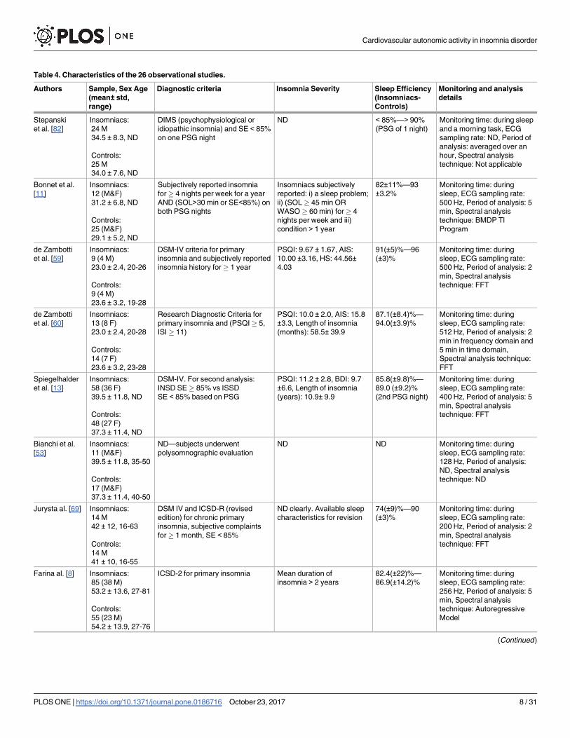
Table 4. Characteristics of the 26 observational studies.
Authors Sample, Sex Age
(mean± std,
range)
Diagnostic criteria Insomnia Severity Sleep Efficiency
(Insomniacs-
Controls)
Monitoring and analysis
details
Stepanski
et al. [82]
Insomniacs:
24 M
34.5 ± 8.3, ND
Controls:
25 M
34.0 ± 7.6, ND
DIMS (psychophysiological or
idiopathic insomnia) and SE < 85%
on one PSG night
ND < 85%—> 90%
(PSG of 1 night)
Monitoring time: during sleep
and a morning task, ECG
sampling rate: ND, Period of
analysis: averaged over an
hour, Spectral analysis
technique: Not applicable
Bonnet et al.
[11]
Insomniacs:
12 (M&F)
31.2 ± 6.8, ND
Controls:
25 (M&F)
29.1 ± 5.2, ND
Subjectively reported insomnia
for� 4 nights per week for a year
AND (SOL>30 min or SE<85%) on
both PSG nights
Insomniacs subjectively
reported: i) a sleep problem;
ii) (SOL� 45 min OR
WASO� 60 min) for� 4
nights per week and iii)
condition > 1 year
82±11%—93
±3.2%
Monitoring time: during
sleep, ECG sampling rate:
500 Hz, Period of analysis: 5
min, Spectral analysis
technique: BMDP Tl
Program
de Zambotti
et al. [59]
Insomniacs:
9 (4 M)
23.0 ± 2.4, 20-26
Controls:
9 (4 M)
23.6 ± 3.2, 19-28
DSM-IV criteria for primary
insomnia and subjectively reported
insomnia history for� 1 year
PSQI: 9.67 ± 1.67, AIS:
10.00 ±3.16, HS: 44.56±4.03
91(±5)%—96
(±3)%
Monitoring time: during
sleep, ECG sampling rate:
500 Hz, Period of analysis: 2
min, Spectral analysis
technique: FFT
de Zambotti
et al. [60]
Insomniacs:
13 (8 F)
23.0 ± 2.4, 20-28
Controls:
14 (7 F)
23.6 ± 3.2, 23-28
Research Diagnostic Criteria for
primary insomnia and (PSQI� 5,
ISI � 11)
PSQI: 10.0 ± 2.0, AIS: 15.8
±3.3, Length of insomnia
(months): 58.5± 39.9
87.1(±8.4)%—
94.0(±3.9)%
Monitoring time: during
sleep, ECG sampling rate:
512 Hz, Period of analysis: 2
min in frequency domain and
5 min in time domain,
Spectral analysis technique:
FFT
Spiegelhalder
et al. [13]
Insomniacs:
58 (36 F)
39.5 ± 11.8, ND
Controls:
48 (27 F)
37.3 ± 11.4, ND
DSM-IV. For second analysis:
INSD SE� 85% vs ISSD
SE < 85% based on PSG
PSQI: 11.2 ± 2.8, BDI: 9.7
±6.6, Length of insomnia
(years): 10.9± 9.9
85.8(±9.8)%—
89.0 (±9.2)%
(2nd PSG night)
Monitoring time: during
sleep, ECG sampling rate:
400 Hz, Period of analysis: 5
min, Spectral analysis
technique: FFT
Bianchi et al.
[53]
Insomniacs:
11 (M&F)
39.5 ± 11.8, 35-50
Controls:
17 (M&F)
37.3 ± 11.4, 40-50
ND—subjects underwent
polysomnographic evaluation
ND ND Monitoring time: during
sleep, ECG sampling rate:
128 Hz, Period of analysis:
ND, Spectral analysis
technique: ND
Jurysta al. [69] Insomniacs:
14 M
42 ± 12, 16-63
Controls:
14 M
41 ± 10, 16-55
DSM IV and ICSD-R (revised
edition) for chronic primary
insomnia, subjective complaints
for� 1 month, SE < 85%
ND clearly. Available sleep
characteristics for revision
74(±9)%—90
(±3)%
Monitoring time: during
sleep, ECG sampling rate:
200 Hz, Period of analysis: 2
min, Spectral analysis
technique: FFT
Farina al. [8] Insomniacs:
85 (38 M)
53.2 ± 13.6, 27-81
Controls:
55 (23 M)
54.2 ± 13.9, 27-76
ICSD-2 for primary insomnia Mean duration of
insomnia > 2 years
82.4(±22)%—
86.9(±14.2)%
Monitoring time: during
sleep, ECG sampling rate:
256 Hz, Period of analysis: 5
min, Spectral analysis
technique: Autoregressive
Model
(Continued )
Cardiovascular autonomic activity in insomnia disorder
PLOS ONE | https://doi.org/10.1371/journal.pone.0186716 October 23, 2017 8 / 31
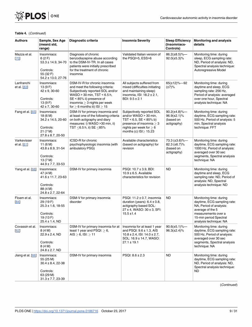
Table 4. (Continued)
Authors Sample, Sex Age
(mean± std,
range)
Diagnostic criteria Insomnia Severity Sleep Efficiency
(Insomniacs-
Controls)
Monitoring and analysis
details
Mazza et al.
[70]
Insomniacs:
6 (2 F)
53.3 ± 14.9, 34-70
Controls:
55 (32 F)
54.2 ± 13.0, 27-76
Diagnosis of chronic
benzodiazepine abuse according
to the DSM-IV-TR. In all cases
patients were initially prescribed
for the treatment of chronic
insomnia
Validated Italian version of
the PSQI>5, ESS>6
86.2(±8.5)%—
92.0(±5.3)%
Monitoring time: during
sleep, ECG sampling rate:
ND, Period of analysis: ND,
Spectral analysis technique:
Autoregressive Model
Lanfranchi
et al. [83]
Insomniacs:
13 (9 F)
42 ± 9, 30-60
Controls:
13 (9 F)
42 ± 7, 30-60
DSM-IV-R for chronic insomnia
and meet the following criteria:
Subjectively reported SOL and/or
WASO > 30 min, TST < 6.5 h,
SE < 85% ii) presence of
insomnia� 3 nights per week
for� 6 months iii) ISI� 15
All subjects suffered from
mixed (difficulties initiating
and maintaining sleep)
insomnia, ISI: 18.2 ± 2.1,
BDI: 9.5 ± 2.1
65(±12)%—92
(±7)%
Monitoring time: during
daytime and sleep, ECG
sampling rate: 256 Hz,
Period of analysis: averaged
over one hour, Spectral
analysis technique: NA
Fang et al. [65] Insomniacs:
18 (6 M)
34.2 ± 14.5, 20-60
Controls:
21 (7 M)
27.8 ± 8.7, 20-50
DSM-IV for primary insomnia and
at least one of the following criteria
on both actigraphy and diary
measures: i) WASO >30 min, ii)
TST�6.5 h, ii) SE�85%
Subjectively reported SOL
and/or WASO > 30 min,
TST < 6.5, SE < 85% b)
presence of insomnia� 3
nights per week for� 6
months (c) ISI� 15.23
90.2(±4.8)%—
96.9(±2.1)%
(based on
actigraphy)
Monitoring time: during
daytime, ECG sampling rate:
500 Hz, Period of analysis: 5
min, Spectral analysis
technique: FFT
Varkevisser
et al. [61]
Insomniacs:
11 (6 M)
43.8 ± 8.9, 31-54
Controls:
13 (7 M)
44.9 ± 7.7, 33-53
ICSD-R for chronic
psychophysiologic insomnia (with
ambulatory PSG)
Available characteristics
(based on actigraphy) for
revision
73.3 (±3.6)%—
82.3 (±6.7)%
(based on
actigraphy)
Monitoring time: during
daytime, ECG sampling rate:
1000 Hz, Period of analysis:
averaged over 30 sec
segments, Spectral analysis
technique: NA
Yang et al. [68] Insomniacs:
47 (4 M)
41.6 ± 11.7, 23-63
Controls:
88 (4 M)
24.8 ± 2.7, 22-64
DSM-IV for primary insomnia PSQI: 10.7 ± 3.9, BDI:
10.9 ± 6.5. Available
characteristics for revision
ND Monitoring time: during
daytime and sleep, ECG
sampling rate: ND, Period of
analysis: ND, Spectral
analysis technique: ND
Floam et al.
[84]
Insomniacs:
29 (19 F)
25.3 ± 1.6, 18-55
Controls:
19 (13 F)
25.4 ± 1.4, ND
DSM-V for primary insomnia
disorder
PSQI: 11.2 ± 0.7, insomnia
duration (years): 6.4 ± 0.8,
actigraphy based SOL:
27 ± 4, WASO: 30 ± 3, SFI:
15.5 ±1.4
ND Monitoring time: during
daytime, ECG sampling rate:
NA, Period of analysis:
average of the 5
measurements over a
15-min period Spectral
analysis technique: NA
Covassin et al.
[62]
Insomniacs:
8 (4 M)
22.9 ± 2.4, ND
Controls:
8 (4 M)
24.8 ± 2.7, ND
DSM-IV for primary insomnia for at
least 1 year and PSQI:� 6,
AIS� 6, ISI:� 11
Insomnia for at least 1 year
and PSQI: 9.6 ± 1.3, AIS
10.8 ± 2.4, ISI: 14.0 ± 2.7,
SOL: 16.9 ± 14.7, WASO:
27.1 ± 19.1
90.6(±5.1)%—
96.3(±2.4)%
Monitoring time: during
daytime, ECG sampling rate:
500 Hz, Period of analysis:
averaged over 30 sec
segments, Spectral analysis
technique: NA
Jiang et al. [66] Insomniacs:
55 (25 M)
30.4 ± 8.4, 22-38
Controls:
63 (29 M)
31.3 ± 7.7, 23-39
DSM-IV for primary insomnia PSQI: 8.6 ± 2.3 ND Monitoring time: during
daytime, ECG sampling rate:
ND, Period of analysis: ND,
Spectral analysis technique:
ND
(Continued )
Cardiovascular autonomic activity in insomnia disorder
PLOS ONE | https://doi.org/10.1371/journal.pone.0186716 October 23, 2017 9 / 31
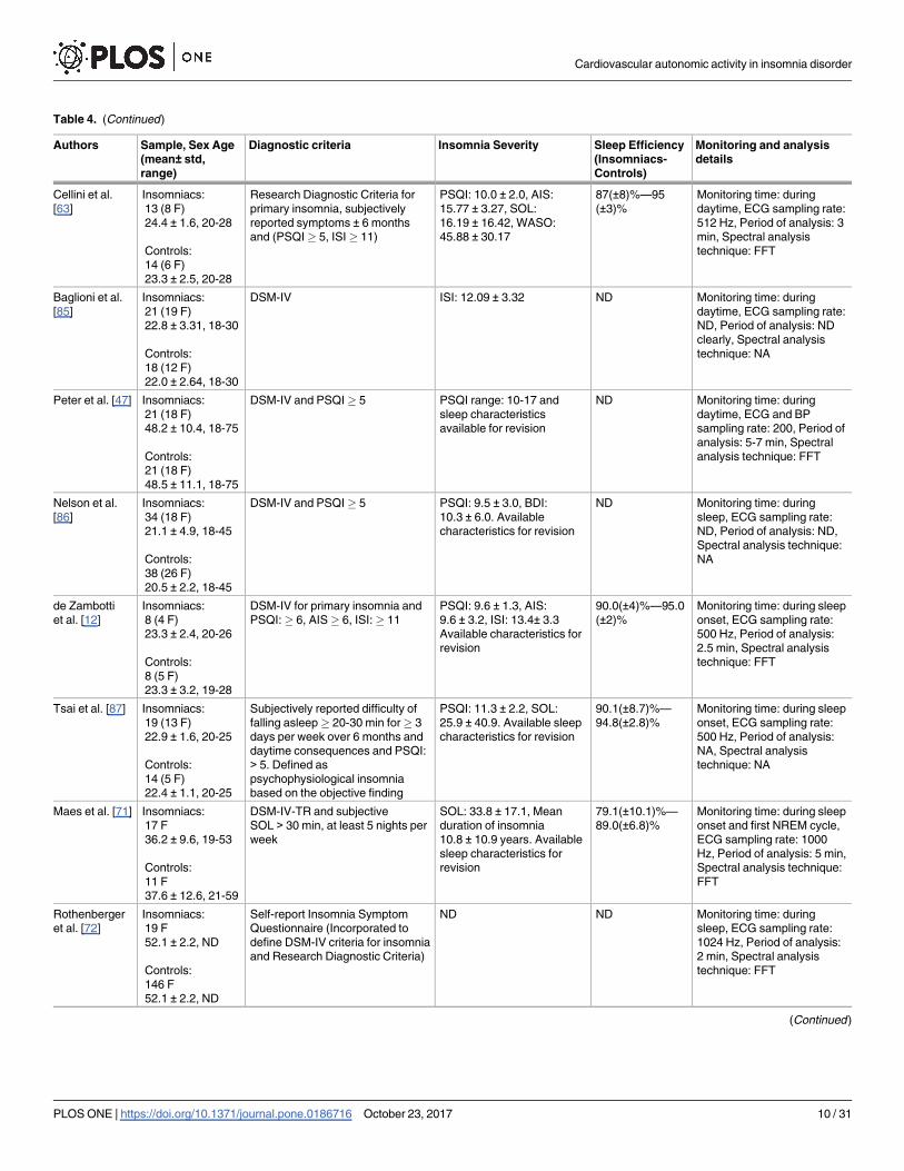
Table 4. (Continued)
Authors Sample, Sex Age
(mean± std,
range)
Diagnostic criteria Insomnia Severity Sleep Efficiency
(Insomniacs-
Controls)
Monitoring and analysis
details
Cellini et al.
[63]
Insomniacs:
13 (8 F)
24.4 ± 1.6, 20-28
Controls:
14 (6 F)
23.3 ± 2.5, 20-28
Research Diagnostic Criteria for
primary insomnia, subjectively
reported symptoms ± 6 months
and (PSQI� 5, ISI� 11)
PSQI: 10.0 ± 2.0, AIS:
15.77 ± 3.27, SOL:
16.19 ± 16.42, WASO:
45.88 ± 30.17
87(±8)%—95
(±3)%
Monitoring time: during
daytime, ECG sampling rate:
512 Hz, Period of analysis: 3
min, Spectral analysis
technique: FFT
Baglioni et al.
[85]
Insomniacs:
21 (19 F)
22.8 ± 3.31, 18-30
Controls:
18 (12 F)
22.0 ± 2.64, 18-30
DSM-IV ISI: 12.09 ± 3.32 ND Monitoring time: during
daytime, ECG sampling rate:
ND, Period of analysis: ND
clearly, Spectral analysis
technique: NA
Peter et al. [47] Insomniacs:
21 (18 F)
48.2 ± 10.4, 18-75
Controls:
21 (18 F)
48.5 ± 11.1, 18-75
DSM-IV and PSQI� 5 PSQI range: 10-17 and
sleep characteristics
available for revision
ND Monitoring time: during
daytime, ECG and BP
sampling rate: 200, Period of
analysis: 5-7 min, Spectral
analysis technique: FFT
Nelson et al.
[86]
Insomniacs:
34 (18 F)
21.1 ± 4.9, 18-45
Controls:
38 (26 F)
20.5 ± 2.2, 18-45
DSM-IV and PSQI� 5 PSQI: 9.5 ± 3.0, BDI:
10.3 ± 6.0. Available
characteristics for revision
ND Monitoring time: during
sleep, ECG sampling rate:
ND, Period of analysis: ND,
Spectral analysis technique:
NA
de Zambotti
et al. [12]
Insomniacs:
8 (4 F)
23.3 ± 2.4, 20-26
Controls:
8 (5 F)
23.3 ± 3.2, 19-28
DSM-IV for primary insomnia and
PSQI:� 6, AIS� 6, ISI:� 11
PSQI: 9.6 ± 1.3, AIS:
9.6 ± 3.2, ISI: 13.4± 3.3
Available characteristics for
revision
90.0(±4)%—95.0
(±2)%
Monitoring time: during sleep
onset, ECG sampling rate:
500 Hz, Period of analysis:
2.5 min, Spectral analysis
technique: FFT
Tsai et al. [87] Insomniacs:
19 (13 F)
22.9 ± 1.6, 20-25
Controls:
14 (5 F)
22.4 ± 1.1, 20-25
Subjectively reported difficulty of
falling asleep� 20-30 min for� 3
days per week over 6 months and
daytime consequences and PSQI:
> 5. Defined as
psychophysiological insomnia
based on the objective finding
PSQI: 11.3 ± 2.2, SOL:
25.9 ± 40.9. Available sleep
characteristics for revision
90.1(±8.7)%—
94.8(±2.8)%
Monitoring time: during sleep
onset, ECG sampling rate:
500 Hz, Period of analysis:
NA, Spectral analysis
technique: NA
Maes et al. [71] Insomniacs:
17 F
36.2 ± 9.6, 19-53
Controls:
11 F
37.6 ± 12.6, 21-59
DSM-IV-TR and subjective
SOL > 30 min, at least 5 nights per
week
SOL: 33.8 ± 17.1, Mean
duration of insomnia
10.8 ± 10.9 years. Available
sleep characteristics for
revision
79.1(±10.1)%—
89.0(±6.8)%
Monitoring time: during sleep
onset and first NREM cycle,
ECG sampling rate: 1000
Hz, Period of analysis: 5 min,
Spectral analysis technique:
FFT
Rothenberger
et al. [72]
Insomniacs:
19 F
52.1 ± 2.2, ND
Controls:
146 F
52.1 ± 2.2, ND
Self-report Insomnia Symptom
Questionnaire (Incorporated to
define DSM-IV criteria for insomnia
and Research Diagnostic Criteria)
ND ND Monitoring time: during
sleep, ECG sampling rate:
1024 Hz, Period of analysis:
2 min, Spectral analysis
technique: FFT
(Continued )
Cardiovascular autonomic activity in insomnia disorder
PLOS ONE | https://doi.org/10.1371/journal.pone.0186716 October 23, 2017 10 / 31

onset insomnia [80, 81]. Monroe [79] reported significantly higher HR 30 minutes before
sleep in “poor” sleepers as compared to “good” sleepers. During sleep, HR of the “poor” sleep-
ers was slightly, but not significantly, higher. Freedman et al. [81] found that sleep-onset
insomniacs had increased HR prior to sleep, but not during sleep. Similarly, Haynes et al. [80]
reported that sleep-onset insomniacs showed a mean HR 4.6 beats per minute, significantly
higher than that of non-insomniacs, whilst the effects of pre-sleep cognitive stress were exam-
ined. Haynes et al. [80], concluded that insomniacs show higher levels of physiological arousal
compared to non-insomniac subjects.
Nocturnal cardiovascular activity. In 1994, Stepanski et al. [82] assessed physiological
activity in patients with objectively documented insomnia using specific American Sleep Dis-
orders Association diagnostic criteria [90]. Subjects with chronic insomnia and normal sleep-
ers slept in the laboratory overnight and were given a stressful performance task in the
morning. HR was assessed for all participants before sleep, during sleep, and in response to
acute stress. Nocturnal HR was significantly higher in insomniacs. The morning after, no dif-
ference was found in HR between the two groups, but HR was significantly higher during the
stressful performance task in insomniacs. These results, regarding increased nocturnal HR,
were confirmed by Bonnet et al. [11]. In particular, sleep and ECG measures were evaluated in
insomnia patients and matched controls. In this study, spectral analysis of HRV revealed sig-
nificant increases in LF and LF/HF and a decrease in HF in insomniacs compared to controls.
Those changes were present across all sleep stages.
Recently, de Zambotti et al. [59] investigated nocturnal cardiovascular modifications in pri-
mary insomniacs compared to healthy controls, focusing on cardiac autonomic functioning.
They found a significant, constant shorter PEP during all sleep stages of the insomniac group
compared to good sleepers. In addition, they found the presence of short PEP to be directly
associated with low quality of sleep assessed by the Pittsburgh Sleep Quality Index (PSQI) and
Table 4. (Continued)
Authors Sample, Sex Age
(mean± std,
range)
Diagnostic criteria Insomnia Severity Sleep Efficiency
(Insomniacs-
Controls)
Monitoring and analysis
details
Schramm et al.
[57]
Insomniacs:
50 (27 M)
46.4 ± 8.6, 30-63
Controls:
36 (17 M)
44.5 ± 8.7, 30-63
DSM-IV for primary insomnia PSQI: 12.11 ± 2.69 83.2(±10.1)%—
89.1(±6.7)%
(from the 2nd
night)
Monitoring time: during
sleep, ECG sampling rate:�
200 Hz, Period of analysis:
8.5 min, Spectral analysis
technique: FFT
Israel et al. [73] Insomniacs:
54 (30 F)
34.6 ± 9.7, ND
Controls:
22 (19 M)
26.5 ± 7.3, ND
DSM-IV for primary insomnia SOL: 26.1 ± 18.3, WASO:
41.4 ± 33.5
85.6(±8.0)%—
90.5(±5.5)%
Monitoring time: during
sleep, ECG sampling rate:
1024 Hz, Period of analysis:
2 min, Spectral analysis
technique: Autoregressive
Model
Abbreviations— AIS: Athens Insomnia Scale, BDI: Beck depression inventory, CBT: Cognitive behavioral therapy, DSM: Diagnostic and Statistical Manual
of Mental Disorders, ECG: electrocardiogram, ESS: Epworth Sleepiness Scale, F: female, FFT: fast Fourier transform, ICSD: International Classification of
Sleep Disorders, INSD: Insomniacs with normal sleep duration, ISI: Insomnia Severity Index, ISSD: Insomniacs with short sleep duration, IV: 4th edition, M:
male, min: minutes, NA: not applicable, ND: not defined, NREM: Non-rapid eye movement sleep, PSG: polysomnography, PSQI: Pittsburgh Sleep Quality
Index, R: revised, SE: sleep efficiency, sec: second, SOL: Sleep onset latency, TR: text revised, TST: Total sleep time, WASO: Wake after sleep onset.
https://doi.org/10.1371/journal.pone.0186716.t004
Cardiovascular autonomic activity in insomnia disorder
PLOS ONE | https://doi.org/10.1371/journal.pone.0186716 October 23, 2017 11 / 31

Table 5. Characteristics of the eight interventional studies.
Authors Sample, Sex Age
(mean±std, range)
Diagnostic criteria Follow-up period
Study type
Intervention Insomnia Severity
Sleep Efficiency
Monitoring and
analysis details
Jobert
et al. [88]
16 (4 M)
(66.7 ± 5.8, ND)
ICSD for chronic or
subcronic
psychophysiological
insomnia
ND placebo-
controlled,
randomized, 3-fold
crossover
bezodiazepin
lormetazepam (1mg)
cyclopyrrolone
zopiclone (7.5mg)
placebo
ND Monitoring time: during
sleep, ECG sampling
rate: 200 Hz, Period of
analysis: averaged
over 30 sec segments,
Spectral analysis
technique: NA
Lo et al.
[74]
18 (7 M)
(43.2 ± 15.4, ND)
Subjective complaints of
difficulty initiating sleep
and/or maintaining sleep
for� 3 months
4 weeks after the
completion of dose
titration open-label
Gabapentin Mean
dose 540 mg Range
200-900 mg
Before:
PSQI: 13.54�
80.00% After:
PSQI: 7.67�
87.17%
Monitoring time: during
sleep, ECG sampling
rate: 400 Hz, Period of
analysis: 10 min,
Spectral analysis
technique: ND
Chung
et al. [64]
Responders:
16 (6 M)
(57.9 ± 10.9, ND)
Non-responders:
10 (4 M)
(59.4 ± 7.4, ND)
ICSD-2 8-week period after
the beginning of
CBT open-label
CBT of 4 sessions,
one every other week
over an eight-week
period
Responders-
Before:
ISI: 19.5 ± 4.0
77.7 ±18.9
After:
ISI: < 8
ND
Non-responders-
Before:
ISI:18.1 ± 4.9
70.8 ± 26.5
After:
ISI: > 8 ND
Monitoring time: during
daytime, ECG
sampling rate: ND,
Period of analysis: 5
min, Spectral analysis
technique: ND
Jarrin
et al. [75]
65 (22 M)
(51.8 ± 10.0, ND)
DSM-IV-TR and ICSD-2
for chronic insomnia
6-week period after
the beginning of
CBT open-label
CBT, frequency of
sessions was not
specified
Before:
ISI: 17.0±4.0
Subjective:
69.4±15.5%
Objective: 80.1
±12.1%
After:
8.8±3.5
Subjective:
83.8±10
Objective:
88.6±8.4
Monitoring time: during
sleep, ECG sampling
rate: ND, Period of
analysis: 2 min,
Spectral analysis
technique: FFT
Litscher
et al. [76]
28 (5 M)
(41.9 ± 14.6, 22-82)
Self-presentation at the
hospital due to insomnia.
AIS ranged from 6-21
10 min before, 20
min during, and 10
min after
stimulation of the
Shenmen acupoint
open-label
Acupuncture by using
the Shenmen
acupuncture point on
the left wrist
Before:
AIS:
12.4±3.6
ND
After:
ND
ND
Monitoring time: during
daytime, ECG
sampling rate: 4096
Hz, Period of analysis:
5 min, Spectral
analysis technique: ND
Wang
et al. [77]
31 (6 M)
(54.3 ± 10.6, 39-82)
Self-presentation at the
hospital due to insomnia
and AIS� 7 and a range
of 7-22
10 min before, 20
min during, and 10
min after
stimulation of the
Shenmen acupoint
open-label
Acupuncture by using
the Shenmen
acupuncture point on
the left ear
Before:
AIS: 14.7±4.4
ND
After:
ND
ND
Monitoring time: during
daytime, ECG
sampling rate: 4096
Hz, Period of analysis:
5 min, Spectral
analysis technique: ND
(Continued )
Cardiovascular autonomic activity in insomnia disorder
PLOS ONE | https://doi.org/10.1371/journal.pone.0186716 October 23, 2017 12 / 31

by the Athens insomnia scale (AIS). In another study by de Zambotti et al. [60], the authors
further studied ANS functioning in insomniacs and confirmed their previous results [59]
regarding shorter PEP during all sleep stages in insomniacs. However, no significant differ-
ences in vagal activity (expressed by: the standard deviation of RR or NN intervals for a desired
period (SDNN); the square root of the mean squared differences of successive NN intervals for
a desired period (RMSSD); and the percentage of successive NN intervals that differ more than
50 ms (pNN50)) were reported between insomniacs and controls. Moreover, pre-sleep RR
intervals duration (over a 5-min window) was positively associated with sleep efficiency (SE)
and negatively associated with wake after sleep onset (WASO) in insomniacs. Spiegelhalder
et al. [13] aimed at investigating the association between insomnia and alterations in polysom-
nographically determined nocturnal HR and HRV. In the insomnia group, results showed a
Table 5. (Continued)
Authors Sample, Sex Age
(mean±std, range)
Diagnostic criteria Follow-up period
Study type
Intervention Insomnia Severity
Sleep Efficiency
Monitoring and
analysis details
Chien
et al. [67]
Experimental group:
34 F (51.09 ± 3.73,
45-55)
Control group:
33 F (50.85 ± 3.73,
45-55)
Primary insomnia
(definition not specified
and Chinese version of
PSQI (CPSQI) > 5
at the 4th week
during treatment,
and after 12 weeks
of treatment cohort
study
lavender
aromatherapy
Experimental
group-
Before:
CPSQI: 9 (8,12)†
ND After:
CPSQI: ND
ND
Control group-
Before:
CPSQI: 11 (9,13)†
ND
After:
CPSQI: ND
ND
Monitoring time: during
daytime, ECG
sampling rate: ND,
Period of analysis: 3
min, Spectral analysis
technique: ND
Tsai
et al. [78]
Patient group:
14 ND
(22.50 ± 1.22, 20-25)
Control group:
14 ND (23.07 ± 1.64,
20-25)
Self-reported insomniacs
who met the DSM-IV
criteria for primary
insomnia and PSQI > 6
2 days later after
intervention (with a
1 week difference
of each other)
open-label
Controlled respiration
at a slow frequency
rate of 0.1 Hz, and a
forced rate of 0.2 Hz
during daytime rest
Patient group:
Before:
PSQI: 11.21±1.97
89.55±9.89
After:
PSQI: ND 94.38
±3.76* &
87.92±12.77**
Control group:
Before:
PSQI: 2.93±1.33
94.99±2.88
After:
PSQI: ND
94.47±2.80* &
93.48±3.24**
Monitoring time: during
daytime and sleep,
ECG sampling rate:
500 Hz, Period of
analysis: 64 secs,
Spectral analysis
technique: FFT
Abbreviations— AIS: Athens Insomnia Scale, CBT: Cognitive behavioral therapy, CPSQI: Chinese Pittsburgh Sleep Quality Index, DSM: Diagnostic and
Statistical Manual of Mental Disorders, F: female, FFT: fast Fourier transform, ICSD: International Classification of Sleep Disorders, ISI: Insomnia Severity
Index, M: male, min: minutes, NA: not applicable, ND: Not defined, PSQI: Pittsburgh Sleep Quality Index, sec: second
*: Paced breathing at 0.1 Hz
**: Paced breathing at 0.2 Hz�: global score†: Median (Interquartile Range) instead of mean value and standard deviation
https://doi.org/10.1371/journal.pone.0186716.t005
Cardiovascular autonomic activity in insomnia disorder
PLOS ONE | https://doi.org/10.1371/journal.pone.0186716 October 23, 2017 13 / 31
lower wake-to-sleep HR difference compared to controls. The SDNN, was also significantly
lower in the insomnia group. The authors [13] characterized the initial group of patients as
subjectively reported insomniacs and they split up the insomnia group according to SE values.
Thus, when restricting their analysis to insomnia patients with objectively determined short
sleep duration (SE<85%), they found decreased HF, as well as decreased RMSSD and pNN50
values. Furthermore, Yang et al. [68] studied the long-term diurnal profile of HRV between
insomniacs and healthy controls. In this study, HRV measures between awake and bed time
were compared. The analysis of complexity indexes of HR dynamics using multiscale entropy
derived measures, showed a considerable decrease in complexity during nighttime in insomni-
acs, compared to healthy subjects [68].
Schramm et al. [57] measured CPC in subjects with insomnia compared to good sleepers.
In insomniacs, they found a lower HFC and a higher LFC on both nights on which subjects
were polysomnographically monitored. In addition, HFC/LFC ratio was lower in both nights
for insomniacs compared to controls reflecting poorer sleep quality. According to the authors
HFC represents a marker of stable sleep while LFC represents a marker of unstable sleep.
Some studies [8, 53, 69, 70], however, have failed to provide differences between insomniacs
and controls over all these cardiovascular measures. In these studies, the cardiovascular differ-
ences are either not seen during all sleep stages or not confirmed for all studied parameters.
For instance, Bianchi et al. [53] when comparing long-term correlations and complexity of the
HRV (using sample and multiscale entropy, Lempel-Ziv complexity, detrended fluctuation
analysis, 1/f slope) during the night did not find significant differences except for a decreased
sample entropy (m = 2, r = 0.2). In another study by Jurysta et al. [69], HF, LF, and LF/HF ana-
lyzed from the first three non-rapid eye movement (NREM)-rapid eye movement (REM)
cycles did not reveal any significant differences between chronic insomniacs and healthy con-
trols. Similarly, no differences in nocturnal LF, HF, LF/HF power were found by Farina et al.
[8] during all sleep stages. In this study, 24 hours of ambulatory (home-based) polysomnogra-
phy (PSG) were recorded. Patients showed modifications of HR (increased) and (increased
normalized low frequency power (LFnorm)), consistent with increased sympathetic activity,
while awake before sleep and during early-stage-N2 (occurring in the first part of the night).
No significant differences between insomniacs and controls could be found during slow-wave
sleep (SWS), REM sleep, and, post-sleep wake. This study raises important questions regarding
the interpretation of the ANS meaning of HRV features. For example, the authors found an
increased RMSSD, LFnorm, and LF/HF in the early-stage N2 in the insomniac group. Using the
traditional interpretations that an increase in HR and the LF/HF ratio are linked with
increased sympathetic activity, seem to be in contradiction with the increase in RMSSD which
is often linked to an increase in parasympathetic activity [8]. In another study by Mazza et al.
[70] sleep modifications induced by chronic benzodiazepine (BDZ) abuse in chronic insomni-
acs were evaluated. In this study, six insomnia patients affected by chronic BDZ abuse were
compared to fifty five normal controls. No significant differences were found in LF, LF/HF
power between BDZ abusers and controls, with the exception of an increased HF component.
In addition, authors reported that abusers, compared to controls, had significantly lower
indexes of electroencephalographic (EEG) arousal in all sleep stages and lower indexes of
NREM sleep instability.
In the area of BP measurements during sleep in insomniacs, the literature is very limited.
Lanfranchi et al. [83] investigated the 24-hour profile of arterial BP (brachial cuff arterial) in
subjects with chronic primary insomnia, and tested the hypothesis that these subjects have
higher nighttime BP, and an attenuation of nocturnal BP dipping compared to good sleepers.
The authors documented significantly higher systolic BP and decreased systolic pressure dip-
ping across the night in primary insomnia patients, compared to controls. However, no
Cardiovascular autonomic activity in insomnia disorder
PLOS ONE | https://doi.org/10.1371/journal.pone.0186716 October 23, 2017 14 / 31

significant differences were observed in the HR and PSG-based profiles (with the exception of
fewer periodic limb movements observed in the insomnia group) between insomniacs and
good sleepers.
Cardiovascular activity during daytime. Several studies have been focused on the diurnal
cardiovascular activity in insomnia population [47, 61, 62, 65, 66, 84]. Fang et al. examined the
differences in HRV and daytime functioning. Five-minute recordings of ECG under paced
breathing were obtained from all participants who were resting in a supine position. Authors
found an increasing trend in LF/HF ratio in insomniacs compared to controls, but the differ-
ences between the two groups did not reach statistical significance [65]. Varkevisser et al. [61]
measured cardiovascular parameters (average HR, RMSSD, PEP) in a group of subjects with
chronic insomnia under strictly controlled constant-routine conditions with continuous wake-
fulness. Although physiologic indexes of arousal were slightly elevated in the insomnia group
relative to the controls, the differences between the groups were not statistically significant.
Floam et al. [84] investigated whether BP differs between individuals with insomnia disorder
and healthy sleepers. Standard oscillometric BP measurements were collected in a seated posi-
tion five times over a 15-min period during the day. Floam et al. [84] did not find any differ-
ences in BP measurements between individuals with insomnia disorder and healthy sleepers
during daytime.
On the other hand, when Yang et al. [68] investigated the long-term diurnal profile of
HRV, differences between insomniacs and healthy controls were seen. In this study, HRV
measures between awake state and bed-time were compared. Compared to controls, insomni-
acs exhibited significant reductions in SDNN, HF, RMSSD, pNN50 during awake period.
Alterations in LF/HF (increase), RMSSD (decrease), HF (decrease) were correlated with per-
ceived sleep questionnaire score, suggesting that according to the authors, altered cardiac auto-
nomic control and physiologic complexity is associated with poor sleep in patients with
insomnia.
Cardiovascular activity during daytime while performing tasks. Several studies exam-
ined HRV during daytime while subjects performed a task [62, 63, 66, 85]. Covassin et al. [62]
focused on cardiovascular re-activity to the task in primary insomnia. Cardiovascular re-activ-
ity is defined as the responsiveness of the cardiovascular system to react to a stressful task. The
task was administered in two sessions, before and after a night of polysomnographic recording.
Results of cardiovascular parameters showed higher HR and lower left ventricular ejection
time values in insomniacs, as compared to controls in the evening. PEP was continuously
reduced in insomniacs. Jiang et al. [66] aimed to examine HRV response to a postural change
manoeuvre, in primary insomniacs and controls. HRV features were computed at the follow-
ing times: seated rest and 0-5 min, 5-10 min and 10-15 min in the standing position. Jiang
et al. [66] reported an attenuated or absent HRV response to postural change in primary
insomnia subjects. Specifically, the increase in LF/HF ratio and the decrease in HF, reflected
that parasympathetic predominance at rest shifted to sympathetic control while standing.
However, this shift was much slower than in the normal controls, according to the authors.
Furthermore, researchers [66] reported a significantly lower LF, SDNN, RMSSD, and HF in
the primary insomnia group than in the normal control group. Two memory tasks and their
association with cardiovascular activity were examined by Cellini et al. [63]. Compared to
healthy controls, insomniacs exhibited shorter PEP, reduced HF and increased RPP at rest.
Similarly, in another study [85] where psychophysiological reactivity to emotional stimuli
using pictures (both related and unrelated to sleep) was examined, cardiac changes were
observed in insomniacs compared to controls. According to the authors [85], enhanced car-
diac vagal tone (defined as pulse-synchronized phase shifts in consecutive cardiac cycles) in
response to all pictures, by people with insomnia compared to good sleepers was found, even
Cardiovascular autonomic activity in insomnia disorder
PLOS ONE | https://doi.org/10.1371/journal.pone.0186716 October 23, 2017 15 / 31

though no significant effects were evidenced for HR responses between the two groups [85]. In
another study by Nelson et al. [86], where the pre-sleep differential content of imagery and ver-
bal thought was investigated, significant differences in HR were found between insomniacs
and controls.
One study failed to report significant cardiovascular changes between insomniacs and
healthy controls during tasks. Peter et al. [47] examined BRS using an exploratory protocol
with paced breathing. The authors found no significant differences in BRS between insomnia
patients and controls.
Sleep onset. Some studies investigated the autonomic changes that characterize wake-to-
sleep transition. De Zambotti et al. [12] examined the cardiovascular activity during the switch
from wakefulness to sleep in insomniacs. The cardiac activity was studied in baseline, as well
as pre- and in post-sleep onset. Results showed higher initial HR (an index primarily modu-
lated by parasympathetic activity at rest) in baseline in the insomniac group, but no differences
between groups in pre- and post- sleep onset. In fact, HR showed a decrease in both groups
during the transition from pre- to post-sleep onset. No significant differences were found
between the two groups regarding HFnorm. The most important result of this study, according
to the authors, is the non significant changes in PEP values across sleep onset in the insomniac
group. This result was interpreted as continuous, unchanged, sympathetic hyperactivation of
insomniacs during sleep onset. In contrast, good sleepers showed the expected trend of
increased PEP values during the transition from wakefulness to sleep. PEP was also signifi-
cantly lower in insomniacs than in normal sleepers in both conditions (pre and post-sleep
onset). These results agree with recent results from Farina et al. [8]. In this study, patients
showed increased HR and LFnorm, while awake before sleep and during early-stage-N2. In
addition, Spiegelhalder et al. [13] reported that insomniacs have a lower wake-to-sleep HR
reduction compared to controls. Sleep onset was also investigated on self-reported insomniacs.
Tsai et al. [87] examined HR dynamics during the sleep onset period, between young self-
reported insomniacs with long sleep latency, and normal controls. Linear regression and non-
linear Hilbert-Huang transform of the HR slope were performed in order to analyze HR
dynamics. Results indicated that a slower drop in HR dynamics during the sleep onset period
seems to be a feature of sleep initiation difficulty. In addition, authors suggest that the magni-
tude of the change in HR during the sleep onset period is associated with the lengths of objec-
tive and subjective sleep-onset latency. In contrast to these studies, Maes et al. [71] failed to
report any significant differences between insomnia female patients and controls preceding
sleep onset, in either HR or HRV (LF, HF, and LF/HF) variables.
Cardiac activity and EEG. Some studies have evaluated cardiac autonomic tone in rela-
tion to sleep. For instance, Jurysta et al. [69] tried to determine the relation if chronic insomnia
alters the relation between HRV and delta sleep EEG power. Results showed that the coherence
between HFnorm and delta EEG was decreased significantly in insomniacs compared to healthy
men. The authors suggested that the decreased coherence between relative vagal cardiac activ-
ity and delta sleep observed in patients with insomnia, in comparison to normal controls,
could suggest a loss of control between brainstem structures, implied in cardiovascular and
sleep controls [69]. Rothenberger et al. [72] examined whether EEG-HRV relationships in
midlife women differ as a function of insomnia. They reported that time-varying correlations
between delta EEG power and HF were stronger in women with self-reported insomnia, com-
pared to healthy controls. Another study by Maes et al. [71] found an association between K-
alpha (K-complex within one, second followed by 8–12 Hz EEG activity) in Stage2 sleep and a
lower HF in SWS in female insomniacs. The authors interpreted the strong association found
between K-alpha in Stage2 sleep and the lower HF as a state of hyperarousal continuing
through sleep.
Cardiovascular autonomic activity in insomnia disorder
PLOS ONE | https://doi.org/10.1371/journal.pone.0186716 October 23, 2017 16 / 31

Stability of sleep and HRV. Israel and colleagues [73] focused on quantifying the short-
term stability of multiple indices of sleep duration, continuity, architecture, and nocturnal
physiology in good sleeper controls and insomniacs. Their results for HF and the LF/HF were
similar for both insomniacs and healthy controls. Additionally, most quantitative EEG
(QEEG) bandwidths, and HRV during sleep, show high short-term stability in good sleepers
and patients with insomnia alike. According to the authors, one night of data is sufficient to
extract reliable estimates of the examined outcomes in studies focused on group differences, or
correlates of QEEG and/or HRV.
Table 6 presents a summary of the findings of the observational studies.
Interventional studies
Pharmacologic treatment. Jobert et al. [88] compared the effect of pharmacologic treat-
ment (benzodiazepine hypnotic lormetazepam and cyclopyrrolone hypnotic zopiclone) on HR
activity in elderly patients with a diagnosis of psychophysiological insomnia. Their results
showed that the relation between sleep and HR remained constant under the influence of a
single dose of lormetazepam or zopiclone. Although both compounds significantly altered the
distribution of sleep stages, no relevant changes in ECG activity were observed when the pro-
portion of the different sleep stages was taken into account. Lo et al. [74] evaluated the benefits
of gabapentin in the treatment of primary insomnia in patients, and the results disagree with
findings reported my Jobert et al. [88]. All insomnia patients in this study [74] received gaba-
pentin treatment for at least 4 weeks. HRV analyses showed a significant increase in HFnorm in
N3 sleep after treatment. In addition, they found a significant decrease in LF/HF ratio and in
LFnorm in N3 sleep after treatment. The authors interpreted these results as a possible increase
in parasympathetic activity, which could be explained by the significant increase in the dura-
tion of N3 sleep which was also observed in this study.
Cognitive-behavioral therapy. Psychological and behavioral therapies are the first-line
treatment for innsomnia disorder [91]. Two studies investigated whether successful non-phar-
macological treatment of insomnia would affect cardiac autonomic activity. The first study by
Chung et al. [64] included 26 insomniacs who underwent four non-pharmacological treatment
sessions over an 8-week period. Non-pharmacological treatment included sleep hygiene,
abdominal breathing, stimulus control therapy, and instruction in progressive muscular relax-
ation, paradoxical intention and cognitive therapy and instructions on guided imagery [64].
The authors [64] observed significant HRV changes in the cognitive behavioural therapy
(CBT) responders’ group (after treatment insomnia severity index (ISI) < 8) but no changes in
the non-responder group. The responders group showed decreased LF and increased HF,
pNN50, and SDNN values. A recent study by Jarrin et al. [75] included 65 patients treated for
chronic insomnia. Patients received CBT over a six week period, and change scores from pre-
to post-treatment derived from the ISI, sleep diary, and PSG were used as indices of sleep
improvement. The study found that sleep improvements following CBT for insomnia are
related with reduced HF and a trend for higher LF/HF ratio. Despite a trend between
improved insomnia symptoms and increased parasympathetic activation, no differences in HF
and LF/HF ratio during either S2 or REM were observed between treatment responders (vs.
non-responders) or remitters (vs. non-remitters). Under the interpretation that reduced HF
and higher LF/HF are associated with a lowered parasympathetic activation, these results do
not seem fully compatible with the interpretation that the decrease in LF and increase in HF,
found by Chung et al. [64], are associated with a decreased sympathetic activation and increase
in parasympathetic tone.
Cardiovascular autonomic activity in insomnia disorder
PLOS ONE | https://doi.org/10.1371/journal.pone.0186716 October 23, 2017 17 / 31

Tab
le6.C
ard
iovascu
lar
featu
res
inin
so
mn
iacs
co
mp
are
dto
co
ntr
ols
.
Au
tho
rsH
RB
Pp
NN
50
SD
NN
RM
SS
DP
EP
RP
PT
PV
LF
LF
LF
/
HF
HF
LZ
CC
PC
Sam
pE
nM
SE
DF
A1/f
BR
S-α
HF
†-
EE
Gδ
HF
-EE
G(K
−α
)
Ste
panski
etal.
[82]□
"
Bonnetetal.
[11]□
"#
"†
"#
†
de
Zam
bott
i
etal.
[60]□
NS
NS
NS
#N
SN
SN
S†
de
Zam
bott
i
etal.[1
2]4
"#
NS
†
Spig
elh
ald
er
etal.
[13]-
subj□
NS
NS
#N
SN
SN
S
Spig
elh
ald
er
etal.
[13]-
obj□
NS
##
#N
S#
Yang
etal.
[68]-
bt□
"N
SN
SN
SN
S#
NS
NS
#
Yang
etal.
[68]-
dt�
NS
##
##
#N
S#
NS
Schra
mm
etal.[5
7]□
#H
FC,
#H
FC
/
LF
C,
"LF
C
Bia
nchie
tal.
[53]□
NS
#N
SN
SN
S
Jury
sta
etal.
[69]□
NS
b*
NS
b*
NS
b*
NS
b*
#coh
Farina
etal.
[8]□
"w
,N2
NS
"esN
2"
esN
2"
w,
esN
2
"esN
2N
S
Mazza
etal.
[70]□
NS
NS
†N
S"
†
Lanfr
anchi
etal.
[83]□
NS
"
Fang
etal.
[65]�
NS
lnN
Sln
NS
NS
ln
Vark
evis
ser
etal.
[61]�
NS
NS
NS
Flo
am
etal.
[84]�
NS
de
Zam
bott
i
etal.
[59]□
NS
#N
Sb
NS
bN
Sb
Jia
ng
etal.
[66]-
seate
d�
NS
##
NS
#N
S#
Jia
ng
etal.
[66]-
postu
ral
change�
NS
NS
NS
NS
#N
S"
Celli
nie
tal.
[63]-
atre
st�
NS
log
NS
log
#lo
g"
log
#lo
g
(Continued
)
Cardiovascular autonomic activity in insomnia disorder
PLOS ONE | https://doi.org/10.1371/journal.pone.0186716 October 23, 2017 18 / 31

Tab
le6.
(Continued
)
Au
tho
rsH
RB
Pp
NN
50
SD
NN
RM
SS
DP
EP
RP
PT
PV
LF
LF
LF
/
HF
HF
LZ
CC
PC
Sam
pE
nM
SE
DF
A1/f
BR
S-α
HF
†-
EE
Gδ
HF
-EE
G(K
−α
)
Celli
nie
tal.
[63]-
atE
SM
T�
NS
log
NS
log
#lo
gN
Slo
gN
Slo
g
Celli
nie
tal.
[63]-
atN
BT�
NS
log
NS
log
NS
log
NS
log
NS
log
Covassin
etal.
[62]�
#
Baglio
nie
tal.
[85]�
NS
Nels
on
etal.
[86]�
"
Pete
retal.
[47]�
NS
LF
BP,
NS
HF
BP
NS
NS
NS
NS
NS
NS
NSαL
F,
NSαH
F,
NS
TF−
BR
S,
NSαT
ota
l
Tsaie
tal.
[87]4
NS
,
#ls
,nls
Maes
etal.
[71]4
NS
NS
NS
NS
#corr
Roth
enberg
er
etal.[7
2]□
NS
†"
corr
Isra
ele
tal.
[73]□
NS
NS
†
Abbre
via
tions—
b:in
both
absolu
teand
norm
aliz
ed
pow
er,
BP
:blo
od
pre
ssure
,bt:
during
bedtim
e,coh:C
ohere
nce
function,corr
:corr
ela
tion,C
PC
:card
iopulm
onary
couplin
g,D
FA
:
detr
ended
fluctu
ation
analy
sis
ofR
Rtim
eseries,dt:
during
daytim
e,E
EGδ:
EE
Gdelta
pow
er,
ELM
T:easy
letterm
em
ory
task,esN
2:early
sta
ge
ofN
2colle
cte
dfr
om
the
firs
tsle
ep
cycle
,H
F:hig
hfr
equency
(ms
2),
HR
:heart
rate
,LF
:lo
wfr
equency
(ms
2),
LF
/HF
:ra
tio
ofLF
and
HF
,ln
:natu
rall
ogarith
mic
transfo
rmation
applie
d,lo
g:lo
garith
mic
transfo
rmation
applie
d,ls
:lin
earslo
pe
ofH
R,LZ
C:Lem
pel-Z
ivcom
ple
xity
ofR
Rtim
eseries,M
SE
:m
ultis
cale
entr
opy,N
BT
:N
-back
task,nls
:non-lin
ear
slo
pe
ofH
R,N
S:non-s
ignific
antdiffe
rence
betw
een
insom
nia
cs
and
contr
ols
,N
2:N
2sle
ep
sta
ge,N
3:N
3sle
ep
sta
ge,P
EP
:pre
-eje
ction
period,pN
N50:perc
enta
ge
ofsuccessiv
eN
Nin
terv
als
thatdiffe
rm
ore
than
50m
s,
RE
M:R
apid
Eye
Movem
entsle
ep,R
MS
SD
:square
rootofth
em
ean
square
ddiffe
rences
ofsuccessiv
eN
Nin
terv
als
,R
PP
:ra
tepre
ssure
pro
duct,
SD
NN
:sta
ndard
devia
tion
ofN
N
inte
rvals
,S
am
pE
n:sam
ple
entr
opy,V
LF
:very
low
frequency
(ms
2),
w:w
ake
†:pow
eris
repre
sente
din
norm
aliz
ed
units
*:firs
tth
ree
NR
EM
-RE
Mcycle
s�:card
iovascula
ractivity
during
daytim
e□ :
noctu
rnalc
ard
iovascula
ractivity
4:card
iovascula
ractivity
during
sle
ep
onset
1/f:slo
pe
ofpow
erla
wre
gre
ssio
nlin
e,K
-alp
ha:K
-com
ple
xw
ithin
one
second
follo
wed
by
8-1
2H
zE
EG
activity
htt
ps:
//doi.o
rg/1
0.1
371/jo
urn
al.p
one.
0186716.t006
Cardiovascular autonomic activity in insomnia disorder
PLOS ONE | https://doi.org/10.1371/journal.pone.0186716 October 23, 2017 19 / 31

Complementary and alternative medicines. Even though complementary and alterna-
tive medicine (CAM) is not the standard treatment for insomnia, studies have shown that
adults use some form of CAM to treat insomnia or trouble with sleeping [92, 93]. Our search
identified the following categories: acupressure-acupuncture treatment, lavender aromather-
apy and paced breathing.
Two studies by Litscher et al. [76] and Wang et al. [77] tested the effect of arm acupuncture
and ear acupressure on HR and HRV. Both studies reported that HR decreased significantly
during and after acupuncture-acupressure stimulation. LF and HF band power increased sig-
nificantly after the stimulation compared to baseline values [76], while LF/HF ratio did not
show significant changes [76, 77]. Authors from both studies suggested that their results could
serve as a basis for further investigations of auricular point stimulation for non-invasive com-
plementary use in treating insomnia.
Chien et al. [67] evaluated the efficacy of lavender aromatherapy on HRV before and after
twelve weeks of treatment, in midlife women with insomnia. ECG recording was performed at
the baseline measurement, in the 4th and after the 12th weeks of follow-up period, in the
experimental and control groups. Results showed a significant decrease in mean HR and
increased SDNN and RMSSD after 20 minutes of lavender inhalation. Moreover, HF was also
increased, while LF and LF/HF ratio remained unchanged. Lavender aromatherapy does not
appear to have a lasting effect on HRV alterations in the long-term follow-up, even though the
insomnia group experienced an improvement in the quality of their sleep.
Tsai et al. [78] aimed at investigating if breathing exercises are an effective behavioural
intervention for insomnia. In this study, HRV under controlled respiration at a slow frequency
rate of 0.1 Hz, and a forced rate of 0.2 Hz during daytime rest was assessed, for both young
self-reported insomniacs and healthy controls. Authors reported reductions in HF during day-
time period, and increased total power of HRV (between 0.0 and 0.4 Hz) in insomniacs com-
pared to controls. In addition, changes in the sleep pattern and subjectively reported sleep
quality observed after paced breathing at 0.1 Hz were observed. SE was increased if insomniacs
practiced slow, paced breathing exercises for 20 minutes before going to sleep.
Table 7 presents a summary of the findings of the interventional studies.
Discussion
Summary of main results
Observational studies. The results of the observational studies included in this review
offer little evidence of a single and consistently reported cardiovascular pattern among patients
with insomnia when compared to healthy controls. However, some consistent findings can be
drawn from the results. Results from several studies suggest alterations in cardiovascular auto-
nomic control [11–13, 57, 59, 60, 62, 63, 66, 68, 86] and physiologic complexity [53, 68] in
insomniacs. An increase in LF and/or LF/HF, sometimes in combination with a decrease in
HF and/or SDNN, RMSSD, pNN50 (interpreted as parasympathetic activity) and shorter PEP,
was interpreted as an increased sympathetic activity. This increased sympathetic activity was
observed during sleep [11, 59, 60], daytime [62, 63], and during sleep onset [8, 12] in insomni-
acs compared to controls. Furthermore, changes in cardiovascular autonomic control were
made evident through decreased parasympathetic activity during sleep [11, 13] and daytime
[63, 66, 68]. Nonetheless, altered sympathovagal balance in insomniacs, compared to controls,
was only reported by two studies. One study reported altered sympathovagal activity during
sleep [11], and one during daytime while performing tasks [63], whilst four studies found no
significant differences between insomniacs and controls in any cardiac measure [47, 61,
65, 73].
Cardiovascular autonomic activity in insomnia disorder
PLOS ONE | https://doi.org/10.1371/journal.pone.0186716 October 23, 2017 20 / 31

All of the studies that investigated the association of cardiac activity and EEG power [69,
71, 72] reported an altered relation between autonomic activity and EEG parameters in insom-
niac patients.
Two studies examined BP differences between insomniacs and controls. Lanfranchi et al.
[83], while recording beat-to-beat non-invasive BP, reported an increased nighttime BP and
an attenuation of nocturnal BP dipping in insomniacs. Floam et al. [84] found no differences
between insomniacs and controls during the day. The number of studies examining BP in
insomniacs compared to control is limited, thus making it unclear whether this behavior is
characteristic of the disorder. In the literature, attenuation of nocturnal BP dipping in insom-
niacs was reported in a recent study by Sieminski et al. [94], where insomnics were compared
to narcoleptic patients. Sieminski et al. reported that “non-dipping” is equally frequent in nar-
coleptic patients and patients with insomnia. It was suggested that observed abnormalities in
circadian changes of BP values are caused by disturbed sleep architecture, rather than by defi-
ciency of hypocretin [94].
Interventional studies. The studies included in this review reveal a wide range of inter-
vention modalities and duration of intervention that was applied for insomnia treatment.
Therefore, a comprehensive and unanimous conclusion regarding insomnia treatment, and
cardiovascular alterations may not be expected. For instance, intervention practices consist of
pharmacological, CBT, acupuncture, lavender aromatherapy, and paced breathing treatments.
Even within each type of intervention, (e.g. pharmacological), different hypnotics have been
used. As a result, different outcomes may be expected since different durations of intervention
have been applied. This, in combination with divergent and heterogeneous insomniac groups,
makes the evaluation of impact of interventions in cardiovascular characteristics in insomnia
Table 7. Cardiovascular features in insomniacs after intervention.
Authors HR pNN50 SDNN RMSSD Total
Power
VLF LF LF/HF HF
Jobert et al. [88]□ NS
Lo et al. [74]□ "N2 "N2 #b,
N3#N3 "b,N3
Chung et al. [64]�-responders NS " " NS " " # NS NS
Chung et al. [64]�-non-
responders
NS NS NS NS NS NS NS NS NS
Jarrin et al. [75]□ NS #S2,
REM
Litscher et al. [76]� # NS " NS "
Wang et al. [77]� # NS NS
Chien et al. [67]� # " " NS NS NSb NS ", NS†
Tsai et al. [78]□� "0.2 Hz
Abbreviations—b: in both absolute and normalized power, HF: high frequency (ms2), HR: heart rate, LF: low
frequency (ms2), LF/HF: ratio of LF and HF, NS: non significant difference compared to before intervention,
N2: N2 sleep stage, N3: N3 sleep stage, pNN50: percentage of successive NN intervals that differ more than
50ms, REM: Rapid Eye Movement sleep, RMSSD: square root of the mean squared differences of
successive NN intervals, SDNN: standard deviation of NN intervals, VLF: very low frequency (ms2), w: wake†: power is represented in normalized units�: cardiovascular activity during daytime□: nocturnal cardiovascular activity
0.2 Hz: paced breathing exercises at 0.2 Hz breath rates
https://doi.org/10.1371/journal.pone.0186716.t007
Cardiovascular autonomic activity in insomnia disorder
PLOS ONE | https://doi.org/10.1371/journal.pone.0186716 October 23, 2017 21 / 31

disorder challenging. Pharmacological studies [74, 88] do not present consistent results. The
results from the two CBT studies [64, 75] included also do not suggest unanimous results.
Studies that applied alternative intervention, such as acupuncture and aromatherapy, showed
significant autonomic changes between insomniacs and controls, although no long-lasting or
long-term follow up effect was found. Interestingly, several studies reported decreased LF [64]
[74], and LF/HF [74], which could be interpreted as sympathetic activity and increased HF
[64, 74, 87], pNN50 [64], and SDNN [64], interpreted as enhanced parasympathetic activity
after intervention. In these studies, responders to intervention present contradicting results
compared with those reported by observational studies (especially those with PSG-defined
insomniacs). Thus, these treatment outcomes may provide additional evidence of the hyper-
arousal concept [7]. Nevertheless, we should note that in each intervention type, the number
of existing studies is limited and therefore the impact of insomnia interventions on cardiovas-
cular activity needs further thorough investigation within well-designed and controlled trials.
Implications of the insomnia heterogeneity
Through careful examination of the reported results, and the testing protocols used in the
examined studies, several explanations may be put forward to account for different expressions
of cardiovascular alterations between insomniacs and controls. These explanations form dif-
ferent perspectives of the same hypothesis. The heterogeneity of the disorder, and the existence
of different phenotypes, could explain the lack of a comprehensive and consistent result
among studies.
As shown in Table 3, over the last decades there has not been a commonly used definition
of insomnia. While all the studies to date have examined insomnia patients using quantitative
criteria (often based on the diagnostic criteria defined in the DSM-IV or ICSD) the definitions
used differ across studies. The populations tested in the above studies are disparate, ranging
from a sample diagnosed with insomnia according to Disorder of Initiating and Maintaining
Sleep (DIMS) (1979) [82], to a study using DSM-V criteria [4] combined with various severity
requirements. The lack of strict and consistent diagnostic criteria increases the heterogeneity
of the sample, and thus reduces the possibility of detecting consistent results across studies. In
most instances, it appeared that findings and resulting opinions derived from a single inter-
viewer using a standard or semi standard clinic interview [8, 78, 85, 86], served as the basis for
diagnostic evaluation. In other studies, the number of interviewers was not clearly mentioned
[12, 57, 59, 65, 66]. Evidence shows that standard clinic interviews, used in practice to diagnose
insomniacs, have very modest inter-rater agreement [95], and Edinger et al. [2] highlighted the
importance and need for a more reliable interview method for the research setting. Further
complicating matters, the DSM-IV-TR gives four diagnostic sub-types for insomnia, whereas
the ICSD-2 lists 11 sub-types. Considering that the diagnostic assessment of insomnia for the
majority of the studies [8, 12, 53, 57, 59, 60, 62, 63, 65, 66, 68, 69, 71–73, 83–87] was relied on
the clinical interviews, and the results of subjective questionnaires, it is not always feasible to
differentiate between paradoxical and psychophysiological insomnia. A PSG recording during
the night measures sleep duration and it can be used to exclude sleep misperception and other
sleep disorders such as sleep-related movement disorders and apnea. However, only a limited
number of studies [11, 13, 82] included PSG defined insomniacs since PSG is not recom-
mended for the diagnosis. Portable solutions for monitoring sleep have a reduced number of
channels and thus yield limited reliability for these issues, and was only used in two studies
[47, 61].
All studies that used PSG-defined insomniacs with low SE (<85%) showed alterations in
cardiac control, expressed by elevated HR [11, 13, 82], increased LF and LF/HF [11]
Cardiovascular autonomic activity in insomnia disorder
PLOS ONE | https://doi.org/10.1371/journal.pone.0186716 October 23, 2017 22 / 31

(interpreted as sympathetic activity), and decreased HF [11, 13], pNN50 [13] (interpreted as
parasympathetic activity). These studies restricted their selection to insomniacs with objec-
tively short sleep duration, and a SE that is lower than 85%. Even though the number of studies
including PSG-defined insomniacs is small, insomniacs with objectively short sleep duration
appear to be the most biologically severe phenotype, as supported by other evidence presented
in [96, 97]. Researchers state that insomnia with objective normal sleep duration is associated
with cognitive-emotional, cortical arousal, and sleep misperception, but not with signs of acti-
vation of both limbs of the stress system, or medical complications. In another study by Vgont-
zas et al. [98], researchers used a cross-sectional design in order to investigate the relation
between hypertension, insomnia, and polysomnographically determined sleep duration in
1741 participants. They found that only insomnia with short sleep duration was linked to
hypertension. Interestingly, neither insomnia without short sleep duration, nor short sleep
duration without any sleep complaints, were significantly associated with hypertension [98].
Another recent study by Miller et al. [99] demonstrated that both insomnia phenotypes based
on objective sleep duration, can be identified without priori cut-offs of sleep duration, and
through cluster analytic statistical techniques. This study provides further support for the
insomnia with objective short sleep duration phenotype, and its associated physiologic mecha-
nisms, such as sympathetic activation [97]. Additionally, autonomic dysfunction and the asso-
ciation between sympathetic nervous system activity and blood pressure have been identified
as one of the indicators of cardiovascular risk [100]. Thus, evidence of hypertension [96–98]
combined with the cardiovascular differences of ANS activity between insomniacs and con-
trols reported in this work may offer a tool for detecting patients at greater cardiovascular risk.
It appears that several studies show that the patient group has a wide range of SE. In most
studies, the standard deviation of sleep efficiency of the insomniac group is two (or more)
times higher than the standard deviation of sleep efficiency of controls [11, 12, 57, 59, 60, 62,
63, 65, 69, 83, 87]. This result, in combination with the chronic aspect, development of the dis-
order over time and its severity, could be an expression of the heterogeneous insomnia group.
Additionally, due to the fact that most of the examined studies were small, comparisons
between the two groups depend on thorough subject selection, in order to maximize group dif-
ferences, and minimize variability. However, in most studies, the normal control groups were
not required to demonstrate a specific SE prior to admission to the study. In many studies, the
average SE between insomniacs and controls did not differ substantially, and in many cases,
SE of the control group was lower than 90% [13, 57, 61, 71]. Moreover, many studies did not
provide information regarding the sleep efficiency of the participants from both groups [47,
53, 66, 68, 72, 84–86], whereas some studies only used actigraphy measures [61, 65]. Several
studies indicate that actigraphy appears to underestimate wake, and thus overestimate sleep
duration [101, 102]. Since insomniacs are shown to constitute such a heterogeneous group,
even under specific selection criteria and laboratory conditions, the presence of different
expressions of cardiovascular alterations between insomniacs and controls may occur.
Variability in medical and psychiatric screening of patients may also account for the differ-
ent expressions of cardiovascular alterations found in the studies. For example, in some studies
[65, 68, 84–87], the patient group did not undergo screening for comorbid sleep disorders, and
medical and psychiatric illnesses, such as apnea, narcolepsy, major depression, etc. In addition,
some studies included patients who: i) occasionally took central nervous system-active medica-
tion [61]; ii) had chronic benzodiazepine abuse [70]; or iii) stopped medication 1-2 weeks
before the study [69]. It is known that medication such as benzodiazepine and antidepressants
influence cardiovascular activity [103, 104]. Other studies included patients with insomnia
and comorbidities, such as (mild) apnea [72][73], and major depression [86]. A recent pro-
spective follow-up cohort study comprising adult patients with obstructive sleep apnea (OSA)
Cardiovascular autonomic activity in insomnia disorder
PLOS ONE | https://doi.org/10.1371/journal.pone.0186716 October 23, 2017 23 / 31

from 17 European countries and Israel, showed that phenotypes with insomnia symptoms
comprised more than half of OSA patients [105]. Given that many medical and psychiatric ill-
nesses are known to have pronounced effects on sleepiness and nighttime sleep, the lack of
adequate screening in either participant group may mask a true difference, or reflect different
cardiovascular changes, between groups using these measures.
Additional factors contributing to the inconsistency of the expressions of cardiovascular
alterations could be sex, age range, as well as the technique used for spectral analysis estimation
and the period of analysis. Studies show that ANS function, and therefore HRV, declines with
aging [106]. Other age-related changes, such as a decline in baroreceptor sensitivity, may result
into compensatory ANS response, which could mask underlying functional deficiencies [106].
The very broad age range of participants in several studies [8, 47, 65, 69] considerably increases
measurement variability in both groups, and decreases the possibility to show group differ-
ences. The HR variation for healthy subjects between 20 and 70 years, was studied by Bonne-
meir et al. [107]. It was found that HRV decreases with age and HRV variation is more likely
in the case of females than men. Some studies have investigated gender and age-related differ-
ences in time and frequency domain measures [108, 109] and non-linear components of HRV.
Furthermore, a significant difference between day and night time, when studying HRV by
means of spectral and time domain analysis, was highlighted in other studies [108, 110].
Regarding the spectral analysis of cardiovascular oscillations, studies have shown that fre-
quency domain analysis based on parametric (AR) or nonparametric (FFT) methods do not
always agree [111, 112]. In addition, changes in the cardiovascular markers observed during
physiological conditions were not equally detected by AR and FFT [111]. The majority of the
reviewed studies [12, 13, 47, 57, 59, 60, 63, 65, 69, 71, 72] used FFT for spectral analysis estima-
tion while some studies [8, 70, 73] used AR. A further complicating factor is the large variation
in the time segments chosen for the computation of the cardiovascular features, varying from
a few seconds [61] to an hour [82]. Furthermore, a low accuracy in the estimation of the HRV
features could have emerged due to physiological or technical artifacts during R-peak detec-
tion, since not all studies performed a visual check of RR intervals.
Limitations
The review was carried out only on papers written in English. Therefore, possibly some rele-
vant findings may have been missed. In addition, statistical meta-analysis of the results of the
included studies was not feasible due to the lack of comparable population, analyses, and car-
diovascular measures.
Conclusion and suggestions
In conclusion, we found that in most of the observational studies (21 out of 26) differences in
cardiovascular activity and HRV-EEG relation between insomniacs and controls were
observed. Based on our review of the literature, we hypothesize that these differences in cardio-
vascular activity can be explained by the heterogeneity of the disorder. The various studies
included in this review describe a heterogeneous insomnia group, including patients with
comorbidities (e.g. depression, OSA), insomniacs who took medication, insomniacs suspected
of misperception of sleep, and PSG-diagnosed insomniacs. Cardiovascular activity in the PSG
subgroup with objectively measured short sleep duration (sleep efficiency <85%) tended to be
more consistent than cardiovascular activity in non-PSG insomniacs. In the literature, studies
show that this subgroup appears to be the most biologically severe phenotype [96, 97] and is
linked to hypertension [98]. Therefore, we hypothesize that autonomic regulation tends to be
consistent between insomniacs, as long as they are grouped into their respective phenotype, as
Cardiovascular autonomic activity in insomnia disorder
PLOS ONE | https://doi.org/10.1371/journal.pone.0186716 October 23, 2017 24 / 31

shown the insomnia subgroup with objectively short sleep duration. When only including
HRV as a measure for autonomic regulation, differences in autonomic activity between insom-
niacs and controls are not so evident among all studies, as also concluded by Dodds et al. [17],
where nearly half of the total of 17 observational studies reported no significant HRV
differences.
Supporting information
S1 Appendix. Search strategy details.
(PDF)
S1 Table. PRISMA checklist.
(PDF)
Author Contributions
Conceptualization: Marina-Marinela Nano, Pedro Fonseca, Rik Vullings, Ronald M. Aarts.
Formal analysis: Marina-Marinela Nano.
Investigation: Marina-Marinela Nano.
Methodology: Marina-Marinela Nano, Pedro Fonseca, Rik Vullings, Ronald M. Aarts.
Project administration: Marina-Marinela Nano.
Supervision: Pedro Fonseca, Rik Vullings, Ronald M. Aarts.
Writing – original draft: Marina-Marinela Nano.
Writing – review & editing: Marina-Marinela Nano, Pedro Fonseca, Rik Vullings, Ronald M.
Aarts.
References1. Ohayon MM. Prevalence of DSM-IV diagnostic criteria of insomnia: distinguishing insomnia related to
mental disorders from sleep disorders; 31(3):333–346. PMID: 9306291
2. Edinger JD, Bonnet MH, Bootzin RR, Doghramji K, Dorsey CM, Espie CA, et al. Derivation of research
diagnostic criteria for insomnia: report of an American Academy of Sleep Medicine Work Group; 27
(8):1567–1596. PMID: 15683149
3. Morin CM, Drake CL, Harvey AG, Krystal AD, Manber R, Riemann D, et al. Insomnia disorder;
1:15026. PMID: 27189779
4. Association AP. Diagnostic and Statistical Manual of Mental Disorders (DSM-5®). American Psychiat-
ric Pub; 2013.
5. Lanfranchi PA, Somers VK. Cardiovascular Physiology: Autonomic Control in Health and in Sleep Dis-
orders. In: Principles and Practice of Sleep Medicine: Fifth Edition; 2010. p. 226–236.
6. Critchley HD, Eccles J, Garfinkel SN. Interaction between cognition, emotion, and the autonomic ner-
vous system; 117:59–77. PMID: 24095116
7. Bonnet MH, Arand DL. Hyperarousal and insomnia: state of the science. Sleep Medicine Reviews.
2010; 14(1):9–15. https://doi.org/10.1016/j.smrv.2009.05.002 PMID: 19640748
8. Farina B, Dittoni S, Colicchio S, Testani E, Losurdo A, Gnoni V, et al. Heart rate and heart rate variabil-
ity modification in chronic insomnia patients. Behavioral Sleep Medicine. 2014; 12(4):290–306. https://
doi.org/10.1080/15402002.2013.801346 PMID: 24128278
9. Janackova S, Sforza E. Neurobiology of sleep fragmentation: cortical and autonomic markers of sleep
disorders. Current Pharmaceutical Design. 2008; 14(32):3474–3480. https://doi.org/10.2174/
138161208786549335 PMID: 19075723
Cardiovascular autonomic activity in insomnia disorder
PLOS ONE | https://doi.org/10.1371/journal.pone.0186716 October 23, 2017 25 / 31
10. Chouchou F, Pichot V, Pepin JL, Tamisier R, Celle S, Maudoux D, et al. Sympathetic overactivity due
to sleep fragmentation is associated with elevated diurnal systolic blood pressure in healthy elderly
subjects: the PROOF-SYNAPSE study; 34(28):2122–2131. PMID: 23756334
11. Bonnet MH, Arand DL. Heart rate variability in insomniacs and matched normal sleepers. Psychoso-
matic Medicine. 1998; 60(5):610–615. https://doi.org/10.1097/00006842-199809000-00017 PMID:
9773766
12. de Zambotti M, Covassin N, De Min Tona G, Sarlo M, Stegagno L. Sleep onset and cardiovascular
activity in primary insomnia. Journal of Sleep Research. 2011; 20(2):318–325. https://doi.org/10.1111/
j.1365-2869.2010.00871.x PMID: 20673289
13. Spiegelhalder K, Fuchs L, Ladwig J, Kyle SD, Nissen C, Voderholzer U, et al. Heart rate and heart rate
variability in subjectively reported insomnia. Journal of Sleep Research. 2011; 20(1 Pt 2):137–145.
https://doi.org/10.1111/j.1365-2869.2010.00863.x PMID: 20626615
14. Stein PK, Pu Y. Heart rate variability, sleep and sleep disorders. Sleep Medicine Reviews. 2012; 16
(1):47–66. https://doi.org/10.1016/j.smrv.2011.02.005 PMID: 21658979
15. Tobaldini E, Nobili L, Strada S, Casali KR, Braghiroli A, Montano N. Heart rate variability in normal and
pathological sleep. Frontiers in Physiology. 2013; 4:294. https://doi.org/10.3389/fphys.2013.00294
PMID: 24137133
16. Calandra-Buonaura G, Provini F, Guaraldi P, Plazzi G, Cortelli P. Cardiovascular autonomic dysfunc-
tions and sleep disorders. Sleep Medicine Reviews. 2016; 26:43–56. https://doi.org/10.1016/j.smrv.
2015.05.005 PMID: 26146026
17. Dodds KL, Miller CB, Kyle SD, Marshall NS, Gordon CJ. Heart rate variability in insomnia patients: A
critical review of the literature; 33:88–100. PMID: 28187954
18. Notarius CF, Floras JS. Limitations of the use of spectral analysis of heart rate variability for the esti-
mation of cardiac sympathetic activity in heart failure. Europace: European Pacing, Arrhythmias, and
Cardiac Electrophysiology: Journal of the Working Groups on Cardiac Pacing, Arrhythmias, and Car-
diac Cellular Electrophysiology of the European Society of Cardiology. 2001; 3(1):29–38. https://doi.
org/10.1053/eupc.2000.0136
19. Goedhart AD, Willemsen G, Houtveen JH, Boomsma DI, De Geus EJC. Comparing low frequency
heart rate variability and preejection period: two sides of a different coin. Psychophysiology. 2008; 45
(6):1086–1090. https://doi.org/10.1111/j.1469-8986.2008.00710.x PMID: 18823417
20. Pichot V, Roche F, Celle S, Barthelemy JC, Chouchou F. HRVanalysis: A Free Software for Analyzing
Cardiac Autonomic Activity; 7:557. https://doi.org/10.3389/fphys.2016.00557 PMID: 27920726
21. Peng CK, Havlin S, Stanley HE, Goldberger AL. Quantification of scaling exponents and crossover
phenomena in nonstationary heartbeat time series; 5(1):82–87. PMID: 11538314
22. Richman JS, Moorman JR. Physiological time-series analysis using approximate entropy and sample
entropy; 278(6):H2039–2049. PMID: 10843903
23. Costa M, Goldberger AL, Peng CK. Multiscale entropy analysis of complex physiologic time series; 89
(6):068102. PMID: 12190613
24. Porta A, Guzzetti S, Montano N, Furlan R, Pagani M, Malliani A, et al. Entropy, entropy rate, and pat-
tern classification as tools to typify complexity in short heart period variability series; 48(11):1282–
1291. PMID: 11686627
25. Kamen PW, Krum H, Tonkin AM. Poincare plot of heart rate variability allows quantitative display of
parasympathetic nervous activity in humans; 91(2):201–208. PMID: 8795444
26. Lempel A, Ziv J. On the Complexity of Finite Sequences; 22(1):75–81.
27. Ziv J, Merhav N. Estimating the Number of States of a Finite-State Source; p. 61–65.
28. Heart rate variability: standards of measurement, physiological interpretation and clinical use. Task
Force of the European Society of Cardiology and the North American Society of Pacing and Electro-
physiology. Circulation. 1996; 93(5):1043–1065. https://doi.org/10.1161/01.CIR.93.5.1043 PMID:
8598068
29. Campbell RL, Langston WG. A comparison of cardiac rate-pressure product and pressure-rate quo-
tient in healthy and medically compromised patients. Oral Surgery, Oral Medicine, Oral Pathology,
Oral Radiology, and Endodontics. 1995; 80(2):145–152. https://doi.org/10.1016/S1079-2104(05)
80193-3 PMID: 7552876
30. Kitamura K, Jorgensen CR, Gobel FL, Taylor HL, Wang Y. Hemodynamic correlates of myocardial
oxygen consumption during upright exercise. Journal of Applied Physiology. 1972; 32(4):516–522.
PMID: 5026501
Cardiovascular autonomic activity in insomnia disorder
PLOS ONE | https://doi.org/10.1371/journal.pone.0186716 October 23, 2017 26 / 31

31. Akselrod S, Gordon D, Ubel FA, Shannon DC, Berger AC, Cohen RJ. Power spectrum analysis of
heart rate fluctuation: a quantitative probe of beat-to-beat cardiovascular control; 213(4504):220–222.
PMID: 6166045
32. Kay SM, Marple SL. Spectrum analysis #8212;A modern perspective; 69(11):1380–1419.
33. Malliani A, Lombardi F, Pagani M. Power spectrum analysis of heart rate variability: a tool to explore
neural regulatory mechanisms.; 71(1):1–2. PMID: 8297682
34. Berntson GG, Bigger JT, Eckberg DL, Grossman P, Kaufmann PG, Malik M, et al. Heart rate variabil-
ity: origins, methods, and interpretive caveats; 34(6):623–648. PMID: 9401419
35. Pagani M, Lombardi F, Guzzetti S, Rimoldi O, Furlan R, Pizzinelli P, et al. Power spectral analysis of
heart rate and arterial pressure variabilities as a marker of sympatho-vagal interaction in man and con-
scious dog; 59(2):178–193. PMID: 2874900
36. Pomeranz B, Macaulay RJ, Caudill MA, Kutz I, Adam D, Gordon D, et al. Assessment of autonomic
function in humans by heart rate spectral analysis; 248(1):H151–153. PMID: 3970172
37. Malliani A, Pagani M, Lombardi F, Cerutti S. Cardiovascular neural regulation explored in the fre-
quency domain; 84(2):482–492. PMID: 1860193
38. Appel ML, Berger RD, Saul JP, Smith JM, Cohen RJ. Beat to beat variability in cardiovascular vari-
ables: noise or music?; 14(5):1139–1148. PMID: 2681319
39. Sleight P, La Rovere MT, Mortara A, Pinna G, Maestri R, Leuzzi S, et al. Physiology and pathophysiol-
ogy of heart rate and blood pressure variability in humans: is power spectral analysis largely an index
of baroreflex gain?; 88(1):103–109. PMID: 7677832
40. Kamath MV, Fallen EL. Power spectral analysis of heart rate variability: a noninvasive signature of car-
diac autonomic function; 21(3):245–311. PMID: 8243093
41. Montano N, Ruscone TG, Porta A, Lombardi F, Pagani M, Malliani A. Power spectrum analysis of
heart rate variability to assess the changes in sympathovagal balance during graded orthostatic tilt; 90
(4):1826–1831. PMID: 7923668
42. Eckberg DL. Sympathovagal Balance; 96(9):3224–3232. PMID: 9386196
43. Porges SW. The Polyvagal Perspective; 74(2):116–143. https://doi.org/10.1016/j.biopsycho.2006.06.
009 PMID: 17049418
44. Parati G, Di Rienzo M, Mancia G. How to measure baroreflex sensitivity: from the cardiovascular labo-
ratory to daily life; 18(1):7–19. PMID: 10678538
45. Robbe HW, Mulder LJ, Ruddel H, Langewitz WA, Veldman JB, Mulder G. Assessment of baroreceptor
reflex sensitivity by means of spectral analysis; 10(5):538–543. PMID: 3666866
46. Pinna GD, Maestri R, Raczak G, La Rovere MT. Measuring baroreflex sensitivity from the gain func-
tion between arterial pressure and heart period; 103(1):81–88. PMID: 12095408
47. Peter JG, Glos M, Blau A, Penzel T, Baumann G, Fietze I. Daytime baroreflex sensitivity in patients
with primary insomnia. Clinical Research in Cardiology: Official Journal of the German Cardiac Soci-
ety. 2011; 100(4):351–358. https://doi.org/10.1007/s00392-010-0253-4
48. Pitzalis MV, Mastropasqua F, Passantino A, Massari F, Ligurgo L, Forleo C, et al. Comparison
between noninvasive indices of baroreceptor sensitivity and the phenylephrine method in post-myo-
cardial infarction patients; 97(14):1362–1367. PMID: 9577947
49. deBoer RW, Karemaker JM, Strackee J. Hemodynamic fluctuations and baroreflex sensitivity in
humans: a beat-to-beat model; 253(3):H680–689. PMID: 3631301
50. Pagani M, Somers V, Furlan R, Dell’Orto S, Conway J, Baselli G, et al. Changes in autonomic regula-
tion induced by physical training in mild hypertension; 12(6):600–610. PMID: 3203964
51. Lucini D, Porta A, Milani O, Baselli G, Pagani M. Assessment of arterial and cardiopulmonary barore-
flex gains from simultaneous recordings of spontaneous cardiovascular and respiratory variability; 18
(3):281–286. PMID: 10726714
52. Hansen C, Wei Q, Shieh JS, Fourcade P, Isableu B, Majed L. Sample Entropy, Univariate, and Multi-
variate Multi-Scale Entropy in Comparison with Classical Postural Sway Parameters in Young Healthy
Adults;11.
53. Bianchi AM, Mendez MO, Ferrario M, Ferini-Strambi L, Cerutti S. Long-term correlations and complex-
ity analysis of the heart rate variability signal during sleep. Comparing normal and pathologic subjects.
Methods of Information in Medicine. 2010; 49(5):479–483. https://doi.org/10.3414/ME09-02-0037
PMID: 20686734
54. Ferrario M, Signorini MG, Magenes G. Comparison between fetal heart rate standard parameters and
complexity indexes for the identification of severe intrauterine growth restriction; 46(2):186–190.
PMID: 17347753
Cardiovascular autonomic activity in insomnia disorder
PLOS ONE | https://doi.org/10.1371/journal.pone.0186716 October 23, 2017 27 / 31

55. Thomas RJ, Mietus JE, Peng CK, Goldberger AL. An electrocardiogram-based technique to assess
cardiopulmonary coupling during sleep; 28(9):1151–1161. PMID: 16268385
56. Ibrahim LH, Jacono FJ, Patel SR, Thomas RJ, Larkin EK, Mietus JE, et al. Heritability of abnormalities
in cardiopulmonary coupling in sleep apnea: use of an electrocardiogram-based technique; 33
(5):643–646. PMID: 20469806
57. Schramm PJ, Thomas R, Feige B, Spiegelhalder K, Riemann D. Quantitative measurement of sleep
quality using cardiopulmonary coupling analysis: a retrospective comparison of individuals with and
without primary insomnia. Sleep & Breathing = Schlaf & Atmung. 2013; 17(2):713–721. https://doi.org/
10.1007/s11325-012-0747-6
58. Bigger JT, Steinman RC, Rolnitzky LM, Fleiss JL, Albrecht P, Cohen RJ. Power law behavior of RR-
interval variability in healthy middle-aged persons, patients with recent acute myocardial infarction,
and patients with heart transplants; 93(12):2142–2151. PMID: 8925583
59. de Zambotti M, Covassin N, Sarlo M, De Min Tona G, Trinder J, Stegagno L. Nighttime cardiac sympa-
thetic hyper-activation in young primary insomniacs. Clinical Autonomic Research: Official Journal of
the Clinical Autonomic Research Society. 2013; 23(1):49–56. https://doi.org/10.1007/s10286-012-
0178-2
60. de Zambotti M, Cellini N, Baker FC, Colrain IM, Sarlo M, Stegagno L. Nocturnal cardiac autonomic pro-
file in young primary insomniacs and good sleepers. International Journal of Psychophysiology: Offi-
cial Journal of the International Organization of Psychophysiology. 2014; 93(3):332–339. https://doi.
org/10.1016/j.ijpsycho.2014.06.014
61. Varkevisser M, Van Dongen HPA, Kerkhof GA. Physiologic indexes in chronic insomnia during a con-
stant routine: evidence for general hyperarousal? Sleep. 2005; 28(12):1588–1596. PMID: 16408419
62. Covassin N, de Zambotti M, Sarlo M, De Min Tona G, Sarasso S, Stegagno L. Cognitive performance
and cardiovascular markers of hyperarousal in primary insomnia. International Journal of Psychophys-
iology: Official Journal of the International Organization of Psychophysiology. 2011; 80(1):79–86.
https://doi.org/10.1016/j.ijpsycho.2011.02.005
63. Cellini N, de Zambotti M, Covassin N, Sarlo M, Stegagno L. Working memory impairment and cardio-
vascular hyperarousal in young primary insomniacs. Psychophysiology. 2014; 51(2):206–214. https://
doi.org/10.1111/psyp.12167 PMID: 24571027
64. Chung S, An H, Park J, Kim H. The Effect of Non-Pharmacological Treatment for Psychophysiological
Insomnia on Cardiovascular Autonomic Regulation Assessed Using Heart Rate Variability. Sleep
Medicine Research. 2011; 2(1):10–15. https://doi.org/10.17241/smr.2011.2.1.10
65. Fang SC, Huang CJ, Yang TT, Tsai PS. Heart rate variability and daytime functioning in insomniacs
and normal sleepers: preliminary results. Journal of Psychosomatic Research. 2008; 65(1):23–30.
https://doi.org/10.1016/j.jpsychores.2008.02.003 PMID: 18582608
66. Jiang Xl, Zhang Zg, Ye Cp, Lei Y, Wu L, Zhang Y, et al. Attenuated or absent HRV response to pos-
tural change in subjects with primary insomnia. Physiology & Behavior. 2015; 140:127–131. https://
doi.org/10.1016/j.physbeh.2014.12.018
67. Chien LW, Cheng SL, Liu CF. The effect of lavender aromatherapy on autonomic nervous system in
midlife women with insomnia. Evidence-Based Complementary and Alternative Medicine: eCAM.
2012; 2012:740813. https://doi.org/10.1155/2012/740813
68. Yang AC, Tsai SJ, Yang CH, Kuo CH, Chen TJ, Hong CJ. Reduced physiologic complexity is associ-
ated with poor sleep in patients with major depression and primary insomnia. Journal of Affective Dis-
orders. 2011; 131(1-3):179–185. https://doi.org/10.1016/j.jad.2010.11.030 PMID: 21195485
69. Jurysta F, Lanquart JP, Sputaels V, Dumont M, Migeotte PF, Leistedt S, et al. The impact of chronic
primary insomnia on the heart rate–EEG variability link. Clinical Neurophysiology: Official Journal of
the International Federation of Clinical Neurophysiology. 2009; 120(6):1054–1060. https://doi.org/10.
1016/j.clinph.2009.03.019
70. Mazza M, Losurdo A, Testani E, Marano G, Di Nicola M, Dittoni S, et al. Polysomnographic findings in
a cohort of chronic insomnia patients with benzodiazepine abuse. Journal of clinical sleep medicine:
JCSM: official publication of the American Academy of Sleep Medicine. 2014; 10(1):35–42.
71. Maes J, Verbraecken J, Willemen M, De Volder I, van Gastel A, Michiels N, et al. Sleep misperception,
EEG characteristics and autonomic nervous system activity in primary insomnia: a retrospective study
on polysomnographic data. International Journal of Psychophysiology: Official Journal of the Interna-
tional Organization of Psychophysiology. 2014; 91(3):163–171. https://doi.org/10.1016/j.ijpsycho.
2013.10.012
72. Rothenberger SD, Krafty RT, Taylor BJ, Cribbet MR, Thayer JF, Buysse DJ, et al. Time-varying corre-
lations between delta EEG power and heart rate variability in midlife women: the SWAN Sleep Study.
Psychophysiology. 2015; 52(4):572–584. https://doi.org/10.1111/psyp.12383 PMID: 25431173
Cardiovascular autonomic activity in insomnia disorder
PLOS ONE | https://doi.org/10.1371/journal.pone.0186716 October 23, 2017 28 / 31

73. Israel B, Buysse DJ, Krafty RT, Begley A, Miewald J, Hall M. Short-term stability of sleep and heart
rate variability in good sleepers and patients with insomnia: for some measures, one night is enough.
Sleep. 2012; 35(9):1285–1291. https://doi.org/10.5665/sleep.2088 PMID: 22942507
74. Lo HS, Yang CM, Lo HG, Lee CY, Ting H, Tzang BS. Treatment effects of gabapentin for primary
insomnia. Clinical Neuropharmacology. 2010; 33(2):84–90. https://doi.org/10.1097/WNF.
0b013e3181cda242 PMID: 20124884
75. Jarrin DC, Chen IY, Ivers H, Lamy M, Vallières A, Morin CM. Nocturnal heart rate variability in patients
treated with cognitive-behavioral therapy for insomnia. Health Psychology: Official Journal of the Divi-
sion of Health Psychology, American Psychological Association. 2016; 35(6):638–641. https://doi.org/
10.1037/hea0000347
76. Litscher G, Cheng G, Cheng W, Wang L, Niu Q, Feng X, et al. Sino-European transcontinental basic
and clinical high-tech acupuncture studies-part 2: acute stimulation effects on heart rate and its vari-
ability in patients with insomnia. Evidence-Based Complementary and Alternative Medicine: eCAM.
2012; 2012:916085. https://doi.org/10.1155/2012/153480
77. Wang L, Cheng W, Sun Z, Xu Y, Cheng G, Gaischek I, et al. Ear acupressure, heart rate, and heart
rate variability in patients with insomnia. Evidence-Based Complementary and Alternative Medicine:
eCAM. 2013; 2013:763631.
78. Tsai HJ, Kuo TBJ, Lee GS, Yang CCH. Efficacy of paced breathing for insomnia: enhances vagal
activity and improves sleep quality. Psychophysiology. 2015; 52(3):388–396. https://doi.org/10.1111/
psyp.12333 PMID: 25234581
79. Monroe LJ. Psychological and physiological differences between good and poor sleepers. Journal of
Abnormal Psychology. 1967; 72(3):255–264. https://doi.org/10.1037/h0024563 PMID: 6045597
80. Haynes SN, Adams A, Franzen M. The effects of presleep stress on sleep-onset insomnia. Journal of
Abnormal Psychology. 1981; 90(6):601–606. https://doi.org/10.1037/0021-843X.90.6.601 PMID:
7320329
81. Freedman RR, Sattler HL. Physiological and psychological factors in sleep-onset insomnia. Journal of
Abnormal Psychology. 1982; 91(5):380–389. https://doi.org/10.1037/0021-843X.91.5.380 PMID:
7142575
82. Stepanski E, Glinn M, Zorick F, Roehrs T, Roth T. Heart rate changes in chronic insomnia. Stress
Medicine. 1994; 10(4):261–266. https://doi.org/10.1002/smi.2460100409
83. Lanfranchi PA, Pennestri MH, Fradette L, Dumont M, Morin CM, Montplaisir J. Nighttime blood pres-
sure in normotensive subjects with chronic insomnia: implications for cardiovascular risk. Sleep. 2009;
32(6):760–766. https://doi.org/10.1093/sleep/32.6.760 PMID: 19544752
84. Floam S, Simpson N, Nemeth E, Scott-Sutherland J, Gautam S, Haack M. Sleep characteristics as
predictor variables of stress systems markers in insomnia disorder. Journal of Sleep Research. 2015;
24(3):296–304. https://doi.org/10.1111/jsr.12259 PMID: 25524529
85. Baglioni C, Lombardo C, Bux E, Hansen S, Salveta C, Biello S, et al. Psychophysiological reactivity to
sleep-related emotional stimuli in primary insomnia. Behaviour Research and Therapy. 2010; 48
(6):467–475. https://doi.org/10.1016/j.brat.2010.01.008 PMID: 20227678
86. Nelson J, Harvey AG. An exploration of pre-sleep cognitive activity in insomnia: imagery and verbal
thought. The British Journal of Clinical Psychology / the British Psychological Society. 2003; 42(Pt
3):271–288. https://doi.org/10.1348/01446650360703384
87. Tsai HJ, Kuo TBJ, Lin YC, Yang CCH. The association between prolonged sleep onset latency and
heart rate dynamics among young sleep-onset insomniacs and good sleepers. Psychiatry Research.
2015; 230(3):892–898. https://doi.org/10.1016/j.psychres.2015.11.030 PMID: 26616304
88. Jobert M, Poiseau E, Jahnig P, Gaillard P, Schulz H. ECG activity in the sleep of insomniac patients
under the influence of lormetazepam and zopiclone. Neuropsychobiology. 1995; 31(4):204–209.
https://doi.org/10.1159/000119195 PMID: 7659203
89. Liberati A, Altman DG, Tetzlaff J, Mulrow C, Gøtzsche PC, Ioannidis JPA, et al. The PRISMA state-
ment for reporting systematic reviews and meta-analyses of studies that evaluate healthcare interven-
tions: explanation and elaboration. BMJ. 2009; 339:b2700. https://doi.org/10.1136/bmj.b2700 PMID:
19622552
90. Diagnostic classification of sleep and arousal disorders. 1979 first edition. Association of Sleep Disor-
ders Centers and the Association for the Psychophysiological Study of Sleep. Sleep. 1979; 2(1):1–
154. PMID: 531417
91. Morin CM, Bootzin RR, Buysse DJ, Edinger JD, Espie CA, Lichstein KL. Psychological and behavioral
treatment of insomnia:update of the recent evidence (1998-2004). Sleep. 2006; 29(11):1398–1414.
https://doi.org/10.1093/sleep/29.11.1398 PMID: 17162986
Cardiovascular autonomic activity in insomnia disorder
PLOS ONE | https://doi.org/10.1371/journal.pone.0186716 October 23, 2017 29 / 31

92. Pearson NJ, Johnson LL, Nahin RL. Insomnia, trouble sleeping, and complementary and alternative
medicine: Analysis of the 2002 national health interview survey data. Archives of Internal Medicine.
2006; 166(16):1775–1782. https://doi.org/10.1001/archinte.166.16.1775 PMID: 16983058
93. Gooneratne NS. Complementary and alternative medicine for sleep disturbances in older adults. Clin-
ics in Geriatric Medicine. 2008; 24(1):121–138, viii. https://doi.org/10.1016/j.cger.2007.08.002 PMID:
18035236
94. Sieminski M, Partinen M. “Non-dipping” is equally frequent in narcoleptic patients and in patients with
insomnia. Sleep and Biological Rhythms. 2016; 14:31–36. https://doi.org/10.1007/s41105-015-0004-z
PMID: 26855609
95. Buysse DJ, Reynolds CF, Kupfer DJ, Thorpy MJ, Bixler E, Manfredi R, et al. Clinical diagnoses in 216
insomnia patients using the International Classification of Sleep Disorders (ICSD), DSM-IV and ICD-
10 categories: a report from the APA/NIMH DSM-IV Field Trial. Sleep. 1994; 17(7):630–637. https://
doi.org/10.1093/sleep/17.7.630 PMID: 7846462
96. Vgontzas AN, Fernandez-Mendoza J, Liao D, Bixler EO. Insomnia with objective short sleep duration:
the most biologically severe phenotype of the disorder. Sleep Medicine Reviews. 2013; 17(4):241–
254. https://doi.org/10.1016/j.smrv.2012.09.005 PMID: 23419741
97. Fernandez-Mendoza J. The insomnia with short sleep duration phenotype: an update on it’s impor-
tance for health and prevention. Current Opinion in Psychiatry. 2016;
98. Vgontzas AN, Liao D, Bixler EO, Chrousos GP, Vela-Bueno A. Insomnia with objective short sleep
duration is associated with a high risk for hypertension. Sleep. 2009; 32(4):491–497. https://doi.org/
10.1093/sleep/32.4.491 PMID: 19413143
99. Miller CB, Bartlett DJ, Mullins AE, Dodds KL, Gordon CJ, Kyle SD, et al. Clusters of Insomnia Disorder:
An Exploratory Cluster Analysis of Objective Sleep Parameters Reveals Differences in Neurocognitive
Functioning, Quantitative EEG, and Heart Rate Variability; 39(11):1993–2004. https://doi.org/10.5665/
sleep.6230 PMID: 27568796
100. Grassi G, Bombelli M, Seravalle G, Dell’Oro R, Quarti-Trevano F. Diurnal blood pressure variation and
sympathetic activity; 33(5):381–385. PMID: 20203684
101. Pollak CP, Tryon WW, Nagaraja H, Dzwonczyk R. How accurately does wrist actigraphy identify the
states of sleep and wakefulness? Sleep. 2001; 24(8):957–965. https://doi.org/10.1093/sleep/24.8.957
PMID: 11766166
102. Taibi DM, Landis CA, Vitiello MV. Concordance of polysomnographic and actigraphic measurement of
sleep and wake in older women with insomnia. Journal of clinical sleep medicine: JCSM: official publi-
cation of the American Academy of Sleep Medicine. 2013; 9(3):217–225.
103. Bastien CH, Fortier-Brochu E, Rioux I, LeBlanc M, Daley M, Morin CM. Cognitive performance and
sleep quality in the elderly suffering from chronic insomnia. Relationship between objective and sub-
jective measures. Journal of Psychosomatic Research. 2003; 54(1):39–49. https://doi.org/10.1016/
S0022-3999(02)00544-5 PMID: 12505554
104. Rodenbeck A, Cohrs S, Jordan W, Huether G, Ruther E, Hajak G. The sleep-improving effects of dox-
epin are paralleled by a normalized plasma cortisol secretion in primary insomnia. A placebo-con-
trolled, double-blind, randomized, cross-over study followed by an open treatment over 3 weeks.
Psychopharmacology. 2003; 170(4):423–428. https://doi.org/10.1007/s00213-003-1565-0 PMID:
13680082
105. Saaresranta T, Hedner J, Bonsignore MR, Riha RL, McNicholas WT, Penzel T, et al. Clinical Pheno-
types and Comorbidity in European Sleep Apnoea Patients. PloS One. 2016; 11(10):e0163439.
https://doi.org/10.1371/journal.pone.0163439 PMID: 27701416
106. Pfeifer MA, Weinberg CR, Cook D, Best JD, Reenan A, Halter JB. Differential changes of autonomic
nervous system function with age in man. The American Journal of Medicine. 1983; 75(2):249–258.
https://doi.org/10.1016/0002-9343(83)91201-9 PMID: 6881176
107. Bonnemeier H, Richardt G, Potratz J, Wiegand UKH, Brandes A, Kluge N, et al. Circadian profile of
cardiac autonomic nervous modulation in healthy subjects: differing effects of aging and gender on
heart rate variability. Journal of Cardiovascular Electrophysiology. 2003; 14(8):791–799. https://doi.
org/10.1046/j.1540-8167.2003.03078.x PMID: 12890036
108. Ramaekers D, Ector H, Aubert AE, Rubens A, Van de Werf F. Heart rate variability and heart rate in
healthy volunteers. Is the female autonomic nervous system cardioprotective? European Heart Jour-
nal. 1998; 19(9):1334–1341. https://doi.org/10.1053/euhj.1998.1084 PMID: 9792258
109. Rajendra Acharya U, Paul Joseph K, Kannathal N, Lim CM, Suri JS. Heart rate variability: a review.
Medical & Biological Engineering & Computing. 2006; 44(12):1031–1051. https://doi.org/10.1007/
s11517-006-0119-0
Cardiovascular autonomic activity in insomnia disorder
PLOS ONE | https://doi.org/10.1371/journal.pone.0186716 October 23, 2017 30 / 31

110. Yamasaki Y, Kodama M, Matsuhisa M, Kishimoto M, Ozaki H, Tani A, et al. Diurnal heart rate variabil-
ity in healthy subjects: effects of aging and sex difference. The American Journal of Physiology. 1996;
271(1 Pt 2):H303–310. PMID: 8760189
111. Silva GJJ, Ushizima MR, Lessa PS, Cardoso L, Drager LF, Atala MM, et al. Critical analysis of autore-
gressive and fast Fourier transform markers of cardiovascular variability in rats and humans; 42
(4):386–396. PMID: 19330268
112. Chemla D, Young J, Badilini F, Maison-Blanche P, Affres H, Lecarpentier Y, et al. Comparison of fast
Fourier transform and autoregressive spectral analysis for the study of heart rate variability in diabetic
patients; 104(3):307–313. PMID: 16186061
Cardiovascular autonomic activity in insomnia disorder
PLOS ONE | https://doi.org/10.1371/journal.pone.0186716 October 23, 2017 31 / 31