measuring changes in gait and vehicle transfer ability ...nroy/courses/shhasp18/papers/p425... ·...
TRANSCRIPT

Vladimir Borisov Voiland School of Chemical and
Bioengineering
Washington State University
Pullman, WA
Gina Sprint, Diane Cook School of Electrical Engineering and
Computer Science
Washington State University
Pullman, WA
{gsprint, cook}@eecs.wsu.edu
Douglas Weeks St. Luke’s Rehabilitation Institute
Spokane, WA
Abstract— Restoration of functional independence in gait and
vehicle transfer ability is a common goal of inpatient
rehabilitation. Currently, ambulation changes tend to be
subjectively assessed by clinicians. To investigate more precise
objective assessment of progress in inpatient rehabilitation, we
quantitatively assessed gait and transfer performances over the
course of rehabilitation with wearable inertial sensors for 20
patients receiving inpatient rehabilitation services. Participant
performance was recorded on a sequence of ambulatory tasks that
closely resemble everyday activities. We developed a custom
software system to process sensor signals and compute metrics
that characterize ambulation performance. We quantified
changes in gait and transfer ability by performing a repeated
measures comparison of the metrics one week apart. Metrics
showing the greatest improvement are walking speed, stride
regularity, acceleration root mean square, walking smoothness,
shank peak angular velocity, and shank range of motion.
Wearable sensor-derived metrics can potentially provide
rehabilitation therapists with additional valuable information to
aid in treatment decisions.
Key words— Accelerometry; ambulatory monitoring; inertial
measurement units; signal processing; wearable sensors.
I. INTRODUCTION
The fundamental goals of inpatient rehabilitation are to
restore function, mobility, and independence. Monitoring of
motor recovery is typically accomplished by clinical
observation using standard clinical rating scales, such as the
Functional Independence Measure (FIM), to determine
independence in activities of daily living at admission and
discharge [1]. Between the admission and discharge FIM
assessments, observations by therapists typically characterize
progress and influence treatment decisions. Because this
approach relies on intuition and subjective observations, it lacks
detailed quantifiable information to characterize patient
movement patterns. To gather more objective measurements of
patients’ abilities, standardized clinical assessments, such as the
Timed Up-and-Go (TUG) test [2], are administered by trained
clinicians. The TUG test measures the time required to rise from
a seated position in a chair, walk out 3 meters, walk back to the
chair, and sit down. Assessments like the TUG provide a high
level overview of patient mobility, but are not sensitive enough
to capture individual limb movements or changes in mobility
and gait features [3]. More precise quantitative measurements
of patient performance during rehabilitation can be collected
via pervasive technology, such as wearable inertial
measurement units (IMUs). Computations based on data
collected from wearable IMU sensors can provide therapists
with measures that are not open to the potential for inter-
observer bias possible with subjective clinical judgments.
These supplementary measurements can identify subtle
performance changes during rehabilitation that are difficult to
observe, such as changes in duration of single and double leg
support. Furthermore, IMUs are an ideal technology for
tracking changes in movement because of their low cost,
portability, reliability, and ease of attachment to the body.
IMUs operate as a self-contained wireless network which can
enable testing outside the lab and for any sequence of tasks.
Also, IMUs do not interfere with the wearer’s movement.
In this paper, we report on a study that utilizes metrics and
visualizations obtained from IMU data to characterize patient
performance in an objective fashion. To produce clinically-
meaningful metrics, we developed a standardized ambulation
performance task, titled the ambulation circuit (AC), which
involves a range of gait and transfer tasks. We fixed the interval
of time over which repeated measurements of AC performance
would be assessed (7 days) in order to quantify changes in
movement parameters over one week of rehabilitation.
II. RELATED WORK
Wearable IMUs have been utilized extensively in healthcare
applications [4], particularly for gait analysis [5] and
rehabilitation [6]. To date only a few studies have focused on
utilizing IMUs to quantify changes in mobility and gait
parameters of impaired populations. These studies have
investigated improvement in gait following surgery, such as hip
arthroplasty surgery [7]; changes in gait after treatment for a
specific injury or illness, such as Parkinson’s Disease [8], [9];
the relationship between changes in longitudinally collected
gait parameters and changes in falls risk [10]; and changes in
daily walking time over the course of rehabilitation for stroke
inpatients [11]. Based on these findings, research quantifying
fine-grained gait and transfer ability changes exhibited during
Measuring Changes in Gait and Vehicle Transfer
Ability During Inpatient Rehabilitation with
Wearable Inertial Sensors
This work is funded in part by National Science Foundation grant 0900781.
Second IEEE PerCom Workshop on Pervasive Health Technologies 2017
978-1-5090-4338-5/17/$31.00 ©2017 IEEE
rehabilitation with wearable inertial sensors represents a new
direction to investigate. Consequently, the current study
extends several areas of research, including IMU data
processing, gait analysis, and rehabilitation research. More
specifically, our work presents the following contributions:
Design and application of an ecological version of the TUG
test (the ambulation circuit).
Computation of novel sensor-based metrics related to
ecological gait and transfer ability (e.g. vehicle transfer and
floor surface metrics).
A framework for measuring changes in IMU metrics for
individual participants and participants as a group.
Insight into the recovery process for a multifarious
population of inpatients (e.g. stroke, brain injury, etc.).
III. METHODS
The study followed a single-arm prospective cohort design
with repeated measures of participant performance on
standardized gait tasks on two different testing sessions
separated by 7 days. The first test session (S1) occurred shortly
after the participant became physically able to walk the distance
required of the gait task (11.15 ± 4.75 days from admission).
The second test session (S2) occurred within the final week of
care (2.65 ± 2.25 days before discharge). During each test
session, participant performance on the ambulation circuit was
recorded two times, producing two separate trials at S1 and two
separate trials at S2. In addition, physical measurements and
information regarding participants’ rehabilitation impairment
and other diagnoses were collected.
A. Participants
Participants were recruited from the inpatient rehabilitation
population at a large inpatient rehabilitation facility. The study
was approved by a regional hospital institutional review board
and all participants gave written informed consent. Twenty
participants (Male = 14, Female = 6), between the ages of 52
and 88 years old (71.55 ± 10.62 years), participated in both
testing sessions of the study. The majority (70%) of participants
required a wheeled walker during both testing sessions. Three
(15%) participants used a cane during both testing sessions.
One participant transitioned from a walker to a cane between
the sessions. Medical record review revealed rehabilitation
diagnoses were varied, with fourteen (70%) participants
undergoing post-stroke rehabilitation. Hemiparesis was present
in 11 post-stroke participants.
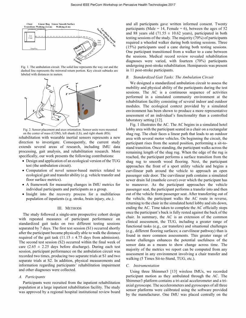
B. Standardized Gait Tasks: The Ambulation Circuit
We designed a standardized ambulation circuit to assess the
mobility and physical ability of the participants during the test
sessions. The AC is a continuous sequence of activities
performed in a simulated community environment at the
rehabilitation facility consisting of several indoor and outdoor
modules. The ecological context provided by a simulated
environment has been shown to produce a more representative
assessment of an individual’s functionality than a controlled
laboratory setting [12].
Fig. 1 illustrates the AC. The AC begins in a simulated hotel
lobby area with the participant seated in a chair on a rectangular
shag rug. The chair faces a linear path that leads to an outdoor
area with several motor vehicles. On beginning the circuit, the
participant rises from the seated position, performing a sit-to-
stand transition. Once standing, the participant walks across the
remaining length of the shag rug. When the edge of the rug is
reached, the participant performs a surface transition from the
shag rug to smooth wood flooring. Next, the participant
approaches the front of a sport utility vehicle and begins a
curvilinear path around the vehicle to approach an open
passenger side door. The curvilinear path contains a simulated
sewer drain lid (manhole cover) over which the participant has
to maneuver. As the participant approaches the vehicle
passenger seat, the participant performs a transfer into and then
out of the vehicle front passenger seat. After transferring out of
the vehicle, the participant walks the AC route in reverse,
returning to the chair in the simulated hotel lobby and sits down,
ending the AC. Time taken to complete the AC officially stops
once the participant’s back is fully rested against the back of the
chair. In summary, the AC is an extension of the common
clinical assessment, the TUG, including a greater range of
functional tasks (e.g., car transfers) and situational challenges
(e.g., different flooring surfaces; a curvilinear pathway) than is
found in more common assessments. This greater range of
motor challenges enhances the potential usefulness of the
sensor data as a means to show change across time. The
majority of the metrics we report can be computed from any
assessment in any environment involving a chair transfer and
walking (5 Times Sit-to-Stand, TUG, etc.).
C. Instrumentation
Using three Shimmer3 [13] wireless IMUs, we recorded
participant motion as they ambulated through the AC. The
Shimmer3 platform contains a tri-axial accelerometer and a tri-
axial gyroscope. The accelerometers and gyroscopes of all three
sensor platforms were calibrated using the software provided
by the manufacturer. One IMU was placed centrally on the
Fig. 1. The ambulation circuit. The solid line represents the way out and the dashed line represents the mirrored return portion. Key circuit subtasks are
labeled with distances in meters.
Fig. 2. Sensor placement and axes orientation. Sensor units were mounted on the center of mass (COM), left shank (LS), and right shank (RS).
Second IEEE PerCom Workshop on Pervasive Health Technologies 2017

lumbar spine at the level of the third vertebrae, near the
individual’s center of mass (COM) [14]. Additionally, one
sensor was placed on each shank, above the ankle and in line
with the tibia. Positioning the sensor along the tibia reduced
mounting error as the sensors were always positioned at
approximately the same angle relative to the sagittal plane.
The flatness of the tibia bone also prevented the sensor from
moving during the activities. The sensor modules were
securely attached to the body with elastic straps. Shank sensor
mounting locations were measured at S1 and S2 for
consistency. Fig. 2 illustrates the shank mounting locations
and axes of the sensors. The accelerometer range was set to ±
2g for the COM sensor and ± 4g for the shanks. The gyroscope
ranges for the shank and COM sensors were set at 500 ⁰/s and
250 ⁰/s, respectively. The data were collected at a sampling
frequency of 51.2 Hz for all sensor platforms. The inertial
movement data and segment times are processed with a
custom Python program designed for the AC data. First, the
timestamps are aligned from the three different sensor
platforms. Next, to correct for the orientation of the shank
sensors along the tibia, the sensor local coordinate system is
transformed to the body coordinate system [15]; a right handed
system with the X-axis along the anterior-posterior body axis,
the Y-axis along the vertical body axis, and the Z-axis along the
medial-lateral body axis. Acceleration data are filtered with a
4th order zero-phase band pass Butterworth filter using cutoff
frequencies of 0.1 Hz and 3 Hz for the COM accelerometer and
0.1 Hz and 10 Hz for the shanks. The gyroscope signals for all
sensors are low passed filtered at 4 Hz.
From the processed data we compute metrics representing
participants’ performance on the AC. AC task durations were
recorded by a researcher using a stopwatch. The times are used
to segment the data into the different tasks for computing
metrics for each of the AC sections. Fig. 3 illustrates the tri-
axial COM acceleration and left and right shank gyroscope data
from a participant partitioned into the key sections of the AC.
D. Computed Metrics
For a unique analysis of sensor-based gait information in a
rehabilitation setting, we compute metrics from three main
components of the AC: the chair sit-to-stand and stand-to-sit
movements at the beginning and ending of the AC, the vehicle
transfer, and the ambulation occurring between the chair and
the vehicle. This ambulation section includes the linear path on
the smooth floor that is used to compute the majority of the gait
cycle metrics. For the ambulation section, an algorithm was
developed to detect the gait cycle events of initial contact,
terminal contact, and mid swing. Initial contact is the moment
the heel strikes the ground and terminal contact is the moment
TABLE I
METRIC DESCRIPTIONS
Category Metric Units Qualitative Description Refer-ence
CAP
Duration 𝑠 Total time to complete the ambulation circuit or a subtask of the ambulation circuit.
Floor surface speed ratio Measures the effect of walking velocity on two different floor surfaces.
Walking speed 𝑚 𝑠⁄ The walking velocity as determined by distance divided by time.
WBM
COM peak angular velocity
° 𝑠⁄ Maximum rotational velocity of the COM around the Z-axis while rising from a seated position in the chair to a standing position.
Root mean square
(RMS) 𝑚 𝑠2⁄ /𝑠
Square root of the mean of the squares of each axes of the acceleration signal on the COM.
Represents the magnitude of the signal (normalized by time). [3]
Smoothness index Ratio of even to odd harmonics of the vertical Y-axis COM acceleration signal. A higher harmonic ratio represents a smoother walking pattern.
[17]
Smoothness of RMS 𝑚 𝑠3⁄ /𝑠 Root mean square of the derivatives of each X, Y, and Z signal. Synonymous with RMS of
jerk (normalized by time). [3]
GF
Cadence 𝑠𝑡𝑒𝑝𝑠 𝑚𝑖𝑛⁄ Step rate as expressed by the number of steps per minute.
Double support percent % Percentage of the gait cycle that both feet are on the ground. Computed as the sum of the
initial double support time and the terminal double support time.
[5],
[16]
Gait cycle time 𝑠 Duration to complete one stride (time between two consecutive initial contacts of the same foot).
[5], [16]
Number of gait cycles The number of complete gait cycles (strides) that occurred.
Shank peak angular
velocity ° 𝑠⁄
Maximum rotational velocity of the shank around the Z-axis during the gait cycle. This
occurs during the swing phase.
Shank range of motion ° Integrated angular velocity for each gait cycle. Provides an estimate of the degrees of shank
movement.
[5],
[16]
Step length 𝑚 Distance between initial contacts of opposite feet. [14]
Step regularity % Expression of the regularity of the acceleration of sequential steps. Computed using the
autocorrelation of the vertical Y-axis of the COM acceleration. [14]
Stride regularity % Expression of the regularity of the acceleration of sequential strides (see step regularity). [14]
Step symmetry % Ratio of step regularity to stride regularity. [14]
CAP = clinical assessments of progress, COM = center of mass, GF = gait features, 𝑚 = meters, 𝑠 = seconds, WBM = whole body movement, ° = degrees.
Fig. 3. Sensor signals recorded during the first half of the AC. The center of
mass (top plot: accelerometer) and shank (bottom plot: gyroscope) sensor
signals were analyzed to quantify the rehabilitation process.
Second IEEE PerCom Workshop on Pervasive Health Technologies 2017
the toes leave contact with the ground. The algorithm operates
on the left and right shank medial-lateral (Z-axis) gyroscope
data. The algorithm utilizes peak detection and thresholding
techniques that were implemented with high accuracy by
previous studies [15], [16]. By locating these key gait events,
the gait cycle is defined (the time interval between two
successive initial contacts of the same leg) and several metrics
related to walking are computed. Table I presents the metrics
we compute and groups the metrics into three categories:
1. Clinical assessments of progress (CAP). CAP metrics are
commonly used approaches for assessing mobility in a
clinical setting by recording the duration of a standardized
activity, such as walking a fixed distance, rising from a
chair, or the TUG assessment.
2. Whole body movement (WBM). WBM metrics are
computed from data collected from the COM sensor. An
example WBM metric is COM peak angular velocity.
3. Gait features (GF). GF are computed from data collected
from the shank sensors. Examples of GF include cadence
and shank range of motion, which are based on the
aforementioned gait cycle event detection algorithm.
E. Data Analysis
Sensor-based metrics are statistically analyzed to identify
clinically significant changes in the repeated measures data.
Detected changes in patient performance offer additional
insights to clinicians, as well as demonstrate the benefit of
sensor-based analysis of rehabilitation. The statistical analyses
we apply to the wearable sensor data at the group and individual
levels are summarized below.
1) Quantifying Group Changes
An effect size (ES) based on Cohen’s 𝑑 for repeated
measures (RM) data is used to quantify the strength of changes
in each of the computed metrics [18]:
𝑑𝑅𝑀 = �̅�𝑆2−�̅�𝑆1
𝑆𝐷 (1)
Where �̅�𝑆1 is the mean group score from data collected at S1,
�̅�𝑆2 is the mean group score from data collected at S2, and 𝑆𝐷
represents the standard error of difference between S1 and S2
scores [18]. The resulting effect sizes, 𝑑𝑅𝑀, are used to evaluate
group changes in gait parameters over the course of one week
of inpatient rehabilitation. Additionally, the confidence
intervals for each ES are computed using a small sample size
approximation with alpha set at 95% [19].
2) Quantifying Individual Changes
At the individual level, changes in gait metrics one week
apart are characterized with the reliable change index (RCI)
[20]:
RCI = 𝑥𝑆2−𝑥𝑆1
𝑆𝐷 (2)
Where 𝑥𝑆1 is an individual participant’s score from data
collected at S1 and 𝑥𝑆2 is the same participant’s score from data
collected at S2. In addition to numeric RCI statistics,
comparison of individuals to the group for change between S1
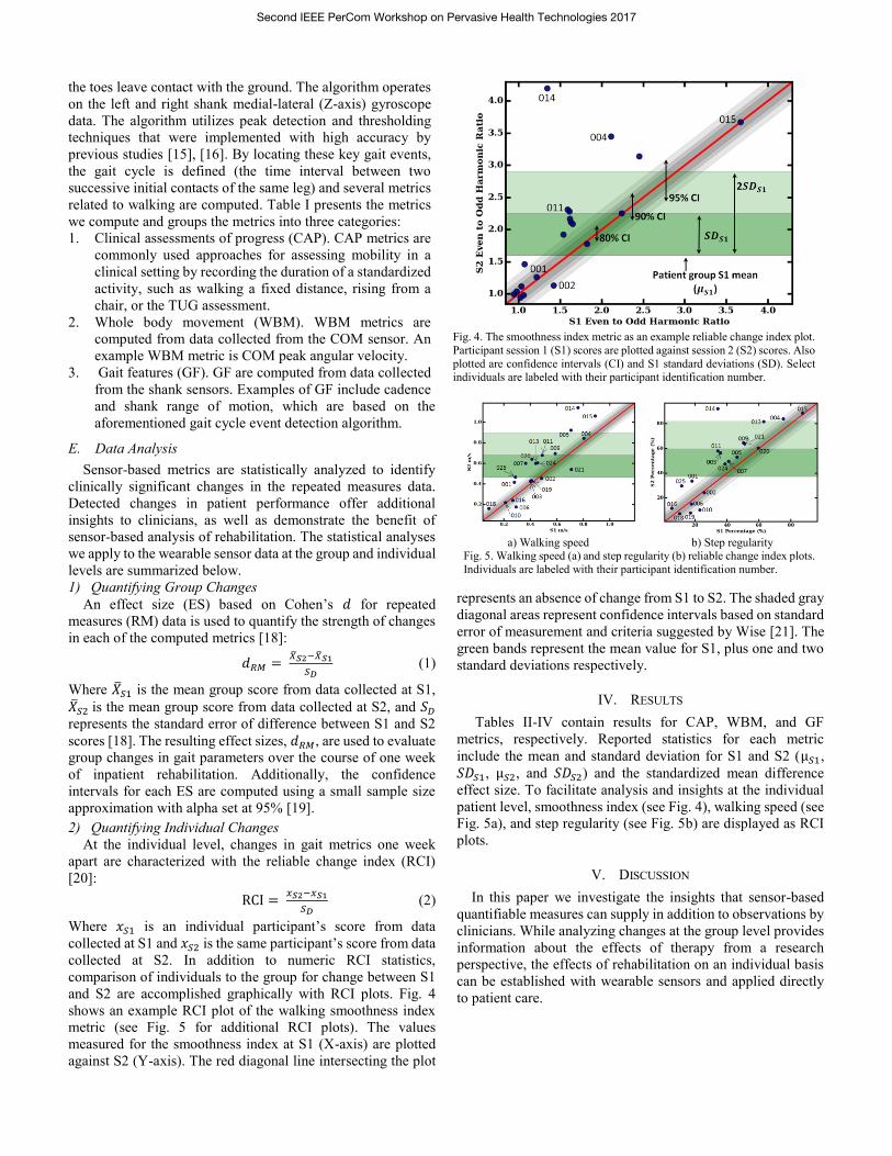
and S2 are accomplished graphically with RCI plots. Fig. 4
shows an example RCI plot of the walking smoothness index
metric (see Fig. 5 for additional RCI plots). The values
measured for the smoothness index at S1 (X-axis) are plotted
against S2 (Y-axis). The red diagonal line intersecting the plot
represents an absence of change from S1 to S2. The shaded gray
diagonal areas represent confidence intervals based on standard
error of measurement and criteria suggested by Wise [21]. The
green bands represent the mean value for S1, plus one and two
standard deviations respectively.
IV. RESULTS
Tables II-IV contain results for CAP, WBM, and GF
metrics, respectively. Reported statistics for each metric
include the mean and standard deviation for S1 and S2 (μ𝑆1,
𝑆𝐷𝑆1, μ𝑆2, and 𝑆𝐷𝑆2) and the standardized mean difference
effect size. To facilitate analysis and insights at the individual
patient level, smoothness index (see Fig. 4), walking speed (see
Fig. 5a), and step regularity (see Fig. 5b) are displayed as RCI
plots.
V. DISCUSSION
In this paper we investigate the insights that sensor-based
quantifiable measures can supply in addition to observations by
clinicians. While analyzing changes at the group level provides
information about the effects of therapy from a research
perspective, the effects of rehabilitation on an individual basis
can be established with wearable sensors and applied directly
to patient care.
Fig. 4. The smoothness index metric as an example reliable change index plot.
Participant session 1 (S1) scores are plotted against session 2 (S2) scores. Also
plotted are confidence intervals (CI) and S1 standard deviations (SD). Select
individuals are labeled with their participant identification number.
a) Walking speed b) Step regularity
Fig. 5. Walking speed (a) and step regularity (b) reliable change index plots.
Individuals are labeled with their participant identification number.
Second IEEE PerCom Workshop on Pervasive Health Technologies 2017

A. Group Responsiveness to Therapy
1) Clinical Assessments of Progress
As a group, AC metrics related to CAP demonstrate
moderate improvements based on the magnitude of effect sizes
(see Table II). For total AC duration, there is a 22.77% decrease
in the total time required to complete the AC. Improved
functionality is confirmed by increased ambulatory capabilities,
completing the curvilinear section of the AC 20.26% faster.
Responsiveness on the vehicle load/unload challenge is
moderate with 27.32% decrease in duration observed on
average. Walking speed is a typical metric for comparison
among populations and for indication of ambulatory
improvements. Group average performance during both
sessions (walking speed S1 0.47 ± 0.22 𝑚/𝑠; walking speed S2
0.57 ± 0.28 𝑚/𝑠) as well as the large responsiveness across
time (see Fig. 5a for an RCI plot) are in agreement with
previous published studies analyzing walking speed in post-
stroke populations [22].
2) Whole Body Movement Metrics
Large levels of responsiveness are observed for RMS during
linear path gait (see Table III). While there is a relationship
between COM RMS and walking speed [17], the large
responsiveness across time is due in part to the participant-
dictated speed of movement. During the vehicle transfer, the ES
for COM RMS during the load and unload tasks suggest
substantial progress from one week of rehabilitation therapy.
Similar changes in COM RMS are also present on the chair
task.
Another WBM metric, the smoothness index of walking (see
Fig. 5), characterizes gait harmonics to quantify cyclical
movements independent of speed [17]. The calculated ES for
change in smoothness index emphasizes the influence
rehabilitation has on developing a more stable pattern of
locomotion. As a group, the participants demonstrate a 25.63%
improvement in the smoothness index.
3) Gait Features
Changes in metrics describing gait quality in terms of
symmetry, regularity, and consistency are observed during the
straight path portion of the AC (see Table IV). During one week
of rehabilitation, the increased walking speed is accompanied
by an average increase of 8.72% in cadence. Another important
outcome is the 4.74% decrease in the amount of double limb
support in the gait cycle. In addition, improvement is observed
in gait consistency, measured with stride and step regularity
(see Fig. 5b for an RCI plot). These metrics indicate that
patients are beginning to produce more consistent walking
patterns over one week, increasing the load carried by the
affected limb.
Changes are also observed in individual leg movements.
Large levels of responsiveness are detected in peak angular
velocity, measured at each shank. Along with faster leg
movements during the swing phase, there is a strong indication
of increased limb range of motion during gait. To perform sub-
group analyses of stroke patients with hemiparesis, each limb is
re-classified as affected (paretic) or unaffected (non-paretic),
instead of left or right. The re-categorization produces a slightly
different ES for shank peak angular velocity and range of
motion. Tracking changes in the affected side of the body offers
additional insight for clinicians treating stroke patients and
injuries affecting one side of the body more than the other side.
TABLE II CLINICAL ASSESSMENTS OF PROGRESS (CAP) METRIC RESULTS
Metric μ𝑆1 𝑆𝐷𝑆1 μ𝑆2 𝑆𝐷𝑆2 Effect
Size
Curvilinear walking
duration 24.43 18.14 19.48 12.23 -1.03*
Duration 177.85 129.53 137.36 88.56 -1.08*
Floor surface speed
ratio 0.75 0.13 0.80 0.14 0.48*
Sit-to-stand duration 6.84 6.10 4.79 3.03 -0.49*
Stand-to-sit duration 12.94 12.05 11.13 7.13 -0.34*
Vehicle challenge duration
47.81 36.36 34.75 28.10 -0.55*
Walking speed 0.47 0.22 0.57 0.28 1.58*
S1 = session 1, S2 = session 2, SD = standard deviation, 𝜇 = mean, * = significant at the 95% confidence level.
TABLE III
WHOLE BODY MOVEMENT (WBM) METRIC RESULTS
Metric μ𝑆1 𝑆𝐷𝑆1 μ𝑆2 𝑆𝐷𝑆2 Effect
Size
COM RMS 0.08 0.06 0.11 0.10 1.87*
COM Smoothness of
RMS 0.75 0.59 1.10 1.04 1.90*
Sit-to-stand RMS 0.65 0.64 0.85 0.67 0.71*
Sit-to-stand peak angular velocity
84.02 37.96 72.46 44.47 -0.37
Smoothness index 1.60 0.65 2.01 0.97 1.82*
Stand-to-sit RMS 0.38 0.45 0.36 0.30 -0.07
Stand-to-sit peak angular velocity
126.87 36.75 118.45 42.08 -0.26
Vehicle load RMS 0.10 0.11 0.16 0.16 1.37*
Vehicle load peak
angular velocity 81.94 36.62 78.27 26.32 -0.10
Vehicle unload RMS 0.16 0.12 0.29 0.37 2.71*
Vehicle unload peak
angular velocity 74.45 47.85 68.45 35.89 -0.15
COM = center of mass, RMS = root mean square, S1 = session 1, S2 =
session 2, SD = standard deviation, 𝜇 = mean, * = significant at the 95% confidence level.
TABLE IV GAIT FEATURES (GF) METRIC RESULTS
Metric μ𝑆1 𝑆𝐷𝑆1 μ𝑆2 𝑆𝐷𝑆2 Effect Size
Cadence 64.88 17.67 70.54 20.32 1.38*
Double support percent 33.79 11.97 32.19 13.72 -0.49*
Gait cycle time 1.96 0.66 1.87 0.68 -0.64*
Affected side peak angular velocity
190.09 67.88 208.79 72.50 1.52*
Affected side shank range of
motion 47.34 13.22 50.02 11.44 1.20*
Left side peak angular velocity 195.98 61.01 213.09 68.84 1.30*
Left side shank range of
motion 47.24 12.66 50.93 12.59 1.73*
Number of gait cycles 18.95 9.32 16.38 5.59 -0.90*
Right side peak angular velocity
217.65 43.80 244.53 51.02 2.02*
Right side shank range of
motion 50.41 10.91 54.70 9.43 1.45*
Step length 0.21 0.07 0.23 0.06 0.64*
Step regularity 37.29 22.36 46.91 28.11 1.31*
Stride regularity 40.88 22.73 51.53 24.45 0.55*
Step symmetry 63.57 27.50 70.80 26.31 0.35
Unaffected side peak angular
velocity 231.38 39.33 255.20 40.62 1.91*
Unaffected side shank range of motion
51.91 11.96 55.69 8.40 1.56*
S1 = session 1, S2 = session 2, SD = standard deviation, 𝜇 = mean, * = significant at the 95% confidence level.
Second IEEE PerCom Workshop on Pervasive Health Technologies 2017

B. Individual Responsiveness to Therapy
RCI analyses suggest that recovery is not consistent for all
patients over one week of inpatient rehabilitation (see Fig. 4 and
5). For example, participant #014 experienced a substantial
amount of recovery compared to the rest of the participants as
assessed through RCI plots. This finding is corroborated by the
conventional method of using the FIM to characterize
functioning at admission. By contrast, participant #015 did not
demonstrate significant change in smoothness of walking or
step regularity. At admission to the inpatient facility the
functional capabilities for this participant were close to
independent, rendering a small window for improvement.
The RCI visualization of performance at the individual level
can track progress by assessing performance on multiple
metrics. For example, a few participants with moderate
responsiveness for walking speed (#007, #015, and #020) did
not show change in the smoothness index metric and vice versa
(#004). Therefore, analysis of multiple metrics, such as
smoothness index along with walking speed, highlights the
differences in individual recovery.
A limitation of this study is the metric computations have not
been laboratory validated; however, all of the algorithms are
derived from previously-published and validated sources.
Another limitation includes the use of human-operated
stopwatch times to segment the AC into its subtasks. The times
recorded by the researchers could impose non-systematic error.
Future work includes recruiting healthy individuals to perform
the AC to provide reference data for comparison to patient data.
VI. CONCLUSION
Inpatient rehabilitation contains a wide spectrum of
challenges that are tackled uniquely by different patients,
depending on their pre-morbid state, injury, drive to improve,
and compensatory strategies. Changes measured in movement
profiles over the course of one week of therapy indicate
wearable IMUs provide a viable platform for gaining insight
into these complex recovery processes. The ambulation circuit
presented in this study allowed data collection to capture
performance of real-world challenges in ecological
environments. Several gait and transfer features exhibit
statistically significant differences in value from session one to
session two, which indicates wearable sensor-derived metrics
may be practical for clinicians to use in addition to observation
to quantify gait and vehicle transfer improvement. Of the
significant metrics, only walking speed does not make use of
wearable inertial sensor data, indicating that wearable sensors
can capture details about changes in movement patterns that
cannot be acquired from standardized subjective clinical
assessments.
ACKNOWLEDGMENT
We wish to thank our therapist collaborators at St. Luke’s
Rehabilitation Institute.
REFERENCES
[1] B. B. Hamilton, C. V. Granger, F. S. Sherwin, M. Zielezny, and J. S.
Tashman, “A Uniform National Data System for Medical Rehabilitation,” in Rehabilitation Outcomes: Analysis and
Measurement, Baltimore, MD: Paul H. Brookes Publishing Company,
1987, pp. 137–150. [2] G. Sprint, D. Cook, and D. Weeks, “Towards Automating Clinical
Assessments: A Survey of the Timed Up and Go (TUG),” Biomed. Eng.
IEEE Rev. In, vol. PP, no. 99, pp. 1–1, 2015. [3] M. Zhang, B. Lange, C.-Y. Chang, A. A. Sawchuk, and A. A. Rizzo,
“Beyond the standard clinical rating scales: fine-grained assessment of
post-stroke motor functionality using wearable inertial sensors,” in Engineering in Medicine and Biology Society (EMBC), 2012 Annual
International Conference of the IEEE, 2012, pp. 6111–6115.
[4] M. M. Rodgers, V. M. Pai, and R. S. Conroy, “Recent Advances in Wearable Sensors for Health Monitoring,” IEEE Sens. J., vol. 15, no. 6,
pp. 3119–3126, Jun. 2015.
[5] A. Salarian, F. B. Horak, C. Zampieri, P. Carlson-Kuhta, J. G. Nutt, and K. Aminian, “iTUG, a Sensitive and Reliable Measure of Mobility,”
IEEE Trans. Neural Syst. Rehabil. Eng., vol. 18, no. 3, pp. 303–310,
2010. [6] V. Bonnet, V. Joukov, D. Kulic, P. Fraisse, N. Ramdani, and G.
Venture, “Monitoring of Hip and Knee Joint Angles Using A Single
Inertial Measurement Unit During Lower-limb Rehabilitation,” IEEE Sens. J., vol. PP, no. 99, pp. 1–1, 2015.
[7] W. Rapp, T. Brauner, L. Weber, S. Grau, A. Mündermann, and T.
Horstmann, “Improvement of walking speed and gait symmetry in older patients after hip arthroplasty: a prospective cohort study,” BMC
Musculoskelet. Disord., vol. 16, no. 1, p. 291, Oct. 2015.
[8] A. Kleiner et al., “The Parkinsonian Gait Spatiotemporal Parameters Quantified by a Single Inertial Sensor before and after Automated
Mechanical Peripheral Stimulation Treatment,” Park. Dis., vol. 2015, 2015.
[9] M. Galli et al., “Timed Up and Go test and wearable inertial sensor: a
new combining tool to assess change in subject with Parkinson’s disease after automated mechanical peripheral stimulation treatment,” Int. J.
Eng. Innov. Technol., vol. 4, pp. 155–163, 2015.
[10] H. Simila, M. Immonen, J. Merilahti, and T. Petakoski-Hult, “Gait analysis and estimation of changes in fall risk factors,” in 2015 37th
Annual International Conference of the IEEE Engineering in Medicine
and Biology Society (EMBC), 2015, pp. 6939–6942. [11] A. K. Dorsch, S. Thomas, X. Xu, W. Kaiser, and B. H. Dobkin,
“SIRRACT: An International Randomized Clinical Trial of Activity
Feedback During Inpatient Stroke Rehabilitation Enabled by Wireless
Sensing,” Neurorehabil. Neural Repair, Sep. 2014.
[12] B. Mcmahon, Work Worth Doing: Advances in Brain Injury
Rehabilitation, 1 edition. Orlando, FL: CRC Press, 1991. [13] A. Burns et al., “Shimmer-A Wireless Sensor Platform for Noninvasive
Biomedical Research,” IEEE Sens. J., vol. 10, no. 9, pp. 1527–1534,
Sep. 2010. [14] R. Moe-Nilssen and J. L. Helbostad, “Estimation of gait cycle
characteristics by trunk accelerometry,” J. Biomech., vol. 37, no. 1, pp.
121–126, 2004. [15] S. Chen, “Gait Feature Extraction from Inertial Body Sensor Networks
for Medical Applications,” University of Virginia, 2013.
[16] A. Salarian et al., “Gait Assessment in Parkinson’s Disease: Toward an Ambulatory System for Long-Term Monitoring,” IEEE Trans. Biomed.
Eng., vol. 51, no. 8, pp. 1434–1443, Aug. 2004.
[17] H. B. Menz, S. R. Lord, and R. C. Fitzpatrick, “Acceleration patterns of the head and pelvis when walking on level and irregular surfaces,” Gait
Posture, vol. 18, no. 1, pp. 35–46, 2003.
[18] L. Wolff Smith and S. N. Beretvas, “Estimation of the Standardized Mean Difference for Repeated Measures Designs,” J. Mod. Appl. Stat.
Methods, vol. 8, no. 2, pp. 600–609, 2009.
[19] W. Viechtbauer, “Approximate Confidence Intervals for Standardized Effect Sizes in the Two-Independent and Two-Dependent Samples
Design,” J. Educ. Behav. Stat., vol. 32, no. 1, pp. 39–60, Mar. 2007.
[20] N. S. Jacobson and P. Truax, “Clinical significance: A statistical approach to defining meaningful change in psychotherapy research,” J.
Consult. Clin. Psychol., vol. 59, no. 1, pp. 12–19, 1991.
[21] E. A. Wise, “Methods for Analyzing Psychotherapy Outcomes: A Review of Clinical Significance, Reliable Change, and
Recommendations for Future Directions,” J. Pers. Assess., vol. 82, no.
1, pp. 50–59, 2004. [22] S. J. Mulroy, T. Klassen, J. K. Gronley, V. J. Eberly, D. A. Brown, and
K. J. Sullivan, “Gait Parameters Associated With Responsiveness to
Treadmill Training With Body-Weight Support After Stroke: An Exploratory Study,” Phys. Ther., vol. 90, no. 2, pp. 209–223, Feb. 2010.
Second IEEE PerCom Workshop on Pervasive Health Technologies 2017