mechanisms underlying rhythmic activities of the · pdf filemechanisms underlying rhythmic...
TRANSCRIPT

MECHANISMS UNDERLYING RHYTHMIC ACTIVITIES OF THE
GASTROINTESTINAL TRACT

MECHANISMS UNDERLYING RHYTHMIC ACTIVITIES OF THE
GASTROINTESTINAL TRACT
BY,
MARC J PISTILLI HBSc
A Thesis
Submitted to the School of Graduate Studies
in Partial Fulfillment of the Requirements
for the Degree
Master of Science
McMaster University
© Copyright by Marc J Pistilli

ii
MASTER OF SCIENCE (2012) McMaster University
(Medical Sciences) Hamilton, Ontario
TITLE: Mechanisms Underlying Rhythmic Activities in the Gastrointestinal Tract
AUTHOR: Marc J Pistilli, HBSc (McMaster University)
SUPERVISOR: Professor Jan D Huizinga PhD
NUMBER OF PAGES: xii, 98

iii
“On no subject in physiology do we meet with so many discrepancies of fact and opinion
as in that of the physiology of the intestinal movements.”
- Bayliss and Starling (1899)

iv
ABSTRACT
The organs of the gastrointestinal (GI) tract display a variety of motor patterns,
involved in grinding, mixing, enhancing absorption and propulsion of nutrients and waste
products. Specialized motor patterns are generated by unique mechanisms inherent to the
GI segment in which they are found. Rhythmic contractions are a feature of most motor
patterns. Slow wave driven peristalsis is an acknowledged motor pattern associated with
interstitial cells of Cajal (ICC) pacemakers, but propulsive motor patterns which are
blocked by tetrodotoxin are seen to be exclusively generated by the enteric nervous
system (ENS). This has not been proven, however, and the origin of rhythmicity of
propulsive motor patterns needs further study, particularly related to a potential role of
the pacemaker ICC found throughout the GI tract. The aim of this study was exploring
the mechanisms which underlie various GI motor patterns, with particular focus on the
origin of rhythmicity of these patterns.
I have demonstrated with manometry and spatiotemporal maps that murine
rhythmic propulsion requires a myogenic pacemaker which is evoked by acetylcholine
and substance P; nitric oxide is not involved. Calcium imaging evidence suggests that the
pacemaker is the ICC of the deep muscular plexus, as these cells rhythmically activate to
substance P. I observed rhythmic contractility patterns in human antrum, pylorus and
duodenum when stimulated with carbachol. The hypothesis emerged that the ENS
modifies the pyloric pacemaker into unique rhythmic patterns. Colonic muscle strip
contractility from the rat has a low frequency rhythmic pattern which is myogenic. This

v
pattern is augmented by the conditioned media from the probiotic E. coli Nissle 1917
through a non-neural mechanism.
The current explanation of entirely ENS generated motor patterns is not accurate.
The ENS plays an important role in stimulating and regulating GI motor patterns in
conjunction with myogenic pacemakers. It is only through acknowledgment of all GI cell
types that we can understand the mechanisms governing motility.

vi
AKNOWLEDGEMENTS
Funding for this project was provided by an operating grant for Dr. Jan Huizinga
from CIHR. I would also like to acknowledge the Ontario Graduate Scholarship and the
Queen Elizabeth II Graduate Scholarship in Science and Technology I received over my
two years.
I have made significant progress both professionally as a student of science but
also personally as I move forward in life. Dr. Jan Huizinga has been of utmost importance
on this journey and for that I am grateful. His dedication to science has left an everlasting
impression on me and helped reinforce my interests in normal physiology. I also want to
thank Jan for the countless opportunities to participate in research, discussions,
publications and presentations. These opportunities are invaluable to my future
employment and success, and have also made graduate school meaningful and enjoyable.
The guidance that my supervisory committee provided was excellent. Both Dr.
Luke Janssen and Dr. Denis Crankshaw helped make this project a realistic and
worthwhile endeavor. I specifically want to thank Dr. Janssen for challenging me over
these two years, as I have learned confidence in myself and the scientific ideas I may
hold. I also want to specifically thank Dr. Crankshaw for fostering the early ideas of
research within me and teaching me techniques that I still use in my research. Without his
guidance and supervision I might never have entered the field of scientific research nor
pursue graduate school.
I want to thank all the members of the Huizinga lab for making it an enjoyable
place to work. I really enjoyed coming in everyday to see you all. I would specifically

vii
like to thank Bobbi-Jo Lowie, Sarah-Lynn Martz and Dr. Sean Parsons for teaching me
the techniques that I used throughout my research and in the preparation of this thesis. I
would also like to thank Amir Khoshdel and George Wright for technical assistance in
performing the motility study and the editing of this document. I want to thank all the
remaining members for the countless discussions we have had and helping me develop a
better understanding of gastrointestinal physiology.
Finally, I would like to thank members of my family, particularly my wife Joanna
and parents for supporting me in countless ways throughout these last two years. Your
patience and understanding have allowed me to focus on my education, and succeed
during this time.

viii
LIST OF FIGURES
Figure 1.1
Location of gastrointestinal interstitial cells of Cajal 2
Figure 1.2
Model of the peristaltic reflex 5
Figure 1.3
Segmentation of the small intestine 8
Figure 3.1
Analysis of organ bath experiments 33
Figure 3.2
Content propulsion in the small intestine 34
Figure 3.3
Effect of distention on the propulsive motor pattern 36
Figure 3.4
Restoration of rhythmic propulsion with carbachol 38
Figure 3.5
Effect of neural blockade on intestinal motor patterns 40
Figure 3.6
Segmentation motor pattern 43
Figure 3.7
Effect of nitric oxide blockade on rhythmic propulsion 44
Figure 3.8
Effect of substance P on rhythmic propulsion 47
Figure 4.1
Effect of substance P on intracellular calcium concentration of neonatal
ICC-DMP 60
Figure 4.2
Effect of long-term substance P exposure on intracellular calcium
concentration of neonatal ICC-DMP 61
Figure 4.3
Effect of substance P exposure on intracellular calcium cycling within
adult ICC-DMP 63
Figure 5.1
Effect of stretch on a human pyloric muscle strip 69
Figure 5.2
Evolving effect of carbachol on the human circular muscle strips 70
Figure 5.3
Stable effect of carbachol on human circular muscle strips 71
Figure 6.1
Effect of neural blockade on high-amplitude low-frequency contractions
of the rat colon muscle strips 78
Figure 6.2
The effects of treated (A) and untreated media (B) on muscle strips from
the colon devoid of mucosa 79

ix
LIST OF ABBREVIATIONS
7TM – seven-transmembrane receptors; G-protein coupled receptors
ACE – angiotensin-converting enzyme
AM – (acetoxymethyl ester)
ANOVA – analysis of variance
AUC – area under the curve
cpm - contractions per minute
ICC – interstitial cells of Cajal
ICC-DMP – deep muscular plexus interstitial cells of Cajal
ICC-MP – myenteric plexus interstitial cells of Cajal
IP3 - inositol 1,4,5-trisphosphate
L-NNA - Nω- Nitro-L-Arginine
mAChR – muscarinic acetylcholine receptors
Mn – muscarinic acetylcholine receptor “n” subtype
nAChR - nicotinic acetylcholine receptors
NEP - neutral endopeptidase
Nissle - Escherichia coli Nissle 1917
NKA – neurokinin A
NKB – neurokinin B
NKn – tachykinin receptor “n” subtype
NO – nitric acid
PAO – phenylarsine oxide
PMC – propulsive motor complex
PMP – propulsive motor pattern
ROI – region of interest
SEM – standard error of the mean
SWAC - Slow wave associated contractions
TTX – tetrodotoxin

x
TABLE OF CONTENTS
DESCRIPTIVE NOTE ii
ABSTRACT iv
ACKNOWLEDGMENTS vi
LIST OF FIGURES viii
LIST OF ABBREVIATIONS ix
1. INTRODUCTION 1
1.1 Anatomy of the small intestine 1
1.2 Motor patterns of the small intestine 1
1.2.1 Peristalsis 3
1.2.2 Segmentation 7
1.3 Interstitial cell of Cajal 10
1.3.1 Interstitial cell of Cajal pacemaking 12
1.3.2 Innervation of interstitial cells of Cajal 13
1.4 Acetylcholine and acetylcholine receptors 14
1.5 Substance P and tachykinin receptors 16
1.6 Objectives 18
1.7 Hypothesis 19
2. METHODOLOGY 20
2.1 Animals 20
2.2 Organ bath technique 21
2.3 Calcium imaging technique 22
2.4 Muscle strip technique 24 2.4.1 Human pyloric region muscle strips 24
2.4.2 Rat colon muscle strips 25
2.5 Solutions, drugs and reagents 26
2.6 Data analysis and statistics 26

xi
3. MECHANISM OF PROPULSION IN THE MOUSE SMALL INTESTINE 28
3.1 Introduction 28
3.2 Methods 30
3.2.1 Organ bath experiments proper 30
3.2.2 Data analysis 31
3.3 Results 32
3.3.1 Characteristics of the propulsive motor pattern 32
3.3.2 Stimulation of the propulsive motor pattern with carbachol 37
3.3.3 Abolishing the propulsive motor pattern with tetrodotoxin 37
3.3.4 Modifying the propulsive motor pattern with L-NNA 41
3.3.5 Modifying the propulsive motor pattern with substance P 46
3.4 Discussion 49
3.4.1 Rhythmic propulsion 49
3.4.2 Segmentation 54
4. CALCIUM OSCILATIONS IN ICC OF THE DEEP MUSCULAR PLEXUS 56
4.1 Introduction 56
4.2 Methods 57
4.2.1 Calcium imaging experiments proper 57
4.2.2 Data analysis 58
4.3 Results 59
4.3.1 Neonatal ICC-DMP 59
4.3.2 Adult ICC-DMP 59
4.4 Discussion 62
5. CONTRACTILE PATTERNS OF THE HUMAN PYLORIC REGION 66
5.1 Introduction 66
5.2 Methods 67
5.2.1 Muscle strip experiments proper 67
5.2.2 Data analysis 67
5.3 Results 68
5.3.1 Effect of stretch on circular muscle strip contractility 68
5.3.2 Effect of carbachol on circular muscle strip contractility 68
5.4 Discussion 72

xii
6. EFFECT OF E. COLI NISSLE 1917 ON RAT COLON CONTRACTILIY 75
6.1 Introduction 75
6.2 Methods 76
6.2.1 Muscle strip experiments proper 76
6.2.2 Data analysis 77
6.3 Results 77
6.4 Discussion 80
7. GENERAL DISCUSSION 83
7.1 Summary of findings 83
7.2 Experimental limitations 86
7.3 Future experiments 88
REFERENCES 90

McMaster University Marc Pistilli
Medical Sciences MSc Thesis
1
1. INTRODUCTION
1.1 Anatomy of the small intestine
The small intestine consists of various layers including mucosa, submucosa, and
circular and longitudinal smooth muscle. These layers work in conjunction with one
another to facilitate digestion, and act as a barrier to the outside (lumen) of the body
(Stockinger et al., 2011). Located between the longitudinal and circular muscle layers is
the myenteric plexus (Fig 1.1) (Komuro, 2006). The plexus consists primarily of enteric
nervous system cell bodies, including sensory neurons (intrinsic primary afferent
neurons) excitatory and inhibitory motor neurons (S-neurons) as well as interneurons
(Costa et al., 2000). Located alongside the neurons is a network of myenteric plexus
interstitial cells of Cajal (ICC-MP) (Komuro, 2006). A second network of interstitial cells
of Cajal (ICC) is found between the submucosa and the circular muscle layer (Fig 1.1);
these cells are termed the deep muscular plexus interstitial cells of Cajal (ICC-DMP)
(Komuro, 2006). Most motor patterns of the small intestine do not rely on central
innervation (Schemann, 2005), thus it is a combination of smooth muscle layers, enteric
neurons and ICC which contribute to this aspect of intestinal function.
1.2 Motor patterns of the small intestine
The musculature of the small intestine facilitates mixing and propulsion of
luminal contents and also plays a role in absorption of nutrients (Gwynne and Bornstein,
2007). These physiological necessities are either fully or partially governed by the various

McMaster University Marc Pistilli
Medical Sciences MSc Thesis
2
Figure 1.1. Location of gastrointestinal interstitial cells of Cajal. The lumen of the gut
(top of figure) is directly contacted by the mucosa sitting atop the submucosa. Below are
the deep muscular plexus and then the circular smooth muscle layers. The myenteric
plexus lies between the circular muscle and the longitudinal smooth muscle layers, which
is all capped off by the serosa. ICC which are underlined are found in the mouse small
intestine; those boxed are found in the rat colon. Figure adapted from Komuro (2006).

McMaster University Marc Pistilli
Medical Sciences MSc Thesis
3
patterns of contraction seen in the small intestine, whereby different patterns yield the
required physiological response. Motility patterns fall into two main categories,
peristalsis and segmentation, both of which are patterns of circular muscle contractions
but have overlapping physiological functions (Huizinga and Lammers, 2009; Gwynne
and Bornstein, 2007).
1.2.1 Peristalsis
Peristalsis is a general term used to define propagating waves. In the
gastrointestinal tract peristalsis is defined as a propagating wave of contraction, where the
wave exhibits either complete or fractional constriction of the lumen (Huizinga and
Lammers, 2009). Peristaltic waves are able to move luminal content along short or long
distances depending on the underlying pattern of contraction. Since peristalsis has a
general definition, any motility pattern consisting of waves of contraction is classified as
such, and thus peristalsis can be broken down into a variety of different patterns. This
generality has led to much confusion, as the term is used as a catch-all for different
“peristaltic” motor patterns with different functions and underlying mechanisms (Spencer
et al., 2001; Huizinga and Lammers, 2009).
A common peristaltic pattern in the small intestine is that of a series both caudad
peristaltic and retroperistaltic contractions. These waves of contractions can travel along
the length of the small intestine but are insufficient in propelling content over long
distances on their own (Wang et al., 2005). These contractile waves are associated with
the slow waves (Hennig et al., 2010; Huizinga et al., 2009; Sanders and Ward, 2006)
generated from the interstitial cells of Cajal of the myenteric plexus (MP) (Der-Silaphet et

McMaster University Marc Pistilli
Medical Sciences MSc Thesis
4
al., 1998). This underlying mechanism has been confirmed using a variety of techniques,
not least of which was the utilization of an ICC-MP knockout model where the slow
waves were not present (Huizinga et al., 1995). Since the aforementioned peristaltic
pattern results in very little net content movement caudad, it is important to make a
distinction between it and peristaltic motility patterns which are primarily propulsive.
Bayliss and Starling (1899) first described the propulsive motor complex as a
bolus preceded by (caudal to) an area of relaxation and followed by (oral to) an area of
contraction. This observation was termed the “law of the intestine” (Spencer et al., 1999).
Research in the field of the enteric nervous system has led to the development of an
underlying mechanism to explain this observed propulsive contraction.
Immunohistochemical evidence shows orally projecting excitatory motor neurons and
caudal projecting inhibitory motor neurons (Fig. 1.2) (Costa et al., 2000). Initiation of
propulsion is by a bolus of content which activates the nearby sensory neurons which in
turn activate the above mentioned neural networks, leading to relaxation caudal and
contraction oral to the bolus (Costa et al., 2000), as the law of the intestine suggests
(Spencer et al., 1999). This mechanism is known as the peristaltic reflex. Assessment of
the peristaltic reflex has shown that although there is neural involvement in a “reflex” to a
stretch of the small intestinal wall, the peristaltic reflex is not supported by in vitro
evidence (Spencer et al., 1999). Specifically, a caudad relaxation was not seen in response
to luminal stretch as the peristaltic reflex suggests (Spencer et al., 1999; White et al.,
1934). Also, apamin, which blocks small conductance calcium-activated potassium

McMaster University Marc Pistilli
Medical Sciences MSc Thesis
5
Figure 1.2. Model of the peristaltic reflex. Grey circles represent sensory neurons
relaying the stretch stimulus from the smooth muscle (elongated structures). Black circles
represent inhibitory and excitatory motor neurons as well as interneurons. Inhibitory
neurons project caudad and excitatory neurons project oral. These pathways are repeated
sequentially along the length of the small intestine. Figure adapted from Costa et al.
(2000).

McMaster University Marc Pistilli
Medical Sciences MSc Thesis
6
channels responsible for inhibitory junction potentials in neurons and smooth muscle
relaxation, had little effect on the propulsive contractions of the small intestine (Spencer
et al., 1999). Thirdly, earlier observations of small intestinal motor patterns observed
contractions which propagated away from the stimulus in both directions in response to a
stretch stimulus (Alvarez and Starkweather 1919), and not the caudal directionality as
suggested by the law of the intestine. Although it is clear there is a role for neurons in
propulsion, the current mechanisms have the glaring omission of being unable to account
for the inherent rhythmicity of the propulsive motor complexes seen in the small
intestine. A new mechanism has been proposed to explain propulsion in the colon
whereby a mixture of both neural inputs and ICC rhythmicity are responsible for
propulsion (Huizinga et al., 2011). This model suggests neurons account for the
inducibility of propulsive patterns and the ICC govern the rhythmicity of the motor
pattern (Huizinga et al., 2011). It is possible that a similar mechanism may be responsible
for propulsion in the small intestine; however the roles of ICC and content propulsion in
this segment of the gastrointestinal system have yet to be properly assessed.
The effects of muscle tone on patterns of propulsion have been partially studied.
Propulsion in the small intestine was originally thought to be an “all-or-none” process
(Kosterlitz and Lees, 1963), whereby a bolus of content eventually stretched the lumen to
the point where a propulsive contraction occurred (Spencer et al., 2001). Spencer et al.,
(2001) showed that propulsive contractions were graded with respect to small intestinal
tone, whereby increases in stretch led to increased force of contraction. The same study
also demonstrated that propulsive contractions propagated caudad even when segments of

McMaster University Marc Pistilli
Medical Sciences MSc Thesis
7
the small intestine were paralyzed pharmacologically with the calcium channel blocker
nifedipine. Conversely, initiation of a propulsive motor complex was prevented by
paralyzing the initial stretch stimulated area. Thus, the current underlying mechanism of
propulsion requires physical distention for initiation but contraction propagation is an
inherent property of the small intestine and does not require further luminal stimulus
(Spencer et al., 2001). However, that study only assessed instantaneous stretch events and
ignored a more physiologically relevant model where luminal distention occurs over large
periods of time, not just acutely. In experiments where a constant distention is applied to
the small intestine over longer periods of time, a rhythmic pattern of propulsion results
with consistent frequency of contractions (Trendelenberg, 1917). The current neural
understanding of propulsion fails to account for the rhythmic nature that propulsive
peristalsis exhibits when the small intestine is distended, leaving much still not
understood about the underlying mechanism of propulsion.
1.2.2 Segmentation
Segmentation is a motility pattern limited to the small intestine, whereas
peristalsis is found throughout the gastrointestinal tract (Wood, 1999). Segmentation is
characterized by circular muscle contractions that are not continuous but regularly spaced
by areas of relaxation. The resulting contractions push the content in the lumen in both
directions resulting in a series of uniform segments (Fig. 1.3) (Cannon, 1902). This
pattern of contractions releases and then is reformed with different circular muscle
sections contracting (Fig. 1.3 (3)). Reformation of regular spaced groups of contracted
small intestine (Fig. 1.3 (4)), hence the original name “rhythmic segmentation”.

McMaster University Marc Pistilli
Medical Sciences MSc Thesis
8
Figure 1.3. Segmentation of the small intestine. (1) A segment of small intestine
containing chyme. Regularly spaced contractions separate the chyme into smaller
segments that are separated by uniform distances (2). The small intestine relaxes and then
segments the chyme again, but areas of contraction are shifted (3). This pattern continues
for many cycles with little net movement of content (4). Figure adapted from Cannon
(1902).

McMaster University Marc Pistilli
Medical Sciences MSc Thesis
9
Segmentation functions to mix intraluminal content and enhance epithelial contact with
the content to promote absorption (Gwynne and Bornstein, 2007). Segmentation has been
seen most consistently using in vivo techniques such as radiological imaging (Cannon,
1902; Wang et al., 2005).
Initiation of the segmentation pattern is not completely understood but has been
described to be highly dependent on intraluminal content (Cannon, 1902). Content may
be involved in the initiation of segmentation either by the intraluminal distention caused
by the content or by direct sensation of particular components of the content, the former
having not been assessed in vitro. It has been shown that the presence of fatty acids
(Gwynne et al., 2004A) or amino acids (Gwynne et al., 2004B) in the lumen of the small
intestine may be responsible (or partially responsible) for the segmentation pattern.
Gwynne and Bornstein (2007) have attempted to develop an in vitro model of
segmentation, employing decanoic acid (a saturated fatty acid) as the initiator of the
pattern. Mechanistically, it was found that inhibitory junction potentials were very
important in the segmentation pattern, since their blockade by apamin resulted in the
segmentation pattern returning to a peristaltic pattern (Gwynne and Bornstein, 2007).
Although there is a neural component to segmentation, a role for slow waves or ICC has
not been ruled out. In fact, the proposed enteric nervous system mechanism of
segmentation fails to account for the rhythmicity that is strongly associated with the
pattern. Furthermore, the in vitro model does not fully resemble the in vivo observations,
as segmentation was only shown to occur in very short segments with only two or three
segmental contractions occurring at any given moment.

McMaster University Marc Pistilli
Medical Sciences MSc Thesis
10
The inherent rhythmicity is an important part of segmentation since the
contractions follow very stable patterns (Cannon, 1902; Henderson 1909). For example it
has been shown that in rabbit small intestine, the number of rhythmic contractions per
minute slows in a linear fashion the further from the pylorus (Alvarez, 1914). The
frequency decay was confirmed in feline (Hightower Jr, 1959), murine (Hightower Jr,
1959) and canine (Puestow, 1931; Castleton, 1934) studies where it was shown that the
decay in frequency ranged between 40-60% in the distal small intestine compared to the
respective duodenum (Hightower Jr, 1959). This pattern is also seen in the small intestine
with the decay in slow wave frequency the more distal from the pylorus (Lammers and
Stephen, 2008). A second similarity between the rhythm of segmentation and slow waves
is the different frequencies seen across species. Comparing slow wave frequencies and
segmentation patterns (Table 1.1), it can be seen that not only the trend in frequencies
across species is the same but that absolute values within species are quite close
(Puestow, 1940). Although only circumstantial, it may be that the slow wave and
segmentation frequencies are linked physiologically by a currently unknown mechanism
as originally suggested by Puestow in 1940.
1.3 Interstitial cell of Cajal
Interstitial cells of Cajal have been described to form fully functioning and
independent networks within the small intestine, and indeed examples such as slow wave
associated contractions is proof of this (Huizinga and Lammers, 2009). However, ICC
have also been though to play the role of a neural-smooth muscle interface, facilitating
motor patterns in conjunction with the enteric nervous system (Ward and Sanders, 2001).

McMaster University Marc Pistilli
Medical Sciences MSc Thesis
11
Table 1.1 Slow wave and segmentation frequencies
Species Segmentation
Frequency
(cycles / min)
Slow Wave
Frequency
(slow waves
/ min)
Reference
Human 11 12 Hightower Jr, 1959; Code et al., 1952;
Lin et al., 2006
Dog 18 17-22 Hightower Jr, 1959; Lin et al., 2000
Rabbit 15-20 14.24 ± 0.44 Alvarez, 1914; Hightower Jr, 1959;
Shea-Donohue et al., 1997
Cat 24 10-15 Hightower Jr, 1959; Dahms et al.,
1987; Lammers and Stephen, 2008

McMaster University Marc Pistilli
Medical Sciences MSc Thesis
12
1.3.1 Interstitial cell of Cajal pacemaking
Smooth muscle contractions of the small intestine are controlled by rhythmic
electrical signals termed slow waves. The slow wave shifts the membrane potential of
myocytes to a potential which increases the open probability of voltage sensitive calcium
channels. When open, these channels allow calcium into the cell and facilitate contraction
(Lee et al., 1999). The slow wave is a rhythmic electrical signal that is generated by the
ICC-MP found in the small intestine (Sanders et al., 2006; Der-Silaphet et al., 1998).
Similarly, the ICC-DMP produce a rhythmic electrical signal not unlike the slow wave
(for simplicity the term “slow wave” will only be used for the electrical signal originating
from the ICC-MP) (Jiménez et al., 1996). The electrical signal generated by the ICC-
DMP has been suggested to be a redundant “back-up” mechanism for controlling small
intestine motility; its true physiological role is unknown. Others have shown that the
network is coupled to both neurons and smooth muscle, and thus may mediate neural
input of small intestinal motor patterns (Iino and Horiguchi, 2006).
Being the most active pacemaker system, the ICC-MP generate a slow wave that
has been shown to correlate with rhythmic contractions of the small intestine. Low
amplitude contractions observed in the gut occur at the same frequency that the ICC-MP
pace (Hennig et al., 2010; Huizinga et al., 2009; Sanders and Ward, 2006). The slow
wave associated contractions (SWAC) are the peristaltic contractions described
previously that are able to traverse the length of the small intestine but with limited ability
for content propulsion.

McMaster University Marc Pistilli
Medical Sciences MSc Thesis
13
Although the function of ICC-DMP has yet to be elucidated, there is some
evidence for its involvement in gut motility. Wang and coworkers (2005) have shown that
destruction of the neural and ICC networks of the deep muscular plexus via Trichinella
spiralis infection leads to a loss of the propulsive motor pattern (not SWAC) described
above. Allowing time for the infection to resolve and the deep muscular plexus (and ICC)
to repair, the propulsive pattern returned. This suggests that the destruction of ICC-DMP
and their associated nerves leads to abnormal motility, since it is this propulsive pattern
which is associated with content propulsion.
A known marker of the slow wave activity is a corresponding calcium oscillation
seen in the ICC associated with the generation of slow waves (Torihashi et al., 2002). The
calcium oscillations of the ICC-MP are synchronized between cells and correlate strongly
with contractions seen in the small intestine (Lowie et al., 2011).
1.3.2 Innervation of interstitial cells of Cajal
Both ICC-MP and ICC-DMP networks are closely associated with nerve endings
and smooth muscle cells, suggesting a possible mechanism for neural regulation of gut
pacemaking and motility (Iino et al., 2004). Specifically, there are more nerve contacts
with the ICC-DMP network than the ICC-MP network, perhaps suggesting it is the
network most responsible for neural input from the enteric nervous system (Inio et al.,
2004). Excitatory nerves associated with the ICC-DMP contain the neuropeptide
substance P (Faussone-Pellegrini, 2006). Additionally, the ICC-DMP in this interaction
express the NK1 - tachykinin receptor (Inio et al., 2004); the subtype at which substance P
is most selective. These nerves also contain acetylcholine with membrane expression of

McMaster University Marc Pistilli
Medical Sciences MSc Thesis
14
muscarinic acetylcholine receptors on ICC-DMP (Ward et al., 2006). Similarly, inhibitory
motor neurons which release nitric oxide are also found in close proximity to the ICC-
DMP (Wang et al., 1999; Ward et al., 2006). There is thus a logical pathway that could
potentially explain neural regulation of small intestinal motility through ICC-DMP
pacemaking via substance P, acetylcholine and nitric oxide messengers; however, the true
biological roles of substance P, acetylcholine and nitric oxide on ICC-DMP have yet to be
elucidated.
1.4 Acetylcholine and acetylcholine receptors
The neurotransmitter acetylcholine plays a prominent role in the excitatory axis of
the enteric nervous system of the small intestine. Acetylcholine is a product of the
enzymatic esterification by choline acetyltransferase of the compounds choline and acetic
acid (Oda, 1999). There are two classes of acetylcholine receptor, those sensitive to
nicotine termed nicotinic acetylcholine receptors (nAChR) and those sensitive to
muscarine termed muscarinic acetylcholine receptors (mAChR) (Lukas et al., 1999;
Caulfield and Birdsall, 1998). There are currently ten nAChR subtypes (α1- α10), all of
which are ligand-gated ion channels (Lukas et al., 1999). Activation of nAChRs upon
binding of acetylcholine opens the channel allowing the passive flow of sodium ions
(Na+) in to the cell and potassium ions (K
+) out of the cell with a net depolarizing effect.
Nicotinic acetylcholine receptors are expressed throughout the enteric nervous system,
both on post-synaptic neurons (mediating acetylcholine signaling) but also on presynaptic
excitatory neurons, facilitating a positive feedback loop whereby acetylcholine activates
nAChRs on presynaptic neurons leading to a further release of acetylcholine (Mandl and

McMaster University Marc Pistilli
Medical Sciences MSc Thesis
15
Kiss, 2007). There are five mAChR subtypes (M1, M2, M3, M4, M5), all of which are
seven-transmembrane (G-protein coupled receptors) receptors (7TM) (Caulfield and
Birdsall, 1998). The odd numbered mAChR subtypes primarily couple to Gq/11 where-as
the even numbered subtypes couple with Gi/o (Alexander et al., 2008). Muscarinic
acetylcholine receptors are found throughout the small intestine, particularly on smooth
muscle cells and ICC. The M2 and M3 receptor subtypes are found on both smooth
muscle cells and ICC, specifically the ICC-DMP (Chen et al., 2007). The cellular
mechanism of action of the M2 and M3 receptor are distinct upon activation, but they
tend to both be excitatory (especially with regards to smooth muscle contraction) in the
gastrointestinal tract (Ehlert et al., 2012). M2 receptors couple to Gi/o, which inhibits
adenylyl cyclase leading to a reduction in the relaxat agent cyclic AMP and therefore
cellular excitation (Ehlert et al., 2012). M3 receptors couple to Gq/11 leading to the
liberation of inositol 1,4,5-trisphosphate (IP3) and the subsequent release of calcium from
intracellular stores (Ehlert et al., 2012), which is part of active smooth muscle and ICC
function (Ehlert et al., 2012; Lowie et al., 2011).
Acetylcholine signal termination is achieved primarily through two mechanisms:
enzymatic breakdown and receptor internalization/desensitization. Once in the synaptic
space, acetylcholine is rapidly broken down by the enzyme acetylcholinesterase.
Acetylcholinesterase breaks down acetylcholine into synaptic inactive acetate and choline
(van Koppen and Kaiser, 2003). The enzymatic breakdown is quite rapid and it has been
suggested to effectively limit the dispersion of acetylcholine past the synapse in which it
was released, preventing inappropriate receptor activation. Pharmacologically,

McMaster University Marc Pistilli
Medical Sciences MSc Thesis
16
acetylcholinesterase inhibitors have been developed, such as neostigmine and
physostigmine, which compete for the active site of the enzyme and prevent the break
down of acetylcholine at the synapse. More convenient for experimentation is the
pharmacological agent carbachol which is an acetylcholine receptor agonist that is not
broken down by acetylcholinesterase (Nishioka et al., 2007), thus avoiding the effects of
this enzyme. However, use of carbachol experimentally limits signal termination.
Concurrently, activation of mAChR (like other seven transmembrane receptors) leads to
the phosphorylation of the active receptor by cytoplasmic 7TM kinases. These
phosphorylation events have the dual capacity to desensitize and internalize mAChRs
(van Koppen and Kaiser, 2003). Desensitization occurs via a conformational change
towards a state which does not interact with G-proteins or ligands as well, thus
desensitizing the agonist-receptor activation response. The same phosphorylation also
leads to the recruitment of β-arrestin, and interaction with clathrin and dynamin leading to
the budding and formation of a clathrin-coated vesicle inside the cell, removing the
receptor from the cell surface and thus terminating the signal (van Koppen and Kaiser,
2003). As with other membrane bound proteins in vesicles they can be targeted for
degradation or recycled back to the cell membrane (Bomberger et al., 2012).
1.5 Substance P and tachykinin receptors
Tachykinins are neuropeptide ligands associated mostly with nerves, but also have
non-neural origin in some instances (Pennefather et al., 2004). The tachykinins are often
associated with excitatory pathways of the nervous system both centrally and peripherally
(Pennefather et al., 2004). There are three tachykinin receptor subtypes and three

McMaster University Marc Pistilli
Medical Sciences MSc Thesis
17
endogenous tachykinins. The subtype nomenclature is simply NK1, NK2 and NK3
(Faussone-Pellegrini, 2006). The three tachykinins are substance P, neurokinin A (NKA)
and neurokinin B (NKB) (Faussone-Pellegrini, 2006). The NK1 receptor is mostly
activated by substance P (Pennefather et al., 2004) but can also be non-selectively
activated by NKA and NKB. The NK2 and NK3 receptors are preferentially activated by
NKA and NKB respectively (Faussone-Pellegrini, 2006). As mentioned previously, there
are substance P producing neurons of the enteric nervous system in the small intestine.
Further evidence of the role of substance P in small intestine motility is that ICC-DMP
express the NK1 receptor which is preferentially activated by substance P, but does not
express the NK2 or NK3 receptors (Shimizu et al., 2008), selective for the other
tachykinins.
Signaling via substance P and NK1 receptors is a highly regulated process
(Kirkwood et al., 2001). Termination of a substance P signal is achieved via two main
mechanisms, substance P metabolism and tachykinin receptor internalization (Kirkwood
et al., 2001). Since substance P is a short polypeptide, it is susceptible to metabolism by a
variety of enzymes and proteases. One of the main substance P metabolizers is the
enzyme enkephalinase (Djokic et al., 1989). Specifically, neutral endopeptidase (NEP),
also known as neprilysin, has been implicated in substance P metabolism at other smooth
muscle junctions, namely in the trachea, but also in the gut (Djokic et al., 1989). Other
enzymes capable of metabolizing substance P include angiotensin-converting enzyme
(ACE) involved in the renin-angiotensin system, serine proteases and acetylcholinesterase
(Djokic et al., 1989). The second method of substance P signal termination is the

McMaster University Marc Pistilli
Medical Sciences MSc Thesis
18
internalization of NK1 receptors on the postsynaptic cell, for example the ICC-DMP of
the small intestine. Upon activation of peptide binding 7TMs, as in the case of tachykinin
receptors, activation of the secondary messenger system feeds back on the 7TMs, leading
to the formation of clathrin-coated pits in proximity to the receptors. Eventually the pit
closes through a process of pinching and budding and the receptor is fully internalized via
endocytosis (Kirkwood et al., 2001; Sagan and Lavielle 2001). Since the receptor is fully
internalized within the cell it is no longer available for activation from extracellular
signaling molecules. In the case of the small intestine, NK1 receptor internalization at
ICC-DMP may facilitate neural signal cessation. Once in the cell, the receptor either
proceeds to the lysosome for degradation or is recycled back to the cell surface to receive
the next signal (Grady et al., 1995). The processes of substance P metabolism and NK1
internalization prevent over stimulation by a signal and contribute to fine tune neural and
effector responses.
1.6 Objectives
To gain a better understanding of myogenic and neural control mechanisms of
rhythmic motor patterns in preparations that have different physiological functions. The
main focus was on the mouse small intestine where rhythmic motor patterns propel
luminal content. I used functional motility studies and calcium imaging specifically on
the mouse small intestine to:
i) Describe the physiological characteristics of rhythmic propulsion.

McMaster University Marc Pistilli
Medical Sciences MSc Thesis
19
ii) Explore the role of neurotransmission as a mechanism for
modulating the pacemaker activity of ICC-DMP and rhythmic
propulsion.
iii) Explore the patterns of ICC-DMP calcium oscillations, paying
particular attention to rhythmic patterns which may be associated with
slow waves, and correlate these oscillations with rhythmic motor
patterns.
1.7 Hypothesis
The general hypothesis is that both myogenic and neurogenic control systems are
involved in rhythmic motor patterns in the gastrointestinal tract. With respect to the
murine small intestine the hypothesis is further articulated as follows:
Based on the significant lack of explanation for the rhythmicity involved in
propulsion by current enteric nervous system models, it is likely that ICC play a role in
this pattern. Interstitial cells of Cajal are already known to supply a rhythmic stimulus to
the smooth muscle in general peristaltic terms and thus could facilitate the rhythmicity
found in propulsion. It may be the ICC-DMP of the small intestine that facilitate this
rhythmicity as the ICC-MP already pace a known pattern of peristalsis (and possibly even
segmentation) in the small intestine. We expect it to be shown using functional organ bath
studies and employing neural blockade just as was previously done in the rat colon
(Huizinga et al., 2011). Perhaps content propulsion in the small intestine originates as is
suggested in the colon, where propulsion is an inducible pattern that is highly regulated
by stretch and the enteric nervous system.

McMaster University Marc Pistilli
Medical Sciences MSc Thesis
20
2. METHODOLOGY
2.1 Animals
All research involving animals was approved by the Animal Research Ethics
Board (AREB) at McMaster University. Animal handling and use was done in
accordance with the standards set out by the Canadian Council on Animal Care (CCAC).
Adult CD-1 mice (Charles River International Inc., Wilmington MA), aged
approximately 16 weeks were housed in conventional cages in the central animal facility
of McMaster University with ad libitum access to food and water under a 12 hour light :
12 hour dark cycle. On the days of organ bath experimentation, animals were fasted for
four hours prior to experimentation, to void the jejunum of content, but were given ad
libitum access to water, animals were not fasted for calcium imaging experiments. For
calcium imaging experiments, neonatal CD-1 mice (Charles River International Inc.,
Wilmington MA) aged 5-15 days were also used. Neonatal mice were housed with their
mother in the same condition as the adult mice. Finally, adult Sprague-Dawley rats,
(Charles River International Inc., Wilmington MA) weighing 200-250 grams, were used
for the in vitro colonic muscle strip experiments. All animals were euthanized by cervical
dislocation. Rats were first anesthetized via chloroform inhalation. Once dead, the
abdominal cavity was opened and the small and large intestines were carefully dissected
out. The intestines were then rinsed with Krebs’ solution in order to remove any excess
blood, and placed into fresh Krebs’ solution to mimic physiological ionic and nutrient
concentrations as well as maintain a constant pH.

McMaster University Marc Pistilli
Medical Sciences MSc Thesis
21
2.2 Organ bath technique
Dissected and cleaned small intestinal segments were required for organ bath
experiments. Intestines were pinned out in a dissecting dish containing fresh Krebs’
(gassed with 95% O2, 5% CO2) under finite longitudinal tension. The mesenteric fat was
cut away with fine point scissors, and then the tissue was cut into a 5 to 8 centimeter
segment, depending on the quality of fat removal and appearance of luminal content
(empty segments were preferred). Each small intestine yielded one high quality, cleaned
segment for organ bath experimentation.
The organ bath apparatus consisted of various parts necessary for keeping the
tissue alive, experimentation and data recording. The main organ bath was filled with 200
mL of fresh, gassed Krebs’ solution. The bath was connected to a water heater/pump
which circulated warm water to the bath in order to heat the Krebs’ solution to 37 oC
while not directly contacting the Krebs’ solution. Adjacent to the organ bath was
reservoir containing room temperature Krebs’ solution. The reservoir was mounted on a
height adjustable track; thus, by raising and lowering the reservoir, increased and
decreased pressures could be simulated. The bottom of the reservoir was attached to a
valve and small “inflow” tube. The valve allowed for the controlled timing of content
flow. The inflow tube was used to cannulate that oral end of the intestinal segment. The
inflow tube was pierced on the side and a second smaller tube was inserted. This smaller
tube was inserted further into the small intestine than the inflow tube. The other end of
the small tube was attached to a fluid pressure recording device (Argon Medical Devices
Inc., Athens TX). The pressure signal was amplified using a Grass LP122 AC/DC

McMaster University Marc Pistilli
Medical Sciences MSc Thesis
22
amplifier (Astro-Med Inc., West Warwick RI). The amplified signal was digitized using
an Axon Instruments MiniDigi 1A digitizer (Molecular Devices, Sunnyvale CA) attached
to a personal computer. Data were recorded using AxoScope 10.3 software (Molecular
Devices, Sunnyvale CA) which maintained a digital copy and was also capable of
performing raw data extraction. Concurrently, an “outflow tube” measuring the same
diameter as the inflow tube was used to cannulate the caudal end of the intestinal
segment. The outflow tube was attached to an empty reservoir containing a second small
pressure recording tube. This pressure recording tube recorded the pressure changes in the
reservoir whenever luminal content was propelled out of the small intestine. A digital
reading of the outflow was achieved in the same manner as the intraluminal pressure
recording. Since both recordings were performed via the same program, a perfect time
match record was created for both. Finally, located above the organ bath, was an HDR-
SR11 digital HD video camera recorder (Sony Corporation, Tokyo Japan), which
recorded the visual contractions of the tissue. Video recordings were started
simultaneously with pressure and outflow recordings to have time matched data
throughout.
2.3 Calcium imaging technique
Both adult and neonatal intestinal preparations devoid of mucosa and submucosa
were required for calcium imaging. An approximately 1 cm segment of small intestine
gathered in the manner stated in section 2.1 was cut open along the mesenteric border. In
a Sylgard 184 silicone elastomer (Dow Corning Corporation, Midland MI) bottom dish
containing continually gassed Krebs’ solution, the resulting approximate 1 cm by 1 cm

McMaster University Marc Pistilli
Medical Sciences MSc Thesis
23
square segment was then pinned under light but finite tension with the mucosal layer
(luminal surface) facing away from the bottom of the dish. In order to expose the ICC-
DMP for imaging, the mucosal and submucosal layers were dissected away using blunt
dissection with forceps. The resulting preparation consisted of an exposed circular muscle
layer with the deep muscular plexus lying above it. The preparation was then returned to
a large reserve of Krebs’ solution and equilibrated for a period of one hour.
The remainder of the experiment was conducted under minimal light conditions as
the imaging agent was light sensitive. Intestinal preparations were removed from the large
Krebs’ reservoir and incubated for 15 minutes with 1 mL Krebs’ solution containing 5
µM Molecular Probes fluo-4 AM (acetoxymethyl ester) (Life Technologies Corporation,
Carlsbad CA), 10 µM probenecid (Sigma-Aldrich, St. Louis MO) and 0.02% pluronic F-
127 (Sigma-Aldrich, St. Louis MO). Pluronic F-127 is a reagent used to aid in the
solubilization of acetoxymethyl esters. Acetoxymethyl esters have difficulty crossing the
cell membrane as well as remaining in aqueous solutions which are required in keeping
cells alive using Krebs’ solution. Fluo-4 AM is a calcium chelating dye which only emits
fluorescent light upon excitation and binding of calcium. Without calcium, the dye does
not emit light even when excited. Upon entry into the cell the acetoxymethyl ester group
of fluo-4 AM is cleaved by non-specific cytosolic esterases, yielding a free fluo-4 which
can not easily transfer across the cell membrane. Finally, probenecid is a nonselective
membrane pump blocker which prevents the active pumping of the fluo-4 dye out of the
cell helping to limit the dye to the cytosol.

McMaster University Marc Pistilli
Medical Sciences MSc Thesis
24
Once the dye was loaded into the cell, tissue preparations were constantly
perfused with fresh Krebs’ solution heated to 37 oC. Intestinal preparations were viewed
using a Nikon eclipse FN1 microscope (Nikon Corporation, Tokyo, Japan) employing
either 20X or 40X objectives with a GFP filter allowing for the excitation of fluo-4 with
light at a wavelength of 488 nm. Digital videos were created using a QuantEM 512SC
camera (Photometrics, Tucson AZ) capturing digital images and recorded with Nikon-
NIS Elements software (Nikon Corporation, Tokyo, Japan) installed on a personal
computer.
2.4 Muscle strip organ bath technique
2.4.1 Human pyloric region muscle strips
Human tissue was acquired from leftover segments from an immunohistochemical
study of ICC at the level of the pyloric interface. Informed consent was obtained from all
patients before samples were taken. Tissue was obtained from patients undergoing
gastrointestinal resections associated primarily with pancreatic cancer. Tissues were
transported to the laboratory in fresh Krebs’ solution where they were transferred to a
dissecting tray. A trained expert in gastrointestinal physiology dissected out two strips,
measuring approximately 10mm x 3mm x 5mm, of antrum, pylorus and duodenum from
each patient sample, oriented along the circular muscle axis.
Dissected muscle strips were tied with thread with a loop at one end and a long
piece at the other. The loop was slipped onto a hook located at the bottom of a 20 mL
organ bath containing fresh Krebs’ solution continually gassed. The long thread was
mounted and tied to a FT-03 force displacement transducer (Astro-Med, Inc., West

McMaster University Marc Pistilli
Medical Sciences MSc Thesis
25
Warwick RI). Signal amplification was achieved using a Grass 7D polygraph (Astro-
Med, Inc., West Warwick RI), captured by acquisition hardware DI 205 and DI 720
(DataQ Instruments, Akron OH) displayed on a computer using Windaq Data acquisition
software (DataQ Instruments, Akron OH). The resulting data were output as force of
contraction over time for the duration of the experiment.
2.4.2 Rat colon muscle strips
Once dissected out of the rat, a 5 cm section of colon was cut away and placed in a
dissecting dish containing fresh gassed Krebs’ solution. The segment was cut along the
mesenteric border and pinned open revealing the mucosal layer. Using forceps, the
mucosal and submucosal layers were removed via blunt dissection, with the muscle layers
remaining. Six full diameter circular muscle strips (approximately 5 mm wide) were cut
from the segment and tied with a loop on one end and a free thread on the other. The loop
was slipped onto a hook located at the bottom of a 20 mL organ bath containing fresh
Krebs’ solution continually gassed. The long thread was strung-up and tied to a FT-03
force displacement transducer (Astro-Med, Inc., West Warwick RI). Signal amplification
was achieved using a Grass 7D polygraph (Astro-Med, Inc., West Warwick RI), captured
by acquisition hardware DI 205 and DI 720 (DataQ Instruments, Akron OH) displayed on
a computer using Windaq Data acquisition software (DataQ Instruments, Akron OH). The
resulting data were output as force of contraction over time for the duration of the
experiment.

McMaster University Marc Pistilli
Medical Sciences MSc Thesis
26
2.5 Solutions, drugs and reagents
All experiments were performed using Krebs’ solution comprised of (in mM):
NaCl 118.1; NaH2PO4 1.0; MgSO4 1.2; CaCl2 2.5; NaHCO3 25, and glucose 11.1;
continually gassed with 95% O2, 5% CO2. All salts were of highest purity from local
sources. All drugs were dissolved in deionized water except for Nω-Nitro-L-Arginine (L-
NNA) in 50 mM HClaq, pluronic F-127 and probenecid in DMSO and fluo-4 in pluronic
F-127/DMSO solution. Drugs were dissolved daily as required except for pluronic F-127
(stored at room temperature), fluo-4 (stored at -20 o
C), tetrodotoxin (TTX) (stored at -20
oC) and methoxyverapimil (stored at 4
oC).
2.6 Data Analysis and Statistics
Raw data from intraluminal pressure recording and luminal outflow recording
were extracted from AxoScope using a personal computer. The parameters of contraction
frequency, amplitude and duration (see section 3.2 for details) were calculated from
pressure recordings. Video recordings were converted to .mov format and transformed
into spatiotemporal maps using a specifically designed ImageJ plugin. The plugin tracked
the diameter changes along the small intestinal segment over time. Spatiotemporal maps
were created along a grey scale where the darker the pixel the smaller the diameter of the
segment at that point in time. Further analysis of these maps can convert the grey scale
directly into numerical diameter changes in millimeters for analysis of baseline diameter,
change in diameter during contraction and degree of tonic contraction.
Calcium imaging videos were analyzed using the region of interest (ROI) function
of the Nikon-NIS Elements software. This function allowed for the encirclement of a cell

McMaster University Marc Pistilli
Medical Sciences MSc Thesis
27
of interest and data output of the intensity of light (as proportional to the amount of fluo-4
dye bound to calcium) versus time of the recording.
Muscle strip contraction data (both human pyloric interface and rat colon) was
analyzed using the WindDaq software used for recording the data. The software allowed
for transformation of contractions on a relative time scale for calculation of frequency.
Likewise, force of contraction data could be exported versus time for graphing purposes
or used directly from the software for individual maximum amplitude calculations.
All data are presented as mean ± standard error of mean (SEM) of n experiments,
where n is a value from a different animal, except in the case of human muscle strips
where n is the number of strips due to the lack in viable tissue (no individual supplied
more than 2 strips). Data means were compared using paired and unpaired Student’s t-test
or one way analysis of variance (ANOVA) employing a Bonferroni correction, where
appropriate.

McMaster University Marc Pistilli
Medical Sciences MSc Thesis
28
3. MECHANISM OF PROPULSION IN THE MOUSE SMALL INTESTINE
3.1 Introduction
Content propulsion in the small intestine has largely been attributed to a
phenomenon known as the peristaltic reflex (Bayliss and Starling, 1899). The peristaltic
reflex is a reflex of the small intestine entirely driven by the enteric nervous system. The
reflex is characterized by caudal projecting inhibitory motor neurons and orally projecting
excitatory motor neurons which when triggered by distention to a bolus of chyme lead to
a relaxation caudal to the bolus and a contraction oral to the bolus (Costa et al., 2000).
This mechanism is repeated along the length of the small intestine creating a wave of
peristalsis, propelling content. Although anatomically possible, there are some questions
as to whether the peristaltic reflex is relevant under physiological conditions (Spencer et
al., 1999; White et al., 1934; Alvarez and Starkweather 1919). Specifically, the reflex is
rarely studied under physiological conditions of large amounts of content but rather
individual distention events which are immediately ceased (Spencer et al., 2001). Since a
constant distention stimulus is rarely studied, the intrinsic rhythm of propulsion is not
usually noticed. However, even when rhythmic propulsion is studied, the aspects of
rhythmicity are not assessed nor commented upon (Holzer et al., 1998), and the role of
other anatomical features of the small intestine (i.e. interstitial cells of Cajal) are ignored
or poorly assessed for their role in propulsive peristalsis (Spencer et al., 2001).
A defining feature of the propulsive motor complex (PMC) is the inherent
rhythmicity with which it is repeated. The propulsive motor pattern (PMP) is a poorly
characterized pattern of the small intestine, which has been seen in guinea-pig (Holzer et

McMaster University Marc Pistilli
Medical Sciences MSc Thesis
29
al., 1998; Spencer et al., 2001) and mouse (Wang et al., 2005). The slow frequency with
which it occurs has been defined as a secondary pattern when compared to the slow wave
associated contractions. It has been shown that a destruction of the ICC and neurons of
the deep muscular plexus is associated with the loss of this lower frequency pattern
(Wang et al., 2005). It is also known that the pattern is sensitive to tachykinin receptor
antagonism and thus, employs substance P in the underlying mechanism (Holzer et al.,
1998). A complete mechanism of rhythmic propulsion remains elusive, as those studying
it to date have either assumed it followed the mechanism of the peristaltic reflex and thus
ignored a more basic assessment of the pattern or only provided preliminary mechanistic
evidence for the pattern. Recently, a mechanism explaining the rhythmicity of content
propulsion of the rat colon has been published where it is suggested that an ICC
pacemaker system independent of the slow wave generating ICC of the colon are
responsible for the low frequency pattern of propulsion (Huizinga et al., 2011). Likewise,
the small intestine contains a fully developed ICC network at the level of the deep
muscular plexus which is independent of the slow wave generating ICC-MP (Komuro,
2006). Interestingly, the ICC-DMP are the cells which were associated with the loss of
rhythmic propulsion as described above (Wang et al., 2005) and, they are the pacemakers
that express high levels of tachykinin receptor subtype NK1 (Faussone-Pellegrini, 2006)
and have a large degree of innervation (Ward et al., 2006).
The objective of this study was to describe the physiological characteristics of
rhythmic propulsion and further assess the mechanism by which rhythmic propulsion
originates in the mouse small intestine.

McMaster University Marc Pistilli
Medical Sciences MSc Thesis
30
3.2 Methods
Adult CD-1 intestinal segments were used for all organ bath experiments.
Information on animal handling and intestinal extraction data can be found in sections 2.1
and 2.2 respectively.
3.2.1 Organ bath experiments proper
Once cannulated and warmed in the organ bath for 30 minutes, simultaneous
intraluminal pressure, luminal outflow and video monitoring of contractions were
initiated. Baseline recordings were done with the reservoir valve closed for 5 minutes.
The reservoir valve was then opened while the reservoir remained in the zero pressure
position for another 5 minutes. The Krebs’ solution reservoir was raised in increments of
1 cm spaced apart by 10 minute recordings up to a total of 3 cm or until rhythmic
propulsion was seen on the intraluminal pressure recording or via visual inspection.
Reservoir heights above 3 cm were not used as the resulting pressure induced a large
degree of intestinal distention which normal contractions could not overcome.
After the entire 10 minute control recording of rhythmic propulsion, the
pharmacological agents used to assess the pattern were, 0.5 µM tetrodotoxin, 0.2 mM L-
NNA, or 10 nM substance P. Tissues that displayed rhythmic propulsion which did not
last the 10 minute recording were treated with carbachol (5 µM). Carbachol (5 µM) was
subsequently added in to tissues that had rhythmic propulsion blocked by TTX in an
attempt to regain the pattern. All drugs were added directly into the organ bath and left in
contact with the intestinal segments for the duration of the experiment.

McMaster University Marc Pistilli
Medical Sciences MSc Thesis
31
3.2.2 Data analysis
All raw data extraction from the intraluminal pressure records was done using the
AxoScope 10.3 software, which was the same software used to record the data during
experimentation. Subsequent transformations of data were done using Microsoft Excel
(Redmond, WA). The following intraluminal pressure parameters were calculated as
follows:
Frequency of PMP = Number of PMC / t (Fig 3.1A)
where PMC is an individual contraction; t is time in minutes.
Maximum Pressure of PMP (Amplitude) = (maxAPMC – baseline) (Fig 3.1A)
(maxASWAC – baseline)
where maxAPMC is the maximum amplitude of PMCs; baseline is the lowest amplitude
reading prior to a PMC; maxASWAC is the average maximum amplitude of slow wave
associated contractions.
Duration of PMC = t1PMC – t0PMC (Fig 3.1 A)
where t0PMC is the time at the beginning of a PMC; t1PMC is the time at the end of a PMC
Data extracted from spatiotemporal maps were from direct measurement of the maps or
image J transformations. The following spatiotemporal map parameters were calculated
as follows:
Frequency of PMP = Number of PMC / t (Fig 3.1 B)
where PMC is an individual contraction; t is time in minutes.
Duration of PMC = t1PMC – t0PMC (Fig 3.1 B)
where t0PMC is the time at the beginning of a PMC; t1PMC is the time at the end of a PMC.

McMaster University Marc Pistilli
Medical Sciences MSc Thesis
32
Velocity = Lgut / (∆tMid) (Fig 3.1B)
where Lgut equals the length of gut that the PMC traveled from start to finish in cm; ∆tMid
is the caudal midpoint PMC duration time minus the oral midpoint duration time in
seconds.
Percent Luminal Occlusion (Amplitude) = (MAXcontraction – Baseline) (Fig 3.1B)
Baseline
where MAXcontraction is the smallest luminal diameter achieved during contraction;
Baseline is the largest luminal diameter achieved during relaxation.
3.3 Results
3.3.1 Characteristics of the propulsive motor pattern
Slow wave associated contractions were readily observed in most organ segments
via changes in intraluminal pressure. More elusive were the low frequency, large
amplitude contractions (Fig 3.2A) which required a distention stimulus for initiation. The
low frequency, large amplitude contractions were associated with content expulsion (Fig.
3.2B) and thus the pattern was termed the propulsive motor pattern; one contraction in
this pattern being referred to as the propulsive motor complex. The slow wave associated
contractions showed small movements of intraluminal content upon visual inspection, but
insufficient force was generated to obtain outflow in response to these contractions. Once
activated, the propulsive motor pattern often persisted at a frequency of 1.0 ± 0.1
contractions per minute (cpm) throughout the duration of the experiment allowing for
pharmacological manipulation.
Spatiotemporal mapping of the entire intestinal segment revealed a distinct
“banding” pattern associated with the propulsive motor complexes. Rhythmic propulsion

McMaster University Marc Pistilli
Medical Sciences MSc Thesis
33
Figure 3.1. Analysis of organ bath experiments. Raw trace output from intraluminal
pressure recording (A) measured as absolute pressure over time. For the effect of a drug,
the same parameters are recorded before and after addition of the compound.
Spatiotemporal map (B) created from video recording of intestinal contractions. The
darker the area on the grey scale, the more contracted the tissue. Time proceeds from left
to right, while gut length proceeds from bottom to top.

McMaster University Marc Pistilli
Medical Sciences MSc Thesis
34
Figure 3.2. Content propulsion in the small intestine. Intraluminal pressure recording
of the propulsive motor pattern (A) and the time-matched outflow of content (B)
associated with the PMCs. Pressure is normalized to the pressure generated by slow-wave
associated contractions (SWAC). Spatiotemporal map (C) reveals the alternating
contractions of the propulsive motor pattern with superimposed slow wave associated
contractions during times of activity. The darker the area on the map along the grey scale
corresponds with smaller luminal diameter or increased level of contraction.
McMaster University Marc Pistilli
Medical Sciences MSc Thesis
35
was seen as a pattern of periods of distinct slow wave associated peristaltic contractions
followed by a period of little to no high frequency contractions (Fig. 3.2C). These bands
occurred at a regular interval of 0.9 ± 0.1 contractions per minute (cpm) under the
conditions of 1-3 cm of intraluminal pressure. The banding pattern was also characterized
as an increase in force of contraction and increase in luminal occlusion compared to
regular slow wave associated contractions. It was the increased force and luminal
occlusion which was able to propel content though the lumen of the small intestine and
achieve outflow.
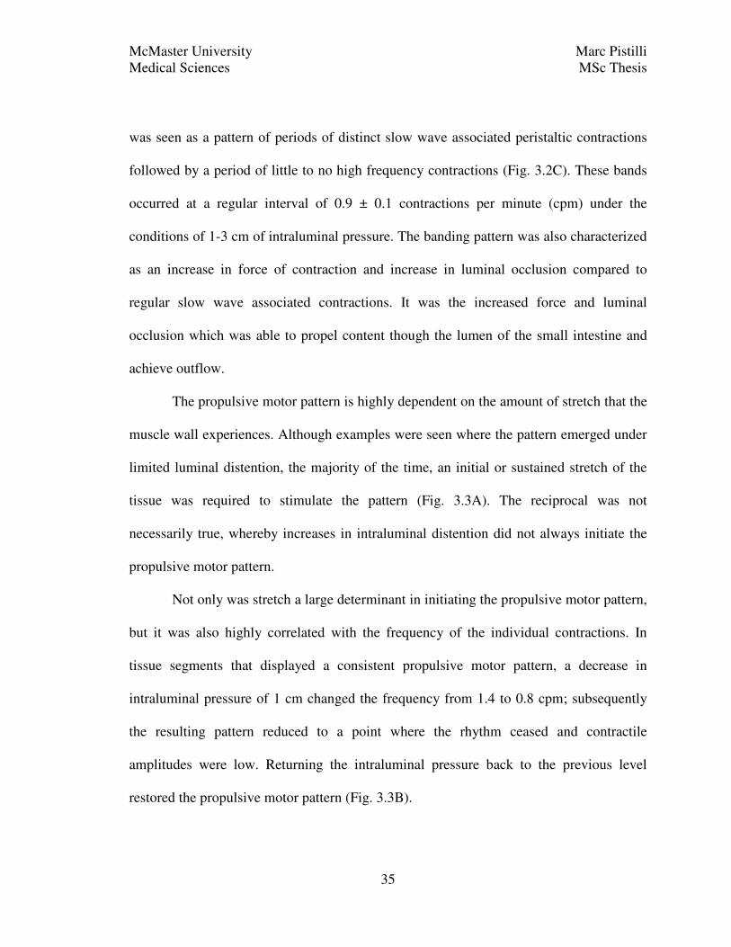
The propulsive motor pattern is highly dependent on the amount of stretch that the
muscle wall experiences. Although examples were seen where the pattern emerged under
limited luminal distention, the majority of the time, an initial or sustained stretch of the
tissue was required to stimulate the pattern (Fig. 3.3A). The reciprocal was not
necessarily true, whereby increases in intraluminal distention did not always initiate the
propulsive motor pattern.
Not only was stretch a large determinant in initiating the propulsive motor pattern,
but it was also highly correlated with the frequency of the individual contractions. In
tissue segments that displayed a consistent propulsive motor pattern, a decrease in
intraluminal pressure of 1 cm changed the frequency from 1.4 to 0.8 cpm; subsequently
the resulting pattern reduced to a point where the rhythm ceased and contractile
amplitudes were low. Returning the intraluminal pressure back to the previous level
restored the propulsive motor pattern (Fig. 3.3B).
McMaster University Marc Pistilli
Medical Sciences MSc Thesis
36
Figure 3.3. Effect of distention on the propulsive motor pattern. A large increase in
intraluminal distention initiated the PMP (A). Reducing intraluminal pressure (B, left
arrow) abolished PMP; restoring luminal distention (B, right arrow) restored the PMP.
Increasing levels of distention (C, both arrows) and corresponding increase in PMP
frequency. Pressure is normalized to the pressure generated by slow wave associated
contractions (SWAC).

McMaster University Marc Pistilli
Medical Sciences MSc Thesis
37
Similarly, a step up by 1 cm of water in intraluminal pressure increased the
frequency from 0.47 cpm to 0.69 cpm (Fig. 3.3C). A further increase in pressure of 1 cm
was able to raise the frequency again to 1.3 cpm (Fig. 3.3C), demonstrating the graded
influence of intraluminal stretch on propulsive motor complex frequency.
3.3.2 Stimulation of the propulsive motor pattern with carbachol
Addition of 5 µM carbachol stimulated the PMP into a consistent high amplitude
pattern (Fig 3.4A) in segments that had previously displayed rhythmic propulsion.
Conversely, the PMP was not initiated via carbachol in tissues that had not exhibited
rhythmic propulsion during the organ bath experimentation. The frequency of carbachol
induced PMCs was almost twice that compared to stretch induced control propulsion
(Table 3.1). Intraluminal pressure recordings indicated no significant difference in
duration or maximum amplitude of control compared to carbachol induced PMCs (Table
3.1). Spatiotemporal mapping (Fig 3.4B) confirmed both the change in frequency and
unaltered duration and amplitude (percent occlusion) to carbachol treatment (Fig 3.4C;
Table 3.1). Mapping also revealed that the velocity of contractions was unaltered between
control and carbachol driven rhythmic propulsion (Table 3.1).
3.3.3 Abolishing the propulsive motor pattern with tetrodotoxin
Blocking of all enteric neural transmission with tetrodotoxin resulted in two main
effects on the PMP. In some cases (2 out of 6) the tissue responded by increasing the
basal tone to the same level as the max amplitude of the PMC (Fig 3.5A). This result
agreed with the analysis of the spatiotemporal maps which showed that the level of

McMaster University Marc Pistilli
Medical Sciences MSc Thesis
38
Figure 3.4. Restoration of rhythmic propulsion with carbachol. Intraluminal pressure
recording (A) showing the recovery of rhythmic propulsion after addition of 5 µM
carbachol (black arrow). Pressure is normalized to the pressure generated by slow wave
associated contractions (SWAC). Control spatiotemporal map (B) with indicated PMCs
(white arrows). PMCs after carbachol stimulation (C; entire duration of the map). The
darker the area on the map along the grey scale corresponds with smaller luminal
diameter or increased level of contraction.

McMaster University Marc Pistilli
Medical Sciences MSc Thesis
39
Table 3.1 The Effect of Carbachol on the Parameters of Rhythmic Propulsion
Intraluminal Pressure Recording
Spatiotemporal Mapping
Control
Carbachol (5 µM) Control Carbachol (5 µM)
Frequency (cpm)
0.9 ± 0.2
n = 4
1.9 ± 0.1**
n = 4
0.9 ± 0.2
n = 3
2.0 ± 0.1**
n = 3
Duration (s)
25.1 ± 5.6
n=4
30.4 ± 1.7
n = 4
55.6 ± 40.3
n = 3
12.3 ± 1.9
n = 3
Amplitude
(%SWAC;
mm)
11.1 ± 3.1
n = 4
17.8 ± 4.3
n = 4
1.3 ± 0.2
n = 3
1.5 ± 0.3
n = 4
Velocity (cm / s)
N/A N/A 0.9 ± 0.2
n = 3
0.7 ± 0.1
n = 4
** p < 0.01; * p < 0.05

McMaster University Marc Pistilli
Medical Sciences MSc Thesis
40
Figure 3.5. Effect of neural blockade on intestinal motor patterns. Intraluminal
pressure recoding (A) shows a tonic increase in pressure in response to the addition of 0.5
mM tetrodotoxin (black arrow). Pressure is normalized to the pressure generated by slow
wave associated contractions (SWAC). Spatiotemporal mapping reveals the abolishment
of PMCs and a shift towards segmentation (B) upon addition of 0.5 µM tetrodotoxin
(white arrow). Addition of 2 µM carbachol (C; white arrow) shifted segmentation to slow
wave associated contractions. The darker the area on the map along the grey scale
corresponds with smaller luminal diameter or increased level of contraction.

McMaster University Marc Pistilli
Medical Sciences MSc Thesis
41
luminal occlusion (amplitude) to PMCs was the same as the contraction induced by TTX
(Table 3.2). In this case, rhythmic propulsion was no longer seen, however, it was not
discernible whether the pattern was abolished or merely overcome by the tonic increase in
basal tone and resultant increase in intraluminal pressure. Conversely, in some intestinal
segments, the PMP was replaced with segmentation like patterns without remnants of the
PMP (Fig 3.5B). Segmentation patterns were identified with two main parameters.
Firstly, the spatiotemporal maps showed neither rhythmic propulsion nor peristaltic slow
wave associated contractions. Secondly, the remaining contractile pattern consisted of
areas of interrupted slow wave associated contractions that were not uniform throughout
the time period (Fig 3.6). Addition of carbachol (2 µM) did not restore the PMP in
segments treated with TTX; however, it did result in a dominant pattern of slow wave
associated contractions (Fig 3.5C). Interestingly, the spatiotemporal map showed a
significant reduction in the frequency of slow wave associated contractions which was
not seen via the intraluminal pressure recording (Table 3.2).
3.3.4 Modifying the propulsive motor pattern with L-NNA
Like acetylcholine, nitric oxide (NO) is a major mediator of intestinal motility,
except on the inhibitory (smooth muscle relaxation) side of the enteric nervous system.
Prevention of NO production by inhibiting nitric oxide synthase with L-NNA increased
the basal pressure of the small intestine and acutely abolished the propulsive motor
pattern (Fig. 3.7A). After a short period, the pattern reemerged superimposed on top of
the increased basal pressure (Fig. 3.7A). The restored PMP had the same frequency,
duration and amplitude of contractions as the control situation (Table 3.3).

McMaster University Marc Pistilli
Medical Sciences MSc Thesis
42
Table 3.2 The Effect of TTX on the Parameters of Rhythmic Propulsion
Intraluminal Pressure Recording
Spatiotemporal Mapping
Control
TTX (0.5 µM) Control TTX (0.5 µM)
Frequency (cpm)
1.2 ± 0.2
n = 3
N/A 0.8 ± 0.1
n = 6
N/A
Duration (s)
28.3 ± 4.7
n = 3
N/A 23.0 ± 3.2
n = 6
N/A
Amplitude (%SWAC;
%Baseline)
13.4 ± 3.1
n = 3
9.6 ± 2.8
n = 3
37.2 ± 3.7
n = 6
39.8 ± 5.4
n = 6
Baseline
(mm) N/A N/A 4.9 ± 0.3
n = 5
4.9 ± 0.5
n = 5
Velocity (cm / s)
N/A N/A 1.48 ± 0.5
n = 5
1.0 ± 0.2
n = 3
Frequency
of SWAC
(cpm)
29.7 ± 2.6
n = 3
23.5 ± 3.3
n = 3
33.9 ± 2.9
n = 6
16.4 ± 5.9*
n = 4
* p < 0.05

McMaster University Marc Pistilli
Medical Sciences MSc Thesis
43
Figure 3.6. Segmentation motor pattern. Appearance of slow wave associated
contractions (A) as measured by spatiotemporal maps. Examples of short duration,
segmental contractions (B, C and D) with the impression of interrupted slow wave
associated contractions, in response to 0.5 µM tetrodotoxin.

McMaster University Marc Pistilli
Medical Sciences MSc Thesis
44
Figure 3.7. Effect of nitric oxide blockade on rhythmic propulsion. Intraluminal
pressure recording (A) revealed both a tonic increase in baseline pressure and
continuation of rhythmic propulsion in the presence of 0.2 mM L-NNA (black arrow).
Pressure is normalized to the pressure generated by slow wave associated contractions
(SWAC). Spatiotemporal mapping (B) consistently revealed the tonic contraction of the
intestine after addition of L-NNA (white arrow) but also the continuation of the PMCs.
The darker the area on the map, along the grey scale, corresponds with reduced luminal
diameter or increased level of contraction.

McMaster University Marc Pistilli
Medical Sciences MSc Thesis
45
Table 3.3 The Effect of L-NNA on the Parameters of Rhythmic Propulsion
Intraluminal Pressure Recording
Spatiotemporal Mapping
Control
L-NNA (0.2
mM)
Control L-NNA (0.2
mM)
Frequency (cpm)
1.1 ± 0.2
n = 5
1.3 ± 0.03
n = 3
1.1 ± 0.2
n = 5
1.3 ± 0.1
n = 3
Duration (s)
25.8 ± 2.3
n = 5
22.4 ± 6.8
n = 3
12.3 ± 2.3
n = 5
9.6 ± 1.2
n = 3
Amplitude
(%SWAC;
%Baseline)
4.5 ± 2.3
n = 5
7.7 ± 2.1
n = 3
31.0 ± 5.3
n = 5
22.0 ± 4.6
n = 5
Proximal Baseline (mm)
N/A N/A 4.6 ± 0.6
n = 5
4.6 ± 0.4
n = 5
Distal Baseline (mm)
N/A N/A 4.3 ± 0.6
n = 5
3.5 ± 0.9
n = 4
Velocity (cm / s)
N/A N/A 0.7 ± 0.3
n = 3
N/A
Frequency of SWAC (cpm)
32.4 ± 5.3
n = 5
34.2 ± 6.9*
n = 4
38.6 ± 6.2
n = 5
42.1 ± 9.2
n = 3
* p < 0.05

McMaster University Marc Pistilli
Medical Sciences MSc Thesis
46
Spatiotemporal mapping of the effect of L-NNA on the propulsive motor pattern
confirmed the results obtained via intraluminal pressure recordings. It is clear that L-
NNA does not abolish the propulsive motor pattern (Fig. 3.7B) but induces an increased
basal contractile state of the circular muscle (Fig. 3.7B). Once again, the resulting PMP
pattern had unaltered parameters compared to control (Table 3.3). Upon visual inspection,
there seemed to be a location bias where the proximal half contracted more than the distal
half of the segment, however empirical data did not support this observation (Table 3.3).
There was a statistically significant increase in slow wave associated contractions as
measured by the intraluminal pressure recording; however, the increase is not likely to be
physiologically relevant due to the absolute magnitude in change. Also, spatiotemporal
mapping did not confirm the statistical significance of this observation.
3.3.5 Modifying the propulsive motor pattern with substance P
In cases where rhythmic propulsion was not as stable (2 out of 6), addition of
substance P normalized the pattern to resemble a more typical propulsive motor pattern
(Fig 3.8A). In the remaining experiments where rhythmic propulsion appeared stable and
typical, substance P had little effect on either the intraluminal pressure recordings (Fig
3.8B) or the spatiotemporal maps (Fig 3.8C and D). The elevated control frequency of the
PMP was significantly reduced by the addition of substance P to a frequency more in line
with typical control parameters. This reduction in frequency was only seen via
intraluminal pressure recordings and not spatiotemporal mapping. All remaining
parameters, measured from both manometry (Table 3.4) and spatiotemporal mapping,

McMaster University Marc Pistilli
Medical Sciences MSc Thesis
47
Figure 3.8. Effect of substance P on rhythmic propulsion. Intraluminal pressure
recording showed a normalization (A) as well as little effect (B) in response to 10 nM
substance P (black arrows), depending on control activity. Spatiotemporal mapping of
control PMP (C) and the marginal effect of 10 nM substance P (D). Addition of 5 µM
carbachol restored (E; black arrow) the PMP in jejunal segments where rhythmic
propulsion was inhibited by 50 nM substance P.

McMaster University Marc Pistilli
Medical Sciences MSc Thesis
48
Table 3.4 The Effect of Substance P on Intraluminal Pressure Parameters of
Rhythmic Propulsion
Control
Substance P
(10 nM)
Sustance P
(50 nM)
Frequency
(cpm) 2.0 ± 0.3
n = 5
1.3 ± 0.1*
n = 5
1.1 ± 0.1*
n = 5
Duration (s)
23.8 ± 2.8
n = 5
31.4 ± 3.2
n = 5
32.5 ± 3.8
n = 5
Amplitude (%SWAC)
10.1 ± 2.5
n = 5
15.7 ± 3.9
n = 5
12.4 ± 3.1
n = 4
Frequency
of SWAC
(cpm)
17.6 ± 2.0
n = 6
24.6 ± 6.3
n = 5
25.7 ± 7.1
n = 5
* p < 0.05
Table 3.5 The Effect of Substance P on Spatiotemporal Mapping of Rhythmic
Propulsion Parameters
Control
Substance P
(10 nM)
Sustance P
(50 nM)
Frequency
(cpm) 1.7 ± 0.3
n = 5
1.6 ± 0.2
n = 6
1.4 ± 0.2
n = 5
Duration (s)
11.8 ± 1.7
n = 5
15.5 ± 2.9
n = 6
18.5 ± 4.2
n = 5
Amplitude (%Baseline)
42.2 ± 4.9
n = 5
39.9 ± 4.1
n = 6
40.8 ± 8.4
n = 5
Baseline (mm)
6.0 ± 0.5
n = 5
5.5 ± 0.5
n = 6
4.8 ± 0.8
n = 5
Velocity (cm / s)
1.4 ± 0.3
n = 5
1.8 ± 0.3
n = 6
1.6 ± 0.4
n = 5
Frequency
of SWAC (cpm)
52.5 ± 1.5
n = 5
50.2 ± 1.7
n = 5
56.6 ± 2.6
n = 5
* p < 0.05

McMaster University Marc Pistilli
Medical Sciences MSc Thesis
49
(Table 3.5) were unchanged in response to both 10 nM and 50 nM substance P. Finally, in
two cases, substance P caused a loss of the PMP which was reversed by the addition of 5
µM carbachol, restoring rhythmic propulsion (Fig 3.8E).
3.4 Discussion
3.4.1 Rhythmic propulsion
The strong, rhythmic nature of small intestinal propulsion is not a novel
observation. This pattern of propulsion has been seen by various researchers often
studying the role of the enteric nervous system on intestinal motility (Costa et al., 2000;
Holzer et al., 1998; Spencer et al., 2001; Spencer et al., 1999). What is novel about
exploring propulsion is characterizing the pattern under more physiological parameters
and acknowledging the inherent rhythmicity.
Rhythmic propulsion is an inducible pattern that requires a stimulus. This can be
thought of as two components, firstly, the physiological stimulus which triggers and
modifies propulsion, and secondly, the underlying mechanism which drives the motor
pattern. I have demonstrated the inducibility of the pattern: providing intraluminal
distention with fluid content can initiate the pattern. Distention is a powerful trigger of
peristaltic contractions which has been documented and assessed rigorously (Tonini et al.,
1989; Grider, 2003). Distention induced propulsion has been mainly attributed to the
peristaltic reflex (Spencer et al., 1999). Under laboratory conditions with short bursts of
distention the peristaltic reflex performs well in explaining the resulting contractile
activity. However, constant distention, which is more physiologically relevant, has only
recently been studied with a growing realization of the rhythmicity with which propulsion
McMaster University Marc Pistilli
Medical Sciences MSc Thesis
50
occurs (Holzer et al., 1998; Huizinga et al., 2011). Distention not only stimulates
rhythmic propulsion, but the degree of distention also modifies the frequency at which it
occurs.
I have demonstrated the graded effect of luminal distention on rhythmic
propulsion. In my experiments an increase in muscular distention resulted in an increased
frequency of propulsive contractions. Similarly, Spencer et al., (2001) using different
experimental conditions, found that there was a graded effect of contractile amplitude and
luminal distention. These two results demonstrate the ability of the small intestine to
modify content propulsion under differing physiological conditions. These findings are in
contrast to the slow wave associated contractions, which are peristaltic but not very
propulsive. The slow wave associated contractions are also inducible but the underlying
pacemaker remains active even in the absence of contractile activity (Huizinga and
Lammers, 2009). Finally, the slow wave associated contraction frequency does not easily
change in response to differing physiological states, but rather exists as an ingrained
feature of the ICC-MP pacemaker system, which can be modified by targeting direct
aspects of the cellular mechanism within ICC (Malysz et al., 2001; Lowie et al., 2011).
Pharmacological experimentation using the small intestine allows for a direct
assessment of the role of the enteric nervous system on motility. Carbachol was used to
mimic the usually stimulatory effects of acetylcholine. Acetylcholine is released from
excitatory motor neurons, which according to the peristaltic reflex model, interacts
directly with the smooth muscle to bring about contraction (Costa et al., 2000). Although
smooth muscle cells express muscarinic acetylcholine receptors (Chen et al., 2007),

McMaster University Marc Pistilli
Medical Sciences MSc Thesis
51
motor neurons additionally project to ICC-DMP, which also express these receptors
(Wang et al., 2003; Ward and Sanders, 2001). Acetylcholine is able to raise the resting
membrane potential of smooth muscles, thereby increasing the likelihood that they reach
threshold and contract (Inoue et al., 1998). The effect of acetylcholine directly on ICC-
DMP remains elusive (Wang et al., 2003). The peristaltic reflex model does not account
for the origin of rhythmicity inherent to the propulsive motor pattern. In my experiments,
carbachol was in contact with the tissue at a constant concentration and yet a rhythmic
pattern persisted. A role of acetylcholine in stimulating the pattern is likely; however, the
persistence of rhythmicity after carbachol addition suggests that it is the target of
carbachol which is the pacemaker, in contrast to the rhythmic release of acetylcholine
from excitatory motor neurons. Carbachol alone was only able to induce the PMP in
segments that were previously propelling content in vitro, which suggests that
acetylcholine is required, but not sufficient in initiating propulsion. This is in contrast to
the rat colon which exhibits a recovery in rhythmic propulsion with the addition of
carbachol, even in the presence of complete neural blockade with lidocaine (Huizinga et
al., 2011).
Nitric oxide in the small intestine is primarily responsible for setting the basal tone
of the muscle layer (Dijkstra et al., 2004). A significant source of nitric oxide is the
inhibitory motor neurons of the enteric nervous system. Nitric oxide is thus an integral
part of the peristaltic reflex facilitating the region of relaxation caudal to the triggering
bolus of content (Grider, 2003). Since the rhythmicity of the propulsive motor pattern
persisted superimposed on top of a tonic increase in baseline pressure in the absence of

McMaster University Marc Pistilli
Medical Sciences MSc Thesis
52
nitric oxide (nitric oxide synthase inhibited by L-NNA), it cannot be caused by rhythmic
nitric oxide release. Since the contractions of the PMP have an amplitude the same as the
tonic increase in pressure, it could be thought that the PMC was achieved just by
removing nitric oxide from a short period of time, which is not the case. Thus, neither
rhythmic acetylcholine nor nitric oxide release can explain the physiologically relevant
rhythmicity of propulsion. Similar to the experiments with carbachol, L-NNA did not
alter any of the PMC parameters, affirming their dependence on the pacemaker and not
the stimulating signal from the enteric nervous system.
Often publications have focused on mechanisms of motility which ignore input
from myogenic sources, and suggest mechanisms which are limited, not entirely based on
all scientific findings or not physiologically relevant (Huizinga and Lammers, 2009). This
oversimplification has led to assumption of the control capacity of enteric neurons for
which there is no evidence, namely the origin of rhythmicity of intestinal propulsion. The
origin of rhythmicity of the propulsive motor pattern is likely myogenic, but this does not
mean that there is no input from the enteric nervous system. Acetylcholine h plays a
prominent excitatory role on intestinal motility and content transit (Kilbinger and
Weihrauch, 1982), which is consistent with the stimulatory role of carbachol on rhythmic
propulsion that I have demonstrated. In my experiments, the enteric nervous system was
required for the PMP because tetrodotoxin abolished the pattern completely. Since
carbachol could not restore rhythmic propulsion after addition of TTX, the underlying
mechanism must be different from that of the rat colon, where carbachol was sufficient in
driving rhythmic propulsion in the presence of neural blockade (Huizinga et al., 2011). It

McMaster University Marc Pistilli
Medical Sciences MSc Thesis
53
is likely then that acetylcholine is required for rhythmic propulsion alongside another
factor such as substance P.
It is unclear whether substance P plays a stimulatory or regulatory role in rhythmic
propulsion. Substance P did not modify any parameters except for frequency. The basal
frequency of the PMP in these experiments was elevated compared to typical control
values. Substance P normalized the frequency and the consistency of the pattern towards
control values. Substance P working through NK1 receptors (primarily found on the ICC-
DMP) is inhibitory with regards to propulsive peristalsis (Holzer et al., 1995). My results
agree with their findings but not entirely with the conclusion. In my case, substance P
reduced frequency, but the higher concentration did not further lower the frequency past
what is typical for rhythmic propulsion in the mouse small intestine. Thus, substance P
can only modify rhythmic propulsion when it is not at a physiologically appropriate
frequency. However, substance P is excitatory at intestinal smooth muscle cells because it
interacts with NK2 receptors expressed on these cells (Holzer et al., 1995). Therefore,
substance P can promote propulsive contractions and simultaneously help set the
pacemaker frequency of these contractions.
The results I have presented above provide strong evidence against the functions
of the peristaltic reflex in rhythmic propulsion. The enteric nervous system does play a
role in rhythmic propulsion from a stimulatory and maintenance capacity, but the
rhythmicity associated with the pattern is myogenic. It is possible that increased
distention is signaled by neurons through acetylcholine release at ICC-DMP leading to
increased frequency of contractions, since the PMP is distention sensitive. Conversely,

McMaster University Marc Pistilli
Medical Sciences MSc Thesis
54
substance P release may counter the excitation to help maintain the pattern and may be
released in response to reductions in distention. The combination of substance P and
acetylcholine to drive propulsive contractions has been seen previously in mouse small
intestine; where mucosal stimulation induced propagation of propulsive peristalsis which
was attenuated, but not abolished, by antagonizing muscarinic acetylcholine receptors or
tachykinin receptors. Using both receptor antagonists concurrently completely abolished
all propagation of the contraction (Grider, 2003). Although nitric oxide plays an
important role in the peristaltic reflex model, it does not play a role in rhythmic
propulsion. Since neither substance P nor acetylcholine can stimulate or recover rhythmic
propulsion alone, it is likely that they are both required for the PMP although whether
they target the ICC-DMP, smooth muscle or both remains unclear.
3.4.2 Segmentation
The primary role of the segmentation motor pattern is to mix intraluminal contents
for greater intestinal absorption (Gwynne et al., 2004B). The underlying pattern shows a
high degree of rhythmicity in vivo (Cannon, 1902; Wang et al., 2005). Using an in vitro
model, I have demonstrated that the segmentation pattern follows the rhythmicity of the
slow wave frequency and is for that, dependent on ICC-MP. Work in our lab
(unpublished) suggests that the rhythmic interference pattern characteristic of
segmentation occurs because of interfering electrical slow waves, but the origin and
mechanisms are undetermined. Gwynne and Bornstein (2007) showed that nutrients, such
as short chain fatty acids, can induce the segmentation motor pattern. This is seen in vivo
where lipid meals require increased processing time and thus a longer measured intestinal

McMaster University Marc Pistilli
Medical Sciences MSc Thesis
55
transit (Higham and Read, 1990). Nutrient induced segmentation was explained as neural
in origin; however, neither ICC nor the origin of rhythmicity in the pattern were properly
assessed (Gwynne and Bornstein, 2007) which is inappropriate, considering it is already
known that the rhythmicity of segmentation follows closely the slow wave frequencies
across species (Table 1). Conversely, I have shown that segmentation can be induced in
the presence of neural blockade with tetrodotoxin, and thus the pattern has a myogenic
origin, whereas, the opposite conclusion was made by Gwynne and Bornstein (2007) who
claimed that segmentation was entirely neurogenic without myogenic input. Gwynne and
Bornstein (2007) employed a guinea-pig model which has low level slow wave activity
compared to other animals, and they admit that it may not represent a true model of
segmentation in all other species. The consistency of cholinergic SWACs in the absence
of neural activity, and the fact that the slow waves clearly underlie segmentation suggests
that these patterns occur on a spectrum which is predominated by SWACSs when
acetylcholine acts at ICC-MP or smooth muscle directly. In vitro, segmentation occurs in
the absence of enteric nervous system activity, however, this does not rule out a potential
physiological role for neurons in segmentation. As mentioned above, the nerves were
shown important for in vitro nutrient induced segmentation. It is thus possible that the
pattern is governed by a myogenic mechanism, but is activated physiologically though
enteric nervous system sensation of specific luminal contents, such as lipids and proteins
(Gwynne et al., 2004B).

McMaster University Marc Pistilli
Medical Sciences MSc Thesis
56
4. CACLIUM OSCILATIONS IN ICC OF THE DEEP MUSCULAR PLEXUS
4.1 Introduction
Excitability of intestinal cells, including neurons, smooth muscle and ICC is often
experimentally signaled by increases in intracellular calcium which outlast the excitatory
event (Tack and Smith, 2004). This calcium signal has given researchers the ability to
study the actions of cells participating in intestinal motility on a cellular level.
Specifically, a rhythmical change in intracellular calcium in ICC-MP is equivalent in
frequency with the frequency and generation of slow waves (Yamazawa and Iino, 2002).
Intracellular calcium measurements have revealed a great degree of synchronicity and
cooperation of ICC-MP in generating and propagating of slow waves (Lowie et al.,
2011). Poorly understood is the role of intracellular calcium oscillations and relations to
ICC-DMP electrical pacemaking. Our lab has preliminary data showing that calcium
activity in ICC-DMP differs from that of the ICC-MP by a great deal, specifically, ICC-
DMP do not flash calcium but rather oscillate it in a polar manner within the cell. Also,
ICC-DMP calcium activity is not synchronized between neighboring cells, whereas ICC-
MP oscillate intracellular calcium as a network at or near an instantaneous manner as
their neighboring ICC (Lowie, 2011).
The ICC-DMP are highly innervated by the enteric nervous system. This
innervation has led many to postulate a transduction role of the ICC-DMP, facilitating a
communication link between nerves and smooth muscle (Iino et al., 2004). Although
anatomically plausible, there are currently no physiological data to provide an
explanation for the high degree of innervation of ICC-DMP (Ward et al., 2006).

McMaster University Marc Pistilli
Medical Sciences MSc Thesis
57
Similarly, it is known that ICC-DMP are innervated by cholinergic (Wang et al., 2003),
tachykinergic (Faussone-Pellegrini, 2006) and nitrergic (Wang et al., 2003) neurons,
however, there are little data reporting how acetylcholine, substance P or nitric oxide
effect ICC-DMP activity. A recent hypothesis by Huizinga and colleagues (2011) has
suggested a dual pacemaker network responsible for rhythmic propulsion in the rat colon.
This hypothesis consists of a summative effect between the slow wave generating ICC
and a second innervated ICC network in the colon. Likewise, the hypothesis may be
extended to the small intestine, which also contains a slow wave generating network of
ICC (ICC-MP) and the highly innervated ICC-DMP network.
4.2 Methods
Adult and neonatal CD-1 intestinal preparations were used for all calcium imaging
experiments. Information on animal handling can be found in sections 2.1. Information on
intestinal preparations and calcium imaging can be found in section 2.3.
4.2.1 Calcium imaging experiments proper
The remainder of the experiment was conducted under minimal light conditions as
the imaging agent was light sensitive. Intestinal preparations were removed from the large
Krebs’ solution reservoir and incubated for 15 minutes with 1 mL Krebs’ solution
containing 5 µM fluo-4 AM (acetoxymethyl ester), 10 µM probenecid and 0.02%
pluronic F-127. Once the dye was loaded, preparations were perfused constantly with
fresh Krebs’ solution gassed with 95% O2; 5% CO2 heated to 37 oC. Intestinal
preparations were viewed using a Nikon eclipse FN1 microscope employing either 20X

McMaster University Marc Pistilli
Medical Sciences MSc Thesis
58
or 40X objectives with a GFP filter allowing for the excitation of fluo-4 with light at a
wavelength of 488 nm. Digital videos were created using a QuantEM 512SC camera
capturing visual images and recorded with Nikon-NIS Elements software loaded on a
personal computer. Upon visual identification of ICC-DMP (actively oscillating calcium
or quiescent), control videos were recorded followed by the addition of agonists
substance P (10 nM), carbachol (5 µM) or phenylarsine oxide (PAO; 0.1 µM), in order to
mimic the activity of the tachykinergic and cholinergic enteric nervous system or reduce
receptor internalization, respectively. A reduced light exposure protocol was also used
where a 0.2 second image was recorded followed by a 0.8 second dark period and
repeated for the duration of the experiment to facilitate longer recording periods.
4.2.2 Data analysis
Data were extracted as the fluorescence intensity versus time of specified cells.
Cells confirmed as ICC-DMP based on their morphology as bipolar, ovoid cells lying in
the same orientation as the circular muscle were circled using the ROI function of the
Nikon-NIS Elements software. The software allowed for the data extraction of multiple
cells from the same field of view, simultaneously. Due to the preliminary nature of the
experiments, no further transformation was done to the data, which is not inconsistent
with previous publications (Lowie et al., 2011). Likewise, the exploratory nature of the
experiments did not present enough data to perform adequate statistical analysis.

McMaster University Marc Pistilli
Medical Sciences MSc Thesis
59
4.3 Results
4.3.1 Neonatal ICC-DMP
The ICC-DMP, of neonatal small intestine, cycled calcium from side to side
within a cell at ever-changing rates. This contrasts with the calcium cycling in ICC-MP
which exhibited a flash of calcium throughout the entire cell body at the same time. The
calcium oscillations did not correspond with those of neighboring ICC-DMP and thus
synchronization of oscillations was not observed. In an interesting experiment, after
addition of 10 nM substance P, both calcium oscillation frequency, as well as baseline
calcium increased (Fig. 4.1A). With regards to baseline [Ca2+
]i, the observed increase
returned back to normal shortly after elevation, but was followed by subsequent increases
in a rhythmic fashion (Fig. 4.1A). In some instances, addition of substance P increased
basal calcium without a return to normal or the presence of rhythmic changes (Fig 4.1B).
By using the reduced exposure protocol, experiments in excess of 10 minutes revealed a
second phasic elevation in calcium longer in duration than those previously seen,
however at a much lower frequency (Fig. 4.2). Coadministration of PAO to reduce NK1
receptor internalization did not reveal any obvious change in duration of low frequency
rhythmic activity initiated by 10 nM substance P.
4.3.2 Adult ICC-DMP
Tissue preparations exposing the deep muscular plexus in adult mice yielded few
active ICC-DMP. Imaging revealed either no obvious ICC-DMP, or showed ICC-DMP
which were not oscillating calcium. The latter group exhibit characteristics of properly
loaded cells, but neither substance P nor acetylcholine initiated calcium activity in these

McMaster University Marc Pistilli
Medical Sciences MSc Thesis
60
Figure 4.1. Effect of substance P on intracellular calcium concentration of neonatal
ICC-DMP. Each line indicates the intensity of fluorescence (relative fluorescence unit -
RFU) of fluo-4 bound to calcium, over time in a separate ICC-DMP. Addition of 10 nM
substance P (black arrow) induced a short-lived rhythmic increase in basal florescent
intensity (A and B). In some instances, the increase in basal florescence was tonic (B).

McMaster University Marc Pistilli
Medical Sciences MSc Thesis
61
Figure 4.2. Effect of long-term substance P exposure on intracellular calcium
concentration of neonatal ICC-DMP. Each line indicates the intensity of fluorescence
(relative fluorescence unit - RFU) of fluo-4 bound to calcium, over time in a separate
ICC-DMP. Addition of 10 nM substance P (black arrow) induced a short-lived low
frequency pattern. Also seen was a second lower frequency pattern of oscillating baseline
calcium levels.

McMaster University Marc Pistilli
Medical Sciences MSc Thesis
62
cells. In ICC-DMP that had active calcium oscillations, substance P increased the
frequency of oscillations and the baseline calcium in segments of the cell (Fig. 4.3B), but
the net effect on the whole cell seems to be unchanged (Fig. 4.3A). The independence of
these cells has yet to be elucidated as preparations have only yielded one or two active
cells at a time and not entire networks as seen with the neonatal preparations.
4.4 Discussion
Although a relatively new technique, intracellular calcium imaging has allowed
for a precise measurement of the activity found in ICC-MP. A similar assessment of ICC-
DMP has not been performed. As mentioned above, an increase in intracellular calcium in
ICC-MP is a marker of the pacemaker potential (Torihashi et al., 2002). ICC-DMP also
generate an electrical signal in some instances (Jiménez et al., 1996), but it is unclear if
this activity is associated with a rise in intracellular calcium. ICC-DMP shifted calcium
concentration from side to side within the cell body rather than flashing a simultaneous
increase in calcium concentration though out the entirety of the cell body like in the ICC-
MP. The origin of calcium in ICC-DMP is currently unknown. There may be an unequal
opening of extracellular calcium channels or intracellular calcium stores, the latter being
the primary source of calcium in ICC-MP activity (Lowie et al., 2011). Since there is
rarely equal calcium activity across an individual ICC-DMP at one time, it is not likely
that this cycling signals electrical activity. The large rhythmic increase in baseline
calcium induced by substance P may signal calcium activity because the increase in
calcium was uniform throughout the cell. Another major difference from ICC-MP is that

McMaster University Marc Pistilli
Medical Sciences MSc Thesis
63
Figure 4.3. Effect of substance P exposure on intracellular calcium cycling within
adult ICC-DMP. The overall fluorescence activity representing intracellular calcium
concentration bound to fluo-4 of a single ICC-DMP (A) and its response to the addition
of 10 nM substance P (black arrow). Fluorescence within the same cell split into thirds
(one line for each; B) enhancing the visibility of calcium activity upon addition of 10 nM
substance P (black arrow).

McMaster University Marc Pistilli
Medical Sciences MSc Thesis
64
the ICC-DMP do not synchronize calcium cycling between neighboring cells. This is
interesting because the cells have a high degree of coupling as judged by the presence of
gap junctions between one another in the network, and also with smooth muscle cells
(Kobilo et al., 2003). The only synchronization seen between neighboring ICC-DMP was
the rhythmic rise in baseline intracellular calcium in response to substance P. This
suggests that ICC-DMP activity may be signaled by changes in baseline calcium and not
the individual internal calcium oscillations. Calcium oscillations did not persist for a very
long time. The termination of the pattern was not a result of tachykinin receptor
internalization, but may be attributed to receptor desensitization (Defea et al., 2000). The
termination could also be due to a complex inhibitory feedback system, from the enteric
nervous system preventing inappropriate cellular actions to absent content or distention
stimuli, as substance P was artificially introduced to the system.
Neonatal ICC-DMP calcium activity was quite abundant, however, the adult ICC-
DMP were rarely spontaneously active. This may be a result of the further development
of both the ICC and neural networks between neonates and adults. The adult ICC-DMP
are highly innervated by both excitatory and inhibitor motor neurons (Wang et al., 2003).
The inhibitory motor neurons may be influencing the lack of activity seen in these cells in
adulthood. Enteric nerves are fully formed by post natal day 10 in mice, however, their
function is not developed to the point of an adult neural system (Foong et al., 2012).
Secondly, it is likely that the ICC-DMP pacemaker is inducible and not always present
like the ICC-MP produced slow wave (Wang, 2005), as seen by the increase in calcium
activity in response to substance P and no spontaneous rhythmicity. This suggests that the

McMaster University Marc Pistilli
Medical Sciences MSc Thesis
65
neural connections form the basis for triggering this inducible pacemaker network.
Excitatory motor neurons respond to input from sensory neurons, in response to stimulus
such as distention or nutrients (Schemann and Mazzuoli, 2010; Mourad and Saadé, 2011).
If the correct stimulus for ICC-DMP network stimulation is not present in the calcium
imaging experiments performed, it is reasonable to expect the cells to be quiescent.
Finding the correct conditions/stimulants of ICC-DMP calcium and pacemaker activity
will allow for a better understanding of how this ICC network performs under
physiological conditions and which motor patterns it may be controlling or modifying.

McMaster University Marc Pistilli
Medical Sciences MSc Thesis
66
5. CONTRACTILE PATTERNS OF THE HUMAN PYLORIC REGION
5.1 Introduction
Understanding the anatomy of the gastrointestinal system is paramount to
understanding how motor functions originate and persist. One of the key cell types
involved in rhythmic motor patterns in both the stomach and the small intestine are the
ICC. The ICC set the frequency of rhythmic motor patterns as well as aid in the
propagation of motor patterns (Huizinga and Lammers, 2009). Animal studies have
shown that the ICC networks found in the antrum of the stomach are completely
independent of the intestinal ICC networks (Wang et al., 2005). Electrical activity is
unable to cross the pylorus either from the stomach to the intestine or vice versa (Wang et
al., 2005). These two findings fit well with our understanding of intestinal and stomach
motor patterns, specifically the high frequency with which contractions occur in the
duodenum compared to those of the antrum (Wang et al., 2005). What remains unclear is
if the motor patterns from the stomach and intestine can interact in other ways other than
through ICC. Also, most of our understanding of anatomical features of the ICC networks
has come from animal models, so studying these cells in the human pyloric region is
important to understanding normal human physiology. The following is a miniature
project designed to assess the rhythmic contractile patterns of smooth muscle strips from
the antrum, pylorus and duodenum. The project was undertaken to correlate contractility
data with the anatomical localization of ICC in the region of the pylorus. I hypothesized
that both the antrum and duodenum would respond to stimulation with a strong rhythmic

McMaster University Marc Pistilli
Medical Sciences MSc Thesis
67
pattern with the antrum displaying a frequency much slower than that found in the
duodenum.
5.2 Methods
Adult circular muscle strips were used for all muscle strip experiments. Methods
on tissue acquisition and muscle strip preparation can be found in section 2.4.1.
5.2.1 Muscle strip experiments proper
Once the tissues were strung up in individual organ baths, they were warmed to
37oC and a resting tension of 10 mN was applied to each. Tissues were allowed a 1 hour
equilibration time and then successive 2 mN stretches were applied to the tissue in 12
minute intervals. After four successive stretches, regardless if phasic contractions were
stimulated, 1 µM carbachol was added to all tissues and remained in contact with the
tissue for 25 minutes.
5.2.2 Data analysis
Due to the dearth in stretch-induced activity in muscle strip experiments, analysis
was only done on carbachol-stimulated contractions. Frequency of contractions was
calculated over a 10 minute interval after a 2 minute equilibration period post carbachol
addition. Where appropriate, a second frequency calculation was done to assess the
reduction/stabilization in tissue contractions. Finally, in pyloric strips exhibiting tonic
contraction caused by 1 µM carbachol, the strength (amplitude) of these contractions was
also calculated, subtracting the preset baseline tension prior to carbachol addition.

McMaster University Marc Pistilli
Medical Sciences MSc Thesis
68
5.3 Results
5.3.1 Effect of stretch on circular muscle strip contractility
Although circular muscle strips responded to stretch, this did not happen in a
consistent manner. Stretching of tissues from 10 – 18 mN did not evoke contractions in
most strips. Figure 5.1 shows an example of increasing stretch and response in the
pylorus where tissues responded to stretch in 2 out of 10 cases. The antrum and duodenal
muscle strips only responded with contractions to stretch in 2 out of 9 and 1 out of 10
strips, respectively.
5.3.2 Effect of carbachol on circular muscle strip contractility
Antrum (5 of 10 strips), pylorus (4 of 10 strips) and duodenum (7 of 10 strips)
muscle strips responded more consistently to carbachol, even in cases where there was no
response to stretch. The distal antrum showed a strong rhythmic pattern at a frequency of
4.3 ± 0.9 cpm (n =4). Following the initial response, the frequency was either maintained
(Fig 5.3A) over a period of 25 minutes or slowed down (Fig. 5.2A) to 2.4 ± 0.7 cpm (n =
5). The pyloric circular muscle strips reacted to carbachol consistently with a marked
increase in tone, not seen in distal antrum or proximal duodenum (Fig 5.2B and Fig
5.3B). The maximal amplitude of the tone was 39.7 ± 6.8 mN, compared to 24.2 ± 4.7
mN initial tone. The tonic contraction progressed into a pattern of stable phasic
contractions with a frequency of 12 ± 3.2 cpm (n=3) (Fig 5.2B). This frequency was
either maintained (Fig 5.3B) or was seen to halve (Fig 5.2B). The duodenal circular
muscle strips reacted to carbachol consistently with a pattern of strong rhythmic
contractile activity with two frequency groups; 12.1 ± 0.5 cpm (n = 5) (Fig 5.2C) or 4.0 ±

McMaster University Marc Pistilli
Medical Sciences MSc Thesis
69
Figure 5.1. Effect of stretch on a human pyloric muscle strip. Data are from a single
pyloric muscle strip presented as force over time. The first stretch (A) induced some
contractions which persisted over time. Additional stretches (B and C) increase the
response time and strength of contractions of the human pylorus.

McMaster University Marc Pistilli
Medical Sciences MSc Thesis
70
Figure 5.2. Evolving effect of carbachol on the human circular muscle strips.
Addition of 1 µM carbachol at the beginning of the traces induced phasic contractions in
both the distal antrum (A) and proximal duodenum (C), but a tonic contraction in the
pyloric strip (B). All initial patterns devolved over time with a reduction in force and
frequency of contractions. Data obtained from the same individual for all three traces.

McMaster University Marc Pistilli
Medical Sciences MSc Thesis
71
Figure 5.3. Stable effect of carbachol on human circular muscle strips. Addition of 1
µM carbachol at the beginning of the traces induced phasic contractions in the antrum
(A), pylorus (B) and proximal duodenum (C) that were consistent over time. The phasic
contractions of the pylorus were superimposed on top of a tonic contraction in this strip.
Data obtained from the same individual for all three traces.
McMaster University Marc Pistilli
Medical Sciences MSc Thesis
72
0.7 cpm (n=3) (Fig 5.3C). These two groups developed over a 25 minute period into a
single rhythmic motor pattern of 1.6 ± 0.2 cpm (n=8) (Fig 5.2C and Fig 5.3C).
5.4 Discussion
The pylorus is a circular muscle sphincter designed to hold content in the stomach
facilitating the physical break-down of content. The pylorus also controls the release of
content into the duodenum (Ramkumar and Schultze 2005). These distinct physiological
processes require differing motor patterns, thus it is not a surprise to find differing
cellular anatomies of the three regions governing these patterns. Rodents have a distinct
separation of pacemaker ICC-MP in the stomach and small intestine separated by the
pylorus which did not contain these cells (Wang et al., 2005). The findings of a
concurrent anatomical study from which muscle strips for my data were provided, did not
have the same distinction in ICC networks in humans. The stomach contained low levels
of ICC-MP, which was also seen in the pylorus itself, unlike the rodent (Wang et al.,
2005). Interestingly, the proximal duodenum did not display ICC-MP or ICC-DMP, as is
typically seen in rodents (Wang et al., 2005). Thus, the similarities between antrum and
pylorus, and the lack of ICC in the duodenum, did not fully explain the distinct motor
patterns that are required by each muscle region.
Although the underlying pacemaker physiology of the human pyloric region was
not as distinct as predicted, the functional contractility data yielded typical distinct
patterns in each of the three regions studied. The stomach requires a great deal of force to
break down large pieces of food into more manageable sizes, which is achieved via
rhythmic low frequency high powered contractions (Cullen and Kelly, 1993). This

McMaster University Marc Pistilli
Medical Sciences MSc Thesis
73
contrasts with the proximal duodenum where motility patterns only need to mix content
to maximize intestinal contact with content for absorption. This is achieved with weak
rhythmic contractions that are at a higher frequency than the stomach (O’Grady et al;
Erickson et al., 2009; Schulze, 2006). The muscle strip data I have presented confirms the
frequency difference between stomach and duodenum, although some duodenal strips had
frequencies closer to that of the stomach. The absolute frequency values compare
favourably with in vivo physiological studies where the stomach and duodenal
contractions occurred at a rate of 2.8 (O’Grady et al., 2010) and 15.8 (Erickson et al.,
2009) per minute, respectively. The fact that there are few ICC in the duodenum of
samples studied suggests that there should not be rhythmic contractions at slow wave
frequencies in these strips, which was not the case. It was difficult to determine a distinct
border between the pylorus and duodenum due to a large population of ICC-MP in this
region (data not published). It is likely that some of these ICC were included in the
muscle strip preparations and thus provided the pacemaker for phasic contractions.
Although anatomically difficult to distinguish, the duodenum strips never exhibited a
tonic contraction, and thus can be claimed confidently to originate from the small
intestine and not the pylorus.
Under normal circumstances, the pylorus remains closed (contracted) far more
than it is open (relaxed) (Desipio et al., 2007). The function of the pylorus is thought not
to be primarily governed by gastrointestinal pacemakers, but rather by direct innervation
(Allescher and Daniel, 1994). This is likely true for rodents where no ICC-MP pacemaker
activities were found. However, my data suggest that in humans, a myogenic pacemaker

McMaster University Marc Pistilli
Medical Sciences MSc Thesis
74
might be an important part of sphincter physiology. The fact that rhythmic activities of
the antrum and pylorus were clearly distinct might be due to a differing neuronal
regulation of the pacemaker activities. Acetylcholine stimulates tonic contraction to keep
the pylorus closed, achieving the compartmentalization required for stomach digestion
(Allescher and Daniel, 1994). Addition of carbachol in my experiments mimicked the
tonic contraction as would be expected from this muscle layer. Interestingly, there were
phasic contractions superimposed on top of the tonic contraction of the pyloric muscle
strips, which has previously been seen in humans via manometry recordings (Friedenberg
et al., 2008). These phasic contractions are likely attributed to the low levels of ICC-MP
expressed (data not published) in the human pylorus. The role of the low level ICC-MP
and resulting phasic contractions in the pylorus is not clear, but it may facilitate low level
release of stomach content into the duodenum between typical pyloric relaxation and
stomach emptying events. Although the rodent model clearly links anatomical features
with motor function, the human pyloric region is not as easily defined, but I have shown
that it still maintains its proper underlying motor patterns in isolated circular muscle
strips.

McMaster University Marc Pistilli
Medical Sciences MSc Thesis
75
6. EFFECT OF E. COLI NISSLE 1917 ON RAT COLON CONTRACTILIY
6.1 Introduction
Symptomology of gastrointestinal disorders almost always involves negative
effects in motility. The most common motility issues are constipation and diarrhea which
effect individuals of all ages (Liu, 2011). Often these symptoms present with no etiology
or as part of a complex disease, such as inflammatory bowel disease, for which the
underlying cause is still unknown (Obermeir et al., 2010). There has been a recent drive
towards understanding the role of intestinal bacteria and normal gastrointestinal health. It
has led to the postulation that probiotics may be used to help promote a healthy intestine-
bacteria symbiosis, and potentially alleviate symptoms of constipation (Valerio et al.,
2010). One such probiotic is Escherichia coli Nissle 1917 (Nissle), which is currently
used in some European countries to help alleviate constipation (Sonnenborn and Schultze,
2009). Nissle has been tested in clinical studies where it increases stool frequency and
thus alleviates constipation compared to control (Sonnenborn and Schultze, 2009).
However, the studies have yet to elucidate how the motility modulating effects occurred.
Nissle has also been studied on in vitro human colonic muscle strips, where it acts to
increase contractile activity (Bär et al., 2009). The same study also showed that the effect
was not neurally mediated and was likely a direct effect on smooth muscle (Bär et al.,
2009). The true biological mechanism of action for Nissle on smooth muscle contractility
is yet to be elucidated. How the increase in contractility might affect the motor patterns of
the colon is currently being studied. The following miniature study was conducted as a
supplement to a motility study being performed using rat colon. The purpose of the mini

McMaster University Marc Pistilli
Medical Sciences MSc Thesis
76
study was to confirm the data of the human study as well as further elucidate the basic
mechanism of action of Nissle on colonic smooth muscle. I hypothesized that the effects
of Nissle would be excitatory on smooth muscle contractions, and that the effect would be
independent of the enteric nervous system, as well as the colonic mucosa and submucosa.
6.2 Methods
Adult Sprague-Dawley rat colonic circular muscle strips were used for all the
following muscle strip experiments. Information on animal handling can be found in
sections 2.1. Information on colonic strip preparation can be found in section 2.4.2.
6.2.1 Muscle strip experiments proper
Once the tissues were strung up in individual organ baths, containing gassed
Krebs’ solution, tissues were warmed to 37oC and a resting tension of 10 mN was applied
to each. Tissues were allowed a 1 hour equilibration time and then stretched back to a 10
mN baseline. Since the contractile pattern of interest was not neurally mediated, 1 µM
lidocaine was added to negate the effects of the enteric nervous system. A 15 minute
control period after lidocaine addition was recorded followed by three successive
additions (1:300 dilution, 1:200 dilution and 1:150 dilution) of either conditioned
(containing dissolved bacterial products) or unconditioned (bacterial growth medium
only) media. Between each addition a 12 minute equilibration and record period occurred.
At the end of the final 12 minute period, 10 µM D-600 was added to halt all contractions.

McMaster University Marc Pistilli
Medical Sciences MSc Thesis
77
6.2.2 Data analysis
Spontaneous contractions were usually present after the 1 hour equilibration
period, thus control data, including frequency, force of contraction (amplitude) and area
under the curve (AUC) were calculated for the final 10 minutes of the 15 minutes control
period. Frequency was calculated as the number of contractions during the 10 minute
recording period divided by 10 minutes. Amplitude was calculated as the average max
force of contraction for those contractions falling in the 10 minute recording period minus
the baseline tension directly before each contraction. Finally, the area under the curve was
calculated as the integral of the force-time recording over the same 10 minute period. The
same procedure was applied for calculating frequency, amplitude and AUC for the 10
minute recording period of each medium addition. A final area under the curve was
calculated after the addition of methoxyverapimil, which was subtracted from each AUC
value calculated to remove the artificially created baseline tension. Data are presented as
mean ± SEM from three animals; the addition of treatment with conditioned and
unconditioned media were performed in triplicate.
6.3 Results
Most muscle strips displayed strong rhythmic contractions after the one hour
incubation period. These high-amplitude low-frequency contractions were unaffected by
neural blockade using lidocaine (Fig. 6.1). The frequency of contractions, after the
addition of lidocaine, was 0.6 ± 0.02 cpm (Fig 6.2). Addition of the two lowest
concentrations of media had no effect on the contractions. The 1:150 dilution of
conditioned media showed

McMaster University Marc Pistilli
Medical Sciences MSc Thesis
78
Figure 6.1. Effect of neural blockade on high-amplitude low-frequency contractions
of the rat colon muscle strips. Representative force versus time graph of a rat colon
circular muscle strip. Addition of 1 µM lidocaine (black arrow) showed no effect on
either the frequency or the force of contractions of the high-amplitude low-frequency
contractions.

McMaster University Marc Pistilli
Medical Sciences MSc Thesis
79
Figure 6.2. The effects of conditioned (A) and unconditioned media (B) on muscle
strips from the colon devoid of mucosa. Tissues were treated with lidocaine (1 µM)
(shown as the first 10 minutes of the figure) and then cumulative additions of treated or
untreated media up to a dilution factor of 1:150 (second 10 minutes of the figure).
Addition of lower concentrations of treated media lasted 30 minutes (not shown; period
represented with the double slash).

McMaster University Marc Pistilli
Medical Sciences MSc Thesis
80
excitatory effects for all parameters calculated (Fig 6.2). Frequency of contractions
increased by 101% ± 39 compared to control, whereas the unconditioned media caused a
decrease of 18% ± 14 of control (Fig 6.2). Conditioned media increased the amplitude of
contractions by 67% ± 11 and the unconditioned media only had a 6% ± 6 effect on
amplitude. Finally, the overall force of contraction increased 172% ± 7 of control in
response to the conditioned media. The unconditioned media increase the overall force by
only 52% ± 14 of control.
6.4 Discussion
The high-amplitude low-frequency contractions responsible for content propulsion
are thought to be governed by a secondary, non-neural pacemaker (Huizinga et al., 2011).
I have confirmed via muscle strip contractility that this is the case, as lidocaine did not
have an effect on these contractions. Physiologically, however, a neural component may
be required for propagation of the contractions, as they are abolished via neural blockade
in a rat colon motility study (Huizinga et al., 2011). However, the same authors showed
that there is still some rhythmic activity in the proximal colon at the same frequency as
the propulsive contractions. It is likely that the lidocaine insensitivity, in my experiments,
is due to the proximal location from where the strips were taken. Interestingly, others
have also observed the neural-independent contractions in human colonic muscle strips
(Gonzalez and Sarna, 2001). Several authors have shown the importance of excitatory
neurotransmitters on propulsion in the colon, and this may attest to the inducible nature of
the pattern. As Huizinga et al., (2011) have demonstrated, colonic propulsion is

McMaster University Marc Pistilli
Medical Sciences MSc Thesis
81
dependent on a myogenic pacemaker, but still requires a cholinergic stimulus to persist,
even when all other neural activity is blocked.
The motility experiments performed in conjunction with the muscle strip
experiments provided support for the use of Nissle to relieve idiopathic constipation.
Martz (2011) showed that there was an increase in frequency and intraluminal pressure
generated by propulsive contractions in the colon, in response to Nissle. The increases in
the parameters of propulsion were superimposed on the decrease in motility seen in
response to the unconditioned media, further emphasizing the excitatory motor effects of
Nissle. My muscle strip data confirmed the results seen in the motility study (Martz,
2011) where frequency, amplitude and overall contractile force were augmented by
Nissle. The motility study did not provide definitive evidence for the mechanism by
which Nissle acts. My study, performed in the presence of lidocaine, and using tissues
devoid of mucosa and submucosa suggests that the conditioned media is interacting
directly with the smooth muscle or the pacemaker cells. Considering the barrier function
of the mucosal layers, it remains uncertain if the results seen via contractility and motility
studies are clinically relevant, since the contractility data were obtained in tissues which
lacked this barrier. Likewise, the bacterial products were exposed to the smooth muscle
layers and not the lumen during the motility study. Conversely, a recent study (Wikoff et
al., 2009) showed that many bacterial products are absorbed and are detectable in the
circulation of the host. These molecules may reach pharmacologically relevant
concentrations, allowing them to interact with the smooth muscle, ICC or enteric nervous
system via the blood steam. This pathway eliminates the condition of direct luminal

McMaster University Marc Pistilli
Medical Sciences MSc Thesis
82
contact with each cell type and renders the mucosal barrier less of an obstacle for these
molecules.
Bacterial products range from simple molecules, like sugars and short chain fatty
acids, to large complex proteins (Macfarlane and Macfarlane, 2012). The molecules
produced specifically by Nissle have not been isolated or characterized. A better
understanding of the Nissle compounds with motility effects could lead to better drugs
and a better understanding of their biological target. For example, a recent study showed
that bacterial products from Bifidobacterium longum NCC 3001 is able to directly interact
with the enteric nervous system (Bercik et al., 2011) and potentially bring about motor
changes through this mechanism. Others have shown that molecules, such as short chain
fatty acids, promote segmentation (Gwynne and Bornstein, 2007; Gwynne et al., 2004B),
reducing propulsion and thus a potential starting point to finding better treatments for
diarrhea. My results have provided the basis for understanding the mechanism of Nissle
products on colonic motility and may spur further research into the pharmacological
targets for treating constipations and diarrhea.

McMaster University Marc Pistilli
Medical Sciences MSc Thesis
83
7. GENERAL DISCUSSION
7.1 Summary of findings
The focus of my research has been on the rhythmicity of motor and contractile
patterns in the mouse small intestine, human stomach, pylorus and small intestine, as well
as the rat large intestine. All segments studied, as well as calcium imaging preparations,
displayed rhythmicity inherent to the organ of study. The origin of rhythmicity was not
always the same across species and locations, but the presence of rhythmic patterns in the
gastrointestinal tract is undeniable.
The major focus was to better understand rhythmic propulsion in the mouse small
intestine. I have shown that the propulsive motor pattern is an inducible pattern that often
responds to intestinal wall distention. The underlying mechanism of rhythmic propulsion,
based on the evidence from my research, requires input from the enteric nervous system,
as well as ICC. Unlike rhythmic propulsion in the colon (Huizinga et al., 2011),
acetylcholine, together with myogenic control systems, are insufficient for the PMP in the
mouse small intestine. It seems that the enteric nervous system must provide both
substance P and acetylcholine for the PMP. Acetylcholine may act directly on ICC-DMP,
as revealed from the calcium imaging data, to regulate the frequency of the pacemaker.
Acetylcholine may also act on ICC-DMP, and perhaps smooth muscle cells, to help bring
the propulsive electrical signal to threshold for smooth muscle contraction. The
appearance of rhythmicity, even in the presence of constant concentrations of carbachol
or substance P, points to a myogenic origin of rhythmicity. Inhibitory nitric oxide does
not play a role in the propulsive motor pattern, as the pattern persisted in the absence of

McMaster University Marc Pistilli
Medical Sciences MSc Thesis
84
nitric oxide production. Excitatory motor neurons are required because blocking neural
transmission abolishes the propulsive motor pattern.
Rhythmic patterns were also prominent in the human antrum, pylorus, duodenum
and rat colonic muscle strips. The human patterns were unique from one another,
confirming the underlying roles of each tissue type. The pattern of ICC expression did not
fully predict the observed contractile patterns, and thus the true origin of rhythmicity of
these patterns remains unclear. The rhythmicity of the low-frequency pattern measured in
the rat colon muscle strips was unaffected by neural transmission blockade, which was
the opposite result from the related motility study. The discrepancy may be a result of the
proximal nature of the strips used, as this is the region under the least amount of
inhibitory neural influence. Finally, the procontractile effects of Nissle on circular muscle
strips were in agreement with the prokinetic effects observed in the colonic motility
study. The mechanism of action remains elusive, however, the procontractile effects are
not likely to be neurogenic as neural transmission was blocked during the experiments.
Final conclusions:
The studies conducted during my thesis work have focused on the interactions
between myogenic and neurogenic control systems. My research and a careful assessment
of the literature have convinced me that only by full recognition of the two control
systems will gastrointestinal motor patterns be fully explained. This is critical for drug
development, since it is important to find targets for modifying motor activities.

McMaster University Marc Pistilli
Medical Sciences MSc Thesis
85
Rhythmic propulsion in the mouse small intestine requires both substance P and
acetylcholine stimulation from enteric motor neurons, but the rhythmicity of the pattern is
myogenic with ICC-DMP likely being the pacemaker. The mouse small intestine
experiments showed that myogenic and neurogenic control systems cannot be seen as
separate systems which are independent and summative. The neural control system can
induce pacemaker activity and so a complicated, but intriguing interdependent control
system governs propulsion.
Unique rhythmic motor patters of the distal antrum, pylorus and proximal
duodenum are not easily explained by simply assuming that separate ICC pacemaker
systems give the three regions their independent activities. The hypothesis from our work
is that ICC networks can be present and similar in regions such as distal antrum and
pylorus, but that their distinct patters are due to differences in innervation of these
pacemaker systems. It has been amply demonstrated (unpublished data) that the pylorus is
uniquely innervated, which is consistent with my findings.
Nissle products have procontracile effects on colonic muscle strips, acting through
a myogenic mechanism. This upholds its use in the treatment of constipation. My study
provided important information on the design of experiments to assess neural control of
motility. I found a contractile pattern that is entirely myogenic. However, the probable
equivalent of this pattern in intestinal motility studies is in part neurogenic. The important
lesson from these experiments is that muscle strips may not have the neuronal circuitry to
modify or elucidate neural control mechanisms. This is not always appreciated as muscle
strips are often used to study neural control of contractile patterns, through electrical

McMaster University Marc Pistilli
Medical Sciences MSc Thesis
86
stimulation of motor neurons. It may be that more physiological and complex neural
responses are not present in isolated muscle strips.
7.2 Experimental limitations
Although in vitro experimentation on the gastrointestinal system has been ongoing
for over 100 years, there are still some limitations to the techniques that are currently
being employed in intestinal motor function studies. My main project required both
intraluminal pressure recordings, as well as spatiotemporal maps of the intestinal
diameter changes. The limitation of using manometry is that in the current setup, I could
only gather pressure data from a single proximal point within the small intestine. Thus, it
is difficult to fully quantify the effect of some of the pressure changes as I do not know if
they were always propulsive. The luminal outflow measurements aided in determining
outflow/propulsion, but due to the small diameter of the mouse small intestine, it was
quite easy for the outflow tube to become obstructed, thus making it seem like there is no
outflow when the pattern of contractions was clearly propulsive. The spatiotemporal
maps were a valuable tool in better understanding the makeup of the propulsive motor
pattern and the peristaltic nature of the associated contraction(s). Unfortunately,
spatiotemporal maps are rendered difficult to assess circular muscle contractions if the
experiment had too many longitudinal muscle contractions, which in the mouse small
intestine occur occasionally under the current experimental design.
Since calcium imaging is a relatively new technique in the field of ICC research,
there are still some aspects limiting the types of experiments which can be performed.
The most significant limitation of calcium imaging is that the calcium chelating dye fluo-

McMaster University Marc Pistilli
Medical Sciences MSc Thesis
87
4 is light sensitive and rapidly loses intensity when being imaged. This limits the duration
that a region with active ICC can be studied. Reducing the light exposure via shutter
manipulation has enabled me to prolong the experiments, however, it is still not ideal for
observing low-frequency rhythmic patterns of calcium enabled fluorescence over time. A
second limitation to specifically studying the ICC-DMP is the lack of baseline activity in
these cells. As mentioned above, the ICC-DMP form an inducible pacemaker and thus are
only activated under specific conditions. Because the exact conditions for active ICC-
DMP are not fully understood, it is difficult to stimulate them and then assess the activity
of these cells via calcium imaging.
Finally, the muscle strip experiments of the human pylorus and the rat colon have
their own limitations. The technique is very useful at studying simple effects of smooth
muscle contractility; however, it does not give a full glimpse of the in vivo activity. Since
circular muscle strips were employed, most longitudinal projecting neurons are severed,
with unknown resulting effects. A major feature of motor patterns of the gastrointestinal
tract is the peristaltic nature of the contractions. By using muscle strips, one can only
understand how the individual region behaves and not how it responds to a propagating
contraction. Finally, a specific limitation to the human pylorus study is the large time
lapse that occurred between the removal of the tissue sample from the patient, and when
the experiments were performed. This was a result of poor coordination between clinical
and basic science teams, as well as the movement of surgeries to an offsite location mid-
way through the study. It is difficult to predict the level of deterioration in the tissue from
the start of surgery to the beginning of the experiments, often eight hours later.

McMaster University Marc Pistilli
Medical Sciences MSc Thesis
88
7.3 Future experiments
The work I have done has provided a better understanding of rhythmic motor
patterns throughout the gastrointestinal tract, in multiple species. There are many avenues
of exploration which can build on the research presented here, both from basic science
and clinical perspectives.
Although inducing rhythmic propulsion in the small intestine does not always
occur in response to distention, in vivo intestinal motility experiments remain a relatively
easy way to further study the propulsive motor patter. From a basic science perspective,
further research must be done to understand the targets of both substance P and
acetylcholine, and how they work cooperatively to induce rhythmic propulsion. From a
human health perspective, probiotics and pharmaceuticals can be tested for their
propulsion-modifying effects, and may help lead to safer and more effective treatments
for dysmotility.
ICC-DMP activity, observed using calcium imaging, holds the potential for a
better understanding of the pacemaker activity of this cell type. Firstly, with regards to
propulsion, a better understanding of how acetylcholine and substance P act on these cells
will assist in replicating the rhythmicity with which the propulsive motor pattern presents
in motility studies. Secondly, a detailed assessment of the cellular mechanism (channels,
receptors etc.) of ICC-DMP could be conducted, just like the studies performed on the
ICC-MP pacemaker network. A better understanding of the cellular mechanism of ICC-
DMP pacemaking could potentially lead to novel targets for the treatment of dysmotility,

McMaster University Marc Pistilli
Medical Sciences MSc Thesis
89
as these are the cells that are likely responsible for content propulsion in the small
intestine.
The pattern of ICC populated within and around the human pylorus has not led to
a simple understanding of the rhythmicity inherent to the motor patterns of each region.
Thus, the opportunity remains to further assess which cell types are responsible for the
inherent rhythmicity seen in these tissues. Ideally, larger sections of human pylorus,
antrum and duodenum would allow for an assessment of the changes in motor pattern
rhythmicity, in relation to the distance from the pylorus. The same could be done using
immunohistochemistry, where one observes the changes in ICC expression at varying
distances from the pylorus. This will allow for a more accurate assessment of which cell
types align with the specific motor patterns of the antrum, pylorus and duodenum,
respectively.
Finally, muscle strip experiments with Nissle on rat colon strips in conjunction
with motility studies have provided the basis for further characterization of the excitatory
effects of the bacterial products. Specifically, it is important to determine which Nissle
products have the observed prokinetic ability and upon which intestinal target they act.
Once again, this will allow for the development of better and more targeted treatments for
dysmotility.

McMaster University Marc Pistilli
Medical Sciences MSc Thesis
90
REFERENCES
Alexander SPH, Mathie A and Peterson JA (2008). Guide to receptors and channels
(GRAC), 3rd
edn. Br J Pharmacol (Suppl. 2): S1-S209.
Allescher HD and Daniel EE (1994) Role of NO in pyloric, antral, and duodenal motility
and its interaction with other inhibitory mediators. Dig Dis Sci 39: 73S-75S.
Alvarez WC (1914) Functional variations in contractions of different parts of the small
intestine. Am J Physiol 35: 177-193.
Alvarez WC and Starkweather E (1919) Conduction in the small intestine. J Physiol 50:
252-265.
Bär F, von Koschitzky H, Roblick U, Bruch HP, Schultze L, Sonnenborn U, Böttner M
and Wenedl T (2009) Cell-free supernatants of Escherichia coli Nissle 1917
modulate human colonic motility: evidence from an in vitro organ bath study.
Neurogastroenterol Motil 21: e16-e17.
Bayliss WM and Starling EH (1899) The movements and innervation of the small
intestine. J Physiol 24: 99-143.
Bercik P, Park AJ, Sinclair D, Khoshdel A, Lu J, Huang X, Deng Y, Blennerhassett PA,
Fahnestock M, Moine D, Berger B, Huizinga JD, Kunze W, McLean PG,
Bergonzelli GE, Collins SM and Verdu EF (2011) The anxiolytic effect of
Bifidobacterim longum NCC3001 involves vagal pathways for gut-brain
communication. Neurogastroenterol Motil 23: 1132-1139.
Bomberger JM, Marameswaran N and Spielman WS (2012) Regulation of GPCR
trafficking by RAMPs. Adv Exp Med Biol 744: 25-37.
Cannon WB (1902) The movements of the intestines studied by means of the Röntgen
rays. Am J Physiol 6: 251-277.
Castleton KB (1934) An experimental study of the movements of the small intestine. Am
J Physiol 107: 641-664.
Caulfield MP and Birdsall NJM (1998) International union of pharmacology. XVII
classification of muscarinic acetylcholine receptors. Pharmacol Rev 50: 279-290.
Chen H, Redelman D, Ro S, Ward SM, Ordög T and Sanders KM (2007) Selective
labeling and isolation of functional classes of interstitial cells of Cajal of human
and murine small intestine. Am J Physiol cell Physiol 292: C497-C507.

McMaster University Marc Pistilli
Medical Sciences MSc Thesis
91
Code CF, Hightower Jr NC and Morlock CD (1952) Motility of the alimentary canal in
man. Review of recent studies. Am J Med 13: 328-351.
Costa M, Brookes SJH and Hennig GW (2000) Anatomy and physiology of the enteric
nervous system. Gut 47: iv15-iv19.
Cullen JJ and Kelly KA (1993) Gastric motor physiology and pathophysiology. Surg Clin
North Am 73: 1145-1160.
Dahms V, Prosser CL and Suzuki N (1987) Two types of ‘slow waves’ in intestinal
smooth muscle of cat. J Physiol 392: 51-69.
Defea K, Schmidlin F, Déry O, Grady EF and Bunnett NW (2000) Mechanisms of
initiation and termination of signaling by neuropeptide receptors: a comparison
with the proteinase-activated receptors. Biochem Soc Trans 28: 419-426.
Der-Silaphet T, Malysz J, Hagel S, Arsenault LA and Huizinga JD (1998) Interstitial cells
of Cajal direct normal propulsive contractile activity in the mouse small intestine.
Gastroenterology 114: 724-736.
Desipio J, Friedenberg FK, Korimilli A, Richter JE, Parkman HP and Fisher RS (2007)
High-resolution solid-state manometry of the antropyloroduodenal region.
Neurogastroenternol Motil 19: 188-195.
Dijkstra G, van Goor H, Jansen PL and Moshage H (2004) Targeting nitric oxide in the
gastrointestinal tract. Curr Opin Investig Drugs 5: 529-536.
Djokic TD, Sekizawa K, Borson DB and Nadel JA (1989) Neutral endopeptidase
inhibitors potentiate substance P-induced contraction in gut smooth muscle. Am J
Physiol 256: G39-G43.
Ehlert FJ, Park KJ and Griffin MT (2012) Muscarinic agonists and antagonists: effects on
gastrointestinal function. Handb Exp Pharmacol 208: 343-374.
Erickson JC, Obioha C, Goodale A, Bradshaw LA and Richards WO (2009) Detection of
small bowel slow wave frequencies from noninvasive biomagnetic measurements.
IEEE Trans Biomed Eng 56: 2181-2189.
Faussone-Pellegrini MS (2006) Relationships between neurokinin receptor-expressing
interstitial cells of Cajal and tachykininergic nerves in the gut. J Cell Mol Med 10:
20-32.

McMaster University Marc Pistilli
Medical Sciences MSc Thesis
92
Foong JP, Nguyen TV, Furness JB, Bornstein JC and Young HM (2012) Myenteric
neurons of the mouse small intestine undergo significant electrophysiological and
morphological changes during postnatal development. J Physiol 590: 2375-2390.
Friedenberg FK, Desipio J, Korimilli A, Bohning M, Sum E, Parkman HP, Richter JE and
Fisher RS (2008) Tonic and phasic pyloric activity in response to CCK-
octapeptide. Dig Dis Sci 53: 905-911.
Gonzalez A and Sarna SK (2001) Neural regulation of in vitro giant contractions in the
rat colon. Am J Physiol Gastrointest Liver Physiol 281: G275-G282.
Grady EF, Garland AM, Gamp PD, Lovett M, Payan DG and Bunnett NW (1995)
Delineation of the endocytic pathway of substance P and its seven-transmembrane
domain NK1 receptor. Mol Biol Cell 6: 509-524.
Grider JR (2003) Neurotransmitters mediating the intestinal peristaltic reflex in the
mouse. J Pharmacol Exp Ther 307: 460-467.
Gwynne RM and Bornstein JC (2007) Mechanisms underlying nutrient-induced
segmentation in isolated guinea pig small intestine. Am J Physiol Gastrointest
Liver Physiol 292: 1162-1172.
Gwynne RM, Dombek A, Chang E, Tan LL and Bornstein JC (2004A) Video mapping
and extracellular recordings studies on nutrient-induced segmentation in isolated
guinea-pig small intestine. Gastroenterology 126: A-223.
Gwynne RM, Thomas EA, Goh SM, Sjovall H and Bornstein JC (2004B) Segmentation
induced by intraluminal fatty acid in isolated guinea-pig duodenum and jejunum.
J Physiol 556: 557-569.
Higham SE and Read NW (1990) Effect of ingestion of fat on ileostomy effluent. Gut 31:
435-438.
Holzer P, Lippe IT, Heinemann A and Barthó L (1998) Tachykinin NK1 and NK2
receptor-mediated control of peristaltic propulsion in the guinea-pig small
intestine in vitro. Neuropharmacology 37: 131-138.
Henderson Y (1909) Acapnia and shock. III. Shock after laparotomy: its prevention,
production and relief. Am J Physiol 24: 66-85.
Hennig GW, Spencer NJ, Jokela-Willis S, Bayguinov PO, Lee HT, Ritchie LA, Ward
SM, Smith TK and Sanders KM (2010) ICC-MY coordinate smooth muscle
electrical and mechanical activity in the murine small intestine.
Neurogastroenterol Motil 22: e138-e151.

McMaster University Marc Pistilli
Medical Sciences MSc Thesis
93
Hightower JR NC (1959) Chapter 98: Motor action of the small bowel. Handbook of
Physiology: a critical comprehensive presentation of physiological knowledge and
concepts, sec. 6 iv The Alimentary Canal, pp 2001-2024. American Physiology
Society: Washington DC.
Holzer P, Schluet W and Maggi CA (1995) Substance P stimulates and inhibits intestinal
peristalsis via distinct receptors. J Pharmacol Exp Ther 274: 322-328.
Huizinga JD and Lammers WJEP (2009) Gut peristalsis is governed by a multitude of
cooperating mechanisms. Am J Physiol Gastrointest Liver Physiol 296: G1-G8.
Huizinga JD, Martz S, Gil V, Wang XY, Jiménez M and Parsons S (2011) Two
independent networks of interstitial cells of Cajal work cooperatively with the
enteric nervous system to create colonic motor patterns. Front Neurosci 5: 93.
Huizinga JD, Thuneberg L, Klüppel M, Malysz J, Mikkelsen HB and Bernstein A (1995)
W/kit gene required for interstitial cells of Cajal and for intestinal pacemaker
activity. Nature 373: 347-349.
Huizinga JD, Zarate N and Farrugia G (2009) Physiology, injury and recovery of
interstitial cells of Cajal: basic and clinical science. Gastroenterology 137: 1548-
1556.
Iino S and Horiguchi K (2006) Interstitial cells of Cajal are involved in neurotransmission
in the gastrointestinal tract. Acta Histochem Cytochem 39: 145-153.
Inoue R, Morita H, Yanagida H and Ito Y (1998) Potentiating actions of lanthanum on
Ach-induced cation current in guinea-pig ileal smooth muscle cells. J Smooth
Muscle Res 34: 69-81.
Iino S, Ward SM and Sanders KM (2004) Interstitial cells of Cajal are functionally
innervated by excitatory motor neurons in the murine intestine. J Physiol 556:
521-530.
Jiménez M, Cayabyab FS, Vergara P and Daniel EE (1996) Heterogeneity in electrical
activity of the canine ileal circular muscle: interaction of two pacemakers.
Neurogastroenterol Motil 8: 339-349.
Kilbinger H and Weihrauch TR (1982) Drugs increasing gastrointestinal motility.
Pharmacology 25: 61-72.

McMaster University Marc Pistilli
Medical Sciences MSc Thesis
94
Kirkwood KS, Bunnett NW, Maa J, Castagliolo I, Liu B, Gerard N, Zacks J, Pothoulakis
C and Grady EF (2001) Deletion of neutral endopeptidase exacerbates intestinal
inflammation induced by Clostridium difficile toxin A. Am J Physiol Gastrointest
Liver Physiol 281: G544-G551.
Kobilo T, Szurszewski JH, Farrugia G and Hanani M (2003) Coupling and innervation
patterns of interstitial cells of Cajal in the deep muscular plexus of the guinea-pig.
Neurogastroenterol Motil 15: 635-641.
Komuro T (2006) Structure and organization of interstitial cells of Cajal in the
gastrointestinal tract. J Physiol 576: 653-658.
Kosterlitz JH and Lees GM (1963) Pharmacological analysis of intrinsic intestinal
reflexes. Pharmacol Rev 16: 301-339.
Lammers WJ and Stephen B (2008) Origin and propagation of individual slow waves
along the intact feline small intestine. Exp Physiol 93: 334-346.
Lee JC, Thuneberg L, Berezin I and Huizinga JD (1999) Generation of slow waves in
membrane potentials is an intrinsic property of interstitial cells of Cajal. Am J
Physiol 277: G409-G423.
Lin AS, Buist ML, Smith NP and Pullan AJ (2006) Modeling slow wave activity in the
small intestine. J Theor Biol 242: 356-362.
Lin X, Hayes J, Peters LJ and Chen JD (2000) Entrainment of intestinal slow waves with
electrical stimulation using intraluminal electrodes. Ann Biomed Eng 18: 582-587.
Liu LW (2011) Chronic constipation: current treatment options. Can J Gastroenterol 25:
22B-28B.
Lowie BJ (2011) Spontaneous calcium oscillations in two networks of interstitial cells of
Cajal in the mouse small intestine. Unpublished Master of Science thesis.
McMaster University, Hamilton Canada.
Lowie BJ, Wang XY, White EJ and Huizinga JD (2011) On the origin of rhythmic
calcium transients in the ICC-MP of the mouse small intestine. Am J Physiol
Gastrointest Liver Physiol 301: G835-G845.
Lukas RJ, Changeux J-P, Le Novere N, Albuqurque EX, Balfour DJ, Berg DK, Bertrand
d, Chiappinelli VA, Clarke PB, Collins AC, Dani JA, Grady SR, Kellar KJ,
Lindstrom JM, Marks MJ, Quik M, Taylor PW and Wonnacott S (1999)
International union of pharmacology XX. Current status of the nomenclature for
nicotinic acetylcholine receptors and their subunits. Pharmacol Rev 51: 397-401.
McMaster University Marc Pistilli
Medical Sciences MSc Thesis
95
Macfarlane GT and Macfarlane S (2012) Bacteria, colonic fermentation, and
gastrointestinal health. J AOAC Int 95: 50-60.
Malysz J, Donnelly G and Huizinga JD (2001) Regulation of slow wave frequency by
IP(3)-sensitive calcium release in the murine small intestine. Am J Physiol
Gastrointest Liver Physiol 280: G439-G448.
Mandl P and Kiss JP (2007) Role of presynaptic nicotinc acetylcholine receptors in the
regulation of gastrointestinal motility. Brain Res Bull 72: 194-200.
Martz SL (2011) Escherichia coli Nissle 1917 conditioned media promotes peristalsis by
increasing colonic motor patterns governed by the ICC-MP and enteric nervous
system. Unpublished Bachelor of Science thesis. McMaster University, Hamilton
Canada.
Mourad FH and Saadé NE (2011) Neural regulation of intestinal nutrient absorption.
Prog Neurobiol 95: 149-162.
Nishioka K, Shibata O, Yamaguchi M, Yoshimura M, Makita T and Sumikawa K (2007)
Succinylcholine potentiates acetylcholine-induced contractile and
phosphatidylionositol responses of rat trachea. J Anesth 21: 171-175.
Obermeier F, Hofmann C and Falk W (2010) Inflammatory bowel diseases: when natural
friends turn into enemies – the importance of CpG motifs of bacterial DNA in
intestinal homeostasis and chronic intestinal inflammation. Int J Inflam 2010:
641910.
Oda Y (1999) Choline acetyltransferase: the structure, distribution and pathologic
changes in the central nervous system.
O’Grady G, Du P, Cheng LK, Egbuji JU, Lammers WJEP, Windsor JA and Pullan AJ
(2010) Origin and propagation of human gastric slow-wave activity define by high
resolution mapping. Am J Physiol Gastrointest Liver Physiol 299: G585-G592.
Pennefather JN, Lecci A, Candenas ML, Patak E, Pinto FM and Maggi CA (2004)
Tachykinins and tachykinin receptors: a growing family. Life Sci 74: 1445-1463.
Puestow CB (1940) Intestinal motility of the dog and man. University of Illinois Press:
Urbana Il.
Puestow CB (1931) Studies on the activity of isolated intestinal segments. Proc Staff
Meetings Mayo Clinic 6: 131-132.

McMaster University Marc Pistilli
Medical Sciences MSc Thesis
96
Ramkumar D and Schultze KS (2005) The pylorus. Neurogastroenterol Motil 17: 22-30.
Sagan S and Lavielle S (2001) Internalization of [3H]substance P analogues in NK-1
receptor transfected CHO cells. Biochem Biophys Res Commun 282: 958-964.
Sanders KM and Ward SM (2006) Interstitial cells of Cajal: a new perspective on smooth
muscle function. J Physiol 576: 721-726.
Schemann M (2005) Control of gastrointestinal motility by the “gut brain” – the enteric
nervous system. J Pediatr Gastroenterol Nutr 41: S4-S6.
Schemann M and Mazzuoli G (2010) Multifunctional mechanosensitive neurons in the
enteric nervous system. Auton Neurosci 153: 21-25.
Schultze K (2006) Imaging and modeling of digestion in the stomach and the duodenum.
Neurogastroenterol Motil 18: 172-183.
Shea-Donohue T, Goldhill JM, Montcalm-Mazzilli E, Colleton C, Piñeiro-Carrero VM
and Sjogren RW (1997) Role of sensory afferents in the myoelectric response to
acute enteric inflammation in the rabbit. Am J Physiol 273: G447-G455.
Shimizu Y, Matsuyama H, Shiina T, Takewaki T and Furness JB (2008) Tachykinins and
their functions in the gastrointestinal tract. Cell Mol Life Sci 65: 295-311.
Sonnenborn U and Schultz J (2009) The non pathogenic Escherichia coli strain Nissle
1917 – features of a versatile probiotic. Microb Ecol Health Dis 21: 122-158.
Spencer NJ, Smith CB and Smith TK (2001) Role of muscle tone in peristalsis in guinea-
pig small intestine. J Physiol 530: 295-306.
Spencer NJ, Walsh M and Smith TK (1999) Does the guinea-pig ileum obey the ‘law of
the intestine’? J Physiol 517: 889-898.
Stockinger S, Hornef MW and Chassin C (2011) Establishment of intestinal homeostasis
during the neonatal period. Cell Mol Life Sci 68: 3699-3712.
Tack J and Smith TK (2004) Calcium imaging of gut activity. Neurogastroenterol Motil
16: 86-95.
Tonini M, Galligan JJ and North RA (1989) Effects of cisapride on cholinergic
neurotransmission and propulsive motility in the guinea pig ileum.
Gastroenterology 96: 1257-1264.

McMaster University Marc Pistilli
Medical Sciences MSc Thesis
97
Torihashi S, Fujimoto T, Trost C and Nakayama S (2002) Calcium oscillation linked to
pacemaking of interstitial cells of Cajal. J Biol Chem 277: 19191-19197.
Trendelenburg P (1917) Physiologische und pharmakologische versuche über die
dünndarmperistaltik. Naunyn Schmiedebergs Arch Pharmacol 81:55-129.
Valerio F, Russo F, de Candia S, Riezzo G, Orlando A, Lonigro SL and Lavermicocca P
(2010) Effects of probiotic Lactobacillus paracasei-enriched artichokes on
constipated patients: a pilot study. J Clin Gastroenterol 44: S49-S53.
van Koppen CJ and Kaiser B (2003) Regulation of muscarinic acetylcholine receptor
signaling. Pharmacol Ther 98: 197-220.
Wang XY, Lammers WJ, Bercik P and Huizinga JD (2005) Lack of pyloric interstitial
cells of Cajal explains distinct peristaltic motor patterns in stomach and small
intestine. Am J Physiol Gastrointest Liver Physiol 289: G539-G549.
Wang XY, Paterson C and Huizinga JD (2003) Cholinergic and nitrergic innervation of
ICC-DMP and ICC-IM in the human small intestine. Neurogastroenterol Motil
15: 531-543.
Wang XY, Sanders KM and Ward SM (1999) Intimate relationship between interstitial
cells of Cajal and enteric nerves in the guinea-pig small intestine. Cell Tissue Res
295: 247-256.
Wang XY, Vannucchi MG, Nieuwmeyer F, Ye J, Faussone-Pellegrini MS and Huizinga
JD (2005) Changes in interstitial cells of Cajal at the deep muscular plexus are
associated with loss of distention-induced burst-type muscle activity in mice
infected by Trichinella spiralis. Am J Pathol 167: 437-453.
Ward and Sanders (2001) Interstitial cells of Cajal: primary targets of enteric motor
innervation. Anat Rec 262: 125-135.
Ward SM, McLaren GL and Sanders KM (2006) Interstitial cells of Cajal in the deep
muscular plexus mediate enteric motor neurotransmission in the mouse small
intestine. J Physiol 573: 147-159.
White HL, Rainey WR, Monaghan B and Harris AS (1934) Observations on the nervous
control of the ileocaecal sphincter and on intestinal movements in an
unanesthetized human subject. Am J Physiol 108: 449-457.
Wikoff WR, Anfora AT, Liu J, Schultz PG, Lesley SA, Peters EC and Siuzdak G (2009)
Metabolomics analysis reveals large effects of gut microflora on mammalian
blood metabolites. Proc Natl Acad Sci U S A 106: 3698-3703.

McMaster University Marc Pistilli
Medical Sciences MSc Thesis
98
Wood JD (1999) Enteric nervous control of motility in the upper gastrointestinal tract in
defensive states. Dig Dis Sci 44: 44S-52S.
Yamazawa T and Iino M (2002) Simultaneous imaging of Ca2+
signals in interstitial cells
of Cajal and longitudinal smooth muscle cells during rhythmic activity in mouse
ileum. J Physiol 538: 823-835.