mesenchymal lineage cells and their importance in b lymphocyte...
TRANSCRIPT

This is a repository copy of Mesenchymal lineage cells and their importance in B lymphocyte niches.
White Rose Research Online URL for this paper:http://eprints.whiterose.ac.uk/132939/
Version: Accepted Version
Article:
Green, A.C. orcid.org/0000-0002-0175-1485, Rudolph-Stringer, V., Chantry, A.D. et al. (2 more authors) (2017) Mesenchymal lineage cells and their importance in B lymphocyte niches. Bone. ISSN 8756-3282
https://doi.org/10.1016/j.bone.2017.11.018
[email protected]://eprints.whiterose.ac.uk/
Reuse
Items deposited in White Rose Research Online are protected by copyright, with all rights reserved unless indicated otherwise. They may be downloaded and/or printed for private study, or other acts as permitted by national copyright laws. The publisher or other rights holders may allow further reproduction and re-use of the full text version. This is indicated by the licence information on the White Rose Research Online record for the item.
Takedown
If you consider content in White Rose Research Online to be in breach of UK law, please notify us by emailing [email protected] including the URL of the record and the reason for the withdrawal request.
Mesenchymal lineage cells and their importance in B lymphocyte niches
Alanna C Green1,2,3,4, Victoria Rudolf-Stringer1,2, Andrew D Chantry3,4, Joy Y Wu5, Louise E
Purton1,2
1. “デ VキミIWミデげゲ Iミゲデキデ┌デW ラa MWSキI;ノ ‘WゲW;ヴIエが Fキデ┣ヴラ┞が VキIデラヴキ;が A┌ゲデヴ;ノキ;
2. TエW Uミキ┗Wヴゲキデ┞ ラa MWノHラ┌ヴミWが DWヮ;ヴデマWミデ ラa MWSキIキミW ;デ “デ VキミIWミデげゲ Hラゲヮキデ;ノが Fキデ┣ヴラ┞が Victoria, Australia
3. Sheffield Myeloma Research Team, Department of Oncology and Metabolism, The
University of Sheffield, Sheffield, UK
4. The Mellanby Centre for Bone Research, Sheffield, UK
5. Division of Endocrinology, Stanford University School of Medicine, Stanford, California,
USA
Note spelling should be in USA English (hematopoietic, not haematopoietic etc)

Abstract

Overview
1. Introduction ........................................................................................................... 5
2. Bone remodelling ......................................................... Error! Bookmark not defined.
3. B lymphopoiesis ..................................................................................................... 5
4. Microenvironment regulation of haematopoiesis .................................................... 7
4.1. Transgenic mouse models as a tool for understanding the bone marrow microenvironment
..........................................................................................................................................8
4.2. Cellular localisation of B lymphocyte niches ............................................................... 10
5. The bone niche(s) for B lymphopoiesis .................................................................. 13
5.1. The importance of osteoblasts ........................................... Error! Bookmark not defined.
5.2. Osteoclasts に direct or indirect regulators? ................................................................ 15
6. Extrinsic factors regulating B lymphopoiesis .......................................................... 16
6.1. CXCL12 ...................................................................................................................... 16
6.2. IL-7............................................................................................................................ 20
6.3. FLT3L ......................................................................................................................... 23
6.4. SCF ............................................................................................................................ 24
6.5. RANKL ....................................................................................................................... 26
6.6. IGF-1 ......................................................................................................................... 25
6.7. VCAM-1 .................................................................................................................... 25
7. Signaling pathways involved in microenvironment regulation of B lymphopoiesis by
mesenchymal cells ................................................................................................... 28
7.1. PTH ........................................................................................................................... 28
7.2. Estrogen .................................................................................................................... 29
7.3. Retinoid .................................................................................................................... 29
8. B lymphocyte stimulation of osteoclastogenesis ................................................... 30
9. Osteoblasts and multiple myeloma ....................................................................... 32
10. Conclusions ........................................................................................................ 33


1. Introduction
Bone is a dynamic organ that is constantly being broken down and replenished in a process
known as bone remodeling. Bone remodelling is carried out by two cell types on the bone
surface; osteoclasts which remove bone via resorption and osteoblasts that subsequently replace
bone by forming a new bone matrix [1]. Bone remodelling is influenced by many cell types within
the bone marrow, most obviously including mature osteoblasts and osteoclasts, but also other
cell types including matrix-embedded osteocytes, osteoblast progenitors, osteoclast precursors,
macrophages, T lymphocytes [1, 2] and B lymphocytes [3]. A large focus in bone biology is the
coupling of osteoblasts and osteoclasts in regulating bone formation. However, osteoblasts have
also been shown to be key regulators of other hematopoietic cells. Of interest in this review is
the contribution of osteoblasts to the regulation of B lymphopoiesis in mice.
2. B lymphopoiesis
B lymphocytes, commonly called B cells, are antibody-producing white blood cells that primarily
function as a part of the adaptive immune system. B lymphopoiesis is the process of mature B
lymphocyte formation, which in sequential stages take part in the bone marrow and spleen in
adult mice. The tightly regulated process of B lymphopoiesis is reliant on intrinsic and extrinsic
stimuli, the latter predominantly being produced by non-hematopoietic microenvironmental
cells.
There are two distinct types of B cells that have different origins in the immune system. The most
primitive B cells (B-1 cells) primarily originate from fetal liver and are sustained through self-
renewal in the periphery [4]. In comparison, conventional B-2 cells (the focus of this review)
mature in the bone marrow and rely greatly on the bone marrow (BM) microenvironment [5-8].
Conventional B lymphopoiesis of B-2 cells begins in the bone marrow in the medullary cavity of
bones from hematopoietic stem cells (HSCs). HSCs are rare, multipotent cells that self-renew to
produce more HSCs and have the capacity to differentiate to form all mature blood cells [9]. The
fate of HSCs is regulated by cell intrinsic mechanisms (e.g. transcription factors and cell cycle

regulators) and additionally influenced by extrinsic factors (e.g. cytokines, growth factors, cell-
cell interactions and extracellular matrix components) produced in their microenvironments.
With respect to lymphopoiesis, HSCs differentiate into common lymphoid progenitors capable of
forming T and B lymphocyte lineages that develop in the thymus and bone marrow and spleen,
respectively. B lymphopoiesis occurs through distinct stages of differentiation: in the bone
marrow, common lymphoid progenitors (CLPs) commit to B-cell-biased lymphoid progenitors
(BLPs), that form early B lymphocyte precursors known as pre-pro-B lymphocytes, followed by
pro-B lymphocytes, pre-B lymphocytes and then immature B lymphocytes, these then migrate
from the bone marrow and mature in the spleen [5]. End-stage B lymphocytes known as plasma
cells can return to the bone marrow [5] (Figure 1). In the bone marrow, early B lymphopoiesis is
reliant on factors expressed by non-hematopoietic cells, with different cell types orchestrating
progression through each stage of early B lymphopoiesis and the homing of terminally
differentiated plasma cells back to the bone marrow.
The first specified B lymphocyte progenitor is the BLP, which are Ly6D+ CLPs that express lower
levels of c-kit compared to the CLPs that generate all lymphoid progenitors (ALPs) [10]. The
transcription factor E2A has been shown to be essential to the generation of BLPs (and
subsequently all B lymphocytes) from ALPs [10]. The expression of B220 identifies pre-pro-B cells
that arise from BLPs [11, 12] (Figure 1). Progression to the pro-B lymphocyte stage is induced by
expression of the transcription factor paired box protein 5 (PAX5) which causes irreversible
commitment to the B cell lineage and expression of CD19 [12, 13]. As committed B cells develop,
variable (V), diverse (D) and joining (J) gene segments of the immunoglobin (Ig) heavy and light
chain loci are shuffled and mutated to create functional B cell antigen receptors (BCRs) and
enhance antibody diversity [14]. The first Ig loci recombination occurs at the pro-B cell stage
where expression of recombination-activating genes (RAG1 and RAG2) initiates Ig heavy chain
rearrangement [14]. In large pre-B lymphocytes the recombined Ig heavy chain pairs with a
surrogate light chain to form the pre-B cell antigen receptor (pre-BCR) which is then trafficked to
the cell surface [15]. Antigen-independent signaling through the pre-BCR is crucial for further
development, stimulating downregulation of RAG1/2, inducing a proliferative burst allowing
clonal expansion of large pre-B cells [14]. Progression to the small pre-B cell stage is marked by
downregulation of the surrogate light chain, and upregulation of RAG1/2 to commence light-
chain rearrangement. Assembly and expression of the recombined Ig light chain leads to pairing
with the rearranged heavy chain and transport to the cell surface of immature B cells, forming
the BCR complex with CD79a ふIェüぶ ;ミS CDΑΓB ふIェéぶ. Immature B cells then go through central
tolerance to ensure they are non-autoreactive [14]. Immature B cells with autoreactive BCRs can
be negatively selected and die by neglect or undergo receptor editing. Positive selection occurs
for immature B cells with unliganded BCRs by phosphoinositide 3-kinase (PI3K) signaling, allowing
progression to a transitional B cell. Transitional B cells then migrate to the spleen but are sensitive
to antigen-induced apoptosis during this phase [16].
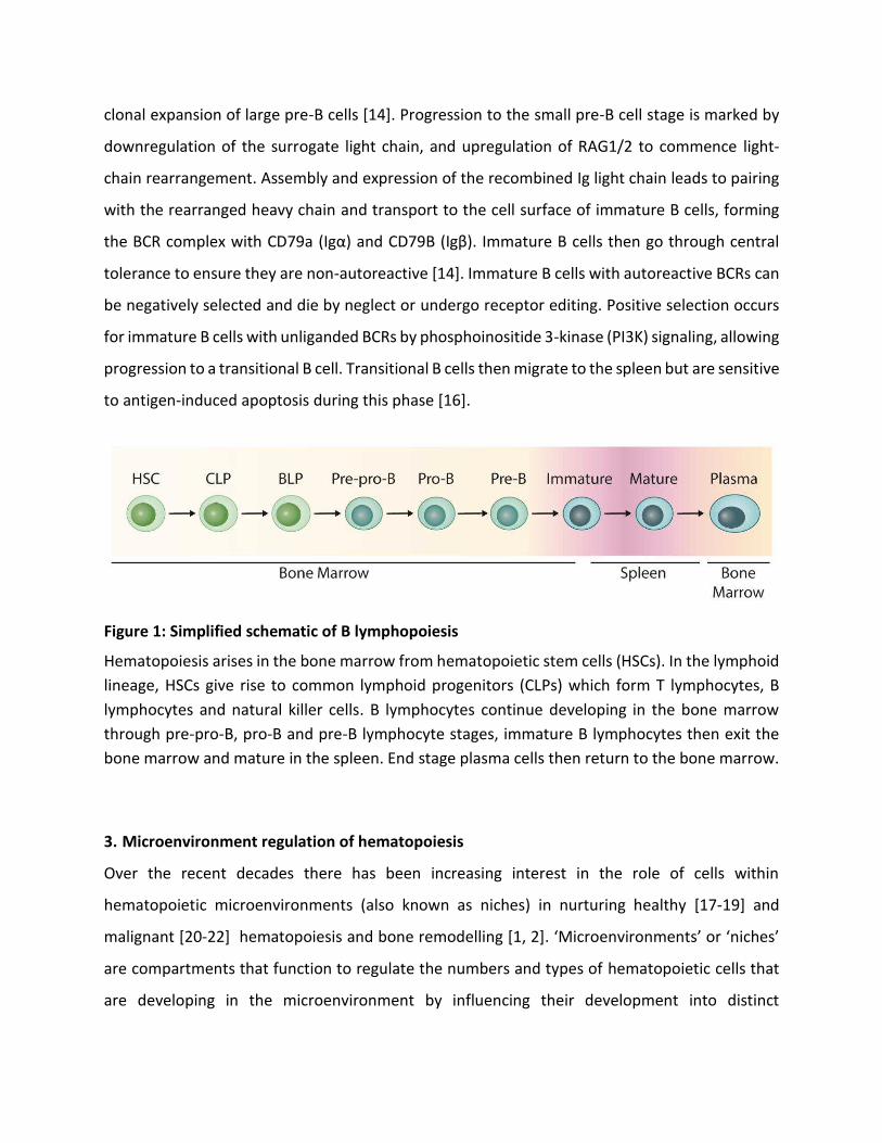
Figure 1: Simplified schematic of B lymphopoiesis
Hematopoiesis arises in the bone marrow from hematopoietic stem cells (HSCs). In the lymphoid
lineage, HSCs give rise to common lymphoid progenitors (CLPs) which form T lymphocytes, B
lymphocytes and natural killer cells. B lymphocytes continue developing in the bone marrow
through pre-pro-B, pro-B and pre-B lymphocyte stages, immature B lymphocytes then exit the
bone marrow and mature in the spleen. End stage plasma cells then return to the bone marrow.
3. Microenvironment regulation of hematopoiesis
Over the recent decades there has been increasing interest in the role of cells within
hematopoietic microenvironments (also known as niches) in nurturing healthy [17-19] and
malignant [20-22] hematopoiesis and bone remodelling [1, 2]く けMキIヴラWミ┗キヴラミマWミデゲげ ラヴ けミキIエWゲげ
are compartments that function to regulate the numbers and types of hematopoietic cells that
are developing in the microenvironment by influencing their development into distinct
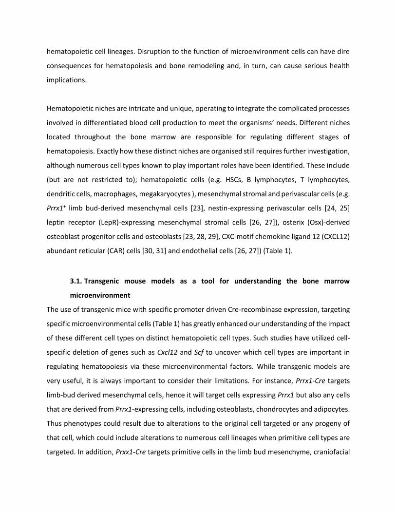
hematopoietic cell lineages. Disruption to the function of microenvironment cells can have dire
consequences for hematopoiesis and bone remodeling and, in turn, can cause serious health
implications.
Hematopoietic niches are intricate and unique, operating to integrate the complicated processes
involved in differentiated blood cell production to meet the organismsげ needs. Different niches
located throughout the bone marrow are responsible for regulating different stages of
hematopoiesis. Exactly how these distinct niches are organised still requires further investigation,
although numerous cell types known to play important roles have been identified. These include
(but are not restricted to); hematopoietic cells (e.g. HSCs, B lymphocytes, T lymphocytes,
dendritic cells, macrophages, megakaryocytes ), mesenchymal stromal and perivascular cells (e.g.
Prrx1+ limb bud-derived mesenchymal cells [23], nestin-expressing perivascular cells [24, 25]
leptin receptor (LepR)-expressing mesenchymal stromal cells [26, 27]), osterix (Osx)-derived
osteoblast progenitor cells and osteoblasts [23, 28, 29], CXC-motif chemokine ligand 12 (CXCL12)
abundant reticular (CAR) cells [30, 31] and endothelial cells [26, 27]) (Table 1).
3.1. Transgenic mouse models as a tool for understanding the bone marrow
microenvironment
The use of transgenic mice with specific promoter driven Cre-recombinase expression, targeting
specific microenvironmental cells (Table 1) has greatly enhanced our understanding of the impact
of these different cell types on distinct hematopoietic cell types. Such studies have utilized cell-
specific deletion of genes such as Cxcl12 and Scf to uncover which cell types are important in
regulating hematopoiesis via these microenvironmental factors. While transgenic models are
very useful, it is always important to consider their limitations. For instance, Prrx1-Cre targets
limb-bud derived mesenchymal cells, hence it will target cells expressing Prrx1 but also any cells
that are derived from Prrx1-expressing cells, including osteoblasts, chondrocytes and adipocytes.
Thus phenotypes could result due to alterations to the original cell targeted or any progeny of
that cell, which could include alterations to numerous cell lineages when primitive cell types are
targeted. In addition, Prxx1-Cre targets primitive cells in the limb bud mesenchyme, craniofacial
mesenchyme and flank mesoderm [32]. Hence mesenchymal cells in the limbs, some calvarial
bones and the sternum will be targeted, but not the vertebrae and thus any local effects will not
be apparent in the spine. Many of the Cre transgenic mice are also not entirely specific to the cell
lineage they are often used to study. For example, Dmp1-Cre is used to target late osteoblasts
and osteocytes but also targets skeletal muscle fibres, some cells in the brain and mesenchymal
cells in the intestine and stomach [33]. While this may not have a direct impact on the bone
microenvironment, changes to skeletal muscle could easily influence mechanical loading and
thus have indirect effects on the bone, being a mechano-sensitive organ. Colヲü1-Cre is used to
study chondrogenic lineage cells but it has also been shown to target some osteoblastic cells.
Seven day old Cラノヲüヱ-Cre:Rosa26LacZ mice exhibited LacZ expression in synovial fibroblasts [34]
and at embryonic day 16.5 LacZ was detected in the periosteum and primary spongiosa in the
appendicular and axial skeleton [35]く Tエ┌ゲが SWゲヮキデW デエW a;Iデ デエ;デ ミWキデエWヴ デエW Cラノヲüヱ ェWミW [34] nor
Cラノヲüヱ-Cre [35] are expressed in osteoblasts, these osteoblastic cells can arise from a cell that
ラミIW W┝ヮヴWゲゲWS Cラノヲüヱ-Cre.
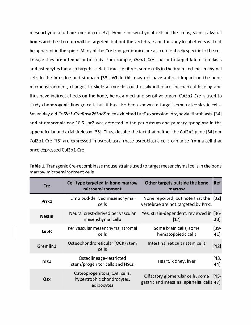
Table 1. Transgenic Cre-recombinase mouse strains used to target mesenchymal cells in the bone
marrow microenvironment cells
Cre Cell type targeted in bone marrow
microenvironment
Other targets outside the bone
marrow
Ref
Prrx1 Limb bud-derived mesenchymal
cells
None reported, but note that the
vertebrae are not targeted by Prrx1
[32]
Nestin Neural crest-derived perivascular
mesenchymal cells
Yes, strain-dependent, reviewed in
[17]
[36-
38]
LepR Perivascular mesenchymal stromal
cells
Some brain cells, some
hematopoietic cells
[39-
41]
Gremlin1 Osteochondroreticular (OCR) stem
cells
Intestinal reticular stem cells [42]
Mx1 Osteolineage-restricted
stem/progenitor cells and HSCs Heart, kidney, liver
[43,
44]
Osx
Osteoprogenitors, CAR cells,
hypertrophic chondrocytes,
adipocytes
Olfactory glomerular cells, some
gastric and intestinal epithelial cells
[45-
47]

Cラノヱü1 2.3kb
(Mouse) Osteoblasts
None reported (the two different
rat-derived Col2.3 strains have
other cell targets, reviewed in [17])
[48]
Cラノヲüヱ
Chondrocytes, synovial fibroblasts,
periosteum and primary spongiosa
in the appendicular and axial
skeleton
None reported
[34,
35]
Tagln
Osteoblasts, majority of CAR cells
and vinous sinusoidal and
arteriolar pericytes
Smooth muscle cells [49,
50]
Osteocalcin Mature osteoblasts, most CAR cells
and arteriolar pericytes
[51]
Dmp1
(9.6kb)*
Late osteoblasts and osteocytes,
subset of CAR cells
Skeletal muscle, brain, intestine,
stomach
[33,
49,
52]
*The 8kb Dmp1-Cre strain also targets some brain cells, reviewed in [17].
3.2. Cellular localisation of B lymphocyte niches
The microenvironment in the bone marrow is known to be essential for commitment to B
lymphopoiesis and the sequential stages of early B lymphopoiesis. From HSC to immature B
lymphocytes ready to exit the bone marrow, each stage is regulated by unique supportive niches.
How these niches are organised for the distinct stages of B lymphocyte development have not
yet been completely defined. We focus here on the regulation of B lymphopoiesis from the CLP
stage onwards.
CLPs have been shown to lose the potential to generate all lymphoid cell types when they express
Ly6D and become BLPs, producing almost exclusively B cells [10]. After commitment to the B
lymphocyte lineage, early B lymphocyte progenitors progress through distinct niches in the bone
marrow. Studies using Cxcl12:GFP [53] knock-in mice have shown that pre-pro-B lymphocytes
localise near CXCL12-abundant reticular (CAR) cells. These cells differentiate into pro-B
lymphocytes and together with Pre-B I cells [54, 55], localise near Interleukin 7 (IL-7)-secreting
cells [54, 55]. The IL-7-producing cells have been described as being spindle-shaped reticular cells

that usually reside in close contact with vessels and express VCAM-ヱ ふCDヱヰヶぶが PDGF‘üが CDヵヴ
(ICAM-1), and BP-1 but not CD31 [54], suggesting these cells are of the mesenchymal lineage.
While the nature of these reticular cells have not yet been determined, it is possible that they
are Osx-Cre-targeted osteoblast progenitor cells, which have been shown to express IL-7 [56].
Pre-B II lymphocytes then migrate away from IL-7-expressing cells [54, 55] to stromal cells that
are scattered throughout the bone marrow. Studies in which pre-B II cells were injected into
hydroxyurea-treated mice suggest that the pre-B II cells home to cells that are located
throughout the bone marrow and express Galectin-1 (GAL1) [54]. Immature B lymphocytes
localise near endothelial cells lining blood vessels [57] prior to these cells exiting the bone
marrow into the circulation. Some immature B lymphocytes are retained in the bone marrow
through cannabinoid receptor 2-mediated adhesion to endothelial cells lining sinusoidal vessels
[57].
The migration of the developing B lymphocytes through the bone marrow to the distinct B
lymphocyte niches relies on the expression of different chemokines (such as CXCL12) and
adhesion molecules, which are required during different stages of B cell development and are
discussed further below. A deregulation of these factors and other B lymphocyte regulatory
factors, including cytokines, that are expressed by distinct B lymphocyte niche cells may result in
changes in BM B lymphopoiesis. Such alterations may include either retention at the niche, or
extrinsically-mediated alterations to genes that are required for B cell development. There are
numerous excellent reviews focusing on the intrinsic regulators of B cell development, in
particular the role of transcription factors that are essential for B lymphopoiesis [12, 58, 59]. Here
we focus on known extrinsic regulators of B lymphopoiesis, with a particular focus on the roles
of mesenchymal-derived cells in these processes.

Figure 2: The bone marrow is the site of early B lymphopoiesis
Early B lymphopoiesis occurs in the bone marrow. Haematopoietic stem cells (HSCs) differentiate
to common lymphoid progenitors (CLPs) and then become the earliest committed B cell
precursor, the pre-pro-B cell. Pre-pro B cells localise next to CXCL12-abundant reticular (CAR)
cells, then as they differentiate into pro-B cells they localise adjacent to IL-7-expressing stromal
cells. Pre-B cells localise near Galectin-1-expressing cells within the bone marrow. Immature B
cells migrate from the bone marrow to mature at the spleen, although some are retained through
interactions with endothelial cells. End stage plasma cells then return to the bone marrow.
Osteoblasts are known to play essential roles in regulating B lymphopoiesis. B lymphocytes can
in turn regulate bone by altering osteoclastogenesis via production of RANKL, which binds to
RANK on the surface of osteoclast precursors. Mature B lymphocytes have also been shown to
be a major source of OPG, thereby inhibiting osteoclastogenesis.
4. The mesenchymal niches for B lymphopoiesis
Mesenchymal cells, including osteoblast lineage cells, are crucial regulators of the B lymphocyte
lineage, influencing both commitment to the B lymphocyte lineage and the progression of the
immature B cells through the early stages of B lymphopoiesis. The importance of osteoblast
lineage cells in B lymphopoiesis has been made apparent by their ability to support all stages of
differentiation from HSCs to immature B cells in vitro, and the impaired formation of B
lymphocyte precursors when osteoblasts are depleted from the bone marrow in vivo. It is
キマヮラヴデ;ミデ デラ ミラデW デエ;デ デエW デWヴマ さosteoblastざ has been used to describe a range of mesenchymal
cell types that give rise to the osteoblast lineage [60]. Furthermore, as discussed above, the use
of different mesenchymal cell lineage Cre strains does not necessarily identify the exact nature
of the mesenchymal cell type involved in a given phenotype due to the potential targeting of
downstream mesenchymal lineage cells in the Cre-targeted strains.
The production of macrophages, granulocytes, megakaryocytes and erythrocytes from HSCs can
occur readily in vitro when permissive cytokines are provided, however, stromal cells are
required as feeder layers for the optimal production of lymphoid cells in vitro. Whitlock et al. [61]
were the first to demonstrate that B lymphocytes could be produced in cultures containing
adherent bone marrow cells, these cultures are now commonly known as Whitlock-Witte
cultures. A range of osteoblast lineage primary cells and cell lines such as S17 and OP9 cells have
since been shown to support the production of B lymphocyte lineage cells in co-culture in vitro
assays [62, 63]. The OP9 stromal cell line is a cell line that was derived from the newborn calvaria
of op/op mice, which lack functional macrophage colony-stimulating factor-1 (M-CSF, also known
as colony-stimulating factor-1, CSF-1)[63, 64]]. The OP9 cell line was originally developed to
improve methods for the differentiation of embryonic stem cells into erythroid, myeloid and B
lymphoid lineages in vitro [63]. The OP9 cell line (and a variant that overexpresses delta-like 1,
OP9-DL1 [65]) have since become routinely used cell lines for in vitro assays of B and T
lymphopoiesis, respectively, from ES cells, adult HSCs and more committed hematopoietic
progenitor cells. While the exact nature of the OP9 cell line is unclear, it can readily give rise to
adipocytes [66], chondrocytes [67] and osteoblasts [68] when cultured with the appropriate

differentiation-inducing media. Furthermore, the immunophenotype of OP9 cells [68] is similar
to that of the population derived from bone marrow that expresses PDGFRü ;ミd Sca-1 (PüS) cells
and gives rise to osteoblasts, adipocytes and chondrocytes in culture [69]. While these cells are
often described as mesenchymal stem cells, their potential to form other mesenchymal lineages
has not been shown, hence they are best termed skeletal stem cells [60].
Furthermore, co-cultures of primary calvaria-derived osteoblasts or the stromal cell line S17 with
HSCs supported the formation of B220+CD19+ B lymphocyte precursors [62]. Zhu et al. [62],
isolated HSCs (GFP-Lin-Sca-1+c-Kit+; LKS+) from the bone marrow of RAG2 GFP NG BAC (B6) mice
(which express GFP in committed early B lymphocyte precursors). When these LKS+ cells were co-
cultured with the primary osteoblast lineage cells, 15% of the progeny of the LKS+ cells became
GFP+, indicating lymphocyte commitment. In comparison, no GFP+ cells were produced when LKS+
cells were co-cultured with the MS-1 endothelial cell line [62]. Stimulation of the osteoblast
lineage cells with PTH for 3 days led to elevated levels of IL-7 and SDF-1 (CXCL12) expressed by
the osteoblast lineage cells. Inhibition of IL-7, SDF-1 or thymic stromal cell-derived lymphopoietin
(TSLP) during PTH stimulation reduced the number of B220+ B lymphocytes. Furthermore,
inhibition of integrin Ƚ4 or VCAM-1 prevented the formation of B220+ cells. This indicates that
osteoblast lineage cells support B lymphopoiesis through the secretion of IL-7, SCF-1 and TSLP
and that VCAM-1/integrin Ƚ4 signalling is also important for osteoblastic-induction of B
lymphopoiesis [62]. The calvarial-derived cells were shown to express Sca-1, CD61, ICAM-1 and
VCAM-1 but lacked expression of endothelial and hematopoietic cell markers. These cells also
expressed osteopontin in culture [62]. The expression of Sca-1 by these cells suggests that they
are also akin to skeletal stem cells [23, 39, 69]. Hence, while these calvarial-derived cells are
clearly of mesenchymal origin, the stage of osteoblast lineage maturation of the cells used in the
cultures remains unclear.
Zhu et al., found that ablation of Col1a1-2.3kb (Col2.3)-targeted osteoblasts in vivo using a
thymidine kinase suicide gene in vivo depleted early B lymphocytes in the bone marrow [62].
When Col2.3-thymidine kinase mice were treated with ganciclovir, the thymidine kinase
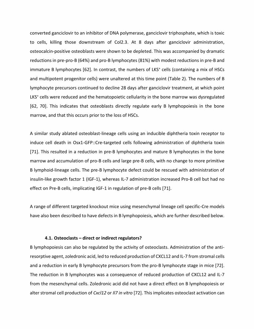
converted ganciclovir to an inhibitor of DNA polymerase, ganciclovir triphosphate, which is toxic
to cells, killing those downstream of Col2.3. At 8 days after ganciclovir administration,
osteocalcin-positive osteoblasts were shown to be depleted. This was accompanied by dramatic
reductions in pre-pro-B (64%) and pro-B lymphocytes (81%) with modest reductions in pre-B and
immature B lymphocytes [62]. In contrast, the numbers of LKS+ cells (containing a mix of HSCs
and multipotent progenitor cells) were unaltered at this time point (Table 2). The numbers of B
lymphocyte precursors continued to decline 28 days after ganciclovir treatment, at which point
LKS+ cells were reduced and the hematopoietic cellularity in the bone marrow was dysregulated
[62, 70]. This indicates that osteoblasts directly regulate early B lymphopoiesis in the bone
marrow, and that this occurs prior to the loss of HSCs.
A similar study ablated osteoblast-lineage cells using an inducible diphtheria toxin receptor to
induce cell death in Osx1-GFP::Cre-targeted cells following administration of diphtheria toxin
[71]. This resulted in a reduction in pre-B lymphocytes and mature B lymphocytes in the bone
marrow and accumulation of pro-B cells and large pre-B cells, with no change to more primitive
B lymphoid-lineage cells. The pre-B lymphocyte defect could be rescued with administration of
insulin-like growth factor 1 (IGF-1), whereas IL-7 administration increased Pro-B cell but had no
effect on Pre-B cells, implicating IGF-1 in regulation of pre-B cells [71].
A range of different targeted knockout mice using mesenchymal lineage cell specific-Cre models
have also been described to have defects in B lymphopoiesis, which are further described below.
4.1. Osteoclasts に direct or indirect regulators?
B lymphopoiesis can also be regulated by the activity of osteoclasts. Administration of the anti-
resorptive agent, zoledronic acid, led to reduced production of CXCL12 and IL-7 from stromal cells
and a reduction in early B lymphocyte precursors from the pro-B lymphocyte stage in mice [72].
The reduction in B lymphocytes was a consequence of reduced production of CXCL12 and IL-7
from the mesenchymal cells. Zoledronic acid did not have a direct effect on B lymphopoiesis or
alter stromal cell production of Cxcl12 or Il7 in vitro [72]. This implicates osteoclast activation can
indirectly regulate B lymphopoiesis through altering bone formation from osteoblast lineage
cells. Interestingly, B lymphocytes can inhibit osteoclastogenesis (and, in turn, influence the
regulation of osteoblasts) by secreting osteoprotegerin (OPG) [73]. Indeed, B cells at different
stages of development, in particular mature B lymphocytes and plasma cells, have been shown
to be the major source of OPG in mouse BM [73]. In inflammatory conditions, B lymphocyte
populations can also secrete RANKL [74].
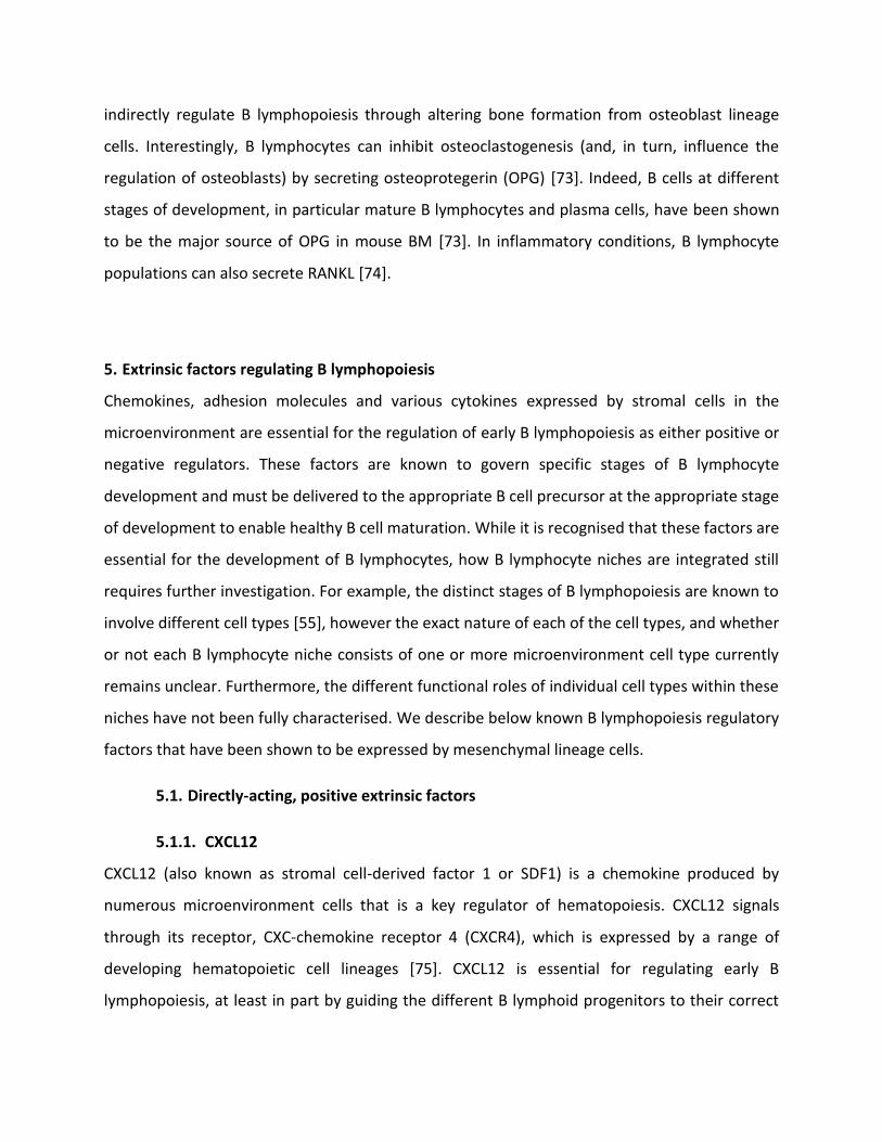
5. Extrinsic factors regulating B lymphopoiesis
Chemokines, adhesion molecules and various cytokines expressed by stromal cells in the
microenvironment are essential for the regulation of early B lymphopoiesis as either positive or
negative regulators. These factors are known to govern specific stages of B lymphocyte
development and must be delivered to the appropriate B cell precursor at the appropriate stage
of development to enable healthy B cell maturation. While it is recognised that these factors are
essential for the development of B lymphocytes, how B lymphocyte niches are integrated still
requires further investigation. For example, the distinct stages of B lymphopoiesis are known to
involve different cell types [55], however the exact nature of each of the cell types, and whether
or not each B lymphocyte niche consists of one or more microenvironment cell type currently
remains unclear. Furthermore, the different functional roles of individual cell types within these
niches have not been fully characterised. We describe below known B lymphopoiesis regulatory
factors that have been shown to be expressed by mesenchymal lineage cells.
5.1. Directly-acting, positive extrinsic factors
5.1.1. CXCL12
CXCL12 (also known as stromal cell-derived factor 1 or SDF1) is a chemokine produced by
numerous microenvironment cells that is a key regulator of hematopoiesis. CXCL12 signals
through its receptor, CXC-chemokine receptor 4 (CXCR4), which is expressed by a range of
developing hematopoietic cell lineages [75]. CXCL12 is essential for regulating early B
lymphopoiesis, at least in part by guiding the different B lymphoid progenitors to their correct
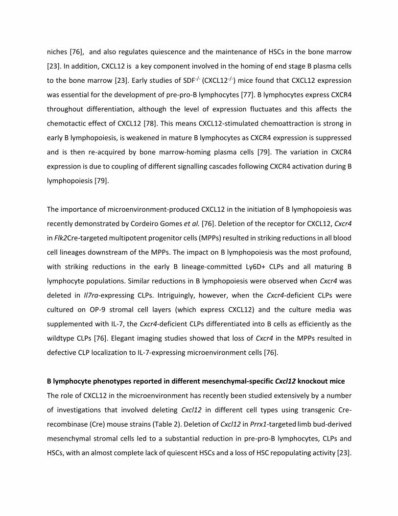
niches [76], and also regulates quiescence and the maintenance of HSCs in the bone marrow
[23]. In addition, CXCL12 is a key component involved in the homing of end stage B plasma cells
to the bone marrow [23]. Early studies of SDF-/- (CXCL12-/-) mice found that CXCL12 expression
was essential for the development of pre-pro-B lymphocytes [77]. B lymphocytes express CXCR4
throughout differentiation, although the level of expression fluctuates and this affects the
chemotactic effect of CXCL12 [78]. This means CXCL12-stimulated chemoattraction is strong in
early B lymphopoiesis, is weakened in mature B lymphocytes as CXCR4 expression is suppressed
and is then re-acquired by bone marrow-homing plasma cells [79]. The variation in CXCR4
expression is due to coupling of different signalling cascades following CXCR4 activation during B
lymphopoiesis [79].
The importance of microenvironment-produced CXCL12 in the initiation of B lymphopoiesis was
recently demonstrated by Cordeiro Gomes et al. [76]. Deletion of the receptor for CXCL12, Cxcr4
in Flk2Cre-targeted multipotent progenitor cells (MPPs) resulted in striking reductions in all blood
cell lineages downstream of the MPPs. The impact on B lymphopoiesis was the most profound,
with striking reductions in the early B lineage-committed Ly6D+ CLPs and all maturing B
lymphocyte populations. Similar reductions in B lymphopoiesis were observed when Cxcr4 was
deleted in Il7ra-expressing CLPs. Intriguingly, however, when the Cxcr4-deficient CLPs were
cultured on OP-9 stromal cell layers (which express CXCL12) and the culture media was
supplemented with IL-7, the Cxcr4-deficient CLPs differentiated into B cells as efficiently as the
wildtype CLPs [76]. Elegant imaging studies showed that loss of Cxcr4 in the MPPs resulted in
defective CLP localization to IL-7-expressing microenvironment cells [76].
B lymphocyte phenotypes reported in different mesenchymal-specific Cxcl12 knockout mice
The role of CXCL12 in the microenvironment has recently been studied extensively by a number
of investigations that involved deleting Cxcl12 in different cell types using transgenic Cre-
recombinase (Cre) mouse strains (Table 2). Deletion of Cxcl12 in Prrx1-targeted limb bud-derived
mesenchymal stromal cells led to a substantial reduction in pre-pro-B lymphocytes, CLPs and
HSCs, with an almost complete lack of quiescent HSCs and a loss of HSC repopulating activity [23].
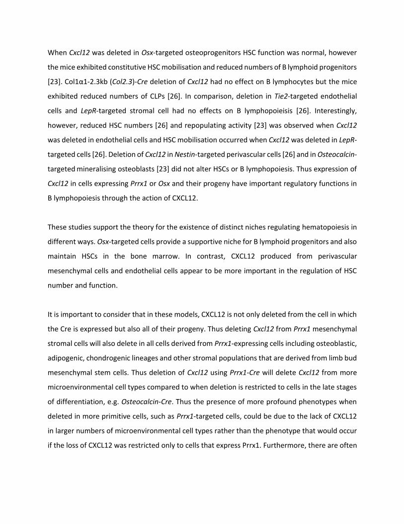
When Cxcl12 was deleted in Osx-targeted osteoprogenitors HSC function was normal, however
the mice exhibited constitutive HSC mobilisation and reduced numbers of B lymphoid progenitors
[23]く Cラノヱüヱ-2.3kb (Col2.3)-Cre deletion of Cxcl12 had no effect on B lymphocytes but the mice
exhibited reduced numbers of CLPs [26]. In comparison, deletion in Tie2-targeted endothelial
cells and LepR-targeted stromal cell had no effects on B lymphopoieisis [26]. Interestingly,
however, reduced HSC numbers [26] and repopulating activity [23] was observed when Cxcl12
was deleted in endothelial cells and HSC mobilisation occurred when Cxcl12 was deleted in LepR-
targeted cells [26]. Deletion of Cxcl12 in Nestin-targeted perivascular cells [26] and in Osteocalcin-
targeted mineralising osteoblasts [23] did not alter HSCs or B lymphopoiesis. Thus expression of
Cxcl12 in cells expressing Prrx1 or Osx and their progeny have important regulatory functions in
B lymphopoiesis through the action of CXCL12.
These studies support the theory for the existence of distinct niches regulating hematopoiesis in
different ways. Osx-targeted cells provide a supportive niche for B lymphoid progenitors and also
maintain HSCs in the bone marrow. In contrast, CXCL12 produced from perivascular
mesenchymal cells and endothelial cells appear to be more important in the regulation of HSC
number and function.
It is important to consider that in these models, CXCL12 is not only deleted from the cell in which
the Cre is expressed but also all of their progeny. Thus deleting Cxcl12 from Prrx1 mesenchymal
stromal cells will also delete in all cells derived from Prrx1-expressing cells including osteoblastic,
adipogenic, chondrogenic lineages and other stromal populations that are derived from limb bud
mesenchymal stem cells. Thus deletion of Cxcl12 using Prrx1-Cre will delete Cxcl12 from more
microenvironmental cell types compared to when deletion is restricted to cells in the late stages
of differentiation, e.g. Osteocalcin-Cre. Thus the presence of more profound phenotypes when
deleted in more primitive cells, such as Prrx1-targeted cells, could be due to the lack of CXCL12
in larger numbers of microenvironmental cell types rather than the phenotype that would occur
if the loss of CXCL12 was restricted only to cells that express Prrx1. Furthermore, there are often

direct and indirect consequences of deletion of genes in a given cell type, hence this may also
indirectly influence B lymphopoiesis in any of these mice.
Table 2. Conditional models targeting cells in the bone marrow microenvironment and the
effects on the B lymphocyte lineage
Genetic model HSCs Progenitors B220+ Pre-pro-B Pro-B Pre-B Ref
Cxcl12
Prrx1-Cre+:Cxcl12 ら/-
HSCs (almost
complete lack of
quiescent HSCs)
repopulating
activity
MPPs,
CLPs, BLPs,
CMPs
[23,
26]
Nestin-Cre+:Cxcl12らっら normal*^ normal normal normal normal [26]
LepR-Cre+:Cxcl12らっら mobilization normal normal normal normal [26]
Osx1-GFP::Cre+:Cxcl12 ら/-
Normal HSC
function,
constitutive
mobilization
normal ND ND [23]
Col1a1(2.3kb) -Cre+:Cxcl12 らっら normal*^# CLPs &
LMPPs normal normal normal [26]
Osteocalcin-Cre+:Cxcl12らっ- normal*^ normal normal normal ND ND [23]
Tie2-Cre+:Cxcl12らっら
HSCs, long-
term repopulating
activity
normal normal normal normal [23,
26]
Il7
Prrx1-Cre+:Il7らっ- normal* normal ND ND [76]
LepR-Cre+:Il7らっ- normal* Ly6D+
CLPs ND ND [76]
Col1a1(2.3kb) -Cre+:Il7 らっ- normal* normal ND ND normal normal [76]
Tie2-Cre+:Il7 らっ- normal ND ND [76]
Scf (Kitl)
Il7-Cre:Scf らっら MPPs ND ND normal normal [76]
Nestin-Cre+:Scfらっ- normal*^ ND normal ND ND ND [27]
Nestin-CreER+:Scfらっら +
tamoxifen normal*^ ND ND ND ND ND [27]
LepR-Cre+:Scfらっ- HSCs ND ND ND ND ND [27]
Col1a1(2.3kb) -Cre+:Scf らっ- normal*^ ND normal ND ND ND [27]

Tie2-Cre+:Cxcl12らっ-
HSCs, long-
term repopulating
activity
ND ND ND ND ND [27]
Gゲü
Osx1-GFP::Cre+:Gsüらっら ND ND normal [56]
Dmp1-Cre+:Gsüらっら normal* normal normal ND ND ND [80]
Pth1r
Col1a1(2.3kb) -Cre+:Pth1r tg/tg
(constitutively active) HSCs ND ND ND ND ND [28]
Rarg
Nestin-Cre+:Rargらっら normal*^ normal normal normal [25]
Osx1-GFP::Cre+:Rargらっら normal*^ normal normal normal normal normal [25]
IGF-1
Osx1-GFP::Cre:Igf1らっら ND ND normal [71]
Osteoblast ablation
Osx1-GFP::Cre:iDTR +
diphtheria toxin normal*^ GMP normal ~ [71]
Col1a1(2.3kb)-Cre+: らTK +
ganciclovir day 21 ND
day 8
day 8
day 8
day 8 [62]
Recombined floxed allele (ら), null allele (-), transgenic allele (tg). Significant changes compared to appropriate
controls are identified by: elevated function or numbers (), lower function or numbers (), normal or not
determined (ND). Haematopoietic stem cell (HSC), multipotent progenitor (MPP), B lymphoid-biased progenitors
(BLPs), common lymphoid progenitor (CLP), lymphoid-primed multipotent progenitor (LMPP), inducible diptheria
toxin receptor (iDTR) and thymidine kinase (TK). *Normal HSC frequency, ^normal repopulating activity. #Data were
based on FACS-isolated HSC transplants, however transplants using whole bone marrow cells showed significantly
impaired lymphoid repopulation. ~Fヴ;Iデキラミ B ┘;ゲ ミラヴマ;ノ ;ミS Fヴ;Iデキラミ Cげ ;ミS Cげげ ┘WヴW キミIヴW;ゲWSく
5.1.2. IL-7
IL-7 is also known to play an essential role in B lymphopoiesis. IL-7 signals through the IL-7
receptor (IL-7R), which is expressed by B lymphocyte precursor populations until the large pre-B
cell stage with subsequent B cell populations lacking expression of the IL-7R [81, 82]. Deletion of
either IL-7 or IL-7R led to reductions in pro-B and pre-B lymphocytes with no change to pre-pro-
B lymphocytes [82, 83]. This suggests the role of IL-7 in B lymphopoiesis occurs subsequent to
CXCL12, from the pro-B cell stage onwards.
IL-7 has been shown to induce the proliferation of pro-B cells, but not pre-pro-B cells, in vitro

(85). In accordance with this, Il7 deficient mice demonstrated a marked loss in the numbers of
pro-B cells and pre-B cells and their later differentiated stages (86, 87). While no changes in pre-
pro B cells were observed in Il7 knockout mice, their differentiation was severely impaired, which
may have resulted in the reductions of the more mature populations (86).
The mechanism of action of IL-7 includes the induction of the expression of myeloid-cell leukemia
sequence 1 (MCL1) in the developing B cell precursors to mediate their survival (88).
Furthermore, it has been reported that overexpression of early B cell factor (EBF) can counteract
the Il7-deficient B cell phenotypes (89).
Elegant lineage tracing studies where Il7-Cre transgenic mice were crossed to Rosa26EYFP mice
demonstrated that the IL-7-expressing cells in the bone marrow were predominantly bipotent
CAR cells that expressed LepR and could give rise to both adipocytes and osteoblasts in vivo [76].
Deletion of Cxcl12 in Il7-Cre-targeted cells resulted in a significant loss in HSCs, CLPs, and
developing B cells in the bone marrow. Interestingly, the numbers of these cells were normal in
the spleen, hence increased mobilization to the spleen was an unlikely cause of the reduced bone
marrow cells [76].
IL-7-dependent B lymphocyte phenotypes that have been reported in different mesenchymal
cell-specific knockout mice.
IL-7 production by osteoblastic cells has been shown to be important for B lymphopoiesis via the
regulatory actions of Gゲü, a downstream mediator of PTHR1 signalling. Deletion of Gゲü キミ Osx-
Cre-targeted osteoblast progenitors leads to severe osteoporosis and reduced B lymphocyte
precursors in the bone marrow [56, 84]. Osx-GFPぎぎCヴWGゲüfl/fl (GsaOsxKO) mice exhibited
significantly reduced proportions of pro-B and pre-B lymphocytes, with no change to pre-pro-B
lymphocytes in their bone marrow [56]く TエW Gゲü-deficient osteoblast lineage cells expressed
significantly reduced levels of IL-7. Administration of IL-7 into GsaOsxKO mice or transplant of
GsaOsxKO bone marrow into wild type mice was able to rescue the B lymphocyte defects [56]. Thus
IL-7 produced by Osx-targeted cells regulates pro-B and pre-B lymphopoiesis.

In their recent study, Cordeiro Gomes et al. [76] deleted Il7 in a range of mesenchymal cell types
and determined the subsequent impact on B lymphopoiesis. Deletion of Il7 in Lepr-Cre-targeted
cells resulted in a significant reduction in BLPs in the mice. This was accompanied by significant
reductions in all downstream B lymphocyte populations, including significantly reduced
peripheral blood B lymphocytes. The numbers of pro-B, pre-B and B220+IgM+ B lymphocytes in
the bone marrow were also significantly reduced in mice lacking Il7 in Prrx1Cre-targeted cells. In
contrast, when Il7 was deleted in mature osteoblasts using Col2.3Cre, no hematopoietic
phenotype was observed. Interestingly, when Il7 was deleted in Tie2-Cre-targeted cells (which
deletes in endothelial and hematopoietic cells [17], there were small, but significant reductions
in the numbers of pro-B and pre-B cells in the bone marrow.
I would include here Aguila, JBMR 2012 showing that osteoblast-specific expression of
IL-7 can rescue the B cell and bone phenotype of IL-7 KO mice:
o J Bone Miner Res. 2012 May;27(5):1030-42. doi: 10.1002/jbmr.1553.
Osteoblast-specific overexpression of human interleukin-7 rescues the bone
mass phenotype of interleukin-7-deficient female mice.
Aguila HL1, Mun SH, Kalinowski J, Adams DJ, Lorenzo JA, Lee SK.
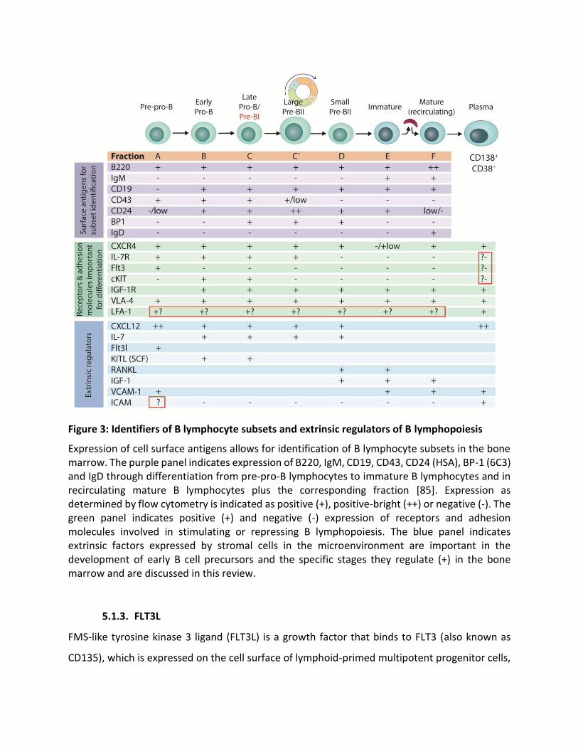
Figure 3: Identifiers of B lymphocyte subsets and extrinsic regulators of B lymphopoiesis
Expression of cell surface antigens allows for identification of B lymphocyte subsets in the bone
marrow. The purple panel indicates expression of B220, IgM, CD19, CD43, CD24 (HSA), BP-1 (6C3)
and IgD through differentiation from pre-pro-B lymphocytes to immature B lymphocytes and in
recirculating mature B lymphocytes plus the corresponding fraction [85]. Expression as
determined by flow cytometry is indicated as positive (+), positive-bright (++) or negative (-). The
green panel indicates positive (+) and negative (-) expression of receptors and adhesion
molecules involved in stimulating or repressing B lymphopoiesis. The blue panel indicates
extrinsic factors expressed by stromal cells in the microenvironment are important in the
development of early B cell precursors and the specific stages they regulate (+) in the bone
marrow and are discussed in this review.
5.1.3. FLT3L
FMS-like tyrosine kinase 3 ligand (FLT3L) is a growth factor that binds to FLT3 (also known as
CD135), which is expressed on the cell surface of lymphoid-primed multipotent progenitor cells,

CLPs and pre-pro B lymphocytes. It is important for early B lymphocyte commitment [5], and acts
synergistically with IL-7 to promote B lymphopoiesis [86, 87]. FLT3L has been shown to be
produced by a range of hematopoietic cell types [88], but is also produced by bone marrow
stromal cells. The nature of the latter cell type(s) is unclear.
Mice deficient in FLT3 or FLT3L exhibited reduced numbers of pre-pro-B and pro-B lymphocytes
in their bone marrow [89, 90]. Furthermore, mice deficient in FLT3L also had a marked reduction
in bone marrow pre-B lymphocytes [90]. FLT3 is expressed by pre-pro-B lymphocytes until their
irreversible commitment to the B lymphocyte lineage, when it is repressed by the expression of
Pax5 [91]. Thus FLT3L is an important regulator of the earliest stages of B lymphopoiesis.
5.1.4. SCF
Stem cell factor (SCF, also known as KIT ligand; KITL) is a cytokine that signals though the c-kit
receptor (also known as CD117), which is a receptor tyrosine kinase that is expressed on the cell
surface of many developing hematopoietic cell types, including HSCs. SCF is primarily expressed
by perivascular cells and exists as membrane-bound or secreted forms [5, 27, 81]. SCF synergizes
with IL-7 in vitro to induce the proliferation of pro-B cells [92]. Furthermore, loss of Scf in Il7-Cre
transgenic mice resulted in a significant reduction in HSCs and MPPs in the bone marrow of the
mice [76].
Neonatal mice deficient in c-kit (termed W/W) do not display any B lymphocyte defects, however
W/W mice die within 1 week of birth due to anemia [93]. When the anemic phenotype was
rescued by administration of erythropoietin, it was found that pro-B and pre-B lymphocyte
numbers decreased with age in these mice [94]. This indicates that SCF is important for B
lymphopoiesis in adults.
A range of mice in which Scf was deleted in microenvironment-specific cell types have recently
been reported [27] (Table 2). These studies focused on the HSC phenotypes of the mice, however,
and to date only the B lymphopoiesis phenotypes have been reported for the mice in which Scf
was deleted in Nestin-expressing or Col2.3-expressing cells. Both mouse strains had normal
numbers of B220+ cells in their bone marrow [27].
5.1.5. IGF-1
IGF-1 is expressed by Osx1-GFP::Cre-targeted cells that binds to the IGF-1 receptor (IGF-1R) on
B lymphocytes to promote transition from the pro-B to the pre-B lympohocyte stage [71]. Igf1
levels are reduced in the bones of Osx1-GFP::Cre:iDTR mice after administration of diphtheria
toxin to ablate Osx1-Cre-targeted cells and mice exhibit defective B lymphopoiesis [71]. Mice
with targeted deletion of IGF-1 using Osx1-GFP::Cre exhibit reduced numbers of pre-B,
immature and mature B lymphocytes in the bone marrow, but increases in cells in fractions B,
Cが Cげ ;ミS Cげげ identifying pro-B and earlier pre-B lymphocytes [71]. This implicates IGF-1 derived
from Osx1-Cre-targeted cells as important in regulating the later stages of B lymphopoiesis
within the bone marrow from fraction D small pre-BII cells.
5.1.6. Sclerostin
For you to consider に one paper demonstrating a B cell defect in mice lacking sclerostin, a
negative regulator of canonical Wnt signalling. Potentially clinically relevant since anti-sclerostin
antibody is in development as an osteoporosis medication:
J Bone Miner Res. 2012 Jul;27(7):1451-61. doi: 10.1002/jbmr.1608. Absence
of sclerostin adversely affects B-cell survival. Cain CJ1, Rueda R, McLelland B, Collette
NM, Loots GG, Manilay JO.
Osteonectin
Luo et al 2014 に SPARC deficiency affects bone marrow stromal cell function, resulting in
impaired B lymphopoiesis. https://www.ncbi.nlm.nih.gov/pubmed/24598056
probably due to reduced osteoblasts
5.1.7. VCAM-1
Adhesion
Tokoyoda に Cxcl12 increases VLA-4 VCAM-1 adhesiveness in pre-pro-B cells
but not pro-B or pre-B cells
Park, S. Y. et al. Focal adhesion kinase regulates the localization and retention of pro-B
cells in bone marrow microenvironments. J. Immunol. 190, 1094に1102 (2013).
Ryan et al 1999 に Vascular cell adhesion molecule-1 and the integrin VLA-4 mediate
adhesion of human B cell precursors to cultured bone marrow adherent cells
https://www.ncbi.nlm.nih.gov/pmc/articles/PMC295504/
5.1.8. Galectin-1
Galectin-1 binds to integrins (including ü41, ü57 and ü47) that are expressed on the cell surface
of a range of cell types [95]. Developing B lymphocytes express ü41 (VLA-4), which also binds to
other cell adhesion molecules including VCAM-1.
Galectin-1 was shown to be expressed in mature osteoblasts in addition to cells located
throughout the bone marrow [54]. The Galectin-1+ cells obtained from flushed bone marrow
were shown to express CD54+ but did not express VCAM-ヱが PDGF‘üが BP-1, Sca-1 or Nestin.
Interestingly, most Galectin-1+ cells obtained from flushed bone marrow were CD31+, but did not
express CD34 or Tie2, suggesting that they were a subset of endothelial cells. In IL-7-GFP reporter
mice, Galectin-1 was not co-expressed with GFP, indicating that IL-7-expressing cells are a
separate cell type to Galectin-1-expressing cells [54].
Galectin-1-deficient mice had normal numbers of the different B lymphocyte populations in vivo
in the steady state. In contrast, however, the recovery of pre-B II cells was significantly delayed
in Gal1-/- mice after treatment with hydroxyurea [95].
5.2. Indirect, positive extrinsic factors
5.2.1. RANKL
Receptor activator of nuclear factor kappa-B ligand [RANKL; also known as osteoprotegrin ligand
(OPGL), encoded by the Tnfsf11 gene] is a protein with important functions in osteoclastogenesis
and B lymphopoiesis. RANKL signals through RANK on osteoclasts, and together with macrophage
colony stimulating factor (M-CSF), stimulates the expression of osteoclastic transcription factors,
including nuclear factors of activated T cells c1 (NFATc1), microphalmia associated transcription
factor (MITF), PU.1 and activator protein 1 (AP-1) [96, 97]. This induces myeloid precursors to
differentiate into mononucleated osteoclasts, which form multinucleated osteoclast by fusing
and finally activate becoming polarised osteoclasts that resorb bone [96, 97].
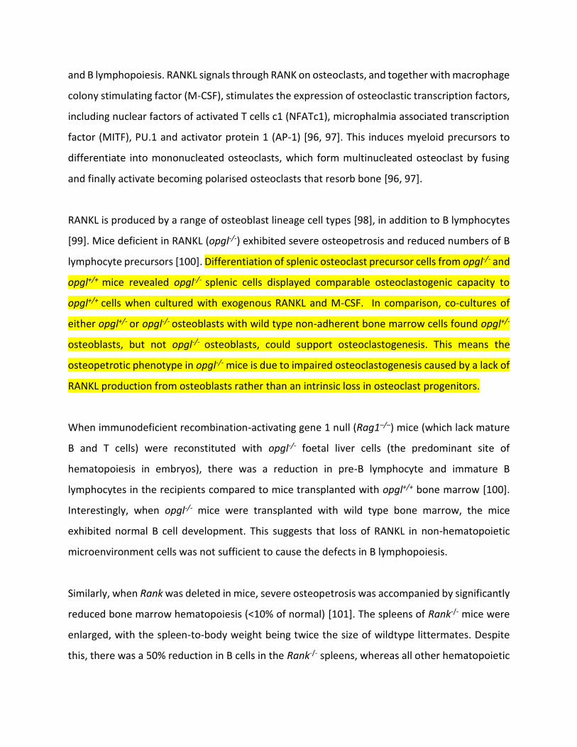
RANKL is produced by a range of osteoblast lineage cell types [98], in addition to B lymphocytes
[99]. Mice deficient in RANKL (opgl-/-) exhibited severe osteopetrosis and reduced numbers of B
lymphocyte precursors [100]. Differentiation of splenic osteoclast precursor cells from opgl-/- and
opgl+/+ mice revealed opgl-/- splenic cells displayed comparable osteoclastogenic capacity to
opgl+/+ cells when cultured with exogenous RANKL and M-CSF. In comparison, co-cultures of
either opgl+/- or opgl-/- osteoblasts with wild type non-adherent bone marrow cells found opgl+/-
osteoblasts, but not opgl-/- osteoblasts, could support osteoclastogenesis. This means the
osteopetrotic phenotype in opgl-/- mice is due to impaired osteoclastogenesis caused by a lack of
RANKL production from osteoblasts rather than an intrinsic loss in osteoclast progenitors.
When immunodeficient recombination-activating gene 1 null (Rag1に/に) mice (which lack mature
B and T cells) were reconstituted with opgl-/- foetal liver cells (the predominant site of
hematopoiesis in embryos), there was a reduction in pre-B lymphocyte and immature B
lymphocytes in the recipients compared to mice transplanted with opgl+/+ bone marrow [100].
Interestingly, when opgl-/- mice were transplanted with wild type bone marrow, the mice
exhibited normal B cell development. This suggests that loss of RANKL in non-hematopoietic
microenvironment cells was not sufficient to cause the defects in B lymphopoiesis.
Similarly, when Rank was deleted in mice, severe osteopetrosis was accompanied by significantly
reduced bone marrow hematopoiesis (<10% of normal) [101]. The spleens of Rank-/- mice were
enlarged, with the spleen-to-body weight being twice the size of wildtype littermates. Despite
this, there was a 50% reduction in B cells in the Rank-/- spleens, whereas all other hematopoietic

cell lineages were normal (T cells, macrophages) or elevated (granulocytes, erythrocytes). Note,
however, that mb1-Cre deletion of Rank in B lymphocytes from the pro-B cell stage onwards did
not result in any defect in B lymphopoiesis in the conditional knockout mice, suggesting that any
effect of perturbing RANKL/RANK signaling on B lymphopoiesis is indirect [102]. Given that both
the opgl-/- and Rank-/- mice had significant osteopetrotic phenotypes, it is likely that the
subsequent changes in the bone marrow microenvironment contributed to the B lymphocyte
defects in these mice.
Discuss paper by Yun et al 2001 に opg-/- mice see Horowitz review
https://www.ncbi.nlm.nih.gov/pubmed/20601290
5.3. Negative extrinsic regulators
OSF-5
o Identification of osteoblast stimulating factor 5 as a negative regulator in the B-lymphopoietic
niche (Fujita et al 2015 Experimental Haematology)
https://www.ncbi.nlm.nih.gov/pubmed/26213229
Each of these factors produced extrinsically to B lymphocytes is important for healthy B
lymphocyte production. Yet how each niche is organised to regulate distinct functions and the
specific cell types involved in each process requires further investigation. Further insights have
been gained by understanding the influence on B lymphopoiesis of signalling pathways of
importance for regulating bone.
6. Signaling pathways involved in microenvironment regulation of B lymphopoiesis by
mesenchymal cells
6.1. PTH
DキゲI┌ゲゲ PTH キミ OBゲ ;ミS デエWミ ノキミニ デラ Jラ┞げゲ ヮ┌HノキI;デキラミゲ H┌デ Sラミげデ HW ヴWヮWデキデキ┗W ┘キデエ ラデエWヴ ゲWIデキラミゲ

Further investigation into the role of PTH in osteoblasts has solidified its significance in regulation
of B lymphopoiesis via IL-7. Osx-Cre-targeted deletion of PTH receptor 1 (PTHR1) [7] or the GS
alpha subunit (GSüぶ Sラ┘ミゲデヴWam signalling mediator [56, 84] caused reductions in bone marrow
pro-B and pre-B lymphocytes and development of severe osteoporosis. This B lymphocyte
phenotype was rescued by administration of IL-7 or by transplanting GSü-deficient bone marrow
into wild type mice [56].
6.2. Estrogen
M;ミラノ;ェ;ゲが “く Cくが OげBヴキWミが Cく Aくが ;ミS Almeida, M. (2013) The role of estrogen and androgen
receptors in bone health and disease. Nat. Rev. Endocrinol. 9, 699に712
https://www.ncbi.nlm.nih.gov/pmc/articles/PMC3971652/
Smithson, G., Beamer, W. G., Shultz, K. L., Christianson, S. W., Shultz, L. D., and Kincade, P. W.
(1994) Increased B lymphopoiesis in genetically sex steroid-deficient hypogonadal (hpg) mice. J.
Exp. Med. 180, 717に720 https://www.ncbi.nlm.nih.gov/pmc/articles/PMC2191601/
Masuzawa, T., Miyaura, C., Onoe, Y., Kusano, K., Ohta, H., Nozawa, S. and Suda, T. (1994)
Estrogen deficiency stimulates B ymphopoiesis in mouse bone marrow. J Clin Invest.
https://www.ncbi.nlm.nih.gov/pmc/articles/PMC295170/
Fujiwara - RANKL (Receptor Activator of NF_B Ligand) Produced by Osteocytes Is Required
for the Increase in B Cells and Bone Loss Caused by Estrogen Deficiency in Mice
6.3. Retinoic acid receptors
Retinoic acid receptors (RARs) are expressed by many cells in the bone marrow
microenvironment, and regulate hematopoiesis (including B lymphopoiesis) [25, 103-106] and
bone remodelling [107-111] through direct and indirect means. Canonical RAR signalling occurs
デエヴラ┌ェエ ミ┌IノW;ヴ ‘A‘ゲ ラa ┘エキIエ デエWヴW ;ヴW デエヴWW ゲ┌Hデ┞ヮWゲき üが é ;ミS á [108]. RARs form
heterodimers with retinoid X receptors (RXRs) and associate with retinoic acid response elements
(RAREs) were they regulate gene transcription. The effect of retinoids on hematopoiesis depends
on the cell types that are exposed and the RAR subtype targeted.

The role of RARs in microenvironment regulation of hematopoiesis has been studied using global
and conditional deletion of RARs in mice. Rara-/- mice do not exhibit defects in bone [109] or
hematopoiesis [105, 112]. Rarb-/- and Rarb2-/- mice appear normal and have no reported skeletal
abnormalities, although these have not been extensively investigated in adult mice, nor have the
hematopoietic phenotypes of these mice been reported [113, 114]. In comparison, Rarg-/- mice
have significantly reduced trabecular bone [109]. Furthermore, Rarg-/- mice have significant
reductions in pro-B and pre-B lymphocytes [25] and have reduced levels of Il7 mRNA in their bone
marrow [109]. Rarg-/- mice also develop a myeloproliferative-like syndrome (MPS) exhibiting
increased granulocyte/macrophage progenitors and granulocytes in bone marrow, peripheral
blood, and spleen [106]. Transplant studies revealed the defect in B lymphopoiesis and the MPS-
like syndrome were due to extrinsic loss of RARá aヴラマ ミラミ-hematopoietic cells. Transplantation
of wild type bone marrow into Rarg-/- mice led to development of the hematopoietic defects,
whereas these defects were resolved when Rarg-/- bone marrow was transplanted into wild type
mice.
The hematopoietic phenotypes have recently been assessed in mice with conditional deletion of
Rarg-/- in nestin and osterix-targeted microenvironmental cells [25]. Deletion of Rarg-/- in osterix-
expressing osteoblast progenitor cells produced no hematopoietic phenotype and B
lymphopoiesis was normal [25]. However, mice with deletion of Rarg in nestin-Cre-targeted cells
had normal numbers of pro-B lymphocytes but significantly reduced numbers of bone marrow
pre-B lymphocytes and immature B220+IgM+ B lymphocytes, in addition to significantly reduced
numbers of peripheral blood B lymphocytes [25]. The effects on early B lymphocytes were not as
profound in Nestin-Cre+:Rargらっら mice as those observed in Rarg-/- mice, suggesting that additional
cell types in the bone marrow are likely involved in the regulation of B lymphopoiesis by RARá.
7. B lymphocyte stimulation of osteoclastogenesis
This review has focused on the importance of bone and osteoblast-lineage cells in supporting B
lymphopoiesis, yet it is also interesting to note that B lymphocytes are capable of influencing
bone mass via stimulation of osteoclastogenesis. Studies in ovariectomized mice revealed that,

along with osteoclast-driven bone loss, the mice exhibited an increase in B lymphopoiesis and an
accumulation of B220+ cells in the bone marrow [115]. Similarly, mice treated with IL-7 to
stimulate B lymphopoiesis also had enhanced osteoclastogenesis and less bone than controls and
Il7-/- mice had higher trabecular bone volume [115]. Numerous cytokines that are normally
suppressed by estrogen are known to contribute to ovariectomy-induced osteoclastogenesis, yet
the increase in B lymphocytes drew light to the idea that B lymphocytes may be positive
regulators of osteoclast formation.
B lymphocytes express RANKL [99] and deletion of RANKL from B lymphocytes using CD19-Cre
protected against ovariectomy-induced trabecular bone loss by impairing osteoclastogenesis [3].
In contrast, non-ovariectomised CD19-Cre+:Tnfsf11らっら mice did not exhibit any bone loss during
the 7 month period of analysis [3]. Ovariectomy did not increase Tnfsf11 expression by B
lymphocytes, but increased the numbers of RANKL-expressing B lymphocytes and sRANKL levels
in the bone marrow, implying a net increase in RANKL due to increased B lymphocyte numbers
[3]. In B lymphocytes the RANKL/OPG axis is regulated by mechanistic target of rapamycin
complex 1 (mTORC1) [116]. Deletion of a negative regulator of mTORC1, tuberous sclerosis
complex 1 (TSC1), in B lymphocytes using CD19-Cre increased RANKL expression and
downregulated OPG in B cells. As a result, CD19-Cre+:Tsc1らっら mice had reduced trabecular bone
mass and increased numbers of osteoclasts ┗キ; ヴWェ┌ノ;デキラミ ラa é-catenin [116]. Under normal
conditions the contribution of B lymphocyte-derived RANKL is not integral for maintaining normal
bone mass, likely due to contributions of many other cell types in the bone marrow
microenvironment. However, a clear role has been established for RANKL produced by B
lymphocytes during estrogen deficiency, suggesting that B cells are capable of contributing to
osteoclast-driven bone loss. This emphasises the potential of B lymphocytes to regulate
osteoclastogenesis, meaning that perturbed function of cells in the B cell lineage can have serious
consequences for bone health. Additional examples of mice in which there are reciprocal
relationships between B lymphocytes, osteoclasts and bone mass include Cntf-/- mice [117] and
others that were recently reviewed by Manilay and Zouali [6].

8. Osteoblasts and multiple myeloma
Multiple myeloma is a malignancy of plasma cells in the bone marrow in which the tumour cells
act on cells in the bone marrow microenvironment to create a favourable niche for tumour
growth. Myeloma causes a destructive bone disease in up to 90% of patients [118] resulting in
bone pain, hypercalcemia and pathological fractures severely reducing quality of life [119], and
increasing the risk of death by 20% [120]. The bone disease develops due to an uncoupling of
bone remodelling. Factors that stimulate formation and activity of osteoclasts sare secreted
directly from myeloma cells and indirectly from osteoblasts following myeloma cell adhesion (e.g.
RANKL, IL-6, IL-1é, IL-3, macrophage inhibitory protein 1ü; MIP-ヱüが tumouヴ ミWIヴラゲキゲ a;Iデラヴ üき
TNFüぶく Tエキゲ results in enhanced resorption, formation of osteolytic lesions and loss of trabecular
bone structure [121]. The bone disease is exacerbated by concurrent release of osteoblast
inhibitory factors (e.g. dickkopf-1; DKK1, secreted frizzled-related protein 2 (SFRP2) and SFRP3)
which block differentiation of osteoblasts and thus impair formation of new bone [122-124]. As
bone is resorbed additional factors that further promote osteoclastogenesis are released from
the bone matrix such as transforming growth factor receptor é (TGFé) which stimulates
osteoclast formation and inhibits any osteoblast differentiation [125-127]. In osteoblasts and BM
stromal cells TGFé increases secretion of factors that stimulate MM growth (e.g. IL-6, insulin-like
growth factor 1; IGF-1) [121, 125]が ヮヴラマラデキミェ デ┌マラ┌ヴ ェヴラ┘デエ ;ミS Wゲデ;Hノキゲエキミェ デエW け┗キIキラ┌ゲ I┞IノWげ
of myeloma progression and bone destruction.
More recently it has also been established that osteoblast-lineage cells and osteoclasts control
myeloma cell dormancy [20] implicating bone cells in chemotherapy evasion and relapse.
Myeloma cell engagement with bone lining cells or osteoblasts induces a dormant state where
cells are resistant to chemotherapeutics that target proliferating cells [20]. These dormant cells
remain in patients with minimal residual disease. The dormant state can be reversed by resorbing
osteoclasts which reactivate dormant myeloma cells facilitating tumour expansion [20].
Chemotherapy resistance in myeloma is also conferred through myeloma adhesion to stromal
cells via VCAM-1:VLA-4 binding and expression of microRNA-15a [128, 129]. While the role of
osteoblasts and osteoclasts in myeloma dormancy is still under investigation, this clearly

demonstrates the processes of bone remodelling and myeloma growth are intricately
intertwined.
9. Conclusions

References
1. Sims, N.A. and T.J. Martin, Coupling Signals between the Osteoclast and Osteoblast: How
are Messages Transmitted between These Temporary Visitors to the Bone Surface?
Front Endocrinol (Lausanne), 2015. 6: p. 41.
2. Sims, N.A. and J.H. Gooi, Bone remodeling: Multiple cellular interactions required for
coupling of bone formation and resorption. Semin Cell Dev Biol, 2008. 19(5): p. 444-
51.
3. Onal, M., et al., Receptor activator of nuclear factor kappaB ligand (RANKL) protein
expression by B lymphocytes contributes to ovariectomy-induced bone loss. J Biol Chem,
2012. 287(35): p. 29851-60.
4. Hardy, R.R., B-1 B cell development. J Immunol, 2006. 177(5): p. 2749-54.
5. Nagasawa, T., Microenvironmental niches in the bone marrow required for B-cell
development. Nat Rev Immunol, 2006. 6(2): p. 107-16.
6. Manilay, J.O. and M. Zouali, Tight relationships between B lymphocytes and the skeletal
system. Trends Mol Med, 2014. 20(7): p. 405-12.
7. Panaroni, C., et al., PTH Signaling in Osteoprogenitors Is Essential for B-Lymphocyte
Differentiation and Mobilization. J Bone Miner Res, 2015. 30(12): p. 2273-86.
8. Panaroni, C., et al., Mesenchymal progenitors and the osteoblast lineage in bone marrow
hematopoietic niches. Curr Osteoporos Rep, 2014. 12(1): p. 22-32.
9. Seita, J. and I.L. Weissman, Hematopoietic stem cell: self-renewal versus differentiation.
Wiley Interdiscip Rev Syst Biol Med, 2010. 2(6): p. 640-53.
10. Inlay, M.A., et al., Ly6d marks the earliest stage of B-cell specification and identifies the
branchpoint between B-cell and T-cell development. Genes Dev, 2009. 23(20): p. 2376-
81.
11. Li, Y.S., et al., Identification of the earliest B lineage stage in mouse bone marrow.
Immunity, 1996. 5(6): p. 527-35.
12. Nutt, S.L. and B.L. Kee, The transcriptional regulation of B cell lineage commitment.
Immunity, 2007. 26(6): p. 715-25.
13. Nutt, S.L., et al., Commitment to the B-lymphoid lineage depends on the transcription
factor Pax5. Nature, 1999. 401(6753): p. 556-62.
14. Nemazee, D., Mechanisms of central tolerance for B cells. Nat Rev Immunol, 2017.
17(5): p. 281-294.
15. Karasuyama, H., et al., The expression of Vpre-B/lambda 5 surrogate light chain in early
bone marrow precursor B cells of normal and B cell-deficient mutant mice. Cell, 1994.
77(1): p. 133-43.
16. Carsetti, R., G. Kohler, and M.C. Lamers, Transitional B cells are the target of negative
selection in the B cell compartment. J Exp Med, 1995. 181(6): p. 2129-40.
17. Joseph, C., et al., Deciphering hematopoietic stem cells in their niches: a critical
appraisal of genetic models, lineage tracing, and imaging strategies. Cell Stem Cell,
2013. 13(5): p. 520-33.
18. Boulais, P.E. and P.S. Frenette, Making sense of hematopoietic stem cell niches. Blood,
2015. 125(17): p. 2621-9.
19. Lo Celso, C., et al., Live-animal tracking of individual haematopoietic stem/progenitor
cells in their niche. Nature, 2009. 457(7225): p. 92-6.
20. Lawson, M.A., et al., Osteoclasts control reactivation of dormant myeloma cells by
remodelling the endosteal niche. Nat Commun, 2015. 6: p. 8983.
21. Noll, J.E., et al., Tug of war in the haematopoietic stem cell niche: do myeloma plasma
cells compete for the HSC niche? Blood Cancer J, 2012. 2: p. e91.
22. Noll, J.E., et al., Myeloma plasma cells alter the bone marrow microenvironment by
stimulating the proliferation of mesenchymal stromal cells. Haematologica, 2014.
99(1): p. 163-71.
23. Greenbaum, A., et al., CXCL12 in early mesenchymal progenitors is required for
haematopoietic stem-cell maintenance. Nature, 2013. 495(7440): p. 227-30.
24. Mendez-Ferrer, S., et al., Mesenchymal and haematopoietic stem cells form a unique
bone marrow niche. Nature, 2010. 466(7308): p. 829-34.
25. Joseph, C., et al., Retinoic Acid Receptor gamma Regulates B and T Lymphopoiesis via
Nestin-Expressing Cells in the Bone Marrow and Thymic Microenvironments. J
Immunol, 2016. 196(5): p. 2132-44.
26. Ding, L. and S.J. Morrison, Haematopoietic stem cells and early lymphoid progenitors
occupy distinct bone marrow niches. Nature, 2013. 495(7440): p. 231-5.
27. Ding, L., et al., Endothelial and perivascular cells maintain haematopoietic stem cells.
Nature, 2012. 481(7382): p. 457-62.
28. Calvi, L.M., et al., Osteoblastic cells regulate the haematopoietic stem cell niche. Nature,
2003. 425(6960): p. 841-6.
29. Zhang, J., et al., Identification of the haematopoietic stem cell niche and control of the
niche size. Nature, 2003. 425(6960): p. 836-41.
30. Omatsu, Y., et al., The essential functions of adipo-osteogenic progenitors as the
hematopoietic stem and progenitor cell niche. Immunity, 2010. 33(3): p. 387-99.
31. Sugiyama, T., et al., Maintenance of the hematopoietic stem cell pool by CXCL12-CXCR4
chemokine signaling in bone marrow stromal cell niches. Immunity, 2006. 25(6): p.
977-88.
32. Logan, M., et al., Expression of Cre Recombinase in the developing mouse limb bud driven
by a Prxl enhancer. Genesis, 2002. 33(2): p. 77-80.
33. Lim, J., et al., Unintended targeting of Dmp1-Cre reveals a critical role for Bmpr1a
signaling in the gastrointestinal mesenchyme of adult mice. Bone Res, 2017. 5: p.
16049.
34. Fosang, A.J., et al., Abundant LacZ activity in the absence of Cre expression in the normal
and inflamed synovium of adult Col2a1-Cre; ROSA26RLacZ reporter mice.
Osteoarthritis Cartilage, 2013. 21(2): p. 401-4.
35. Sakai, K., et al., Stage-and tissue-specific expression of a Col2a1-Cre fusion gene in
transgenic mice. Matrix Biol, 2001. 19(8): p. 761-7.
36. Tronche, F., et al., Disruption of the glucocorticoid receptor gene in the nervous system
results in reduced anxiety. Nat Genet, 1999. 23(1): p. 99-103.
37. Battiste, J., et al., Ascl1 defines sequentially generated lineage-restricted neuronal and
oligodendrocyte precursor cells in the spinal cord. Development, 2007. 134(2): p. 285-
93.

38. Trumpp, A., et al., Cre-mediated gene inactivation demonstrates that FGF8 is required
for cell survival and patterning of the first branchial arch. Genes Dev, 1999. 13(23): p.
3136-48.
39. Zhou, B.O., et al., Leptin-receptor-expressing mesenchymal stromal cells represent the
main source of bone formed by adult bone marrow. Cell Stem Cell, 2014. 15(2): p. 154-
68.
40. Plum, L., et al., Enhanced leptin-stimulated Pi3k activation in the CNS promotes white
adipose tissue transdifferentiation. Cell Metab, 2007. 6(6): p. 431-45.
41. Scott, M.M., et al., Leptin targets in the mouse brain. J Comp Neurol, 2009. 514(5): p.
518-32.
42. Worthley, D.L., et al., Gremlin 1 identifies a skeletal stem cell with bone, cartilage, and
reticular stromal potential. Cell, 2015. 160(1-2): p. 269-84.
43. Kuhn, R., et al., Inducible gene targeting in mice. Science, 1995. 269(5229): p. 1427-9.
44. Park, D., et al., Endogenous bone marrow MSCs are dynamic, fate-restricted participants
in bone maintenance and regeneration. Cell Stem Cell, 2012. 10(3): p. 259-72.
45. Chen, J., et al., Osx-Cre targets multiple cell types besides osteoblast lineage in postnatal
mice. PLoS One, 2014. 9(1): p. e85161.
46. Rodda, S.J. and A.P. McMahon, Distinct roles for Hedgehog and canonical Wnt signaling
in specification, differentiation and maintenance of osteoblast progenitors.
Development, 2006. 133(16): p. 3231-44.
47. Song, L., et al., Loss of wnt/beta-catenin signaling causes cell fate shift of preosteoblasts
from osteoblasts to adipocytes. J Bone Miner Res, 2012. 27(11): p. 2344-58.
48. Dacquin, R., et al., Mouse alpha1(I)-collagen promoter is the best known promoter to
drive efficient Cre recombinase expression in osteoblast. Dev Dyn, 2002. 224(2): p. 245-
51.
49. Zhang, J. and D.C. Link, Targeting of Mesenchymal Stromal Cells by Cre-Recombinase
Transgenes Commonly Used to Target Osteoblast Lineage Cells. J Bone Miner Res, 2016.
50. Boucher, P., et al., LRP: role in vascular wall integrity and protection from
atherosclerosis. Science, 2003. 300(5617): p. 329-32.
51. Zhang, M., et al., Osteoblast-specific knockout of the insulin-like growth factor (IGF)
receptor gene reveals an essential role of IGF signaling in bone matrix mineralization. J
Biol Chem, 2002. 277(46): p. 44005-12.
52. Lu, Y., et al., DMP1-targeted Cre expression in odontoblasts and osteocytes. J Dent Res,
2007. 86(4): p. 320-5.
53. Ara, T., et al., A role of CXC chemokine ligand 12/stromal cell-derived factor-1/pre-B cell
growth stimulating factor and its receptor CXCR4 in fetal and adult T cell development
in vivo. J Immunol, 2003. 170(9): p. 4649-55.
54. Mourcin, F., et al., Galectin-1-expressing stromal cells constitute a specific niche for pre-
BII cell development in mouse bone marrow. Blood, 2011. 117(24): p. 6552-61.
55. Tokoyoda, K., et al., Cellular niches controlling B lymphocyte behavior within bone
marrow during development. Immunity, 2004. 20(6): p. 707-18.
56. Wu, J.Y., et al., Osteoblastic regulation of B lymphopoiesis is mediated by Gs{alpha}-
dependent signaling pathways. Proc Natl Acad Sci U S A, 2008. 105(44): p. 16976-81.
57. Pereira, J.P., et al., Cannabinoid receptor 2 mediates the retention of immature B cells
in bone marrow sinusoids. Nat Immunol, 2009. 10(4): p. 403-11.
58. Rothenberg, E.V., Transcriptional control of early T and B cell developmental choices.
Annu Rev Immunol, 2014. 32: p. 283-321.
59. LeBien, T.W. and T.F. Tedder, B lymphocytes: how they develop and function. Blood,
2008. 112(5): p. 1570-80.
60. Askmyr, M., et al., What is the true nature of the osteoblastic hematopoietic stem cell
niche? Trends Endocrinol Metab, 2009. 20(6): p. 303-9.
61. Whitlock, C.A., D. Robertson, and O.N. Witte, Murine B cell lymphopoiesis in long term
culture. J Immunol Methods, 1984. 67(2): p. 353-69.
62. Zhu, J., et al., Osteoblasts support B-lymphocyte commitment and differentiation from
hematopoietic stem cells. Blood, 2007. 109(9): p. 3706-12.
63. Nakano, T., H. Kodama, and T. Honjo, Generation of lymphohematopoietic cells from
embryonic stem cells in culture. Science, 1994. 265(5175): p. 1098-101.
64. Yoshida, H., et al., The murine mutation osteopetrosis is in the coding region of the
macrophage colony stimulating factor gene. Nature, 1990. 345(6274): p. 442-4.
65. Schmitt, T.M., et al., Induction of T cell development and establishment of T cell
competence from embryonic stem cells differentiated in vitro. Nat Immunol, 2004. 5(4):
p. 410-7.
66. Wolins, N.E., et al., OP9 mouse stromal cells rapidly differentiate into adipocytes:
characterization of a useful new model of adipogenesis. J Lipid Res, 2006. 47(2): p. 450-
60.
67. Sugiki, T., et al., Hyaline cartilage formation and enchondral ossification modeled with
KUM5 and OP9 chondroblasts. J Cell Biochem, 2007. 100(5): p. 1240-54.
68. Gao, J., et al., Characterization of OP9 as authentic mesenchymal stem cell line. J Genet
Genomics, 2010. 37(7): p. 475-82.
69. Morikawa, S., et al., Prospective identification, isolation, and systemic transplantation
of multipotent mesenchymal stem cells in murine bone marrow. J Exp Med, 2009.
206(11): p. 2483-96.
70. Visnjic, D., et al., Hematopoiesis is severely altered in mice with an induced osteoblast
deficiency. Blood, 2004. 103(9): p. 3258-64.
71. Yu, V.W., et al., Distinctive Mesenchymal-Parenchymal Cell Pairings Govern B Cell
Differentiation in the Bone Marrow. Stem Cell Reports, 2016. 7(2): p. 220-35.
72. Mansour, A., et al., Osteoclast activity modulates B-cell development in the bone
marrow. Cell Res, 2011. 21(7): p. 1102-15.
73. Li, Y., et al., B cells and T cells are critical for the preservation of bone homeostasis and
attainment of peak bone mass in vivo. Blood, 2007. 109(9): p. 3839-48.
74. Pietschmann, P., et al., Immunology of Osteoporosis: A Mini-Review. Gerontology, 2016.
62(2): p. 128-37.
75. Nagasawa, T., CXC chemokine ligand 12 (CXCL12) and its receptor CXCR4. J Mol Med
(Berl), 2014. 92(5): p. 433-9.
76. Cordeiro Gomes, A., et al., Hematopoietic Stem Cell Niches Produce Lineage-Instructive
Signals to Control Multipotent Progenitor Differentiation. Immunity, 2016. 45(6): p.
1219-1231.
77. Egawa, T., et al., The earliest stages of B cell development require a chemokine stromal
cell-derived factor/pre-B cell growth-stimulating factor. Immunity, 2001. 15(2): p.
323-34.
78. Honczarenko, M., et al., SDF-1 responsiveness does not correlate with CXCR4 expression
levels of developing human bone marrow B cells. Blood, 1999. 94(9): p. 2990-8.
79. Palmesino, E., et al., Differences in CXCR4-mediated signaling in B cells.
Immunobiology, 2006. 211(5): p. 377-89.
80. Fulzele, K., et al., Myelopoiesis is regulated by osteocytes through Gsalpha-dependent
signaling. Blood, 2013. 121(6): p. 930-9.
81. Mercier, F.E., C. Ragu, and D.T. Scadden, The bone marrow at the crossroads of blood
and immunity. Nat Rev Immunol, 2012. 12(1): p. 49-60.
82. von Freeden-Jeffry, U., et al., Lymphopenia in interleukin (IL)-7 gene-deleted mice
identifies IL-7 as a nonredundant cytokine. J Exp Med, 1995. 181(4): p. 1519-26.
83. Peschon, J.J., et al., Early lymphocyte expansion is severely impaired in interleukin 7
receptor-deficient mice. J Exp Med, 1994. 180(5): p. 1955-60.
84. Wu, J.Y., et al., Gsalpha enhances commitment of mesenchymal progenitors to the
osteoblast lineage but restrains osteoblast differentiation in mice. J Clin Invest, 2011.
121(9): p. 3492-504.
85. Hardy, R.R., et al., Resolution and characterization of pro-B and pre-pro-B cell stages in
normal mouse bone marrow. J Exp Med, 1991. 173(5): p. 1213-25.
86. Sitnicka, E., et al., Complementary signaling through flt3 and interleukin-7 receptor
alpha is indispensable for fetal and adult B cell genesis. J Exp Med, 2003. 198(10): p.
1495-506.
87. Sitnicka, E., et al., Critical role of FLT3 ligand in IL-7 receptor independent T
lymphopoiesis and regulation of lymphoid-primed multipotent progenitors. Blood,
2007. 110(8): p. 2955-64.
88. Brasel, K., et al., Expression of the flt3 receptor and its ligand on hematopoietic cells.
Leukemia, 1995. 9(7): p. 1212-8.
89. Mackarehtschian, K., et al., Targeted disruption of the flk2/flt3 gene leads to
deficiencies in primitive hematopoietic progenitors. Immunity, 1995. 3(1): p. 147-61.
90. Sitnicka, E., et al., Key role of flt3 ligand in regulation of the common lymphoid
progenitor but not in maintenance of the hematopoietic stem cell pool. Immunity, 2002.
17(4): p. 463-72.
91. Holmes, M.L., et al., Repression of Flt3 by Pax5 is crucial for B-cell lineage commitment.
Genes Dev, 2006. 20(8): p. 933-8.
92. McNiece, I.K., K.E. Langley, and K.M. Zsebo, The role of recombinant stem cell factor in
early B cell development. Synergistic interaction with IL-7. J Immunol, 1991. 146(11):
p. 3785-90.
93. Takeda, S., T. Shimizu, and H.R. Rodewald, Interactions between c-kit and stem cell
factor are not required for B-cell development in vivo. Blood, 1997. 89(2): p. 518-25.
94. Waskow, C., et al., Viable c-Kit(W/W) mutants reveal pivotal role for c-kit in the
maintenance of lymphopoiesis. Immunity, 2002. 17(3): p. 277-88.
95. Espeli, M., et al., Impaired B-cell development at the pre-BII-cell stage in galectin-1-
deficient mice due to inefficient pre-BII/stromal cell interactions. Blood, 2009.
113(23): p. 5878-86.
96. Martin, T.J., Historically significant events in the discovery of RANK/RANKL/OPG. World
J Orthop, 2013. 4(4): p. 186-97.
97. Quinn, J.M. and M.T. Gillespie, Modulation of osteoclast formation. Biochem Biophys
Res Commun, 2005. 328(3): p. 739-45.
98. Xiong, J., et al., Matrix-embedded cells control osteoclast formation. Nat Med, 2011.
17(10): p. 1235-41.
99. Kanematsu, M., et al., Prostaglandin E2 induces expression of receptor activator of
nuclear factor-kappa B ligand/osteoprotegrin ligand on pre-B cells: implications for
accelerated osteoclastogenesis in estrogen deficiency. J Bone Miner Res, 2000. 15(7):
p. 1321-9.
100. Kong, Y.Y., et al., OPGL is a key regulator of osteoclastogenesis, lymphocyte development
and lymph-node organogenesis. Nature, 1999. 397(6717): p. 315-23.
101. Dougall, W.C., et al., RANK is essential for osteoclast and lymph node development.
Genes Dev, 1999. 13(18): p. 2412-24.
102. Perlot, T. and J.M. Penninger, Development and function of murine B cells lacking RANK.
J Immunol, 2012. 188(3): p. 1201-5.
103. Purton, L.E., Roles of retinoids and retinoic acid receptors in the regulation of
hematopoietic stem cell self-renewal and differentiation PPAR Research, 2007.
2007(87934).
104. Purton, L.E., I.D. Bernstein, and S.J. Collins, All-trans retinoic acid delays the
differentiation of primitive hematopoietic precursors (lin-c-kit+Sca-1(+)) while
enhancing the terminal maturation of committed granulocyte/monocyte progenitors.
Blood, 1999. 94(2): p. 483-95.
105. Purton, L.E., et al., RARgamma is critical for maintaining a balance between
hematopoietic stem cell self-renewal and differentiation. J Exp Med, 2006. 203(5): p.
1283-93.
106. Walkley, C.R., et al., A microenvironment-induced myeloproliferative syndrome caused
by retinoic acid receptor gamma deficiency. Cell, 2007. 129(6): p. 1097-110.
107. Green, A.C., et al., Retinoic acid receptor signalling directly regulates osteoblast and
adipocyte differentiation from mesenchymal progenitor cells. Exp Cell Res, 2017.
350(1): p. 284-297.
108. Green, A.C., T.J. Martin, and L.E. Purton, The role of vitamin A and retinoic acid receptor
signaling in post-natal maintenance of bone. J Steroid Biochem Mol Biol, 2016. 155(Pt
A): p. 135-46.
109. Green, A.C., et al., RARgamma is a negative regulator of osteoclastogenesis. J Steroid
Biochem Mol Biol, 2015. 150: p. 46-53.
110. Conaway, H.H., et al., Retinoids inhibit differentiation of hematopoietic osteoclast
progenitors. FASEB J, 2009. 23(10): p. 3526-38.
111. Conaway, H.H., et al., Retinoids stimulate periosteal bone resorption by enhancing the
protein RANKL, a response inhibited by monomeric glucocorticoid receptor. J Biol Chem,
2011. 286(36): p. 31425-36.
112. Kastner, P. and S. Chan, Function of RARalpha during the maturation of neutrophils.
Oncogene, 2001. 20(49): p. 7178-85.
113. Mendelsohn, C., et al., Retinoic acid receptor beta 2 (RAR beta 2) null mutant mice
appear normal. Dev Biol, 1994. 166(1): p. 246-58.
114. Luo, J., et al., Mice lacking all isoforms of retinoic acid receptor beta develop normally
and are susceptible to the teratogenic effects of retinoic acid. Mech Dev, 1995. 53(1):
p. 61-71.
115. Miyaura, C., et al., Increased B-lymphopoiesis by interleukin 7 induces bone loss in mice
with intact ovarian function: similarity to estrogen deficiency. Proc Natl Acad Sci U S A,
1997. 94(17): p. 9360-5.
116. Xu, S., et al., Activation of mTORC1 in B Lymphocytes Promotes Osteoclast Formation
via Regulation of beta-Catenin and RANKL/OPG. J Bone Miner Res, 2016. 31(7): p.
1320-33.
117. Askmyr, M., et al., Ciliary neurotrophic factor has intrinsic and extrinsic roles in
regulating B cell differentiation and bone structure. Sci Rep, 2015. 5: p. 15529.
118. Kumar, S.K., et al., Multiple myeloma. Nat Rev Dis Primers, 2017. 3: p. 17046.
119. Melton, L.J., 3rd, et al., Fracture risk with multiple myeloma: a population-based study.
J Bone Miner Res, 2005. 20(3): p. 487-93.
120. Saad, F., et al., Pathologic fractures correlate with reduced survival in patients with
malignant bone disease. Cancer, 2007. 110(8): p. 1860-7.
121. Walker, R.E., et al., Myeloma bone disease: pathogenesis, current treatments and future
targets. Br Med Bull, 2014. 111(1): p. 117-38.
122. Tian, E., et al., The role of the Wnt-signaling antagonist DKK1 in the development of
osteolytic lesions in multiple myeloma. N Engl J Med, 2003. 349(26): p. 2483-94.
123. Oshima, T., et al., Myeloma cells suppress bone formation by secreting a soluble Wnt
inhibitor, sFRP-2. Blood, 2005. 106(9): p. 3160-5.
124. Giuliani, N., et al., Production of Wnt inhibitors by myeloma cells: potential effects on
canonical Wnt pathway in the bone microenvironment. Cancer Res, 2007. 67(16): p.
7665-74.
125. Matsumoto, T. and M. Abe, TGF-beta-related mechanisms of bone destruction in
multiple myeloma. Bone, 2011. 48(1): p. 129-34.
126. Lu, A., et al., Inhibition of Transforming Growth Factor-beta Activation Diminishes
Tumor Progression and Osteolytic Bone Disease in Mouse Models of Multiple Myeloma.
Am J Pathol, 2016. 186(3): p. 678-90.
127. Nyman, J.S., et al., Combined treatment with a transforming growth factor beta
inhibitor (1D11) and bortezomib improves bone architecture in a mouse model of
myeloma-induced bone disease. Bone, 2016. 91: p. 81-91.
128. Damiano, J.S., et al., Cell adhesion mediated drug resistance (CAM-DR): role of integrins
and resistance to apoptosis in human myeloma cell lines. Blood, 1999. 93(5): p. 1658-
67.
129. Hao, M., et al., Bone marrow stromal cells protect myeloma cells from bortezomib
induced apoptosis by suppressing microRNA-15a expression. Leuk Lymphoma, 2011.
52(9): p. 1787-94.