metachronous renal vein and artery injure after percutaneous nephrostolithotomy
TRANSCRIPT
CASE REPORT Open Access
Metachronous renal vein and artery injure afterpercutaneous nephrostolithotomyChaojun Wang, Shanwen Chen*, Fuqing Tang and Baihua Shen
Abstract
Background: Percutaneous nephrostolithotomy is important approach for kidney stones removal. A percutaneousnephrostomy drainage tube placement is an effective method to stop venous bleeding. Occasionally, the cathetercan pierce into the renal parenchyma, and migrate into the renal vein even to the vena cava.
Case presentation: A 66-year-old woman underwent a percutaneous nephrostolithotomy for kidney staghorn stonecomplicating severe bleeding. A computed tomography angiography showed the percutaneous nephrostomydrainage tube inside the renal vein. The percutaneous nephrostomy drainage tube was withdrawn 3 cm back to therenal parenchyma/sinus/pelvis in stages with the surgical team on standby. Seven days later, the patient developedsevere hematuria. Computed tomography angiography demonstrated the pseudoaneurysm located near thepercutaneous nephrostomy drainage tube. Pseudoaneurysm is embolized successfully.
Conclusion: Our case shows intravenous misplacement of the nephrostomy tube and subsequent pseudoaneurysmafter percutaneous nephrostolithotomy. To our knowledge, this seems to be the first documentation of major bleedingfrom the injury to both renal vein and artery. The percutaneous nephrostomy drainage tube can be withdrawn back tothe renal parenchyma/sinus/pelvis in stages with the surgical team on standby, and the withdrawn distance may varyaccording to patient and catheter position.
Keywords: Percutaneous nephrostolithotomy, Misplacement, Pseudoaneurysm, Computed tomography angiography
BackgroundPercutaneous nephrostolithotomy (PCNL) was introducedby Fernström and Johansson in 1976 [1], and it hasremained as important approach for kidney stones re-moval since its inception. Venous bleeding during per-cutaneous procedures is mild and resolves spontaneouslyor responds to simple maneuvers such as placement alarge caliber nephrostomy tube into the tract [2]. Severebleeding-complications of percutaneous renal surgery arecommonly arterial in nature [3,4]. Hemorrhage caused bypseudoaneurysms usually occurs in the postoperativeperiod and can be managed best by selective angiographicembolization. Occasionally nephrectomy is required forrefractory bleeding. To our knowledge, major bleedingfrom injury to both the renal vein and artery has not beenreported. We report one case of intravenous misplace-ment of the nephrostomy tube and subsequent pseudoa-neurysms following percutaneous nephrostolithotomy.
Case presentationA 66-year-old woman underwent a PCNL for a staghornstone in the left kidney. Access to the excretory systemwas achieved by fascial dilators, and a safety guide wirewas used during the procedure. An ultrasonic energysource was used to shatter the stone. Severe venous blee-ding was noted during the fragmentation process. Theprocedure was interrupted; a percutaneous nephrostomydrainage tube (PNDT) was inserted and closed in order tocontrol bleeding within the excretory system.PNDT was reopened on the second postoperative day,
and intense bleeding was observed through the drainagetube, which was immediately closed. The patient pre-sented with normal blood pressure (128/79 mmHg) anda decreased hemoglobin (7.3 g/dL). The patient wastransfused with four units of blood, and she remainedhemodinamically stable.On the second post-operative day, a computed tomog-
raphy angiography showed the PNDT in the left renalvein (Figure 1). Vascular flow was monitored throughregular Doppler ultrasonography. Doppler ultrasound
* Correspondence: [email protected] of Urology, the First Affiliated Hospital, Medical of College,Zhejiang University, No. 79 Qing Chun road, Hangzhou 310003, China
© 2013 Wang et al.; licensee BioMed Central Ltd. This is an open access article distributed under the terms of the CreativeCommons Attribution License (http://creativecommons.org/licenses/by/2.0), which permits unrestricted use, distribution, andreproduction in any medium, provided the original work is properly cited.
Wang et al. BMC Urology 2013, 13:69http://www.biomedcentral.com/1471-2490/13/69

showed that there was no renal vein or vena cava throm-bosis. The patient was taken to the operating room, andthe PNDT was withdrawn 3 cm back to the renal sinus/parenchyma under fluoroscopy control (the contrast ma-terial through the catheter when we withdrew the cath-eter) with the surgical team on standby ready to intervene.24 hours later the PNDT was repositioned in the collec-ting system under fluoroscopy through the same way.Bleeding of the PNDT immediately slowed down and theurine became clear during the next day. The patientwas discharged on the sixth post-operative day with ahemoglobin level of 9.3 g/dL.On the eighth post-operative day, the patient deve-
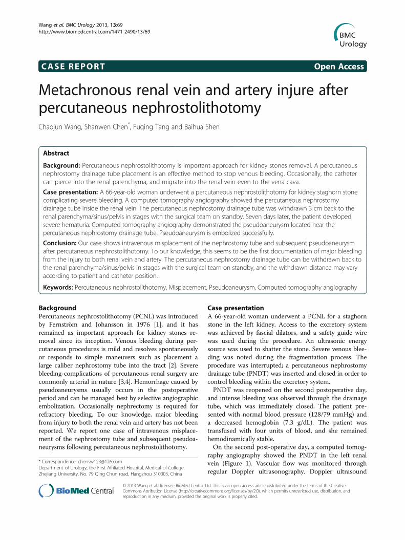
loped gross hematuria and severe pain of the left loin. Asignificant drop of the hemoglobin level was noted (from9.3 g/dL to 6.8 g/dL) but she remained hemodynamicallystable and the coagulation parameters were within normallimits. An abdominal computed tomography scan revealeda large left perinephric hematoma. She was initially treatedconservatively with bed rest and transfusions but grosshematuria persisted. After failed resuscitation, a computedtomography angiography was arranged, which revealed apseudoaneurysm in the left renal artery (Figure 2). A se-lective left renal angiogram was arranged in order to
achieve endovascular control of the bleeding vessel. Activebleeding from the pseudoaneurysm at a branch of theleft renal artery with contrast extravasation was shown(Figure 3). The bleeding was embolized selectively withtwo 2 mm*2 cm metal coils. Twenty-four hours later,hematuria ceased and the patient remained hemodyna-mically stable.Following above procedure, there was no further gross
hematuria. On the thirtieth post-operative day, a nephros-togram revealed the tract had sealed completely, and thePNDT was removed uneventfully. After an 18-month fol-low up, her post-operative hemoglobin and serum crea-tinine remained normal.
DiscussionA PNDT placement in the collecting system followingPCNL is a routine practice. It is an effective method tostop venous bleeding [5]. Occasionally, the catheter canpierce into the renal parenchyma, and migrate into therenal vein even to the vena cava [5]. The proximity ofthe renal vein to the renal pelvis and major posterior ca-lices predisposes them to injury during PCNL [6]. Ourpatient had complex staghorn renal calculi involving therenal pelvis. Concomitant infection and inflammation
Figure 1 Axial contrast enhanced CT and volume rendering image of CT angiography. A-B) Axial contrast enhanced CT and volumerendering image of CT angiography demonstrate percutaneous nephrostomy drainage tube inside the left renal vein; C-D) Axial contrastenhanced CT and volume rendering image of CT angiography reveal percutaneous nephrostomy drainage tube inside the left renal pelvis afterthe percutaneous nephrostomy drainage tube was withdrawn in stages.
Wang et al. BMC Urology 2013, 13:69 Page 2 of 5http://www.biomedcentral.com/1471-2490/13/69
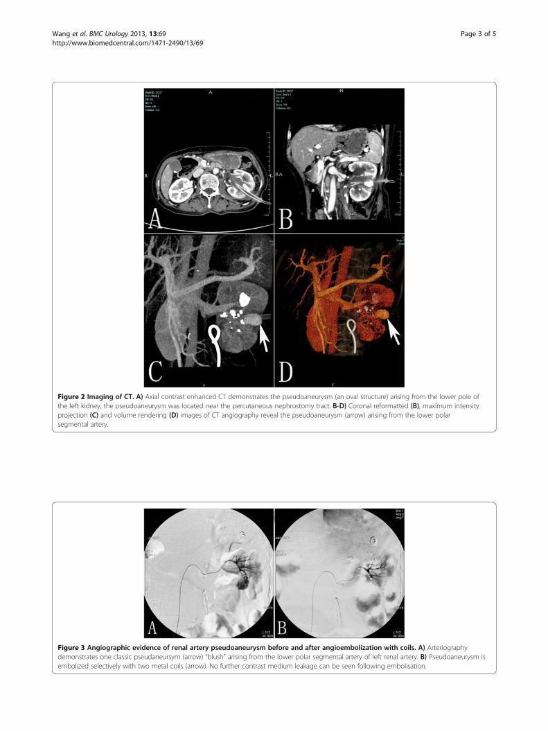
Figure 2 Imaging of CT. A) Axial contrast enhanced CT demonstrates the pseudoaneurysm (an oval structure) arising from the lower pole ofthe left kidney, the pseudoaneurysm was located near the percutaneous nephrostomy tract. B-D) Coronal reformatted (B), maximum intensityprojection (C) and volume rendering (D) images of CT angiography reveal the pseudoaneurysm (arrow) arising from the lower polarsegmental artery.
Figure 3 Angiographic evidence of renal artery pseudoaneurysm before and after angioembolization with coils. A) Arteriographydemonstrates one classic pseudaneursym (arrow) “blush” arising from the lower polar segmental artery of left renal artery. B) Pseudoaneurysm isembolized selectively with two metal coils (arrow). No further contrast medium leakage can be seen following embolisation.
Wang et al. BMC Urology 2013, 13:69 Page 3 of 5http://www.biomedcentral.com/1471-2490/13/69

may have made the renal pelvic wall more friable andsusceptible to the injury.The mechanism of injury was different in resulting from
dilating too medially, overzealous tract dilation, injury to acaliceal infundibulum, and pushing too hard on a renalpelvic stone with the ultrasound lithotrite probe as in ourpatient. Understanding since the renal pelvis is a delicatestructure with a thin muscular layer and there is no struc-tural support for it, special care should be taken to stonesin the pelvis or lodged at the ureteropelvic junction. Byusing the ultrasound probe gingerly with firm but gentleintermittent pressure against the stone and without jerkyto and fro motions can prevent the stone or the instru-ment from perforating the collecting system.Abdallah Geara presented a case of visualization of the
renal vein during pyelography after nephrostomy, andexamination by microscopy showed the presence of tearsin the fornix of the pelvic cavity that extend into the kid-ney parenchyma [7]. However, significant venous injuriesduring percutaneous renal surgery probably are underdiagnosed [8]. Since contrast material generally is notinjected during percutaneous procedures to prevent ex-travasation from obscuring the fluoroscopic image, majorinjuries may be missed. Clotting of these venous injuriesmay occur during the operation and, therefore, the injurymay not be evident on nephrostograms performed at theconclusion of the procedure or at follow-up.In cases where misplacement of the tube is detected, de-
pending on the postoperative time elapsed, relocationof the nephrostomy tube under fluoroscopy is stronglyrecommended, and the surgical team must stand ready tooperate in case an open emergency procedure is required[9]. Our case illustrates that control on the hemorrhagecould be achieved with the catheter being withdrawn instages, and laparotomy could be avoided despite of theperforation into the major renal vein. If the hemorrhagecould not be controlled, in Gupta M’experience, the tam-ponading nephrostomy catheter is optimally suited for thispurpose [8]. Holding mild tension on the catheter againstthe site of injury, the balloon can be further inflated ifnecessary until extravasation into the vein ceases. How-ever, those cases involved prolonged balloon inflation andrisked pressure necrosis. Our case highlights the impor-tance of prompt diagnosis of renal-vein perforation anddemonstrates that this can be managed conservatively bythe catheter being withdrawn in stages or a tamponadingnephrostomy catheter.This study is the fifth report in the literature regarding
misplacement of a nephrostomy tube into the vascularsystem [8-10], yet, it is the first report under the guideof ultrasound, and is also the first report with such acomplication of vein and artery following PCNL.It is an uncommon case of renal artery pseudoaneu-
rysm because of its delayed occurrence after invasive
procedure. The percutaneous tract disrupts the normalvessel wall and a pseudoaneurysm is formed from thetissues surrounding the high-pressure arterial system,resulting in recanalisation between the intravascular andextravascular space that produces a pulsating, encapsu-lated hematoma. The vascular compromise is initially con-trolled by a combination of decreased blood flow, arteryspasm, coagulation, and a tamponading effect of the sur-rounding tissue, which initially prevents hemorrhage andleads to its nonrecognition in the operating room. Asthese temporizing measures begin to degrade, the patientbecomes more active, the blood flow to this vessel in-creases and the pseudoaneurysm may eventually grow andbecomes unstable. With erosion into the pelvicaliceal sys-tem, gross hematuria thus occurs. Post-PCNL pseudoa-neurysm is usually located in the peripheral arteries. Thismight explain the absence of significant bleeding intraop-eratively and postoperatively. This is supported by the factthat our patient angiography identified that the pseudoa-neurysm was located near the punctured cavity.The natural history of pseudoaneurysm ranges from
spontaneous resolution to acute rupture. By far, the mostcommon presentation is gross hematuria. Patients areoften clinically stable and can present with nonspecificsymptoms including flank pain, hypertension, arterialbruits, deterioration of renal function and worseninglower urinary tract symptoms. Patients usually presentin the first few weeks (ranging from 1 day to 5 months)postoperatively.The diagnosis of intrarenal pseudoaneurysm is challen-
ging. Clinical diagnosis can be done through invasive(angiography) and non-invasive (angio CT or Dopplerultrasonography) methods. Arteriography remains thegold standard diagnostic tool, although multiplanar con-trast enhanced computed tomography is suitable alter-natives. The advantages of angiography in this settinginclude high sensitivity in identifying the pseudoaneurysm(which usually appears as a round or oval structure arisingfrom the main renal artery or one of its branches) and thepotential to achieve simultaneous endovascular manage-ment of these lesions, with success rates exceeding 90%.10Superselective embolisation is highly efficient in achievingpseudoaneurysm occlusion through the injection of a per-manent agent at the fistulous point. Materials such asethanol, metal coil, gel foam particles and N-butyl-2-cyanoacrylate have been successfully used for embolisation[11,12]. No significant difference was found between thepreangioembolization and postangioembolization serumcreatinine, similar to that reported by Martin et al. [11].Pseudoaneurysm appears as a focal area of high attenu-ation with a similar enhancement to the adjacent vessels.The advantage of computed tomography imaging is thatthese modalities not only can diagnose pseudoaneu-rysms but can also detect other possible intra-abdominal
Wang et al. BMC Urology 2013, 13:69 Page 4 of 5http://www.biomedcentral.com/1471-2490/13/69

pathologies that may be responsible for the presentingsymptoms. A three-dimensional reconstruction can beuseful for differentiating a pseudoaneurysm from urinoma,which may, at times, be difficult to distinguish from eachother.
ConclusionsOur case shows intravenous misplacement of the nephro-stomy tube and subsequent pseudoaneurysm after per-cutaneous nephrostolithotomy. To our knowledge, thisseems to be the first documentation of major bleedingfrom the injury to both renal vein and artery. The percu-taneous nephrostomy drainage tube can be withdrawn3 cm back to the renal parenchyma/sinus/pelvis in stageswith the surgical team on standby, and the withdrawn dis-tance may vary according to patient and catheter position.
ConsentWritten informed consent was obtained from the patientfor publication of this manuscript and accompanyingimages. A copy of the written consent is available for re-view by the Editor-in-Chief of this journal.
AbbreviationsPCNL: Percutaneous nephrostolithotomy; PNDT: Percutaneous nephrostomydrainage tube.
Competing interestsThe authors declare that they have no competing interests.
Authors’ contributionsCW and FT cared for the patient and drafted the report. BS cared for thepatient. SC revised and approved the final version of the manuscript. Allauthors reviewed the report and approved the final version of themanuscript.
AcknowledgementsLanguage editor Shuo Wang edited our manuscript.
Received: 29 July 2013 Accepted: 27 November 2013Published: 5 December 2013
References1. Ferström I, Johansson B: Percutaneous pyelolithotomy: a new extraction
technique. Scand J Urol Nephrol 1976, 10:257–259.2. Galek L, Darewicz B, Werel T, Darewicz J: Hemorrhagic complications of
percutaneous lithotripsy: original methods of treatment. Int Urol Nephrol2000, 32:231–233.
3. Clayman RV, Surya V, Hunter D, et al: Renal vascular complicationsassociated with the percutaneous removal of renal calculi. J Urol 1984,132:228–230.
4. Ugras M, Gunes A, Baydinc C: Severe renal bleeding caused by a rupturedrenal sheath: case report of a rare complication of percutaneousnephrolithotomy. BMC Urol 2002, 2:10.
5. Winfield HN, Weyman P, Clayman RV: Percutaneous nephrostolithotomy:complications of premature nephrostomy tube removal. J Urol 1986,136:77–79.
6. Sampaio FJ: The dilemma of the crossing vessel at the ureteropelvicjunction: precise anatomic study. J Endourol 1996, 10:411–415.
7. Geara A, Kamal L, El-Imad B, El-Sayegh S: Visualization of the renal veinduring pyelography after nephrostomy: a case report. J Med Case Rep2010, 23:93.
8. Gupta M, Bellman GC, Smith AD: Massive hemorrhage from renal veininjury during percutaneous renal surgery: endourological management.J Urol 1997, 157:795–797.
9. Mazzucchi E, Mitre A, Brito A, Arap M, Murta C, Srougi M: Intravenousmisplacement of the nephrostomy catheter following percutaneousnephrostolithotomy: two case reports. Clinics (Sao Paulo) 2009, 64:69–70.
10. Shaw G, Wah TM, Kellett MJ, Choong SK: Management of renal-veinperforation during a challenging percutaneous nephrolithotomy.J Endourol 2005, 19:722–723.
11. Martin X, Murat FJ, Feitosa LC, et al: Severe bleeding afternephrolithotomy: results of hyperselective embolization. Eur Urol 2000,37:136–139.
12. Lee KL, Stoller ML: Minimizing and managing bleeding afterpercutaneous nephrolithotomy. Curr Opin Urol 2007, 17:120–124.
doi:10.1186/1471-2490-13-69Cite this article as: Wang et al.: Metachronous renal vein and arteryinjure after percutaneous nephrostolithotomy. BMC Urology 2013 13:69.
Submit your next manuscript to BioMed Centraland take full advantage of:
• Convenient online submission
• Thorough peer review
• No space constraints or color figure charges
• Immediate publication on acceptance
• Inclusion in PubMed, CAS, Scopus and Google Scholar
• Research which is freely available for redistribution
Submit your manuscript at www.biomedcentral.com/submit
Wang et al. BMC Urology 2013, 13:69 Page 5 of 5http://www.biomedcentral.com/1471-2490/13/69