mhc class i antigen processing and presenting machinery ...ecules, peptides generated by the...
TRANSCRIPT

Vol. 105, Issue 16 | August 21, 2013
DOI:10.1093/jnci/djt184Advance Access publication July 12, 2013
1172 Review | JNCI
© The Author 2013. Published by Oxford University Press. All rights reserved. For Permissions, please e-mail: [email protected].
Review
MHC Class i Antigen Processing and Presenting Machinery: Organization, Function, and Defects in Tumor CellsPatrizia Leone, Eui-Cheol Shin, Federico Perosa, Angelo Vacca, Franco Dammacco, Vito Racanelli
Manuscript received January 15, 2013; revised March 21, 2013; accepted June 13, 2013.
Correspondence to: Vito Racanelli, MD, Department of Internal Medicine and Clinical Oncology, University of Bari Medical School, Policlinico, 11 Piazza G Cesare, 70124 Bari, Italy (e-mail: [email protected]).
The surface presentation of peptides by major histocompatibility complex (MHC) class I molecules is critical to all CD8+ T-cell adap-tive immune responses, including those against tumors. The generation of peptides and their loading on MHC class I molecules is a multistep process involving multiple molecular species that constitute the so-called antigen processing and presenting machin-ery (APM). The majority of class I peptides begin as proteasome degradation products of cytosolic proteins. Once transported into the endoplasmic reticulum by TAP (transporter associated with antigen processing), peptides are not bound randomly by class I molecules but are chosen by length and sequence, with peptidases editing the raw peptide pool. Aberrations in APM genes and proteins have frequently been observed in human tumors and found to correlate with relevant clinical variables, including tumor grade, tumor stage, disease recurrence, and survival. These findings support the idea that APM defects are immune escape mechanisms that disrupt the tumor cells’ ability to be recognized and killed by tumor antigen–specific cytotoxic CD8+ T cells. Detailed knowledge of APM is crucial for the optimization of T cell–based immunotherapy protocols.
J Natl Cancer Inst;2013;105:1172–1187
In normal cellular physiology, proteins regularly undergo a pro-cess of turnover in which they are degraded and replaced by newly synthesized proteins. The degradation of most cellular proteins occurs by one of two major proteolytic pathways: the lysosomal pathway and the ubiquitin-proteasome pathway (1). The lysosomal pathway degrades proteins taken up by endocyto-sis (from the extracellular environment) and by autophagy (from the cytosol); in this pathway, bacteria, bacterial antigens, para-sites or long-lived bulk proteins, particularly membrane-bound proteins, are delivered to endosomes, which become increasingly acidic as they progress into the interior of the cell, eventually fusing with lysosomes (2). In contrast, the ubiquitin-proteasome pathway is mainly involved in degradation of cytosolic proteins (3), such as regulatory proteins (short-lived proteins that are eliminated soon after completing their functions); misfolded and damaged proteins (4), including defective ribosomal products (ie, newly synthesized proteins degraded within minutes of their syn-thesis) (5,6); mutated proteins in cancer cells; and virus-derived proteins in infected cells. In both pathways, cellular proteins are cleaved into oligopeptide fragments that are presented to T cells by molecules of the major histocompatibility complex (MHC). Whereas peptides derived from proteins degraded by the lyso-somal pathway are primarily presented by MHC class II mol-ecules, peptides generated by the ubiquitin-proteasome pathway are presented by MHC class I molecules (7). A major exception to this rule is cross-presentation, a process specific to profes-sional antigen-presenting cells, whereby peptides derived from proteins that have entered the lysosomal pathway gain access to MHC class I molecules (8).
In humans, there are three main (and several minor) MHC class I molecules, which are also called by their gene name, human leukocyte antigen (HLA). The main function of class I molecules, which are expressed on the plasma membrane of most cell types, is to display these peptides to cytotoxic CD8+ T cells in support of their crucial activity of immune surveillance. Peptides derived from normal cellular (self) proteins are regularly ignored by CD8+ T cells, whereas those from mutated proteins and from the non-self proteins of viruses and other intracellular pathogens are not ignored but trigger an adaptive immune response through binding to the T-cell receptor (TCR). MHC class I molecules also function in the innate immune system by serving as ligands of inhibitory killer cell immunoglobulin-like receptors (KIRs) on natural killer (NK) cells. NK cells have the unique ability to recognize and non-specifically kill cells lacking self MHC class I molecules. Because all healthy nucleated cells express self MHC class I molecules, inhibitory KIRs ensure that NK cells do not attack normal cells but eliminate infected and tumor cells (which may have reduced MHC class I molecule expression) (9). Because not all infections or cancers reduce MHC class I expression, the role of these proteins in the adaptive immune response is fundamental.
For MHC class I molecules to present self and nonself peptides to CD8+ T cells, the peptides must first be produced by proteolysis in the ubiquitin-proteasome pathway. Proteins are targeted for degradation in this pathway by the covalent attachment of multiple copies of the 76-residue protein ubiquitin to free amino groups (always near the ε-amino group of Lys). Ubiquitination involves a ubiquitin-activating enzyme (E1), a ubiquitin-conjugating enzyme (E2), a substrate-specific ubiquitin-protein ligase (E3), and in some

JNCI | Review 1173jnci.oxfordjournals.org
cases an additional conjugation factor (E4) (10). The breakdown of polyubiquitinated proteins and the processing of the resulting peptides until they are presented on the cell surface involve multiple molecular species, including the proteasome in its constitutive and immunoproteasome forms, peptide transporters (TAP1 and TAP2), endoplasmic reticulum chaperones (calnexin, calreticulin, ERp57, and tapasin), and the Golgi apparatus. Acting in concert, these proteins, multimeric protein complexes, and organelles make up what is called the MHC class I antigen processing and presentation machinery (APM) (11–15). Defects in the function or expression of APM components affect the formation of MHC class I peptide complexes and their recognition by CD8+ T cells (and NK cells). This review describes the structure and key functions of the proteasome and immunoproteasome, dissects the four main tasks of antigen processing and presentation, lists APM changes that have been observed in tumors, and explores the possible clinical significance of these defects with a special focus on their potential role in tumor cells’ ability to escape immunosurveillance.
Proteasomes and immunoproteasomes: Structure, Components, and FunctionsThe proteasome is a multimeric protein complex found in both the cytosol and nucleus (16–18). Structurally, it has a cylindrical shape and contains both a catalytic core and regulatory particles (Figure 1). The catalytic core, called the 20S proteasome, is com-posed of four stacked heptameric rings that produce a barrel-shaped
structure with a central gate. The two outer rings each contain seven α subunits (α1–α7) that interact with regulatory particles and create a physical barrier to regulate access to the gate (17). The two inner rings each contain seven β subunits (β1–β7), three of which (β1 or δ, β2 or Z, and β5 or MB1) have threonine-protease catalytic centers with different cleavage specificities: β1 has caspase-like activity (cleavage after acid residues); β2 has trypsin-like activity (cleavage after basic residues); and β5 has chymotrypsin-like activ-ity (cleavage after hydrophobic residues) (19,20).
The proteasomal gate is normally closed by the N-termini of the seven α subunits to keep the proteasome in a proteolytically inactive state and to prevent unregulated protein degradation. The N-terminus of subunit α3 sticks out the most into the gateway, interacting with every other α subunit (17,21). Cleavage of this N-terminus, which occurs upon conformational rearrangements caused by the attachment of regulatory particles to the α rings, opens the gate, permits the access of substrates, and activates the proteasome (17,21).
Regulatory particles bind to one or both ends of the 20S proteasome. The major regulatory particle, called the 19S regulator (or PA700), binds to the 20S proteasome to form the 26S proteasome (22). The 19S regulator consists of 17 distinct subunits, 9 in a “base: subcomplex and 8 in a “lid” subcomplex (23). The lid contains binding sites for both polyubiquitinated proteins and enzymes that disassemble and recycle ubiquitin chains, called deubiquitinating enzymes. The base interacts with the α rings of the 20S proteasome; it triggers gate opening, unfolds
Figure 1. Proteasome composition

Vol. 105, Issue 16 | August 21, 20131174 Review | JNCI
protein substrates, and catalyzes protein translocation into the 20S proteasome (24,25). These functions require metabolic energy, and, indeed, the base contains six ATPase subunits.
In addition to the 19S regulator, other regulatory particles, named PA28αβ (26), PA200 (27), and PI31 (28;29) can bind the 20S proteasome and form proteasomal isoforms. These alternative reg-ulators bind the α rings of the 20S proteasome just as the 19S regu-lator does, but in an ATP-independent way. PA28αβ is a heptameric complex composed of α and β subunits; upon binding with the 20S proteasome, it increases the catalytic activity of the cleavage sites and facilitates the opening of the proteasome gate (30). PA28αβ is particularly abundant in immune tissues, and it is induced by inter-feron (IFN) γ and infection. PA200 is the most recent proteasome activator to be discovered (27). The original description of this pro-teasome activator proposed it to be involved in DNA repair, pos-sibly by recruiting proteasomes to double-strand breaks. Following gamma irradiation, PA200 forms hybrid proteasomes with 19S regulator–20S proteasome–PA200 that accumulate on chromatin, leading to an increase in proteolytic activity (27). PI31 is a cellular regulator with inhibitory function that competes with PA28αβ and PA200 for binding with the 20S proteasome (28,29).
Three of the 20S proteasome’s β subunits (δ, Z, and MB1) may be replaced by functionally different counterparts named low molecular weight protein (LMP) 2 (also called β1i), LMP7 (β5i), and LMP10 (β2i), respectively (31–33). Proteasomes incorporating LMP2, LMP7, and LMP10 are called immunoproteasomes because they develop under conditions of intensified immune response. Indeed, they are induced in the majority of cells by stimulation with type I (α and β) IFN (34;35) and type II (γ) IFN (34,35) (Figure 1). Cells exposed to IFN in the context of an inflammatory process are not the only cells to contain immunoproteasomes. These are also expressed in a constitutive manner in lymphoid organs such as the spleen, lymph nodes, and thymus (36). Interestingly, dendritic cells were recently found to have approximately equal amounts of proteasomes and immunoproteasomes when immature and only immunoproteasomes when mature (37).
Compared with 20S proteasomes, immunoproteasomes display a weaker ability to cleave peptides after acidic residues but a bet-ter ability to cleave after basic and hydrophobic residues (38,39). Immunoproteasomes also serve functions besides antigen process-ing. They generate biologically active proteins (such as cytokines) that are involved in inflammatory processes (40) and in T-cell dif-ferentiation, survival, and function during thymocyte development (41). In addition, immunoproteasomes are thought to have a role in cell differentiation because they are constitutively expressed in mouse ocular lens and brain (42), which are immune-privileged sites with no apparent need to generate class I peptide ligands.
Four Main Tasks of MHC Class i Antigen Processing and PresentationWhen polyubiquitinated proteins reach the proteasome (or immu-noproteasome), a complex cellular process begins that prepares antigens for presentation on MHC class I molecules. This process consists of four main tasks: 1) peptide generation and trimming; 2) peptide transport; 3) assembly of the MHC class I loading com-plex; and 4) antigen presentation (11–15) (Figure 2).
Peptide Generation and TrimmingWhen the proteasome is activated, ubiquitinated proteins pass through the gate, unfold, spread along it, and lose the polyubiq-uitin chain (deubiquitination) through the action of deubiquit-inating enzymes. Proteins are then broken down into peptides ranging from 2 to 25 residues, which are released to the cytosol. The particular peptide repertoire that is generated, in terms of amino acid sequence, length, and quantity (39), varies depending on whether the proteasome or immunoproteasome is involved. Both are able to generate MHC class I epitopes, but dramatic differences exist in the efficiency at which a given epitope is generated. For instance, immunodominant epitopes of infec-tious organisms have recently been found to be more effectively produced by the immunoproteasome. In particular, in experi-ments in which HeLa cells were infected with vaccinia virus expressing the hepatitis B virus core antigen, generation of the hepatitis B virus core antigen141–151 epitope required the immu-noproteasome with subunit LMP7 (43). Moreover, in knock-out mice lacking the three immunoproteasome subunits, dendritic cells could not present several major MHC class I epitopes, and the epitope repertoire was 50% different from that of wild-type mice (44).
A minority of intracellular proteins are cleaved in protea-some-independent pathways that also generate peptides for MHC class I presentation (45–47). For instance, peptides with a C-terminal lysine are generated by proteasomes with very low efficiency given that lysine is not a preferred proteasomal cleavage site (48). These peptides, which may represent important T-cell epitopes, may, however, be generated by additional proteases such as tripeptidyl peptidase II (TPPII), a cytosolic aminopeptidase with endoproteolytic activity able to cleave after lysine residues (49). This protease is essential for the generation of the immu-nodominant HLA-A3– and HLA-A11–restricted HIV-1 Nef73-82 epitope (50). Another cytosolic protease involved in the direct production of MHC class I peptides is insulin-degrading enzyme, a metallopeptidase that generates epitopes from the melanoma-associated antigen A3 (51), an immunogenic protein highly expressed by several human tumors (52). Nardilysin and thimet oligopeptidase (TOP) are two additional cytosolic endopeptidases that are required, either together or alone, for the generation of T-cell epitopes from the Epstein–Barr virus nuclear antigen 3C, the melanoma antigen recognized by T cells 1 (MART-1), and the preferentially expressed antigen of melanoma (PRAME). TOP operates as a C-terminal trimming peptidase, whereas nardilysin contributes to both the C-terminal and N-terminal generation of the epitopes (53).
Peptides produced in the cytosol are further trimmed by enzymes within the endoplasmic reticulum (ER) (45,54,55) to fit into the groove of the MHC class I molecules. One of these enzymes, an ER aminopeptidase called ERAP1 (ER aminopeptidase associated with Ag processing 1), is considered a “molecular ruler” because of its substrate preference (56): ERAP1 preferentially trims peptides of 9 to 16 residues but spares those of 8 to 9 residues, the typical length for MHC class I binding (56). ERAP1 prefers peptides with hydrophobic C-termini and is induced by type I and II IFNs (57). Recent work has also shown that the ER dipeptidase angiotensin-converting enzyme can make the final C-terminus peptide cut (58).

JNCI | Review 1175jnci.oxfordjournals.org
Peptide TransportPeptides generated by proteasome-dependent and -independent pathways are actively transported from the cytosol into the ER by the transporter associated with antigen processing (TAP). This heterodimeric complex is composed of the two half-transporters, TAP1 and TAP2, that are members of the ATP-binding cassette transporter family. Both TAP1 and TAP2 contain a hydrophobic transmembrane domain and a cytosolic nucleotide-binding domain (59). TAP forms a transmembrane pore in the ER membrane whose opening and closing depend on ATP binding and hydrolysis, respectively (ATP switch model) (60–62).
TAP transports most efficiently peptides of a certain length (8–12 residues) and with hydrophobic or basic C-termini that bind to MHC class I molecules (63–68). Transport of longer peptides occurs with reduced efficiency (64–66,68). These longer peptides, which do not fit the class I binding groove, can be further trimmed
in the ER lumen or, alternatively, can be transported back to the cytosol where they are trimmed by cytosolic peptidases and recycle back to the ER in a TAP-dependent fashion for association with MHC class I molecules (69).
Assembly of the MHC Class I Loading ComplexPeptides transported into the ER by TAP are loaded onto nas-cent MHC class I molecules with the assistance of four chaperone proteins: calnexin (70), the thiol oxidoreductase ERp57 (71,72), calreticulin (73), and tapasin (74;75). These proteins, along with MHC class I molecules and TAP, form the MHC class I load-ing complex that combines the activities of peptide transport and loading onto MHC molecules (70–78). Specifically, a newly synthesized MHC class I heavy chain, translocated into the ER, acquires a Glc1Man9GlcNAc2 glycan moiety that serves as a recog-nition element for the membrane-bound chaperones calnexin and
Figure 2. The four main tasks of major histocompatibility complex (MHC) class I antigen processing and presentation: 1) Peptide generation and trimming; 2) peptide transport; 3) assembly of the MHC class loading complex; 4) antigen presentation.

Vol. 105, Issue 16 | August 21, 20131176 Review | JNCI
calreticulin (79,80). The MHC class I heavy chain interacts with calnexin., which both facilitates its complete folding and, by act-ing in concert with ERp57 (81), ensures its correct oxidation (82). At this point, the heavy chain is conformationally recognizable by β2microglobulin (83). Their binding triggers the release of cal-nexin (83,84). The resulting conformational changes give the heavy chain/β2microglobulin heterodimer an “open” form that interacts with calreticulin (73). Peptide binding requires the additional par-ticipation of TAP, which assures a pool of free peptides, and tapasin. This chaperone bridges class I molecules to TAP, allowing peptides to gain access to the ER for class I binding (73,85). It also stabilizes heavy chain/β2microglobulin heterodimers (75,86) and optimizes the class I peptide load (87).
Antigen PresentationAfter the binding of a peptide, the ER-resident chaperones are released and the peptide-MHC class I complex leaves the ER within vesicles that traverse the Golgi apparatus, migrate to the cell membrane, and fuse with it, so that the bound peptide in the MHC class I groove is exposed extracellularly. Here the peptide-MHC class I complex may be recognized by and interact with the cognate TCR on CD8+ T cells. Peptides derived from unmutated (self) proteins are normally ignored by CD8+ T cells, whereas those derived from mutated or pathogen-derived (nonself) proteins are not. Using this system of intracellular surveillance, CD8+ T cells play a crucial role in eradicating viruses and other intracellular pathogens and also exert potent antitumor activity.
Defects in Class i APM in Human TumorsDefects in the expression and function of APM components have been found in various solid and hematologic tumors. They occur individually or in combination, and the frequency and nature of the defect vary substantially according to tumor type (Table 1). The molecular mechanisms underlying these defects have been partly identified for some components only and seem to take place at the genetic and epigenetic levels (Table 2). There is also some evidence that transcriptional and post-transcriptional defects may occur.
Defects in Proteasome Subunit Expression PatternsAlterations of proteasome subunits have been identified only recently, thanks to the availability of monoclonal antibodies that permit semiquantitative analyses. Downregulation of one or more of the 20S proteasome’s β subunits δ, MB1, and Z is characteristic of colorectal (88), bladder (89), and ovarian (90) carcinomas, as well as medulloblastoma (91). Similarly, downregulation of one or more of the inducible subunits (LMP2, LMP7, and LMP10) is prominent in acute myeloid leukemia (92), in carcinoma of the head and neck (93,94), esophagus (95–97), stomach (98), colo-rectum (88,99,100), kidney (101,102), bladder (89,103), prostate (104), cervix (105,106), ovary (90), and breast (107;108), and in astrocytoma (109), medulloblastoma (91), neuroblastoma (110), and melanoma (111,112). We have also found that the constitu-tive subunits δ, Z, and MB1 and the immunoproteasome subu-nits LMP2 and LMP10 are progressively lost in premalignant and malignant plasma cells from bone marrow of patients with mono-clonal gammopathy of undetermined significance (MGUS) and
multiple myeloma (MM) (113). It should be noted that interpreta-tion of data related to the expression of immunoproteasome subu-nits is quite complex, given that they are likely unexpressed under basal conditions but are induced after exposure of cells to IFN-γ. Furthermore, no data are available regarding the expression of immunoproteasome subunits in most normal tissues. Therefore, in several cases it is not possible to establish whether the expres-sion of an immunoproteasome subunit in malignant cells is a normal phenotype and its lack of expression is a downregulation process or whether such lack is a normal phenotype and its expres-sion reflects regulatory abnormalities.
Little is known about the molecular basis of the defects in proteasome components. Mutations at coding microsatellites of genes encoding LMP7 have been detected in gastric cancer (98). Single nucleotide polymorphisms in the LMP2 and LMP7 genes have been identified in cervical carcinoma (105,114). In prelimi-nary experiments, we found that treatment of myeloma cells with decitabine, a potent DNA methyltransferase inhibitor, restored the expression of several proteasome subunits, suggesting that promoter methylation alterations and epigenetic regulation were involved (113). Loss of IFN-γ–mediated upregulation of LMP2 in one renal cell carcinoma cell line has been associated with the lack of IFN regulatory factor 1 and signal transducer and activator of transcription 1 (STAT1) binding activities, as well as of Janus associated kinase (JAK)1, JAK2, and STAT1 phosphorylation (115). More recently, loss of IFN-γ–mediated upregulation of LMP2 and LMP10 in melanoma cell lines has been found to be caused by a deletion of the JAK2 gene on chromosome 9 (116).
Defects in TAP1 and TAP2Low to undetectable levels of TAP1 and/or TAP2 mRNA and/or protein have been reported in primary cells and cell lines from several tumors, including carcinomas of the head and neck (93,94,117–119), esophagus (95–97,120), stomach (98), pancreas (121), colorectum (88,99,100,122–124), breast (108,125–127), and cervix (105,106,128–130); renal cell (101,102), prostate (104) and bladder (89,103) cancer; and melanoma (111,112,131–133), astrocytoma (109,134), medulloblastoma (91), neuroblastoma (110), acute myeloid leukemia (92), and multiple myeloma (113). In several tumor cell lines in which TAP was downregulated, its levels were restored by IFN-γ treatment (135,136). At the genetic level, mutations in TAP genes that resulted in loss of expression or in expression of a nonfunctional protein have been observed in colorectal (88,100,123), cervical (114,129,137), gastric (98), and lung (138) carcinomas. Methylation of the TAP1 gene promoter has also been found in cervical carcinoma (105). A post-transla-tional downregulation of TAP2 has been observed in a melanoma cell line that does not express TAP1 because of a frameshift muta-tion that generates a stop codon in the TAP1 gene (139). In this cell line, transcription of TAP1 and TAP2 genes proceeds nor-mally, but, in the absence of TAP1 translation, the TAP2 protein is unstable and is lost from the cell; these results suggest that TAP expression is regulated through a mechanism of coordinated sta-bilization of the TAP heterodimeric complex. Moreover, as for immunoproteasome subunits, loss of JAK2 has been shown to impair the IFN-γ inducibility of TAP1 and TAP2 in melanoma cell lines. (116).

JNCI | Review 1177jnci.oxfordjournals.org
Tab
le 1
. E
xpre
ssio
n d
efec
ts in
an
tig
en p
roce
ssin
g a
nd
pre
sen
tin
g m
ach
iner
y co
mp
on
ents
, by
tum
or
typ
e*
Bo
dy
reg
ion
Tum
or
typ
eδ
MB
1Z
LMP
2LM
P7
LMP
10TA
P1
TAP
2E
RA
P1-
2C
LXC
LTT
PN
ER
p57
HLA
-Iβ 2
mR
efer
ence
Hea
d an
d ne
ckH
ead
and
neck
squ
amou
s ce
ll ca
rcin
oma
P, M
■■
■■
118
Hea
d an
d ne
ck s
quam
ous
cell
carc
inom
a P
.■
■■
■.
■93
Hea
d an
d ne
ck s
quam
ous
cell
carc
inom
a P, M
■■
■■
117
Max
illar
y si
nus
squa
mou
s ce
ll ca
rcin
oma P
■■
■■
■■
142
Lary
ngea
l squ
amou
s ce
ll ca
rcin
oma P
■■
■■
■■
■■
94
Ora
l squ
amou
s ce
ll
carc
inom
a P
■■
170
Ora
l squ
amou
s ce
ll
carc
inom
a P
■14
4
Lung
Lung
car
cino
ma P
■14
0G
astr
oint
estin
al
syst
emE
soph
agus
squ
amou
s ce
ll ca
rcin
oma P
■■
■■
■■
95
Eso
phag
us s
quam
ous
cell
carc
inom
a P, M
■■
120
Eso
phag
us s
quam
ous
cell
carc
inom
a P
■■
■96
Eso
phag
us s
quam
ous
cell
carc
inom
a P
■■
■■
■■
■97
Gas
tric
ade
noca
rcin
oma P
, M■
146
Gas
tric
ade
noca
rcin
oma P
■■
■■
■■
98Pa
ncre
atic
car
cino
ma P
■■
■12
1Pa
ncre
atic
car
cino
ma P
■14
0C
olor
ecta
l car
cino
ma P
■■
■12
3C
olor
ecta
l car
cino
ma P
, M■
■■
■■
■99
Col
orec
tal c
arci
nom
a P■
■■
■■
■■
■■
■■
■■
■88
Col
orec
tal c
arci
nom
a P■
■■
■■
■■
■■
■10
0C
olor
ecta
l car
cino
ma P
, M■
■■
■12
2C
olor
ecta
l car
cino
ma P
■■
■12
4G
enito
urin
ary
syst
emR
enal
car
cino
ma P
■■
■■
■■
102
Ren
al c
arci
nom
a P, M
■■
■■
■10
1B
ladd
er c
arci
nom
a P■
■■
■■
■■
103
Bla
dder
car
cino
ma P
■■
■■
■■
■■
89Pr
osta
te c
arci
nom
a P■
■■
■■
■■
■■
■10
4C
ervi
cal s
quam
ous
cell
carc
inom
a P
■■
■■
■■
■■
■■
■10
5
Cer
vica
l squ
amou
s ce
ll ca
rcin
oma P
■■
■■
■■
■■
■10
6
Cer
vica
l ade
noca
rcin
oma P
■■
■■
■■
■■
■10
6O
varia
n ca
rcin
oma M
■19
2O
varia
n ca
rcin
oma P
■14
5O
varia
n ca
rcin
oma P
■14
0O
varia
n ca
rcin
oma P
■■
■■
■■
90
(Tab
le c
ontin
ues)
Vol. 105, Issue 16 | August 21, 20131178 Review | JNCI
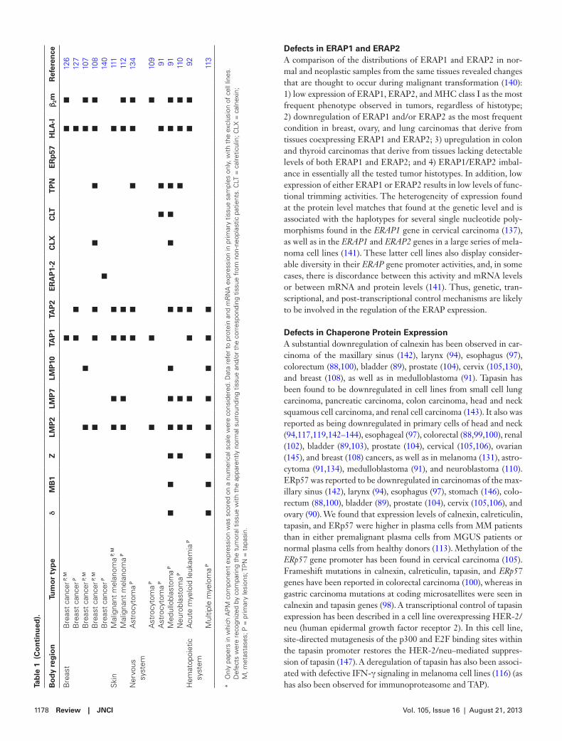
Defects in ERAP1 and ERAP2A comparison of the distributions of ERAP1 and ERAP2 in nor-mal and neoplastic samples from the same tissues revealed changes that are thought to occur during malignant transformation (140): 1) low expression of ERAP1, ERAP2, and MHC class I as the most frequent phenotype observed in tumors, regardless of histotype; 2) downregulation of ERAP1 and/or ERAP2 as the most frequent condition in breast, ovary, and lung carcinomas that derive from tissues coexpressing ERAP1 and ERAP2; 3) upregulation in colon and thyroid carcinomas that derive from tissues lacking detectable levels of both ERAP1 and ERAP2; and 4) ERAP1/ERAP2 imbal-ance in essentially all the tested tumor histotypes. In addition, low expression of either ERAP1 or ERAP2 results in low levels of func-tional trimming activities. The heterogeneity of expression found at the protein level matches that found at the genetic level and is associated with the haplotypes for several single nucleotide poly-morphisms found in the ERAP1 gene in cervical carcinoma (137), as well as in the ERAP1 and ERAP2 genes in a large series of mela-noma cell lines (141). These latter cell lines also display consider-able diversity in their ERAP gene promoter activities, and, in some cases, there is discordance between this activity and mRNA levels or between mRNA and protein levels (141). Thus, genetic, tran-scriptional, and post-transcriptional control mechanisms are likely to be involved in the regulation of the ERAP expression.
Defects in Chaperone Protein ExpressionA substantial downregulation of calnexin has been observed in car-cinoma of the maxillary sinus (142), larynx (94), esophagus (97), colorectum (88,100), bladder (89), prostate (104), cervix (105,130), and breast (108), as well as in medulloblastoma (91). Tapasin has been found to be downregulated in cell lines from small cell lung carcinoma, pancreatic carcinoma, colon carcinoma, head and neck squamous cell carcinoma, and renal cell carcinoma (143). It also was reported as being downregulated in primary cells of head and neck (94,117,119,142–144), esophageal (97), colorectal (88,99,100), renal (102), bladder (89,103), prostate (104), cervical (105,106), ovarian (145), and breast (108) cancers, as well as in melanoma (131), astro-cytoma (91,134), medulloblastoma (91), and neuroblastoma (110). ERp57 was reported to be downregulated in carcinomas of the max-illary sinus (142), larynx (94), esophagus (97), stomach (146), colo-rectum (88,100), bladder (89), prostate (104), cervix (105,106), and ovary (90). We found that expression levels of calnexin, calreticulin, tapasin, and ERp57 were higher in plasma cells from MM patients than in either premalignant plasma cells from MGUS patients or normal plasma cells from healthy donors (113). Methylation of the ERp57 gene promoter has been found in cervical carcinoma (105). Frameshift mutations in calnexin, calreticulin, tapasin, and ERp57 genes have been reported in colorectal carcinoma (100), whereas in gastric carcinoma mutations at coding microsatellites were seen in calnexin and tapasin genes (98). A transcriptional control of tapasin expression has been described in a cell line overexpressing HER-2/neu (human epidermal growth factor receptor 2). In this cell line, site-directed mutagenesis of the p300 and E2F binding sites within the tapasin promoter restores the HER-2/neu–mediated suppres-sion of tapasin (147). A deregulation of tapasin has also been associ-ated with defective IFN-γ signaling in melanoma cell lines (116) (as has also been observed for immunoproteasome and TAP).B
od
y re
gio
nTu
mo
r ty
pe
δM
B1
ZLM
P2
LMP
7LM
P10
TAP
1TA
P2
ER
AP
1-2
CLX
CLT
TP
NE
Rp
57H
LA-I
β 2m
Ref
eren
ce
Bre
ast
Bre
ast
canc
er P
, M■
■■
126
Bre
ast
canc
er P
■■
■12
7B
reas
t ca
ncer
P, M
■■
■■
107
Bre
ast
canc
er P
, M■
■■
■■
■■
108
Bre
ast
canc
er P
■14
0S
kin
Mal
igna
nt m
elan
oma P
, M■
■■
■■
111
Mal
igna
nt m
elan
oma P
■■
■■
■■
112
Ner
vous
sy
stem
Ast
rocy
tom
a P■
■■
■■
134
Ast
rocy
tom
a P■
■■
109
Ast
rocy
tom
a P■
■■
91M
edul
lobl
asto
ma P
■■
■■
■■
■■
■■
■■
91N
euro
blas
tom
a P■
■■
■■
■■
110
Hem
atop
oiet
ic
syst
emA
cute
mye
loid
leuk
aem
ia P
■■
■■
■■
92
Mul
tiple
mye
lom
a P■
■■
■■
■■
■11
3
* O
nly
pape
rs in
whi
ch A
PM
com
pone
nt e
xpre
ssio
n w
as s
core
d on
a n
umer
ical
sca
le w
ere
cons
ider
ed. D
ata
refe
r to
pro
tein
and
mR
NA
exp
ress
ion
in p
rimar
y tis
sue
sam
ples
onl
y, w
ith t
he e
xclu
sion
of
cell
lines
. D
efec
ts w
ere
reco
gniz
ed b
y co
mpa
ring
the
tum
oral
tis
sue
with
the
app
aren
tly n
orm
al s
urro
undi
ng t
issu
e an
d/or
the
cor
resp
ondi
ng t
issu
e fr
om n
on-n
eopl
astic
pat
ient
s. C
LT =
cal
retic
ulin
; CLX
= c
alne
xin;
M
, met
asta
ses;
P =
prim
ary
lesi
ons;
TP
N =
tap
asin
.
Tab
le 1
(C
on
tin
ued
).

JNCI | Review 1179jnci.oxfordjournals.org
Defects in MHC Class I Molecule ExpressionMHC class I molecules are integral membrane proteins of 45 kDa, but the full-length gene product can be naturally shed from cells. Moreover, soluble isoforms of 43, 39, and 35 kDa exist in serum and urine (148–151). The 39 kDa truncated isoform lacking the trans-membrane domain results from alternative mRNA splicing (152), whereas the 35 kDa isoform also lacking the cytoplasmic domain is the product of metalloprotease cleavage (151,153,154). Soluble MHC class I molecules have been detected in plasma both com-plexed to and free of β2 microglobulins (151,153,155–159).
Aberrations in MHC class I molecule expression regard both integral membrane forms and secreted soluble forms. Defects in the surface expression of MHC class I molecules have been demon-strated in a large variety of human tumors. The molecular mecha-nisms underlying these changes vary according to the tumor type, and different mechanisms can lead to the same alteration in surface expression (160,161). These alterations can be genetic (at the gene or chromosome level) or regulatory (at the transcriptional level) and range from total loss or downregulation of all class I molecules to selective losses of HLA class I haplotypes or alleles.
The total loss of MHC class I expression from the cell surface is associated with mutations in the β2 microglobulin gene, micro-satellite instability, defects in peptide formation and transport (as a result of alterations in other APM components, usually LMPs,
TAP, and tapasin), deficient peptide loading of MHC molecules, and hypermethylation of MHC gene promoters. Total loss has been described in colorectal carcinoma (100,122,162–164), gastric carci-noma (98,165), melanoma (164,166–169), oral squamous cell car-cinoma (170), laryngeal carcinoma (164,171), cervical carcinoma (105,128), esophageal squamous cell carcinoma (96), breast cancer (172), and astrocytoma (134).
Locus-specific downregulation is due to the transcriptional regulation of particular MHC genes and may be caused by the loss of DNA binding factors required for optimal promoter acti-vation (173,174). Locus-specific downregulation has been found in colorectal carcinoma (163,175), cervical and laryngeal carci-noma (171,176), and melanoma (177). Total loss or locus-specific downregulation of MHC class I molecules has been described in mos-, myc-, ras-, and HER-2/neu–transformed murine and human cell lines, confirming that multiple signal transduc-tion pathways control MHC class I molecule expression either directly or through the regulation of other APM components (147,178–182).
Allele-specific MHC class I defects result from point mutations in or partial deletions of MHC genes, chromosomal breakage, or somatic recombination. These defects have been detected in colon carcinoma (163), melanoma (183), cervical carcinoma (129,183), laryngeal carcinoma (171,176), and astrocytoma (134).
Table 2. Molecular mechanisms underlying changes in major histocompatibility class I antigen processing and presenting machinery components*
Molecular mechanism Affected molecules Tumor Reference
GeneticLoss of heterozygosity HLA-I (6p21.3), β2m (15q) Head and neck squamous cell
carcinoma118,184
HLA-I (6p21.3) Laryngeal carcinoma 171HLA-I (6p21.3) Colorectal carcinoma 100
Gene mutationPoint mutation β2m Colorectal carcinoma 162Frameshift β2m Colorectal carcinoma 162Frameshift (start codon, splice-site) β2m Colorectal carcinoma 122, 100Frameshift TAP1, TAP2 Colorectal carcinoma 88, 100Frameshift HLA-I, CLX, CLT, TPN, ERp57 Colorectal carcinoma 100Frameshift β2m Gastric carcinoma 165Frameshift (start codon deletion,
stop codon generation)β2m Melanoma 166
Coding microsatellite β2m Colorectal carcinoma 88Coding microsatellite β2m, LMP7, TAP1, TAP2, CLX, TPN Gastric adenocarcinoma 98Single nucleotide polymorphism TAP1, TAP2, LMP2, LMP7, ERAP1 Cervical carcinoma 137, 114, 141Defective allele TAP1 Lung cancer 138Defective allele TAP1, TAP2, HLA-A Cervical carcinoma 129
Epigenetic, transcriptional, post-transcriptional, post-translational
Gene promoter methylation HLA-I Esophagus squamous cell carcinoma 96HLA-I Gastric adenocarcinoma 98TAP1, LMP7, ERp57, TPN Cervical squamous cell carcinoma 105
Gene promoter mutation ERAP1 Melanoma (cell lines) 141E2F1-mediated gene promoter regulation TPN HER-2/neu+ fibroblasts (cell line) 147Frameshift mutation in TAP1 gene TAP2 Melanoma (cell line) 139IFN-γ signal transduction pathway defects
Lack of IRF1 and STAT1 binding to gene promoter
TAP1, LMP2 Renal cell carcinoma (cell line) 115
JAK2 deletion LMP2, LMP10, TAP1, TAP2, TPN, HLA-I Melanoma (cell lines) 116
* CLT = calreticulin; CLX = calnexin; E2F1 = E2F transcription factor 1; HER-2/neu = human epidermal growth factor receptor 2; IRF1 = interferon regulatory transcription factor 1; JAK2 = janus associated kinase 2; STAT1 = signal transducer and activator of transcription 1;TPN = tapasin.

Vol. 105, Issue 16 | August 21, 20131180 Review | JNCI
Haplotype-specific MHC class I loss has been associated with loss of heterozygosity on chromosome 6 due to total or partial deletion of the chromosome, chromosomal nondisjunction or mitotic recombination (160,161,164,173). It has been documented in laryngeal carcinoma and colorectal carcinoma (171), head and neck carcinoma (118,184), melanoma (164,183), and pancreatic adenocarcinoma (185).
Aberrations in levels of soluble MHC class I molecules in malig-nant diseases have been investigated in a few studies. Low levels have been described in gastric cancer (186) and melanoma (187). High levels have been observed in Japanese patients with pancreatic cancer (188). In addition, high levels of soluble MHC class I and β2 microglobulin have been reported in MM (158,159,189,190), chronic myelogenous leukemia (151), acute myeloid leukemia (153,155), myelodysplastic syndrome (155,190), and non-Hodgkin lymphoma (155) by our group and other groups.
Clinical Meaning of Class i APM DefectsAbnormalities in the expression of APM components, especially TAP and MHC class I, are of particular clinical interest because of their strict link with disease aggressiveness and clinicopathological outcome (Table 3). For instance, downregulation of TAP expres-sion is more frequent in metastatic than in primary melanoma lesions and in nevi. TAP1, in particular, is an independent prognos-tic factor for melanoma metastases (112), and it is never lost in pri-mary melanoma lesions undergoing spontaneous regression (131). Downregulation of MHC class I expression associates with primary melanoma lesion thickness, advanced stage of disease, and reduced time to disease progression (111). The APM expression profiles of stage III and IV melanoma (as graded according to the American Joint Committee on Cancer) can be used to distinguish patients into two groups that differ in survival (191). Downregulation of MHC class I, TAP1, and TAP2 is also associated with breast can-cer lesion grading, given that it is more frequently observed in high-grade (G2 and G3) than in low-grade (G1) lesions (127). In the same tumor, primary lesions with positive estrogen receptor or progesterone receptor status express lower levels of TAP2 than those with negative estrogen receptor or progesterone receptor status (108).
A connection between changes of APM components and clini-cal course has also been described in astrocytoma (109,134), gas-tric (146), colorectal (99,124), bladder (89), prostate (104), cervical (106,128), ovarian (90,145,192), head and neck (93,94,117,119, 142,144), and esophageal squamous carcinoma (95–97). In particu-lar, high-stage bladder carcinoma displays lower levels of immu-noproteasome components than low-stage urothelial carcinoma; higher expression of delta and lower expression of calreticulin are associated with lower survival in urothelial carcinoma and in all types of bladder carcinoma (89). Downregulation or loss of calnexin and MHC class I molecules correlates with higher Gleason grade and early prostate cancer recurrence (104). Partial MHC class I loss is statistically associated with decreased overall survival of patients with cervical carcinoma (106). In the same tumor, TAP1 and ERAP1 loss is associated with decreased overall and disease-free survival, and ERAP1 downregulation is an independent predictor for worse overall and disease-free survival in multivariable analysis (106).
LMP2, LMP7, TAP1, TAP2, and MHC class I expression rates in primary head and neck squamous cell carcinoma were found to pre-dict overall survival, and the level of LMP7 expression was inversely associated with disease recurrence at 2 years (93). The loss or down-regulation of MHC class I, TAP1, LMP7, calnexin, and ERp57 in esophageal squamous carcinoma was directly associated with tumor grade and lymph node status (95). APM component deficiencies occur more frequently in Ki-ras–mutated colorectal carcinoma lesions, and APM abnormalities in combination with Ki-ras muta-tions appear to be associated with disease stage (99).
Regarding hematological tumors, a negative correlation between proteasome subunit levels and clinical progression of MGUS to MM has been demonstrated by our group (113). Levels of soluble MHC class I and β2 microglobulin have been reported to correlate with poor prognosis in MM (158,159,189,190), chronic myelog-enous leukemia (151), acute myeloid leukemia (153,155), myelod-ysplastic syndrome (155,190), and non-Hodgkin lymphoma (155).
immunological Consequences of APM DefectsThe mechanisms underlying the above-mentioned clinical associa-tions are likely to be immunologic. They reflect the negative effect of APM dysfunction, caused by numerous possible defects in the generation and expression of trimolecular class I β2 microglobu-lin–peptide complexes, on immune recognition of tumor cells. First, downregulation of proteasome subunit expression can inhibit the processing of antigens in the cytoplasm, thus decreasing the efficiency of epitope generation. Second, variations in proteasome (or immunoproteasome) subunit ratios may modify the character-istics of presented peptides, thus altering the tumor cell antigen repertoire. Third, TAP abnormalities may reduce the translocation of peptides into the ER, resulting in decreased formation of sta-ble MHC class I molecule–peptide complexes or in expression of “peptide-free” MHC class I molecules. Fourth, changes in chap-erone protein levels may hamper proper loading and assembly of MHC class I molecules, thus altering their maturation and stability. Finally, loss of cell surface β2 microglobulin and MHC class I mol-ecules may cause their accumulation in the extracellular milieu as soluble forms. All these events can have profound consequences on CD8+ T-cell and NK cell immune responses against tumors: only mature MHC class I molecules with a peptide in their bind-ing cleft are recognized by T-cell receptors, activating T-cell cyto-toxicity (adaptive immune response) (193), and cells lacking MHC class I molecules on their cell surface are unable to bind inhibitory KIR and therefore are subject to NK cell killing (innate immune response) (9).
The impact of MHC class I APM defects on the human immune system can be studied in type I bare lymphocyte syn-drome (BLS) (194,195). Type I BLS is a rare immunodeficiency syndrome mostly caused by mutations in TAP (194–196). Similarly to MHC class I KbDb-deficient mice, type I BLS patients have reduced plasma membrane levels of MHC class I molecules and low numbers of CD8+ αβ T cells. Their NK cells are cytotoxic upon activation but less cytotoxic than those of normal healthy donors in resting conditions (194,197–199). In line with the type I BLS model, examination of different tumors has revealed that

JNCI | Review 1181jnci.oxfordjournals.org
Table 3. Clinical correlates of changes in major histocompatibility class I antigen processing and presenting machinery components, by tumor type*
Tumor ProteinClinical or histopathological
correlate Correlation Reference
Head and neck squamous cell carcinoma
LMP7 Disease recurrence at 2 years Inverse 93LMP2, LMP7, TAP2 Overall survival DirectTAP1, TAP2, TPN, HLA-I Primary lesions vs metastases Greater down-regulation
in metastases117
HLA-I† Disease-free survival DirectMaxillary sinus squamous cell
carcinomaβ2m T stage, TNM staging system Direct 142TPN Tumor grade DirectTPN, HLA-I† Disease-free survival Direct
Laryngeal squamous cell carcinoma
HLA-I Disease recurrence Inverse 94HLA-I Disease-specific death InverseLMP2, β2m, HLA-I† Disease-free survival Directβ2m, HLA-I† Cause-specific survival Direct
Oral squamous cell carcinoma TPN Tumor grade Inverse 144TPN 5-year survival Direct
Esophageal carcinoma LMP7, TAP1, CLX, TPN, ERp57, HLA-I
Tumor grade Inverse 97
LMP7, TAP1, CLX, TPN, ERp57, HLA-I
Depth of tumor invasion Inverse
LMP7, TAP1, CLX, ERp57, HLA-I
Lymph node involvement Inverse
CLX Tumor vascular invasion InverseEsophageal squamous cell
carcinomaTAP1, HLA-I Tumor grade Inverse 96HLA-I Depth of tumor invasion InverseLMP2, TAP1, HLA-I Lymph node involvement InverseLMP2, HLA-I Tumor stage, I–IV Inverse
Gastric adenocarcinoma ERp57 Depth of tumor invasion Inverse 146ERp57 Tumor stage InverseERp57 Survival, postoperative Direct
Colorectal carcinoma LMP2, LMP7, TAP1, TPN, β2m, HLA-I
Tumor stage Inverse 99
LMP2, LMP7, TAP1, TPN, β2m, HLA-I
Ki-ras mutations Inverse
TAP1 Lymph node involvement Inverse 124TAP1 Tumor grade Inverse
Bladder carcinoma LMP2, LMP7, LMP10, CLX Tumor stage Inverse 89Δ Overall survival DirectCLX Overall survival Inverse
Prostate carcinoma CLX Gleason score ≥7 Inverse 104CLX, HLA-I Early disease recurrence Inverse
Cervical carcinoma LMP2, LMP7, LMP10, TAP1,TAP2, CLX, CLT, TPN, ERp57, ERAP1, HLA-I
Depth of tumor invasion (>15 mm)
Inverse 106
LMP2, LMP7 Lymph node involvement DirectTAP1, ERAP1†, HLA-I Overall survival DirectTAP1†, ERAP1 Disease-free survival Direct
Ovarian carcinoma HLA-I Primary lesions vs metastases Loss in metastases 192TAP1, TPN Tumor stage Inverse 145TAP1, TAP2, TPN, β2m Tumor grade InverseTAP1, TAP2, TPN Lymph node involvement Inverseβ2m M stage, TNM staging system InverseTAP1, TAP2, TPN, β2m, HLA-I Survival DirectMB1† Disease-specific survival Inverse 90LMP7 Disease-specific survival Direct
Breast carcinoma TAP1, CLX, β2m Primary lesions vs metastases Greater down-regulation in metastases
108
TAP1, TAP2 Tumor stage, AJCC InverseTAP2 Nuclear grade InverseTAP2 Estrogen receptor and
progesterone receptorInverse
TAP1, TAP2, HLA-I Tumor grade Inverse 127Melanoma β2m Overall survival Direct 191
TAP1†, TAP2 M stage, TNM staging system Inverse 112
(Table continues)

Vol. 105, Issue 16 | August 21, 20131182 Review | JNCI
the extent of CD8+ T-cell infiltration directly correlates with the expression of several APM components (94,108,112,124,142,145) and that, in some cases, the lack of cytotoxic CD8+ T-cell rec-ognition is associated with the downregulation of specific APM components (113,119). Indeed, defective generation of MHC class I–peptide complexes (eg, surface expression of peptide-free MHC class I complexes) might impair the activation of CD8+ T cells if this requires direct CD8+ T-cell priming by tumor cells instead of mediation by dendritic cell–dependent cross-priming (200). In the same way, expansion at the tumor site of previously primed CD8+ T cells and successful recognition of tumor cells by effector CD8+ T cells might be weakened by the reduced expres-sion of MHC class I–peptide complexes on the tumor cell mem-brane. Furthermore, β2 microglobulin and MHC class I molecules released from the surface of tumor cells may cause apoptosis of activated CD8+ T cells (201), as suggested by in vitro experiments (202–205) and by the finding that injection of appropriate MHC class I–peptide complexes into tumor-bearing mice suppressed T cell–mediated control of tumor growth (206,207). Besides these “quantitative” effects, the strength of the IFN-driven process of proteasome–immunoproteasome replacement might shape the tumor cell antigen profile and compromise ongoing CD8+ T-cell responses against dominant epitopes.
With respect to NK cells, if the expression of MHC class I mol-ecules on the surface of tumor cells is reduced, one might expect an enhancement of NK cell–mediated killing because of a decline in inhibitory KIR-mediated effects. However, there are examples in which NK cell activity against tumors is reduced (208,209). This may occur because soluble MHC class I molecules released from the tumor induce NK cell apoptosis or impair NK cell cytotoxicity by binding CD8 or members of the inhibitory receptor superfamily [reviewed in (201)].
As already mentioned, APM components, in addition to their immunological roles, participate in activities essential for cell sur-vival, cell cycle progression, and inhibition of apoptosis. These include the control of quality of newly synthesized proteins in the ER and the degradation of proteins tagged by ubiquitin. This means that two opposing selection forces shape the APM pheno-type of tumor cells. On one hand, for tumor cells to survive, normal APM processes of protein degradation and ER function must be active. On the other hand, the function of these pathways sustains the generation of MHC class I–peptide complexes recognized by CD8+ T cells, thus exposing the tumors to negative immune selec-tion. As a result, the tumor is subject to immunoediting, whereby
those tumor cells with selective APM defects (not essential for cell survival) survive but cells with widespread defects in most APM components are eliminated.
Conclusions and PerspectivesGreater knowledge about the molecular mechanisms underly-ing APM defects may shed light on the mechanisms of tumor progression and ultimately help to develop personalized immu-nological approaches for cancer treatment. Ideally, a means to upregulate APM components by immunotherapy protocols should be investigated. Pharmacological manipulation of tumor cells may be feasible, although the upregulation of surface MHC class I expression can promote CD8+ T cell–mediated killing and simultaneously hinder lysis by NK cells. Thus, a fine tuning of this pathway is needed to increase the overall level of tumor cell recognition by the host immune system. A method of antigen delivery that bypasses the requirements for both transport and proteolysis may also be considered for targeting APM-deficient tumors. Therefore, further studies should be directed at investi-gating strategies to modulate in vivo APM expression in tumor cells.
References 1. Ciechanover A. Intracellular protein degradation: from a vague idea thru
the lysosome and the ubiquitin-proteasome system and onto human dis-eases and drug targeting. Biochim Biophys Acta. 2012;1824(1):3–13.
2. Watts C. The endosome-lysosome pathway and information generation in the immune system. Biochim Biophys Acta. 2012;1824(1):14–21.
3. Glickman MH, Ciechanover A. The ubiquitin-proteasome proteo-lytic pathway: destruction for the sake of construction. Physiol Rev. 2002;82(2):373–428.
4. Goldberg AL. Protein degradation and protection against misfolded or damaged proteins. Nature. 2003;426(6968):895–899.
5. Schubert U, Anton LC, Gibbs J, Norbury CC, Yewdell JW, Bennink JR. Rapid degradation of a large fraction of newly synthesized proteins by proteasomes. Nature. 2000;404(6779):770–774.
6. Yewdell JW, Schubert U, Bennink JR. At the crossroads of cell biology and immunology: DRiPs and other sources of peptide ligands for MHC class I molecules. J Cell Sci. 2001;114(Pt 5):845–851.
7. Yewdell JW. Not such a dismal science: the economics of protein syn-thesis, folding, degradation and antigen processing. Trends Cell Biol. 2001;11(7):294–297.
8. Joffre OP, Segura E, Savina A, Amigorena S. Cross-presentation by den-dritic cells. Nat Rev Immunol. 2012;12(8):557–569.
9. Thielens A, Vivier E, Romagne F. NK cell MHC class I specific recep-tors (KIR): from biology to clinical intervention. Curr Opin Immunol. 2012;24(2):239–245.
Tumor ProteinClinical or histopathological
correlate Correlation Reference
TAP1 Spontaneous regression Positive 131LMP2, LMP7, TAP2, HLA-I Tumor thickness Inverse 111LMP2, LMP7, HLA-I Tumor stage InverseHLA-I Time to disease progression Direct
Astrocytoma LMP2, TAP1, β2m Tumor grade, WHO II–IV Inverse 109HLA-I Tumor grade, WHO) Inverse 134
* AJCC = the American Joint Committee on Cancer; CLT = calreticulin; CLX = calnexin; TNM = the TNM Classification of Malignant Tumors; TPN = tapasin; WHO = World Health Organization.
† Independent prognostic factor for that variable
Table 3 (Continued).

JNCI | Review 1183jnci.oxfordjournals.org
10. Koegl M, Hoppe T, Schlenker S, Ulrich HD, Mayer TU, Jentsch S. A novel ubiquitination factor, E4, is involved in multiubiquitin chain assembly. Cell. 1999;96(5):635–644.
11. Antoniou AN, Powis SJ, Elliott T. Assembly and export of MHC class I peptide ligands. Curr Opin Immunol. 2003;15(1):75–81.
12. Jensen PE. Recent advances in antigen processing and presentation. Nat Immunol. 2007;8(10):1041–1048.
13. Kloetzel PM. Antigen processing by the proteasome. Nat Rev Mol Cell Biol. 2001;2(3):179–187.
14. Sant A, Yewdell J. Antigen processing and recognition. Curr Opin Immunol. 2003;15(1):66–68.
15. Yewdell JW. The seven dirty little secrets of major histocompatibility complex class I antigen processing. Immunol Rev. 2005;207:8–18.
16. Goldberg AL. Functions of the proteasome: the lysis at the end of the tunnel. Science. 1995;268(5210):522–523.
17. Kloetzel PM. The proteasome and MHC class I antigen processing. Biochim Biophys Acta. 2004;1695(1–3):225–233.
18. Rechsteiner M, Hoffman L, Dubiel W. The multicatalytic and 26 S pro-teases. J Biol Chem. 1993;268(9):6065–6068.
19. Dick TP, Nussbaum AK, Deeg M, et al. Contribution of proteasomal beta-subunits to the cleavage of peptide substrates analyzed with yeast mutants. J Biol Chem. 1998;273(40):25637–25646.
20. Heinemeyer W, Fischer M, Krimmer T, Stachon U, Wolf DH. The active sites of the eukaryotic 20 S proteasome and their involvement in subunit precursor processing. J Biol Chem. 1997;272(40):25200–25209.
21. Groll M, Bajorek M, Kohler A, et al. A gated channel into the proteasome core particle. Nat Struct Biol. 2000;7(11):1062–1067.
22. Voges D, Zwickl P, Baumeister W. The 26S proteasome: a molecu-lar machine designed for controlled proteolysis. Annu Rev Biochem. 1999;68:1015–1068.
23. Glickman MH, Rubin DM, Fu H, et al. Functional analysis of the protea-some regulatory particle. Mol Biol Rep. 1999;26(1–2):21–28.
24. Braun BC, Glickman M, Kraft R, et al. The base of the protea-some regulatory particle exhibits chaperone-like activity. Nat Cell Biol. 1999;1(4):221–226.
25. Strickland E, Hakala K, Thomas PJ, DeMartino GN. Recognition of mis-folding proteins by PA700, the regulatory subcomplex of the 26 S protea-some. J Biol Chem. 2000;275(8):5565–5572.
26. Hill CP, Masters EI, Whitby FG. The 11S regulators of 20S proteasome activity. Curr Top Microbiol Immunol. 2002;268:73–89.
27. Ustrell V, Hoffman L, Pratt G, Rechsteiner M. PA200, a nuclear pro-teasome activator involved in DNA repair. EMBO J. 2002;21(13): 3516–3525.
28. Cutchen-Maloney SL, Matsuda K, Shimbara N, et al. cDNA cloning, expression, and functional characterization of PI31, a proline-rich inhibi-tor of the proteasome. J Biol Chem. 2000;275(24):18557–18565.
29. Zaiss DM, Standera S, Holzhutter H, Kloetzel P, Sijts AJ. The proteasome inhibitor PI31 competes with PA28 for binding to 20S proteasomes. FEBS Lett. 1999;457(3):333–338.
30. Whitby FG, Masters EI, Kramer L, et al. Structural basis for the activation of 20S proteasomes by 11S regulators. Nature. 2000;408(6808):115–120.
31. Griffin TA, Nandi D, Cruz M, et al. Immunoproteasome assembly: coop-erative incorporation of interferon gamma (IFN-gamma)-inducible subu-nits. J Exp Med. 1998;187(1):97–104.
32. Groettrup M, Standera S, Stohwasser R, Kloetzel PM. The subunits MECL-1 and LMP2 are mutually required for incorporation into the 20S proteasome. Proc Natl Acad Sci U S A. 1997;94(17):8970–8975.
33. Nandi D, Woodward E, Ginsburg DB, Monaco JJ. Intermediates in the formation of mouse 20S proteasomes: implications for the assembly of pre-cursor beta subunits. EMBO J. 1997;16(17):5363–5375.
34. Aki M, Shimbara N, Takashina M, et al. Interferon-gamma induces dif-ferent subunit organizations and functional diversity of proteasomes. J Biochem. 1994;115(2):257–269.
35. Shin EC, Seifert U, Kato T, et al. Virus-induced type I IFN stimulates generation of immunoproteasomes at the site of infection. J Clin Invest. 2006;116(11):3006–3014.
36. Stohwasser R, Standera S, Peters I, Kloetzel PM, Groettrup M. Molecular cloning of the mouse proteasome subunits MC14 and MECL-1:
reciprocally regulated tissue expression of interferon-gamma-modulated proteasome subunits. Eur J Immunol. 1997;27(5):1182–1187.
37. Macagno A, Gilliet M, Sallusto F, Lanzavecchia A, Nestle FO, Groettrup M. Dendritic cells up-regulate immunoproteasomes and the proteasome regulator PA28 during maturation. Eur J Immunol. 1999;29(12):4037–4042.
38. Gaczynska M, Rock KL, Spies T, Goldberg AL. Peptidase activities of proteasomes are differentially regulated by the major histocompatibility complex-encoded genes for LMP2 and LMP7. Proc Natl Acad Sci U S A. 1994;91(20):9213–9217.
39. Toes RE, Nussbaum AK, Degermann S, et al. Discrete cleavage motifs of constitutive and immunoproteasomes revealed by quantitative analysis of cleavage products. J Exp Med. 2001;194(1):1–12.
40. Chen W, Norbury CC, Cho Y, Yewdell JW, Bennink JR. Immuno-proteasomes shape immunodominance hierarchies of antiviral CD8(+) T cells at the levels of T cell repertoire and presentation of viral antigens. J Exp Med. 2001;193(11):1319–1326.
41. Melnikova VI, Sharova NP, Maslova EV, Voronova SN, Zakharova LA. Ontogenesis of rat immune system: proteasome expression in different cell populations of the developing thymus. Cell Immunol. 2010;266(1):83–89.
42. Singh S, Awasthi N, Egwuagu CE, Wagner BJ. Immunoproteasome expression in a nonimmune tissue, the ocular lens. Arch Biochem Biophys. 2002;405(2):147–153.
43. Sijts AJ, Ruppert T, Rehermann B, Schmidt M, Koszinowski U, Kloetzel PM. Efficient generation of a hepatitis B virus cytotoxic T lymphocyte epitope requires the structural features of immunoproteasomes. J Exp Med. 2000;191(3):503–514.
44. Kincaid EZ, Che JW, York I, et al. Mice completely lacking immuno-proteasomes show major changes in antigen presentation. Nat Immunol. 2012;13(2):129–135.
45. Craiu A, Akopian T, Goldberg A, Rock KL. Two distinct proteolytic processes in the generation of a major histocompatibility complex class I-presented peptide. Proc Natl Acad Sci U S A. 1997;94(20):10850–10855.
46. Hammer GE, Kanaseki T, Shastri N. The final touches make perfect the peptide-MHC class I repertoire. Immunity. 2007;26(4):397–406.
47. Reits E, Griekspoor A, Neijssen J, et al. Peptide diffusion, protection, and degradation in nuclear and cytoplasmic compartments before antigen presentation by MHC class I. Immunity. 2003;18(1):97–108.
48. Benham AM, Gromme M, Neefjes J. Allelic differences in the relationship between proteasome activity and MHC class I peptide loading. J Immunol. 1998;161(1):83–89.
49. Geier E, Pfeifer G, Wilm M, et al. A giant protease with potential to substi-tute for some functions of the proteasome. Science. 1999;283(5404):978–981.
50. Seifert U, Maranon C, Shmueli A, et al. An essential role for tripeptidyl peptidase in the generation of an MHC class I epitope. Nat Immunol. 2003;4(4):375–379.
51. Parmentier N, Stroobant V, Colau D, de DP, Morel S, Chapiro J, et al. Production of an antigenic peptide by insulin-degrading enzyme. Nat Immunol. 2010;11(5):449–454.
52. Caballero OL, Chen YT. Cancer/testis (CT) antigens: potential targets for immunotherapy. Cancer Sci. 2009;100(11):2014–2021.
53. Kessler JH, Khan S, Seifert U, et al. Antigen processing by nardilysin and thimet oligopeptidase generates cytotoxic T cell epitopes. Nat Immunol. 2011;12(1):45–53.
54. Brouwenstijn N, Serwold T, Shastri N. MHC class I molecules can direct proteolytic cleavage of antigenic precursors in the endoplasmic reticulum. Immunity. 2001;15(1):95–104.
55. Fruci D, Niedermann G, Butler RH, van Endert PM. Efficient MHC class I-independent amino-terminal trimming of epitope precursor pep-tides in the endoplasmic reticulum. Immunity. 2001;15(3):467–476.
56. Chang SC, Momburg F, Bhutani N, Goldberg AL. The ER aminopeptidase, ERAP1, trims precursors to lengths of MHC class I peptides by a “molecu-lar ruler” mechanism. Proc Natl Acad Sci U S A. 2005;102(47):17107–17112.
57. Shin EC, Seifert U, Urban S, et al. Proteasome activator and antigen-processing aminopeptidases are regulated by virus-induced type I inter-feron in the hepatitis C virus-infected liver. J Interferon Cytokine Res. 2007;27(12):985–990.
58. Shen XZ, Billet S, Lin C, et al. The carboxypeptidase ACE shapes the MHC class I peptide repertoire. Nat Immunol. 2011;12(11):1078–1085.

Vol. 105, Issue 16 | August 21, 20131184 Review | JNCI
59. Schmitt L, Tampe R. Structure and mechanism of ABC transporters. Curr Opin Struct Biol. 2002;12(6):754–60.
60. Arora S, Lapinski PE, Raghavan M. Use of chimeric proteins to investigate the role of transporter associated with antigen processing (TAP) structural domains in peptide binding and translocation. Proc Natl Acad Sci U S A. 2001;98(13):7241–7246.
61. Higgins CF, Linton KJ. The ATP switch model for ABC transporters. Nat Struct Mol Biol. 2004;11(10):918–926.
62. Parcej D, Tampe R. ABC proteins in antigen translocation and viral inhibi-tion. Nat Chem Biol. 2010;6(8):572–580.
63. Herget M, Baldauf C, Scholz C, et al. Conformation of peptides bound to the transporter associated with antigen processing (TAP). Proc Natl Acad Sci U S A. 2011;108(4):1349–1354.
64. Koopmann JO, Post M, Neefjes JJ, Hammerling GJ, Momburg F. Translocation of long peptides by transporters associated with antigen processing (TAP). Eur J Immunol. 1996;26(8):1720–1728.
65. Momburg F, Roelse J, Howard JC, Butcher GW, Hammerling GJ, Neefjes JJ. Selectivity of MHC-encoded peptide transporters from human, mouse and rat. Nature. 1994;367(6464):648–651.
66. Momburg F, Roelse J, Hammerling GJ, Neefjes JJ. Peptide size selection by the major histocompatibility complex-encoded peptide transporter. J Exp Med. 1994;179(5):1613–1623.
67. Momburg F, Hammerling GJ. Generation and TAP-mediated transport of peptides for major histocompatibility complex class I molecules. Adv Immunol. 1998;68:191–256.
68. Schumacher TN, Kantesaria DV, Heemels MT, shton-Rickardt PG, Shepherd JC, Fruh K, et al. Peptide length and sequence specificity of the mouse TAP1/TAP2 translocator. J Exp Med. 1994;179(2):533–540.
69. Roelse J, Gromme M, Momburg F, Hammerling G, Neefjes J. Trimming of TAP-translocated peptides in the endoplasmic reticulum and in the cytosol during recycling. J Exp Med. 1994;180(5):1591–1597.
70. Diedrich G, Bangia N, Pan M, Cresswell P. A role for calnexin in the assembly of the MHC class I loading complex in the endoplasmic reticu-lum. J Immunol. 2001;166(3):1703–179.
71. Hughes EA, Cresswell P. The thiol oxidoreductase ERp57 is a com-ponent of the MHC class I peptide-loading complex. Curr Biol. 1998;8(12):709–712.
72. Morrice NA, Powis SJ. A role for the thiol-dependent reductase ERp57 in the assembly of MHC class I molecules. Curr Biol. 1998;8(12):713–76.
73. Sadasivan B, Lehner PJ, Ortmann B, Spies T, Cresswell P. Roles for cal-reticulin and a novel glycoprotein, tapasin, in the interaction of MHC class I molecules with TAP. Immunity. 1996;5(2):103–114.
74. Momburg F, Tan P. Tapasin-the keystone of the loading complex optimiz-ing peptide binding by MHC class I molecules in the endoplasmic reticu-lum. Mol Immunol. 2002;39(3–4):217–233.
75. Ortmann B, Copeman J, Lehner PJ, et al. A critical role for tapasin in the assembly and function of multimeric MHC class I-TAP complexes. Science. 1997;277(5330):1306–1309.
76. Dick TP, Bangia N, Peaper DR, Cresswell P. Disulfide bond isomeriza-tion and the assembly of MHC class I-peptide complexes. Immunity. 2002;16(1):87–98.
77. Ortmann B, Androlewicz MJ, Cresswell P. MHC class I/beta 2-microglob-ulin complexes associate with TAP transporters before peptide binding. Nature. 1994;368(6474):864–867.
78. Wright CA, Kozik P, Zacharias M, Springer S. Tapasin and other chaperones: models of the MHC class I loading complex. Biol Chem. 2004;385(9):763–778.
79. Hammond C, Braakman I, Helenius A. Role of N-linked oligosaccharide recognition, glucose trimming, and calnexin in glycoprotein folding and quality control. Proc Natl Acad Sci U S A. 1994;91(3):913–917.
80. Wada I, Imai S, Kai M, Sakane F, Kanoh H. Chaperone function of cal-reticulin when expressed in the endoplasmic reticulum as the membrane-anchored and soluble forms. J Biol Chem. 1995;270(35):20298–20304.
81. Oliver JD, Roderick HL, Llewellyn DH, High S. ERp57 functions as a subunit of specific complexes formed with the ER lectins calreticulin and calnexin. Mol Biol Cell. 1999;10(8):2573–2582.
82. Ellgaard L, Helenius A. ER quality control: towards an understanding at the molecular level. Curr Opin Cell Biol. 2001;13(4):431–437.
83. Degen E, Cohen-Doyle MF, Williams DB. Efficient dissociation of the p88 chaperone from major histocompatibility complex class I mol-ecules requires both beta 2-microglobulin and peptide. J Exp Med. 1992;175(6):1653–1661.
84. Nossner E, Parham P. Species-specific differences in chaperone interaction of human and mouse major histocompatibility complex class I molecules. J Exp Med. 1995;181(1):327–337.
85. Lehner PJ, Surman MJ, Cresswell P. Soluble tapasin restores MHC class I expression and function in the tapasin-negative cell line .220. Immunity. 1998;8(2):221–231.
86. Grandea AG, III, Lehner PJ, Cresswell P, Spies T. Regulation of MHC class I heterodimer stability and interaction with TAP by tapasin. Immunogenetics. 1997;46(6):477–483.
87. Williams AP, Peh CA, Purcell AW, McCluskey J, Elliott T. Optimization of the MHC class I peptide cargo is dependent on tapasin. Immunity. 2002;16(4):509–520.
88. Kloor M, Becker C, Benner A, et al. Immunoselective pressure and human leukocyte antigen class I antigen machinery defects in microsatellite unsta-ble colorectal cancers. Cancer Res. 2005;65(14):6418–6424.
89. Cathro HP, Smolkin ME, Theodorescu D, Jo VY, Ferrone S, Frierson HF Jr. Relationship between HLA class I antigen processing machinery component expression and the clinicopathologic characteristics of bladder carcinomas. Cancer Immunol Immunother. 2010;59(3):465–472.
90. Leffers N, Gooden MJ, Mokhova AA, et al. Down-regulation of protea-somal subunit MB1 is an independent predictor of improved survival in ovarian cancer. Gynecol Oncol. 2009;113(2):256–263.
91. Raffaghello L, Nozza P, Morandi F, et al. Expression and functional analy-sis of human leukocyte antigen class I antigen-processing machinery in medulloblastoma. Cancer Res. 2007;67(11):5471–5478.
92. Hoves S, Aigner M, Pfeiffer C, Laumer M, Obermann EC, Mackensen A. In situ analysis of the antigen-processing machinery in acute myeloid leukaemic blasts by tissue microarray. Leukemia. 2009;23(5):877–85.
93. Meissner M, Reichert TE, Kunkel M, et al. Defects in the human leuko-cyte antigen class I antigen processing machinery in head and neck squa-mous cell carcinoma: association with clinical outcome. Clin Cancer Res. 2005;11(7):2552–2560.
94. Ogino T, Shigyo H, Ishii H, et al. HLA class I antigen down-regulation in primary laryngeal squamous cell carcinoma lesions as a poor prognostic marker. Cancer Res. 2006;66(18):9281–9289.
95. Liu Q, Hao C, Su P, Shi J. Down-regulation of HLA class I antigen-processing machinery components in esophageal squamous cell car-cinomas: association with disease progression. Scand J Gastroenterol. 2009;44(8):960–969.
96. Qifeng S, Bo C, Xingtao J, Chuanliang P, Xiaogang Z. Methylation of the promoter of human leukocyte antigen class I in human esophageal squa-mous cell carcinoma and its histopathological characteristics. J Thorac Cardiovasc Surg. 2011;141(3):808–814.
97. Ayshamgul H, Ma H, Ilyar S, Zhang LW, Abulizi A. Association of defective HLA-I expression with antigen processing machinery and their association with clinicopathological characteristics in Kazak patients with esophageal cancer. Chin Med J (Engl). 2011;124(3):341–346.
98. Hirata T, Yamamoto H, Taniguchi H, et al. Characterization of the immune escape phenotype of human gastric cancers with and without high-frequency microsatellite instability. J Pathol. 2007;211(5):516–523.
99. Atkins D, Breuckmann A, Schmahl GE, et al. MHC class I antigen process-ing pathway defects, ras mutations and disease stage in colorectal carci-noma. Int J Cancer. 2004;109(2):265–273.
100. Dierssen JW, de Miranda NF, Ferrone S, et al. HNPCC versus sporadic microsatellite-unstable colon cancers follow different routes toward loss of HLA class I expression. BMC Cancer. 2007;7:33.
101. Atkins D, Ferrone S, Schmahl GE, Storkel S, Seliger B. Down-regulation of HLA class I antigen processing molecules: an immune escape mecha-nism of renal cell carcinoma? J Urol. 2004;171(2 Pt 1):885–889.
102. Seliger B, Atkins D, Bock M, et al. Characterization of human lym-phocyte antigen class I antigen-processing machinery defects in renal cell carcinoma lesions with special emphasis on transporter-associated with antigen-processing down-regulation. Clin Cancer Res. 2003;9(5):1721–1727.

JNCI | Review 1185jnci.oxfordjournals.org
103. Romero JM, Jimenez P, Cabrera T, et al. Coordinated downregulation of the antigen presentation machinery and HLA class I/beta2-microglobulin complex is responsible for HLA-ABC loss in bladder cancer. Int J Cancer. 2005;113(4):605–610.
104. Seliger B, Stoehr R, Handke D, et al. Association of HLA class I antigen abnormalities with disease progression and early recurrence in prostate cancer. Cancer Immunol Immunother. 2010;59(4):529–540.
105. Hasim A, Abudula M, Aimiduo R, et al. Post-transcriptional and epi-genetic regulation of antigen processing machinery (APM) compo-nents and HLA-I in cervical cancers from Uighur women. PLoS One. 2012;7(9):e44952.
106. Mehta AM, Jordanova ES, Kenter GG, Ferrone S, Fleuren GJ. Association of antigen processing machinery and HLA class I defects with clinico-pathological outcome in cervical carcinoma. Cancer Immunol Immunother. 2008;57(2):197–206.
107. Gobbi G, Mirandola P, Micheloni C, et al. Expression of HLA class I anti-gen and proteasome subunits LMP-2 and LMP-10 in primary vs. meta-static breast carcinoma lesions. Int J Oncol. 2004;25(6):1625–1629.
108. Liu Y, Komohara Y, Domenick N, et al. Expression of antigen process-ing and presenting molecules in brain metastasis of breast cancer. Cancer Immunol Immunother. 2012;61(6):789–801.
109. Mehling M, Simon P, Mittelbronn M, et al. WHO grade associated down-regulation of MHC class I antigen-processing machinery components in human astrocytomas: does it reflect a potential immune escape mecha-nism? Acta Neuropathol. 2007;114(2):111–119.
110. Raffaghello L, Prigione I, Bocca P, et al. Multiple defects of the antigen-processing machinery components in human neuroblastoma: immuno-therapeutic implications. Oncogene. 2005;24(29):4634–4644.
111. Kageshita T, Hirai S, Ono T, Hicklin DJ, Ferrone S. Down-regulation of HLA class I antigen-processing molecules in malignant melanoma: asso-ciation with disease progression. Am J Pathol. 1999;154(3):745–754.
112. Kamarashev J, Ferrone S, Seifert B, et al. TAP1 down-regulation in pri-mary melanoma lesions: an independent marker of poor prognosis. Int J Cancer. 2001;95(1):23–28.
113. Racanelli V, Leone P, Frassanito MA, et al. Alterations in the antigen pro-cessing-presenting machinery of transformed plasma cells are associated with reduced recognition by CD8+ T cells and characterize the progres-sion of MGUS to multiple myeloma. Blood. 2010;115(6):1185–1193.
114. Mehta AM, Jordanova ES, van Wezel T, et al. Genetic variation of anti-gen processing machinery components and association with cervical carci-noma. Genes Chromosomes Cancer. 2007;46(6):577–586.
115. Dovhey SE, Ghosh NS, Wright KL. Loss of interferon-gamma induc-ibility of TAP1 and LMP2 in a renal cell carcinoma cell line. Cancer Res. 2000;60(20):5789–5796.
116. Respa A, Bukur J, Ferrone S, et al. Association of IFN-gamma signal trans-duction defects with impaired HLA class I antigen processing in melanoma cell lines. Clin Cancer Res. 2011;17(9):2668–2678.
117. Bandoh N, Ogino T, Katayama A, et al. HLA class I antigen and trans-porter associated with antigen processing downregulation in metastatic lesions of head and neck squamous cell carcinoma as a marker of poor prognosis. Oncol Rep. 2010;23(4):933–939.
118. Feenstra M, Veltkamp M, van Kuik J, et al. HLA class I expression and chromosomal deletions at 6p and 15q in head and neck squamous cell car-cinomas. Tissue Antigens. 1999;54(3):235–245.
119. Lopez-Albaitero A, Nayak JV, Ogino T, et al. Role of antigen-processing machinery in the in vitro resistance of squamous cell carcinoma of the head and neck cells to recognition by CTL. J Immunol. 2006;176(6):3402–3409.
120. Milano F, Guarriera M, Rygiel AM, Krishnadath KK. Trastuzumab medi-ated T-cell response against HER-2/neu overexpressing esophageal ade-nocarcinoma depends on intact antigen processing machinery. PLoS One. 2010;5(8):e12424.
121. Pandha H, Rigg A, John J, Lemoine N. Loss of expression of antigen-pre-senting molecules in human pancreatic cancer and pancreatic cancer cell lines. Clin Exp Immunol. 2007;148(1):127–135.
122. de Miranda NF, Nielsen M, Pereira D, et al. MUTYH-associated polyposis carcinomas frequently lose HLA class I expression—a com-mon event amongst DNA-repair-deficient colorectal cancers. J Pathol. 2009;219(1):69–76.
123. Kaklamanis L, Townsend A, Doussis-Anagnostopoulou IA, Mortensen N, Harris AL, Gatter KC. Loss of major histocompatibility complex-encoded transporter associated with antigen presentation (TAP) in colorectal can-cer. Am J Pathol. 1994;145(3):505–509.
124. Kasajima A, Sers C, Sasano H, et al. Down-regulation of the antigen pro-cessing machinery is linked to a loss of inflammatory response in colorectal cancer. Hum Pathol. 2010;41(12):1758–1769.
125. Alimonti J, Zhang QJ, Gabathuler R, Reid G, Chen SS, Jefferies WA. TAP expression provides a general method for improving the recognition of malignant cells in vivo. Nat Biotechnol. 2000;18(5):515–20.
126. Kaklamanis L, Leek R, Koukourakis M, Gatter KC, Harris AL. Loss of transporter in antigen processing 1 transport protein and major histocom-patibility complex class I molecules in metastatic versus primary breast cancer. Cancer Res. 1995;55(22):5191–5194.
127. Vitale M, Rezzani R, Rodella L, et al. HLA class I antigen and transporter associated with antigen processing (TAP1 and TAP2) down-regulation in high-grade primary breast carcinoma lesions. Cancer Res. 1998;58(4):737–742.
128. Cromme FV, Airey J, Heemels MT, et al. Loss of transporter protein, encoded by the TAP-1 gene, is highly correlated with loss of HLA expres-sion in cervical carcinomas. J Exp Med. 1994;179(1):335–340.
129. Fowler NL, Frazer IH. Mutations in TAP genes are common in cervical carcinomas. Gynecol Oncol. 2004;92(3):914–921.
130. Ritz U, Momburg F, Pilch H, Huber C, Maeurer MJ, Seliger B. Deficient expression of components of the MHC class I antigen processing machin-ery in human cervical carcinoma. Int J Oncol. 2001;19(6):1211–1220.
131. Dissemond J, Gotte P, Mors J, et al. Association of TAP1 downregulation in human primary melanoma lesions with lack of spontaneous regression. Melanoma Res. 2003;13(3):253–258.
132. Seliger B, Ritz U, Abele R, et al. Immune escape of melanoma: first evi-dence of structural alterations in two distinct components of the MHC class I antigen processing pathway. Cancer Res. 2001;61(24):8647–8650.
133. Yang T, McNally BA, Ferrone S, Liu Y, Zheng P. A single-nucleotide dele-tion leads to rapid degradation of TAP-1 mRNA in a melanoma cell line. J Biol Chem. 2003;278(17):15291–15296.
134. Facoetti A, Nano R, Zelini P, et al. Human leukocyte antigen and antigen processing machinery component defects in astrocytic tumors. Clin Cancer Res. 2005;11(23):8304–8311.
135. Ayalon O, Hughes EA, Cresswell P, et al. Induction of transporter associ-ated with antigen processing by interferon gamma confers endothelial cell cytoprotection against natural killer-mediated lysis. Proc Natl Acad Sci U S A. 1998;95(5):2435–2440.
136. Ma W, Lehner PJ, Cresswell P, Pober JS, Johnson DR. Interferon-gamma rapidly increases peptide transporter (TAP) subunit expres-sion and peptide transport capacity in endothelial cells. J Biol Chem. 1997;272(26):16585–16590.
137. Mehta AM, Jordanova ES, Corver WE, et al. Single nucleotide polymor-phisms in antigen processing machinery component ERAP1 significantly associate with clinical outcome in cervical carcinoma. Genes Chromosomes Cancer. 2009;48(5):410–418.
138. Chen HL, Gabrilovich D, Tampe R, Girgis KR, Nadaf S, Carbone DP. A functionally defective allele of TAP1 results in loss of MHC class I anti-gen presentation in a human lung cancer. Nat Genet. 1996;13(2):210–213.
139. Seliger B, Ritz U, Abele R, et al. Immune escape of melanoma: first evi-dence of structural alterations in two distinct components of the MHC class I antigen processing pathway. Cancer Res. 2001;61(24):8647–8650.
140. Fruci D, Giacomini P, Nicotra MR, et al. Altered expression of endoplas-mic reticulum aminopeptidases ERAP1 and ERAP2 in transformed non-lymphoid human tissues. J Cell Physiol. 2008;216(3):742–749.
141. Kamphausen E, Kellert C, Abbas T, et al. Distinct molecular mechanisms leading to deficient expression of ER-resident aminopeptidases in mela-noma. Cancer Immunol Immunother. 2010;59(8):1273–1284.
142. Ogino T, Bandoh N, Hayashi T, Miyokawa N, Harabuchi Y, Ferrone S. Association of tapasin and HLA class I antigen down-regulation in primary maxillary sinus squamous cell carcinoma lesions with reduced survival of patients. Clin Cancer Res. 2003;9(11):4043–4051.
143. Seliger B, Schreiber K, Delp K, et al. Downregulation of the constitutive tapasin expression in human tumor cells of distinct origin and its transcrip-tional upregulation by cytokines. Tissue Antigens. 2001;57(1):39–45.

Vol. 105, Issue 16 | August 21, 20131186 Review | JNCI
144. Jiang Q, Pan HY, Ye DX, Zhang P, Zhong LP, Zhang ZY. Downregulation of tapasin expression in primary human oral squamous cell carcinoma: association with clinical outcome. Tumour Biol. 2010;31(5):451–459.
145. Han LY, Fletcher MS, Urbauer DL, et al. HLA class I antigen process-ing machinery component expression and intratumoral T-Cell infiltrate as independent prognostic markers in ovarian carcinoma. Clin Cancer Res. 2008;14(11):3372–3379.
146. Leys CM, Nomura S, LaFleur BJ, et al. Expression and prognostic signifi-cance of prothymosin-alpha and ERp57 in human gastric cancer. Surgery. 2007;141(1):41–50.
147. Bukur J, Herrmann F, Handke D, Recktenwald C, Seliger B. Identification of E2F1 as an important transcription factor for the regulation of tapasin expression. J Biol Chem. 2010;285(40):30419–30426.
148. Aultman D, Adamashvili I, Yaturu K, et al. Soluble HLA in human body fluids. Hum Immunol. 1999;60(3):239–244.
149. Puppo F, Scudeletti M, Indiveri F, Ferrone S. Serum HLA class I anti-gens: markers and modulators of an immune response? Immunol Today. 1995;16(3):124–127.
150. Puppo F, Indiveri F, Scudeletti M, Ferrone S. Soluble HLA antigens: new roles and uses. Immunol Today. 1997;18(4):154–155.
151. Tabayoyong WB, Zavazava N. Soluble HLA revisited. Leuk Res. 2007;31(2):121–125.
152. Krangel MS. Secretion of HLA-A and -B antigens via an alternative RNA splicing pathway. J Exp Med. 1986;163(5):1173–1190.
153. Demaria S, Schwab R, Gottesman SR, Bushkin Y. Soluble beta 2-microglob-ulin-free class I heavy chains are released from the surface of activated and leukemia cells by a metalloprotease. J Biol Chem. 1994;269(9):6689–6694.
154. Demaria S, Bushkin Y. Soluble HLA proteins with bound peptides are released from the cell surface by the membrane metalloproteinase. Hum Immunol. 2000;61(12):1332–1338.
155. Albitar M, Vose JM, Johnson MM, et al. Clinical relevance of soluble HLA-I and beta2-microglobulin levels in non-Hodgkin’s lymphoma and Hodgkin’s disease. Leuk Res. 2007;31(2):139–145.
156. Bangia N, Ferrone S. Antigen presentation machinery (APM) modulation and soluble HLA molecules in the tumor microenvironment: do they pro-vide tumor cells with escape mechanisms from recognition by cytotoxic T lymphocytes? Immunol Invest. 2006;35(3–4):485–503.
157. Dobbe LM, Stam NJ, Neefjes JJ, Giphart MJ. Biochemical complexity of serum HLA class I molecules. Immunogenetics. 1988;27(3):203–210.
158. Perosa F, Luccarelli G, Prete M, Ferrone S, Dammacco F. Increased serum levels of beta2m-free HLA class I heavy chain in multiple myeloma. Br J Haematol. 1999;106(4):987–994.
159. Perosa F, Prete M, Luccarelli G, Dammacco F. Size variants of beta-2-mi-croglobulin-free human leucocyte antigen class I heavy chain make differ-ent contributions to its serum increase in multiple myeloma. Br J Haematol. 2003;120(1):36–43.
160. Cabrera T, Lopez-Nevot MA, Gaforio JJ, Ruiz-Cabello F, Garrido F. Analysis of HLA expression in human tumor tissues. Cancer Immunol Immunother. 2003;52(1):1–9.
161. Garrido F, Algarra I. MHC antigens and tumor escape from immune sur-veillance. Adv Cancer Res. 2001;83:117–158.
162. Bicknell DC, Kaklamanis L, Hampson R, Bodmer WF, Karran P. Selection for beta 2-microglobulin mutation in mismatch repair-defective colorectal carcinomas. Curr Biol. 1996;6(12):1695–1697.
163. Cabrera T, Collado A, Fernandez MA, et al. High frequency of altered HLA class I phenotypes in invasive colorectal carcinomas. Tissue Antigens. 1998;52(2):114–123.
164. Cabrera T, Maleno I, Collado A, Lopez Nevot MA, Tait BD, Garrido F. Analysis of HLA class I alterations in tumors: choosing a strategy based on known patterns of underlying molecular mechanisms. Tissue Antigens. 2007;69(Suppl 1):264–268.
165. Yamamoto H, Perez-Piteira J, Yoshida T, et al. Gastric cancers of the microsatellite mutator phenotype display characteristic genetic and clini-cal features. Gastroenterology. 1999;116(6):1348–1357.
166. Benitez R, Godelaine D, Lopez-Nevot MA, et al. Mutations of the beta2-microglobulin gene result in a lack of HLA class I molecules on melanoma cells of two patients immunized with MAGE peptides. Tissue Antigens. 1998;52(6):520–529.
167. Hicklin DJ, Wang Z, Arienti F, Rivoltini L, Parmiani G, Ferrone S. beta2-Microglobulin mutations, HLA class I antigen loss, and tumor progression in melanoma. J Clin Invest. 1998;101(12):2720–2729.
168. Serrano A, Tanzarella S, Lionello I, et al. Rexpression of HLA class I anti-gens and restoration of antigen-specific CTL response in mela-noma cells following 5-aza-2’-deoxycytidine treatment. Int J Cancer. 2001;94(2):243–251.
169. White CA, Thomson SA, Cooper L, et al. Constitutive transduction of peptide transporter and HLA genes restores antigen processing function and cytotoxic T cell-mediated immune recognition of human melanoma cells. Int J Cancer. 1998;75(4):590–595.
170. Tang Q, Zhang J, Qi B, Shen C, Xie W. Downregulation of HLA class I molecules in primary oral squamous cell carcinomas and cell lines. Arch Med Res. 2009;40(4):256–263.
171. Maleno I, Lopez-Nevot MA, Cabrera T, Salinero J, Garrido F. Multiple mech-anisms generate HLA class I altered phenotypes in laryngeal carcinomas: high frequency of HLA haplotype loss associated with loss of heterozygosity in chromosome region 6p21. Cancer Immunol Immunother. 2002;51(7):389–396.
172. Cabrera T, Angustias FM, Sierra A, et al. High frequency of altered HLA class I phenotypes in invasive breast carcinomas. Hum Immunol. 1996;50(2):127–134.
173. Garrido F, Ruiz-Cabello F, Cabrera T, et al. Implications for immunosur-veillance of altered HLA class I phenotypes in human tumours. Immunol Today. 1997;18(2):89–95.
174. Soong TW, Hui KM. Locus-specific transcriptional control of HLA genes. J Immunol. 1992;149(6):2008–2020.
175. Soong TW, Hui KM. Regulation of the expression of major histocompat-ibility complex class I genes in human colorectal cancer. Cancer Detect Prev. 1991;15(3):231–239.
176. Cabrera T, Salinero J, Fernandez MA, Garrido A, Esquivias J, Garrido F. High frequency of altered HLA class I phenotypes in laryngeal carcino-mas. Hum Immunol. 2000;61(5):499–506.
177. Griffioen M, Ouwerkerk IJ, Harten V, Schrier PI. HLA-B locus-specific downregulation in human melanoma requires enhancer A as well as a sequence element located downstream of the transcription initiation site. Immunogenetics. 2000;52(1–2):121–128.
178. Ashrafi GH, Tsirimonaki E, Marchetti B, et al. Down-regulation of MHC class I by bovine papillomavirus E5 oncoproteins. Oncogene. 2002;21(2):248–259.
179. Griffioen M, Peltenburg LT, van Oorschot DA, Schrier PI. C-myc represses transiently transfected HLA class I promoter sequences not locus-specifically. Immunobiology. 1995;193(2–4):238–47.
180. Herrmann F, Lehr HA, Drexler I, et al. HER-2/neu-mediated regulation of components of the MHC class I antigen-processing pathway. Cancer Res. 2004;64(1):215–220.
181. Seliger B, Harders C, Lohmann S, et al. Down-regulation of the MHC class I antigen-processing machinery after oncogenic transformation of murine fibroblasts. Eur J Immunol. 1998;28(1):122–133.
182. Vertegaal AC, Kuiperij HB, Houweling A, Verlaan M, van der Eb AJ, Zantema A. Differential expression of tapasin and immunoproteasome subunits in adenovirus type 5- versus type 12-transformed cells. J Biol Chem. 2003;278(1):139–46.
183. Seliger B. Molecular mechanisms of MHC class I abnormalities and APM components in human tumors. Cancer Immunol Immunother. 2008;57(11):1719–1726.
184. Feenstra M, Verdaasdonk M, van der Zwan AW, de WR, Slootweg P, Tilanus M. Microsatellite analysis of microdissected tumor cells and 6p high density microsatellite analysis in head and neck squamous cell carci-nomas with down-regulated human leukocyte antigen class I expression. Lab Invest. 2000;80(3):405–414.
185. Torres MJ, Ruiz-Cabello F, Skoudy A, et al. Loss of an HLA haplotype in pancreas cancer tissue and its corresponding tumor derived cell line. Tissue Antigens. 1996;47(5):372–381.
186. Shimura T, Hagihara M, Yamamoto K, et al. Quantification of serum-sol-uble HLA class I antigens in patients with gastric cancer. Hum Immunol. 1994;40(3):183–186.
187. Westhoff U, Fox C, Otto FJ. Soluble HLA class I antigens in plasma of patients with malignant melanoma. Anticancer Res. 1998;18(5B):3789–3792.

JNCI | Review 1187jnci.oxfordjournals.org
188. Shimura T, Tsutsumi S, Hosouchi Y, et al. Clinical significance of soluble form of HLA class I molecule in Japanese patients with pancreatic cancer. Hum Immunol. 2001;62(6):615–619.
189. Carbone E, Neri P, Mesuraca M, et al. HLA class I, NKG2D, and natural cytotoxicity receptors regulate multiple myeloma cell recognition by natu-ral killer cells. Blood. 2005;105(1):251–258.
190. Leleu X, Le FG, Facon T, et al. Total soluble HLA class I and soluble HLA-G in multiple myeloma and monoclonal gammopathy of undeter-mined significance. Clin Cancer Res. 2005;11(20):7297–7303.
191. Anichini A, Mortarini R, Nonaka D, et al. Association of antigen-process-ing machinery and HLA antigen phenotype of melanoma cells with sur-vival in American Joint Committee on Cancer stage III and IV melanoma patients. Cancer Res. 2006;66(12):6405–6411.
192. Norell H, Carlsten M, Ohlum T, et al. Frequent loss of HLA-A2 expres-sion in metastasizing ovarian carcinomas associated with genomic haplo-type loss and HLA-A2-restricted HER-2/neu-specific immunity. Cancer Res. 2006;66(12):6387–6394.
193. Germain RN, Margulies DH. The biochemistry and cell biology of anti-gen processing and presentation. Annu Rev Immunol. 1993;11:403–450.
194. Cerundolo V, de la SH. Description of HLA class I- and CD8-deficient patients: Insights into the function of cytotoxic T lymphocytes and NK cells in host defense. Semin Immunol. 2006;18(6):330–336.
195. Shrestha D, Szollosi J, Jenei A. Bare lymphocyte syndrome: an opportunity to discover our immune system. Immunol Lett. 2012;141(2):147–157.
196. Gadola SD, Moins-Teisserenc HT, Trowsdale J, Gross WL, Cerundolo V. TAP deficiency syndrome. Clin Exp Immunol. 2000;121(2):173–178.
197. de la SH, Houssaint E, Peyrat MA, et al. Human peptide transporter defi-ciency: importance of HLA-B in the presentation of TAP-independent EBV antigens. J Immunol. 1997;158(10):4555–4563.
198. Vugmeyster Y, Glas R, Perarnau B, Lemonnier FA, Eisen H, Ploegh H. Major histocompatibility complex (MHC) class I KbDb -/- deficient mice possess functional CD8+ T cells and natural killer cells. Proc Natl Acad Sci U S A. 1998;95(21):12492–12497.
199. Zimmer J, Donato L, Hanau D, et al. Activity and phenotype of natural killer cells in peptide transporter (TAP)-deficient patients (type I bare lym-phocyte syndrome). J Exp Med. 1998;187(1):117–122.
200. van der Bruggen P, van den Eynde BJ. Processing and presentation of tumor antigens and vaccination strategies. Curr Opin Immunol. 2006;18(1):98–104.
201. Campoli M, Ferrone S. Tumor escape mechanisms: potential role of soluble HLA antigens and NK cells activating ligands. Tissue Antigens. 2008;72(4):321–334.
202. Contini P, Ghio M, Merlo A, et al. Soluble HLA class I/CD8 ligation trig-gers apoptosis in EBV-specific CD8+ cytotoxic T lymphocytes by Fas/Fas-ligand interaction. Hum Immunol. 2000;61(12):1347–1351.
203. Contini P, Ghio M, Poggi A, et al. Soluble HLA-A,-B,-C and -G molecules induce apoptosis in T and NK CD8+ cells and inhibit cytotoxic T cell activity through CD8 ligation. Eur J Immunol. 2003;33(1):125–134.
204. Puppo F, Contini P, Ghio M, et al. Soluble human MHC class I mol-ecules induce soluble Fas ligand secretion and trigger apoptosis in activated CD8(+) Fas (CD95)(+) T lymphocytes. Int Immunol. 2000;12(2):195–203.
205. Zavazava N, Kronke M. Soluble HLA class I molecules induce apoptosis in alloreactive cytotoxic T lymphocytes. Nat Med. 1996;2(9):1005–1010.
206. Goldberg J, Shrikant P, Mescher MF. In vivo augmentation of tumor-spe-cific CTL responses by class I/peptide antigen complexes on microspheres (large multivalent immunogen). J Immunol. 2003;170(1):228–235.
207. O’Herrin SM, Slansky JE, Tang Q, et al. Antigen-specific block-ade of T cells in vivo using dimeric MHC peptide. J Immunol. 2001;167(5):2555–2560.
208. Costello RT, Sivori S, Marcenaro E, et al. Defective expression and func-tion of natural killer cell-triggering receptors in patients with acute mye-loid leukemia. Blood. 2002;99(10):3661–3667.
209. Pierson BA, Miller JS. CD56+bright and CD56+dim natural killer cells in patients with chronic myelogenous leukemia progressively decrease in number, respond less to stimuli that recruit clonogenic natural killer cells, and exhibit decreased proliferation on a per cell basis. Blood. 1996;88(6):2279–2287.
FundingThis work was supported by the Italian Association for Cancer Research (AIRC) through an Investigator Grant (no. 7055) and a Molecular Clinical Oncology Program grant (no. 9965) and by the European Seventh Framework Programme (EU FPT7, OVER-MyR, grant no. 278706) to AV. PL is recipient of a fellowship from the OVER-MyR, grant no. 278706.
NotesThe sponsors of this study are public or nonprofit organizations that support science in general; they had no role in gathering, analyzing, or interpreting the data. The authors are fully responsible for the content and editorial decisions for this manuscript.
Affiliations of authors: Department of Internal Medicine and Clinical Oncology, University of Bari Medical School, Bari, Italy (PL, FP, AV, FD, VR); Laboratory of Immunology and Infectious Diseases, Graduate School of Medical Science and Engineering, KAIST, Daejeon, Republic of Korea (E-CS).