mhc class i mhc class ii structure of mhc antigens: mhc ... · in class ii mhc molecules as well,...
TRANSCRIPT

MHC class I MHC class II Structure of MHC antigens: MHC class I antigens consist of a transmembrane heavy chain (α chain) that is non-covalently associated with β2-microglobulin. Membrane proximal domain (α3) of the heavy chain has structural homology to immunoglobulin constant region domain. The N-terminal α1 and α2 domains fold to provide the peptide binding cleft of MHC class I antigens. MHC class II antigens consist of two transmembrane proteins. Again, the membrane proximal domains have homology to immunoglobulin constant region domain. The N-terminal α1 and β1 domain of each chain interact and fold to provide the peptide binding cleft of the Class II MHC antigens

A top-down view of the peptide binding cleft of MHC antigens. The peptide binding cleft for MHC class I antigens is relatively narrower at the ends and accommodate 8-10 amino acid residues long peptides. In contrast, due to non-covalent association between the N-terminal domains of the two chains of the class II antigens, the peptide binding cleft is open at the ends. This permits binding of larger peptides, typically 13-20 aa long.
MHC class I MHC class II

Interaction between T cell receptor and MHC-peptide complex: The T cell receptors binds to the peptide-MHC complex in a diagonal fashion. The CDR1 and CDR2 regions of the α and β chains of the T cell receptor bind to the α-helical walls of the peptide binding cleft. The CDR3 regions on the other hand bind to the peptide present in the cleft. This composite binding to the peptide-MHC complex by T cell receptor is the basis of the biological phenomenon known as MHC restriction
T cell receptor
Peptide-MHC complex

A T cell that is activated to recognize peptide x in association with MHCa
will not recognize peptide x in association with MHCb. Also, this T cell will not recognize peptide y in association with MHCa.

The basis of CD 8 and CD4 T cell recognition of cells expressing MHC class I and Class II antigens respectively: The CD8 molecule ( also known as T cell co-receptor) expressed on T cells interacts with the α3 domain of class I MHC antigens expressed on the surface of antigen presenting or pathogen infected cells. In contrast CD4 molecule on T cells interacts with membrane proximal domain of the MHC class II antigens. These interactions provide the structural basis of the preferential association of the CD8 and CD4 T cells with cells expressing the MHC class I and class II antigens respectively.

Tissue distribution of MHC antigens: MHC class I molecules are expressed on all nucleated cells, although they are most highly expressed on hematopoietic cells. MHC class II antigens are normally expressed only on a subset of hematopoietic cells and on thymic stromal cells, although they may be expressed by other cell types upon exposure to cytokine IFN-γ. * In humans, activated T cells express MHC class II molecules, whereas in mice all T cells are MHC class II-negative. ϮIn the brain most cell types are MHC class II-negative except microglia, which are related to macrophages, that are MHC class II-positive

Antigen processing and presentation pathway for peptide binding to MHC class I

Cross-presentation of cellular antigens on MHC molecules: Certain subsets of dendritic cells are efficient in capturing exogenous proteins or cellular debris and loading peptides derived from them onto class I MHC molecules. The exact pathways of this presentation is not fully elucidated. One route may involve the translocation of the ingested proteins from phagolysosome into the cytosol for degradation by the proteasome, the resultant peptides then passing through TAP into the endoplasmic reticulum, where they load onto MHC class I molecules in the usual way. Other route may be direct transport from the phagolysosome into a vesicular loading compartment, without passage through cytosol, where peptides bind to mature MHC class I molecules

Experimental evidence for cross-presentation of cell-associated antigens: The first evidence for cross-presentation of cell-associated antigens was found in studies of minor histocompatibility antigens. These are non-MHC molecules that can elicit strong immune responses between mice of different backgrounds. Thus, when spleen cells from B10 mice H-2b type were used to immunize Balb/c mice of H-2bxd (which express both b and d MHC types) Balb/c mice generated cytotoxic T cells reactive against minor antigens of B10 background. Some of these cytotoxic T cells recognized minor antigens presented by B10 cells. Others recognized minor B10 antigens when presented by cells of H2d type only. These results meant that CD8 T cells were activated in vivo by recognizing the minor B10 antigens presented by Balb/c host’s own H-2d molecules. In other words, minor histocompatibility antigens must have become transferred from the immunizing B10 cells to host’s dendritic cells.

Genetic organization of MHC: In humans, the MHC gene cluster is called HLA (human leukocyte antigen). In mice, it is called H-2 (for histocompatibility ). Gene organization is similar in both species, with separate clusters of MHC class I genes (red) and MHC class II genes (yellow). The class I genes encode the α chain of the respective class I antigens, HLA-A, -B and -C. The other subunit β2m is not encoded in the MHC. The class II genes include genes for both α and β subunits (designated A and B) of the MHC class II molecules , HLA-DR, -DQ and -DP. Also in the class II region are genes encoding the TAP1:TAP2 transporter, LMP genes encoding the proteasome subunits, genes encoding the DM and D0 molecules and the gene encoding TAP binding protein Tapasin.
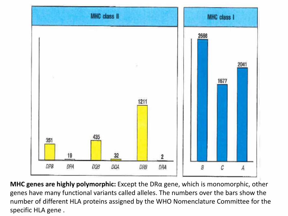
MHC genes are highly polymorphic: Except the DRα gene, which is monomorphic, other genes have many functional variants called alleles. The numbers over the bars show the number of different HLA proteins assigned by the WHO Nomenclature Committee for the specific HLA gene .

Allelic variation occurs mostly within the peptide binding region: Variability plots of the amino acid sequences of MHC molecules show that the variation arising from genetic polymorphism is restricted to the amino terminal α1 and α2 domains of MHC class I molecules. These are the domains that form the peptide binding cleft. Moreover, this variability is further clustered in amino acid positions on the floor of the cleft or inward from the helical walls.

In class II MHC molecules as well, the allelic variability is clustered in the α1 and β1 domains of MHC class II molecules, that form the peptide binding cleft. In this figure the variability for HLA-DR molecule is shown. The variability occurs only in the β chain since the α chain in this molecule is monomorphic. On the other hand, in HLA-DQ and –DP molecules, variability occurs in both chains and is restricted to the N-terminal domains only.
Slides 1-13 were reproduced from Janeway’s Immunobiology, 9th edition, Chapters 4 and 6
Summary MHC antigens bind to short peptides from proteins and present them on the cell surface for recognition by T cells. MHC class I molecules present peptides to CD8 T cells and MHC class II present peptides to CD4 T cells. T cells are activated only when they see peptide-MHC complexes on the surface of dendritic cells. Activated CD8 T cells then kill virus infected cells or tumor cells that present the same MHC-peptide complexes on the cell surface. Activated CD4 T cells provide help to other cells including CD8 T cells, macrophages and B cells
Multiple MHC class I and class II antigens are encoded in the individuals genome. Moreover, genes at each of these MHC loci are highly polymorphic. This results in as many as six MHC class I antigens and class II antigens being expressed by an individual. This ensures that an individual can present a diversity of peptides to T cells to ensure protection from pathogens. At the population level, high MHC polymorphism is advantageous such that some individuals of a population will always be able to respond to a given pathogen. This ensures survival of the population from challenges by new pathogens.
Cross-presentation is an important mechanism underlying the activation of T cells against viruses and tumors.
T cell recognition of virus-infected or tumor cells involves interaction with both MHC and the peptide bound in the cleft of the MHC molecule. This understanding underscores the importance of MHC antigens in tumor immune surveillance by T cells.

Garrido F et al Immunology Today 1997; 2: 89-95

Garrido F et al Immunology Today 1997; 2: 89-95

MHC expression in cells from regressing and progressing metastases from two melanoma patients
Carretaro R et al. Int J Cancer 2012; 131: 387-395

Representative staining for T cells in regressing and progressing metastases from same melanoma patient . Upper micrographs are from progressing metastases and lower from regressing metastases. These results show strong infiltration of regressing tumors by CD4 and CD8 T cells
Carretaro R et al. Int J Cancer 2012; 131: 387-395

Densities of infiltrating CD45RO (memory) T cells and CD8 T cells correlate with disease-free survival in colon cancer patients
Pages F et al J Clin Onco 2009; 27: 5944-5951

Densities of infiltrating CD45RO (memory) T cells and CD8 T cells correlate with overall survival in colon cancer patients
Pages F et al J Clin Onco 2009; 27: 5944-5951

These results provide robust evidence that HLA class I expression in tumors is necessary for tumor regression by T cell mediated immunotherapy. T cell infiltration of tumors is a useful biomarker for predicting clinical outcomes for cancer patients.

Adaptive immunity can control dormant tumor cells Clinical evidence has repeatedly pointed to the existence of dormant tumor cells

Adaptive immunity can control dormant tumor cells. Boebel, CM et al. Nature 2007; 903-908


Certain DNA and chromatin modifiers and cytokines can induce MHC expression in tumor cells that show loss of MHC class I expression

Induction of MHC class I expression by DNA methyl transferase (DNMT) inhibitor 5-azacytidine and by histone deacetylase (HDAC) inhibitor trichostatin A (TSA).

Induction of antigen processing presentation pathway and interferon-γ pathway molecules by 5-azacytidine and trichostatin A (TSA).
Simova J et al Br J Cancer 2011; 105:1533-41

Garrido F et al Immunology Today 1997; 2: 89-95
Soft versus Hard lesions