mhc class i stability is modulated by cell surface
TRANSCRIPT

pharmaceutics
Article
MHC Class I Stability is Modulated by Cell SurfaceSialylation in Human Dendritic Cells
Zélia Silva 1,†, Tiago Ferro 1,2,†, Danielle Almeida 1, Helena Soares 3 ,José Alexandre Ferreira 4,5 , Fanny M. Deschepper 1, Paul J. Hensbergen 6, Martina Pirro 6 ,Sandra J. van Vliet 7, Sebastian Springer 8 and Paula A. Videira 1,2,*
1 UCIBIO, Departamento Ciências da Vida, Faculdade de Ciências e Tecnologia, Universidade Nova de Lisboa,2829-516 Caparica, Portugal; [email protected] (Z.S.); [email protected] (T.F.);[email protected] (D.A.); [email protected] (F.M.D.)
2 CDG & Allies – PPAIN- Congenital Disorders of Glycosylation & Allies - Professionals and PatientAssociations International Network, 2829-516 Caparica, Portugal
3 Human Immunobiology and Pathogenesis, CEDOC-Chronic Diseases Research Centre, NOVA MedicalSchool, Faculdade de Ciências Médicas, Universidade Nova de Lisboa, 1150-082 Lisbon, Portugal;[email protected]
4 Experimental Pathology and Therapeutics Group, Portuguese Institute of Oncology,4200-162 Porto, Portugal; [email protected]
5 Porto Comprehensive Cancer Center (P.ccc), 4200-072 Porto, Portugal6 Center for Proteomics and Metabolomics, Leiden University Medical Center,
2300 RC Leiden, The Netherlands; [email protected] (P.J.H.); [email protected] (M.P.)7 Amsterdam UMC, Vrije Universiteit Amsterdam, Department of Molecular Cell Biology and Immunology,
Cancer Center Amsterdam, Amsterdam Infection and Immunity Institute, De Boelelaan 1117,1081 HzAmsterdam, The Netherlands; [email protected]
8 Department of Life Sciences and Chemistry, Jacobs University, 28759 Bremen, Germany;[email protected]
* Correspondence: [email protected]; Tel.: +351-212948530† These authors contributed equally to this work.
Received: 15 January 2020; Accepted: 6 March 2020; Published: 10 March 2020�����������������
Abstract: Maturation of human Dendritic Cells (DCs) is characterized by increased expression ofantigen presentation molecules, and overall decreased levels of sialic acid at cell surface. Here, weaimed to identify sialylated proteins at DC surface and comprehend their role and modulation. Massspectrometry analysis of DC’s proteins, pulled down by a sialic acid binding lectin, identified moleculesof the major human histocompatibility complex class I (MHC-I), known as human leucocyte antigen(HLA). After desialylation, DCs showed significantly higher reactivity with antibodies specific forproperly folded MHC-I-β2-microglobulin complex and for β2-microglobulin but showed significantlower reactivity with an antibody specific for free MHC-I heavy chain. Similar results for antibodyreactivities were observed for TAP2-deficient lymphoblastoid T2 cells, which express HLA-A*02:01.Using fluorescent peptide specifically fitting the groove of HLA-A*02:01, instead of antibody staining,also showed higher peptide binding on desialylated cells, confirming higher surface expression ofMHC-I complex. A decay assay showed that desialylation doubled the half-life of MHC-I moleculesat cell surface in both DCs and T2 cells. The biological impact of DC´s desialylation was evaluated inco-cultures with autologous T cells, showing higher number and earlier immunological synapses, andconsequent significantly increased production of IFN-γ by T cells. In summary, sialic acid contentmodulates the expression and stability of complex MHC-I, which may account for the improvedDC-T synapses.
Keywords: dendritic-cells; antigen-presentation; MHC-I; immunogenicity; T-cell response; cancer-vaccines
Pharmaceutics 2020, 12, 249; doi:10.3390/pharmaceutics12030249 www.mdpi.com/journal/pharmaceutics

Pharmaceutics 2020, 12, 249 2 of 21
1. Introduction
Dendritic cells (DCs) are the most potent antigen-presenting cells, bridging the innate and adaptiveimmune responses [1,2]. T cell stimulation is essential for setting up immune responses, which are onlyachieved by proper DC maturation. The development of immunotherapies based on DCs has longresorted to ex vivo differentiation from the monocyte precursors [3,4], especially due to their relativehigh abundance in peripheral blood compared to DCs. DC-based anticancer vaccines productionfollows these steps: autologous CD14+ monocytes derived from peripheral blood of cancer patientsare differentiated in vitro with GMCSF and IL-4 into immature DCs. After, immature DCs are loadedwith tumor antigens and then further matured through cytokine based maturation cocktails [4,5].Mature DCs are characterized by increased expression of antigen presentation molecules (e.g., MajorHistocompatibility Complex (MHC) class-I and –II), co-stimulatory receptors (e.g., CD40, CD80, CD86)and cytokine production. The antigen presentation by DCs depends on their migration into lymphnodes, and on the establishment of an intricate cell-cell communication that culminates in the formationof the immunological synapse with T cells [6]. Cytotoxic CD8+ T cell engagement starts by DCsdelivering the first signal through MHC-I. This early signaling step is considered to be of low affinity,as T cells simply “touch and go” DCs, unless a specific peptide presented via MHC molecules isrecognized by the T cell receptor (TCR) [7,8]. Typically, MHC-I presents peptides generated by thedegradation of endogenous proteins by the proteasome. These peptides are transported into theendoplasmic reticulum, where they are assembled with MHC-I heavy chain· complexed with β2microglobulin (β2m) and the resulting peptide:MHC-I (pMHC-I) transit to the cell surface. The peptideor the β2m may then dissociate from the heterotrimer complex resulting in the appearance of freeMHC-I heavy chain at the cell surface [9]. Free MHC-I heavy chains have reduced cell surface half-life,they do not activate T cells and they are typically internalized to enable the assembly with new peptides.Effector T cell activation is only triggered by the assembled heterotrimer and it can be evaluated by thesecretion of interferon (IFN)-γ, a hallmark of T cell-mediated immune responses [10,11].
The cell surface of all immune cells is coated by a complex assortment of glycans, characteristicallydecorated at their terminal position by sialic acids. These sialic acids comprise a broad family ofsugars derived from neuraminic acid, catalyzed by specific sialyltransferases. Due to their terminalposition, sialic acid-containing glycoproteins are frequently recognized by lectin receptors, whichinclude selectins and siglecs that modulate many biological processes such as cell signaling, cell-cellinteractions and migration [12]. The cell surface of human DCs has a high content of α2,6-sialylatedstructures, due to the expression of ST6Gal-I sialyltransferases [13–15]. The levels of sialic acid atDCs surface were shown to modulate the maturation status of both murine and human DCs [16].Previous results from our group showed that the temporary removal of surface sialic acids by sialidasetreatment increased the ability of human monocyte-derived DCs to activate T cells and to provideantigen-specific anti-tumoral responses [17]. Desialylated DCs have higher expression of MHC-I,MHC-II and co-stimulatory molecules (e.g., CD80, CD86), and higher secretion of pro-inflammatorycytokines, which altogether contribute to a superior TH1 anti-tumoral response. ST6Gal-I knockoutDCs also show increased capacity to activate T cells, further supporting that the ability of DCs toinduce TH1 anti-tumoral responses is improved by sialic acid removal [16,17]. Initially thought asmere induction of DC maturation [16] the fact is that the molecular mechanisms behind this outcomeare still poorly characterized.
In the present work, we have analyzed the molecular mechanism underlying sialic acid-inducedmodulation in DCs. We identified MHC-I as one of the major α2,6-sialylated molecules expressedin DCs. We showed that desialylated DCs have increased the number and stability of MHC-I heavychain I-β2m complexes at the cell surface. At the molecular level, we propose that increased stabilityof MHC-I at the cell surface is due to differences in MHC-I turnover. We propose that there is aslower dissociation of MHC-I-β2m-peptide complexes, lower endocytosis, and lower degradation.The resulting increased stability of MHC-I complexes at the cell surface may contribute to theirsuperior immunogenicity in eliciting CD8+ T cell-mediated responses. Our work paves the way to

Pharmaceutics 2020, 12, 249 3 of 21
new biochemical investigations aiming to get further insights into how the removal of α2,6-sialic acidsimpacts the expression and regulation of MHC-I on human DCs, which can be exploited to developanti-tumoral cell based-therapies.
2. Materials and Methods
2.1. Generation of Human DCs
Peripheral blood mononuclear cells (PBMCs) were isolated from buffy coats of healthy anonymousvolunteers provided by Instituto Português do Sangue e da Transplantação (IPST), after written andinformed donor consent IMP.74.52.4, according to the directive 2004/23/EC on setting standards ofquality and safety for the donation, procurement, testing, processing, preservation, storage, anddistribution of human tissues and cells (Portuguese Law 22/2007, June 29), with the approval of theethics committee of IPST 30072015 Monocytes were selected using anti-CD14 coated immunomagneticbeads (Miltenyi Biotech, Germany), and differentiated for 5 days into DCs, using Roswell ParkMemorial Institute (RPMI)-1640 medium, supplemented with 10% fetal bovine serum (FBS), 2 mMGlutaMAX, 1 mM sodium pyruvate, non-essential amino acids and 100 µg/mL penicillin/streptomycin,all from Gibco/Thermo Fisher Scientific (Waltham, MA, USA) in the presence of human recombinantGM-CSF (1000 U/mL) and IL-4 (750 U/mL) (Miltenyi Biotech, Germany) [17,18]. When appropriate,DCs maturation was induced after differentiation by the addition of 5 µg/mL of lipopolysaccharide(LPS) or 1000 U/mL of IL-1β, IFN-γ or TNF-α.
2.2. Cell Lines
The HLA-A*02:01 positive leukemia cell line T2 (ATCC® CRL-1992™) was kindly provided byProfessor Y. van Kooyk (VU University Medical Center, Amsterdam, The Netherlands). Cells were keptin culture using RPMI-1640 medium supplemented with 10% FBS, 2 mM GlutaMAX and 100 µg/mLpenicillin/streptomycin. The colon cancer carcinoma cancer cell line SW948 (ATCC® CCL-237™) waskindly provided by Professor Fabio Dall’Olio Department of Experimental Diagnostic and SpecialtyMedicine, University of Bologna, Bologna, Italy). Cells were cultured in Leibovitz’s L-15 mediumsupplemented with 10% FBS.
2.3. Sialidase Treatment
Enzymatic removal of sialic acid from the cell surface was performed using sialidase fromClostridium perfringens (Roche Diagnostics, Basel, Switzerland). Cells (5 × 10⁶/mL) were treated using100 mU sialidase, in RPMI-1640 media, for 60 min, at 37 ◦C. When appropriate, enzymatic removal ofsialic acid from total cell lysates was performed with 20 mU of sialidase per 100 µg of total protein, indigestion buffer (50 mM sodium acetate, 5 mM CaCl2, pH 5.5), overnight at 37 ◦C.
2.4. Anti-MHC-I antibodies
Table 1 shows the list of monoclonal antibodies against major human histocompatibility complexclass I (MHC-I) that were used in this study.
Pharmaceutics 2020, 12, 249 4 of 21
Table 1. List of anti-MHC-I antibodies used in this study.
Antibody Clone Specificity Technique Source Reference
W6/32
Recognizes antigenicdeterminant common to
HLA-A, B and C antigenswhen in their
three-dimensionalconfiguration
FC/WBandIP
ImmunoTools,Germany/Pierce,Thermo Fisher
Scientific (Waltham,MA, USA)
[19]
HC10
Recognizes free HLA class Iheavy chains. HC10 reacts
mostly with HLA-B andHLA-C heavy chains andsome HLA-A (HLA-A10,
HLA-A28, HLA-A29,HLA-A30, HLA-A31,HLA-A32, HLA-A33)
FCandWB
Hybridomasupernatants [20]
BBM.1 Recognizes both free andclass I bound β2m
FCandWB
Hybridomasupernatants [21]
BB7.2
Recognizes the α subunit ofHLA-A2 which is a subset of
MHC-class I moleculesencoded by A*02 alleles
FC BD Bioscience (CA,USA) [22]
246-B8.E7Recognizes monomorphic
determinant of human MHC-I antigens (HLA-A, B, and C)
CMThermo Fisher
Scientific (Waltham,MA, USA)
Not found
Abbreviations: MHC-I, major human histocompatibility complex class I, HLA, human leucocyte antigen, FC, FlowCytometry; WB, Western blot; CM, Confocal Microscopy; IP, immunoprecipitation.
2.5. Flow Cytometry
Anti-MHC-I monoclonal antibodies are listed in Table 1 (see above). Mouse monoclonalhybridoma supernatants, HC10 and BBM.1, were used in a 1:4 dilution, and commercial antibodieswere used following vendor instructions. For flow cytometry analysis, we also stained cells withFITC-conjugated Sambucus nigra (SNA) lectin (Vector Labs) and the fluorescently FITC-labeledpeptides NLVPKFITCVATV and SIINFEKFITCL (Genecust). Cells were harvested and washed beforestaining with appropriate fluorophore-conjugated antibodies for flow cytometry. For peptide staining,a final concentration of 5 µM was used. Staining was performed at 4 ◦C for 30 min. Cells were thenwashed and fixed in 2% paraformaldehyde. Acquisition of data was performed on an Attune AcousticFocusing Cytometer (Applied Biosystems, Waltham, MA, USA) and the FlowJo software version 10.0.5(TreeStar, San Carlos, CA, USA) was used to analyze the mean fluorescent intensity (MFI) and cellpercentages. The MFI represents the median fluorescence as calculated by the software. For eachstaining condition, the respective MFI of unstained/isotype control was subtracted.
2.6. Confocal Laser Scanning Microscopy
To perform confocal laser scanning microscopy, cells were plated on 12-mm diameterpolylysine-coated glass coverslips and incubated for 5 min at room temperature. Coverslipswere then centrifuged at 100 × g for 1 min to promote cell adhesion, fixed for 30 min with 4%paraformaldehyde (PFA) and washed using 1% bovine serum albumin (BSA) in PBS. Mouse anti-humanHLA-ABC, clone 246-B8.E7 (see Table 1, above) was used for staining human MHC-I, followed bya fluorescently-conjugated secondary antibody. FITC-conjugated SNA lectin was also used to stainα2,6-linked sialic acids on the cell surface. Images were acquired on a Zeiss LSM710 confocal microscope(Zeiss, Oberkochen, Germany). Illustrative confocal cross-section pictures were selected after Z-stackingprocessing. Staining intensity was analytically quantified using the corrected total cell fluorescence(CTCF) = Integrated Density − (Area of selected cell ×Mean fluorescence of background readings).

Pharmaceutics 2020, 12, 249 5 of 21
2.7. Protein Extraction, Immunoprecipitation, and Western Blotting
Whole cell lysates were obtained using Immunoprecipitation (IP) lysis buffer and the concentrationof protein determined using the BCA Protein Assay Kit, all from Pierce, Thermo Fisher Scientific(Waltham, MA, USA). For protein affinity separation, an SNA-agarose column (EY Labs, San Mateo,CA, USA) was loaded with whole cell lysates. Unbound proteins were eluted using PBS and boundproteins were eluted using 0.1 M lactose in PBS. The collected bound protein fraction was concentratedusing an Amicon Ultracel 3K column (Millipore) and both fractions were stored for further use.
IP of MHC-I protein were obtained from total protein extracts using clone W6/32 [16] (see Table 1,above) and Direct IP Kit from Pierce.
For Western blotting, protein samples were separated in SDS-PAGE gels, transferred to 0.45 µmnitrocellulose membrane (GE Healthcare Life Sciences, Piscataway, NJ, USA) and blocked with 7.5%non-fat milk. To detect MHC-I, an anti-HLA-ABC (W6/32) antibody and HC10 hybridoma supernatantwere used as primary antibodies. In both cases, goat anti-mouse IgG1 heavy chain horseradishperoxidase (HRP) conjugate (Abcam, Cambridge, UK) was used as the secondary antibody. Forlectin blotting, membranes were blocked overnight with 1% CarboFree (Vector Labs, Burlingame, CA,USA) and HRP conjugated-SNA lectin was used to probe the membrane. The Amersham ECL PrimeWestern Blotting Detection Reagent (GE Healthcare Life Sciences, Piscataway, NJ, USA) was used asa developing reagent. β-actin was used as endogenous control to quantify the relative amount ofprotein expression.
2.8. Mass Spectrometry and Bioinformatics
For glycoproteomics analysis, IP of MHC-I protein from DCs was separated on a 4–12% gradientSDS-PAGE gel (Thermo Fisher Scientific) under reducing conditions and subsequently stained withSimplyBlue Safe Stain (Thermo Fisher Scientific) and washed with distilled water. Protein bands ofinterest (between 38 and 49 kDa) were excised from the gel and proteins were subsequently in-geldigested with trypsin (Promega, WI, USA) after reduction (10 mM 1,4-dithiothreitol (Sigma Aldrich))and alkylation (50 mM iodoacetamide (Sigma Aldrich).
For LC-MS/MS analysis, tryptic digests were separated by online C18 nano-HPLC MS/MS with anEasy nLC 1000 gradient HPLC system (Thermo, Bremen, Germany) coupled to an Orbitrap FusionLumos mass spectrometer (Thermo), as previously described [23]. Fractions were loaded onto ahomemade precolumn (100 µm × 15 mm; Reprosil-Pur C18-AQ 3 µm, Dr. Maisch, Ammerbuch,Germany) and eluted via a homemade analytical nano-HPLC column (15 cm × 50 µm; Reprosil-PurC18-AQ3 µm) with a gradient from 10% to 40% of solvent B (20/80/0.1 water/acetonitrile/FA v/v/v) for20 min. The nano-HPLC column was drawn to a tip of ∼5 µm, which acted as the electrospray needleof the MS source. The Lumos mass spectrometer was operated in data-dependent Higher-energyCollisional Dissociation (HCD) MS/MS (top-10 mode) using at a normalized collision energy of 32%and recording of the MS2 spectrum in the Orbitrap. At the master scan (MS1) level, the resolution was120,000 and the scan range was m/z 400−1500 at an AGC target of 400,000 with maximum injectiontime of 50 ms. Dynamic exclusion was applied after n = 1, with exclusion duration of 10 s, with masstolerance of 10 ppm. For MS2, charge states 2−5 were included and precursors were isolated with thequadrupole with an isolation window of 1.2 Da. The MS2 scan resolution was 30,000 with an AGCtarget of 50,000 and maximum injection time of 60 ms.
For proteomics analysis, 10 µg of the protein pool was separated by 4–16% gradient SDS/PAGE(BioRad) under reducing conditions; the bands were excised from the gels, and proteins were reducedwith 5 mM 1,4-dithiothreitol (Sigma Aldrich) for 40 min. at 60 ◦C, alkylated with 10 mM iodoacetamide(Sigma Aldrich) for 45 min. in the dark and digested with trypsin (Promega, WI, USA) overnightat 37 ◦C for mass spectrometry (MS) analysis. Protein identification was performed as previouslydescribed [24] Tryptic digests were separated with a C18 Pepmap (Dionex) column on an Ultimate3000 (Dionex/LC Packings, Sunnyvale, CA) nano-HPLC, and fractions were collected with a Probot(Dionex/LC Packings, Sunnyvale, CA) directly onto a matrix-assisted laser desorption ionization

Pharmaceutics 2020, 12, 249 6 of 21
(MALDI) plate. MS was performed on a 4800 MALDI-TOF/TOF Analyzer (Applied Biosystems, FosterCity, CA). The MS and MS/MS spectra acquired were processed and analyzed by the Global ProteinServer Workstation (Applied Biosystems). LC-MALDI-MS/MS runs were done in duplicates. Proteinidentification was achieved with a search performed against the Swiss-Prot protein database (March2009, 428,650 entries) for Homo sapiens. The final list includes proteins queried using the “RetrieveID/mapping” tool of UniProtKB for membrane glycoproteins with extracellular domain prone toexhibit or secreted glycoproteins, as previously described [25]. The presence of glycosylation sites wasconfirmed in silico with NetNglyc 1.0 server, an artificial neural network that examines the sequencecontext of Asn-X-Ser/Thr (where X is not Pro) sequons [26], and with NetOglyc 4.0 server, that producesneural network predictions of mucin-type GalNAc O-glycosylation sites in mammalian proteins [27].Glycoproteins biological and molecular functions were annotated based on gene ontology (GO) termsusing STRAP 1.5 (Software Tool for Rapid Annotation of Proteins: Cardiovascular Proteomics Center,Boston University School of Medicine, Boston, MA, USA).
2.9. Peptide Stability Assays
For peptide stability binding assays, T2 cells were incubated with HLA-A*02:01 matching peptides(YLEPGPVTA from gp100 or NLVPMVATV from CMVpp65 antigens) in concentrations ranging from1 to 100 µM, and 10 µg/mL of β2m (Sigma-Aldrich, St. Louis, MO, USA), in RPMI-1640 medium.Controls without peptide were performed in parallel. The sialic acid removal was performed with100 mU sialidase for 1 h at 37 ◦C immediately before peptide incubation. For sialic acid addition otherexperiments, cells were incubated in the same conditions, with 2.1 mU recombinant human ST6Gal-Iand 50 µM CMP-sialic acid (Sigma-Aldrich), instead of sialidase. Recombinant ST6Gal-I was a kind giftfrom Professor Joseph Lau (Roswell Park Comprehensive Cancer Center, Buffalo, NY, USA), preparedas described elsewhere [28]. After 3 h, cells were harvested and analyzed by flow cytometry for theexpression of MHC-I, using an anti-HLA-A2 antibody (BB7.2).
2.10. Surface MHC-I Stability Assays
DCs or T2 cells were treated with 5 µg/mL of the Golgi-export inhibitor Brefeldin A (BFA)(Sigma-Aldrich) and incubated at 37 ◦C, in the presence of RPMI-1640 medium, for 5 h. When treatingcells with sialidase, 100 mU of this enzyme was added simultaneously with BFA. After 1 h, the sialidasewas removed by centrifugation and the cells were plated again with RPMI-1640 supplemented with5 µg/mL BFA during the course of the experiments. At each time point, cells were harvested andthe cell surface expression of MHC-I molecules was assessed by staining cells with an anti-HLA-A2antibody (BB7.2) and analyzed by flow cytometry.
2.11. Gene Expression Assays
Total RNA was extracted from 1–5 × 10⁶ DCs or T2 cells using the NZY Total RNA Isolationkit (NZYTech, Lisbon, Portugal). Genomic DNA was removed using the RNase-Free DNase Set(Qiagen, Germany), following manufacturers’ instructions. RNA concentration from each sample wasdetermined spectrophotometrically and then reverse transcribed into cDNA using random primersand the High Capacity cDNA Reverse Transcription Kit (Applied Biosystems, Foster City, CA).
Real-time PCR was performed in a Rotor-Gene 6000 (Corbett Life Science, Sydney, Australia) usingTaqMan® Fast Universal PCR Master Mix. The assays containing both TaqMan® probes and primers(Thermo Fisher Scientific, Waltham, MA, USA) for a specific gene were: HLA-A (human leukocyteantigen A allele; Hs01058806_g1), β-actin (4352935E) and GAPDH (glyceraldehyde 3-phosphatedehydrogenase; 4333764F) were used. Each reaction was performed in duplicate.
Messenger RNA (mRNA) expression was normalized using the geometric mean of the expressionof two endogenous controls, β-actin and GAPDH, as a reference. The relative expression of eachgene was calculated using the cycle threshold (Ct) method and compared accordingly to the 2−∆∆Ct

Pharmaceutics 2020, 12, 249 7 of 21
equation [29]. The efficiency of the amplification reaction for each primer/probe was above 95% (asdetermined by the manufacturer).
2.12. DC: T Cell Interaction/ Doublet Analysis
For DC: T cell doublet analysis, DCs were generated as previously described and autologousCD8+ T cells were isolated by immunomagnetic separation using anti-human CD8 beads (Miltenyi,Germany). DCs were fluorescently labeled with CellTrace™ Far Red (Thermo Fisher) and CD8+ Tcells were fluorescently labeled with CellTrace™ carboxyfluorescein succinimidyl ester (CFSE, ThermoFisher) and co-cultured (1.1 × 10⁶ cells/mL) in a 96-well plate. The ratio of DCs to T cells was 1:10 in atotal volume of 200 µL of RPMI-1640 medium. After 6 or 24 h of co-culture, cells were collected, gentlypippeted and analyzed using an Attune Acoustic Focusing Cytometer (Applied Biosystems, USA). Thegating strategy excluded single cells, and DC: T cell doublets were positively identified when the twofluorescences were simultaneously co-localized.
2.13. Cytokine Production Evaluation
To evaluate intracellular cytokine production, DCs were co-cultured with autologous CD8+ T cells(1:10 ratio) for 24 or 48 h, as described in the previous section. BFA was used to block cytokine secretionfor the last 4 h of co-culture within the referred period. Cells were then fixed and permeabilized usingthe BD Cytofix/CytopermTM kit (BD Pharmingen, CA, USA). Cell staining was performed using aPE-conjugated anti-human IFN-γ antibody, clone B27 (Biolegend) and a PerCP-conjugated anti-humanCD8 antibody, clone HIT8a (Immunotools, Germany). After washing steps, cells were analyzed byflow cytometry. Untreated DCs were used as control.
2.14. Statistical Analysis
Statistical analysis was performed using the GraphPad Prism 6.0 software (GraphPad Software,La Jolla, CA, USA). Unless otherwise stated, statistical significance (p value) was calculated usingthe two-tailed paired t-test. Statistical significance was defined as p < 0.05 (*), p < 0.01 (**) and p <
0.001 (***).
3. Results
3.1. MHC Class I Molecules on Human Dcs are Sialylated
The expression of α2,6-sialic acids by human DCs was evaluated by the reactivity with SNAlectin, which preferentially recognizes α2,6-linked sialic acids. Data showed that DCs are highlyα2,6 sialylated. During maturation, the α2,6 sialylation pattern is altered, decreasing significantlywith IFN-γ and being only slightly modulated with the pro-inflammatory stimulus IL-1β, TNF-αand LPS (Figure 1A). The data is in agreement with previous reports [13,14] and suggests that whensurface sialylated proteins change their sialic acid content, they also change their behavior. To identifyα2,6-sialylated proteins expressed by DCs, we immunoprecipitated proteins from total DCs lysatesusing the SNA lectin. Isolated SNA-binding proteins were identified by Mass Spectrometry (Figure 1Band Supplementary Figure S1) and the proteins with higher scores (Table S1) were matched, associatedby Gene Ontology and sorted accordingly to different biochemical functions. Considering onlymembrane-associated proteins, the main cellular function of the eluted fraction was related to immunesystem processes (GO: 0002376) (26% of all identified proteins) (Figure 1C). The protein with higherscore sequence coverage were alleles of the HLA-A molecules, followed by HLA-B and HLA-C alleles,all known for their high degree of polymorphism and sequence homology (Table S1).

Pharmaceutics 2020, 12, 249 8 of 21
1
Figure 1. Mass spectrometry analysis of SNA-binding proteins isolated from DCs reveals keyimmune-related proteins. (A) The reactivity of Sambucus nigra (SNA) lectin to α2,6-sialylated glycanson the surface of human DCs was quantified by flow cytometry after different maturation stimuli:IFN-γ, IL-1β, TNF-α and LPS. Unstimulated DCs were used as control. Values presented are mean ±SEM (N = 6). Statistically significant differences are indicated by asterisks (* p ≤ 0.05). (B) Schematicrepresentation of the steps followed to identify α2,6-sialylated proteins from DCs. Whole cell lysatesof human DCs were immunoprecipitated through a SNA-binding column. The eluted proteins wereanalyzed by mass spectrometry and the corresponding identified scores were matched and associatedwith Gene Ontology (GO) entries. (C) Distribution of the identified sialylated proteins by their molecularfunction. Pie chart represents different molecular functions of the identified proteins, according to theGO entries. Immune system processes were highlighted.
To evaluate the sialylation of MHC-I molecules, these were immunoprecipitated from DCs ofdifferent individuals and probed with SNA lectin on Western blot. Immunoprecipitates showed astrong SNA binding, demonstrating the presence of α2,6-sialylated structures on MHC class I heavychains (Figure 2A). To further confirm that MHC-I molecules in DCs were sialylated, whole cell lysateswere treated with sialidase, electrophoresed and stained with an anti-HLA-ABC antibody W6/32 (seeTable 1) on Western blot (Figure 2B). The resulting blots showed a band with a molecular weight ofapproximately 40–45 kDa, similar to the predicted molecular weight of the MHC-I heavy chain; themolecular weight of this band was decreased when proteins were desialylated, suggesting that MHC-Iheavy chain is decorated with sialic acid (Figure 2B). Decreased molecular weight of the MHC-I heavychain after sialidase treatment of whole cell lysate was also confirmed with the antibody HC10, whichis specific for the free MHC-I heavy chain (Supplementary Figure S2). These results suggest thatα2,6-sialylation is a common feature among different heavy chain alleles of MHC-I.
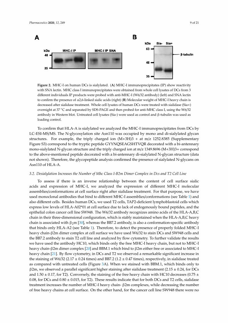
Pharmaceutics 2020, 12, 249 9 of 21Pharmaceutics 2020, 12, x 9 of 20
329 Figure 2. MHC-I on human DCs is sialylated. (A) MHC-I immunoprecipitates (IP) show reactivity 330 with SNA lectin. MHC class I immunoprecipitates were obtained from whole cell lysates of DCs 331 from 3 different individuals IP products were probed with anti-MHC-I (W6/32 antibody) (left) and 332 SNA lectin to confirm the presence of α2,6-linked sialic acids (right) (B) Molecular weight of MHC-I 333 heavy chain is decreased after sialidase treatment. Whole cell lysates of human DCs were treated 334 with sialidase (Sia+) overnight at 37 ºC and separated by SDS-PAGE and then probed for anti-MHC 335 class I, using the W6/32 antibody in Western blot. Untreated cell lysates (Sia-) were used as control 336 and β-tubulin was used as loading control. 337
To confirm that HLA-A is sialylated we analyzed the MHC-I immunoprecipitates from DCs by 338 LC-ESI-MS/MS. The N-glycosylation site Asn110 was occupied by mono and di-sialylated glycan 339 structures. For example, the triply charged ion (M+3H)3 + at m/z 1252.8385 (Supplementary Figure 340 S3) correspond to the tryptic peptide GYYNQSEAGSHTVQR decorated with a bi-antennary 341 mono-sialylated N-glycan structure and the triply charged ion at m/z 1349.8696 (M+3H)3+ 342 correspond to the above-mentioned peptide decorated with a bi-antennary di-sialylated N-glycan 343 structure (data not shown). Therefore, the glycopeptide analysis confirmed the presence of sialylated 344 N-glycans on Asn110 of HLA-A. 345
3.2. Desialylation Increases the Number of Mhc Class I-Β2m Dimer Complex in Dcs and T2 Cell Line 346 To assess if there is an inverse relationship between the content of cell surface sialic acids and 347
expression of MHC-I, we analyzed the expression of different MHC-I molecular 348 assemblies/conformations at cell surface right after sialidase treatment. For that purpose, we have 349 used monoclonal antibodies that bind to different MHC-I assemblies/conformations (see Table 1) 350 and also different cells. Besides human DCs, we used T2 cells, TAP2-deficient lymphoblastoid cells 351 which express low levels of HLA-A02*01 at cell surface due to lack of endogenously bound peptides, 352 and the epithelial colon cancer cell line SW948. The W6/32 antibody recognizes amino acids of the 353 HLA-A,B,C chain in their three-dimensional configuration, which is stably maintained when the 354 HLA-A,B,C heavy chain is associated with β2m [30], whereas the BB7.2 antibody, is also a 355 conformation-specific antibody that binds only HLA-A2 (see Table 1). Therefore, to detect the 356 presence of properly folded MHC-I heavy chain-β2m dimer complex at cell surface we have used 357 W6/32 to stain DCs and SW948 cells and the BB7.2 antibody to stain T2 cell line and analyzed by flow 358 cytometry. To further validate the results we have used the antibody HC10, which binds only the 359 free MHC-I heavy chain, but not to MHC-I heavy chain-β2m dimer complex [20]and BBM.1 which 360 bind to β2m either free or associated to MHC-I heavy chain [21]. By flow cytometry, in DCs and T2 361 we observed a remarkable significant increase in the staining of W6/32 (2.17 ± 0.24 times) and BB7.2 362 (1.2 ± 0.47 times), respectively, in sialidase treated as compared with untreated cells (Figure 3A). 363 When we stained with BBM.1, which binds only to β2m, we observed a parallel significant higher 364 staining after sialidase treatment (2.15 ± 0.24, for DCs and 1.50 ± 0.17, for T2). Conversely, the 365 staining of the free heavy chain with HC10 decreases (0.75 ± 0.08, for DCs and 0.80 ± 0.015, for T2). 366 These results indicate that for both DCs and T2 cells, sialidase treatment increases the number of 367 MHC-I heavy chain- β2m complexes, while decreasing the number of free heavy chains at cell 368 surface. On the other hand, for the cancer cell line SW948 there were no significant differences in the 369 staining for any of the antibodies for MHC-I molecules, demonstrating that the increase in MHC-I 370
Figure 2. MHC-I on human DCs is sialylated. (A) MHC-I immunoprecipitates (IP) show reactivitywith SNA lectin. MHC class I immunoprecipitates were obtained from whole cell lysates of DCs from 3different individuals IP products were probed with anti-MHC-I (W6/32 antibody) (left) and SNA lectinto confirm the presence of α2,6-linked sialic acids (right) (B) Molecular weight of MHC-I heavy chain isdecreased after sialidase treatment. Whole cell lysates of human DCs were treated with sialidase (Sia+)overnight at 37 ◦C and separated by SDS-PAGE and then probed for anti-MHC class I, using the W6/32antibody in Western blot. Untreated cell lysates (Sia-) were used as control and β-tubulin was used asloading control.
To confirm that HLA-A is sialylated we analyzed the MHC-I immunoprecipitates from DCs byLC-ESI-MS/MS. The N-glycosylation site Asn110 was occupied by mono and di-sialylated glycanstructures. For example, the triply charged ion (M+3H)3 + at m/z 1252.8385 (SupplementaryFigure S3) correspond to the tryptic peptide GYYNQSEAGSHTVQR decorated with a bi-antennarymono-sialylated N-glycan structure and the triply charged ion at m/z 1349.8696 (M+3H)3+ correspondto the above-mentioned peptide decorated with a bi-antennary di-sialylated N-glycan structure (datanot shown). Therefore, the glycopeptide analysis confirmed the presence of sialylated N-glycans onAsn110 of HLA-A.
3.2. Desialylation Increases the Number of Mhc Class I-B2m Dimer Complex in Dcs and T2 Cell Line
To assess if there is an inverse relationship between the content of cell surface sialicacids and expression of MHC-I, we analyzed the expression of different MHC-I molecularassemblies/conformations at cell surface right after sialidase treatment. For that purpose, we haveused monoclonal antibodies that bind to different MHC-I assemblies/conformations (see Table 1) andalso different cells. Besides human DCs, we used T2 cells, TAP2-deficient lymphoblastoid cells whichexpress low levels of HLA-A02*01 at cell surface due to lack of endogenously bound peptides, and theepithelial colon cancer cell line SW948. The W6/32 antibody recognizes amino acids of the HLA-A,B,Cchain in their three-dimensional configuration, which is stably maintained when the HLA-A,B,C heavychain is associated with β2m [30], whereas the BB7.2 antibody, is also a conformation-specific antibodythat binds only HLA-A2 (see Table 1). Therefore, to detect the presence of properly folded MHC-Iheavy chain-β2m dimer complex at cell surface we have used W6/32 to stain DCs and SW948 cells andthe BB7.2 antibody to stain T2 cell line and analyzed by flow cytometry. To further validate the resultswe have used the antibody HC10, which binds only the free MHC-I heavy chain, but not to MHC-Iheavy chain-β2m dimer complex [20] and BBM.1 which bind to β2m either free or associated to MHC-Iheavy chain [21]. By flow cytometry, in DCs and T2 we observed a remarkable significant increase inthe staining of W6/32 (2.17 ± 0.24 times) and BB7.2 (1.2 ± 0.47 times), respectively, in sialidase treatedas compared with untreated cells (Figure 3A). When we stained with BBM.1, which binds only toβ2m, we observed a parallel significant higher staining after sialidase treatment (2.15 ± 0.24, for DCsand 1.50 ± 0.17, for T2). Conversely, the staining of the free heavy chain with HC10 decreases (0.75 ±0.08, for DCs and 0.80 ± 0.015, for T2). These results indicate that for both DCs and T2 cells, sialidasetreatment increases the number of MHC-I heavy chain- β2m complexes, while decreasing the numberof free heavy chains at cell surface. On the other hand, for the cancer cell line SW948 there were no

Pharmaceutics 2020, 12, 249 10 of 21
significant differences in the staining for any of the antibodies for MHC-I molecules, demonstratingthat the increase in MHC-I levels induced by sialidase removal seems to be restricted to leucocytes(Figure 3A). The efficient enzymatic removal of sialic acids from the cell surface was always confirmedby lectin staining and flow cytometry analysis (results not shown).
Pharmaceutics 2020, 12, x 10 of 20
levels induced by sialidase removal seems to be restricted to leucocytes (Figure 3A). The efficient 371 enzymatic removal of sialic acids from the cell surface was always confirmed by lectin staining and 372 flow cytometry analysis (results not shown). 373
To further investigate the influence of sialic acid removal on the distribution of MHC-I 374 molecules at the cell surface, DCs were stained with 246-B8.E7, which binds to MHC-I heavy 375 chain-β2m complex (see Table 1) and imaged by confocal microscopy after sialidase treatment. The 376 expression of MHC-I molecules increased homogenously at cell surface after sialic acid removal, 377 while SNA lectin binding is nearly absent (Figure 3B). Image processing with matching staining 378 intensity showed significantly increased fluorescence of MHC-I molecules on sialidase-treated cells. 379 In fact, there was a statistically significant increase of MHC-I CTCF on desialylated human DCs, 380 compared with untreated cells (CTCF 229,699 ± 136,268 vs. 60,644 ± 16,127) (Figure 3C). 381
382 Figure 3. Desialylation increases MHC class I molecules in DCs and T2 cell line but not in the SW948 383 colon cancer cell line. (A) DCs, T2 cell line, and SW948 cell line were treated with sialidase for 1 h at 384 37ºC and analyzed by flow cytometry for MHC-I staining (using the HC10 (free heavy chain); β2m 385 (BBM.1 antibody) and the complex heavy chain+β2m (BB7.2 for T2 and W6/32 antibody for MoDCs 386 and SW948) (B) Confocal microscopy pictures of immature human DCs probed for surface staining 387 of MHC-I (W6/32 antibody). Human DCs were treated with sialidase and prepared on coverslips for 388 confocal microscopy. A range of z-stack images was collected from different cells and processed to 389 include mean staining intensity. Scale bar equals 20 μm. (C) The fluorescence of MHC-I by confocal 390 microscopy was quantified accordingly to the corrected total cell fluorescence (CTCF) method on at 391 least 14 different cells per slide, as described in the Methods section. Values presented are mean ± 392 SEM (N≥5). Statistically significant differences are indicated by asterisks (* p ≤ 0.05, ** p ≤ 0.01, *** p ≤ 393 0.001). 394
3.3. MHC Class I Molecules Show Increased Peptide Stability at the Cell Surface after Desialylation 395 To investigate the mechanisms that regulate the immediate increase in MHC-I expression at the 396
DCs surface, after sialidase treatment, a TAP-deficient cell line (T2) was used to study MHC-I 397 stability. T2 cells are HLA-A*02:01 positive cells, expressing low levels of this MHC-I allele at cell 398 surface due to their inability to efficiently load peptides into the endoplasmic reticulum (ER), 399
Figure 3. Desialylation increases MHC class I molecules in DCs and T2 cell line but not in the SW948colon cancer cell line. (A) DCs, T2 cell line, and SW948 cell line were treated with sialidase for 1 h at37 ◦C and analyzed by flow cytometry for MHC-I staining (using the HC10 (free heavy chain); β2m(BBM.1 antibody) and the complex heavy chain+β2m (BB7.2 for T2 and W6/32 antibody for MoDCsand SW948) (B) Confocal microscopy pictures of immature human DCs probed for surface stainingof MHC-I (W6/32 antibody). Human DCs were treated with sialidase and prepared on coverslips forconfocal microscopy. A range of z-stack images was collected from different cells and processed toinclude mean staining intensity. Scale bar equals 20 µm. (C) The fluorescence of MHC-I by confocalmicroscopy was quantified accordingly to the corrected total cell fluorescence (CTCF) method on atleast 14 different cells per slide, as described in the Methods section. Values presented are mean ± SEM(N ≥ 5). Statistically significant differences are indicated by asterisks (* p ≤ 0.05, ** p ≤ 0.01, *** p ≤ 0.001).
To further investigate the influence of sialic acid removal on the distribution of MHC-I moleculesat the cell surface, DCs were stained with 246-B8.E7, which binds to MHC-I heavy chain-β2m complex(see Table 1) and imaged by confocal microscopy after sialidase treatment. The expression of MHC-Imolecules increased homogenously at cell surface after sialic acid removal, while SNA lectin binding isnearly absent (Figure 3B). Image processing with matching staining intensity showed significantlyincreased fluorescence of MHC-I molecules on sialidase-treated cells. In fact, there was a statisticallysignificant increase of MHC-I CTCF on desialylated human DCs, compared with untreated cells (CTCF229,699 ± 136,268 vs. 60,644 ± 16,127) (Figure 3C).
3.3. MHC Class I Molecules Show Increased Peptide Stability at the Cell Surface after Desialylation
To investigate the mechanisms that regulate the immediate increase in MHC-I expression at theDCs surface, after sialidase treatment, a TAP-deficient cell line (T2) was used to study MHC-I stability.T2 cells are HLA-A*02:01 positive cells, expressing low levels of this MHC-I allele at cell surface due to

Pharmaceutics 2020, 12, 249 11 of 21
their inability to efficiently load peptides into the endoplasmic reticulum (ER), hindering the formationof stable peptide:MHC-I:β2m complexes [31]. In the presence of peptides that match the MHC-I heavychain pocket fold, the complexes of MHC-I and β2m are stabilized, resulting in increased expressionat the cell surface of T2 cells. To assess the effect of desialylation, the CMVpp65 (NLVPMVATV) andgp100 (YLEPGPVTA) peptides, which match the HLA-A*02:01 pocket [32,33] were used in T2 cellstreated or not treated with sialidase. As shown in Figure 4, sialidase treated T2 cells show a generalhigher staining of HLA-A*A02 molecules at the cell surface, when incubated with either peptide. Forthe gp100 peptide experimental assay, a statistically significant increase in HLA-A*A02 moleculeswas observed when desialylated T2 cells were incubated with peptide concentrations of 1 µM (MFIfold increase 2.21 ± 0.38 vs. 1.05 ± 0.06) and 10 µM (MFI fold increase 2.59 ± 0.35 vs. 1.29 ± 0.10),compared to untreated cells (Figure 4A). Similarly, desialylated T2 cells incubated with CMVpp65peptide, also showed a significant increase in HLA-A*A02 molecules at the cell surface (Figure 4B,D) forall peptide concentrations. Additional experiments using the recombinant sialyltransferase ST6Gal-Ifor extrinsic α2,6-sialylation of T2 cells followed by incubation with the CMVpp65 peptide showedreduced expression of MHC-I at the cell surface after treatment (Figure 4C).
1
Figure 4. MHC class I molecules stability at cell surface after desialylation. T2 cells were used to studyMHC-I stability after sialic acid removal from cell membrane. T2 cells were incubated with either(A) gp100 peptides or (B) CMVpp65 peptides, which match the HLA-A*02:01 pocket and stabilizethe peptide-MHC-I-β2 complex. Cells were then probed with anti-HLA-A*02 antibody (BB7.2) andanalyzed by flow cytometry. (C) T2 cells were extrinsically sialylated with a recombinant ST6Gal-Ienzyme and incubated with the CMVpp65 peptide. Cells were then probed by flow cytometry forHLA-A*02 (using the BB7.2 antibody) expression at the cell surface. Graphs show normalized meanfluorescent intensity (MFI) values of at least 4 independent experiments. (D) Representative flowcytometry histograms of T2 cells untreated (in blue) or after sialidase treatment (in orange) and incubatedwith different concentrations of CMVpp65 peptide. (E) The fluorescently labeled NLVPKFITCVATVpeptide was used to stain HLA-A2 on desialylated T2 cells and the fluorescently labeled SIINFEKFITCLpeptide was used as irrelevant control. The quantification of the mean fluorescence intensity offluorescently labeled peptides presented by T2 cells is also presented. Values presented are mean ± SDof at least N = 4 independent experiments. Statistically significant differences are indicated by asterisks(* p ≤ 0.05, ** p ≤ 0.01).
These results suggest that the stability of HLA-A*A02-peptide complexes on the cell membraneis influenced by cell surface sialylation. To exclude the possibility that desialylation lead to anincreased anti-MHC-I antibody binding affinity (reactivity), we used a fluorescently labeled peptide,the CMVpp65 peptide NLVPKFITCVATV, that matches and binds to the HLA-A*A02 pocket to assess

Pharmaceutics 2020, 12, 249 12 of 21
surface MHC-I expression. When incubating desialylated T2 cells with fluorescently labeled CMVpp65peptide, we could observe a significant increase of peptide presentation via MHC-I (MFI 10,560 ± 142vs. 7586 ± 130), compared to untreated cells. This increase was not observed using an irrelevant(SIINFEKFITCL) peptide. These results are aligned with the flow cytometric quantification and confirmthat desialylation increases the expression of MHC-I molecules at the cell surface. The increasedexpression after desialylation indicates that MHC-I is a potential molecular target for sialidase enzymes.
3.4. MHC Class I Molecules Show Slower Turnover after Desialylation
While results from the previous section suggest that the stability of MHC-I at the cell surface isimproved after desialylation, they do not exclude the possibility that increased protein export andincorporation into cell membrane may also be responsible for the increased cell surface levels of MHC-I,after desialylation. To assess this, we blocked the export of nascent glycoproteins to the cell surface onboth T2 cells and HLA-A*A02+ DCs during sialidase treatment. For blocking protein export, we usedBFA, which disrupts the Golgi apparatus [34], preventing MHC-I molecules and other proteins frombeing exported to the cell surface. Cell surface HLA-A*A02 molecules were then analyzed by flowcytometry at defined time points after treatment, as a readout indicative of internalization kinetics(turnover) or decay rate (membrane stability) (Figure 5). As shown in Figure 5A,C, in T2 cells the decayor internalization rate of desialylated HLA-A*A02 molecules is remarkably slower than non-treatedcells. Namely, 2 h after treatment the HLA-A*A02 levels remain approximately like the initial value. Incontrast, considering the same time point, fully sialylated T2 cells cultured in the presence of BFA loseapproximately 50% of their surface HLA-A*A02 molecules, with an estimated half-life of t1/2 ~ 3 h.
1
Figure 5. Effect of sialic acid removal on MHC class I molecules internalization. Cells were incubatedwith Brefeldin A (BFA) and treated with sialidase for 1 h, after which sialidase was removed butBFA remained for the course of the experiment. The cell surface expression of MHC-I molecules wasassessed over time by staining cells with an anti-HLA-A*02 antibody (BB7.2) and analyzed by flowcytometry in (A) T2 cells (N = 3) or (B) immature DCs (N = 5). (C) Representative histograms ofantibody staining of untreated and sialidase treated human DCs over time (from 1 h to 4 h). (D) Whole

Pharmaceutics 2020, 12, 249 13 of 21
cell lysates of sialidase treated and untreated DCs were obtained at each time point and probed forMHC-I (W6/32) and β-actin (loading control) by Western blot. Lysates from each experimental setup(untreated vs. sialidase treated cells) were loaded into different gels and processed in parallel. Therelative quantification of MHC-I expression was calculated comparing to the expression of β-actin(N = 5). (E) HLA-A gene expression was assessed by qPCR following sialidase treatment on DCs (N = 7)and T2 cells (N = 3). (F) Schematic representation of the suggested effect of sialidase treatment on themolecular events that occur during MHC-I turnover. After synthesis and assembly in the ER, peptide:MHC-I complex (pMHC-I) is formed by a heavy chain, non-covalently bound to β2m chain and aspecific peptide. In pMHC-I, the heavy chain is further glycosylated in the Golgi and then exportedto cell surface. Part of the MHC-I molecules that are endocytosed is recycled back to the cell surface,while others are degraded. Upon extrinsic desialylation, we suggest that there is a slower dissociationof β2m chain and of specific peptide, probably slowing internalization by endocytose and decreasingdegradation. Values presented as mean ± SEM. Statistically significant differences are indicated byasterisks (* p ≤ 0.05, ** p ≤ 0.01, *** p ≤ 0.001).
In DCs, the decay or internalization rate of desialylated HLA-A*A02 molecules is also remarkablyslower than in non-treated cells. However, DCs appear to have different HLA-A*A02 turnover kinetics,under the same experimental conditions (Figure 5B). Right after sialidase treatment (1 h), in thepresence of BFA, DCs show a 1.5-fold increase in the content of HLA-A*A02 at the cell surface. Incontrast, fully sialylated cells lose approximately 50% of cell surface MHC-I after 1 h indicating thatthe half-life of MHC-I molecules on fully sialylated DCs is t1/2 ~ 1 h.
To assess the total MHC-I content of the cells submitted to protein export blockade with BFA,whole-cell lysates of DCs were prepared at each time point and probed with an anti-HLA-ABC antibodyon Western blot. As shown in Figure 5D, on desialylated DCs, the total amount of MHC-I proteinremained constant during the entire course of the experiment. These results suggest that even thoughby flow cytometry, we observed an initial 1.5-fold increase in the membrane expression of HLA-A2, thetotal amount of MHC-I in the cell remained unaltered on desialylated cells. These results contrast tothe amounts of MHC-I protein on untreated cells, where MHC-I is significantly degraded readily overtime (Figure 5D), evidencing reduced protein stability. On untreated cells, the membrane expression ofHLA-A2 approaches 50% of initial expression level, as measured by flow cytometry (Figure 5B), whichis concordant with the total MHC-I expression observed by Western blot (Figure 5D).
We also found evidence that total MHC-I is not upregulated on cell membrane due to increasedgene expression, following desialylation of cell surface. Analysis of HLA-A expression by qPCR showsa statistically significant reduction in gene expression on both DCs and T2 desialylated cells, comparedto respective untreated cells (Figure 5E). In sialidase treated DCs, HLA-A showed a 4-fold decreasedgene expression (mRNA copies 1540 ± 468 vs. 6044 ± 2499 for untreated cells). In T2 cells, which havea lower basal expression level of MHC-I molecules, desialylated cells showed a nearly 2-fold reducedHLA-A gene expression (mRNA copies 1895 ± 61 vs. 2889 ± 95 for untreated cells) (Figure 5E). Geneexpression analysis indicates that increased MHC-I molecules seen at cell surface after desialylationare not due to increased gene expression.
Altogether, our results indicate that the membrane stability and the half-life of MHC-I moleculesare modulated by their intrinsic sialic acid content, suggesting that pMHC-I complexes are more stableon the cell surface when the heavy chain is desialylated. As shown in Figure 5F after synthesis andassembly in the ERpMHC-I is further glycosylated in the Golgi and exported to cell surface. The half-lifeof this complex at the cell surface varies for different cell types. There is a dynamic exchange betweenMHC-I molecules at the surface and intracellular endocytosis compartments. Loss/dissociation ofthe β2m chain and of specific peptide can occur either in the cell surface or endocytic compartment.Part of the MHC-I molecules that are endocytosed is recycled back to the cell surface, while others aredegraded [35]. We suggest that when desialylated there is a slower dissociation of β2m and/or specificpeptide from the heavy chain, lower internalization by endocytose and lower degradation.

Pharmaceutics 2020, 12, 249 14 of 21
3.5. DC:T Cell Doublet Formation and T Cell Activation are Improved by Desialylated DCs
The ability of DCs to prime T cells requires activation signals that include the recognition ofspecific MHC-I-bound peptides by the TCR. The DC:T cell synapse promotes TCR clustering andsustained signaling. A sustained TCR signaling essential to promote T cell activation requires at least20 h of contact between DC and T cell [36], which can be reduced in the presence of co-stimulation [37].Since desialylated DCs have a higher content of cell surface MHC-I and co-stimulatory moleculeson their surface [16,17], we reasoned that this may increase their ability to interact with T cells andprime them, promoting antigen-specific cytotoxic responses. Thus, we assessed the level of interactionbetween desialylated DCs and autologous CD8+ T cells and verified if these interactions would lead toa functional T cell activation. For that purpose, co-cultures of fluorescently labeled DCs and autologousCD8+ T cells were established and a doublet analysis by flow cytometry was performed as a measureto assess conjugates/interactions between DCs and T cells (Figure 6A). The gating strategy includedthe selection of doublet events and considered a DC:T cell positive interaction when both fluorescenceswere detected on the same event by flow cytometry. As shown in Figure 6B, when DCs are desialylated,the percentage of DCs that establish interactions with CD8+ T cells (i.e. the doublets) in relation toall DCs detected is significantly higher at 6 h (% 23.3 ± 2.2 vs. 14.4 ± 1.8 for untreated DCs) and 24 h(% 73.9 ± 10.6 vs. 44.9 ± 8.2 for untreated DCs). In relation to all cells, the percentage of DC:T cellinteractions are also significantly higher when DCs are previously desialylated at 6 h (% cells 9.6 ±1.9 vs. 3.8 ± 0.3 for untreated cells) and 24 h (% cells 7.1 ± 0.7 vs. 3.6 ± 0.8) (Figure 6C). We alsocalculated the population formed by doublets DC:T cell within the total of population that formeddoublets. Similarly, desialylated DCs show a statistically significantly higher ability to form doubletswith autologous CD8+ T cells either after 6 h (% of doublets 77.3 ± 5.7 vs. 59.5 ± 5.2 for untreatedcells) or 24 h in co-culture (% of doublets 89.8 ± 3.4 vs. 75.6 ± 3.1 for untreated cells) (SupplementaryFigure S4A). Interestingly, the number of DC:DC doublets significantly decreases in relation to thetotal of doublets, when DCs were desialylated (Supplementary Figure S4B). Considering that theimmunological synapse is stable for many hours, the results suggests that desialylation increasesthe number of specific interactions between DC and CD8+ T cells, strengthening over time. Whendetermining the MFI of the fluorescence attributed to T cells (CFSE)-within DC:T cell doublets, a higherfluorescence is observed for desialylated DCs (Supplementary Figure S4C). These results suggest thateach desialylated DCs interacts with more CD8+ T cells than untreated DCs.
The production of cytokines is one of the hallmarks of T cell activation. While IL-2 remains themost important cytokine driving the autocrine T cell expansion and activation, we sought to assessthe intracellular production of IFN-γ, the major TH1 cytokine involved in anti-viral and anti-tumoralresponses, upon the establishment of the immunological synapse with desialylated DCs. After 24 hin co-culture, the intracellular levels of IFN-γ were similar on T cells primed with fully sialylated ordesialylated DCs (Figure 6D). However, co-culturing these cells for 48 h elicits a statistically significantincrease in the intracellular staining of IFN-γ (% of doublet CD8+ T cells 82.0 ± 4.7 vs. 67.2 ± 1.8 foruntreated DCs), where there are more CD8+ T cells becoming activated by interaction with desialylatedDCs. Moreover, we found evidence that the expression of IFN-γ is dependent on the conjugateformation (data not shown). The population of single T cells expressing intracellular IFN-γ is lower,even when co-cultured with desialylated DCs. This result suggests a relationship between conjugateformation and IFN-γ production, which is improved by desialylated DCs.

Pharmaceutics 2020, 12, 249 15 of 21
1
Figure 6. Co-cultures of DCs (untreated or sialidase treated) with autologous CD8+ T cells. (A) Gatingstrategy used to identify DC:T cell interactions. Co-cultures of FarRed®-labeled DCs with CFSE-labeledautologous CD8+ T cells were analyzed by flow cytometry. Single cells were excluded and doubletidentification was considered when the two fluorescences co-localize. (B) Percentage of DC:T celldoublets in relation to all DCs.. The number of doublet DC:T cell and of the number of total DCs (asevaluated by staining with FarRed fluorescence) was quantified by flow cytometry at 6 h and 24 h ofco-culture. The graph represents the percentage of DC:T cell doublets in relation to the total number ofDCs (N = 6). (C) Percentage of DC:T cell doublets in relation to all cells.. The number of doublet DC:Tcell and of the number of total events was quantified by flow cytometry at 6 h and 24 h of co-culture.The graph represents the percentage of DC:T cell doublets in relation to the total number of cells(doublet and non-doublet) (N = 6). (D) Intracellular IFN-γ production was evaluated at 24 h and 48 hof co-culture. The left panel shows the gating strategy, which considers doublet populations of DC:Tcells stained with the anti-human CD8 (HIT8a) antibody and with anti-IFN-γ (B27). The right panelshows a graph representing the percentage of CD8+ cells that express IFN-γ in relation to the totalnumber of DC:T cells interacting (N = 5). Values presented as mean ± SEM. Statistically significantdifferences are indicated by asterisks (* p ≤ 0.05, ** p ≤ 0.01).
4. Discussion
Nearly all cell surface proteins involved in antigen presentation and T cell activation, bear atsome point sialylated glycans and are potential targets for the enzymatic removal of sialic acids on thecell surface. Cell surface receptors whose expression or signaling was already shown to be altered,following desialylation, include co-stimulation molecules (e.g., CD80, CD86) and innate immunityreceptors [16,38]. Indeed, the activity of innate immune receptors (e.g., TLR-2, -3 and -4) is dependenton and potentiated by the activity of intrinsic sialidases [38,39]. Similarly, increased immune cellinteraction with extracellular matrix components (e.g., ICAM-1), is associated with the activity of cellmembrane-bound sialidases and lower levels of sialic acids [40].
MHC-I is expressed by all nucleated cells and plays a key role in driving antigen-specific immuneresponses by presenting peptides to CD8+ T cells. As virtually all cell surface proteins are glycosylated,MHC-I molecules were also described to include highly conserved glycosylation sites [41] In humans,the consensual glycosylation site is located on the heavy chain in the loop connecting the α1 andα2 domains, which form the binding groove for peptide presentation [42] However, proper glycanidentification and characterization of this glycoprotein in human DCs is still lacking. Our resultsshowed that the expression level of MHC-I at the cell surface is sensitive to the enzymatic removalof sialic acids, and SNA lectin reactivity against MHC-I molecules immunoprecipitated using apan-HLA-ABC class I antibody (W6/32) suggests that sialylation is a common feature of differentclass I alleles. It also suggests that the degree of sialylation can differ between different alleles and

Pharmaceutics 2020, 12, 249 16 of 21
may have a role in differential peptide presentation. The degree of sialylation of a specific allele maymodulate the immune response, mainly by glycan steric effects, since the binding pocket (fold) forpeptide presentation is in close proximity to the glycan chain [43].
Immature human DCs, which typically have lower expression of MHC-I than mature DCs, areknown to be highly sialylated [16]. Flow cytometry analysis of DCs after sialidase treatment showeda statistically significant upregulation of MHC-I signal at the cell surface, both on T2 cells and DCs.Even though target protein glycosylation can affect antibody binding to certain epitopes [44,45],improved MHC-I signal due to increased antibody affinity and staining against desialylated cells seemsimplausible, since there was no improvement on MHC-I signal for desialylated SW948. (Figure 3A).Moreover, we used fluorescently labeled peptides, which are better presented via HLA*A02:01 ondesialylated T2 cells, and also observed increased signal suggesting increased MHC-I molecules at thecell surface, which further validates our observations. When using gp100 and CMVpp65 peptides tostabilize desialylated MHC-I on T2 cells, we could not find significant differences in MHC-I MFI for thehighest peptide concentration of gp100, between treated and untreated cells. This result suggests thatgp100 and CMVpp65 peptides have different binding affinities for the MHC-I pocket. Using artificialneural networks to calculate peptide-MHC-I binding [46,47], the predicted affinity of gp100 peptide forthe HLA-A02 pocket is 135.23 nM, in contrast with the higher binding affinity of the CMVpp65 peptide(for the same allele) of 25.85 nM. Considering this, we may infer that the stability of desialylated MHC-Imolecules at the cell surface is also influenced, as expected, by the peptide affinity that stabilizes thepocket for presentation.
We hypothesized that the increased expression of MHC-I on desialylated DCs and T2 cells wouldbe due to reduced internalization and degradation and higher membrane stability. To address this, wetreated DCs and T2 cells with BFA, which blocks protein export from the Golgi apparatus but does notnegatively affect the incorporation of recycling vesicles into cell membrane [48] Even though BFA isa well-known inhibitor of clathrin-mediated endocytosis, MHC-I turnover was already shown to bemediated by clathrin-independent endocytosis mechanisms [49,50]. Furthermore, although the smallGTPases Arf1 and Arf5 are BFA sensitive it seems very unlikely that the traffic of MHC-I recyclingvesicles and endosomes is affected since it is dependent on Arf6, which is not affected by BFA [51,52].
We have followed the expression of HLA-A*A02 at cell surface over time in a BFA decay assay.The untreated DCs lose approximately 50% of cell surface MHC-I after 1 h indicating that the half-lifeof MHC-I molecules on fully sialylated DCs is t1/2 ~ 1 h agreement with previous studies showing thatMHC-I molecules have a short half-life (< 2 h) at the cell surface of DCs [35]. After desialylation, theinitial 1.5-fold increase in the HLA-A*A02 expression may be explained either due to the presenceof intracellular recycling vesicles, containing previously synthesized MHC-I molecules which areincorporated on cell membrane [49] by BFA-insensitive mechanisms, or other mechanisms regulatingsurface expression of membrane-bound proteins. Considering that in DCs there is evidence thatapproximately 50–60% of MHC-I molecules reside in recycling vesicles [35,49], our results suggestthat desialylated MHC-I molecules, originating from recycling vesicles, are the main contributorsto the increased surface expression of HLA-A2 molecules after sialidase treatment. These recyclingvesicles may be responsible for the characteristically and exclusive antigen cross-presentation feature ofDCs [53]. These mechanisms may have a role in the regulation of MHC-I turnover and expression at thecell surface, with the ability to insert additional MHC-I molecules on the cell membrane, which bypassde novo protein synthesis. In fact, antigenic peptide stimulation of DCs for priming of T cells has shownthat cross-presented peptides are bound to recycled MHC-I molecules [54–56]. We can then assumethat increased anti-tumoral activity and T cell-mediated cytotoxicity following sialidase treatment onDCs may be partially due to increased cross-presentation of peptides that bind to recycled, desialylated,MHC-I molecules. Thus, by blocking nascent proteins from the Golgi towards cell surface, we couldobserve that MHC-I show increased stability on cell membrane after desialylation, possibly being ableto deliver a stronger activation signal to T cells. Furthermore, there seems to be a downregulationof HLA-A gene expression, possibly by negative feedback mechanisms due to increased levels at cell

Pharmaceutics 2020, 12, 249 17 of 21
surface. Thus MHC-I levels at the cell surface are probably sensed by cellular mechanisms, whichregulate their expression level accordingly.
It was already shown that unstimulated DCs can form immunological synapses with T cells, in theabsence of antigen presentation [57]. These transient synapses can elicit changes in phosphorylationand Ca2+ signaling on T cells. Here, we assessed the synapse formation with T cells driven by theenzymatic removal of sialic acids from DCs surface, which was already shown to promote bettercytotoxic responses. We found that desialylated DCs could establish earlier immunological synapsesand activate autologous T cells, measured by the intracellular secretion of IFN-γ. We postulated thatthese improved immunological synapses are mediated by increased cell surface stability of MHC-I,delivering stronger signaling through the TCR for the activation of T cells. This results in a highernumber of interactions and conjugates formed, which are enough to achieve the intracellular signalinglevel required to produce IFN-γ.
Considering these results, there seems to be evidence that the content of sialic acid on DCs’proteins is a key regulator of their membrane stability, possibly indicating that desialylation leads tothe accumulation of proteins on the cell membrane due to diminished internalization. Altogether,we infer that glycosylation has a key role in the regulation of MHC-I expression, with a shortage ofsialic acids stabilizing this protein complex at the cell surface and increasing antigen presentation.Lower levels of glycosylation, and particularly sialylation of immune receptors, may lead to decreasedturnover and endocytosis, increasing their expression at the cell surface.
However, other cell surface molecules bearing sialylated motifs may also pose as targets for theenzymatic removal of terminal sialic acids. Considering this, the desialylation of a regulatory surfaceprotein on DCs may influence MHC-I expression and traffic to the cell surface, and a more detailedanalysis of the mechanisms regulating MHC-I expression should be pursued.
5. Conclusions
The development of more efficient dendritic cell vaccines to be used as cancer immunotherapiesrelies on the unraveling of cellular mechanisms that can be modulated during ex vivo DCs production.Glycan engineering, particularly variation of sialic acid contents arose as a promising tool to be used inthe modulation of monocyte-derived DC’s immunogenicity. Our results point to an important role ofMHC-I sialic acid content in the modulation of its presence and stability at the cell surface and, hence,on the establishment of earlier and stronger synapses with T cells. Thus, modulation of sialic acidcontent on MHC-I may have an important contribution to the improvement of T cell function. If weunderstand better the underlying mechanisms and molecular targets impacted by sialic acid content,we will be closer to finding new ways to fine-tune DC’s function in clinical applications.
Supplementary Materials: The following are available online at http://www.mdpi.com/1999-4923/12/3/249/s1,Figure S1: Ray diagram highlighting the key steps of the proteomics workflows used in this study, Figure S2:MHC-I heavy chain on human DCs is sialylated, Figure S3: Sialylated glycopeptide from HLA-A, Figure S4:Co-culture of FarRed®-labeled desialylated DCs with CFSE-labeled autologous CD8+ T cells, Table S1: List ofproteins identified by nano LC-MALDI-MS/MS on SNA column elution from human DCs.
Author Contributions: Conceptualization, P.A.V, S.S., S.J.v.V.; Data Curation, Z.S., T.F., F.M.D., Funding acquisition,P.A.V.; Methodology, Z.S., T.F., D.A., F.M.D., M.P., H.S.; Supervision, P.A.V.; Resources, P.A.V., H.S., J.A.F.; P.J.H.,S.J.v.V.; Investigation, P.A.V., S.S., T.F., Z.S.; Project administration, P.A.V., Z.S.; writing-original draft, Z.S., T.F.,P.A.V., S.S.; Writing-review & editing, Z.S., T.F., H.S., J.A.F., Validation; P.A.V., S.S., H.S.; S.J.v.V., P.J.H.; Visualization,T.F., Z.S., D.A. All authors have read and agreed to the published version of the manuscript.

Pharmaceutics 2020, 12, 249 18 of 21
Funding: This work was funded by the Portuguese Foundation for Science and Technology (fellowshipPD/BD/52472/2014, awarded to T.F.; assistant researcher position CEECIND/03186/2017, awarded to J.A.F.),and part of it has been developed in the framework of “aDVANCe” project (Ref. 38870), supported by thePortuguese program COMPETE- Operational Programme for Competitiveness Factors under the Incentivesfor Research and Technological Development System, and co-funded by the European Regional DevelopmentFund (ERDF). The European Commission, through Horizon 2020 GlyCoCan program, Marie-Skłodowska CurieEuropean Training Network, under grant agreement number 676421 supported Fanny M. Deschepper and MartinaPirro. This work was also supported by the Applied Molecular Biosciences Research Unit-UCIBIO which isfinanced by national funds from FCT/MCTES (UID/Multi/04378/2013) and co-financed by the ERDF under thePT2020 Partnership Agreement (POCI-01-0145-FEDER-007728). GenMedcare research agreement.
Acknowledgments: Portuguese Foundation for Science and Technology (fellowships PD/BD/52472/2014, awardedrespectively to T.F.), the framework of “aDVANCe” project (Ref. 38870), supported by the Portuguese programCOMPETE- Operational Programme for Competitiveness Factors under the Incentives for Research andTechnological Development System, and co-funded by the European Regional Development Fund (ERDF).The European Commission, through Horizon 2020 GlyCoCan program, Marie-Skłodowska Curie EuropeanTraining Network, under grant agreement number 676421. Applied Molecular Biosciences Research Unit-UCIBIOwhich is financed by national funds from FCT/MCTES (UID/Multi/04378/2013) and co-financed by the ERDF underthe PT2020 Partnership Agreement (POCI-01-0145-FEDER-007728). GenMedcare research agreement.
Conflicts of Interest: The authors declare no conflict of interest. The sponsors had no role in study design; in thecollection, analysis, and interpretation of data; in the writing of the report; and in the decision to submit the articlefor publication.
References
1. Rossi, M.; Young, J.W. Human Dendritic Cells: Potent Antigen-Presenting Cells at the Crossroads of Innateand Adaptive Immunity. J. Immunol. 2005, 175, 1373–1381. [CrossRef]
2. Palucka, K.; Banchereau, J. Dendritic Cells: A Link Between Innate and Adaptive Immunity. J. Clin. Immunol.1999, 19, 12–25. [CrossRef]
3. Anguille, S.; Smits, E.L.; Lion, E.; Van Tendeloo, V.F.; Berneman, Z.N. Clinical use of dendritic cells for cancertherapy. Lancet Oncol. 2014, 15, e257–e267. [CrossRef]
4. Garg, A.D.; Coulie, P.G.; Van den Eynde, B.J.; Agostinis, P. Integrating Next-Generation Dendritic CellVaccines into the Current Cancer Immunotherapy Landscape. Trends Immunol. 2017, 38, 577–593. [CrossRef][PubMed]
5. Bol, K.F.; Schreibelt, G.; Gerritsen, W.R.; de Vries, I.J.M.; Figdor, C.G. Dendritic Cell-Based Immunotherapy:State of the Art and Beyond. Clin. Cancer Res. 2016, 22, 1897–1906. [CrossRef] [PubMed]
6. Förster, R.; Schubel, A.; Breitfeld, D.; Kremmer, E.; Renner-Müller, I.; Wolf, E.; Lipp, M. CCR7 coordinates theprimary immune response by establishing functional microenvironments in secondary lymphoid organs.Cell 1999, 99, 23–33. [CrossRef]
7. Valitutti, S.; Miller, S.; Cella, M.; Padovan, E.; Lanzavecchia, A. Serial triggering of many T-cell receptors by afew peptide-MHC complexes. Nature 1995, 375, 148–151. [CrossRef] [PubMed]
8. Matsui, K.; Boniface, J.; Reay, P.; Schild, H.; Fazekas de St. Groth, B.; Davis, M. Low affinity interaction ofpeptide-MHC complexes with T cell receptors. Science 1991, 254, 1788–1791. [CrossRef]
9. Hansen, T.H.; Leet, D.R. Mechanism of Class I Assembly with β2 Microglobulin and Loading with Peptide.Adv. Immunol. 1997, 64, 105–137.
10. Banchereau, J.; Briere, F.; Caux, C.; Davoust, J.; Lebecque, S.; Liu, Y.J.; Pulendran, B.; Palucka, K.Immunobiology of dendritic cells. Annu. Rev. Immunol. 2000, 18, 767–811. [CrossRef]
11. Mellman, I.; Steinman, R.M. Dendritic cells: Specialized and regulated antigen processing machines. Cell2001, 106, 255–258. [CrossRef]
12. Varki, A.; Gagneux, P. Multifarious roles of sialic acids in immunity. Ann. N. Y. Acad. Sci. 2012, 1253, 16–36.[CrossRef] [PubMed]
13. Bax, M.; García-Vallejo, J.J.; Jang-Lee, J.; North, S.J.; Gilmartin, T.J.; Hernández, G.; Crocker, P.R.; Leffler, H.;Head, S.R.; Haslam, S.M.; et al. Dendritic cell maturation results in pronounced changes in glycan expressionaffecting recognition by siglecs and galectins. J. Immunol. 2007, 179, 8216–8224. [CrossRef] [PubMed]
14. Videira, P.A.; Amado, I.F.; Crespo, H.J.; Algueró, M.C.; Dall’Olio, F.; Cabral, M.G.; Trindade, H. Surfacealpha 2–3— and alpha 2–6–sialylation of human monocytes and derived dendritic cells and its influence onendocytosis. Glycoconj. J. 2008, 25, 259–268. [CrossRef]

Pharmaceutics 2020, 12, 249 19 of 21
15. Cabral, M.G.; Piteira, A.R.; Silva, Z.; Ligeiro, D.; Brossmer, R.; Videira, P.A. Human dendritic cells containcell surface sialyltransferase activity. Immunol. Lett. 2010, 131, 89–96. [CrossRef]
16. Crespo, H.J.; Guadalupe Cabral, M.; Teixeira, A.V.; Lau, J.T.Y.; Trindade, H.; Videira, P.A. Effect of sialic acidloss on dendritic cell maturation. Immunology 2009, 128, e621–e631. [CrossRef]
17. Silva, M.; Silva, Z.; Marques, G.; Ferro, T.; Gonçalves, M.; Monteiro, M.; van Vliet, S.J.; Mohr, E.; Lino, A.C.;Fernandes, A.R.; et al. Sialic acid removal from dendritic cells improves antigen cross-presentation andboosts anti-tumor immune responses. Oncotarget 2016, 7, 41053–41066. [CrossRef]
18. Carrascal, M.A.; Severino, P.F.; Guadalupe Cabral, M.; Silva, M.; Ferreira, J.A.; Calais, F.; Quinto, H.; Pen, C.;Ligeiro, D.; Santos, L.L.; et al. Sialyl Tn-expressing bladder cancer cells induce a tolerogenic phenotype ininnate and adaptive immune cells. Mol. Oncol. 2014, 8, 753–765. [CrossRef]
19. Barnstable, C.J.; Bodmer, W.F.; Brown, G.; Galfre, G.; Milstein, C.; Williams, A.F.; Ziegler, A. Production ofmonoclonal antibodies to group A erythrocytes, HLA and other human cell surface antigens-new tools forgenetic analysis. Cell 1978, 14, 9–20. [CrossRef]
20. Stam, N.J.; Spits, H.; Ploegh, H.L. Monoclonal antibodies raised against denatured HLA-B locus heavy chainspermit biochemical characterization of certain HLA-C locus products. J. Immunol. 1986, 137, 2299–2306.
21. Brodsky, F.M.; Bodmer, W.F.; Parham, P. Characterization of a monoclonal anti-β2-microglobulin antibodyand its use in the genetic and biochemical analysis of major histocompatibility antigens. Eur. J. Immunol.1979, 9, 536–545. [CrossRef] [PubMed]
22. Brodsky, F.M.; Parham, P.; Barnstable, C.J.; Crumpton, M.J.; Bodmer, W.F. Monoclonal antibodies for analysisof the HLA system. Immunol. Rev. 1979, 47, 3–61. [CrossRef] [PubMed]
23. Pirro, M.; Schoof, E.; van Vliet, S.J.; Rombouts, Y.; Stella, A.; de Ru, A.; Mohammed, Y.; Wuhrer, M.; vanVeelen, P.A.; Hensbergen, P.J. Glycoproteomic Analysis of MGL-Binding Proteins on Acute T-Cell LeukemiaCells. J. Proteome Res. 2019, 18, 1125–1132. [CrossRef] [PubMed]
24. Ferreira, J.A.; Daniel-Da-Silva, A.L.; Alves, R.M.P.; Duarte, D.; Vieira, I.; Santos, L.L.; Vitorino, R.; Amado, F.Synthesis and optimization of lectin functionalized nanoprobes for the selective recovery of glycoproteinsfrom human body fluids. Anal. Chem. 2011, 83, 7035–7043. [CrossRef]
25. Azevedo, R.; Silva, A.M.N.; Reis, C.A.; Santos, L.L.; Ferreira, J.A. In silico approaches for unveiling novelglycobiomarkers in cancer. J. Proteomics 2018, 171, 95–106. [CrossRef]
26. Lam, P.V.N.; Goldman, R.; Karagiannis, K.; Narsule, T.; Simonyan, V.; Soika, V.; Mazumder, R. Structure-basedComparative Analysis and Prediction of N-linked Glycosylation Sites in Evolutionarily Distant Eukaryotes.Genom. Proteom. Bioinform. 2013, 11, 96–104. [CrossRef]
27. Steentoft, C.; Vakhrushev, S.Y.; Joshi, H.J.; Kong, Y.; Vester-Christensen, M.B.; Schjoldager, K.T.; Lavrsen, K.;Dabelsteen, S.; Pedersen, N.B.; Marcos-Silva, L.; et al. Precision mapping of the human O-GalNAcglycoproteome through SimpleCell technology. EMBO J. 2013, 32, 1478–1488. [CrossRef]
28. Nasirikenari, M.; Veillon, L.; Collins, C.C.; Azadi, P.; Lau, J.T.Y. Remodeling of marrow hematopoietic stemand progenitor cells by non-self ST6Gal-1 sialyltransferase. J. Biol. Chem. 2014, 289, 7178–7189. [CrossRef]
29. Livak, K.J.; Schmittgen, T.D. Analysis of Relative Gene Expression Data Using Real-Time Quantitative PCRand the 2−∆∆CT Method. Methods 2001, 25, 402–408. [CrossRef]
30. Parham, P.; Barnstable, C.J.; Bodmer, W.F. Use of a monoclonal antibody (W6/32) in structural studies ofHLA-A.,B.,C., antigens. J. Immunol. 1979, 123, 342–349.
31. Smith, K.D.; Lutz, C.T. Peptide-dependent expression of HLA-B7 on antigen processing-deficient T2 cells. J.Immunol. 1996, 156, 3755–3764. [PubMed]
32. Skipper, J.C.; Gulden, P.H.; Hendrickson, R.C.; Harthun, N.; Caldwell, J.A.; Shabanowitz, J.; Engelhard, V.H.;Hunt, D.F.; Slingluff, C.L., Jr. Mass-spectrometric evaluation of HLA-A*0201-associated peptides identifiesdominant naturally processed forms of CTL epitopes from MART-1 and gp100. Int J. Cancer 1999, 82, 669–677.[CrossRef]
33. Hoppes, R.; Oostvogels, R.; Luimstra, J.J.; Wals, K.; Toebes, M.; Bies, L.; Ekkebus, R.; Rijal, P.; Celie, P.H.N.;Huang, J.H.; et al. Altered Peptide Ligands Revisited: Vaccine Design through Chemically ModifiedHLA-A2–Restricted T Cell Epitopes. J. Immunol. 2014, 193, 4803–4813. [CrossRef]
34. Donaldson, J.G.; Lippincott-Schwartz, J.; Klausner, R.D. Guanine nucleotides modulate the effects of brefeldinA in semipermeable cells: Regulation of the association of a 110-kD peripheral membrane protein with theGolgi apparatus. J. Cell Biol. 1991, 112, 579–588. [CrossRef] [PubMed]
Pharmaceutics 2020, 12, 249 20 of 21
35. Ackerman, A.L.; Cresswell, P. Regulation of MHC Class I Transport in Human Dendritic Cells and theDendritic-Like Cell Line KG-1. J. Immunol. 2003, 170, 4178–4188. [CrossRef] [PubMed]
36. Iezzi, G.; Karjalainen, K.; Lanzavecchia, A. The duration of antigenic stimulation determines the fate of naiveand effector T cells. Immunity 1998, 8, 89–95. [CrossRef]
37. Müller, S.; Demotz, S.; Bulliard, C.; Valitutti, S. Kinetics and extent of protein tyrosine kinase activation inindividual T cells upon antigenic stimulation. Immunology 1999, 97, 287–293. [CrossRef]
38. Amith, S.R.; Jayanth, P.; Franchuk, S.; Finlay, T.; Seyrantepe, V.; Beyaert, R.; Pshezhetsky, A.V.; Szewczuk, M.R.Neu1 desialylation of sialyl α-2,3-linked β-galactosyl residues of TOLL-like receptor 4 is essential for receptoractivation and cellular signaling. Cell. Signal. 2010, 22, 314–324. [CrossRef]
39. Amith, S.R.; Jayanth, P.; Franchuk, S.; Siddiqui, S.; Seyrantepe, V.; Gee, K.; Basta, S.; Beyaert, R.;Pshezhetsky, A.V.; Szewczuk, M.R. Dependence of pathogen molecule-induced Toll-like receptor activationand cell function on Neu1 sialidase. Glycoconj. J. 2009, 26, 1197–1212. [CrossRef]
40. Feng, C.; Zhang, L.; Almulki, L.; Faez, S.; Whitford, M.; Hafezi-Moghadam, A.; Cross, A.S. EndogenousPMN sialidase activity exposes activation epitope on CD11b/CD18 which enhances its binding interactionwith ICAM-1. J. Leukoc. Biol. 2011, 90, 313–321. [CrossRef]
41. Ryan, S.O.; Cobb, B.A. Roles for major histocompatibility complex glycosylation in immune function. InSeminars in Immunopathology; Springer: Berlin, Germany, 2012; Volume 34, pp. 425–441.
42. Bjorkman, P.J.; Saper, M.A.; Samraoui, B.; Bennett, W.S.; Strominger, J.L.; Wiley, D.C. Structure of the humanclass I histocompatibility antigen, HLA-A2. Nature 1987, 329, 506–512. [CrossRef] [PubMed]
43. Ryan, S.O.; Cobb, B.A. Host glycans and antigen presentation. Microbes Infect. 2012, 14, 894–903. [CrossRef][PubMed]
44. Cole, K.S.; Steckbeck, J.D.; Rowles, J.L.; Desrosiers, R.C.; Montelaro, R.C. Removal of N-linked glycosylationsites in the V1 region of simian immunodeficiency virus gp120 results in redirection of B-cell responses to V3.J. Virol. 2004, 78, 1525–1539. [CrossRef] [PubMed]
45. Huso, D.L.; Narayan, O.; Hart, G.W. Sialic acids on the surface of caprine arthritis-encephalitis virus definethe biological properties of the virus. J. Virol. 1988, 62, 1974–1980. [CrossRef] [PubMed]
46. Lundegaard, C.; Lamberth, K.; Harndahl, M.; Buus, S.; Lund, O.; Nielsen, M. NetMHC-3.0: Accurate webaccessible predictions of human, mouse and monkey MHC class I affinities for peptides of length 8–11.Nucleic Acids Res. 2008, 36, W509–W512. [CrossRef]
47. Andreatta, M.; Nielsen, M. Gapped sequence alignment using artificial neural networks: Application to theMHC class I system. Bioinformatics 2015, 32, 511–517. [CrossRef]
48. Miller, S.G.; Carnell, L.; Moore, H.P.H. Post-Golgi membrane traffic: Brefeldin A inhibits export from distalGolgi compartments to the cell surface but not recycling. J. Cell Biol. 1992, 118, 267–283. [CrossRef]
49. Adiko, A.C.; Babdor, J.; Gutiérrez-Martínez, E.; Guermonprez, P.; Saveanu, L. Intracellular transport routesfor MHC I and their relevance for antigen cross-presentation. Front. Immunol. 2015, 6, 1–11. [CrossRef]
50. Naslavsky, N.; Weigert, R.; Donaldson, J.G. Characterization of a nonclathrin endocytic pathway: Membranecargo and lipid requirements. Mol. Biol. Cell 2004, 15, 3542–3552. [CrossRef]
51. Blander, J.M. The comings and goings of MHC class I molecules herald a new dawn in cross-presentation.Immunol. Rev. 2016, 272, 65–79. [CrossRef]
52. Zeeh, J.C.; Zeghouf, M.; Grauffel, C.; Guibert, B.; Martin, E.; Dejaegere, A.; Cherfils, J. Dual specificity of theinterfacial inhibitor brefeldin A for arf proteins and Sec7 domains. J. Biol. Chem. 2006, 281, 11805–11814.[CrossRef] [PubMed]
53. Neefjes, J.; Sadaka, C. Into the intracellular logistics of cross-presentation. Front. Immunol. 2012, 3, 1–6.[CrossRef] [PubMed]
54. Reid, P.A.; Watts, C. Cycling of cell-surface MHC glycoproteins through primaquine-sensitive intracellularcompartments. Nature 1990, 346, 655–657. [CrossRef] [PubMed]
55. Pfeifer, J.D.; Wick, M.J.; Roberts, R.L.; Findlay, K.; Normark, S.J.; Harding, C.V. Phagocytic processing ofbacterial antigens for class I MHC presentation to T cells. Nature 1993, 361, 359–362. [CrossRef]

Pharmaceutics 2020, 12, 249 21 of 21
56. Chiu, I.; Davis, D.M.; Strominger, J.L. Trafficking of spontaneously endocytosed MHC proteins. Proc. Natl.Acad. Sci. USA 1999, 96, 13944–13949. [CrossRef]
57. Li, G.; Abediankenari, S.; Kim, Y.J.; Campbell, T.B.; Ito, S.; Graham-Evans, B.; Cooper, S.; Broxmeyer, H.E.;Steinman, R.; Steinman, R.; et al. TGF-beta combined with M-CSF and IL-4 induces generation of immuneinhibitory cord blood dendritic cells capable of enhancing cytokine-induced ex vivo expansion of myeloidprogenitors. Blood 2007, 110, 2872–2879. [CrossRef]
© 2020 by the authors. Licensee MDPI, Basel, Switzerland. This article is an open accessarticle distributed under the terms and conditions of the Creative Commons Attribution(CC BY) license (http://creativecommons.org/licenses/by/4.0/).