microbial genetics 428l/528l - laboratory course … genetics 428l/528l - laboratory course dr....
TRANSCRIPT

MIC et al. 428/528L D. Baltrus
Microbial Genetics 428L/528L - Laboratory Course Dr. David Baltrus
Spring 2015 INTRODUCTION
Welcome to the exciting world of Microbial Genetics. MIC428L/528L is going to be your opportunity to experience the hands-on experimental part of microbial genetics. It is important to remember that the field of Microbial Genetics consists of an extremely large area; in this laboratory we will expose you to some of the major theories and techniques used daily in thousands of microbial genetics research labs around the world. In fact, most of the experiments you will perform are used regularly in the research of Dr. Baltrus.
Microbial Genetics 428L/528L is a constantly evolving course. Most students take the lecture (MIC428R/528R) and lab simultaneously. Although the laboratory runs concurrently with the lecture, it is impossible to match the laboratory exercises with the lecture material due to the different rates with which the information can be imparted to you and the limited laboratory periods available. At several points the laboratory utilizes techniques not yet covered in lecture. In addition, several laboratory periods are devoted to the DNA sequence analysis module taught in the Bioscience Learning Center (BLC) located in the Henry Koffler building. LABORATORY GOALS
The overall goal of this laboratory course is to expose you to realistic microbial genetics research. Some laboratory periods will be short. Occasionally you will be required to come in during a non-scheduled lab time or day to continue the experimental protocol. Experience over the past several years re-enforces the maxim that your success and failure is directly related to your preparation and carefulness in the laboratory. IT IS CRITICAL THAT YOU READ CAREFULLY READ THE EXPERIMENTAL PLANS PRIOR TO THE LAB PERIODS IN ORDER TO BE PREPARED. Prior to each new experiment section, you will have a short open book quiz posted on D2L that MUST BE COMPLETED PRIOR to the start of that new experiment section. There will be no quiz for experiment section 1 – Common Laboratory Techniques, thus there will be a total of six (6) quizzes worth 90 points. - It is important that you do not memorize the specific steps/procedures of the various protocols, as we often modify protocols to improve the results. -To maximize what you get out of this laboratory course, it is critical that you gain a strong understanding of the general terminology, concepts and methodology used in this laboratory - Although each group will perform the same experiments, results often differ. Since each

MIC et al. 428/528L D. Baltrus group receives identical cultures, DNA, etc., differences observed can be traced back to the care and attention used by the different groups. - On several occasions you or your partner must come in on a non-regularly scheduled lab period. Unlike people, bacteria never rest, so they grow (or do not grow) dependent on the environment they are exposed to, thus as a microbial geneticist you are now on the schedule of the bacteria you will be working with in the lab. - IF YOU HAVE TWO (2) CONSECUTIVE LABORATORY PERIODS OF UNEXCUSED ABSENCES THEN YOU WILL BE AUTOMATICALLY DROPPED FROM THE CLASS. - THERE WILL BE NO MAKE-UP LABORATORIES FOR UNEXCUSED ABSENCES.
Overall, I hope that you enjoy your journey into microbial genetics, and that it excites and stimulates ideas and questions. Just maybe it might also inspire some of you to become microbial geneticists, or at least further open your eyes to the world of microbiology.
Dr. David Baltrus
MIC et al. 428/528L D. Baltrus
OVERVIEW OF LABORATORY EXERCISES
Experiment Number
Topic
1 Common Laboratory Techniques 2 Genotype and Phenotype 3 Mutation and Transformation 4 Plasmid Isolation 5 Gene Expression 6 Transposon Mutagenesis 7 Isolation of Chromosomal DNA 8 Phenotypic Evolution
INSTRUCTOR: Dr. David Baltrus Office: Marley 821C Phone: 626-8215 Laboratory: Marley 817/811 Email: [email protected] GRADUATE TEACHING ASSISTANTS (GTAs): Section 1 Mana Ohkura [email protected] Section 2 Noelle Espinosa [email protected] Section 3 Rousel Orozco [email protected]

MIC et al. 428/528L D. Baltrus
Grading and Lab Reports
GRADING There are a total of 1,000 points available for 428L/528L. There are 7 required lab write-ups for this course (Experiment No. 1 requires you to turn in your plates and is worth 50 points). The purpose of the lab reports is to re-enforce experimental concepts. Each lab report is worth 100 points. Thus, there are 750 points available for your lab reports. There is a written lab midterm exam worth 125 points total. There is a written lab final exam worth 125 points total. There are six (7) open book quizzes, one to be taken prior to each new experiment section (Excluding experiment no. 1), each quiz is worth 15 points. Thus, there are 105 points available for your quizzes. Additionally, 110 points will be awarded by your GTA based on attendance, preparedness for each lab, AND attitude. Lab Reports 750 pts. Lab Midterm Exam 125 pts. Lab Final Exam 125 pts. Experiment Section Quizzes (7 total) 105 pts. Lab Effort 110 pts. Total available points
1,215 pts.
LAB REPORT FORMAT
Lab reports serve several functions, they: 1) Re-‐enforce the goals of the
experiment you just performed; and 2) Allow us to gauge how well you understand the experiments, how well you are able to interpret the data/results and how you relate your results to goals of the experiment. The ability to write concisely and intelligently is required. Writing every detail, hoping you cover what the GTA is looking for is not an acceptable lab report; you should concisely and clearly show your complete understanding of the entire experiment (including but not limited to: methods, results, goals and overall reason for experiment as it relates to microbial genetics).
Although you are working in pairs/partners, each person is required to turn in their
own lab report for each of the required lab reports. Use the format below as a model for ALL lab reports for this course. As previously stated, the lab reports need to short, concise and

MIC et al. 428/528L D. Baltrus clear and must include: 1. Title 2. Author 3. Purpose of the experiment (1 paragraph) Explain why the experiment was performed and what was the goal(s). 4. Methodology (1-2 paragraphs) I do NOT want you to re-type the protocol. What I want is a brief description of the logic for the procedure. 5. Data Include all data for the experiment. This includes but is not limited to: results, raw numerical data, calculations, etc. For example, what frequency of various mutations did you see? 6. Conclusions (1 paragraph) What does the data tell you. 7. Selected questions These should help you review the labs and point out key concepts. Typed lab reports must be turned in by the date provided by your GTAs. Lab reports are to be turned in at the beginning of lab on the day they are due. Late lab reports will NOT be accepted unless you have made prior arrangements with your GTAs. YOUR GTA HAS COMPLETE DISCRETION OVER WHETHER TO ACCEPT LATE REPORTS, RE-‐GRADE EXAMS OR REPORTS AND YOUR LAB EFFORT GRADE.

MIC et al. 428/528L D. Baltrus GUIDELINES FOR SCHOLASTIC ETHICS This course operates under the U of A Code of Academic Integrity as described on the Dean of Student Offices website at the following address: http://deanofstudents.arizona.edu/codeofacademicintegrity -Students must write their own reports. -Students must follow all written and verbal instructions. -Students must adhere to course requirements as specified in the syllabus. -Students caught cheating will be given a zero for the quiz, exam or report. -Students who plagiarize will be given a zero for the course. ADVICE TO SENIORS PLANNING TO GRADUATE
This course is meant to be one of your final laboratory experiences before graduation. Soon you will be in the real world (private industry, government or academia). Honesty and hard work are EXPECTED—excuses are not well tolerated. Enjoy the lab and be sure to give your best effort- it‘s always good practice. SPECIAL NEEDS AND ACCOMODATIONS
Students needing special accommodations or special services should contact the Learning Disabilities programs/SALT (1010 N. Highland Ave., Tucson, AZ 85721, (520) 621-1242, http://www.salt.arizona.edu/) and/or the Disability Resource Center (1224 E. Lowell St., Tucson, AZ 85721, (520) 621-3268 V/TTY, Fax: (520) 621-9423, [email protected]). The needs for specialized services must be documented, verified by these units, and presented to the instructor before the end of the second week of class. We will do everything we can to enhance your learning experience. WITHDRAWALS
Students withdrawing from this course must notify the instructor prior to non-attendance in classes and execute drop or withdrawal procedures in accordance with the U of A General Catalog. Any student failing to attend class in two or more successive classes is subject to automatic withdrawal if arrangements have not been made previously. INCOMPLETES
Any incomplete given must be verified with a written agreement with the
student that specifies the work to be done and a timetable of completion.
QUESTIONS, COMMENTS AND CRITICISMS
I am happy to discuss any aspect of the lecture/laboratory at any time. Please come see or email me anytime you have questions or conceptual difficulties (or email me and we

MIC et al. 428/528L D. Baltrus will schedule a meeting time). I am often present in the lab sessions, so feel free to ask me questions during the time.
My goal for this course is introduce you to the wonderful world of microbial genetic research. Quality research is not easy and often takes time and patience, and sometimes experiments fail for reasons we do not initially understand. However, part of being a good scientist is the ability to dissect experimental protocols to figure out how to improve them. WHEN IT COMES TO MICROBIAL GENETICS, THEORY AND REALITY ARE OFTEN TO VERY DIFFERENT THINGS, WHAT SHOULD WORK PERFECTLY IN THEORY OFTEN DOES NOT WORK IN REALITY. If an experiment does not work, it is critical that you understand the reason, methods and goal of the experiment, so that you can dissect the protocols and determine how to improve the experiment.

MIC et al. 428/528L D. Baltrus
Date Day Laboratory Exercise 1/19 M 1/20 T
NO LAB
1/21 W 1/22 R
Experiment No. 1: Common Techniques
Experiment No. 8: Phenotypic Evolution
1/26 M 1/27 T
Experiment No. 2: Phenotype and Genotype
1/28 W 1/29 R
Experiment No. 2: Phenotype and Genotype
Experiment No. 3:
Mutation and Transformation
Experiment No. 8:
Phenotypic Evolution
2/2 M 2/3 T
Experiment No. 3: Mutation and
Transformation
2/4 W 2/5 R
Experiment No. 3: Mutation and
Transformation
Experiment No. 4:
Plasmid Isolation
Experiment No. 8:
Phenotypic Evolution
2/9 M 2/10 T
Experiment No. 4: Plasmid Isolation
2/11 W 2/12 R
Laboratory Midterm Exam Experiment No. 8: Phenotypic Evolution
2/16 M 2/17 T
Experiment No. 5: Gene Expression
2/18 W 2/19 R
Experiment No. 5: Gene
Expression
Experiment No. 6:
Transposon Mutagenesis
Experiment No. 8: Phenotypic Evolution
2/23 M 2/24 T
Sequence Analysis Project Experiment No. 6: Transposon Mutagenesis
2/25 W 2/26 R
Seq Analysis Project
Experiment No. 6:
Transposon Mutagenesis
Experiment No. 8: Phenotypic Evolution
3/2 M 3/3 T
Seq Analysis Project
Experiment No. 6: Transposon Mutagenesis
3/4 W 3/5 R
Seq Analysis Project
Experiment No. 6:
Transposon Mutagenesis
Experiment No. 8: Phenotypic Evolution
3/9 M 3/10 T
Seq Analysis Project
Experiment No. 6: Transposon Mutagenesis
3/11 W 3/12 R
Experiment No. 7: Culture Inoculations
3/16 – 3/19 Spring Break
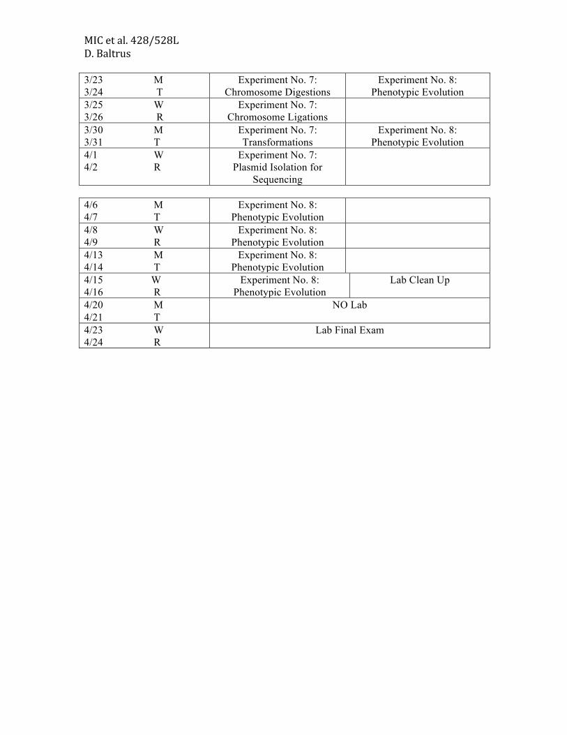
MIC et al. 428/528L D. Baltrus 3/23 M 3/24 T
Experiment No. 7: Chromosome Digestions
Experiment No. 8: Phenotypic Evolution
3/25 W 3/26 R
Experiment No. 7: Chromosome Ligations
3/30 M 3/31 T
Experiment No. 7: Transformations
Experiment No. 8: Phenotypic Evolution
4/1 W 4/2 R
Experiment No. 7: Plasmid Isolation for
Sequencing
4/6 M 4/7 T
Experiment No. 8: Phenotypic Evolution
4/8 W 4/9 R
Experiment No. 8: Phenotypic Evolution
4/13 M 4/14 T
Experiment No. 8: Phenotypic Evolution
4/15 W 4/16 R
Experiment No. 8: Phenotypic Evolution
Lab Clean Up
4/20 M 4/21 T
NO Lab
4/23 W 4/24 R
Lab Final Exam
MIC et al. 428/528L D. Baltrus
INTRODUCTION TO EXPERIMENTAL SYSTEMS In this course you will be working with several different bacterial strains. A brief
description of some of the strains we will be using is given below. Pseudomonas chlororaphis strain 30-84
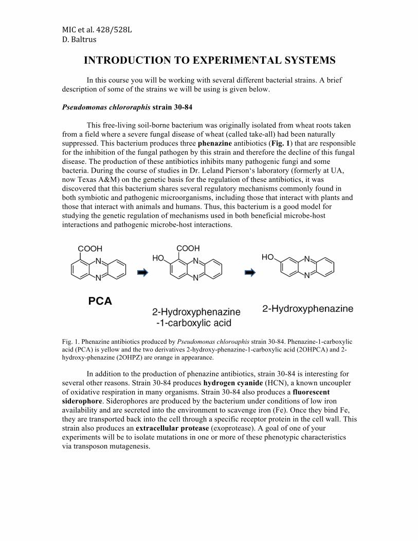
This free-living soil-borne bacterium was originally isolated from wheat roots taken from a field where a severe fungal disease of wheat (called take-all) had been naturally suppressed. This bacterium produces three phenazine antibiotics (Fig. 1) that are responsible for the inhibition of the fungal pathogen by this strain and therefore the decline of this fungal disease. The production of these antibiotics inhibits many pathogenic fungi and some bacteria. During the course of studies in Dr. Leland Pierson‘s laboratory (formerly at UA, now Texas A&M) on the genetic basis for the regulation of these antibiotics, it was discovered that this bacterium shares several regulatory mechanisms commonly found in both symbiotic and pathogenic microorganisms, including those that interact with plants and those that interact with animals and humans. Thus, this bacterium is a good model for studying the genetic regulation of mechanisms used in both beneficial microbe-host interactions and pathogenic microbe-host interactions.
Fig. 1. Phenazine antibiotics produced by Pseudomonas chloroaphis strain 30-84. Phenazine-1-carboxylic acid (PCA) is yellow and the two derivatives 2-hydroxy-phenazine-1-carboxylic acid (2OHPCA) and 2-hydroxy-phenazine (2OHPZ) are orange in appearance.
In addition to the production of phenazine antibiotics, strain 30-84 is interesting for several other reasons. Strain 30-84 produces hydrogen cyanide (HCN), a known uncoupler of oxidative respiration in many organisms. Strain 30-84 also produces a fluorescent siderophore. Siderophores are produced by the bacterium under conditions of low iron availability and are secreted into the environment to scavenge iron (Fe). Once they bind Fe, they are transported back into the cell through a specific receptor protein in the cell wall. This strain also produces an extracellular protease (exoprotease). A goal of one of your experiments will be to isolate mutations in one or more of these phenotypic characteristics via transposon mutagenesis.

MIC et al. 428/528L D. Baltrus Pseudomonas stutzeri strain 28a24 Pseudomonas stutzeri is a highly diverse species of great physiological and ecological versatility and widespread geographic distribution. Members of this species have been shown to be involved in nitrification and denitrification processes as well as in the degradation of environmental pollutants. This strain was originally isolated from soil and found to be highly competent for natural transformation. It is used in the Baltrus lab because introduction of a megaplasmid, originally isolated from Pseudomonas syringae pv. lachrymans into this strain disrupts various phenotypes such as motility, biofilm formation, and antibiotic sensitivity. Pseudomonas sp. Strain UB246 This strain was originally isolated from river water in France. Although not much is known about it’s phenotypic characteristics, it is a phylogenetic outgroup for the common plant pathogen P. syringae and is also related to both P. fluorescens and P. chloroaphis. Like P. syringae, this strain possesses a type III secretion system where transcription is controlled by the sigma factor HrpL. Pseudomonas syringae pv. tomato DC3000 This is a model bacterial phytopathogen that is used in many labs to dissect virulence and host responses. This strain is the causative agent of bacterial speck in tomato, but has become very useful for lab studies because it’s one of the rare bacterial pathogens that can infect and cause symptoms in Arabidopsis (the lab model system for plant genetics). This parent of this strain was isolated in Norwich UK in 1961, with DC3000 being a rifampicin resistant isolate of this original strain. Virulence in planta for DC3000 is due to the presence of a type III secretion system and associated effector proteins as well as secreted toxins such as coronatine. Virulence factors in DC3000 are regulated by a cascade of different transcriptional units, with the HrpR and HrpS proteins activating in conjunction with σ54 to upregulate transcription of HrpL. HrpL is an alternative σ54 factor that binds to a promoter (termed the hrp-box) upstream of virulence genes to trigger expression. Pseudomonas syringae pv. lachrymans DAB885, DBL242, DAB895 Both strains are closely related (with nearly identical sequences at a variety of housekeeping genes) and were isolated as a causative agent of angular leaf spot of cucumber. However, the genome of DAB885 naturally contains an additional 1Mb megaplasmid compared to DBL242. Moreover, this megaplasmid is self-transmissible and was mated from DAB885 to DBL242 to create DAB895. These strains will be used over the course of experiment 8 in order to test for differences in evolutionary potential due to the megaplasmid.

MIC et al. 428/528L D. Baltrus Escherichia coli strain DH5
Strain DH5is an engineered strain of E. coli that is widely used for cloning experiments, in fact it is used almost weekly in Dr. Baltrus‘s research. Its genotype is given below. F-, recA1, endA1, hsdR17, supE44, thi-1, gyrA96, relA1, (argF-lacZYA), I169, 80lacZM15, -. This strain has 3 very useful characteristics: 1. RecA-. It is defective in homologous recombination. So if it contains a plasmid with genes homologous to those on its chromosome NO recombination can occur. 2. HsdR-. There is NO host restriction system functioning in this strain. Therefore, introduced DNA will not be degraded by restriction enzymes in the cell. 3. lacZM15. In E. coli the lacZ gene encodes for the enzyme -galactosidase that is only active as a homotetramer (composed of four identical monomers). Each monomer by itself is inactive, and is composed of two parts LacZ-alpha and LacZ-omega. The lacZM15 mutation lacks a small 92 amino acid portion (LacZ-alpha) of the lacZ gene that is responsible for causing the -galactosidase monomers to bind together. Thus, monomers of -galactosidase are produced but they cannot bind together to form the active form. Thus, the strain is phenotypically Lac-. However, if a specific type of engineered plasmid is introduced into the strain (such as pUC18) that encodes the missing 92 amino acid portion of lacZ (LacZ-alpha), then the -galactosidase monomers can form the active -galactosidase tetramer and the strain becomes Lac+. This phenomenon, called -complementation (Fig. 2, pg. 14) is extremely useful in cloning experiments, as you will see.

MIC et al. 428/528L D. Baltrus

MIC et al. 428/528L Experiment No. 1
EXPERIMENT NO. 1 COMMON LABORATORY PROCEDURES
Section 1, 1/21/2014 W Section 2, 1/22/2014 R Section 3, 1/21/2014 W
INTRODUCTION
During this course, it is imperative that you are able to perform accurately laboratory techniques such as streaking plating and dilution plating. In addition, the accurate use of micropipettors is an often overlooked but critical skill. Most of you have performed these techniques in prior laboratory courses and are proficient. Therefore, in this exercise you have the opportunity to demonstrate your proficiency to us. In lieu of a lab report, your plates will be scored for technique. MATERIALS (each person) LB broth culture of a mixture of Pseudomonas chloroaphis strain 30-84 & 30-84Z (prepared immediately prior to class) 5 LB + X-gal agar plates 28oC Incubator I. STREAK PLATING
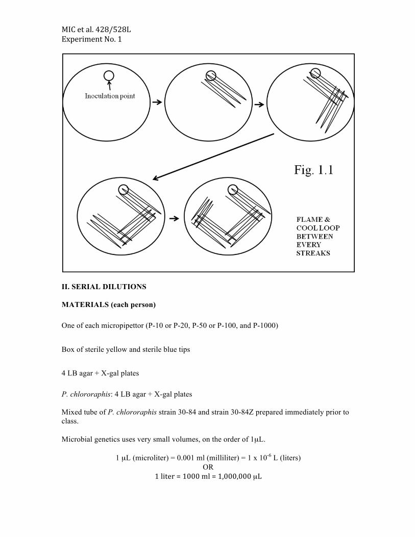
When doing genetics, it is important to be able to isolate pure colonies composed of identical clones that do not contain contaminating cells from other populations. EXPERIMENTAL PROTOCOL 1. Label your plate with your name & section no. 2. Using a sterilized loop, place a loopful of the strain mixture onto one side of the LB agar + X-gal plate. 3. Using the technique shown below (Fig. 1.1), streak the loopful out for single colonies. 4. Place your labeled plates in the box at the front of the room. (Your GTA will look for isolated colonies of both strains)
MIC et al. 428/528L Experiment No. 1
II. SERIAL DILUTIONS MATERIALS (each person) One of each micropipettor (P-10 or P-20, P-50 or P-100, and P-1000) Box of sterile yellow and sterile blue tips 4 LB agar + X-gal plates P. chlororaphis: 4 LB agar + X-gal plates Mixed tube of P. chlororaphis strain 30-84 and strain 30-84Z prepared immediately prior to class. Microbial genetics uses very small volumes, on the order of 1µL.
1 µL (microliter) = 0.001 ml (milliliter) = 1 x 10⁻⁶ L (liters) OR
1 liter = 1000 ml = 1,000,000 µL

MIC et al. 428/528L Experiment No. 1 Micropipettors are essential for microbial genetics. They are used for physiology, biochemistry, genetics, etc. It is important that you are competent with their usage. USAGE & CARE OF MICROPIPETTORS There are several different sizes of micropipettors. Please treat these instruments with care. Their reliable operation is critical for the success of several of your experiments. PRINCIPLE OF OPERATION
This instrument is designed to be held in the hand and operated by the thumb. The plunger stroke is divided into two parts. The longer calibrated stroke ends at the FIRST STOP, the shorter stroke ends at the SECOND STOP (Fig. 1.2). VOLUME ADJUSTMENT 1. To select the desired volume, loosen the top locknut by turning it counterclockwise. 2. To reduce the volume, turn the thumb knob clockwise. Turning the thumb knob counterclockwise will increase the volume (Fig. 1.3). 3. Set the desired volume on the digital display to correspond to the arrow mark located on the base of the window frame. 4. The selected volume is fixed by tightening the locknut clockwise (check to see that tightening the locknut has not altered the setting).
Fig. 1.2

MIC et al. 428/528L Experiment No. 1
OPERATING INSTRUCTIONS 1. Attach a clean sterile tip firmly to the barrel of the instrument. 2. Depress the thumb knob to the first stop. 3. Immerse the tip approximately 3 mm into the sample. 4. Smoothly & slowly return the thumb knob to the release position (DO NOT let it snap back). 5. Withdraw the tip from the solution, do not wipe the tip.
Fig. 1.3

MIC et al. 428/528L Experiment No. 1 6. Place tip against the side of the receiving tube. 7. Smoothly depress the thumb knob to the first stop, pause & then depress the knob to the second stop. 8. With the knob still held at the second stop, withdraw the tip from the receiving tube. 9. Return the knob to the release position (DO NOT let it snap back). 10. Remove the tip by hand or by pushing the tip ejector button. AIDS TO REPRODUCIBILITY & ACCURACY 1. Try to affect the same speed for both uptake & delivery of all samples. 2. Always depress the thumb knob to the proper stop prior to taking up sample. 3. Insert the tip to the same depth each time & keep the micropipettor as vertical as possible. EXPERIMENTAL PROTOCOL
You have a tube containing a mixture of two derivatives of P. chlororaphis. One derivative is the wild-type strain 30-84 while the second is the -galactosidase reporter strain 30-84Z. Your goal is to isolate each of the strains and determine their population densities (cfu/ml).
Each person does this independently. Label your plates with your name & section no. Using the procedure given below (Fig. 1.4), serially dilute the broth culture and spread onto TWO (2) LB agar + X-‐gal plates at both 10-‐7 and 10-‐8. Place your labeled plates in the box at the front of the room. (Your GTA will store the plates until we count them during the next lab period).

MIC et al. 428/528L Experiment No. 1
Fig. 1.4

MIC et al. 428/528L Experiment No. 1
EXPERIMENT NO. 1 – SELECTED QUESTIONS 1. You have performed the indicated series of dilutions. For each, give the titer of the original culture (show your calculations). A. Tube 10 µL 990 µL PBS
300 µL 700 µL PBS 200 µL 800 µL LB broth
100 µL LB agar plate Colonies on plate after 24 hr = 2
Titer of original culture = __________________ cfu/ml. B. Tube 100 µL 900 µL H2O
1 µL 9999 µL LB broth 1 µL 49 µL LB broth
200 µL 800 µL LB broth 100 µL LB agar plate
Colonies on plate after 24 hr = 130 Titer of original culture = ______________________ cfu/ml. C. Tube 10 µL 490 µL PBS
1 µL 99 µL PBS 100 µL 1900 µL PBS
500 µL 500 µL PBS 100 µL LB agar plate
Colonies on plate after 24 hr = 353
Titer of original culture = ____________________ cfu/ml.
2. Why do you plate on duplicate plates for each dilution (think statistics)?

MIC et al. 428/528L Experiment No. 1
EXPERIMENT NO. 8 PHENOTYPIC EVOLUTION
Section 1, 1/21/2014 W Section 2, 1/22/2014 R Section 3, 1/21/2014 W
Over the course of this semester, your group will passage one strain of bacteria
each week and evaluate phenotypic variation that arises over the course of the semester. Section 1 will passage strain DAB885, Section 2 will passage strain DBL242, Section 3 will passage DAB895. You will passage the strains and streak out whole populations to single colonies on W/R. On M/T of each week you will observe phenotypes that arise across those single colonies. This experiment is based off of phenotypic evolution seen within Pseudomonas fluorescens described within Travisano and Rainey 1998 (found at link below and on D2L):
http://www.nature.com/nature/journal/v394/n6688/abs/394069a0.html For this first week, you will transfer 5uL of liquid culture of your section’s strain
to 5mL of KB media supplemented with Rifampicin 50 and Tetracycline 10. You will place this test tube into a rack in the 28 degree incubator and leave it for 1 week. After transfer to the test tube, you will streak your strain to single colonies on KB plates supplemented with Rifampicin 50 and Tetracyline 10.

MIC et al. 428/528L Experiment No. 2
1
EXPERIMENT NO. 2 PHENOTYPIC VS. GENOTYPIC VARIATION
Section 1, 1/26/2015 M Section 2, 1/27/2015 T Section 3, 1/26/2015 M
PHENOTYPIC VARIATION Phenotype – the observable properties of an organism, produced by the genotype in conjunction with the environment. All of the phenotypic characteristics of a bacterium (or any living organism) can be related to the expression of specific genes within the chromosome or extrachromosomal elements such as plasmids. The ability of a bacterium to utilize specific nutrients, to grow in specific niches, to be pathogenic, symbiotic, etc. is dependent upon the appropriate expression of specific genes or groups of genes. The expression of many of these genes is responsive to the bacterium‘s environment. Many environmental factors, including nutrition, temperature, and osmolarity influence bacterial gene expression and therefore the phenotype of the bacterium. These are called phenotypic changes. Remember, P. chlororaphis strain 30-84 produces several secondary metabolites such as phenazine antibiotics, HCN, a siderophore and an exoprotease. Keep this in mind as you examine your plates after incubation. We will only look at some of these phenotypes. Strain 30-84Z contains a phzB::lacZ genomic fusion and thus expresses -galactosidase in place of phenazines. MATERIALS (per team) LB agar plates of Pseudomonas chlororaphis strains 30-84, 30-84Z & 30-84gacA 1 LB agar plate and 1 M9 agar plate 1 KMB agar plate 1 KMB + FeCl3 agar plate (3 ml 0.5 M FeCl3 per liter) 1 Skim milk agar plate 2 semi-solid motility agar plates 1 LB + X-gal agar plate and 1 M9 + X-gal agar plate

MIC et al. 428/528L Experiment No. 2
2
28OC incubator EXPERIMENTAL PROTOCOL I. Examination of the effect of medium type, iron and carbon source
A. Effect of medium type (Fig 2.1) 1. Streak the cultures of P. chlororaphis 30-84 onto one (1) LB agar plate and one (1)
M9 agar plate. Do the same for the culture of 30-84Z but use plates with X-gal. 2. Incubate the plates at 28oC. 3. Examine the phenotypes after 24 h.
Fig. 2.1 Effect of medium type on the soil-borne bacterium P. chlororaphis 30-84 and its derivative 30-84Z. B. Effect of iron
1. Streak strain 30-84Z onto one (1) KMB* agar plate and one (1) KMB + FeCl3 agar
plate. Incubate at 28oC. (*KMB = King‘s Medium B which is a medium low in available iron).
MIC et al. 428/528L Experiment No. 2
3
C. Exoprotease production 1. Streak strain 30-84, 30-84Z & 30-84gacA onto 1/3 of a skim milk plate. Incubate
at 28OC. D. Motility
1. Using a pipette tip, scrape P. chloroaphis 30-‐84 so that there is a little dab of bacteria on the tip. Stab center of motility plate with this pipette tip. Repeat with & 30-84gacA. BE VERY CAREFUL HANDLING MOTILITY PLATE AND DO NOT TURN MOTILITY PLATE UPSIDE DOWN!!!!!.
Section 1, 1/27/2012 T Section 2, 1/28/2012 W Section 3, 1/27/2012 T ****NOTE THAT YOU NEED TO COME IN BRIEFLY ON AN ODD DAY TO EXAMINE THE PLATES. 1. Examine and RECORD the phenotypes of each bacterial culture on ALL plates, noting any differences. Think about what the results indicate to you about the plasticity of phenotypic expression.
MIC et al. 428/528L Experiment No. 2
4
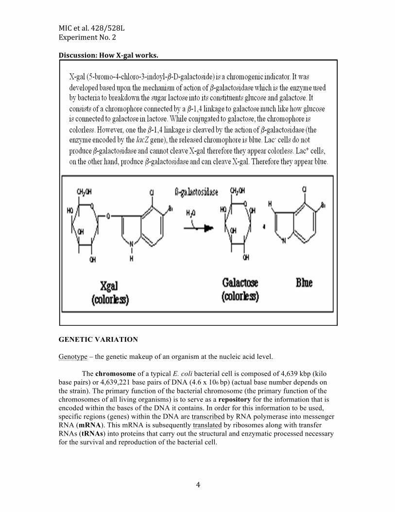
Discussion: How X-‐gal works.
GENETIC VARIATION Genotype – the genetic makeup of an organism at the nucleic acid level.
The chromosome of a typical E. coli bacterial cell is composed of 4,639 kbp (kilo base pairs) or 4,639,221 base pairs of DNA (4.6 x 10⁶ bp) (actual base number depends on the strain). The primary function of the bacterial chromosome (the primary function of the chromosomes of all living organisms) is to serve as a repository for the information that is encoded within the bases of the DNA it contains. In order for this information to be used, specific regions (genes) within the DNA are transcribed by RNA polymerase into messenger RNA (mRNA). This mRNA is subsequently translated by ribosomes along with transfer RNAs (tRNAs) into proteins that carry out the structural and enzymatic processed necessary for the survival and reproduction of the bacterial cell.

MIC et al. 428/528L Experiment No. 2
5
In addition, many bacteria contain additional genetic information that resides on self-replicating extrachromosomal elements called plasmids. It is often easiest to think of plasmids as accessory pieces of DNA that contain genetic information non-essential to basic cellular metabolism but which provide the host bacterial cell with additional metabolic or physiological properties. Examples of these properties include the ability to utilize unusual carbon sources or to produce specific toxins important in pathogenesis, etc. Plasmids can be of many different sizes. Some plasmids (R factors) carry genes providing resistance to various antibiotics.
Surprisingly, although the chromosome serves as a repository for genetic information, it is not an unchanging structure. Spontaneous mutations (i.e. changes in the basic DNA sequence) arise at a frequency of ~1 x 10⁻⁸ per generation.
Any change in the sequence of bases in the DNA of a cell is called a mutation. Many factors in the environment can cause mutations in DNA sequences, including physical factors such as ultraviolet light and radiation; chemical factors such as nitrous acid and benzene; and genetic factors such as transposable elements and some bacteriophages.
These spontaneous mutations may occur within the DNA sequence of the bacterial chromosome or within the DNA sequence of the plasmids contained within the bacterial cell. The majority of mutations are detrimental to the bacterial cell and a cell containing these mutations is selected against. Mutations can also be neutral, that is, they do not alter the fitness of the cell. Occasionally, a mutation can confer a selective advantage to the cell

MIC et al. 428/528L Experiment No. 2
6
depending on the particular environmental conditions. In this case, the mutated cell will not only survive, but in many cases it will replace the original ―wild-typeǁ‖ or parent strain. Changes in the basic DNA sequences in a bacterial cell are called genotypic changes. Discussion: mechanism of action of Rifampicin.
Rifampicin is an antibiotic. It is produced by some species of Streptomyces. It binds
to the -subunit of RNA polymerase and blocks initiation of transcription. Rifampicin does not block elongation of the RNA polymerase complex if transcription has already initiated. Therefore, it has been widely used as a mechanism to block de novo transcription during physiological studies.
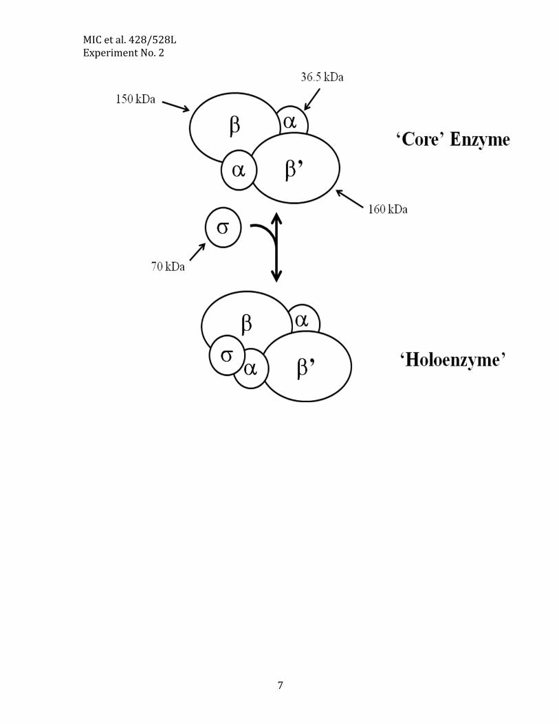
The structure of the E. coli RNA polymerase RNA polymerase from E. coli consists of five protein subunits. It is one of the largest
enzymes known and can easily be seen by electron microscopy. Its subunits are encoded by rpoA (α), rpoB (β), rpoC (β’), and rpoD (σ). Mutations within rpoB are responsible for Rifampicin resistance (RifR).
MIC et al. 428/528L Experiment No. 2
7

MIC et al. 428/528L Experiment No. 2
8
EXPERIMENT NO. 2 – SELECTED QUESTIONS Phenotypic Variation 1. What was the effect of medium type on pigmentation in strain 30-84 and 30-84Z? What is the difference between these strains? 2. The availability of iron (Fe3+) in the environment is very low (~1 x 10-17 M). Therefore, siderophore production by bacteria can be critical for successful growth in nature. Many bacteria in nature produce siderophores, and often one bacterium will out-compete another bacterium for iron. What could explain this observation? 3. How do skim milk plates allow detection of proteases? How does semi-soild motility agar allow for the detection of motility? Genotypic Variation 1. Define the following two terms in regards to phenotype. A. Selection. B.Screening.

MIC et al. 428/528L Experiment No. 3
1
EXPERIMENT NO. 3 MUTATION AND NATURAL TRANSFORMATION
Section 1, 1/28/2015 W Section 2, 1/29/2015 R Section 3, 1/28/2015 W
Mutation
Mutations normally occur during DNA replication because polymerases make errors while copying the chromosome. Although a large majority of mutations are actually corrected by the polymerases themselves as well as dedicated DNA repair machinery, some occasionally slip through. Mutations can be single base pair changes as well as additions/deletions of nucleotides. Mutations can also occur outside of chromosomal replication as a product of environmental stress (UV light, some antibiotics, etc…). As discussed within the last lab, these mutations are genotypically different from the defined wild type sequence, but likely only have phenotypic effects IF A) the region is transcribed B) the transcribed region is capable of changing a phenotype within the measured environment C) the mutation is non-synonymous.
During growth in liquid culture, bacteria divide in a binary way (one cell gives rise to two cells). If all cells within a culture start out genotypically identical, even if only one mutation occurs during growth, the number mutant cells at the end of a growth cycle will be determined by when in the growth cycle that mutation occurs. If only ONE mutation occurs over the course of a growth curve, but occurs during an early division, there will be many mutants at the end of growth because each mutant cell will divide several times. Alternatively, if a single mutation occurs during the last possible cell division, there will be only one mutant cell within the whole population. VARIABILITY IN MUTANT NUMBERS AT THE TIME OF MEASURING IS DUE TO DIFFERENT APPEARANCE TIMES OF THE UNDERLYING MUTATIONS DURING GROWTH. Variability across bacterial cultures in the number of mutants roughly follows a Poisson distribution and mutation rates can be estimated by fitting the observed numbers to this mathematical distribution. If a single population is plated out, there is only a set number of mutants within that culture. In this case, any variability is due to randomness and should be normally distributed.
Fig. 1. The Spontaneous Mutation Model. Variability from culture to culture in number of colonies is due to the underlying mutations occurring at different times during cell growth. In each case, there is only one mutant within each population.

MIC et al. 428/528L Experiment No. 3
2
Natural Transformation
Many bacteria have the capability to import DNA from the extra-cellular environment and incorporate it into their own genomes. One of the definitive traits of bacteria that are competent for natural transformation is the presence of dedicated proteins and enzymes to transport this DNA into the cell and protect this DNA from degradation. Oftentimes, the trick to making bacterial competent for natural transformation is finding the right environmental conditions to turn on regulatory pathways controlling genes involved in DNA importation. Once the DNA is present within the cell, almost all bacteria have enzymes necessary (i.e. RecA) to recombine this DNA into homologous (matching sequence) regions on the chromosome. As little as 25bp of homologous DNA is sufficient for some level of transformation to occur but even a single nucleotide difference between imported DNA and the chromosome can disrupt this process. Natural transformation can occur without much bacterial growth, and so any variation between plates should be normally distributed. Streptomycin and Rifampicin Resistance Streptomycin inhibits bacterial growth by binding to the 16s rRNA subunit, and interfering with the binding of formyl-methionyl-tRNA to the 30s subunit to inhibit protein synthesis. Spontaneous mutations to streptomycin resistance are possible within P. stutzeri, and usually occur within the rpsL gene. Rifampicin inhibits bacterial growth by binding to the active site of RNA polymerase and preventing transcription. Spontaneous mutations to rifampicin resistance occur quite readily within P. stutzeri (>100 fold higher rates than to streptomycin resistance), and usually occur at the rpoB gene. The rpsL and rpoB genes are found in close proximity to each other within the P. stutzeri genome.
MATERIALS (per team) Overnight culture of 1mL P. stutzeri DAB273 in Saltwater LB 1mL of Saltwater LB 100ng of P. stutzeri DBL509 strepR/rifS DNA 100ng of P. stutzeri DBL455 strepR/rifR DNA 100ng of P. syringae DC3000 strepR/rifR DNA
MIC et al. 428/528L Experiment No. 3
3
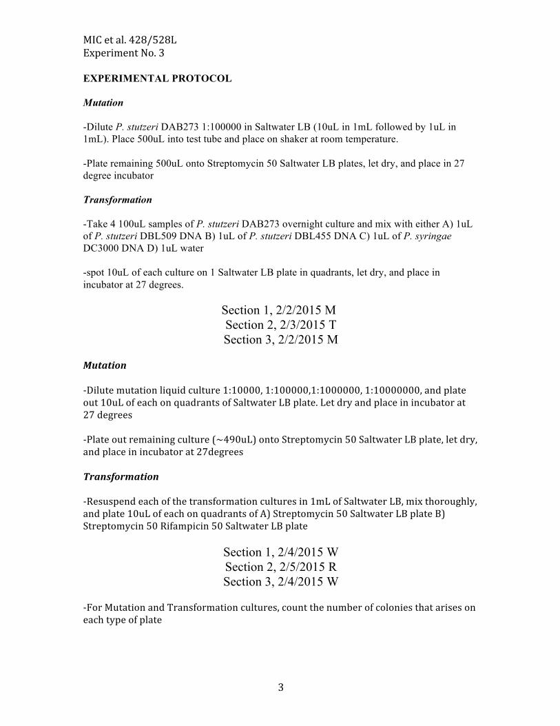
EXPERIMENTAL PROTOCOL Mutation -Dilute P. stutzeri DAB273 1:100000 in Saltwater LB (10uL in 1mL followed by 1uL in 1mL). Place 500uL into test tube and place on shaker at room temperature. -Plate remaining 500uL onto Streptomycin 50 Saltwater LB plates, let dry, and place in 27 degree incubator Transformation -Take 4 100uL samples of P. stutzeri DAB273 overnight culture and mix with either A) 1uL of P. stutzeri DBL509 DNA B) 1uL of P. stutzeri DBL455 DNA C) 1uL of P. syringae DC3000 DNA D) 1uL water -spot 10uL of each culture on 1 Saltwater LB plate in quadrants, let dry, and place in incubator at 27 degrees.
Section 1, 2/2/2015 M Section 2, 2/3/2015 T Section 3, 2/2/2015 M
Mutation -‐Dilute mutation liquid culture 1:10000, 1:100000,1:1000000, 1:10000000, and plate out 10uL of each on quadrants of Saltwater LB plate. Let dry and place in incubator at 27 degrees -‐Plate out remaining culture (~490uL) onto Streptomycin 50 Saltwater LB plate, let dry, and place in incubator at 27degrees Transformation -‐Resuspend each of the transformation cultures in 1mL of Saltwater LB, mix thoroughly, and plate 10uL of each on quadrants of A) Streptomycin 50 Saltwater LB plate B) Streptomycin 50 Rifampicin 50 Saltwater LB plate
Section 1, 2/4/2015 W Section 2, 2/5/2015 R Section 3, 2/4/2015 W
-‐For Mutation and Transformation cultures, count the number of colonies that arises on each type of plate

MIC et al. 428/528L Experiment No. 3
4
EXPERIMENT NO. 3 – SELECTED QUESTIONS
1. Why is it important to dilute your start fluctuation assays with a very small number of cells?
2. When DBL332 is transformed with DNA from P. stutzeri 23a24 DNA, and selected on streptomycin, why can you isolate both strepR/rifS colonies and strepR/rifR colonies?
3. Is the transformation rate with P. syringae DC3000 DNA lower than with P. stutzeri DNA? If so (or not) why do you think this is?
MIC et al. 428/528L Experiment No. 4
1
EXPERIMENT NO. 4 ISOLATION OF PLASMID DNA
Section 1, 2/4/2015 W Section 2, 2/5/2015 R Section 3, 2/4/2015 W
INTRODUCTION
Plasmids are small extrachromosomal elements that are separate from the bacterial chromosome but depend on cellular enzymes for replication. Plasmids often confer unique properties on the cells that contain them. Plasmids come in all different sizes, copy numbers, and may contain many different types of genes. Many plasmids have been engineered to be useful in microbial genetics. Plasmid Nomenclature 1. Plasmids are named beginning with a lower case p to indicate the name refers to a plasmid and not to a bacterial strain (e.g. pUC18, pKKC259, pKT2, etc). 2. The letters in front of the plasmid number usually refers to the individuals who engineered the plasmid (e.g. pDBL632 indicates it was constructed by Dr. David Baltrus (the L is for Lab, and this is the 632nd strain in my lab collection). 3. Bacteria containing a known plasmid are named by the strain name followed by the plasmid name in parentheses (e.g. E. coli DH5 (pUC18), or DH5 (pKKCRec1452). Plasmid DNA Isolation
In comparison to most bacterial chromosomes, plasmids are small in size. They exist in cells as supercoiled (tightly twisted) molecules and are relatively unaffected by many procedures that denature chromosomes and proteins. In fact, we use the fact that plasmids are resistant to shearing (breaking) as part of our isolation methodology.
The basic principle of all plasmid DNA isolations is the same: break open the cells, remove everything you do not want, and what is left is predominantly plasmid DNA. The procedure we will use is a modified alkaline lysis procedure. We will use a kit from Fermentas Corp. for this procedure. The procedure involves re-suspending bacterial cells in a buffer that stabilizes nucleic acids and then a buffer containing a cell wall degrading enzyme (Lysozyme) that lyses the cells by breaking down the peptidoglycan backbone. Proteins are next degraded using an alkaline protease solution under alkaline conditions to denature the chromosomal DNA and proteins. The pH is returned to neutrality which causes the chromosome and proteins to collapse and cell debris is pelleted by centrifugation. The plasmid DNA, being supercoiled and small, remains intact and relatively unaffected. This solution has high salt and causes the ds (double-stranded) plasmid DNA to stick to the spin

MIC et al. 428/528L Experiment No. 4
2
column. The plasmid DNA is washed of RNA and other contaminants and dried. Then the column is placed in a sterile tube and the plasmid DNA eluted from the resin in pure water that lacks any contaminating salt. Thus, when you are done, column wash will contain nearly pure plasmid DNA. In this exercise you will compare yields for two versions high copy number plasmid (pDBL632-1 or -6) with an oriR6K origin of replication
MATERIALS 3 ml LB + Kan30 broth overnight of S17 (pDBL632-1 or -6) –high copy number plasmid Fermentas GeneJetTM Plasmid Miniprep Kit Sterile 1.5 ml microfuge tubes Micropipettors Sterile tips EXPERIMENTAL PROTOCOL Isolation of plasmid DNA from bacteria using a modified Fermentas GeneJetTM Plasmid Miniprep Kit Protocol is given on the next page.

MIC et al. 428/528L Experiment No. 4
3
Modified Protocol for Fermentas GeneJetTM Plasmid Miniprep Kits
MIC et al. 428/528L Experiment No. 4
4
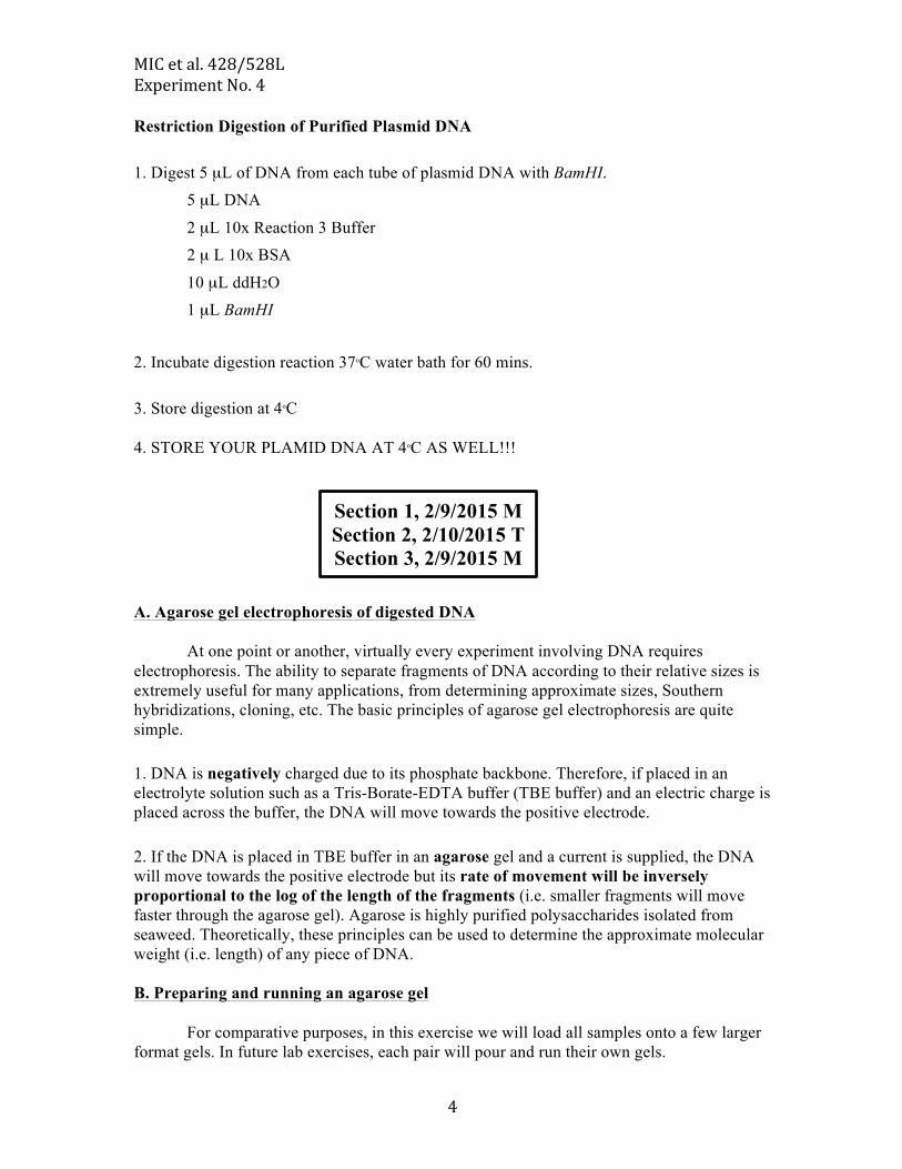
Restriction Digestion of Purified Plasmid DNA 1. Digest 5 µL of DNA from each tube of plasmid DNA with BamHI.
5 µL DNA
2 µL 10x Reaction 3 Buffer 2 µ L 10x BSA
10 µL ddH2O 1 µL BamHI
2. Incubate digestion reaction 37oC water bath for 60 mins. 3. Store digestion at 4oC 4. STORE YOUR PLAMID DNA AT 4oC AS WELL!!!
A. Agarose gel electrophoresis of digested DNA
At one point or another, virtually every experiment involving DNA requires electrophoresis. The ability to separate fragments of DNA according to their relative sizes is extremely useful for many applications, from determining approximate sizes, Southern hybridizations, cloning, etc. The basic principles of agarose gel electrophoresis are quite simple. 1. DNA is negatively charged due to its phosphate backbone. Therefore, if placed in an electrolyte solution such as a Tris-Borate-EDTA buffer (TBE buffer) and an electric charge is placed across the buffer, the DNA will move towards the positive electrode. 2. If the DNA is placed in TBE buffer in an agarose gel and a current is supplied, the DNA will move towards the positive electrode but its rate of movement will be inversely proportional to the log of the length of the fragments (i.e. smaller fragments will move faster through the agarose gel). Agarose is highly purified polysaccharides isolated from seaweed. Theoretically, these principles can be used to determine the approximate molecular weight (i.e. length) of any piece of DNA. B. Preparing and running an agarose gel
For comparative purposes, in this exercise we will load all samples onto a few larger format gels. In future lab exercises, each pair will pour and run their own gels.
Section 1, 2/9/2015 M Section 2, 2/10/2015 T Section 3, 2/9/2015 M

MIC et al. 428/528L Experiment No. 4
5
Below are the procedures for preparing, loading, running, staining and photographing gels. Preparing gels. Assemble the small horizontal gel tray using the rubber spacers as demonstrated by your GTA. There is pre-melted agarose in the 55oC water bath.
1. Place comb in gel tray. Pour approximately 35-40 ml of melted 1% agarose into the tray (Fig. 3.1). (The goal is to pour a thin gel, not a thick one). Let harden (it turns opaque, almost chalky).
2. 2. Carefully remove the comb by rocking it back and forth. Remove the rubber end caps being CAREFUL not to tear the wells.
3. 3. Place the gel in the gel box and fill it with 1x TBE buffer until the gel is just submerged.
Loading gels.
1. Add 2 µL Tracking dye to each BamHI digest (Total volume = 22 µL). DO NOT ADD TRACKING DYE TO YOUR STOCK PLASMID PREPS!!!
2. Take 5µL of your stock, undigested plasmid culture and place in separate tube. Add 15µL water and 2µL Tracking dye (Total volume = 22 µL).
3. Load gel with one lane plasmid digest, one lane undigested plasmid. Include 5 µL Kb

MIC et al. 428/528L Experiment No. 4
6
ladder and 10 µL λ HindIII as size standards. NOTE: These standards do not require the addition of tracking dye.
Running gels.
1. Carefully place gel top on and connect electrodes to power supply. Run gel at 90V until blue tracking dye is ¼ inch from the bottom of gel. Do not electrocute yourself – major loss of points!
C. Visualization of DNA products on gel using EtBr.
DNA is so small that we cannot visualize it with our own eyes. In order to visualize the DNA on the gel we use a stain called ethidium bromide (EtBr). EtBr is an intercalating agent that fits between the stacked bases of DNA. EtBr fluoresces under UV light. Due to the large number of gels we will add EtBr to the gels in a small volume for staining. We will use the UV gel box and digital camera to capture images of yours gels. The GTAs will help you use the equipment. ALWAYS WEAR GLOVES WHEN WORKING WITH EtBr BECAUSE IT IS A CARCINOGEN!!!!! Notch your gel (check with your GTA) so you can tell which photograph is yours! EtBr Staining.
1. Using disposable gloves, place gel into a tray containing EtBr in TBE. 2. Let stain for 10 to 20 mins. 3. Visualize bands using a short-wavelength (254 nm) UV light box. 4. Photograph gel using digital camera (make 2 copies of the pictures).

MIC et al. 428/528L Experiment No. 4
7
See Appendix 1 for more information on restriction enzymes. See Appendix 2 at end of exercise regarding molecular weight standards.

MIC et al. 428/528L Experiment No. 4
8
APPENDIX 1: RESTRICTION ENZYMES (RESTRICTION ENDONUCLEASES)
Many molecular techniques are based on the now simple notion of cutting and joining DNA molecules. In this exercise, you will be exposed to some of the basic procedures used in molecular cloning. In addition, some of the techniques we have used in previous sections will be used as part of the recombinant experiment you will do. Before we begin, a short discussion of the terms and procedures used in recombinant DNA work is necessary. There are four classes of restriction enzymes currently known. Type or Class II enzymes are primarily used in recombinant DNA work. This class of enzyme recognizes a specific sequence of base pairs in double-stranded DNA and cleaves once on each strand. There are 100‘s of known restriction enzymes. We use these enzymes to easily manipulate DNA in cloning experiments. The other classes not used in recombinant DNA work are:
-Type or Class I enzymes cut DNA randomly, far from their recognition sequences. -Type or Class III enzymes cut outside of their recognition sequences and require two such sequences in opposite orientations within the same DNA molecule to accomplish cleavage. -Type or Class IV enzymes recognize modified, typically methylated DNA.
Enzyme Nomenclature
Restriction enzymes are named for the bacterium they were isolated from. For example, EcoRI was the first restriction enzyme isolated from Escherichia coli = Eco + RI = EcoRI. The first three letters are italicized (or underlined) because they are derived from the genus and species name of the bacterium. HindIII = third restriction enzyme isolated from strain d of Haemophilus influenzae. Commonly used enzymes:
Enzyme Bacterium Isolated From Recognition Sequence BglII Bacillus globigii 5‘ – A‘ GATCT – 3‘
3‘ – TCTAG‘ A – 5‘ EcoRI Escherichia coli RY-13 5‘ – G‘ AATTC – 3‘
3‘ – CTTAA‘ G – 5‘ EcoRV Escherichia coli J62PLG74 5‘ – GAT‘ ATC – 3‘
3‘ – CTA‘ TAG – 5‘ HindIII Haemophilus influenzae Rd 5‘ – A‘ AGCTT – 3‘
3‘ – TTCGA‘ A – 5‘ PstI Providencia stuartii 5‘ – CTGCA‘ G – 3‘
3‘ – G‘ ACGTC – 5‘ SalI Streptomyces albus G 5‘ – G‘ TCGAC – 3‘
3‘ – CAGCT‘ G – 5‘ SmaI Serratia marcescens 5‘ – CCC‘ GGG – 3‘
3‘ – GGG‘ CCC – 5‘

MIC et al. 428/528L Experiment No. 4
9
As you can see, each one of these restriction endonucleases or enzymes recognizes a different sequence of base pairs. Enzymes such as EcoRI and HindIII leave 5‘ overlaps, SmaI and EcoRV leave blunt ends, and PstI leaves a 3‘ overhang when it digests DNA. IMPORTANT: Only DNA fragments with complementary ends can be joined together. The exception is blunt-ended fragments, which can be joined to any other blunt-ended fragment. However, the re-joining (ligation) of blunt ended fragments is much less efficient. Discussion: Restriction enzyme function
The restriction enzyme PstI will be used as an example. PstI is a protein that binds to
DNA. When it binds to DNA containing its recognition sequence it creates asymmetric nicks in each strand of the DNA. PstI happens to create 3‘ overhanging ends. Once the DNA is cleaved the affinity between PstI and the DNA is weakened and it dissociates and the cycle begins again.

MIC et al. 428/528L Experiment No. 4
10
APPENDIX 2: DISCUSSION: DNA SIZE STANDARDS Measuring fragment sizes using the KB Ladder Standard.
By including size standards on your gel, it is possible to determine the size of any
unknown fragment of DNA. The basic idea is that the logarithm of a molecule‘s length is proportional to its migration velocity over a broad range. For a typical gel this relationship can be plotted on semilog paper to give the curve shown below (Fig. 3.3).
Fig. 3.3 Standard Curve of DNA size versus fragment migration
Thus, the length of any unknown fragment can be determined by co-electrophoresis with other fragments of known length. The fragments of known length (the size standard) can be plotted to yield a standard curve (Fig. 3.3) on which the unknown fragment can be interpolated, resulting in an estimate of its length. In the example shown in the figure, using the size standard has resulted in the curve illustrated. Two fragments of unknown length were also run on the same gel. One migrated 8 mm and the second 15 mm. By interpolating their distances onto the curve we can tell that the larger fragment is 4,500 bp in length and the shorter one 2,300 bp long. Note that the curve is not linear at the ends. Also gels are only accurate to + 10%.
We often use one of two size standards, a Kb Ladder or bacteriophage λ DNA
which has been digested with HindIII.

MIC et al. 428/528L Experiment No. 4
11
EXPERIMENT NO. 4 – SELECTED QUESTIONS 1. What physical property allows us to isolate plasmid DNA as opposed to chromosomal DNA? 2. Why should you always include antibiotics in the medium when cultures are grown for plasmid isolation? 3. What is in the Tracking Dye/Loading Buffer?

MIC et al. 428/528L Experiment No. 5
1
EXPERIMENT NO. 5 GENE EXPRESSION
Section 1, 2/16/2015 M Section 2, 2/17/2015 T Section 3, 2/16/2015 M
Bacterial operons are tightly regulated through the action of transcription factors called σ factors. The action of these σ factors are in turn regulated by inducer and repressor molecules as well as activator proteins. Gene expression within bacterial cells is the product of a multi-layered cascade of regulatory proteins and molecules acting together, with these cascades consisting of multiple interacting/interfering σ factors and regulatory molecules. Virulence gene expression within the bacterial phytopathogen P. syringae is the product of a complicated regulatory module consisting of numerous sigma factors and inducers/repressors. At the top of the cascade is the GacAS two component system which triggers expression of the NtrC-like regulators HrpR and HrpS. HrpRS work together to recruit σ54 to a promoter upstream of the alternative σ54 transcription factor HrpL. HrpL then binds to a promoter upstream of virulence genes (called the hrp-box) in order to transcribe these genes. Little is known about the environmental signals that ultimately trigger expression of HrpL, other than these signals reflect the cellular environment inside a leaf (low pH, low Iron, presence of sugars like fructose/sucrose). In this lab will use chromosomal uidA fusions to HrpRS, HrpL, and Avrpto (a virulence gene) in P. syringae strain to investigate the effects of gene knockouts and strain backgrounds on expression of this regulatory cascade. We will also use a plasmid borne hrpL:lacZ to investigate the differences between regulation on the chromosome and plasmids as well as differences in reporter genes. MATERIALS (per team) 1mL LB media containing Pseudomonas syringae strain DC3000 + 1) gacA:uidA 2) hrpRS:uidA 3) hrpL:uidA 4) avrpto:uidA 1mL LB media containing Pseudomonas syringae strain DC3000:ΔhrpRS 1) hrpRS:uidA 2) hrpL:uidA 3) avrpto:uidA 1mL LB media containing Pseudomonas syringae strain DC3000:ΔhrpL 1) hrpRS:uidA 2) hrpL:uidA 3) avrpto:uidA 1mL LB media containing Pseudomonas syringae strain DC3000:ΔhrpRS + 1) hrpL:lacZ 1mL LB media containing Pseudomonas syringae strain DC3000 + 1) hrpL:lacZ 1mL LB media containing Pseudomonas sp. Strain UB246 + 1)hrpL:lacZ 2 KB agar plates + Xgluc 2 M9 (+10Mm Fructose) agar plates + Xgluc
MIC et al. 428/528L Experiment No. 5
2
2 M9 (+10Mm Glycerol) agar plates +Xgluc 1 KB agar plates + Xgal 1 M9 (+10Mm Fructose) agar plates + Xgal 1 M9 (+10Mm Glycerol) agar plates +Xgal 28OC incubator EXPERIMENTAL PROTOCOL I. Examination of the effect of medium type and strain background on gene expression on solid agar plates
1. Streak isolates of each uidA parent strain onto plates containing each of the media
types (M9 Fructose, M9 Glycerol, KB) containing Xgluc
2. Streak isolates of DC3000 and UB246 containing hrpL:lacZ, as well as DC3000 containing gacA:uidA onto plates containing Xgal.
3. Incubate the plates at 28oC. 4. Examine the phenotypes after 48 h.
Section 1, 2/18/2014 W Section 2, 2/19/2014 R Section 3, 2/18/2014 W 1. Examine and RECORD the phenotypes of each bacterial culture on ALL plates, noting any differences. Think about what the results indicate to you about the genetics of the regulatory cascade, gene expression, and the ability of each reporter gene to demonstrate gene expression.
MIC et al. 428/528L Experiment No. 5
3
EXPERIMENT NO. 5 – SELECTED QUESTIONS
1. Draw out the regulatory cascade of strain DC3000
2. Is gene expression of reporters from a plasmid higher or lower than from the chromosome? Why?
3. Does the gacA:uidA reporter behave the same with both types of reporter substrate (Xgal and Xgluc)? Why or why not?
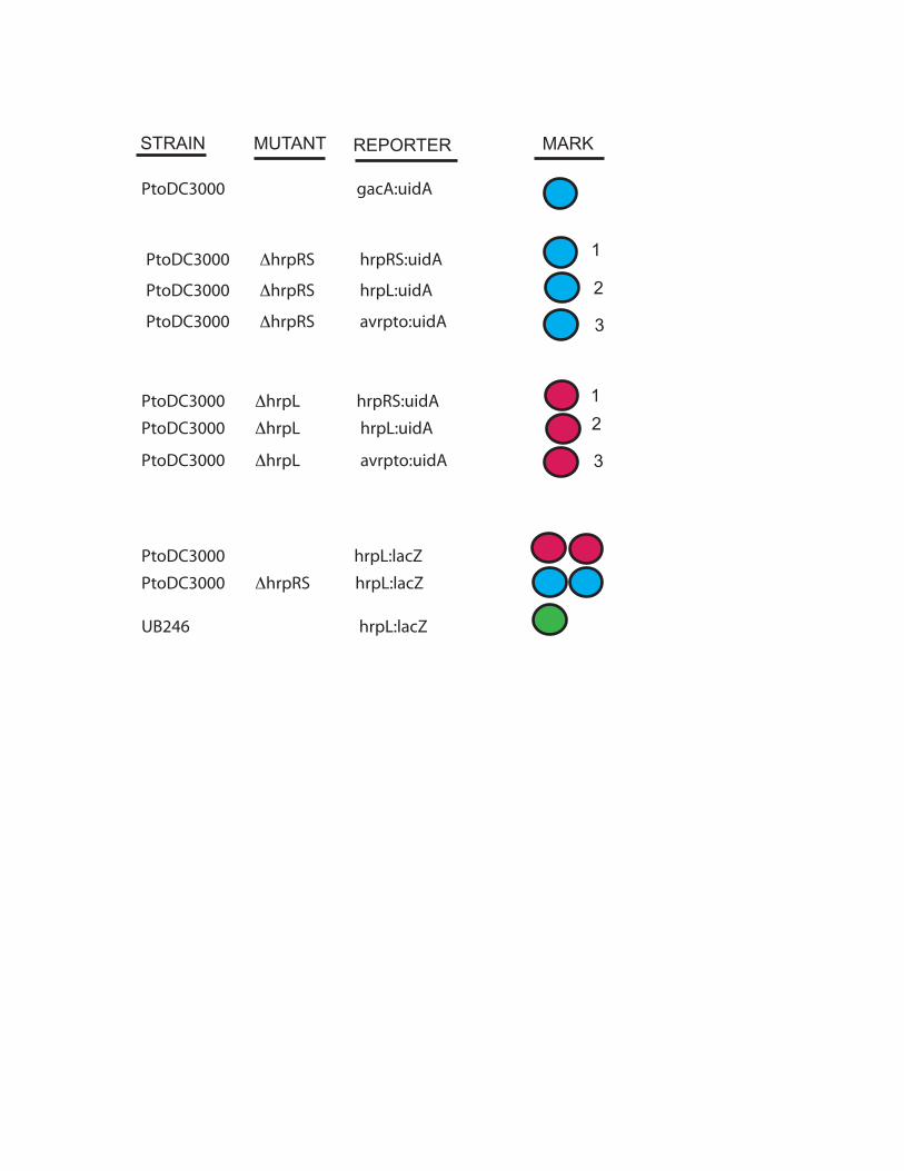
PtoDC3000 gacA:uidA
PtoDC3000 ΔhrpRS hrpRS:uidA
PtoDC3000 ΔhrpRS hrpL:uidA
PtoDC3000 ΔhrpRS avrpto:uidA
PtoDC3000 ΔhrpL hrpRS:uidA
PtoDC3000 ΔhrpL hrpL:uidA
PtoDC3000 ΔhrpL avrpto:uidA
PtoDC3000 hrpL:lacZ
PtoDC3000 ΔhrpRS hrpL:lacZ
UB246 hrpL:lacZ
STRAIN MUTANT REPORTER MARK
1
1
2
3
2
3

MIC et al. 428/528L Experiment No. 6
1
EXPERIMENT NO. 6 TRANSPOSON MUTAGENESIS
Section 1, 2/16/2015 M Section 2, 2/17/2015 T Section 3, 2/16/2015 M
INTRODUCTION
In many cases, the loss of a particular phenotype leads to our understanding of how something works. This was the basis of the ―Mutant Methodologyǁ‖ pioneered by George Beadle many years ago. The approach of finding a phenotype of interest and then determining what is responsible for the phenotype is often referred to as ‘Forward Genetics.’ This mutational approach is dependent on the occurrence of mutations in the genome of the organism under study. In bacteria, the average mutation rate in the average gene is 0.00000001, or 1 x 10⁻⁸. This is a rather low frequency event and we would have to observe a lot of bacteria in order to have a chance to see a visible mutation. To increase the mutation rate, scientists usually use a mutagen to increase the mutation rate to 1 x 10⁻⁵ to 1 x 10⁻³. Therefore, mutagenesis plays a critical role in our understanding of all living things. Even today, with the power of genomics and proteomics, the study of mutants is essential.
In this experiment, you will isolate mutants of Luteibacter that have altered phenotypes and levels of gene expression. What is exciting about this experiment is the opportunity to identify new mutant phenotypes. Most genetic laboratories (prokaryotic and eukaryotic) use transposable elements (TEs) carried by plasmids for mutagenesis experiments. Plasmids often serve as extremely useful vectors for microbial genetics. Plasmids can be grouped into different classes based on many properties. One useful one is host range. Plasmid host ranges: narrow versus wide High copy narrow host range plasmids. These plasmids can only replicate n specific hosts (e.g. E. coli) or in hosts that encode a specific gene product required for replication. This class of plasmids usually exists in large numbers of copies in each cell (30-300). Low copy broad host range plasmids. These plasmids can replicate in a wide range of hosts (e.g. E. coli, Pseudomonas, etc.) but maintain themselves at only a few copies per cell (1-4).
Plasmids can often be transferred from cell-to-cell via conjugation. The ability to
conjugate into a new host is separate from the ability to replicate in the new host cell. Thus, some plasmids can be conjugated into a different genus of bacteria but will be unable to replicate once they enter the new cell. This seeming suicide by the plasmid resulted in their being termed suicide plasmids. We take advantage of suicide plasmids for transposon mutagenesis. For example, the plasmid we will use, pRL27, is a narrow host-range plasmid requires the pir gene product π in order to replicate. Thus, it can only replicated in strains of E. coli or closely-related bacteria that contain the pir gene. We will be using E. coli strain S17-1 λpir for matings. The pir region has been to this strain added using λ phage. The
MIC et al. 428/528L Experiment No. 6
2
chromosome of this strain has also been engineered to contain all tra genes and structural genes required for pilus formation and plasmid mobilization.
Transposable Elements Transposable elements are distinct pieces of DNA that are capable, as their name implies, of transposing, or ―hoppingǁ‖ from one piece of DNA into another. One commonly used Tn for bacterial mutagenesis is the natural transposable element Tn5. Background on Tn5 Transposon Tn5 is a composite transposon, consisting of 2 identical (except for one base change) inverted repeat sequences (IS50L & IS50R) flanking a core region containing a kanamycin resistance (KmR) gene. The right inverted repeat, IS50R, also contains a gene named tnpA that encodes a transposase enzyme. The transposase in conjunction with the inverted repeat sequences are responsible for allowing Tn5 to ―hop or transpose―from one site in a piece of DNA to another site in the same or a different piece of DNA. This transposition is not dependent on RecA-mediated homologous recombination. However, transposons cannot replicate themselves and are dependent on the DNA they are inserted into for replication. We will discuss Tn5 in depth in the lecture course. Tn5 advantages 1. Many chemical or physical mutagens, such as UV light, cause point mutations that occasionally still allow some gene function to remain. These gene mutations are referred to as ‘leaky’. Mutations created by Tn5 are not ‘leaky’ since Tn5 inserts 5.7 kb of DNA into a gene. Tn5 also can have polar effects on downstream genes, preventing their expression as well. 2. Chemical and physical mutagens often cause multiple mutations. Thus, it is not always simple to know which mutation is responsible for the altered phenotype. Useful transposons such as Tn5 usually transpose once. 3. When Tn5 inserts into the chromosome it creates a KmR marker at that site. Therefore, the mutated region of the chromosome can be cloned directly by selecting for KmR. This has also been called marking a mutation site.
We will use an engineered transposon based on Tn5 in conjunction with the suicide plasmid pRL27 (Fig. 5.1)

MIC et al. 428/528L Experiment No. 6
3
Features of pRL27: 1. Contains inverted repeats (IRs). These are the sequences recognized by the transposase encoded by tnpA. During transposition, transposase cleaves the dsDNA at each end of the IRs and moves the region between into another piece of DNA (i.e. the chromosome). 2. The Tn lacks the Tn5 tnpA transposase gene. This gene is carried by the plasmid outside of the region that is transposed. This means that once transposition has occurred, there is no tnpA present to cause any subsequent transpositions to occur, resulting in a very stable mutation. 3. The tnpA gene contains several mutations that render the transposase it encodes hyperactive. That means it will transpose the Tn at a much higher frequency than the wild type transposase. 4. Contains neophosphotransferase activity (npt) within the transposon. Thus, the location of the insertion is ‗marked‘ by kanamycin antibiotic resistance (KmR). 5. The Tn contains an oriR6K origin of replication. This sequence is inactive unless the Tn is present in a cell containing the pir gene encoding the π replication protein. 6. The plasmid contains an oriT sequence (origin of transfer) that enables it to be ‗mobilized‘ into new bacterial cells.

MIC et al. 428/528L Experiment No. 6
4
Overview of experiment: Luteibacter strain DBL564 is a rifampicin resistant mutant of a strain originally isolated living inside of endophytic fungi living inside of the needles Platycladus orientalis trees (related to Juniper). These bacteria alter fungal phenotypes such as production of plant hormones, lignin degradation, and cellulose degradation. We don’t know how this symbiosis is established or what bacterial genes influence these phenotypes. This transposon mutagenesis will be the first step to dissecting the genetic basis of these interactions, and you will look for genes that influence protease production and motility (as well as any other interesting phenotypes that come along). You will perform the following conjugation (Fig. 6.2). The net result will be to introduce the modified Tn5 into Luteibacter strain DBL564. Examine the steps closely as several events must take place for this to work. As convoluted as this may seem initially, it works very well in the lab.
(Biparental Mating) The E. coli donor strain DBL330 contains a pir gene so pRL27 can replicate. It also has tra genes to enable pRL27 to be mobilized into the recipient Luteibacter cell, which lacks the pir gene.
Once inside the recipient, the plasmid cannot replicate and is lost. However, the transposase can transpose a section of pRL27 (between inverted repeats) from the plasmid into the host chromo-some before the plasmid is lost. This results in the creation of a Kanamycin (KmR) marked muta-tion in DBL564.
Figure 6.2 Conjugating pRL27 into Luteibacter strain DBL564
DBL330 (pRL27)DBL564 (RifR)
DBL330 (pRL27)
DBL564 (RifR)
DBL564 + Tn5 (RifR KmR)
KmR

MIC et al. 428/528L Experiment No. 6
5
MATERIALS LB + Km25 O/N culture of DBL330(pRL27) LB O/N culture of Luteibacter DBL564 Sterile microfuge tubes Sterile glass test tubes LB agar plates 8 LB agar + Rif50, Kan25 plates Sterile nitrocellulose squares Tabletop centrifuge Next steps Sterile toothpicks in glass petri plates Semisolid motility plates Skim Milk Agar Plates LB + Kan25 plates

MIC et al. 428/528L Experiment No. 6
6
Experimental Protocol
Section 1, 2/16/2015 M Section 2, 2/17/2015 T Section 3, 2/16/2015 M
1. Available are O/N cultures of E. coli DBL330(pRL27) donor in LB + Kan25, and Luteibacter recipient in LB These were inoculated from fresh O/Ns early this morning and are shaking at 37oC and 28oC, respectively. 2. For each conjugation mixture, aliquot 500 uL ml of each into 2 sterile 1.5 ml microfuge tubes (Fig. 6.3). 3. Microfuge 1 minute. Remove supernatants by a quick wrist flick into a waste container. Add 200 µl LB, resuspend via gentle pipetting, microfuge 1 additional minute. 4. Re-suspend the cell pellets at the bottom of the tubes in 200 µl LB via gentle pipetting. Spot 50 µl of the suspensions onto nitrocellulose filters on an LB plate. Include a filter with donor alone and recipient alone (Fig. 6.4). Once the liquid has been absorbed, incubate plate O/N at 28oC.
DBL564
DBL453
DBL330
500uL
500uL
Figure 6.3. Preparation of donor and recipient strains for conjugation

MIC et al. 428/528L Experiment No. 6
7
Figure 6.4 Transposon Mutagenesis 50 uL DBL 564 alone
50 uL DBL 330 alone
50 uL DBL 564 +DBL330
DBL 564 alone
DBL 330 alone
DBL 564 alone
DBL 330 alone
LB + Rif50 Kan50 PlatesIncubate at 28 degrees C
MIC et al. 428/528L Experiment No. 6
8
Section 1, 2/18/2015 W Section 2, 2/19/2015 R Section 3, 2/18/2015 W
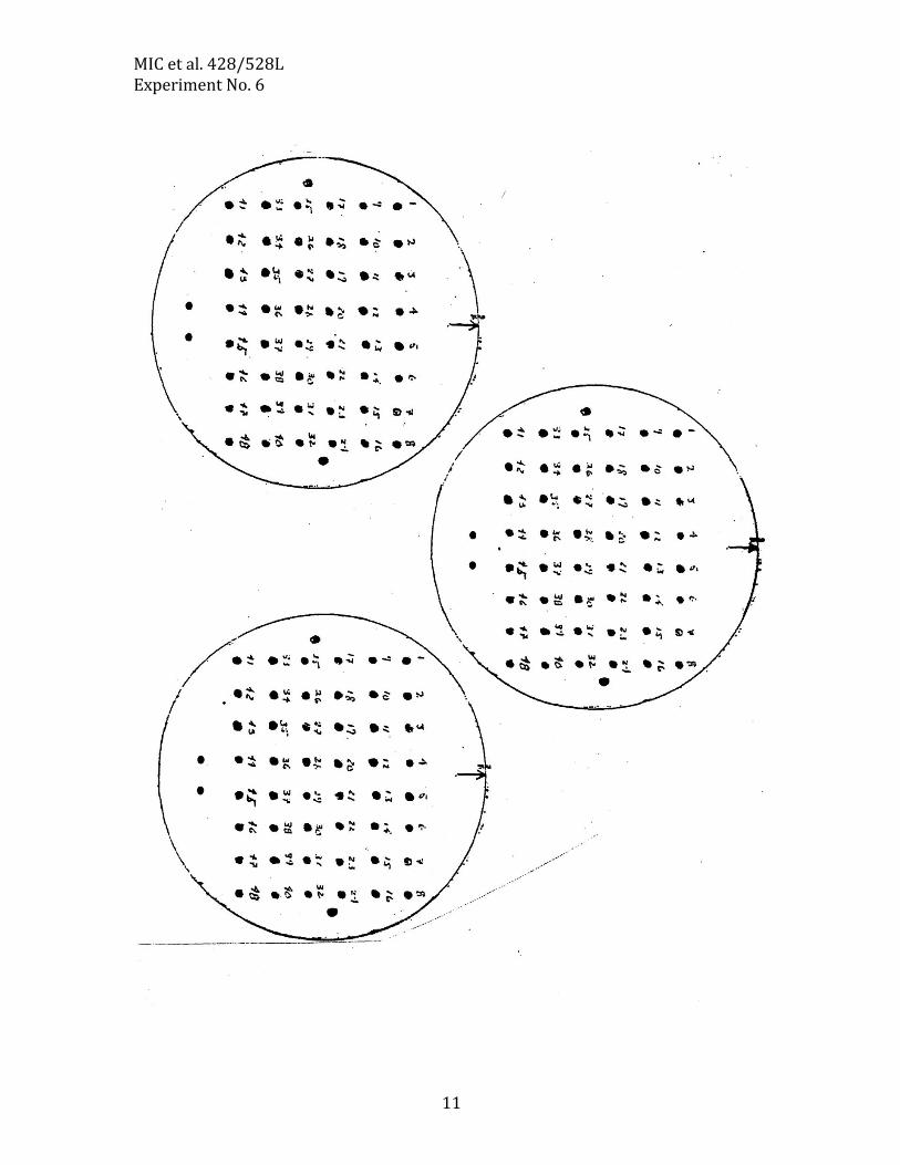
1) Using ETOH-flamed forceps, carefully lift and place each filter in labeled large sterile glass tubes. Add 2 ml sterile ddH2O and vortex until the cells are removed from the filters. 2) Spread 100 µl from the suspensions of DBL330 (pRL27) alone onto 1 LB agar + Rif50, Kan25 plate. Repeat for the Luteibacter DBL564 alone tube. Incubate plates @ 28oC. 3) Spread 100 µl from each mating filter suspensions onto 3 LB + Rif50, Kan25 plates each (6 plates total). Place all at 28oC Store remaining filter suspensions @ 4oC so you can plate more if needed later. Check plates for the next several lab periods: Patch colonies onto LB + Rif 50 Kan25 plates. You should patch at least 10 master plates. Section 1, 2/23/2015 W to 3/11/2015 W Section 2, 2/24/2015 R to 3/12/2015 R Section 3, 2/23/2015 W to 3/5/2015 W ---Examine your plates, record what you see. Do you see any unusual colony phenotypes on the LB + Rif50Kan25 plates? Possible phenotypes include colonies that appear less/more “goopy”, have different colors, or those that grow slow. The diameter of the patch can be altered, etc. Record your findings carefully. DO NOT DISCARD YOUR PLATES TOO QUICKLY! Remember, many phenotypes are only visible under specific conditions or on specific media. Therefore, the next step is to look for specific phenotypic differences as compared to wild type strain DBL564. We will use different media as we will be looking for different things. ---Using sterile toothpicks, begin to patch at least 500 colonies from your mating plates onto various media (Fig. 6.5 on Next Page) using the grid pattern provided. If you carefully patch your cells onto the grids, then you can save time by replica-pronging the colonies onto the subsequent screening media. Replica-pronging Protocol

MIC et al. 428/528L Experiment No. 6
9
We have a limited number of replica-pronging devices. They basically are metal rectangles with a handle on the top and 48 stainless steel prongs on the bottom. Used correctly, you can transfer 48 colonies at once to multiple medium plates. 1. Pour 95% ETOH into a glass Petri dish. Dip the pronger into the ETOH and pass it through a flame (do not hold it in the flame). 2. Once the ETOH had burned off, set the pronger lightly onto a plain LB agar plate to cool the prongs (~10 sec.). 3. Carefully line up the pronger on top of the colonies on the first master plate (Pl 1). Jiggle it gently to adsorb cells to the prongs. 4. Lift it up and lightly place on the first screening plate, Skim Milk agar. Then the second, motility plate, then passage to another LB + Kan25 plate and place at 37oC. Continue until all plates of that series are done. The last plate to be pronged is another LB + Kan25 plate. 5. Once a set of plates is completed, wash the prongs with water on a paper towel. 6. Replace pronger in ETOH and repeat for the next series of plates (Pl 2, etc.). 7. When done, wash the pronger thoroughly and place it back in the cabinet. ---Incubate plates @ 28oC. During this time, you will need to come to the labs to patch your colonies. NOTE: The plate numbers and patch number determine your mutant’s identification. For example, if patch 37 on plate set 3 looks interesting in some way, then the mutant is 3-37.

MIC et al. 428/528L Experiment No. 6
10
Sterile toothpick
Replica Stamp to Skim Milk and Motility
LB + Kn50
LB + Kn50
Fig. 6.5 Screening Transposon Mutations
Skim Milk
Skim Milk
MIC et al. 428/528L Experiment No. 6
11

MIC et al. 428/528L Experiment No. 6
12
---It is imperative as a good scientist that you keep accurate records. A Nobel Prize-worthy mutant means nothing if it cannot be recovered. ---Make copies of the score sheet (see previous page). ---Score your plates. Note the numbers of colonies on each plate and any interesting or unusual phenotypes. Make accurate notes, circle the interesting mutants on the stock LB + Kan25 plates (last ones patched) and refrigerate them. ---Replica-prong all of your mutants from your 10 master plates onto new LB + Kan25
plates as necessary to keep them fresh. ---Make glycerols of all your mutants. See Appendix 1 for the protocol. ---Re-test your interesting mutants to verify the phenotype. Wait until you have several interesting ones and re-test all at once. IMPORTANT: THE POSSIBILITY OF SEEING SOMETHING INTERESTING IS DIRECTLY PROPORTIONAL TO THE NUMBER OF EXCONJUGANTS YOU PATCH. WE WILL BE USING SELECTED MUTANTS FOR OTHER EXERCISES THIS SEMESTER. BE SURE TO CAREFULLY ANNOTATE YOUR PHENOTYPES, AND KEEP FRESH LB+Gm25 PLATES OF EACH INTERESTING MUTANT.
Please have your collection of interesting mutants (5-10)
ready by 3/11 (3/12 T)

MIC et al. 428/528L Experiment No. 6
13
DBL564 Transposon Mutagenesis Score Sheet: Plate No.________ Patch No. SM Motility LB+ Rif
Kan Notes
1 2 3 4 5 6 7 8 9
10 11 12 13 14 15 16 17 18 19 20 21 22 23 24 25 26 27 28 29 30 31 32 33 34 35 36 37 38 39 40 41 42 43 44 45 46 47 48

MIC et al. 428/528L Experiment No. 6
14
EXPERIMENT NO. 5 – SELECTED QUESTIONS 1. Once the transposon has transposed from the suicide vector into the chromosome, what keeps it from hopping over and over and over and over and over? 2. Would you expect to see increases and decreases in motility in mutants? 3. You notice that one colony of DBL564::pRL27 grows slowly on LB plates. What might this tell you about the effect of the transposon insertion?
MIC et al. 428/528L Experiment No. 6
15
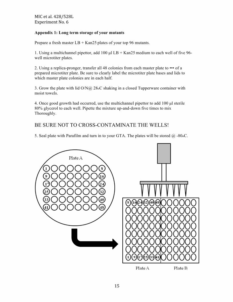
Appendix 1: Long term storage of your mutants Prepare a fresh master LB + Kan25 plates of your top 96 mutants. 1. Using a multichannel pipettor, add 100 µl LB + Kan25 medium to each well of five 96- well microtiter plates. 2. Using a replica-pronger, transfer all 48 colonies from each master plate to ••• of a prepared microtiter plate. Be sure to clearly label the microtiter plate bases and lids to which master plate colonies are in each half. 3. Grow the plate with lid O/N@ 28oC shaking in a closed Tupperware container with moist towels. 4. Once good growth had occurred, use the multichannel pipettor to add 100 µl sterile 80% glycerol to each well. Pipette the mixture up-and-down five times to mix Thoroughly. BE SURE NOT TO CROSS-CONTAMINATE THE WELLS! 5. Seal plate with Parafilm and turn in to your GTA. The plates will be stored @ -80oC.

MIC et al. 428/528L Experiment No. 7
1
EXPERIMENT NO.7
ISOLATION OF CHROMOSOMAL DNA
Section 1, 3/23/2015 M Section 2, 3/24/2015 T Section 3, 3/23/2015 M
INTRODUCTION Many types of research that address a myriad of biological questions require the isolation of the genomic DNA of an organism. This is a prerequiste for many experiments, from DNA sequencing analysis of a genome to cloning specific regions or genes to construction of a genomic library of the organism to the performance of metagenomic studies. In Experiment No. 6 we used a novel modified Tn5 transposon to generate mutants of Luteibacter with interesting phenotypes. Review of pRL27 Review the introduction of Experiment No. 6. The transposon carried by pRL27 has many useful properties. 1. Inverted repeats (IRs). 2. No tnpA transposase gene 3. A hyperactive transposase 4. Neophophotransferase activity (npt = KmR). 5. An oriR6K origin of replication. Experimental overview We will isolate total genomic DNA from a couple of your mutants and digest it with a restriction enzyme. We will self-ligate the fragments and transform the ligated mix into a special strain of E. coli that contains the pir gene encoding the replication protein. This will allow only the single chromosomal fragment that contains the Tn to replicate as a plasmid. Next, we will isolate the plasmid and use DNA primers specific to the ends of the Tn to sequence into the adjacent chromosomal DNA. The sequence generated will be compared to the GenBank database to try to identify the gene disrupted by the Tn. The isolation of chromosomal DNA is the critical first step for many experimental procedures. One common goal of all chromosomal isolation protocols is to avoid shearing the chromosomal DNA. The large chromosome is fragile and can be sheared easily into pieces. In fact, in spite of our best efforts, some chromosomal DNA will be sheared-there is

MIC et al. 428/528L Experiment No. 7
2
simply no way to avoid it. Most methods utilize a gentle lysis step using a detergent (e.g. SDS) and an enzyme (e.g. Proteinase K) that degrades proteins. Unwanted proteins bound to the DNA are usually removed by the addition of chloroform (CHCL3), a nonpolar chemical that denatures them. Finally, the genomic DNA is precipitated using the nonpolar solvent isopropanol or ethanol (EtOH) and excess salt is removed with a 70% EtOH wash. DNA precipitation is based on the fact that DNA has a strong negatively charged phosphate backbone-it is very polar. If a polar molecule is placed in a less polar solvent such as isopropanol or ethanol, it will try to minimize the amount of surface area exposed to the solvent. Thus it will collapse into a ball, and hence precipitate. To facilitate chromosomal isolation, we will be using the Gentra Puregene Yeast/Bact. Kit from Qiagen, Inc. (www.qiagen.com). This kit is speedy and produces good quality chromosomal DNA for restriction digestions. You and/or your partner must come in and inoculate 3 ml LB + Km50 overnight cultures (O/Ns) of an interesting mutants. Incubate the tubes shaking at 28oC.
Section 1, 3/25/2015 W Section 2, 3/26/2015 Th Section 3, 3/25/2015 W
MATERIALS Overnights of strain 30-84::RL27 mutants Gentra Puregene Yeast/Bact. Kits (1 per mutant) Pipettor (P-1000, P-200, P-20) Sterile tips Sterile microfuge tubes Microcentrifuges 80OC water bath 65OC water bath 37OC water bath EXPERIMENTAL PROTOCOL We will be following the instruction that come with the Gentra Puregene Yeast/Bact. Kit (next page).

MIC et al. 428/528L Experiment No. 7
3

MIC et al. 428/528L Experiment No. 7
4
MIC et al. 428/528L Experiment No. 7
5
It is important to be careful in each step or subsequent ones will not work. Chromosomal Digestions Now that we have isolated genomic DNA from several mutants, we need to digest it and re-ligate it to create a plasmid containing the modified Tn and adjacent chromosomal DNA. Fig. 1 illustrates the principles of the next step of the experiment. The intact chromosomal DNA will be digested with a restriction enzyme (BamHI) that cleaves within the chromosomal DNA on each side of the Tn at the sequence (5’-GGATCC-3’). There are no BamHI sites within the Tn. The distance from the insertion where BamHI cuts is dependent on the location of the recognition sequence. Therefore, the enzyme may cut close to the Tn or far away. The calculated size a BamHI fragments of DNA is given below.
46 = 4,096 bps Where: 4 = number of different bases (A, C, G, or T). 6 = length of the recognition sequence (6 nts) bps = basepairs So, on average BamHI will digest the chromosome approximately every 5 kb. This means for a genome of 6 Mb (megabases), a BamHI digestion will create 1,200 fragments (6 Mb = 6,000,000 bps divided by 5,000 bps/cut = 1,200 cuts = number of fragments for a circular piece of DNA). We will next self-ligate the fragments into circles. The key point is that only one of these 1,200 circles of chromosomal DNA will contain the Tn which has the -dependent R6K origin of replication and KanR. If we transform all of this DNA into an E. coli strain that contains the pir gene that encodes the replication protein, only the one circular DNA piece will replicate. This plasmid contains the modified Tn plus the adjacent region of the chromosomal DNA!
Figure 1.

MIC et al. 428/528L Experiment No. 7
6
PROTOCOL 1. Set up the following digestion for each of your chromosomal DNAs:
7 µL chromosomal DNA 3 µL 10x Reaction 3 Buffer 18 µL H2O 2 µL BamHI enzyme 30 µL total volume
2. Incubate the digestion in the 37OC water bath for 2 hours. 3. Store the remaining digested DNAs at 4OC until the next lab. NOTE: Restriction enzymes stocks are extremely sensitive to temperature fluctuations. They must be kept as close to -20oC as possible at all times. Please remove them from the ice buckets as little as possible, and immediately return them to the freezer when you are done with them

MIC et al. 428/528L Experiment No. 7
7
Section 1, 3/30/2015 M Section 2, 3/31/2015 T Section 3, 3/30/2015 M
1. Pour and run 6 µL of the completed digestions on an agarose gel (see below). 2. Set up ligations with 5 µL of the completed digestions as follows:
5 µL BamHI-digested DNA 4 µL 5x Ligase Buffer 9 µL H2O 2 µL DNA Ligase 20 µL total volume
3. Incubate ligations at 15OC overnight. 4. Next day store ligation mixes at 4OC.
Section 1, 4/6/2015 M Section 2, 4/7/2015 T Section 3, 4/6/2015 M
TRANSFORMATION OF E. coli WITH LIGATED DNA Many bacteria in nature naturally take up exogenous DNA from their environment. This is thought to be a mechanism for acquistion of nucleotides as the DNA is often degraded, and may help bacteria acquire new traits if the DNA provides a beneficial trait. However, the genetic workhorse, E. coli, does not naturally take up exogenous DNA. It can be made competent, that is, it can be treated to make it able to take up DNA by the following procedure. Below is the basic protocol for making E. coli competent. However, we will provide you with competent cells. Procedure for making E. coli competent. Grow a culture of E. coli to early log phase growth (OD600 ~0.3). Centrifuge the culture at 4OC to pellet the cells. Resuspend the cells in 1/2 volume ice cold 50 mM calcium chloride (CaCl2) and sit on ice overnight. The following day, centrifuge the cells and resuspend the pellet in 1/10 volume ice cold 50 mM CaCl2. Immediately add 1.5 ml 80% sterile glycerol

MIC et al. 428/528L Experiment No. 7
8
per 10 ml cells on ice and mix gently. Aliquot 0.5 ml into chille microfuge tubes, cap and drop into liquid nitrogen to quick-‐‑freeze the cells. Store aliquoted competent cells at -‐‑80OC until ready for use. Thaw amount needed on ice. Discard any unused cells as they cannot be refrozen. However, recovery of the single circular fragment out of all of the possible circular genomic fragments is difficult and the rate-‐‑limiting step to the procedure. Therefore, this year we will try electroporating the DNA into special electrocompetent E. coli cells. ELECTROPORATION PROTOCOL Ligation Cleanup 1. To ligation, add 300 µL GeneJet Wash Solution. 2. Load onto GeneJet column. Spin 1 minute, discard flow-through. 3. Add 750 µL GeneJet Wash Solution. Spin 1 minute, discard flow-through. 4. Spin dry column 2 minutes to dry thoroughly. 5. Add 20 µL 55OC sterile water, let sit 2 mins. 6. Spin column 3 mins. Keep flow through as this is your ligated DNA. Electroporation 1. Add 2 µμL of ligation mix to a 100uL chilled tube of electrocompetent E. coli Pir+ cells. To a separate tube of E. coli add 1uL of positive control plasmid. 2. Transfer this 102uL (or 101uL for positive control) to a chilled electroporation cuvettes. 3. Electroporate using electroporator, setting 1. Write down numbers after the machine shocks the cells. 4. Immediately add 300uL Room temperature LB buffer to cuvette. Pipette this 400uL total (LB plus electroporation reaction) to the 1.5mL eppendorf tube. 5. Incubate while shaking at 37OC for 60 min. 1. Spread 400 µμL of solution onto LB + Kan25 plates. Incubate at 37OC for 1-‐‑3 days.

MIC et al. 428/528L Experiment No. 7
9
Section 1, 4/7/2015 T NOTE: You must come in on an odd day! Section 2, 4/8/2015 W NOTE: You must come in on an odd day! Section 3, 4/7/2015 T NOTE: You must come in on an odd day!
1. Check transformation plates. 2. Pick 2 colonies from each transformation and:
Touch a new LB + Kan50 plate, and Inoculate a 3 ml LB + Kan50 broth overnight of each.
3. Incubate the tubes shaking at 37OC and the plates at 37OC.
Section 1, 4/8/2015 M Section 2, 4/9/2015 T Section 3, 4/8/2015 M
ISOLATION OF PLASMID DNA FOR SIZING AND SEQUENCING Now the goal is to isolate the plasmid DNA from the transformed E. coli DH5pir cells. This is to allow us to determine:
1. How large is the plasmid? 2. The DNA sequence of the chromosomal DNA adjacent to the Tn.
Prepare plasmid DNA from each overnight culture according to the protocol used in Exercise 4.

MIC et al. 428/528L Experiment No. 7
10
Please use your best skills during this procedure. Good DNA is essential for successful DNA sequencing. The presence of contaminating salt will inhibit the sequencing reaction resulting in poor or no readable sequence. Turn in your plasmid preps (well-labeled with name, clone #, etc.). We will determine the concentration of your DNA. The DNA sequencing center requires at least 50 ng/l DNA (400 ng total DNA) for sequence analysis. If necessary, you will have to re-make the plasmid DNA. This is why we keep a plate of the clones.
MIC et al. 428/528L Experiment No. 7
11
Experiment No. 7 Selected Questions
1. Explain the role of each of the following reagents used in chromosomal DNA isolation: A. SDS. B. Ethanol (EtOH). 2. A 25-bp piece of double stranded DNA when placed into sterile water will assume a linear form. Why?

MIC et al. 428/528L Experiment No. 7
1
EXPERIMENT NO.8
OBSERVING MICROBIAL EVOLUTION
INTRODUCTION Given their large population sizes and rapid growth rates, microbial populations can adapt quite readily to new environments. Furthermore, their small size means that cells can sense and respond to fine scaled environmental gradients. Since the beginning of the semester, you have been passaging and plating strains of P. syringae in static (not shaking) liquid media. You have observed what these cultures have looked like each week, and have plated out samples of these cultures to identify colony variants that have arisen over this time course. This experiment was based on a paper (by Mike Travisano and Paul Rainey http://www.nature.com/nature/journal/v394/n6688/abs/394069a0.html ) which first investigated this adaptive evolution using Pseudomonas fluorescens strain SBW25. These first experiments showed a rapid diversification of colony types in the same exact media that you have been passaging your cells in. The adaptive radiation occurs because the lack of shaking of the media enables an oxygen gradient to form. Because of this gradient, you can observe mutants of P. fluorescens which form a mat (or pellicle) on the top of the media (high oxygen) and some that are found at the bottom of the media (low oxygen). If you let the cultures sit for long enough, the mat will collapse under it’s own weight and you’ll see an intermediate mass in the culture. The wrinkly spreader phenotype arises due to cellulose overexpression and modification by P. fluorescens which enables mat formation. At a more direct level, many of the mutations that cause the wrinkly spreader phenotype affect cyclic di-GMP levels, which is a key signaling component across bacteria. This experiment has been repeated across bacterial species (and even in P. syringae), and typically you can find phenotypes that look like wrinkly spreaders after a certain number of passages. The strains you have been passaging are all versions of P. syringae pathovar lachrymans and are all pathogenic on cucumbers. For reasons outside the scope of this lab, there are reasons to believe that cyclic di-GMP levels may differ across these strains and thus maybe the wrinkly spreader phenotype will be different than previously reported. I actually don’t know what the outcome “should” be, you are the first ones to carry out these experiments for this period of time with these strains. I’m interested to see what happened.
MIC et al. 428/528L Experiment No. 7
2
Section 1, 4/6/2015 M Section 2, 4/7/2015 T Section 3, 4/6/2015 M
MATERIALS 3 KB plates with Kan25 and Tet10 You should have streaked the last passage to single colonies one week ago. Observe these single colonies and note any differences you see compared to the original isolate. You should have been documenting what these colonies looked like through time, but a plate of the original isolate will also be provided for comparison. Across the single colonies, what is the frequency of the different colony types? Find at least two (three if possible) different colony types on your plates. Streak each of these morphologically different colonies to single colonies on their own plates KB Kan Tet plates. Place at 27 degrees C.
Section 1, 4/82015 W Section 2, 4/9/2015 R Section 3, 4/8/2015 W
MATERIALS 4 Test Tubes containing 5mL KB liquid media (with Kan25/Tet10). 1 Plate of the “ancestral” strain Observe colony morphology on the plates you streaked out on M/T. Does the morphology match what you observed on the original plate you picked the colony from? If so, that means that the phenotype is heritable (inheritable) and thus the morphological change is due to an underlying genetic mutation. Pick each of your morphologically different colonies to a separate test tube containing 5mL media. Also pick a sample of the original, unevolved strain to a separate test tube. Place in the 27 degree incubator.
Section 1, 4/13/2015 M Section 2, 4/14/2015 T Section 3, 4/13/2015 M
Observe the test tubes that you inoculated last W/R and compare these to the “ancestral” controls. Do the any of the cultures appear to perform differently in liquid culture from each other or from the controls?

MIC et al. 428/528L Experiment No. 7
3
Section 1, 4/15/2015 W Section 2, 4/16/2015 R Section 3, 4/15/2015 W
Observe the test tubes that you inoculated last W/R and compare these to the “ancestral” controls. Do the any of the cultures appear to perform differently in liquid culture from each other or from the controls?
MIC et al. 428/528L Experiment No. 7
4
Experiment No. 7 Selected Questions
1. What would happen to these cultures if you shook them during the course of passages? 2. Cyclic di-GMP is an important signaling molecule for a variety of other phenotypes besides mat formation. Do you think these other phenotypes are affected (are there phenotypic tradeoffs) in the wrinkly spreader cells?