microbiolog & immunolg phd program - eth z · lymphocyte trafficking is a finely tuned...
TRANSCRIPT

5
Microbiology & Immunolgy
9
Special thanks to our sponsors:
5th
Student Retreat
Microbiology & Immunolgy
PhD Program
Davos, Switzerland
9th
– 11th
September 2012

2

3
Dear Students,
We would like to welcome you to the 2012 PhD student retreat of the MIM PhD Program in the
Youthpalace Davos in the canton Graubünden.
The following three days should give you the opportunity to discuss your research project with fellow
PhD Students in the field of Microbiology, Immunology and Virology. We have a broad scientific
program with 20 oral and 30 poster presentations of PhD projects from the three different fields.
Moreover we are happy to have 3 guest speakers who will give new insights about current research
projects in immunology & virology. In addition, to broaden our view of other research fields than the
MIM research field, we have a guest speaker from the department of architecture from the ETH.
Apart from the scientific part, we will have a small party on the last evening and a short hiking trip on
Tuesday afternoon. Use these 3 days to discuss your PhD Project and the ones of others and feel free
to give as much feedback as you want to presentations, posters and us, as organizers of the retreat.
We hope we arranged an interesting and entertaining program and that all of you, whether
newcomers in the first year or 2nd
and 3rd
year PhD-Students, enjoy the three days with us in Davos.
We are looking forward to meeting all of you and would like to thank you for your participation in
this year’s students retreat.
Best wishes,
The 2012 Organizing Committee
Thomas, Pascal, Jonas, Tess, Dominik, Julia & Leon

4
General Information
Accomodation in Davos
Jugendherberge Davos
Youthpalace
Horlaubenstrasse 27
7260 Davos (GR)
Tel: +41 81 410 19 20
Fax: +41 81 410 19 21
www.youthhostel.ch/davos
Travel Information
• Train Zurich to Davos: Train leaves Zurich HB, 09.09.2012 at 8:37, arrives at Davos Dorf at
10:50. (CHANGE TRAIN at Landquart) – Meeting point/time: large clock (meeting point) in
the main hall of the Zurich Main Train Station (ZHB) by 8:15 am, Sunday 9th
• Train Davos to Zurich: Train leaves Davos Dorf, 11.09.2012 at 14:06, arrives at 16:23.
(CHANGE TRAIN at Landquart)
Organizing Team
Contact information City Tel.No. Email
Dominik Aschenbrenner (Bellinzona) 078 923 5729 [email protected]
Tess Brodie (Bellinzona) 079 459 4396 [email protected]
Thomas Edinger (Zürich) 076 743 0342 [email protected]
Jonas Müller (Zürich) 076 707 6136 [email protected]
Julia Noack (Bellinzona) 078 915 1253 [email protected]
Leontios Pappas (Bellinzona) 078 758 9466 [email protected]
Pascal Ziltener (Zürich) 076 790 1788 [email protected]

5
MIM Retreat 2012 Program Overview
*packed lunch will be provided after breakfast. During the hike, luggage can be stored in the youthostel.
Time/date Sunday, 9th Monday, 10th Tuesday, 11th
7
7:30 – 8:30 Breakfast
8 8:37 Departure Zürich
10:50 Arrival Davos
8:00 - 9:00 Breakfast
8:30 – 10:30
Presentation Session 3 9 9:00 - 13:00
Hike and lunch*
10
10:30 – 11:00 Coffee Break
11 11:00 – 12:00
Arrival and Welcome
11:00 – 12:00
Presentation Session 4
12 12:00 – 13:00 Lunch 12:00 – 13:30 Lunch
13 13:00 – 14:00
Presentation Session 1 13:30 – 14:30
Dr. Linda Schädler 14 14:00 – 15:00
Prof. Jens Stein
14:06 Departure Davos
16:23 arrival Zürich HB
14:30 – 15:00 Coffee Break
15 15:00 – 15:30 Coffee Break 15:00 – 16:00
Prof. Daniel Pinschewer 15:30 – 16:30
Presentation Session 2 16
16:30 – 17:30
Prof. Fulvio Reggiori 17
18 18:00 – 19:30 Dinner
18:30 – 20:00 Dinner
19
19:30 – 21:30
Poster Session 2
20 20:00 – 22:00
Poster Session 1
21
21:30 Party
22

6
Talks & Posters
Guest Speakers
I. Prof. Jens Stein
Theodor Kocher Institute
University of Bern
Bern Switzerland
E-mail: [email protected]
CURRICULUM VITAE (EXCERPTS)
Personal data
Nationality German
Date and Place of birth 12.11.1968 in Aachen (Germany)
Academic career
Since 07/2010 Lecturer (“Dozent”), Theodor Kocher Institute, University of Bern,
Switzerland
2004 – 2010 Group leader (“Oberassistent”), Theodor Kocher Institute, University of Bern,
Switzerland; Venia docendi in Immunology (April 2010)
2003 – 2004 Young Investigator “Ramón y Cajal”, Department of Immunology and
Oncology, National Center for Biotechnology (CNB)-CSIC, Madrid, Spain
1999 – 2003 Postdoctoral fellow in the laboratory of Carlos Martínez-A., Department of
Immunology and Oncology, National Center for Biotechnology (CNB)-CSIC,
Madrid, Spain
1999 Ph.D. awarded from the Université Louis Pasteur, Strasbourg, France Mention
“Très honorable avec félicitations”
1995 –1999 Ph. D. thesis at the Center for Blood Research (CBR) and Department of
Pathology, Harvard Medical School (HMS), Boston, USA and Institut de
Génétique et Biologie Moléculaire et Cellulaire (IGBMC), University Louis
Pasteur, Strasbourg, France
Title: “Mechanisms of lymphocyte homing to peripheral lymph nodes”
Supervisors: Dr. U. H. von Andrian, CBR and HMS, and Dr. Diane Mathis,
IGBMC, University Louis Pasteur, France
1995 Diploma in Molecular Biology and Bioengineerin awarded from Ecole
Supérieure de Biotechnologie de Strasbourg (ESBS), University Louis Pasteur,
Strasbourg, France

7
1995 Diploma thesis at LeukoSite Inc., Cambridge, USA Title: “A11: A new
chemokine receptor & CC-CKR-1: Functional characterization”
Supervisor: Dr. C. R. Mackay, LeukoSite, Inc., Cambridge, USA
1992 – 1995 Ecole Supérieure de Biotechnologie de Strasbourg (ESBS), University Louis
Pasteur, Strasbourg, France
1989 – 1992 Basic biology studies (“Vordiplom”), University of Karlsruhe, Germany
Abstract
Molecular mechanisms directing lymphocyte migration during immune surveillance
An essential feature of the adaptive immune system is the continuous lymphocyte trafficking through
the body in search of foreign antigen, known as immune surveillance. Lymphocytes are in permanent
movement from blood into lymphoid organs such as spleen and lymph nodes. Within these organs,
lymphocytes screen antigen-presenting cells for their cognate antigen. Recirculation is completed by
migration of lymphocytes back to blood via lymphatic vessels. Continuous trafficking makes
lymphocytes one of the most motile mammalian cell types, migrating hundreds of micrometer every
day within tissue.
Lymphocyte trafficking is a finely tuned mechanism, which is in large part regulated by adhesion
receptors of the integrin family and G-protein-coupled receptor (GPCR), notably chemokine
receptors. Chemokines are small secreted polypeptides highly expressed in spleen and lymph nodes
and play an important role in the selective recruitment of blood-borne lymphocytes into lymphoid
tissue. Also, chemokines are responsible for lymphocyte segregation into T and B cell areas inside
lymphoid tissue. Our research encompasses three complementary lines of investigations. A major
focus is the study of molecular mechanisms of lymphocyte migration to and within PLN, with a
special emphasis on chemokine receptor signaling. As examples, we study the regulators of small
GTPases and their effect on in vitro and in vivo motility. Second, we are examining T cell-dendritic cell
(DC) interactions during immune responses using transgenic mice. Finally, we are applying novel
imaging techniques for the study of these events, mainly intravital microscopy (IVM), twophoton
microscopy (2PM) and, as of recent, Optical Projection Tomography (OPT) and selective plane
illumination microscopy (SPIM).

8
II. Prof. Fulvio Reggiori
Department of Cell Biology, Cell Microscopy Center and Institute of Biomembranes
University Medical Centre (UMC) Utrecht
Utrecht, the Netherlands
E-mail: [email protected]
CURRICULUM VITAE (EXCERPTS)
Personal data
Citizenship: Swiss
Birthday and place: 26 November 1970, Varese (Italy)
Languages: Native Italian, fluent English (spoken and written), fluent French
(spoken and written), Portuguese (spoken), basic knowledge in
German and Dutch (spoken and written)
Education
1985 - 1989 General Certificate of Education in the Cantonal High School of Bellinzona (TI,
Switzerland). General Certificate of Education work in chemistry: “Tri-dimensional
configuration of trypsin and pepsin”.
1989 - 1993 Undergraduate student at the Institute of Biochemistry, University of Fribourg
(Switzerland). Graduation work: “Detection of remodeling enzyme activity in yeast
Saccharomyces cerevisiae, a protein which catalyses the lipid change in GPI anchors”
(Supervisor: Prof. A. Conzelmann).
1994 - 1997 PhD student in the laboratory of Prof. Andreas Conzelmann, Institute of
Biochemistry, University of Fribourg (Switzerland). PhD thesis: “Remodelings of GPI
anchor lipid moiety in Saccharomyces cerevisiae”
Professional experience
1997 - 1998 Postdoctoral position in the laboratory of Prof. Andreas Conzelmann, Institute of
Biochemistry, University of Fribourg (Switzerland)
1998 - 2001 Postdoctoral position in the laboratory of Dr. Hugh Pelham, MRC Laboratory of
Molecular Biology, Cambridge (United Kingdom)
2001 - 2005 Postdoctoral position in the laboratory of Prof. Daniel Klionsky, Life Sciences
Institutes and Department of Molecular, Cellular, and Developmental Biology,
University of Michigan, Ann Arbor (USA)
2005 – 2011 Assistant Professor (permanent position), Department of Cell Biology, UMC Utrecht,
Utrecht University (The Netherlands)
2011 – now Associate Professor (permanent position), Department of Cell Biology, UMC
Utrecht, Utrecht University (The Netherlands)
Fellowships, awards and grants
1991 Arturo e Marguerite Lang Found fellowship
1991 Felix Leeman Found fellowship
1991 Achille Isella Found fellowship
1991 Helsinn fellowship
1993 "Association des anciens etudiants en chimie et biochimie UNI Fribourg" fellowship

9
1993 CIBA-GEIGY prize for the best 1993/1994 graduation curriculum in chemistry and
biochemistry at the University of Fribourg
1998 Swiss National Science Foundation fellowship (1 year)
Title: Retrieval transport inbetween Golgi compartments
1999 EMBO long-term fellowship (2 years)
Title: Retrieval transport inbetween Golgi compartments
2001 EMBO long-term fellowship (1 year)
Title: Identification of the triggering event in the sequestration step of the Cvt
pathway
2002 Swiss National Science Foundation fellowship for advanced researchers (2 years)
Title: Identification of the triggering event in the sequestration step of the Cvt
pathway
2006 Utrecht University High Potential grant with Dr. Xander de Haan
(5 years, 1’000’000 €)Title: Hosting coronavirus infection: Subversion of the cellular
machinery of autophagy
2006 Netherlands Organization for Health Research and Development (ZonMW)-VIDI grant
(5 years, 600’000 €)
Title: Dissection of the molecular mechanism of autophagy
2007 ZonMW medium investment grant (500’000 €)
Title: Ultrasensitive DeltaVision Microscope real time (RT) technology for biomedical
investigations at the subcellular level
2007 Earth and Life Sciences (ALW) open program grant with Prof. Bernd Helms
(3 years, one postdoctoral fellow + 66’600 €)
Title: Regulation of autophagy by the cellular lipidome
2008 Utrecht University Short Stay Fellowship for PhD students from China and India with
Nian Liu (3 months) Title: Unveiling new cellular roles of autophagy
2010 ECHO Project Grant form Chemical Sciences (CW) (3 years, 260’000 €)
Title: The role of the Atg9 reservoirs in autophagosome biogenesis
2010 DFG-NWO cooperation grant with Prof. Christian Ungermann (3 years, 2 PhD
students and 141’500 €)
Title: Regulation of autophagosome fusion with vacuoles
2011 Earth and Life Sciences (ALW) open program grant (241’206 €)
Title: Regulation of the autophagosome completion by phosphatidylinositol-3-
phosphatases
2011 ZonMW VENI postdoctoral fellowship with prof. Jos van Strijp for Jovanka Besteborer
(3 years, 250’000 €) Title: Outside-In: A role for the extracellular recognition receptors
in intracellular immunity
2012 Utrecht University Short Stay Fellowship for PhD students from China and India for
Shanshan Wong (3 months)
10
Abstract
Coronaviruses hijack LC3-I-positive EDEMosome membranes for replication
Fulvio Reggiori, Department of Cell Biology, University Medical Centre Utrecht, Utrecht, The
Netherlands
Coronaviruses (CoVs) are enveloped positive-stranded RNA viruses that infect the mammalian
respiratory and gastrointestinal tracts with mechanisms that are poorly characterized. The relevance
of this family of viruses has considerably increased due to their recent emerging as the cause of the
severe acute respiratory syndrome (SARS). In 2003, an epidemic outbreak of the SARS CoVs (SARS-
CoV) in South East Asia and Canada sickened more than 8,000 people and killed nearly 800 of them.
Two additional new human CoVs, HCoV-NL63 and HCoV-HKU-1, have recently been discovered and
because this family of viruses is characterized by frequent host-shifting events including zoonosis
(animal-to-human), CoVs are a threat for the human health. Unfortunately, an effective therapy
against these pathogens, which also causes considerable losses in the livestock industry, does not
exist.
Upon entering the host cells, CoVs induce the formation of double-membrane vesicles
(DMVs). These structures are vital for the virus because harboring the viral replication and
transcription complexes. The subcellular origin of these DMVs remains unknown but a recent study
has indicated that they could be derived from the endoplasmic reticulum (ER). The ER is the organelle
where proteins are translated and folded before being sent to the extracellular space, plasma
membrane and other subcellular organelles. When proteins fail to acquire their final correct
conformation, they are rapidly removed from the ER by a system known as the ER-associated
degradation (ERAD). Under normal growing conditions, the activity of this degradative pathway is
maintained at minimal levels to avoid the disposal of correctly folded proteins. It has been shown
that this is achieved by eliminating two positive ERAD regulators from the ER lumen through a
transport route has been named the ERAD tuning and involve vesicle called EDEMosomes. The
mechanism underlying this pathway has still to be characterized but we have recently demonstrated
that the mouse hepatitis virus (MHV), the CoVs that we use as a model, hijacks the EDEMosomes to
generate its replicative DMVs. The molecular principles of this hijacking, however, remain completely
unknown. The elucidation of these principles has become of primary interest because we have
shown that inhibition of the ERAD tuning completely blocks MHV infection. This discovery has thus
unveiled a possible way of therapeutically fight CoVs infections. Importantly, initial observations
indicate that SARS-CoV is probably using the same expedient to invade host cells.

11

12
III. Dr. Linda Schädler
Institute for the History and Theory of Architecture (gta)
Department of Architecture, Swiss Federal Institute of Technology (ETH) Zurich
Zurich, Switzerland
E-mail: [email protected]
CURRICULUM VITAE
Since 2012 Postdoc Research Associate, Institute for the History and Theory of Architecture
gta, ETH Zurich, (Chair of the History of Art and Architecture, Prof. Dr. Philip
Ursprung)
Since 2001 Freelance art critic, Neue Zürcher Zeitung and author of essays for exhibition
catalogues from the late 19th to 21st century
2010 – 2012 Research Associate, Kunstmuseum Basel
2008 – 2011 Ph.D. „Der Blick von der Seite“ – James Colemans Werk und die anamorphotische
Perspektive“
2003 – 2008 Assistant Curator / Curator, Kunsthaus Zürich Exhibitions:
– „Rivoluzione! Italienische Moderne von Segantini bis Balla“ (2008/09) – „Félix
Vallotton – Idylle am Abgrund“ (2007/08)
– „Rodin“ (2007)
– „Gefrorene Augenblicke. Von Vallotton bis Hubbard/Birchler“
(2006/07)
– „The Expanded Eye. Sehen – entgrenzt und verflüssigt“ (2006) – „Fest
der Farbe. Die Sammlung Merzbacher---Mayer“ (2006)
– „The Art of the Archive. Fotografien aus dem Los Angeles Police Department“
and „Miroslav Tich ý“ (2005)
– „Monets Garten“ (2004/05)
2000 – 2003 Tutor, Institute of Art History, University of Zurich (Prof. Dr. Franz Zelger), and
Institut of German Philology, University of Zurich (Prof. Dr. Barbara Naumann)
1996 – 2003 Studies in German Philology, Art History, Mass Communication and Media
Research, University of Zurich. Master Thesis: „Künstlerische Reflexionen –
Eduardo Chillidas Hommagen an Alberto Giacometti“
Research Focus Art of the 20st and 21st century
Interdisciplinary researches (text---image---relation, ekphrasis, forms of
staging in contemporary art)
Discourse of presence (in multimedia installations, voice).
13
Research Abstract
From Boxes to Bodies.
The Artistic Approaches of Donald Judd and Bruce Nauman in Comparison
Dr. des. Linda Schädler
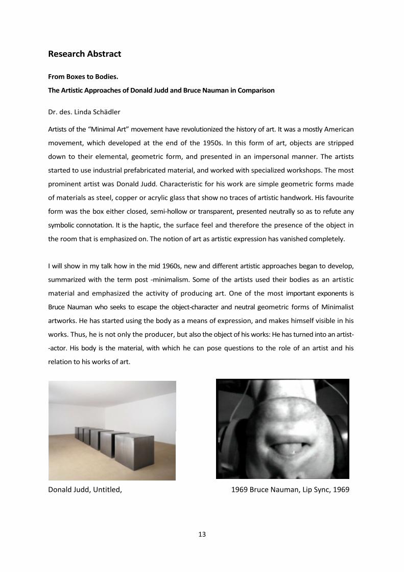
Artists of the “Minimal Art” movement have revolutionized the history of art. It was a mostly American
movement, which developed at the end of the 1950s. In this form of art, objects are stripped
down to their elemental, geometric form, and presented in an impersonal manner. The artists
started to use industrial prefabricated material, and worked with specialized workshops. The most
prominent artist was Donald Judd. Characteristic for his work are simple geometric forms made
of materials as steel, copper or acrylic glass that show no traces of artistic handwork. His favourite
form was the box either closed, semi-hollow or transparent, presented neutrally so as to refute any
symbolic connotation. It is the haptic, the surface feel and therefore the presence of the object in
the room that is emphasized on. The notion of art as artistic expression has vanished completely.
I will show in my talk how in the mid 1960s, new and different artistic approaches began to develop,
summarized with the term post -minimalism. Some of the artists used their bodies as an artistic
material and emphasized the activity of producing art. One of the most important exponents is
Bruce Nauman who seeks to escape the object-character and neutral geometric forms of Minimalist
artworks. He has started using the body as a means of expression, and makes himself visible in his
works. Thus, he is not only the producer, but also the object of his works: He has turned into an artist-
-actor. His body is the material, with which he can pose questions to the role of an artist and his
relation to his works of art.
Donald Judd, Untitled, 1969 Bruce Nauman, Lip Sync, 1969

14
IV. Prof. Daniel Pinschewer
Department of Pathology and Immunology &
W.H.O. Collaborating Centre for Vaccine Immunology University of Geneva C.M.U.
Geneva, Switzerland
E-mail: [email protected]
CURRICULUM VITAE (EXCERPTS)
Personal Data
Date and place of birth: August 21st 1974, Zurich, Switzerland
Nationality: Swiss
Citizenship: Bern
Professional training and positions
Per 09/2013 Full professorship, University of Geneva, CH, Faculty of Medicine, Department of
Pathology and Immunology
2008- Associate member of the W.H.O. Collaborating Centre for Vaccine Immunology,
University of Geneva, Switzerland
2007- Associate Professor (stipendiary professorship of the Swiss National Science
Foundation, SNF), University of Geneva, CH
2004-2007 Principle investigator / independent group leader, Institute of Experimental
Immunology, University Hospital of Zurich, CH
2002- 2004 Postdoctoral fellow, Institute of Experimental Immunology, University of Zurich, CH,
laboratory of Profs. R. Zinkernagel and H. Hengartner
2001-2002 Postdoctoral fellow, The Scripps Research Institute, La Jolla, CA, USA, laboratory of
Prof. J. C. de la Torre
1998 - 2000 Medical doctorate, Institute of Experimental Immunology, University of Zurich, CH,
laboratory of Profs. R. Zinkernagel and H. Hengartner
Education, degrees and habilitation
2006 Venia legendi (habilitation) in “Infektionsimmunologie” (immunology of infectious
diseases) at the Medical Faculty of the University of Zurich, Switzerland
2004 United States Medical Licensure Examination (USMLE) step 1
2001 Medical doctorate, University of Zurich, Switzerland
2000 United States Medical Licensure Examination (USMLE) step 2
2000 Swiss medical degree (Staatsexamen)
1994 – 2000 Medical School, University of Zurich, Switzerland
1993 Matura type A (Greek, Latin)
1987 – 1993 High School, Zurich, Switzerland
1981 – 1987 Primary School, Zurich, Switzerland

15
Former nominations for faculty appointments
2007 W2 professorship (associate professor, tenure-track), Institute of Immunology,
Ludwig Maximilians University Munich, Germany
2006 Principle Investigator, Center for Molecular Medicine of the Austrian Academy of
Sciences, Vienna, Austria
Prizes and awards
2011 Pfizer Research Prize for Infectiology, Rheumatology and Immunology (CHF 30’000
jointly with Dr. L. Flatz)
2010 Prix Leenaards 2010 pour la promotion de la recherche scientifique (CHF 600'000,
jointly with Prof. B. Marsland, University of Lausanne)
2010 Loeffler-Frosch-Prize of the “Society for Virology” (Gesellschaft für Virologie)
(EUR 5'000)
2007 Stipendiary professorship of the Swiss National Science Foundation
2001 Fellowship of the Gebert Rüf Stiftung, Switzerland
2001 Semesterprämie (thesis with distinction), Medical Faculty, University of Zurich
Research Abstract
Prof. Pinschewer’s laboratory has a longstanding interest in virus-host relationship, viral
pathogenesis and antiviral immunity. Additionally, they recently developed a research branch in
virally vectored vaccines. For their research they genetically engineer lymphocytic choriomeningitis
virus (LCMV) by reverse genetics techniques they have developed, and test immune response and
pathogenesis of the designed viruses. In his talk he will provide an overview over some of the most
recent studies published from the lab, with particular emphasis on vaccine vectors and the newly
discovered role of damage-associated patterns in driving antiviral immunity.

16
Overview Sessions
Session / Day Chair Person Start End Duration [min]
Sunday
Student Session 1 • Julia Noack
• Leontios Pappas
13:00 14.00 60
Guest Speaker1 • Pascal Ziltener 14:00 15:00 60 Jens Stein
Student Session 2 • Dominik Aschenbrenner
• Thomas Edinger
15:30 16:30 60
Guest Speaker 2 • Julia Noack 16:30 17:30 60 Fulvio Reggiori
Poster Session 1 • Drinks/Snacks 20:00 22:00 120
Monday
Student Session 3 • Jonas Müller
• Thomas Edinger
• Pascal Ziltener
8:30 10:30 120
Student Session 4 • Tess Brodie
• Leontios Pappas
11:00 12:00 60
Guest Speaker 3 • Dominik Aschenbrenner 13:30 14:30 60 Linda Schädler
Guest Speaker 4 • Jonas Müller 15:00 16:00 60 Daniel Pinschewer
Poster Session 2 • Drinks/Snacks 19:30 21:30 120

17
List of oral presentations
Student Session 1 - Sunday 13:00– 14:00 page
1. Thomas Edinger 19
2. Mattia Garbani 20
3. Eva Potthoff 21
Guest Speaker 1: Prof. Jens Stein 14:00 – 15:00 6-7
Student Session 2 – Sunday 15:30 – 16:30 page
4. Pascal Ziltener 22
5. Jonas Müller 23
6. Eveline Kindler 24
Guest Speaker 2: Prof. Fulvio Reggiori 16:30 – 17:30 8-10
Session 3 – Monday 8:30- 10:30 page
7. Boas Felmy 25
8. Moira Prati 26
9. Julia Noack 27
10. Leontios Pappas 28
11. Kathrin Moor 29
12. Anugraha Mathew 30
13. Giuseppina Fascellaro 31
14. Tess Brodie 32
Session 4 – Monday 11:00-12:00 page
15. Gustavo Gers-Huber 33
16. Lennart Schada von Borzyskowski 34
17. David Aebischer 35
18. Dominik Aschenbrenner 36
Guest Speaker 3: Dr. Linda Schädler 13:30-14:30 12-13
Guest Speaker 4: Prof. Daniel Pinschewer 15:00 - 16:00 14-15

18
List of Poster Presentations
Poster Session 1 – Sunday 20:00 – 22:00 page
19. Anna Müller 37
20. Ismeta Curkic 38
21. Lucia Reh 39
22. Ute Greczmiel 40
23. David Plaza 41
24. Marie-Theres Pohl 42
25. Nicolas Ponroy 43
26. Sarah Gabriel 44
27. Lisa Metzger 45
28. Christine Vogel 46
29. Kristina Poljak 47
30. Niels van der Velden 48
31. Emanuel Stiegeler 49
32. Roman Huber 50
33. Alexandra Ozga (Guest Student) 51
34. Markus Pieczyk (Guest Student) 52
Poster Session 2 – Monday 19:30 – 21:30 page
35. Jenna Denyes 53
36. Valentina Vongrad 54
37. Erica Russo 55
38. Walther Hänseler 56
39. Mary-Aude Rochat 57
40. Martina Stöckli 58
41. Florian Ryffel 59
42. Ana Chavez Steenbock 60
43. Alexey Dudnik 61
44. Thomas Liechti 62
45. Stefanie Schmieder 63
46. Dominik Müller 64
47. David Beauparlant 65
48. Raoul Rosenthal 66

19
Abstracts PhD Students
[1]
Receptor for activated C kinase 1 is a cellular host factor required for influenza A virus entry
Thomas H. Ludersdorfer1, Thomas O. Edinger*1, Christian Wisskirchen1, Patricia Nigg1, Karin Boucke2,
Eva Moritz1, Urs Greber2, Silke Stertz1 and Jovan Pavlovic1
1University of Zürich, Institute of Medical Virology, Winterthurerstrasse 190, 8057 Zürich Switzerland; 2University of Zürich Institute of Molecular Life Science, Winterthurerstrasse 190,
8057 Zürich, Switzerland.
*corresponding email address: [email protected]
Influenza A virus has a small genome with limited coding capacity and therefore strongly
depends on cellular factors for its replication. As a consequence, current research deals with
the identification of host factors that play a functional role in the entry process of influenza A
virus. Especially due to high mutational rates of influenza strains and the emergence of
broad resistance against common antivirals. RACK1 is a highly conserved intracellular
scaffold protein that binds activated protein kinase C beta II (PKCβII) and thereby increase
PKCβII mediated substrate phosphorylation. Previously, PKCβII had been shown to be of
importance for influenza A virus (IAV) infection. Furthermore, RACK1 had been identified as
an interaction partner of the viral matrix protein M1. We therefore investigated the role of
RACK1 during influenza A virus life cycle.
Here we demonstrate that RACK1 knockdown has tremendous effects on the influenza A
virus entry into lung epithelial cells. Rack1 knockdown resulted in decreased viral titers after
IAV infection with different viral strains. Using a virus-like particle entry assay we showed
impaired viral entry in siRACK1-treated cells. By performing confocal analysis of RACK1
knockdown cells, we could see dysfunction in the endosomal localization and unusual
microtubule and actin organization. Finally electron microscope imaging revealed trapped
viral particles below the cellular membrane after RACK1 knockdown. All together we could
show that RACK1 is a crucial host factor for the entry process of influenza a virus by
exhibiting essential functions within the cellular endocytic machinery and cytoskeleton
organization.

20
[2]
Dendritic cells targeting and improved cell penetration in allergen-specific
immunotherapy
Garbani Mattia, Rhyner Claudio, Crameri Reto
Swiss Institute for Allergy and Asthma Research (SIAF), Molecular Allergology, University of
Zürich, Davos, Switzerland
Todays therapies for allergic diseases aim to induce a switch towards a TH1/Treg dominated
immune response by challenging the patient with increasing doses of allergen. This
approach has two major disadvantages: the duration of the treatment and the anaphylactic
side-effects.
The Modular Antigen Translocation (MAT) Vaccine consist of a cell penetration peptide
linked to a part of invariant-Chain (Li). In the cell Li targets the immunogenic protein directly
to MHC-II, therefore i) less antigen is needed and ii) less injections are necessary. MAT
vaccines are potentially internalized in every cell and do not specifically target antigen
presenting cells. Moreover they show the undesired feature of migrating to the nucleus. The
aim of the project is to create a new generation of vaccines where the TAT peptide is
substituted or supplemented by newly discovered peptides, lacking nuclear localization
sequence or able to specifically target dendritic cells.
By confocal microscopy and flow cytometry it was shown that, when added exogenously and
fused to a protein of interest, CPP512 triggers the internalization of the construct into HeLa
cells without driving it towards the nucleus. Additionally, using Green Fluorescent Protein
fusions and fluorescent labeling, we were able to confirm the specific targeting of the DC-
specific peptide pep3. Pep3 constructs were specifically binding to DCs, but not to other
investigated cell types like CaCo-2 and THP1.
In the next stage the efficacy of the modified versions of the MAT vaccine will be tested in
vivo in mouse models of allergy.

21
[3]
Investigating single cell adhesion by means of FluidFM
Eva Potthoff1, Orane Guillaume-Gentil1, Tomaso Zambelli2, Julia Vorholt1
1 Institute of Microbiology, ETH Zurich, Wolfgang-Pauli-Strasse 10, 8093 Zurich, Switzerland
2 Institute for Biomedical Engineering, ETH Zurich, Gloriastrasse 35, 8092 Zurich,
Switzerland
Adhesion to natural or engineered surfaces, mediated by a complex interplay between
specific and non-specific interactions, is the initial step for survival, growth and biofilm
formation. Adhesion forces are dynamically regulated depending on environmental
conditions like shear forces or the metabolic state of the cell. A detailed understanding of the
adhesion phenomena is therefore highly relevant for instance in industrial and bio medical
applications. Interaction forces of cell surfaces have been studied extensively using a variety
of methods. Among those, force spectroscopy approaches using atomic force
microscopy (AFM) provided novel information on adhesion and nanomechanical cell
properties. In this project we are using fluidic force microscopy (FluidFM), a newly
developed technique combining the accurate forcecontrolled positioning of an AFM with
the universality of microfluidics. FluidFM consists of an AFM cantilever with an integrated
microsized channel. Under-pressure applied through the microfluidic system allows
aspiration and therewith immobilization of a target cell to the cantilever and detachment
from a surface to measure adhesion forces. In contrast to standard AFM force spectroscopy,
FluidFM technology enables measurements of cell adhesion forces without prior time
consuming biochemical functionalization of the cantilever. Therefore the spectrum of
examinable cells is enlarged, measurement time is reduced and the possibility of multiple
usage for serial experiments is offered. High spatiotemporal resolution of the FluidFM
technology opens new perspectives for unraveling the details of cell-surface interactions.

22
[4]
The role of innate immunity in control of Legionella pneumophila lung infection
Pascal Ziltener, Stefan Weber and Annette Oxenius
Institute for Microbiology, ETH Zürich, Wolfgang-Pauli-Str. 10, 8093 Zürich, Switzerland.
Though amoeba are the environmental niche for Legionella pneumophila, the bacteria can
also replicate in alveolar macrophages upon transmission to the lung via aerosols, and
have the potential to cause a severe form of pneumonia known as Legionnaires’ Disease.
The intracellular replication of L. pneumophila depends on the injection of multiple
effector molecules into the host cell cytoplasm via a type IV secretion system (T4SS).
These effectors promote phagocytosis, inhibit fusion of the phagosome with lysosomes
and thus allow the bacteria to establish a replicati on permissive compartment. Alveolar
macrophages are able to detect the activity of the T4SS with cytosolic pattern recognition
receptors such as NLRC4, which in combination with MyD88-dependent signals leads to
inflammatory secretion of IL-1β. This innate immune response is believed to be important
for the control of primary L. pneumophila infection, resulting in the recruitment of
neutrophils and subsequent clearance of bacteria.
Though reduced L. pneumophila clearance has been shown in mice lacking various innate
immune molecules, NLRC4-/- effector including, Caspase-1-/- and IFNγR-/- mice, we show
here that the bacterial load in the lung five days post intranasal infection is increased 10-fold
more in mice lacking TNF production in macrophages, monocytes and neutrophils (mTNF
mice), and in MyD88-/- mice. Furthermore, macrophages lacking TNF production are as
permissive for intracellular L. pneumophila replication as NLRC4 deficient macrophages.
Further investigation of the in vivo immune effector mechanisms mediated by TNF will
enhance our understanding of how L. pneumophila infection is controlled by the innate
immune system.
23
[5]
Engineering methylotrophy
Jonas Müller, Carlos Mora, Boris Litsanov, Patrick Kiefer, Julia A. Vorholt
Institute of Microbiology, D-BIOL, ETH Zurich, Switzerland
This study aims at engineering an artificial, methylotrophic bacterium suited for the
conversion of methanol into value-added products. Methylotrophic bacteria utilize methanol
and other reduced C1 compounds as their sole carbon and energy source, a process for
which they have evolved a number of specialized enzymes and pathways. A common
feature of all methylotrophs is the initial oxidation of methanol to formaldehyde used to
produce energy and biomass.
We have inserted genes encoding the necessary enzymes into the well characterized non-
methylotroph E. coli in order to reprogram it for methylotrophy. Given the biological
constraints of this organism, NAD-linked methanol dehydrogenases were selected. For the
incorporation of formaldehyde into biomass, the endogenous enzymes of the pentose-
phosphate pathway were complemented with the only two heterologous enzymes needed to
close the RuMP-cycle in E. coli: hexulose-6-phosphate synthase (Hps) and phospho-hexulo-
isomerase (Phi).

24
[6]
Impact of mRNA 5’-cap methylation on type-I interferon innate immune responses
Eveline Kindler, Cristina Gil, Matthias Habjan, Roland Züst, Luisa Cervantes-Barragan, Burkhard Ludewig, and Volker Thiel
Institute of Immunobiology, Kantonal Hospital St. Gallen, St. Gallen, Switzerland [email protected]
Innate immune recognition of pathogen-associated molecular patterns facilitates the
distinction between immunological self and non-self. In case of cytoplasmic viral RNA,
detection is mediated by the cytoplasmic receptors RIG-I and Mda5. Following sensing of
viral RNA, type-I interferons (IFN-I) are produced which then results in the expression of
interferon-stimulated genes (ISGs) with antiviral functions. However, viruses have evolved
mechanisms to avoid the presentation of these immunogenic structures to cytoplasmic RNA
sensors in order to evade innate immune recognition.
We have previously shown that many cytoplasmic viruses encode mRNA capping and 5’-cap
methylation enzymes to provide viral mRNA 5’-cap structures with methyl groups at the
capping guanosine N-7- and at the ribose 2’O-position that mimic cellular mRNAs. Viruses
with a deficiency in 2’O methylation display increased IFN-I induction and are extremely
sensitive to the restriction of viral replication mediated by IFIT-1 (ISG-56). Here we extend
this finding to mRNA 5’-cap N7-methylation by analyzing N7-methyltransferase-deficient
Murine-Hepatitis-Viruses. Infection with these mutant coronaviruses enhanced Mda5-
dependent IFN-I production and displayed increased sensitivity to IFN-I treatment in
macrophages. In addition, replication of N7-MTase-deficient viruses was abolished in wild-
type mice, but restored (i) in IFN-I receptor-deficient mice, (ii) in Mda5- and TLR7-deficient
mice, and (iii) in IFIT-1-deficient mice. These results demonstrate that viral mRNA 5’-cap
methylation has a pivotal role in evading innate immune recognition of viral mRNA by
Mda5, and restriction if viral replication by IFIT-1.

25
[7]
Resolving Salmonella-triggered inflammation in time and space
Boas Felmy, Wolf-Dietrich Hardt
ETHZ HCI G409
Wolfgang-Pauli-Str. 10
8093 Zurich, Switzerland
Salmonella enterica enterica serovar Typhimurium (S. Typhimurium) can be transmitted by
contaminated food and is a major cause of foodborne diseases all over the world. After
ingestion and transport to the gut, a part of the Salmonella population invades the intestinal
epithelium and activates caspase-1 which leads to the maturation and release of pre-formed
cytokines. Eventually, these mediate an inflammation of the gut which enables
S. Typhimurium to outcompete the microbiota. Inflammation is mainly maintained by pro-
inflammatory gene expression, of which the nuclear factor (NF)-κB signaling pathway
is a key control element.
We are interested in identifying the cell type up-regulating pro-inflammatory gene
expression as a response to S. Typhimurium infection. There are two possibilities. Either
epithelial cells, shortly after being invaded, or lamina propria cells are activated and
eventually express proinflammatory genes. This process can be monitored using in vivo
microscopy. Mice
expressing a GFP-labeled NF-κB variant are going to be infected with S. Typhimurium. The
pathogen’s entry into intestinal epithelial cells and the intracellular localization of NF-κB
will be tracked. Translocation of NF-κB to the nucleus indicates activation of pro-
inflammatory gene expression. Additional information about this process will be provided
by quantitative real-time PCR arrays, immunofluorescence stainings and in situ
hybridization. By now, all methods are established and we will apply them for the time-
resolved analyses during infection.

26
[8]
Targeting the EMPD of IgE-switched memory B cells
Moira Prati, Claudio Rhyner, Reto Crameri
Swiss Institute for Allergy and Asthma Research (SIAF), University of Zürich, Davos
Allergen-specific IgE is the key molecule in allergic diseases and it can be expressed
either as membrane-bound (mIgE) or as secreted IgE (sIgE) form. mIgE contains, in
contrast to sIgE, the so called extracellular membrane-proximal domain (EMPD) as
integral part of the B cell receptor. In previous work the EMPD region of murine mIgE
was targeted with monoclonal antibodies and shown to induce apoptosis of IgE memory B
cells in vitro and to inhibit IgE synthesis in vivo. sIgE can also be targeted with non-
anaphylactic anti-IgE mAb’s (e.g. OmalizumabTM) resulting in a drastic reduction of serum
IgE levels. We think that specific targeting of mIgE on memory B cells is a useful
strategy for prophylactic therapeutic interventions. With this study we aim therefore to
demonstrate that anti-EMPD mAbs might be used to isolate IgE-switched memory B cells
from blood of allergic patients. Usually the frequency of IgE+ B cells is very low, however
preliminary results have shown that about 0.3 % of the total B cell can be targeted with the
monoclonal hAbC20. With this approach we want to generate clones producing human
allergen-specific mAbs of the IgE isotype. These initial studies can facilitate the
discovery and development of new therapeutics, vaccines, and diagnostics, thus
providing new insights into allergy-related disorders.

27
[9]
Mechanisms regulating the recovery from acute ER stress in mammalian cells
Julia Noack, Riccardo Bernasconi, Maurizio Molinari
Institute of Research in Biomedicine Via Vincenzo Vela 6
6500 Bellinzona
The endoplasmic reticulum (ER) is an essential station in the maturation of secretory and membrane proteins where correct folding and quality control are assisted by various chaperones and enzymes. If protein folding fails, the terminally misfolded polypeptide is degraded by processes called ER associated degradation (ERAD): the misfolded protein is delivered to the dislocon, a protein complex in the ER membrane regulating transport of the misfolded protein across the ER membrane, polyubiquitylation and proteasomal degradation. Mutation of polypeptide sequences, variations in the ER environment or in the ER cargo load may result in induction of so called unfolded protein responses (UPR) which eventually leads to 1) inhibit ion of global transcription/translation, 2) selective up-regulation of ER chaperones, 3) increase in the size of the ER by enhanced lipid-biogenesis. While severe chronic stress usually leads to cell death, cells can recover from or adapt to acute stress or mild forms of chronic stress. In yeast, the expansion of the ER during ER stress is counterbalanced by ER-specific autophagy (ER-phagy) and this mechanism is crucial for the survival of the cell. It remains to be established by which mechanisms the luminal content and the size of the ER return to the initial state in the recovery phase after an acute stress in mammalian cells. Here we show that after having experienced ER stress, the cells remove excess ER chaperones by different, specific mechanisms. While some ER chaperones, like HERP, are degraded by the proteasome, others are removed from the ER by autophagy or autophagy-like mechanisms.

28
[10]
Molecular bases and clonal dynamics of broadly neutralizing antibody responses to
Influenza
Leontios Pappas, Davide Corti, Blanca Fernandez, Chiara Silacci, Mathilde Perez, Debora
Pinna, Andrea Minola, Laurent Perez, David Jarossay, Federica Sallusto, Antonio
Lanzavecchia
Institute for Research in Biomedicine, Bellinzona, Switzerland
The generation of a B cell memory response following pathogen exposure or immunization is
crucial in establishing effective host antibody responses to invading organisms. Despite recent
progress in the field, the molecular signatures of antibodies to highly conserved regions of a
pathogen and the dynamics of an antibody response following immunogen contact both in the
human and mouse still remain largely undescribed.
In the first part of our work, we developed an in vitro quantitative assay to measure the
frequency and specificity of mouse memory B cells after infection or vaccination with influenza,
allowing for analysis of the murine antibody response at a clonal level. Furthermore, we
determined the effects of administering broadly neutralizing antibody against the stem of
influenza HA proteins in mitigating disease pathology and directing the development of B cell
memory in infected or vaccinated animals.
Finally, our study examines the preferential genomic rearrangements and structural constraints of
antibody-antigen interactions that characterize broadly neutralizing antibodies isolated from
different compartments of the B cell and plasma cell memory response to influenza, focusing on
antibodies with a strong potency of binding and neutralizing virus of different subtypes.
Ultimately, we aim to elucidate the cellular and molecular mechanisms that direct the generation
of a specific, highly potent memory response by B cells or plasmocytes.

29
[11]
The ability of adaptive immunity to protect animals with bacterial killing deficiencies
from Salmonella infection
Kathrin Moor, Emma
Slack Institut für
Mikrobiologie ETH
Zürich, HCI G413
Wolfgang-Pauli-Strasse
10 8093 Zürich
The mammalian gastrointestinal tract harbors an enormous number of microorganisms, the
microbiota. Under homeostasis, the microbiota lives in symbiosis with the host by
providing nutritional benefits, facilitating the maturation of the intestinal immune system
and inhibiting pathogen growth in the gut (colonization resistance). The intestinal immune
system has an essential role in limiting tissue invasion by the resident microbiota. Direct
contact between intestinal bacteria and the epithelial cell surface is limited through the
production of a mucus layer, antimicrobial peptides and the secretion of IgA. Salmonella
Typhimurium is a gram-negative enteropathogenic bacterium causing diarrheal disease.
By employing two different Type-3 secretion systems the S. Typhimurium invades into
gut tissue, triggers inflammation and can overcome colonization resistance. Mice
deficient in bacterial recognition, phagocyte anti-bacterial activity and cytokine production
are highly susceptible to Salmonella infections. Mucosal vaccination by gavage of
peracetic-killed attenuated Salmonella leads to the induction of an intestinal IgA
response that is associated with decreased pathology and lower bacterial loads in the
mesenteric lymph nodes upon re-infection with a strain of identical serotype. In this
project wildtype mice and genetically immunodeficient mice will be immunized by mucosal
vaccination in order to achieve protection from wildtype Salmonella. Further it will be
investigated whether the immunization of mucosally-vaccinated mice can be improved by
additional induction of a T cell response by systemic vaccination. In addition, neutrophil
oxidative-burst responses in the presence of Salmonella-specific IgA or IgG will be
assessed.
30
[12]
A NOVEL IRON UPTAKE PATHWAY IN B. CENOCEPACIA H111
Anugraha Mathew, Aurelien Carlier, Leo Eberl
Department of Microbiology, Institute of Plant Biology, University of Zurich
Contact: Leo Eberl, [email protected]
Iron uptake in B.cenocepacia H111 is mediated mainly by two siderophores, pyochelin and
ornibactin. Our preliminary investigation on the importance of these siderophore systems
revealed that H111 defective in the production of both siderophores was able to grow
under iron limited conditions. This raised an intriguing question: What is the alternative, high
affinity uptake strategy developed by this pathogen to scavenge iron and thrive in the
absence of siderophores?
While analysing different Burkholderia genomes to address this question, we encountered a
putative iron transport locus which we named as fep locus/system (BCAL2298 –
BCAL2301 in B.cenocepacia J2315), which is conserved across all sequenced
Burkholderia species. Being the only locus encoding putative iron transport proteins in
some Burkholderia species and its similarity to the well characterised iron uptake
system in the yeast S.cerevisiae implied the possible involvement of this locus in iron
uptake. A conditional mutation was introduced in the fep locus in a siderophore mutant of
H111 to investigate its role in growth and iron uptake. The mutants failed to grow under
iron limited conditions and radiolabeled iron transport assays showed that these mutants
were highly impaired in iron acquisition compared to the siderophore mutants. In addition,
heterologous expression of the locus in an iron uptake mutant of E.coli restored its
growth in iron limited conditions. This finding provides initial evidence of a novel iron
uptake pathway in Burkholderia.

31
[13]
Adapting to stress – investigating the dynamic of the mycobacterial pupylome
Giuseppina Fascellaro
Institute of Medical Microbiology, University of Zurich
Gloriastrasse 30/32, 8006 Zürich
Mycobacteria belong to a restricted group of bacteria that make use of a posttranslational
tagging system termed pupylation. This unique pathway results in the covalent
modification of proteins with the small ubiquitin-like protein Pup, thereby targeting them for
degradation by the bacterial proteasome in a manner akin to eukaryotic ubiquitin system. In
Mycobacterium tuberculosis (Mtb) the Pup-proteasome system was shown to be
essential for stress resistance and persistence in mice. However, the
mechanisms underlying these observations remain unclear to date.
In order to gain insights in the role of pupylation for the lifestyle of Mtb the projects aims to
identify proteins that are specifically modified with Pup under stress conditions. Initial
analyses will be performed using the model organism M. smegmatis (Msm) exposed to
various stress conditions in vitro, i.e. carbon starvation, acidic stress, nitrosative stress,
oxidative stress, heat stress. By expressing a Strep-tagged variant of Pup in Msm pupylated
proteins are purified by affinity chromatography and are subsequently identified by mass
spectrometry exploiting the characteristic +243 Da mass shift attributed to trypsinized
pupylated peptides. A reference pupylome has already been established under non-stress
conditions consisting of about 100 proteins with confirmed pupylation sites.
Approximately half of these proteins are involved in intermediary metabolism and
respiration pathways. About 5% of them are involved in regulation, lipid metabolism,
virulence, adaptation or detoxification.
In a second step the presumeable involvement of the Pup-proteasome system in
regulating the response to stress conditions Mtb faces inside the host an infection model
using murine bone-marrow-derived macrophages will be used.

32
[14]
Phenotype and distribution of allergen specific cells in diverse CD4 T cell subsets from allergic and non allergic individuals
Tess Brodie, Elena Brenna, Antonio Lanzavecchia, Federica Sallusto
Institute for Research in Biomedicine, Via Vincenza Vela 6, 6500, Bellinzona, CH
Phenotypic and functional heterogeneity is the hallmark of effector and memory T cells.
Upon antigenic stimulation, naïve CD4+ T cells make choices to become TH1, TH2, or
TH17 cells, or even regulatory T cells. In addition to differences in cytokine repertoire,
memory CD4+ T cells exhibit diversity in homing: central memory cells patrol lymphoid
organs while effector memory cells act as sentinels in peripheral tissues such as the skin
and the gut. To link the phenotype and function of the different memory T cell subsets
with their antigen specificity, we have developed a high throughput cellular screening
method and are applying this method, called T cell library, to dissect the T cell response
to allergens. We report data on the distribution and responsiveness of allergen-specific T
cells in the different subsets in allergic and non-allergic individuals for house dust mite.
Understanding the dynamics of allergen-specific T cells in naïve, effector and memory
Th2 subsets can give insight to the mechanisms of pathology for allergic individuals and
tolerance for non-allergic individuals
33
[15]
APOBEC3 restriction of HIV-1 Vif mutants in a humanized mouse model
Gustavo Gers-Huber1, Annette Audigé1, Marc Nischang1, Li Duo1, Mary-Aude Rochat1,
Viviana Simon2 & Roberto F. Speck1
1University Hospital of Zurich, University of Zurich,
Switzerland. 2 Mount Sinai School of Medicine, New York, NY,
USA
Background: If left unchecked, one or more of the seven cellular APOBEC3 enzymes
deaminate the genome of retroviruses during reverse transcription, reducing viral
infectivity. HIV-1 neutralizes some, but not all APOBEC3 proteins by Vif-mediated
proteasomal degradation. Partially active Vif alleles are found in patients, suggesting that
complete APOBEC3 neutralization is not necessary for viral replication. We hypothesize that
suboptimal Vif activity is essential for HIV-1 diversification and potentially acts as one of
the main driver of the HIV-1 pandemic.
Methods: Here we examined the fitness of wild-type (WT) and Vif-mutant viruses in NSG
humanized mice (immunodeficient mice transplanted with human CD34+ cells) over a 3
months period. We selected three Vif-mutant viruses that selectively fail to neutralize
APOBEC3G (E45G), APOBEC3F (W11R) or both (144AAA). Plasma viremia (RNA
copies/ml) and CD4/CD8 ratios were measured over time and served as surrogate markers
for viral replication and pathogenicity. Real-time quantitative PCR and western blot assays
were used to measure A3F/A3G expression at the transcript and protein level, respectively.
Statistical analyses were performed (MannWhitney tests).
Results: WT virus and the E45G and W11R mutant viruses replicated and caused a profound
decrease of CD4 T-cells over time as compared to the uninfected animals. However, the
replication of the E45G mutant virus decreased significantly over time as compared to the
WT (P=0.0039) and the W11R mutant. In contrast, in the mice infected with the Vif mutant
144AAA, there was no replication and no reduction of the CD4/CD8 ratio. APOBEC3F/G mRNA
and protein levels differed between the CD34+ donor cells which were used for the
reconstitution, but there was no correlation between viral load and APOBEC3F/G
expression levels for any group of infected mice in vivo. Donor cells supported replication of
WT and Vif mutant viruses with the exception of Vif 144AAA which also failed to efficiently
spread in vivo. Experiments to examine the viral diversity associated with this phenotype are
on-going.
Conclusions: The humanized mouse model provided us with an in vivo system mirroring
the complexity of the lymphoid system in humans. We found that suboptimal
neutralization of APOBEC3 affected viral replication in this in vivo HIV infection model. HIV-1
Vif may, thus, act as a virulence factor impacting on HIV/AIDS pathogenesis in humans by
modulating viral replication and diversification.

34
[16]
Design and implementation of synthetic CO2-fixation pathways
Lennart Schada von Borzyskowski, Ramon Weishaupt, Julia Vorholt, Tobias J. Erb
Institute of
Microbiology ETH
Zürich
Wolfgang-Pauli-Strasse
10 8093 Zürich
Vorholt
Group HCI
F430
The problem of steadily rising atmospheric CO2 concentrations requires society to find novel
creative ideas to control this greenhouse gas. Previous efforts in this field have been focused
on the optimization of existing natural pathways (e.g., the Calvin cycle in plants) that usually
limit carbon flux.
In contrast to such classical biotechnological efforts, the emerging field of synthetic biology
provides an alternative approach to address this question. Here, completely novel pathways of
improved functionality are intended to be created by the combination of different enzymatic
reactions from different sources or organisms.
This project aims at creating a novel “synthetic” CO2 fixation pathway in bacterial host
organisms by combining enzymes from different metabolic contexts. In order to assemble the
synthetic pathway, the corresponding genes and genetic elements are currently introduced
with a specific plasmid assembler technique. An operating synthetic CO2-fixing pathway will
be demonstrated by enzyme assays and 13C isotope labeling techniques combined with
high resolution mass spectrometry metabolomics.
The synthetic pathway will be optimized using experimentally-guided metabolome network
modeling approaches and targeted knock-outs in the host organism. Subsequently, the
CO2- fixing organism will be selected for growth with CO2 and the CO2-fixing strain will be
compared to the wildtype, before the modified organisms will be forced to exclusively use this
synthetic pathway for carbon assimilation.
Such synthetic CO2-fixation pathways could find applications in biotechnology and “green”
chemistry (e.g., the production of biomass or biopharmaceuticals) or in environmental
protection (e.g., customized control of CO2 emissions).

35
[17]
Chronic skin inflammation facilitates immune priming by dendritic cells
David Aebischer, Tamara Ršthlin and Cornelia Halin
Institute of Pharmaceutical Sciences, ETH Zürich, Switzerland
It is becoming increasingly clear that besides infection also sterile inflammation can activate
the immune system, but the latter process remainspoorly understood. Here we used a
mouse model of psoriasis, namely, K14-VEGF-A transgenic mice, to investigate how the
presence of sterile chronic skin inflammation affects the induction of adaptive immunity
indraining lymph nodes (dLNs).We observed that lymphatic drainage to LNs wasincreased
during chronic skin inflammation. Furthermore, dendritic cell (DC) migration from inflamed
skin to dLNs was strongly enhanced and correlatedwith increased CCR7 expression inskin-
resident DCsand increased DC responsiveness to CCL21 chemokine. DCs inLNs draining
chronically inflamed skin expressed higher levels of MHC and co-stimulatory molecules and
produced more IL-12, as compared to DCspresent in LNs draining control skin. Consequently,
DCs isolated from inflammation-dLNswere more potent in inducing T cell proliferation and
cytokine production inin vitro co-culture experiments. Finally, we observed in vivo thatthe
induction of T cell immunity towards an injected protein antigen or a topically applied contact
sensitizer was significantly stronger when T cell priming had occurred ininflammation-dLNs,
rather than in LNs draining uninflamed skin.
Collectively, our findings show that the induction of adaptive immunity is facilitated in LNs
draining sites of chronic skin inflammation, due to a more mature phenotype and increased
numbers of DCs found ininflammation-dLNs. In psoriasis, this might favor the induction of T
cell responses towards skin-derived self-antigen or explain why psoriatic patients frequently
develop another T cell-mediated autoimmune disease, namely,psoriatic arthritis.

36
[18]
Regulation of cytokine gene expression in human TH17 cells: a role for IL-2 and
pSTAT5 in the activation-dependent down-regulation of IL-17 production
Dominik Aschenbrenner, Christina Zielinski, Federico Mele, Antonio Lanzavecchia and
Federica Sallusto
Institute for Research in
Biomedicine Vial Vincenzo
Vela 6
6500 Bellinzona
[email protected]; [email protected]
TH17 cells represent a distinct lineage of CD4+ T helper cells characterized by the production
of IL-17A, IL-17F and IL-22 that are involved in autoimmunity as well as host defense.
Groundbreaking studies showed that IL-6 and TGF-β initiate mouse TH17 differentiation,
which is reinforced by IL-23, leading to acquisition of pathogenic activity in models of
autoimmunity.
In humans, TH17 differentiation is induced by IL-6, IL-1β, and IL-23, while TGF-β may
play enhancing or inhibitory roles depending on the concentration used during stimulation.
Using an in vitro T cell priming method we recently found that Candida albicans primes
TH17 cells producing IFNγ but no IL-10 in an IL-1β-dependent fashion, while
Staphylococcus aureus primes TH17 cells producing IL-17 and IL-10 but no IFNγ in an
IL-1β-independent fashion. These pathogen-specific cytokine signatures were also
detected in ex vivo isolated memory TH17 cells, indicating that the in vitro priming
recapitulates the essential elements of the in vivo priming. Production of IL-10 by
Staphylococcus aureus–specific TH17 clones was dependent on the activation state
and was inhibited when re-stimulation was performed in the presence of IL-1β. We also
show that IL-1β inhibited IL-10 production by memory TH17 cells while in vivo blockade of
IL1β led to an increase in IL-10 production. Finally, following re-stimulation , TH17 cells
transiently down-regulated IL-17 production through a mechanism that involved IL-2-
induced activation of STAT5 and decreased expression of RORγt. These findings
demonstrate the existence of at least two distinct human TH17 subsets that differ in IFNγ
and IL-10 production, microbial specificity, and priming requirements. They also identify IL-
1β and IL-2 as pro- and anti-inflammatory regulators of human TH17 cells.

37
Abstracts for Poster Presentations
[19]
Contribution of IL-1 family cytokines in inducing SopE-mediated mucosal inflammation
Anna Müller, Wolf-Dietrich Hardt
Institut für Mikrobiologie ETH
Zürich, HCI G415 Wolfgang-
Pauli-Strasse 10 8093 Zürich
Salmonella Typhimurium is a gram-negative enteropathogenic bacterium causing diarrheal
disease. By employing two different Type-3 secretion systems (TTSS1 and TTSS2), the
pathogen triggers gut inflammation in two independent ways. In the TTSS1-dependent
pathway, S. typhimurium injects a cocktail of TTSS1 effector protein into intestinal epithelial cells,
leading to gut tissue invasion of the pathogen and subsequent inflammation. Within the pool of
TTSS1 effector proteins, SipA and SopE are involved in the induction of inflammation. SopE is a
known inducer of caspase-1, a central component in various signaling pathways regulating innate
immune defense. Activation of caspase-1 is achieved by assembly of the inflammasome, a multi-
protein complex stimulated by extra- and intracellular danger signals. Once activated, caspase-1
enables processing and release of IL-1 family cytokines (IL-1α, IL1β, IL-18 and IL-33) which are key
inducers of the inflammatory response. According to that, it is likely that SopE induced gut
inflammation is triggered via this caspase-1/IL-1 family cytokine axis. However the respective
contribution of the different IL-1 family members to SopE induced inflammation is not well
understood. By examination of typical cytokine profiles of the early phase of inflammation
employing Real-time PCR arrays and ELISA-based protein expression analysis, this project
focuses on the identification of key IL-1 family members involved in the initiation of SopE induced
gut inflammation. In addition, the use of suitable knock-out mice and bone marrow chimeras will
enable the identification of sender and recipient cells of this first wave of caspase1/IL-1 cytokine
signaling.
38
[20]
Bistable gene expression of Salmonella virulence factors: Mechanism and role in disease
Ismeta Curkic, Alexander Sturm, Wolf-Dietrich Hardt
Institute of Microbiology, ETH Zürich, Wolfgang-Pauli Strasse 10
The pathogenic bacterium Salmonella Typhimurium (S. Tm) infections are regarded
as a major cause of diarrhea worldwide. S. Tm is mainly transmitted by contaminated food or
drinking water and establishes the disease by a series of virulence factors, including the
Type Three Secretion System (TTSS-1) that is a protein translocation complex facilitating
the uptake into epithelial gut cells. The TTSS-1 is encoded on a genomic island (Salmonella
pathogenicity island 1, SPI-1), which in addition to the protein translocation complex
encodes for effector proteins and transcription factors, such as hilA, hilC and hilD. However,
it was found that virulence factors encoded on SPI-1 are only expressed within a
subpopulation of an isogenic S. Tm population and that their expression can be regulated by
environmental stimuli. Bistable gene expression, i.e. the existence of two different
phenotypes in isogenic organisms, is driven by regulators, which vary in concentration as a
result of stochastic fluctuations in their gene expression. The expression of ttss-1 is
controlled by a complex regulatory cascade that includes effector proteins, such as HilA,
HilC, HilD, HilE and RtsA as well as a number of global regulators not exactly and
exclusively linked to virulence.
With this project, we aim to study the regulatory cascade leading to ttss-1 expression
and analyze the phenotype switch by single cell assays using fluorescence microscopy and
flow cytometry. Identifying the key players of bistability will improve the understanding of the
mechanism behind the bistable switch and its molecular function.

39
[21]
Investigating the inhibitory potential of Antiretroviral Therapies on HIV-1 cell-to-cell
transmission
Lucia Reh, Irene Abela, Peter Rusert, Huldrych F. Günthard and Alexandra Trkola
Universität Zürich, Institut für Medizinische Virologie, Winterthurerstrasse 190, 8057 Zürich
The Human Immunodeficiency Virus type 1 (HIV-1), causative agent of the Acquired Immune
Deficiency Syndrome (AIDS), uses two different modes of viral transmission: free virus
spread and cell-to-cell transmission. While viral spread via cell-cell contacts is a very
efficient, if not dominant step for virus spread in vitro, its role in vivo has only been
insufficiently addressed.
The seven major classes of Antiretroviral Therapies (ART) currently in use act by targeting
viral enzymes like the reverse transcriptase (RT), integrase and protease or the viral entry
process into the host cell. While combined administration of these antiretroviral drugs
succeed in reducing viral loads in patients below detection levels, they still do not achieve
complete cure from HIV infections due to the virus’s capacity to form reservoirs in long-lived
or latently infected cells.
Recently, a decreased potency of RT and protease inhibitors to specifically block HIV cell-to-
cell transmission has been reported. However, it remains unclear if this effect results from
differences in viral load, virus maturation or in the entry mechanisms between the two
transmission modes. For that reason, we seek to more closely investigate the inhibitory
potential of a broad panel of different ART on free virus and cell-to-cell transmission and to
study the molecular basis of the observed disparities.

40
[22]
How chronic viral infections shape T cell responses
Ute Greczmiel
Institut für Mikrobiologie, ETH Zürich
Wolfgang-Pauli-Strasse 10, HCI G405
8093 Zürich
The murine lymphocytic choriomeningitis virus (LCMV) is often used as a model to study
virus-specific CD4 and CD8 T cell responses during acute and chronic infections.
Depending on the strain and the dose, an acute LCMV infection (low dose) and a chronic
persistent infection (high dose) can be distinguished.
During a chronic LCMV infection the CD8+ T cells are functionally impaired i.e. their cytokine
production is repressed. One reason for this is the missing translocation of the transcription
factor NFAT into the nucleus upon TCR stimulation. This project will address the question
whether there are further differences in TCR signaling during chronic infections and whether
the functionality of the virus-specific CD8+ T cells can be improved by restoring specific TCR
signaling components and what consequences this might have on the host physiology.
A further focus is on the attempt to visualize possible differences in the interaction dynamics
between LCMV infected APCs and CD8+ T cells during acute and chronic infection using in
vivo live microscopy.
During a chronic LCMV infection, not only CD8+ T cells are affected but also the
differentiation of CD4+ T cells is altered. During acute infections these cells differentiate
mainly into Th1 helper cells whereas they preferably differentiate into follicular helper T cells
(TFH) during chronic infections. The aim of the project is to further elucidate the role of these
cells in the production of virus-specific antibodies leading to eventual control of persistent
LCMV infection.

41
[23]
Warfare in the microbial world: The basidiomycete defensome
David F. Plaza, Stefanie Schmieder, Silvia Bleuler, Markus Künzler, Markus Aebi.
Institute of Microbiology, Swiss Federal Institute of Technology Zurich (ETHZ)
Wolfgang-Pauli Strasse 10, HCI F420, Zurich
Fungi undergo complex symbiotic and antagonistic interactions with other organisms in nature.
As an example, fungal mycelium (M) and fruiting bodies (FBs) are preyed by fungivorous
vertebrates and invertebrates. As defense against their predators, fungi produce secondary
metabolites and proteins that interfere with the development of these organisms. In the inky cap
mushroom Coprinopsis cinerea, several defense lectins and protease inhibitors have
previously been found to be specifically and constitutively expressed in FBs. In this project, we
aim at the identification of protein-encoding genes in C. cinerea whose expression is induced in
the vegetative mycelium upon predation by fungal feedingnematodes using RNA Sequencing
by Oligonucleotide Ligation and Detection (SOLiD). As a first step towards this goal, we
compared the transcriptomes of young FBs and unchallenged M to identify differentially
expressed genes. The first striking observation was that most of the protein-encoding genes
were expressed under these conditions (88% and 91% in FBs and M, respectively). Second,
the fruiting body-specific expression of genes coding for previously identified defense proteins,
including C. cinerea lectin 2 (CCL2) and protease inhibitor of Coprinopsis 1 (PIC1), was
confirmed. Third, several genes encoding cytoplasmic and secreted proteins with a predicted
RicinB fold were shown to be differentially expressed. Finally, potential cytoplasmic pattern
recognition receptors showing WD40 domains are specifically expressed in mycelium. Currently,
the transcriptomes of C. cinerea mycelium challenged with various fungivorous nematodes or
subjected to mechanical damage are sequenced. A preliminary analysis of this data will be
presented.

42
[24]
Discovering novel host factors involved in influenza A virus entry
Marie-Theres Pohl, Thomas Edinger and Silke Stertz
Institute of Medical Virology, University of Zurich, 8057 Zurich, Switzerland.
Influenza A virus (IAV) is a constant threat to public health. Many IAV strains comprise
resistance against one or both of the currently FDA-approved groups of antiviral drugs
emphasizing the need for novel antivirals. The focus of our research is the development of
compounds targeting host factors rather than viral proteins which are less likely to encounter
resistance. Recently, five genome-wide siRNA screens have been performed to identify
human host factors associated with IAV infection. Surprisingly, there was little overlap
between primary hits of those screens, and only a fraction of the identified genes were
validated. We aim to revisit the primary hits of the five siRNA screens to determine and
characterize novel host factors that are associated with the entry of IAV. We are focusing on
51 primary hits lacking experimental validation, which are common of two or more screens.
To identify factors relevant for the IAV entry process we are using two independent virus-like
particle-based (VLP) entry assays. Identified entry factors will be characterized for their
specific function during influenza A virus entry and druggable functions of confirmed entry
factors will be determined to assign their potential as novel antiviral drug targets. Preliminary
data indicate that the fibroblast growth factor receptor 2 (FGFR2) and the family of Polo-like
kinases (PLK) are host factors of IAV infection. Knockdown of the respective candidates
resulted in decreased viral titers, confirming their importance during viral infection. Further
experiments will indentify the mechanisms underlying the need of IAV for those host factors.

43
[25]
Title:
Different Anti-Cytomegalovirus Profiles of Statins and Their Potential for Combination Therapy With
Ganciclovir
Abstract:
Several statins exert vasculoprotective effects but also antiviral activities. Except fluvastatin, little is
known about the potential of statins against human cytomegalovirus (HCMV), a causal atherogenic
agent. Here, we compared the in vitro anti-CMV activity of three lipophilic (atorva-, fluva- and
simvastatin) and one hydrophilic statin (pravastatin) alone and in combination with ganciclovir,
based on 3 doses of statins that were responsible for 50% (IC50), 20% (IC20) or no cell growth
inhibition (SD), in human endothelial cells (EC) and fibroblasts. Statins other than fluvastatin
significantly reduced HCMV titers in both cell types by at least 1 log with IC20 at 6dpi. With IC50, the
hydrophilic pravastatin showed the strongest anti-CMV activity (3 log reduction), followed by
lipophilic atorva- and simvastatin, fluvastatin exhibited only limited effects. Whereas statins did not
affect HCMV entry or type I IFN response, atorva-, simva- and pravastatin blocked the accumulation
of immediate early antigens after 1dpi onwards and completely abrogated early and late antigens
expression. In metabolite rescue experiments, mevalonate almost completely abrogated the anti-
CMV activity of all statins, whereas cholesterol failed to counteract the effects.
Geranylgeranylpyrophosphate partially reversed the activity of some statins, suggesting an
involvement of the non-sterol isoprenoids arm of the pathway. Finally, when used in combination
with ganciclovir, anti-CMV activities were additive at most doses, although synergy was observed at
some concentration, being the result of interference with two distinct steps of the viral replication
cycle. Statins, as part of cholesterol-lowering treatment, might act as prophylaxis and pre-emptive
therapy for HCMV.
Nicolas Ponroy, Aline Taveira, Nicolas J. Mueller*, Anne-Laure Millard*
*contributed equally
Division of Infectious Diseases and Hospital Epidemiology, University Hospital, Zürich; Switzerland

44
[26]
The tolerogenic effect of Bcl-2 inhibition in allo-transplantation is mediated by
enrichment of regulatory T cells
Sarah S. Gabriel 1,2, Jin Chen 1,2, Andrew Bushell 3, Thomas Fehr 1,2, Pietro E. Cippà 1,2
1 Institute of Physiology, University of Zürich, Winterthurerstrasse 190, 8057 Zürich
2 Division of Nephrology, University Hospital Zürich
3 Nuffield Department of Surgical Sciences, University of Oxford, John Radcliffe Hospital,
Oxford, UK
Corresponding author: [email protected]
Background/Aim: Inhibition of anti-apoptotic Bcl-2 family members by the BH3-mimetic
ABT-737 has been shown to suppress allogeneic immune responses in transplantation. In
this study we investigated the contribution of Tregs to the tolerogenic effect of ABT-737 in
combination with costimulation blockade.
Methods: Lymphocytes from FoxP3-GFP transgenic mice were used to assess the effect of
ABT-737 on naïve and allo-stimulated lymphocytes in vitro. Expression-levels of anti-
apoptotic genes in ex vivo sorted Tregs were analyzed by qRT-PCR. In vivo, ABT-737 was
combined with donor specific transfusion and costimulation blockade prior to skin
transplantation and the relative enrichment of Tregs was monitored. Furthermore, the
contribution of Tregs to tolerance induction in a non-myelosuppressive, mixed chimerism
protocol using ABT-737 has been shown by Treg depletion.
Results: We have shown that naïve and activated Tregs are resistant to ABT-737 in vitro
and in vivo, leading to a relative enrichment of Tregs. This effect is not mediated by the
calcineurin-NFAT pathway as previously described in activated T cells. Furthermore,
increasing numbers of Tregs potentiate the effect of an established tolerance induction
protocol when it is combined with ABT-737. Tregs mediate also the tolerogenic effect in a
new non-myeolosuppressive protocol using ABT-737, since tolerance to allograft was lost
when CD4, CD25 or GITR expressing cells were depleted in vivo.
Conclusion: Tregs are resistant to Bcl-2 inhibitors in contrast to effector T cells. In future,
therapeutic protocols for immune-suppression or tolerance induction on the basis of Tregs
could take advantage of this fact.
45
[27]
Novel regulatory elements acting on the PhyR-NepR-σEcfG cascade in
Methylobacterium extorquens PA1
Lisa C. Metzger, Anne Francez-Charlot and Julia A. Vorholt
Institute of Microbiology, ETH Zürich, Wolfgang-Pauli-Str. 10, 8093 Zürich
The general stress response of Alphaproteobacteria was recently described to be regulated
by a conserved partner-switching mechanism which involves the alternative sigma factor
σEcfG, the anti-sigma factor NepR and the anti-sigma factor antagonist PhyR. In this system,
the response regulator PhyR acts by sigma factor mimicry: PhyR N-terminal domain
resembles σEcfG and upon phosphorylation of PhyR receiver domain, it sequesters NepR
thereby releasing σEcfG to initiate the transcription of stress-related genes. Little is known
about the activation of the PhyR-NepR-σEcfG cascade. As a response regulator, PhyR is
expected to act in concert with one or several cognate histidine kinase(s). In line with this
hypothesis, putative histidine kinases are encoded at the phyR locus in several
Alphaproteobacteria.
We are interested in how signals are sensed and transduced to PhyR in Methylobacterium
extorquens, where the cascade was first described. In this organism, no histidine kinase is
encoded at the phyR locus and no obvious candidate is present elsewhere in the genome. In
order to identify components acting on the PhyR cascade, a forward genetic screen was
performed. Among the candidates found was a gene encoding a single domain response
regulator (SD-RR). The single domain response regulator deletion mutant showed a severe
defect in coping with various stresses. Complementation with a non-phosphorylatable
version suggests that phosphorylation is required for the activity of SD-RR.

46
[28]
A forward genetic in planta screen to identify plant-protective traits of Sphingomonas
sp. Fr1 against Pseudomonas syringae
C. VOGEL, G. INNEREBNER, J. ZINGG, E. GRANATO, J.A. VORHOLT
Institute of Microbiology, ETH Zurich, Wolfgang-Pauli-Strasse, 8093 Zurich, Switzerland
Members of the genus Sphingomonas belong to the predominant phyllosphere colonizers of
several plant species. Plant-indigenous Sphingomonas have been shown to protect
Arabidopsis thaliana against the bacterial leaf pathogen Pseudomonas syringae DC3000.
We have developed a forward genetic in planta screen to identify genes of the beneficial
Sphingomonas sp. Fr1 necessary for this effect. About 6000 mini-Tn5 mutants of
Sphingomonas sp. Fr1 were assayed for a defect in plant protection against a luxCDABE-
tagged P. syringae DC3000 derivative in a space-saving 24-well plate system.
Bioluminescence of the pathogen was used as readout for pathogen colonization and
allowed the identification of Sphingomonas sp. Fr1 mutants that had lost the ability to restrict
pathogen growth. Of these mutants, 17 were validated as plant protection defective, yet
colonization competent. A subset of mutants was evaluated in a previously described
standard microbox system and plants were confirmed to show enhanced disease
phenotypes relative to those inoculated with the parental strain as control. In conclusion, the
established screening protocol allowed us to identify a number of genes in Sphingomonas
sp. Fr1 that affect the outcome of pathogen proliferation on A. thaliana.
47
[29]
Functional characterization of yeast oligosaccharyltransferase complex
Kristina Poljak
Institute of Microbiology
ETH Zurich
HCI F412, Wolfgang-Pauli-Strasse 10
N-linked protein glycosylation is the most frequent post-translational modification of proteins.
It is a highly conserved process and occurs in pro- and eukaryotic cells.
Oligosaccharyltransferase (OST) is the enzyme that covalently links a pre-assembled
oligosaccharide to the amide of asparagine residues located in the acceptor sequon AsN-X-
Ser/Thr of a large number of proteins. In prokaroytes and protozoan eukaryotes, this enzyme
is composed of a single polypeptide and this protein is conserved among all domains of life.
In multicellular eukaryotes, up to 7 additional subunits of oligosaccharyltransferase have
been identified. Specific mutations in all of the oligosaccharyltransferase subunits will be
generated and their effect on the process of N-linked protein glycosylation and assembly of
the OST complex in yeast will be evaluated in vivo. For this analysis, an MS-based
quantitative determination of N-glycosylation site occupancy will be used.

48
[30]
Toxicity-based identification of novel nematotoxic defense effector proteins from
mushroom forming fungi
Niels van der Velden
Institut f. Mikrobiologie
Wolfgang-Pauli-Strasse 10
8093 Zürich
Fungi are heterotrophs and therefore encounter many organisms that compete for the same
substrate. In addition they are confronted with parasites and predators that use the fungus
themself as substrate. One major strategy of fungi to protect themselves against competitors
and fungivores is the production of toxins. These toxins can range from secondary
metabolites, peptides to whole proteins. Toxins against competitors are mainly secreted,
since competitors most often do not come in direct contact with the fungus, while toxins
against fungivores are mostly retained intracellular, mainly of protein origin, highly up
regulated in the fruiting body and sometimes induced upon predation.
In our group we focus on the protein-mediated defense of fungi against animal-predators, in
particular fungivorous nematodes. To identify novel protein toxins that could play a role in
defense against nematodes I will screen fruiting body extracts of several mushroom species,
of which the genome has been sequenced, for nematotoxic activities. I will purify the toxic
proteins using a variety of analytical techniques including chromatography and identify the
respective proteins by mass spectrometry. The identified proteins will be produced
recombinantly and their toxic mechanisms will be further analyzed. This will contribute to a
better understanding of the interaction of fungi with fungivores and may lead to the
identification of novel approaches for the control of animal parasites.

49
[31]
Stiegeler, E., Mann, A. M., Friedrich, N., Trkola, A.
Institut für Medizinische Virologie
Winterthurerstrasse 190
8057 Zürich
Although first described some 30 years ago the Human Immunodeficiency Virus remains a
threat to human health, with over 33 million infected people worldwide. Viral loads in infected
individuals can be maintained at low levels with combination antiretroviral therapies (cART)
and thus the viral spread can be decreased. Since cART is not available to all infected
individuals due to high costs and challenging logistics in developing countries, there is an
obvious need for HIV infection prevention. The first step in the HIV life cycle is cell entry and
therefore an attractive target for intervention. Upon binding to the primary receptor CD4
structural rearrangements in the viral envelope protein (env), which is composed of three
heavily glycosylated glycoprotein (gp) 120 molecules each non-covalently associated with a
gp41 molecule, occur. These rearrangements finally lead to co-receptor (CCR5 or CXCR4)
binding and fusion pore formation. Env is the only target for a neutralizing antibody response.
However, high mutation rates, extensive glycosylation and conformational masking of env
assure rapid and efficient immune evasion of HIV. Nevertheless, env specific broadly
neutralizing antibodies (bNAb) have been described in the literature. Most prominent targets
of bNAbs are the CD4 binding site, N-linked glycans on gp120 and the membrane proximal
external region of gp41. In this project we apply the Designed Ankyrin Repeat Protein
(DARPin) technology to develop DARPins which specifically bind to the vulnerable sites on
the HIV envelope. Rendering env incapable of mediating cell entry, these DARPins might be
applied as topically administered microbicides able to block HIV entry and hence to reduce
HIV spread.

50
[32]
Identification of inflammation-promoting factors in the melanoma microenvironment
Roman Huber, Samuel Gehrke, Emmanuel Contassot, Lars E. French
UniversityHospital Zurich Division of Dermatology Gloriastrasse 30, GLM J10 8006 Zurich
Inflammation is known to promote malignancy and metastasis. The inflammatory cytokine
IL-1β has been shown in certain experimental set-ups to increase tumor invasiveness and
metastasis indicating that IL-1β/IL-1βR is involved in tumor progression. Our results
demonstrate, that melanoma themselves do not produce IL-1β, but induce IL-1β secretion in
antigen presenting cells in the tumor-microenvironment. Triggers released by necrotic
melanoma cells activate the NLRP3 inflammasome, but not the NLRP1 inflammasome in
antigen presenting cells with subsequent Caspase-1 activation and IL-1β secretion, which
was shown with shRNA strategy and Caspase-1 inhibition. The responsible factor is bigger
than 100 kDa and needs to be internalized to induce oligomerization of NLRP3 proteins. We
show that the alarmin HMGB1 (25 kDa) released by necrotic melanoma cells is in a complex
bigger than 100 kDa and when knocked-out in necrotic cells, IL-1β secretion is decreased in
antigen-presenting cells. These results indicate, that necrotic melanoma cells release
(among other PAMP`s or DAMP`s) HMGB1, that is internalized by antigen presenting cells of
the tumor microenvironment with subsequent activation of the NLRP3 inflammasome,
Caspase-1 cleavage and finally, secretion of IL-1β.

51
[33]
Quantitative analysis of single cell distribution inside entire lymph nodes using laser
sheet microscopy
Aleksandra J. Ozga1, Jorge Ripoll
1, Miroslav Hons
1, Renzo Danuser
1, Jürgen Mayer
2, Jim Swoger
2,
James Sharpe2, Jens V. Stein
1
1 Theodor Kocher Institute, University of Bern, Freiestrasse 1, 3012 Bern, Switzerland
2 Centre for Genomic Regulation (CRG), C/ Dr. Aiguader 88, 08003 Barcelona, Spain
Peripheral lymph nodes (PLNs) and other lymphoid organs perform central functions as a structural
interface between the innate and adaptive immune systems. The precise three-dimensional (3D)
localization of a ntigen (Ag)-presenting cells and rare specific lymphocytes (approx. 1-10 cells/PLN) is
central forthe modeling of relevant molecular and cellular interactions during adaptive immune
responses. Thus far, the overall internal organization of lymphoid organ microenvironments and
cellular interactions have been mainly determined by two-dimensional tissue sectioning, whereby 3D
information is lost, while rare events are difficult to detect.
Laser sheet microscopy, such as selective plane illumination microscopy (SPIM), is a fluorescence
imaging technique suited for optical sectioning of whole biological samples at single cell resolution,
yet it has not been applied in immunological research thus far. Here, we have used a customized
SPIM setup to vi sualize low numbers of adoptively transferred immune cells and their spatial relation
to vascular network structure s inentire murine PLNs.Our findings show that SPIM has the resolution to
reliably and reproducibly identify fluorescently labeled single cells within an entire, fixed PLN.
Anatomical landmarks such as high endothelial venulesor lymphatic vessels can be easily stained
with the use of fluorescently coupled monoclonal antibodies. Visual information about
3Dlocalization of the cells , such as the distance between cells and anatomical landmarks,can be
quantified with appropriate software. In summary, SPIM, byenabling tracking of very small amount of
cells, opens the door to a detailed “optical footprinting” of initial events ofadaptive immune
response with physiologically low Ag-specific cell numbers.

52
[34]
DOCK8 is a regulator of T cell migration and CD4+ effector T cell differentiation
Markus Pieczyk1, Yoshihiko Tanaka
2,3,4, Yoshinori Fukui
2,3,4, Jens V. Stein
1
1Theodor Kocher Institute, University of Bern, 3012 Bern, Switzerland;
2Division of Immunogenetics,
Department of Immunobiology and Neuroscience, Medical Institute of Bioregulation and 3Research
Center for Advanced Immunology, Kyushu University, Fukuoka, Japan; 4Core Research for Evolutional
Science and Technology (CREST), Japan Science and Technology Agency, Tokyo, Japan
Corresponding author: [email protected]
The initiation of an efficient adaptive immune response requires migration and activation of
lymphocytes within secondary lymphoid organs. The recently described DOCK family member DOCK8
is a guanine exchange factor for the small GTPase Cdc42, which is involved in maintaining cell
polarity. DOCK8 mutations in mice result in impaired immunological synapse (IS) formation of B
cells, as well as impaired germinal center formation and long-lived antibody production. In
humans, lack of DOCK8 causes combined immunodeficiency. Here, we investigated the function of
DOCK8 during T cell migration, T cell – dendritic cell (DC) interactions, T cell activation and
effector T cell differentiation. Chemotaxis assays and short time homing experiments revealed
that DOCK8 participates to a minor degree in CD4+ T cell migration through confined
environments such as endothelial barriers. However, 2-photon intravital microscopy analysis
showed that DOCK8 deficiency had no effect on the interstitial migration velocity of T cells in the
lymph node parenchyme. During adaptive immune responses, the ongoing interaction of CD4+ T
cells with DCs and cognate B cells is central for the generation and maintenance of follicular
helper T (Tfh) cells. Flow cytometric analysis after immunization revealed strongly decreased Tfh
cell maintenance in the absence of DOCK8. In contrast to DOCK8-deficient B cells, DOCK8-deficient
CD4+ T cells showed normal LFA-1 translocation to the IS interface in vitro and sustained
engagement with DCs in vivo, suggesting diverting functions of DOCK8 in different lymphocyte
subsets. In summary, our data uncover that DOCK8 participates in CD4+ T cell migration through
confined environments, and for efficient maintenance of Tfh cells.

53
[35]
Recovery and detection of bacterial pathogens using phage receptor mediated
nanoparticles.
Denyes J.M., Klumpp J., Loessner M. J.
Institute of Food, Nutrition and Health Schmelzbergstrasse 7 B36 8092 Zurich, Switzerland
Bacteriophages compete in a highly heterogeneous environment filled with many diverse
bacteria and other phages. Phages must recognize an appropriate host reliably and rapidly,
to improve chances of productive infections, and to decrease the amount of time spent
between hosts in the environment. However, is the presence of a receptor always a
guarantee of a sensitive cell, or do a considerable number of phage adsorb irreversibly to
non-sensitive hosts? The aim of this project was to investigate this phage host cell interaction
in greater detail. This investigation was carried out on the level of host range, whole phage
adsorption, and the binding range of purified tail fibers. Phage JDA12 is a newly isolated,
virulent broad host range Cronobacter sakazakii Myovirus. The phage is capable of
productively infecting 68% of a C. sakazakii strain collection, which was the broadest host
range of a collection of 30 novel isolated C. sakazakii phages. It is tentatively classified as a
RB16 like T-even phage, and has an identified bacterial receptor. While it is of interest as a
result of the broad host range, it readily absorbs even to bacterial strains which it cannot
infect. The bacterial receptor of these strains was sequenced, and no amino acid differences
were identified to explain the difference in adsorption of the phage to the cell. The binding
range of the purified tail fiber more closely mimicked the adsorption of the phage than the
host range of the phage. This disconnect between phage host range and purified tail fiber
binding range is an interesting phenomenon, and warrants further investigation. This is of
special interest, as this phage has been selected for biotechnological applications involved
with specific detection of bacteria.

54
[36]
Valentina Vongrad, Claudia F. Althaus, Barbara Niederöst, Francesca Di Giallonardo, Beda
Joos, Philip Rieder, Huldrych F. Günthard, Marek Fischer & Karin J. Metzner
HIV-1 Derived Small Noncoding RNAs Inhibit Virus Replication
Small noncoding RNAs (sncRNAs) are key players in the regulatory pathway called RNA
interference. HIV-1 derived sncRNAs are expressed at very low levels in infected cells,
therefore, a new method, based on a sequence targeted enrichment strategy, was developed
in our laboratory. SncRNA libraries from in vitro HIV-1 infected primary macrophages and
CD4+ T cells were generated, which led to 216 unique HIV-1 specific sncRNA clones
distributed throughout the whole HIV-1 genome, 21 of them in antisense orientation.
The impact of these viral sncRNAs (vsRNAs) on the viral life cycle was assessed by
monitoring HIV-1 infection in vsRNA transfected primary cells, macrophages and CD4+ T
cells. Exemplary vsRNAs have been chosen based on their frequencies and molecular
characteristics.
Individual sense/antisense vsRNA hybrids from the env region, which potentially act like
small-interfering RNAs, showed strong and persistent inhibition of HIV-1 replication of up to
90% in macrophages and of up to 75% in primary CD4+ T cells from two different donors.
They act in a sequence-specific manner, since replication of another virus isolate containing
mutations in the potential target sites of these sense/antisense vsRNA hybrids could not be
inhibited. Tested separately as single-stranded vsRNAs, they showed no or little effect on
HIV-1 replication in primary cells.
We have tested several identified vsRNAs for their potential to inhibit virus replication.
Although, single molecules of single-stranded vsRNAs seem not to have HIV-1 inhibitory
potential, hybrids of sense and antisense single-stranded vsRNAs show strong inhibition of
HIV-1 replication in primary macrophages and CD4+ T cells. These potent vsRNAs might
play a role as intrinsic factors to regulate viral replication.
55
[37]
Investigating dendritic cell migration through lymphatic vessels
Erica Russo, Institute of Pharmaceutical Sciences, ETH Zurich
Migration of dendritic cells (DCs) via lymphatic vessels to draining lymph nodes (dLNs) is
crucial for the induction of effective adaptive immune responses during infection and
vaccination, but only few molecules involved in this migration step have been identified to date.
The most prominent one is the chemokine CCL21, which is constitutively expressed in
lymphatic endothelial cells (LECs) and guides DCs into lymphatic vessels and dLNs. Tissue
inflammation is generally thought to enhance DC migration either directly, by stimulating
DCs, or indirectly, by up-regulating adhesion molecules and chemokines in lymphatic
endothelial cells (LECs). In fact, some LEC-expressed adhesion molecules, namely, ICAM-1,
VCAM-1 and L1CAM, have recently been shown to mediate DC migration into afferent
lymphatics. The aim of my thesis project is to identify and characterize more (adhesion)
molecules involved in DC migration into or within lymphatic vessels under steady state and
inflammation.
The s tarting point of my first project will be a recent microarray-based gene
expression study , which we performed on LECs isolated by FACS sorting from inflamed
and resting murine skin. This study has provided us with a wealth of information on
adhesion molecules, chemokines and other candidate genes that presumably are
expressed in LECs in vivo and upregulated during inflammation. In a first step, we will
validate the expression of interesting candidate genes - particularly genes with a presumed or
reported role in leukocyte migration or adhesion – in LECs in vitro and in vivo. Subsequently,
candidate genes will be further characterized in functional in vitro assays, such as
leukocyte –LEC adhesion assays, or leukocyte- L EC transmigration assays, performed in
presence of blocking antibodies or small molecule-inhibitors of the gene of interest. The
presumed in vivo role of a gene of interest inDC migration into lymphatic vessels will be
studied in mice by adoptive transfer experiments or FITC painting experiments,
performed in presence of blocking antibodies or using gene-targeted mice.
In a second project,I plan to make use of an intravital microscopy (IVM) model, which
was recently established in our group, to image the migration of DCs into or within
lymphatic vessels in vivo in real time. In this model, LPS-matured, yellow- fl u o resce
nt DCs are injected into the ear skin of mice with red-fluorescent vasculature,and their
migration within lymphatics is subsequently imaged by timelapse confocal microscopy.
Using this setup, we plan to further study the involvement of integrins and their ligands in DC
migration within lymphatic vessels. To this end, imaging experiments will be performed in
presence of blocking antibodies (against LFA-1, Mac-1, ICAM-1 or VCAM-1) or using DCs
derived from integrin-, talin-, or kindlin-deficient mice.

56
[38]
Prevention of silencing in gene therapy of p47phox-deficient form of
chronic granulomatous disease (CGD)
W. Hänseler, C. Brendel, G. Santili, S. Cowley, A. Thrasher, M. Grez, RA. Seger, J.
Reichenbach, U. Siler
University Children's Hospital Zürich, Div. of Immunology, Steinwiesster. 75, 8032 Zürich
Recent clinical Chronic Granulomatous Disease (CGD) gene therapy (GT) trial brought proof-
of-concept but was accompanied by transactivation and transgene silencing. The ubiquitous
chromatin opening element (UCOE) comprises the sequences of two divergent oriented promoters
and is known to have anti-silencing properties. We generated deletion constructs of the UCOE
element. A screen of these constructs for anti-silencing properties in highly silencing P19 cells
provided us with two constructs protecting the adjacent SFFV promoter from silencing. To develop a
silencing resistant GT vector for the p47phox-deficient form of CGD we combined one UCOE
element with a myelospecific (miR223 or cathepsinG/c-fes chimeric) or with the SFFV promoter
in a lentiviral SIN vector to drive p47phox expression. In P19 cells, the SFFV promoter was
protected from silencing by a UCOE deletion construct. Upon transduction of p47phox-/- iPSCs,
silencing of SFFV was prevented by the adjacent UCOE deletion construct. Both myeloid
promoters showed minimal activity in undifferentiated cells. Both UCOE-protected myeloid
promoters continously gained activity upon differentiation to monocytes and macrophages.
Furthermore all constructs restored ROS production in monocytes and macrophages.

57
[39]
Promising role of Toll-like receptor (TLR) agonist in concert with compounds acting
directly at the transcriptional level for purging the latent reservoir of HIV
Mary-Aude Rochat, Erika Schlaepfer, Roberto F. Speck
Division of Infectious Diseases and Hospital Epidemiology
University Hospital Zurich
Rämistrasse 100
CH-8091 Zürich, Switzerland
Anti-retroviral treatment, while efficient in suppressing HIV replication, will not cure HIV
infection; its interruption results in rapid HIV rebound from the latent reservoir. Eradication of
this latent reservoir is mandatory for a definite cure. We hypothesize that triggering the
immune system in concert with transcriptional enhancers will be a promising concept for
purging the latent reservoir, mostly by HIV-specific CD8+-cytotoxic T-cells.
Compounds acting at transcriptional level were screened in latently infected T-cells (J-Lat)
for their potency to induce HIV replication with a minimal toxicity. TLR8agonist acts primarily
on myeloid dendritic cells (mDC), resulting in a favourable microenvironment for mounting an
immune response. Thus, we tested our concept in co-cultures of mDC and J-Lat cells. We
found that Prostratin (PKC activator) and TLR8agonist activated HIV transcription moderately
(-'10% and -'2%, respectively). However, combining TLR8agonist with Prostratin resulted in
a synergistic reactivation of -'30%.
TNF-α, produced by mDC, Prostratin and co-stimulatory stimulation, were the main
effectors in this model.
We postulate that mDC activation will increase HIV reactivation and induce a TH1 response
essential for fighting HIV. Addition of transcriptional enhancers will result in a synergistic
activation of latently infected cells. The next step will be an in vivo proof-of-concept in
humanized mice.
58
[40]
Interaction of the basidiomycete Coprinopsis cinerea with bacteria
Martina Stöcklia, Claire Stanleyb, Pauli Kallioa, Markus Künzlera, Andrew deMellob, Markus
Aebia
aInstitut für Mikrobiologie, Wolfgang-Paulistrasse 10, 8093 Zürich
bInstitut für Chemie- und Bioingenieurwissenschaften, Wolfgang-Paulistrasse 10, 8093
Zürich
The interaction of bacteria with fungi is diverse and prevalent in many environments. These
interactions for example play an important role in certain human infections as well as in
biological control of plant diseases. But the basis of these interactions is often not well
understood. To study the interaction of individual hyphae of the basidiomycetous model
organism Coprinopsis cinerea with different bacteria a microscopic approach is used. For
this purpose a microfluidic device manufactured from the biocompatible polymer
polydimethylsiloxane serves as a useful tool to unravel hyphae and allows studying single
hyphae in a defined environment.
Many gram-negative bacteria produce different N-acyl-homoserine lactones which serve as
signal molecules. These bacteria are able to sense their population density by sensing the
level of these diffusible signal molecules and coordinate expression of several genes in
response to a cell density threshold. This process is termed quorum-sensing. It is known that
several microorganisms as well as plants and animal cells are able to degrade these signal
molecules. The genome of Coprinopsis cinerea contains a putative lactonase that is 2.9
times higher expressed in the mycelium compared to the fruiting body. The ability of this
lactonase to degrade N-acyl homoserine lactones and a potential role in bacteria fungi
interaction is tested.

59
[41]
Towards in situ Metabolomics of Phyllosphere Bacteria
Florian Ryffel, Patrick Kiefer, Julia Vorholt
ETH Zürich
Institut für Mikrobiologie
Wolfgang-Pauli-Strasse
10 8091 Zürich
The phyllosphere represents a large habitat for a plethora of different microbial species
such as bacteria, in particular Proteobacteria and Gram positives, as well as fungi. It is a
stressful and dynamic ecosystem, in which many forms of symbiotic interactions between
the host and microorganisms, as well as interactions among microorganisms can be
expected. Recent advancements in proteomic and genomic techniques revealed first
insights into phenotypic traits and their underlying regulatory basis, for example cell density
dependent regulation. That being said, very little is currently known on the metabolic
prerequisites to successfully establish colonization in the phyllosphere since LC-MS based
metabolomics of phyllosphere colonizing bacteria as they grow in their natural habitat has
not been done so far. Here we developed methods to access the in situ metabolome of
bacterial model strains cultivated on Arabidopsis thaliana leaf surfaces. The analytical
part is done by high performance liquid chromatography coupled to high resolution mass
spectrometry (HPLCMS). Control measurements have shown that we can obtain a
representative core metabolome data set using as little as 6*105 bacterial cells, which can
be harvested from a single Arabidopsis leaf. The resulting phyllosphere bacterial
metabolome data will improve our understanding of the bacterial physiology on plant leaf
surfaces.
60
[42]
The role of CD4+ Foxp3+ T regulatory cells in induction of TH-17 differentiation during
oral candidiasis
Ana Chavez Steenbock, Salomé Leibundgut
Institut f. Mikrobiologie
HCI G 439
Wolfgang-Pauli-Str. 10
8093 Zuerich
CD4+ FoxP3+ T cells (Tregs) are characterized by having a regulatory function important for
promoting suppression of inflammatory responses. Some of the mechanisms that contribute
to the regulatory function comprise secretion of anti-inflammatory cytokines such as IL-10,
which in turn inhibits the synthesis of pro-inflammatory cytokines. During systemic infection
with the fungal pathogen Candida albicans IL-10 deficiency leads to a reduced colonization
and faster clearance of the fungi. This suggests that during infection the presence of anti-
inflammatory cytokines such as IL-10 and Tregs are important for suppressing the immune
response and protecting the host from immunopathology. More recently Tregs have been
implicated in the induction and differentiation of TH-17 cells, which are required for protection
towards C. albicans. The differentiation of naïve T cells into TH-17 cells is of particular
importance for the immune response and clearance of fungal pathogens, such as C.
albicans. The latter suggests Tregs also have a role in stimulating the immune response by
inducing differentiation of TH-17 cells. This project proposes to better understand and clarify
how IL-10 and Tregs may stimulate and suppress the immune response and the
differentiation of TH-17 cells during C. albicans oral infection. An IL-10 deficient mouse strain
(IL-10 KO) and a further transgenic strain in which Tregs can be depleted (DEREG) will be
used to assess the immune response towards C. albicans. Activation, differentiation and
activity of TH-17 cells will be analyzed by flow cytometry and histology during the course of
oral infection. Ultimately this study will help to elucidate the properties and function of Tregs
and IL-10 during oral C. albicans infection for generation of a protective immune response
and fungal clearance in the tongue.

61
[43]
Production of syringolin-like compounds by Photorhabdus luminescens
Alexey Dudnik, Laurent Bigler, and Robert Dudler
University of Zurich
Institute of Plant Biology
Zollikerstrasse 107
8008 Zurich
e-mail: [email protected]
Syrbactins are cyclic peptide derivatives which are known to inhibit the eukaryotic
proteasome by irreversible covalent binding to its catalytic sites. The only two members of
this family characterized to date, syringolin A and glidobactin A, are secreted by certain
strains of Pseudomonas syringae pv. syringae and strain K481-B101 from the order
Burkholderiales, respectively. Syrbactins are the products of mixed non-ribosomal
peptide/polyketide synthases encoded by gene clusters with a characteristic architecture.
Similar, but not identical gene clusters are present in several other bacterial genomes, including
that of Photorhabdus luminescens subsp. laumondii TT01, which is therefore hypothesized
to be able to produce a syrbactin-type proteasome inhibitor. Here we report the cloning of
the putative syrbactins synthetase encoding gene cluster of P. luminescens into a cosmid
vector and its heterologous expression in Pseudomonas putida. Analysis of culture
supernatants of transformed Ps. putida by HPLC and mass spectrometry revealed the
presence of glidobactin A, indicating that the syrbactins-like gene cluster of P. luminescens
encodes a glidobactin A synthetase and that this organism has the capacity to synthesize
glidobactin A.

62
[44]
The antiviral activity of HIV-specific antibodies
Thomas Liechti1 , Lucy Reynell1 and Alexandra Trkola1
Institute of Medical Virology, University of Zürich
Besides neutralizing the virus HIV-specific antibodies can mediate antiviral activity by
inducing effector mechanisms of the immune system through the interaction between their
constant Fc region and Fc receptor (FcR)-bearing immune cells. So has antibody-dependent
cellular cytotoxicity (ADCC) been suggested to have a decisive impact in HIV infection as
by this mechanism antibody-opsonized HIV infected cells are recognized and killed by FcγRIIIa-
expressing NK cells. Interestingly, the magnitude of NK cell responses correlates inversely
with HIV disease progression and evasion from NK cell-mediated responses can be
detected in HIV-infected individuals. Despite these lines of evidence formal proof that
ADCC is a protective mechanism in HIV infection is still missing.
Antibodies mediating ADCC are elicited in HIV infection, however to date we lack
information on the timing, potency and breadth of this response and which specific
epitopes are recognized. On a functional level, the stochiometric requirements of
ADCC and FcR triggering have not been resolved. Defining how many antibodies need to
bind to a target cell (and therefore how many viral antigens need to be expressed) in order
to trigger NK killing will be important to determine the impact of ADCC in vivo.
In the past measuring HIV specific ADCC responses has proven difficult. Besides
fluctuation in NK effector activity also the target cells represent a problem as HIV infected
cells have variable envelope expression and rapidly lose viability upon infection. To
overcome this coating CD4 positive target cells with recombinant gp120 has been
frequently employed, however this method does only allow to measure a fraction of
envelope specific antibodies namely those that are not directed to the CD4 binding site and
recognize monomeric gp120. The aim of my thesis is to develop a high-throughput in
vitro system with envelopetransfected target cells to detect and quantify the ADCC
response of plasma from HIVinfected patients. This system will allow us to control the
surface envelope density to determine the stochiometry of ADCC. Furthermore using
mutant envelopes we will be able to map specificities of autologous and heterologous
ADCC responses.

63
[45]
A laboratory model system for assessing the role of fruiting body lectins in fungal
defense against predators
Stefanie S Schmieder, David F Plaza, Silvia Bleuler-Martinez, Markus Aebi, Markus
Künzler ETH Zürich,Institut f. Mikrobiologie,Wolfgang-Pauli-Str. 10 8093 Zuerich
Lectins are low molecular weight proteins that specifically and reversibly bind carbohydrates.
Among their diverse physiological roles, one universally described function is their
participation as recognition and effector molecules in the innate defense of multicellular
organisms against all kinds of predators.
In the fungal kingdom, a large number of lectins with different specificities have been
isolated from fruiting and resting (sclerotia) bodies of multicellular representatives of the
phyla Basidiomycota and Ascomycota. Various lines of evidence suggest that these lectins
play a role as effector molecules in the defense against predators. Upon ingestion of the
host cytoplasm, the fungal lectins bind to specific glycoconjugates in the digestive tract of
the predators which leads to inhibition of development and eventually killing of the predator
by a yet unknown mechanism. Toxicity of fruiting body lectins was so far assayed using
model organisms e.g. Caenorhabditis elegans.
To demonstrate the ecological significance of these findings, we established a laboratory
model system for assaying the toxicity of these lectins towards fungivorous nematodes
such as Aphelenchus avenae. In this system, we use the filamentous fungus, Ashbya
gossypii, as a host. This fungus can easily be manipulated to express single or multiple
exogenous lectins in the vegetative mycelium. Using this system, we could demonstrate
a severe reduction in population growth of Aphelenchus avenae as a result of the
expression of a single fruiting body lectin in the mycelium of the host fungus. Our
results show that fungivores are susceptible to at least some of the fruiting body lectins.

64
[46]
Does MxA exert its antiviral activity through interaction with the Semliki
Forest virus helicase nsp2?
Dominik Müller*, Patricia Nigg*, Christian Wisskirchen, Jovan Pavlovic
Institute of Medical Virology University of Zürich Winterthurerstrasse 190
CH-8057 Zürich. *Ph.D. Program in Microbiology and Immunology,
University of Zurich, 8057 Zürich, Switzerland
MxA is a key player in the interferon mediated antiviral activity against many
RNA viruses including influenza A virus (IAV) and Semliki Forest virus (SFV). For SFV,
a positive stranded RNA virus of the togaviridae family, it was shown tha t MxA
res t r ic ts the v i ra l rep l ica t ion a t the leve l o f transcription and inhibits the
amplification of a SFV-based replicon system in the absence of a l l v ira l
st ructura l prote ins. The exact molecular mechanism for inhibit ion of SFV
replication is stil l unclear. Recently, however, we could show that MxA interacts
with the two cellular helicases UAP56 and its paralogue URH49. UAP56 plays a
pivotal role for efficient replication of IAV. This helicase is recruited by the IAV
polymerase complex and is required for nuclear export of nascent viral RNAs.
Moreover, down regulation of UAP56 leads to accumulation of double stranded
RNA and triggers the interferon type I response through activation of PKR. One
possible explanation for the antiviral activity of MxA is thus interference with the
activity of RNA helicases essential for viral replication. In contrast to IAV, SFV
codes for its own RNA helicase termed nsp2.
The aim of this project is to assess the interaction of MxA with nsp2 in vitro
with recombinant protein in AlphaScreen assays and cell culture with help of the
mammalian two-hybrid assay and with the split-GFP system. We will present
preliminary AlphaScreen data. Further experiments will include measuring the
influence of MxA on the RNA unwinding activity of nsp2.

65
[47]
Functional adaptation of the HIV envelope during disease progression
David Beauparlant*1,2, Peter Rusert1, Jaqueline Weber1, Therese Uhr1, Huldrych Günthard3
and Alexandra Trkola1
1University of Zürich, Institute of Medical Virology, Winterthurerstrasse 190, 8057 Zürich
Switzerland; 2PhD program in Microbiology and Immunology; 3Division of infectious Diseases
and Hospital Epidemiology, University Hospital Zurich, 8006 Zurich
*correspondence: [email protected]
The HIV envelope is continuously evolving during disease progression. While it mutates to
evade neutralizing antibody pressure it nevertheless needs to maintain entry functionality.
Escape from antibody neutralization involves both mutation of the antibody epitope and
increasing structural camouflage by V1V2-loop shielding and glycan positioning. Switching of
co-receptor affinity from CCR5 (found preferentially in early infections) to CXCR-4 (found
only in late infections) may also occur during disease progression and can lead to increased
virulence. The ‘arms race’ that exists within a single patient between the autologous virus
and humoral immune system results in continuous evolution of both virus and antibodies
through the course of infection.
To investigate the interplay between HIV-1 and the humoral immune response during
disease progression, longitudinal samples of patient virus will allow us to compare various
functional traits of the envelope protein in the autologous system.

66
[48]
Crotonyl-CoA reductase/ carboxylase (CCR) catalyzes the reductive carboxylation
of crotonyl-CoA with NADPH as co-factor yielding ethylmalonyl-CoA. If no CO2 is
present crotonyl-CoA is reduced to butyryl-CoA [1]. CCR and its homologs provide
building blocks (e.g. ethylmalonyl-CoA, chloroethylmalonyl-CoA, hexylmalonyl-CoA,
etc.) in polyketide (antibiotic) biosynthesis, which makes them attractive candidates
for introducing modifications into the polyketide backbone [2]. However, to be able
to utilize CCR in biotechnology, a good understanding of the mechanism is
desired to facilitate enzyme engineering attempts.
Investigations on the reaction mechanism of CCR identified the formation of a
transient intermediate between the substrates, NADPH and crotonyl-CoA, during
catalysis. This intermediate was further characterized by spectrophotometry, NMR and
MS indicating that it exist as a free species (non-enyzme bound) in the absence of CO
2. The collected data challenges the mechanism of reductive carboxylation as it was
originally proposed [1] and, more importantly: the chemistry of NAD(P)H.
[1] Erb TJ, Brecht V, Fuchs G, Müller M, Alber BE (2009) PNAS 106: 8871 -6 [2] Erb TJ (2011) Appl. Environ. Microbiol. 77:8466-77

67

The MIM Retreat 2012 is sponsored by
68
The MIM Retreat 2012 is sponsored by