microbiota thematernal microbiotadrivesearly … · signatures that are consistent with adapting...
TRANSCRIPT

22. American Diabetes Association, Diabetes Care 24, 775–778(2001).
23. S. Marshall, O. Nadeau, K. Yamasaki, J. Biol. Chem. 279,35313–35319 (2004).
ACKNOWLEDGMENTS
O.L., G.W.H., and R.L.H. designed all experiments; the optogeneticsexperiments were also designed by Y.A. and J.E.S., and theelecotrophysiological experiments were also designed by I.H.O.L. performed all experiments and analyzed all data but theelectrophysiology (I.H.) and the optogenetics (J.E.S.). Theoptogenetics data were analyzed by J.E.S., Y.A., and O.L. O.L.,
G.W.H., and R.L.H. wrote the manuscript. We thank G. Schütz forproviding the aCaMKII-CreERT2 mice, J. L. Bedont for help with insitu hybridization experiments, and R. H. White for assistance withmating and genotyping. All data necessary to understand andassess the conclusions of the manuscript are in the body of thepaper and in the supplementary materials. All primary data arearchived on a secure server located in the Department ofNeuroscience at Johns Hopkins University (JHU). All data will bemade available upon request. G.W.H. receives a share of royaltyreceived by the university on sales of the CTD 110.6 antibody,which are managed by JHU. The research was supported byNIH (grant R01NS036715 to R.L.H. and grants R01DK6167,
N01-HV-00240, and P01HL107153 to G.W.H.) and the NationalInstitute on Drug Abuse Intramural Research Program (Y.A.).
SUPPLEMENTARY MATERIALS
www.sciencemag.org/content/351/6279/1293/suppl/DC1Materials and MethodsFigs. S1 to S9References (24–29)
29 September 2015; accepted 5 February 201610.1126/science.aad5494
MICROBIOTA
The maternal microbiota drives earlypostnatal innate immune developmentMercedes Gomez de Agüero,1* Stephanie C. Ganal-Vonarburg,1* Tobias Fuhrer,2
Sandra Rupp,1 Yasuhiro Uchimura,1 Hai Li,1 Anna Steinert,1 Mathias Heikenwalder,3
Siegfried Hapfelmeier,4 Uwe Sauer,2 Kathy D. McCoy,1* Andrew J. Macpherson1*†
Postnatal colonization of the body with microbes is assumed to be the main stimulus topostnatal immune development. By transiently colonizing pregnant female mice, we showthat the maternal microbiota shapes the immune system of the offspring. Gestationalcolonization increases intestinal group 3 innate lymphoid cells and F4/80+CD11c+
mononuclear cells in the pups. Maternal colonization reprograms intestinal transcriptionalprofiles of the offspring, including increased expression of genes encoding epithelialantibacterial peptides and metabolism of microbial molecules. Some of these effects aredependent on maternal antibodies that potentially retain microbial molecules and transmitthem to the offspring during pregnancy and in milk. Pups born to mothers transientlycolonized in pregnancy are better able to avoid inflammatory responses to microbialmolecules and penetration of intestinal microbes.
During pregnancy, the eutherian fetus in-habits a largely sterile environment inutero, protected from infections by mater-nal immunity. Rejection of the allogeneicfetus is avoided through maternal and fe-
tal vascular separation, the immune privilegedstatus of the placental trophoblast, and gesta-tionalmaternal tolerancemechanisms (1). At birth,the situation changes dramatically as body sur-faces becomeprogressively colonizedwithmicrobes,directly exposing the immature neonatal immunesystem to potential pathogens (2, 3). Despitecontinued protection from the immunoglobulinsand antibacterial peptides in milk, the conse-quence of this transition for human health is thatmost of the worldwide mortality in children upto 5 years old is due to infectious disease (4–6).Immune system development is both prepro-
grammed in neonatal tissues and driven later byexposure to pathogenic and nonpathogenic mi-crobes (3). Germ-free mice have low immuno-
globulin concentrations; lymphopenia of lymphoidstructures; reduced bonemarrow leukocyte pools;and aberrant innate and adaptive immune func-tions (7, 8). It has beenwidely assumed thatmostmicrobiota-driven immune alterations are post-natal effects induced by the neonate’s ownmicro-biota (2, 9, 10). Here, we challenge this assumptionby asking how the maternal microbiota in preg-nancy alone affects the early postnatal immunesystem of the offspring.To achieve gestation-only colonization under
conditionswhere themice deliver their pups spon-taneously at term, we used a system in whichpregnant dams are transiently colonized withgenetically engineered Escherichia coli HA107(11). Because this strain does not persist in theintestine, pregnant dams become germ-free againbefore term and naturally deliver germ-free pups(fig. S1A). Although E. coli is a minor componentof the adult human microbiota, it is commonerin the neonatal intestine (12) and a frequent causeof human neonatal sepsis (13).
Gestation-only colonization shapesthe intestinal mucosal innateimmune composition
Gestation-only colonization with E. coli HA107altered the numbers of early postnatal intestinalinnate leukocytes in wild-type C57BL/6 mice. Atpostnatal day 14, there was an increase in small
intestinal innate lymphoid cell (ILC) proportionsand total numbers compared with germ-free con-trols, particularly the NKp46+RORgt+ ILC3 sub-set (Fig. 1, A and B, and fig. S1B). Small intestinalNKp46+RORgt+ ILC3 are described in germ-freemice (14), but persistently increased followingtransient gestational colonization, reaching amax-imum in 14- to 21-day-old pups: This increasepersisted even after weaning (Fig. 1C and fig. S1C),consistent with increased small intestinal ILC3content of colonized compared with germ-freemice (15) and the microbiota-dependent mod-ulation of RORgt expression in this subset (16).Increases in the expression of the cytokineinterleukin-22 (IL-22) in this population havebeen observed following permanent colonizationor the introduction of segmented filamentousbacteria to themicrobiota (17, 18). Total numbersof IL-22–expressing cells increased in line withthe increased NKp46+RORgt+ ILC3 numbers asa result of gestational colonization, although in-dividual IL-22 expression levels did not change,likely because the pups were born and raisedgerm-free (fig. S1, D to F).There was also an increase in the small and
large intestinal F4/80+CD11c+ mononuclear cells(iMNCs) in day 14 (d14) pups born to gestation-only colonized dams (Fig. 1, D and E, and fig. S2,A to C), whereas the F4/80+CD11c–macrophages,F4/80loCD11c+ dendritic cells (DCs), and the CD103+
or CD11b+ DC subpopulations were not signifi-cantly affected (Fig. 1, D and E, and fig. S2, B to E).The gestational effects on increased F4/80+CD11c+
iMNCswere alsomaximal betweenpostnatal days14 to 21, and they persisted until at least 8 weeksof age in the colon (Fig. 1F). Gestational coloniza-tion causedno significant changes in small intestinalILC2 numbers (fig. S3, A and B) or in other earlypostnatal innate leukocyte populations in eithersystemic or intestinal tissues (table S1). These re-sults showed that temporary colonization of apregnant dam has long-term consequences forcertain populations of innate lymphoid andmono-nuclear cells in the intestines of her offspring.We next sought to verify that the effects of
gestational E. coli on early postnatal innate leu-kocytes would also be seen with animals stablycolonized by a different microbiota both in themother and after birth. We compared C57BL/6animals carrying the defined altered Schaedlerflora (ASF) of eight microbes with germ-free con-trols. Both small intestinal NKp46+ ILC3 and in-testinal F4/80+CD11c+ MNC populations wereincreased in pups born to stably colonized ASF
1296 18 MARCH 2016 • VOL 351 ISSUE 6279 sciencemag.org SCIENCE
1Maurice Müller Laboratories (DKF), Universitätsklinik fürViszerale Chirurgie und Medizin Inselspital, Murtenstrasse35, University of Bern, 3010 Bern, Switzerland. 2Institute ofMolecular Systems Biology, Swiss Federal Institute ofTechnology (ETH) Zürich, 8093 Zürich, Switzerland. 3Divisionof Chronic Inflammation and Cancer, German Cancer ResearchCenter (DKFZ), Heidelberg, Germany. 4Institute for InfectiousDiseases, University of Bern, 3010 Bern, Switzerland.*These authors contributed equally to this work. †Correspondingauthor: E-mail: [email protected]
RESEARCH | RESEARCH ARTICLES
on M
arch
18,
201
6D
ownl
oade
d fr
om o
n M
arch
18,
201
6D
ownl
oade
d fr
om o
n M
arch
18,
201
6D
ownl
oade
d fr
om o
n M
arch
18,
201
6D
ownl
oade
d fr
om o
n M
arch
18,
201
6D
ownl
oade
d fr
om o
n M
arch
18,
201
6D
ownl
oade
d fr
om o
n M
arch
18,
201
6D
ownl
oade
d fr
om

mothers compared with germ-free controls (fig.S4, A and B). Colonization of adult germ-freeC57BL/6 animals with an ASF microbiota for21 days also selectively increased intestinalNKp46+
ILC3 and F4/80+CD11c+ iMNC populations (fig.S4, C and D, and table S2). Given that the ASFmicrobiota does not contain Proteobacteria, weconcluded that the innate leukocyte alterationsseen through gestation-only colonization withE. coli are also present in mice colonized with adefined microbiota, dominated by Bacteroidesdistasonis (19). Nevertheless, given the altera-tions in adaptive immunity when germ-freemiceare permanently colonized with amicrobiota (8),we next assessed the extent of adaptive immunechanges after gestation-only colonization.
Maternal gestational colonizationdoes not affect adaptive immunecomposition of pups
We found that gestation-only colonization didnot alter relative or absolute populations of B orT cells during development in the bone marrow,spleen, or thymus (fig. S5, A and B). Intestinal
and systemic CD4 or CD8 T cell numbers, T cellactivation status (table S3), CD4 subpopulations(fig. S5, C to G), and intestinal microarchitecture(fig. S6) were also generally unaffected. Becauseall the neonatal mice in these experiments weregerm-free, we concluded that the well-knownmicrobiota-driven effects of amplification of Band T cell numbers and resulting reorganizationof lymphoid structures result from postnatal col-onizationwith an endogenousmicrobiota (19–21).
Maternal microbiota induces intestinaltranscriptional reprogramming in offspring
Many functions of the neonatal intestine are de-velopmentally regulated, including transport ofnutrients, salts, and water; barrier function; andsecretion of antibacterial peptides and mucus(22). Because different aspects of intestinal devel-opment determine the ability of the neonate totolerate an incoming microbiota, we questionedwhether the changes in innate leukocytes aftergestational colonization were part of a muchwider range of adaptations triggered bymaternalexposure to intestinal microbes. We carried out
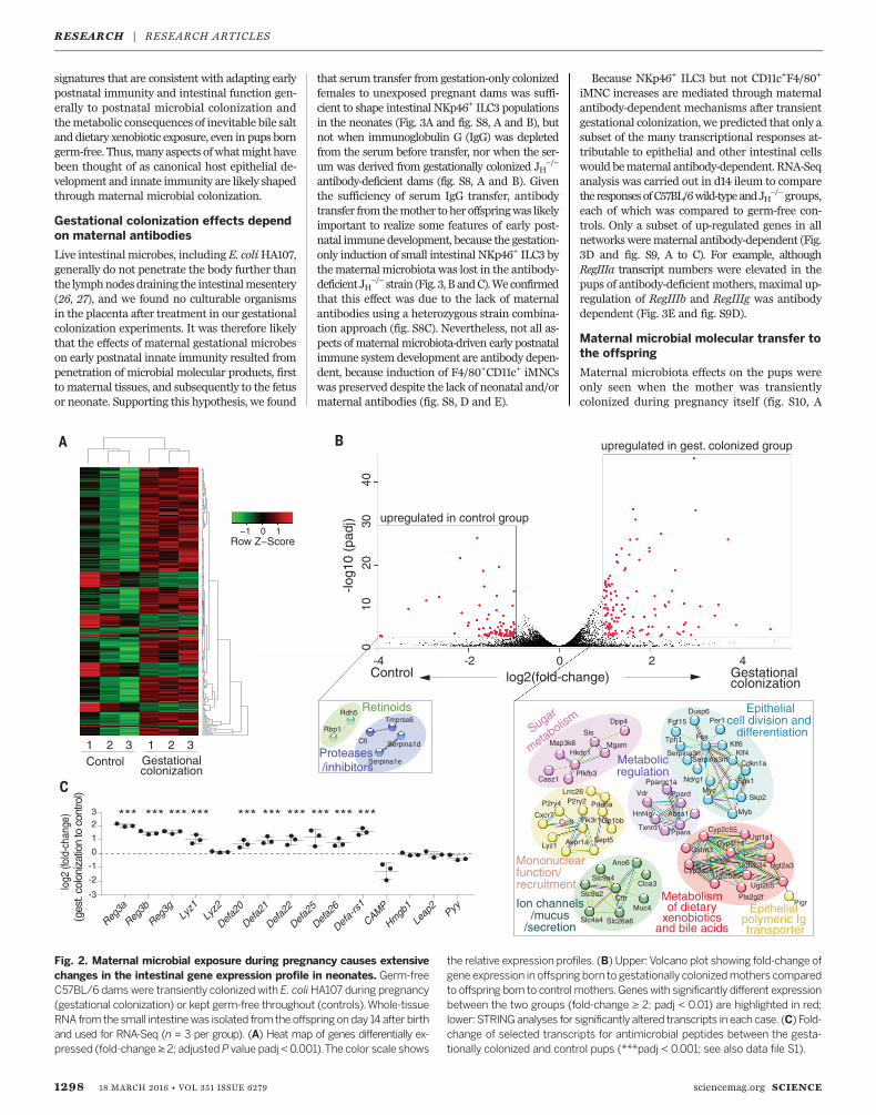
RNA sequencing (RNA-Seq) analysis of wholesmall intestinal mucosal RNA from neonates atday 14. Unsupervised analysis showed a series ofconsistent transcriptional changes in the pupsborn to gestation-only colonized dams comparedwith controls (Fig. 2A). The genetic and proteininteractions inferred from differentially expressedtranscripts (23) included up-regulated gene net-works for cell division and differentiation,mucusand ion channels, and the polymeric immuno-globulin receptor and mononuclear recruitment,as well as for metabolism of xenobiotics, bileacids, complex lipids, and sugars (Fig. 2B). Thesedifferentially expressed genes included signif-icantly increased overall expression of signaturegenes for the different Paneth cell, goblet cell, andearly/late enterocyte precursor epithelial lineages(24, 25) (fig. S7, A to C). Transcripts for the C-lectinReg family and antibacterial defensin peptideswere also significantly increased in the pups ofgestation-only colonized dams compared withcontrols (Fig. 2C and fig. S7D).These results show that the maternal micro-
biota drives wide-rangingmucosal transcriptional
SCIENCE sciencemag.org 18 MARCH 2016 • VOL 351 ISSUE 6279 1297
Fig. 1. Maternal microbial exposure during pregnancy shapes the frequen-cy of intestinal innate lymphoid and mononuclear cell populations in theoffspring. Germ-free C57BL/6 dams were transiently colonized with E. coliHA107 (gestational colonization) or kept germ-free throughout (controls). Alloffspring were analyzed by flow cytometry at day 14 after birth unless indi-cated. (A) Representative dot plots showing Lin– (CD19–CD3–) small intestinallamina propria lymphocytes (upper row) and Lin–RORgt+NKp46– ILC3 (lowerrow). (B) Absolute numbers (geometric mean, sample number n ≥ 5) of theindicated Lin– small intestinal ILC populations. (C) Absolute numbers of
small intestinal Lin–NKp46+RORgt+ ILC3 at indicated time points after birth.Data represent geometricmean±SD,n=3 to 10per timepoint. (D)Representativedot plots showing Lin–MHC-II+ colon lamina propria intestinal mononuclear cells(iMNCs) (upper row) and Lin–MHC-II+CD11c+F4/80lo iMNCs (lower row). (E) Ab-solute numbers (geometric mean, n ≥ 5) of indicated Lin–MHC-II+ iMNCpopulations in the colon. (F) Absolute numbers (geometricmean ± SD, n= 3 to10 per time point) of colon Lin–MHC-II+CD11c+F4/80+ iMNCs at different timepoints after birth. Data are each representative of four independent experi-ments or show pooled data from four experiments. *P ≤ 0.05; **P ≤ 0.01.
RESEARCH | RESEARCH ARTICLES
signatures that are consistent with adapting earlypostnatal immunity and intestinal function gen-erally to postnatal microbial colonization andthemetabolic consequences of inevitable bile saltand dietary xenobiotic exposure, even in pups borngerm-free. Thus,many aspects ofwhatmight havebeen thought of as canonical host epithelial de-velopment and innate immunity are likely shapedthrough maternal microbial colonization.
Gestational colonization effects dependon maternal antibodies
Live intestinalmicrobes, including E. coliHA107,generally do not penetrate the body further thanthe lymphnodesdraining the intestinalmesentery(26, 27), and we found no culturable organismsin the placenta after treatment in our gestationalcolonization experiments. It was therefore likelythat the effects of maternal gestational microbeson early postnatal innate immunity resulted frompenetration of microbial molecular products, firstto maternal tissues, and subsequently to the fetusor neonate. Supporting this hypothesis, we found
that serum transfer from gestation-only colonizedfemales to unexposed pregnant dams was suffi-cient to shape intestinal NKp46+ ILC3 populationsin the neonates (Fig. 3A and fig. S8, A and B), butnot when immunoglobulin G (IgG) was depletedfrom the serum before transfer, nor when the ser-um was derived from gestationally colonized JH
−/−
antibody-deficient dams (fig. S8, A and B). Giventhe sufficiency of serum IgG transfer, antibodytransfer from themother toher offspringwas likelyimportant to realize some features of early post-natal immune development, because the gestation-only induction of small intestinal NKp46+ ILC3 bythematernalmicrobiota was lost in the antibody-deficient JH
−/− strain (Fig. 3, B andC).Weconfirmedthat this effect was due to the lack of maternalantibodies using a heterozygous strain combina-tion approach (fig. S8C). Nevertheless, not all as-pects ofmaternal microbiota-driven early postnatalimmune system development are antibody depen-dent, because induction of F4/80+CD11c+ iMNCswas preserved despite the lack of neonatal and/ormaternal antibodies (fig. S8, D and E).
Because NKp46+ ILC3 but not CD11c+F4/80+
iMNC increases are mediated through maternalantibody-dependent mechanisms after transientgestational colonization, we predicted that only asubset of the many transcriptional responses at-tributable to epithelial and other intestinal cellswould bematernal antibody-dependent. RNA-Seqanalysis was carried out in d14 ileum to comparethe responses ofC57BL/6wild-type andJH
−/−groups,each of which was compared to germ-free con-trols. Only a subset of up-regulated genes in allnetworkswerematernal antibody-dependent (Fig.3D and fig. S9, A to C). For example, althoughRegIIIa transcript numbers were elevated in thepups of antibody-deficient mothers, maximal up-regulation of RegIIIb and RegIIIg was antibodydependent (Fig. 3E and fig. S9D).
Maternal microbial molecular transfer tothe offspring
Maternal microbiota effects on the pups wereonly seen when the mother was transientlycolonized during pregnancy itself (fig. S10, A
1298 18 MARCH 2016 • VOL 351 ISSUE 6279 sciencemag.org SCIENCE
Sugar
metabolism
Ion channels /mucus
/secretion
Mononuclear function/recruitment
Epithelial cell division and
differentiation
Metabolicregulation
Epithelial polymeric Ig transporter
Metabolism of dietary xenobiotics
and bile acids
Dusp6Per1Dpp4 Fgf15
Sis
Klf6FosTph1
MgamMap3k6Serpina3n Klf4Hkdc1
Casz1
Serpina3m Cdkn1aPfkfb3
Ndrg1 Sgk1Ppargc1aLrrc26
Skp2Myc
Pde6a
Vdr PpardP2ry2P2ry4
Gp1bbMybHnf4gCxcr7
PparaCcl9
Sept5
Abca1
Lyz1
Txnrd1
Cyp4f14
Pik3r1
Avpr1a
Cyp2c55Ugt1a1
Ano6
Gstm3Ces2c
Slc9a4
Ugt2a3
Clca3
Cyp3a25
Slc9a2 Cftr
Ugt2b34
Ugt2b5
Muc4
Ugt2b35
Pla2g2f
Slc4a4 Slc26a6
Pigr
log2
(fol
d-ch
ange
) (g
est.
colo
niza
tion
to c
ontro
l)
Reg3a
Reg3b
Reg3g
Lyz1
Lyz2
Defa2
0
Defa2
1
Defa2
2
Defa2
5
Defa2
6
Defa-
rs1
CAMP
Hmgb
1
Leap
2Pyy
-2
-1
0
1
23 *** *** *** *** *** *** *** *** *** ***
1 2 3 1 2 3Control Gestational
colonization
Rdh5
Rbp1Tmprss6
Cfi Serpina1d
Serpina1eProteases /inhibitors
Retinoids
-3
-4 -2 0 2 4
010
2030
40
log2(fold-change)
-log1
0 (p
adj)
Control Gestational colonization
upregulated in gest. colonized group
upregulated in control group−1 0 1
Row Z−Score
Fig. 2. Maternal microbial exposure during pregnancy causes extensivechanges in the intestinal gene expression profile in neonates. Germ-freeC57BL/6 dams were transiently colonized with E. coliHA107 during pregnancy(gestational colonization) or kept germ-free throughout (controls).Whole-tissueRNA from the small intestinewas isolated from the offspringonday 14after birthand used for RNA-Seq (n = 3 per group). (A) Heat map of genes differentially ex-pressed (fold-change≥ 2; adjustedP value padj < 0.001).The color scale shows
the relative expression profiles. (B) Upper:Volcano plot showing fold-change ofgene expression in offspring born to gestationally colonizedmothers comparedto offspring born to controlmothers.Genes with significantly different expressionbetween the two groups (fold-change ≥ 2; padj < 0.01) are highlighted in red;lower: STRINGanalyses for significantly altered transcripts in each case. (C) Fold-change of selected transcripts for antimicrobial peptides between the gesta-tionally colonized and control pups (***padj < 0.001; see also data file S1).
RESEARCH | RESEARCH ARTICLES

and B). In mice, maternal IgG is transferredacross the placenta and through intestinal up-take from the milk (28), so we used litter-swapexperiments to distinguish between antenataland postnatal effects of the maternal microbiota.Although there was a nonsignificant trend to-ward increased small intestinal NKp46+ ILC3 inneonates born to an unmanipulated mother andnursed by a gestation-only colonized mother,both in utero gestation and postnatal nursing bydams that had been colonized during pregnancywere necessary for significant ILC3 induction(Fig. 4A).These results imply that maternal microbiota-
derived compounds are transferred fromthemoth-er to the offspring and that this process is increasedin the presence of maternal antibodies. We nextconsidered antibody-enhanced retention of bacte-rial products in themother and antibody-mediatedtransfer of bacterial products. The first of theseeffects was demonstrated by following 14C elim-ination from metabolically labeled E. coli HA107in wild-type and antibody-deficient mice. Thepresence of antibodies significantly increasedretention of 14C-labeledmolecules in themesenteric
lymph nodes, spleen, liver, and serum for at least36 hours compared with antibody-deficient con-trols (Fig. 4, B andC, and fig. S11, A toD):Microbialmolecular exposure of the placenta and the fetuswas also enhanced at embryonic day 16 (E16) (Fig.4, D and E). We also found significantly increasedradioactivity originating from maternal micro-bial molecules in the milk, and from postnatalintestinal mucosa and liver of wild-type pups(Fig. 4, F to I). This shows that maternal anti-bodies enhance the retention and transmissionofmicrobialmolecules, although effects other thandirect microbial molecular binding cannot beexcluded. To verify that these compounds arereally ofmicrobial origin rather than the productsof secondary metabolism in the mother, we grewHA107 on [13C]glucose so that bacterial com-pounds became fully labeled with 13C, as judgedby mass spectrometry–shift data. Intestinal 13C-labeled metabolite levels were equivalent wheth-er or not the mother expressed antibodies (fig.S12); however, after intestinal administration of13C-labeled HA107 in C57BL/6 intravenously (i.v.)primed mice, serum contained bacterial metab-olites comigrating with IgG that were absent
from the serum of antibody-deficient mice (fig.S13 and data file S5). Even if HA107 was onlydelivered through the intestinal route, which doesnot induce high-affinity serum IgG (11), therewas evidence of low-affinity IgG coating of E. colithat was absent from serum of untreated germ-free controls or from HA107-treated JH-deficientmice (fig. S14). We therefore concluded that ei-ther sterile bacterial fragments or small moleculescan potentially be bound to maternal IgG afterintestinal exposure.Given that increases in NKp46+ ILC3 and com-
ponents of the mucosal transcriptome were anti-body dependent, whereas F4/80+CD11c+ iMNCswere induced by gestational colonization even inpups of antibody-deficient dams, we assumedthat a number of molecular ligand–receptor sys-tems are driving different aspects of neonataladaptation in response to the maternal micro-biota. Toll-like receptor ligand signaling was notessential for the effect (fig. S15, A and B). Therewas an extensive range of bacterial-derived (13C-labeled) molecules passed from the mother tothe offspring (Fig. 5A and fig. S16), some of whichalso reached neonatal tissues (fig. S16). These
SCIENCE sciencemag.org 18 MARCH 2016 • VOL 351 ISSUE 6279 1299
Fat storage
Cholesterol derived hormone
production
Cholesterol synthesis
Inflammatory signals
Sugar
met
aboli
sm
Metabolism of dietary
xenobiotics/bile acids
Metabolicregulation
Sugar
metabolism Metabolic
regulation
Metabolism of dietary xenobiotics/bile acids
Ion channels/mucus /secretionIon
channels /mucus /secretion
n= 228
n= 85
n=95
Antibody-dependent networks
Antibody-independent
networks
Networks in the absence of antibodies
Sqle
Lss
Sc4mol Cyp51
Idi1
Ryr1
Lrat
Bst1
Defa1
Hmgcr Il1rl1
Rhof
Il1a
Mmp7
Vav2
Abcg5 Adipoq
HpPcsk9
FgaSlc22a1
Slc36a2Retn
CfdHgdHabp2
Dpp4Mgam
Adcy1Vipr1
Atp1b1
Ccl25
Agtr2
Anpep
Ggt1Gpx2
Ppara
Cyp2j13
Hnf4g
Acsm3
Cyp2c66
Cyp2c55Akr1c14
GykAkr1b7
Cyp2j6Ugt2b35 Plb1
Khk
Ugt2b34Slc2a5
Cyp2d26
Slc5a1Cyp3a13
Slc26a6 Sult2b1Sult1d1
Fmo4
Hkdc1
ControlGestational colonization
WT
JH-/-
RORγ t
NK
p46
101
102
103
104
Abs
olut
e ce
ll nu
mbe
r ***
JH-/-WT
n.s.
Treh
Ang4
Sis
Defa20
Oas3H2-Q1
Clca3
Pla2g2a
Tff2Cyp4f14
Chdh
Dmbt1
Zg16
Cyp2c65
Pnliprp2
Cyp3a11
Rdh7Cyp3a25
Ugt2a3
10.97 1.58
56.4 31.0
8.03 3.79
50.1 38.1
9.60 1.18
56.5 32.8
7.54 1.09
57.2 34.2
ControlGestationalcolonization
WT (Gestational colonization vs control) (Gestational colonization vs control)
JH-/-
WTJH -/-
Lo
g2
(fo
ld- c
ha
ng
e)(g
est.
colo
niza
tion
t o c
ontro
l)
-4
-2
0
2
4 *** *** *** *** **
Reg3a Reg3b Reg3g CAMP Hmgb1
WT
Abs
olut
e ce
ll nu
mbe
r
103
104
105 **
Control
Gestationalcolonization HA107 serum
NKp46+ ILC3
NKp46+ ILC3
Fig. 3. Sufficiency of serum transfer and requirement for maternalantibodies for gestational colonization effects. (A) Pregnant germ-freeC57BL/6 dams were transiently colonized (gestational colonization), leftgerm-free (control), or injected i.v. with serum from a germ-free donor pre-viously gavaged with E. coliHA107 (HA107 serum).Total Lin–RORgt+NKp46+
ILC3 (geometric mean, n ≥ 2) in d14 offspring small intestine. (B to E)Germ-free C57BL/6 (WT,wild type) or JH
−/− damswere colonizedwith E. coliHA107 during pregnancy (gestational colonization) or kept germ-free (con-trol). (B) Representative flow cytometry dot plots showing Lin–RORgt+NKp46+
ILC3 in d14 small intestine. (C) Total numbers (geometric mean, n ≥ 3) of
Lin–RORgt+NKp46+ ILC3 in d14 small intestine. (D and E) RNA-Seq of d14distal small intestinal RNA in offspring of JH
−/− and wild-type dams (n = 3 pergroup). (D) STRING analysis comparing genes significantly enriched aftergestational colonization versus germ-free in either WT (black circle) or JH
−/−
(blue circle) strains. Total number of differentially regulated genes areshown. (E) Fold-change of expression of indicated genes in offspring fromgestationally colonized compared to control mothers in WTor JH
−/− strains(***padj < 0.001, see also data files S2 and S3). Data are representative oftwo (A) and three (B and C) independent experiments. (A and C) **P ≤ 0.01;***P ≤ 0.001.
RESEARCH | RESEARCH ARTICLES

1300 18 MARCH 2016 • VOL 351 ISSUE 6279 sciencemag.org SCIENCE
n.d.Amino SugarsAmino Acids and DerivativesMonosaccharidesTrisaccharidesOligosaccharidesDisaccharidesFatty Acid EstersFatty Acids and ConjugatesGlycerolipidsGlycerophospholipids
log2(fold-change)Control Gestational C-HA10713
103
104
105
Abs
olut
ece
llnu
mbe
r * *
Control waterColonization
NOD ligandsSCFAs
RIG-I ligands
Gestational treatment:
Control Colonization
I3CSolvent
Gestational treatment:
WT
WT
-log1
0(p-
valu
e)
-log1
0(p-
valu
e)
WT Milk JH Milk-/-
-2 0 2 40
0.5
1
1.5
2
2.5
3
3.5
4
4.5
10
-2 0 2 40
0.5
1
1.5
2
2.5
3
3.5
4
4.5
10
12
49
73
,!"#
$!!"#
76
319
238
Tissue Compound Sum formula WT JH-/-
fold-change p-value fold-change p-value
Milk Kynurenine [+10] C10H12N2O3 7.807 0.0325 2.529 > 0.05
Indolelactic acid [+11] C11H11NO3 6.790 0.0341 n.d.
5-Hydroxy-L-tryptophan [+11] C11H12N2O3 5.248 0.0308 1.791 > 0.05
Hydroxykynurenamine [+9] C9H12N2O2 4.973 0.0278 0.974 > 0.05
Hydroxykynurenine [+10] C10H12N2O4 4.539 0.0406 1.457 > 0.05
5-Methoxytryptamine [+11] C11H14N2O 4.181 0.0380 1.190 > 0.05
5-Methoxytryptophol [+11] C11H13NO2 3.808 0.0109 1.235 > 0.05
Indole-5,6-quinone [+8] C8H5NO2 2.024 0.0019 1.307 0.0057
Neonatal liver Hydroxykynurenine [+10] C10H12N2O4 2.394 < 0.0001 1.679 0.0015
Neonatal spleen Hydroxykynurenine [+10] C10H12N2O4 2.754 < 0.0001 1.656 0.0015
log2(fold-change)Control Gestational C-HA10713
NKp46+ ILC3
NKp46+ ILC3
Abs
olut
ece
llnu
mbe
r
103
104
105
Fig. 5. Profiles of maternal microbial molecules shaping the neonatalimmune system. (A to C) Pregnant germ-free wild-type or JH
−/− mice weregavaged on E10 and E12 with unlabeled E. coli HA107, and on E15 and E16 with13C-labeled E. coli HA107. Maternal milk (gestationally colonized, n = 6 to 8;germ-free control, n = 2) was analyzed by mass spectrometry. (A and B)Volcano plots of fold-change analysis betweenmilk from treated and untreatedWT (A) and JH
−/− (B) dams. Metabolite compound classes are color-coded.Inset histogram: percentage 13C labeling for each significantly altered com-pound (fold-change ≥ 2; P ≤ 0.05): unlabeled (white), ≥25% labeled (gray),100% labeled (black). A total of 395 potentially 13C-labeled compounds were
significantly enriched in the milk of WTdams. (C) Fully labeled metabolites ofthe AhR ligand class present in d1 to d3 milk of WT and JH
−/− femalesgestationally exposed to 13C-HA107. See also data file S4. (D) Pregnantgerm-free C57BL/6 mice were gavaged with indole-3-carbinol (I3C) or sol-vent, or gestationally colonized.Total numbers of Lin–RORgt+NKp46+ ILC3 inthe d14 offspring small intestine (geometric mean, n ≥ 6). (E) Pregnantgerm-free C57BL/6 mice were exposed to short-chain fatty acids (SCFAs),NOD1/2 ligands, or RIG-I ligand or gestationally colonized. Total numbers ofLin–RORgt+NKp46+ ILC3 in d14 offspring small intestine (geometric mean, n ≥3). Data are representative of three (D) independent experiments. *P ≤ 0.05.
102
103
104
105
Mother: Foster:
ctrl HA107 HA107 ctrlctrl HA107 HA107ctrl
Parental MLN
*****
*
***
WT Back-ground
JH-/-
Placenta, E16
******
DP
M/g
*
Back-ground
120h36h8h
*
Abs
olut
e ce
ll nu
mbe
r WTAntibody-deficient
102
103
104
105
Parental liver
DP
M/g
*
Back-ground
120h36h8h
*
10 2
10 3
10 4
10 5
DP
M/g
102
103
104*
DP
M/g
102
103
104
105 ***
WT Back-ground
JH-/-
Fetus, E16
******
p=0.1
WT Back-ground
JH-/-
Maternal milk, d1/2
**
DP
M/g
102
103
104
Offspring liver
DP
M/g
Back-ground
d10/11d6/7d1102
103
104
105
*
Offspring spleen
DP
M/g
Back-ground
d10/11d6/7d1102
103
104
******
***
Offspring small intestine
DP
M/g
Back-ground
d10/11d6/7d1102
103
104
NKp46+ ILC3
WTAntibody-deficient
Fig. 4. Maternal microbial molecules reach the offspring during preg-nancy and after birth. (A) Pregnant germ-free C57BL/6 mice were colo-nized with E. coli HA107 or germ-free (ctrl). Half of each litter was swappedto another group for fostering at birth.Total numbers (geometric mean, n ≥8) of Lin–RORgt+NKp46+ ILC3 in the offspring small intestine on postnatalday 14. (B and C) Adult germ-free WTor Rag1−/− (antibody-deficient) micereceived three gavages of 1010 colony-forming units (CFU) of unlabeled E. coliHA107 and one gavage of 1010 CFU of 14C-labeled E. coli HA107. Radio-activity in the indicated tissues of WT and Rag1−/− adults was monitored
over time. (D to F) Pregnant germ-free C57BL/6 WT or JH−/− mice were
gavaged with 1010 CFU of 14C-labeled E. coli HA107 on E14. Placental or fetaltissues at E16 (D and E) or milk at d1/2 after birth (F) fromWTor JH
−/− micewas analyzed for radioactivity. (G to I) Postnatal tissues from WT offspringwere analyzed for persistence of transferred radioactive maternal microbialproducts. Geometric means are shown. Open circles show backgroundscintillation in offspring from nongavaged mice (B to I). Results are repre-sentative of three (A) or two (D to I) independent experiments. *P ≤ 0.05;**P ≤ 0.01; ***P ≤ 0.001.
RESEARCH | RESEARCH ARTICLES
bacterial-derived metabolites present in milk oroffspring tissues from gestationally colonizedmiceincluded natural microbial ligands for the arylhydrocarbon receptor (AhR) or their precursors(Fig. 5, A to C, and table S4) (29). Most of thesebacterial metabolites, including the fully labeledAhR ligands (Fig. 5C), were not enriched in themilk of treated JH
−/− mice (Fig. 5, B and C), al-though these data do not prove that these mol-
ecules are necessarily bound to the antibodies fortransfer. Because AhR-deficientmice have a com-pound phenotype (30), and strain combinationexperiments reveal globally nonredundant signal-ing pathways, we took the approach of treatingpregnant germ-free mice with authentic ligandsfor AhR, short-chain fatty acids, nucleotide-binding oligomerization domain (NOD) ligandsand the retinoic acid–inducible gene I (RIG-I)
ligand. Of these, only the AhR ligand (indole-3-carbinol, I3C) increased NKp46+ ILC3 in theoffspring of the treatedmothers (Fig. 5, D and E).This occurred even in the absence of antibodies,although to a significantly lower extent (fig. S17,A and B). Although this shows that early postnatalNKp46+ ILC3 numbers are increased in responseto aryl hydrocarbons, antibodies are not essentialfor the effect provided that a sufficient dose of
SCIENCE sciencemag.org 18 MARCH 2016 • VOL 351 ISSUE 6279 1301
0
20
40
60
CF
U/o
rgan
PBS JM83 PBS JM83Challenge:
***
Bacteria translocation MLNE. coli JM83
Bacteria translocation MLN B. fragilis
0
20
40
60 **
A B
D
-2 0 2
010
2030
40
log2 (fold-change)
-log1
0 (p
adj)
ControlGestational colonization
50
CF
U/o
rgan
upregulated in gestational
colonized group
upregulated in control
group
Man2b2
Naga
Man2b1Fuca1
Lgmn
Dlk1Dgat2
Fabp4
HexaAmdhd2
Gpihbp1
St3gal5
Cfd
Ces1d
Fuca2
Renbp
Galns
Gusb
Ctsa
Npl
Neu1
Glb1
Gpx3
Slc17a5
G6pcGalk1
Gsta2Cryz
Gaa St3gal1
Ddit3 Hpgds
Bmp7 Cyp2e1Hmox1
Ftl1 Slc46a1
Slc40a1Hif3a
Apoc1Itgb5 Apoc3
Diras1
LtfHgd
Apoc4Spp2
Igfals
Lrrc10b
Apoa4
Entpd3
Fga
Lrrn1
Cfi
Rtn4r
FgbSaa1
Ttr
Pde4c
C9F10
Sucnr1
Rras
Galr2Sst
Cell cycle regulation
Signaling by GPCR
Amino sugar and nucleotide metabolism
Lipid metabolism
Glycosaminoglycan metabolism
Lysosome activity
Adaptation to microbial colonization
Detoxification of oxidative stress
Purinemetabolism
Fignl1Sass6
Zwilch
Lrr1Dsn1
Casc5Kif4Xpo1Sgol2
DckIqgap3 Nuf2
Cenpi Kntc1Kif20a
CenpfKif24 Bub1Ska1
Sgol1
Ckap2 Cenpe
Aurkb
Shcbp1
Bub1b Ccnb2
Tpx2
Spag5
Ccnb1Gca
Asf1b Hirip3Cdc25c
Tex30Nupl1Nup160
Cse1lMlf1ip Nek2 Pbk
Esco2
Topbp1
Cdkn3
AI448607
Ttk Tmem48
Cenpq
Mad2l1
Pola1
Ercc6l
Kif18a
Ankrd32
Cenpk
Rfc4TpteDtl
Exo1 Orc1Mcm8
Rrm2Chek1 Atr
Ccl2
Fbxo5
Atad2Haus6Cdc6 Dbf4Ncapg2
Ccna2
Smc2
Ckap2l Ccne2
E2f8Depdc1a
Noxin
Slc16a12
Insig1 Mki67
Mis18bp1Kif11
Kiaa1524
Hells
Smc4
Kif15
Top2a
Nusap1
AunipHnrnpa1
Ncapg
Kif20b
Aqp4
Aspm
HmmrBlm
Neil3
Rad54l
G2e3
Ppat
Racgap1Prc1
Kif14Plk4
Cep55
Arhgap11aGen1
Paf
Dkc1
Vav3
Polq
Dlgap5
BC055324Ect2Gbp1
Kif23Parpbp
Ska3
Stil
Pla2g4aDiap3
NcaphTrim59
Nox1 HmgcrPrr11 Aurka Kif18b
Melk Rps6ka6
Mitotic prometaphase,, metaphase and anaphase
APC and cell cycle regulation
Cellular growth and proliferationCell death and survival
Centrosome and chromatin
organization
Cell cycle check point
Control Gestational colonization
Gestationaltreatment:
E
Reg3a Reg3b Reg3g
C
Rag1-/-
****
PBS JM83 PBS JM83 PBS JM83 PBS JM83
Bacteria translocation MLNE. coli JM83
Challenge:
GF GF ASF d21 GF
Rag-/-
c-/-
**
GF
0
10
20
30
40
50
CF
U/o
rgan
Control Gestational colonization
B. fragilis B. fragilisChallenge:
Gestationaltreatment:
During pregnancy
101
102
103
104
105
# re
ads 19x
37x
25x
3x
3x
3x
******
PBSJM83PBSJM83
Control
Gestational colonization
Challenge
Fig. 6. Microbial exposure during pregnancy functionally protects theoffspring. (A, B, D, and E) Germ-free C57BL/6 dams were gestationallycolonized or kept germ-free. (A) Day 14 pups were challenged with 105 CFU ofE. coli JM83 or phosphate-buffered saline (PBS). Bacterial titers in mesentericlymph nodes (MLN) were determined at 18 hours (x ± SD, n ≥ 5). (B) Day 14pups were challenged with 1010 CFU of B. fragilis or PBS. Bacterial titers inMLNs were determined at 18 hours (x ± SD, n ≥ 10). (C) Adult germ-freeC57BL/6 Rag1−/− or Rag2−/−gc−/− were colonized for 21 days with ASFor keptgerm-free before challenge with 107 CFUof E. coli JM83 or PBS. Bacterial titers
at 18 hours in MLNs are shown (x ± SD, n ≥ 5). (D and E) Day 14 pups werechallenged with 105 CFU of E. coli JM83 or PBS (n = 3 per group) before smallintestinal RNA-Seq. 18 hours later. STRING analyses for differentially expressedtranscripts (fold-change ≥ 2; padj < 0.01, data file S6) compare challengedpups born to germ-free or gestationally colonizedmothers. (E) Read number ofindicated genes in E. coli– or PBS-challenged offspring (geometricmean, n= 3,***padj < 0.001; see also data files S7 and S8). Data are representative of three(A and C) or two (B) independent experiments. (A to C) *P ≤ 0.05; **P ≤ 0.01;***P ≤ 0.001.
RESEARCH | RESEARCH ARTICLES

AhR ligand is transmitted from themother to heroffspring. Indeed, we found that ILC3 increasesinduced by an endogenous ASF microbiota inadult mice were also antibody independent, pre-sumably because endogenous colonization pro-vides a sufficient dose of bacterial ligands (fig.S17C). We concluded that maternal antibodiesassist the transfer of microbial compounds tothe offspring, but are not independently requiredto increase ILC3 numbers. Given the diversity ofmaternal microbial molecular transfer, it remainsprobable that other microbial molecular speciescan also drive early postnatal adaptation.
Gestational colonization effects oninnate immune precursors
The elevated number of intestinal NKp46+ ILC3and F4/80+CD11c+ iMNCs in the offspring bornto gestation-only colonized dams may result fromamplified precursor populations or increased pro-liferation of the mature intestinal population.Neonatal ILC3 precursors (14, 31) were not in-creased in the liver or intestine of E17 fetusesfrom gestationally HA107-colonized mice (fig.S18, A toD).We did detect increased proliferativecapacity of small intestinalNKp46+ ILC3 isolatedfrom 14-day-old pups born to gestation-onlycolonized dams (fig. S18, E and F). F4/80+CD11c+
iMNCs stem from CD11b+CX3CR1intLy6C+ mono-cytes (32, 33), which were significantly increasedin the colon lamina propria of 14-day-old pupsfrom gestationally colonized dams (fig. S18, Gand H).
Functional impact of gestationalcolonization on the early postnatalimmune system
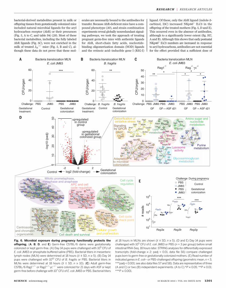
To test whether the integrity of the early post-natal intestine to live microbial challenge wasimproved by gestational colonization, we chal-lenged pups with the replication-competentparent strain of HA107, E. coli JM83. Despiteequal E. coli cecal colonization at 18 hours, onlythe pups of gestationally colonized mothers ordams treated with the AhR ligand I3C couldavoid translocation of JM83 to the mesentericlymph nodes (Fig. 6A and fig. S19, A and B).Because presence of HA107-specific antibodiesmight contribute to E. coli–primed protection,we confirmed these results by challenge withBacteroides fragilis, where HA107-induced anti-bodies do not cross-react (Fig. 6B and fig. S19C).To verify the role of ILC3 in intestinal integrity
(34), we exploited the fact that ILC3 can be in-duced in adults independently of B cells and anti-bodies (fig. S17C and table S5). Comparison ofRag−/− and Rag−/−gc
−/− mice (which lack ILCsas well as B and T cells) showed that ILCs wererequired to mediate the microbiota-driven pro-tection from bacterial translocation during chal-lenge with JM83 (Fig. 6C).Systemic immune responsiveness is also likely
shaped by gestational colonization, as tumor ne-crosis factor–a and IL-6 proinflammatory cytokineproduction was reduced in the pups’ splenocytesafter intraperitoneal lipopolysaccharide (fig.S20). Because ILC3 are a very minor population
in the spleen, the mechanism is likely to be quitedistinct from the gestational effects on intestinalfunction.These functional readouts of gestational colo-
nization only show some of the potential benefitsof maternal microbial molecular exposure in thepups. After challengewith replication-competentE. coli, small intestinal RNA-Seq analysis showedexpression of antioxidant and lysosomal enzymenetworks in the pups of gestation-only colonizeddams, whereas control pups had wide-rangingexpression signatures for cellular proliferation,cytoskeletal organization, and ribosome biosynthe-sis (Fig. 6D and fig. S21A). Expression of somegenes for antimicrobial peptides induced bygestational colonization also increased furtherafter intestinal bacterial challenge (Fig. 6E andfig. S21B).
Conclusion
The maternal microbiota prepares the newbornfor host-microbial mutualism. This results frommicrobial molecular transfer because in our ex-perimental system, live microbes are no longerpresent at birth; we do not detect live microbesin the placenta or the neonate; and the result canbe recapitulated with sterile serum transfer. Inother words, maternal antibodies not only pro-tect the neonate through pathogen neutraliza-tion (4, 5), but also have a more general effectpromoting microbial molecular transfer. Short-chain fatty acids from microbes are known toshape the adult immune system (35, 36).We showhere that ligands for the AhR, known to driveILC3 expansion (37) and limit adult bacterialtranslocation (38), can be derived from the ma-ternal microbiota and shape the composition andfunction of early postnatal immunity.Nevertheless,AhR ligands are unlikely to be the onlymolecularmechanism involved in gestational microbialshaping.Secretory antibodies in the milk are known
to delay the maturation of the early postnatalimmune system and determine long-term intes-tinal microbiota composition (39–41). Here weshow that maternal antibodies also enhance mi-crobial molecular levels in the fetus and the neo-nate. Themolecular constituents of the maternalmicrobiota are able to ready neonatal innate im-munity in time for the tsunami of microbes thatsuccessively colonize the intestine (42, 43). Althoughthese studies were focused on benign microbes, theimmune morphogenesis driven by the maternalmicrobiota is likely also to benefit young mam-mals when they encounter pathogens. Postnatalmicrobial colonization is a pivotal early event inautonomous host-microbialmutualism. Fortunately,the maternal microbiota and maternal immunityprepare the neonate for its inevitable challenges.
REFERENCES AND NOTES
1. R. M. Samstein, S. Z. Josefowicz, A. Arvey, P. M. Treuting,A. Y. Rudensky, Cell 150, 29–38 (2012).
2. M. Fulde, M. W. Hornef, Immunol. Rev. 260, 21–34 (2014).3. H. Renz, P. Brandtzaeg, M. Hornef,Nat. Rev. Immunol. 12, 9–23 (2012).4. P. Brandtzaeg, Vaccine 21, 3382–3388 (2003).5. R. M. Zinkernagel, N. Engl. J. Med. 345, 1331–1335
(2001).
6. GBD 2013 Mortality and Causes of Death Collaborators, Lancet385, 117–171 (2015).
7. T. Olszak et al., Science 336, 489–493 (2012).8. L. V. Hooper, D. R. Littman, A. J. Macpherson, Science 336,
1268–1273 (2012).9. A. M. Kabat, N. Srinivasan, K. J. Maloy, Trends Immunol. 35,
507–517 (2014).10. S. Rakoff-Nahoum et al., Proc. Natl. Acad. Sci. U.S.A. 112,
1929–1936 (2015).11. S. Hapfelmeier et al., Science 328, 1705–1709 (2010).12. F. Bäckhed et al., Cell Host Microbe 17, 690–703 (2015).13. M. J. Bizzarro et al., J. Pediatr. 166, 1193–1199 (2015).14. S. Sawa et al., Science 330, 665–669 (2010).15. S. L. Sanos et al., Nat. Immunol. 10, 83–91 (2009).16. C. Vonarbourg et al., Immunity 33, 736–751 (2010).17. T. Sano et al., Cell 163, 381–393 (2015).18. K. Atarashi et al., Cell 163, 367–380 (2015).19. M. B. Geuking et al., Immunity 34, 794–806 (2011).20. A. J. Macpherson, N. L. Harris, Nat. Rev. Immunol. 4, 478–485
(2004).21. K. Smith, K. D. McCoy, A. J. Macpherson, Semin. Immunol. 19,
59–69 (2007).22. J. Pácha, Physiol. Rev. 80, 1633–1667 (2000).23. D. Szklarczyk et al., Nucleic Acids Res. 43, D447–D452
(2015).24. D. Grün et al., Nature 525, 251–255 (2015).25. M. L. Robinette et al., Nat. Immunol. 16, 306–317 (2015).26. A. J. Macpherson, T. Uhr, Science 303, 1662–1665 (2004).27. M. L. Balmer et al., Sci. Transl. Med. 6, 237ra66 (2014).28. V. Ghetie, E. S. Ward, Immunol. Today 18, 592–598
(1997).29. C. Esser, A. Rannug, Pharmacol. Rev. 67, 259–279 (2015).30. B. Stockinger, P. Di Meglio, M. Gialitakis, J. H. Duarte,
Annu. Rev. Immunol. 32, 403–432 (2014).31. M. Cherrier, S. Sawa, G. Eberl, J. Exp. Med. 209, 729–740
(2012).32. S. Tamoutounour et al., Eur. J. Immunol. 42, 3150–3166
(2012).33. C. C. Bain et al., Nat. Immunol. 15, 929–937 (2014).34. G. F. Sonnenberg et al., Science 336, 1321–1325 (2012).35. N. Arpaia et al., Nature 504, 451–455 (2013).36. Y. Furusawa et al., Nature 504, 446–450 (2013).37. E. A. Kiss et al., Science 334, 1561–1565 (2011).38. G. F. Sonnenberg, D. Artis, Nat. Med. 21, 698–708 (2015).39. D. R. Kramer, J. J. Cebra, J. Immunol. 154, 2051–2062
(1995).40. N. L. Harris et al., J. Immunol. 177, 6256–6262 (2006).41. E. W. Rogier et al., Proc. Natl. Acad. Sci. U.S.A. 111, 3074–3079
(2014).42. R. I. Mackie, A. Sghir, H. R. Gaskins, Am. J. Clin. Nutr. 69,
1035S–1045S (1999).43. Y. R. Nobel et al., Nat. Commun. 6, 7486 (2015).
ACKNOWLEDGEMENTS
We thank W.-D. Hardt, C. Mueller, and D. Candinas for criticalinput and support. J. Limenitakis and I. Keller helped withbioinformatic analyses. The data presented in this manuscriptare tabulated in the main paper and in the supplementary materials;sequencing data are available via www.ebi.ac.uk/ena/data/view/PRJEB12398. E. coli HA107 is available from A.J.M. undera material transfer agreement with the University of Bern. TheClean Mouse Facility is supported by the Genaxen Foundation,Inselspital and the University of Bern. This work was funded bythe Swiss National Science Foundation (SNSF 310030B_160262,SNF Sinergia CRSII3_136286, and SNSF Sinergia CRSII3_154414)to A.J.M. K.D.M is supported by a grant from the SNSF(SNSF310030_134902) and the European Research Council(ERC, FP/2007-2013) Agreement no. 281785. S.C.G.-V. was fundedby a Marie Curie Intra-European Fellowship (FP7-PEOPLE-2013-IEFProject No. 627206). S.H. was funded by SNSF grant 310030_138452and the ERC (FP/2007-2013) Agreement no. 281904. The authorshave no conflicts of interest.
SUPPLEMENTARY MATERIALS
www.sciencemag.org/content/351/6279/1296/suppl/DC1Materials and MethodsFigs. S1 to S21Tables S1 to S5References (44–53)Data Files S1 to S8
28 August 2015; accepted 22 January 201610.1126/science.aad2571
1302 18 MARCH 2016 • VOL 351 ISSUE 6279 sciencemag.org SCIENCE
RESEARCH | RESEARCH ARTICLES
DOI: 10.1126/science.aad2571, 1296 (2016);351 Science
et al.Mercedes Gomez de AgüerodevelopmentThe maternal microbiota drives early postnatal innate immune
This copy is for your personal, non-commercial use only.
clicking here.colleagues, clients, or customers by , you can order high-quality copies for yourIf you wish to distribute this article to others
here.following the guidelines
can be obtained byPermission to republish or repurpose articles or portions of articles
): March 17, 2016 www.sciencemag.org (this information is current as of
The following resources related to this article are available online at
/content/351/6279/1296.full.htmlversion of this article at:
including high-resolution figures, can be found in the onlineUpdated information and services,
/content/suppl/2016/03/16/351.6279.1296.DC1.html can be found at: Supporting Online Material
/content/351/6279/1296.full.html#ref-list-1, 23 of which can be accessed free:cites 53 articlesThis article
/cgi/collection/immunologyImmunology
subject collections:This article appears in the following
registered trademark of AAAS. is aScience2016 by the American Association for the Advancement of Science; all rights reserved. The title
CopyrightAmerican Association for the Advancement of Science, 1200 New York Avenue NW, Washington, DC 20005. (print ISSN 0036-8075; online ISSN 1095-9203) is published weekly, except the last week in December, by theScience
on M
arch
18,
201
6D
ownl
oade
d fr
om