micrococcaceae. family micrococcaceae includes 4 genera: planococcus – free living saprophyte...
Post on 20-Dec-2015
221 views
TRANSCRIPT
Micrococcaceae
Family Micrococcaceae
Includes 4 genera:Planococcus – free living saprophyteMicrococcus – free living saprophyteStomatococcus – normal flora on surface of
primates and other mammalsStaphylococcus – normal flora on surface of
primates and other mammalsAll, except Planococcus have been isolated
from clinically significant sources
Family Micrococcaceae
Propensity to cause disease: Stomatococci are part of the normal oral flora and
are now an emerging pathogen in immuno-compromised patients.
Micrococci become pathogens when they are accidentally introduced into a susceptible host.
Staphylococci have long been recognized as important human pathogens. The most commonly isolated pathogenic species in order of pathogenicity are S. aureus, S. epidermidis, and S. saprophyticus.

Family Micrococcaceae
Morphology and General Characteristics Gram positive cocci which may lose the ability to
retain their Gram positive staining characteristics with age.
May occur singly, in pairs, tetrads (common for Micrococci), or in clusters (Staphylococci – staphyle means bunch of grapes and this arrangement is due to the tendency of the organism to divide in different planes)
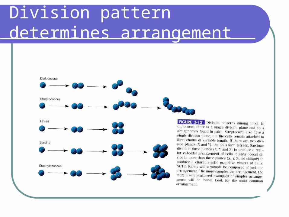
Division pattern determines arrangement
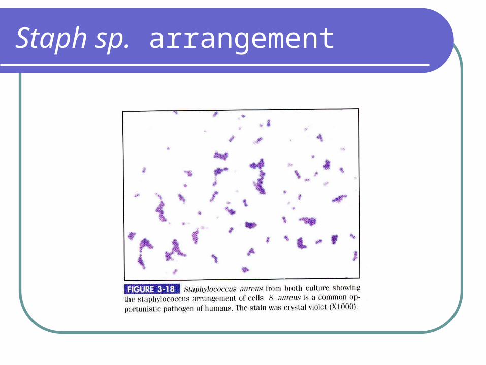
Staph sp. arrangement
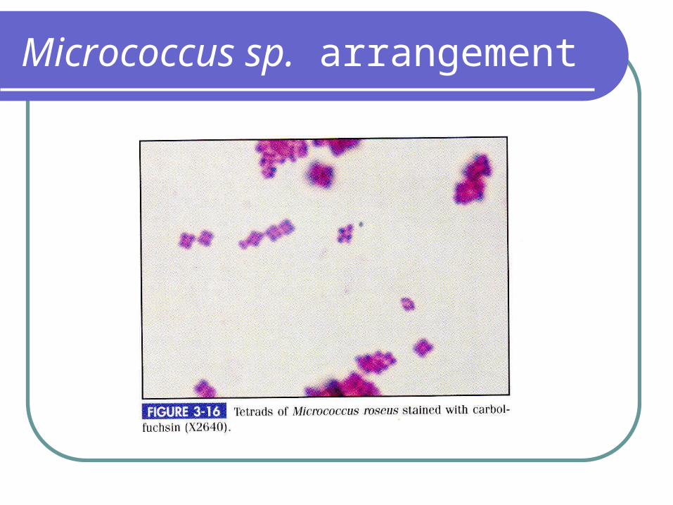
Micrococcus sp. arrangement
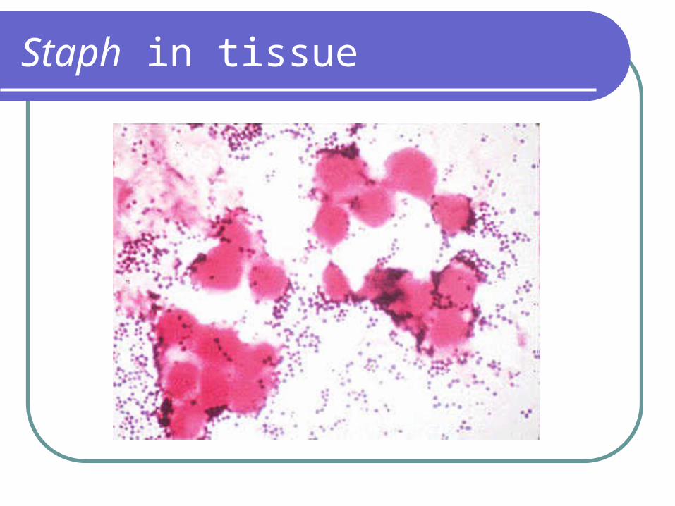
Staph in tissue
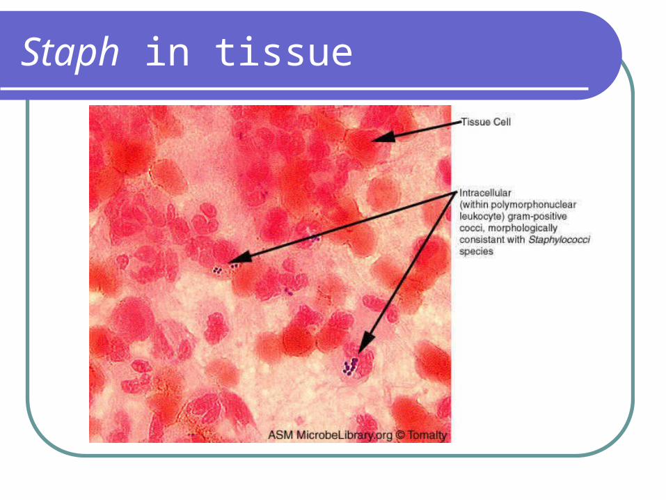
Staph in tissue
Family Micrococcaceae
Will grow on most lab media that will support the growth of Gram positive organisms. Within 24 hours smooth, circular colonies with a buttery consistency will grow.
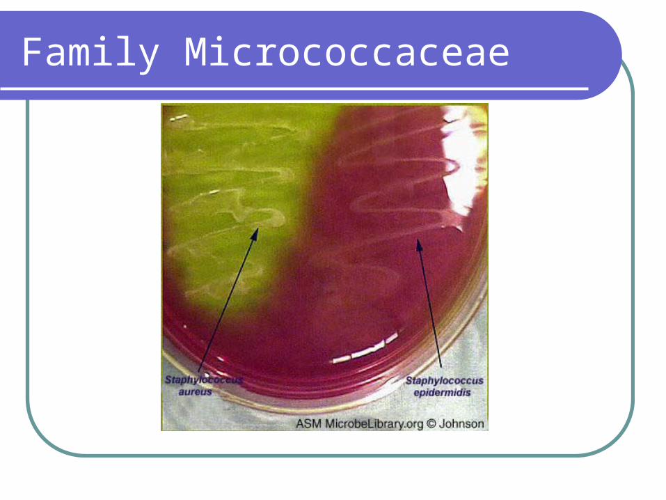
S. aureus classically has a golden or yellow pigmentation, but many clinical isolates have a creamy or white pigmentation.
S. epidermidis produces white colonies M. luteus produces colonies with bright yellow
pigmentation
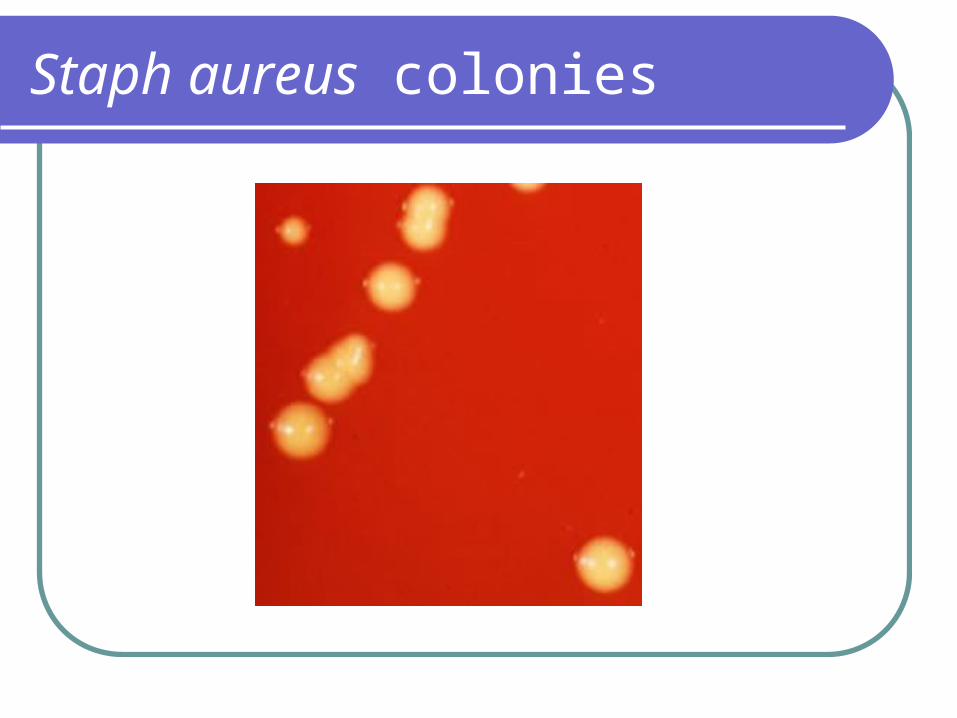
Staph aureus colonies
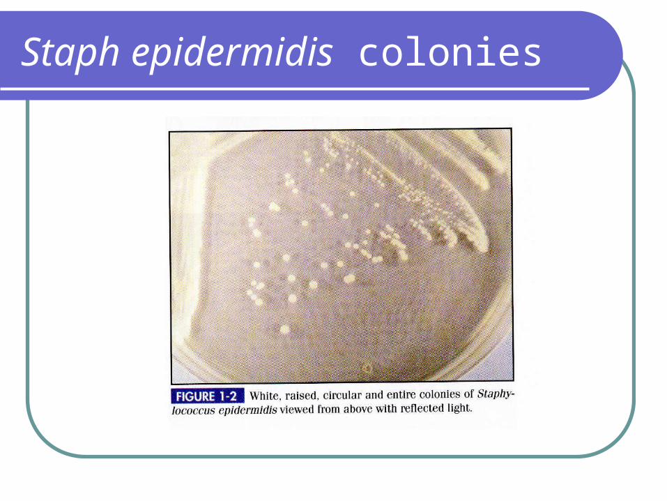
Staph epidermidis colonies
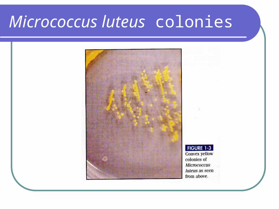
Micrococcus luteus colonies
Family Micrococcaceae
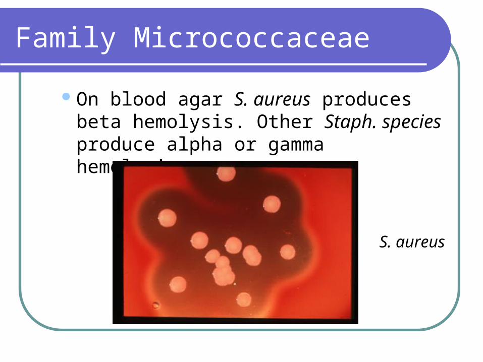
On blood agar S. aureus produces beta hemolysis. Other Staph. species produce alpha or gamma hemolysis.
S. aureus

Family Micrococcaceae
Selective media may be used to isolate Staph. from specimens likely to be contaminated with other bacterial flora.
Phenylethyl alcohol (PEA) – inhibits Gram negative bacteria
Family Micrococcaceae
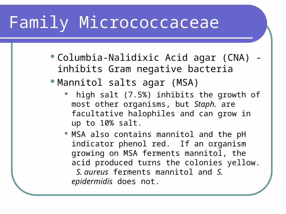
Columbia-Nalidixic Acid agar (CNA) - inhibits Gram negative bacteria
Mannitol salts agar (MSA) high salt (7.5%) inhibits the growth of most other
organisms, but Staph. are facultative halophiles and can grow in up to 10% salt.
MSA also contains mannitol and the pH indicator phenol red. If an organism growing on MSA ferments mannitol, the acid produced turns the colonies yellow. S. aureus ferments mannitol and S. epidermidis does not.
Family Micrococcaceae

Family Micrococcaceae
Biochemical identificationStomatococcus versus all others in the
familyStomatococci are catalase –Micrococci and Staphylococci are catalase +
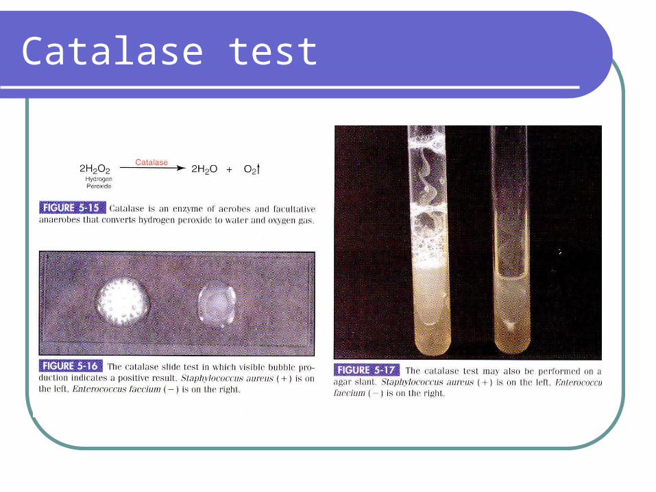
Catalase test
Family Micrococcaceae
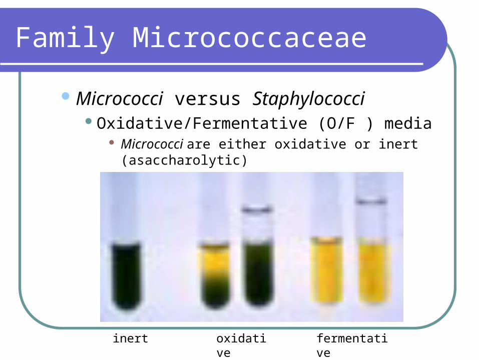
Micrococci versus StaphylococciOxidative/Fermentative (O/F ) media
Micrococci are either oxidative or inert (asaccharolytic)
Staphylococci are fermentative
inert oxidative fermentative
Family Micrococcaceae
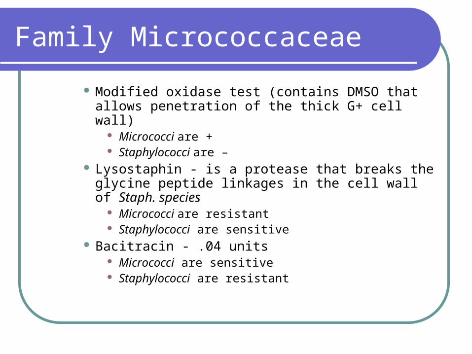
Modified oxidase test (contains DMSO that allows penetration of the thick G+ cell wall)
Micrococci are + Staphylococci are –
Lysostaphin - is a protease that breaks the glycine peptide linkages in the cell wall of Staph. species
Micrococci are resistant Staphylococci are sensitive
Bacitracin - .04 units Micrococci are sensitive Staphylococci are resistant
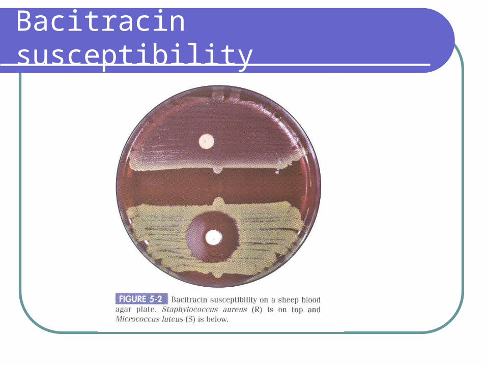
Bacitracin susceptibility
Family Micrococcaceae
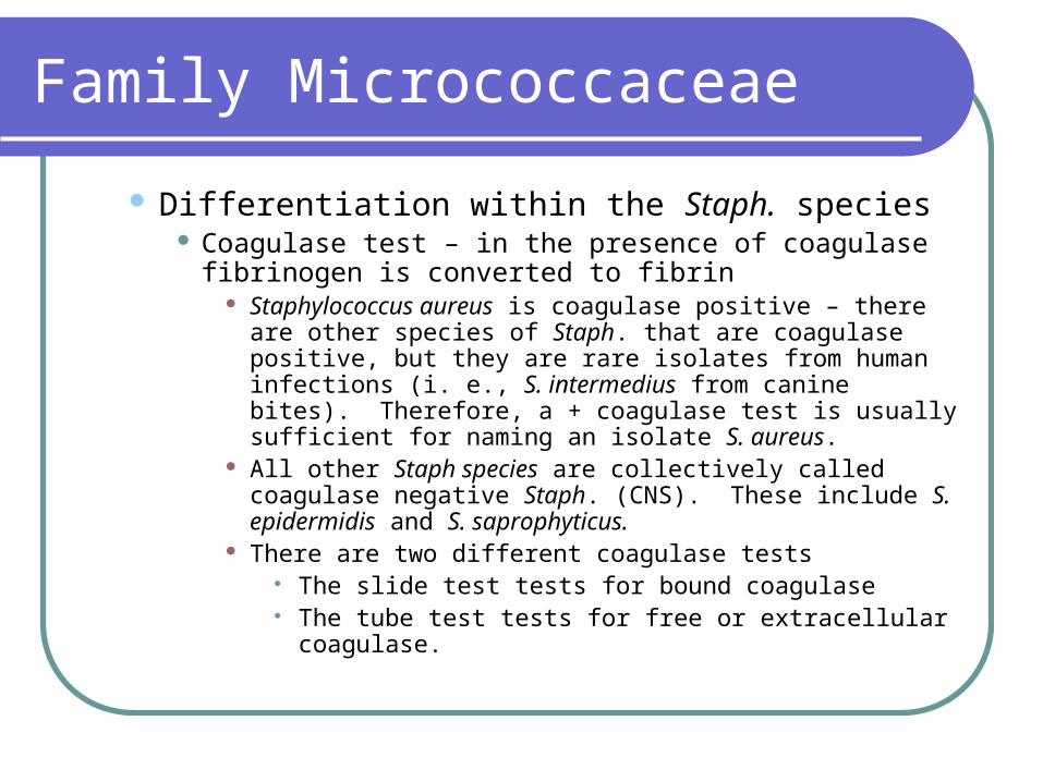
Differentiation within the Staph. species Coagulase test – in the presence of coagulase fibrinogen
is converted to fibrin Staphylococcus aureus is coagulase positive – there are
other species of Staph. that are coagulase positive, but they are rare isolates from human infections (i. e., S. intermedius from canine bites). Therefore, a + coagulase test is usually sufficient for naming an isolate S. aureus.
All other Staph species are collectively called coagulase negative Staph. (CNS). These include S. epidermidis and S. saprophyticus.
There are two different coagulase tests The slide test tests for bound coagulase The tube test tests for free or extracellular coagulase.
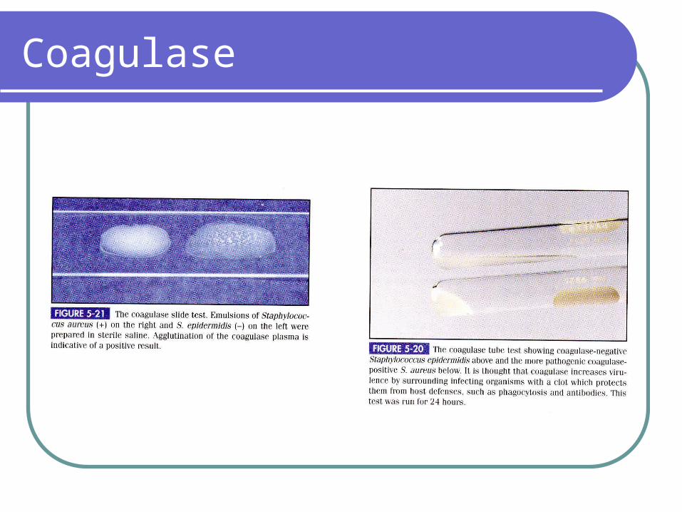
Coagulase

Family Micrococcaceae
Mannitol fermentation S. aureus and some S. saprophyticus are positive S. epidermidis is negative
DNAse S. aureus is positive S. epidermidis and S. saprophyticus are negative

DNAse test
Family Micrococcaceae
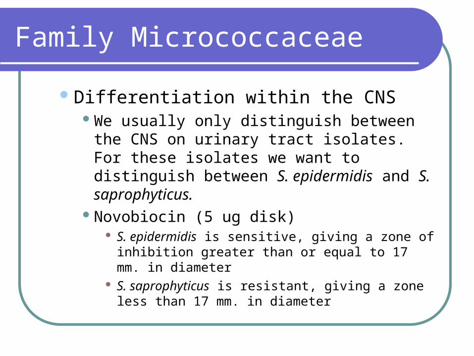
Differentiation within the CNSWe usually only distinguish between the CNS on
urinary tract isolates. For these isolates we want to distinguish between S. epidermidis and S. saprophyticus.
Novobiocin (5 ug disk) S. epidermidis is sensitive, giving a zone of inhibition
greater than or equal to 17 mm. in diameter S. saprophyticus is resistant, giving a zone less than
17 mm. in diameter
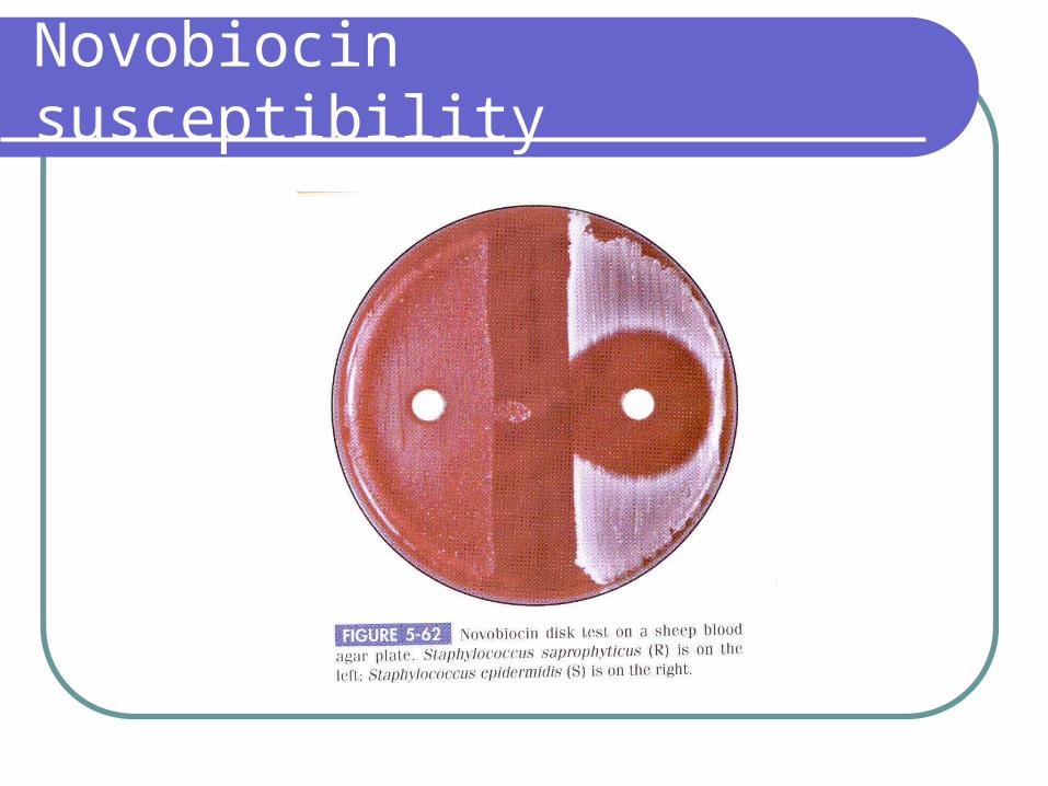
Novobiocin susceptibility
Family Micrococcaceae
Other ways to identify Staph. species as the causative agents of an infection Rising antibody titer to Staph. teichoic acids. Teichoic
acids are part of the cell wall of Staph. and other gram positive organisms and they vary in structure depending upon the organism. A rising titer to Staph. teichoic acids may be used to confirm a diagnosis of Staph. endocarditis.
Phage typing – Different strains of S. aureus and S. epidermidis are placed into different groups based on their susceptibility to infection by different bacteriophages. Used in epidemiologic investigations.

Family Micrococcaceae
Epidemiologic investigations may also identify different strains of S. aureus based on the type of antigenic polysaccharide capsule produced.
Mechanisms of pathogenicity – unless otherwise stated, these refer to S. aureus only Capsule – allows the organism to resist
phagocytosis. Some only form capsules “in vivo”. Teichoic acids – for all Staph. species - may be
involved in adherence as a fibronectin binding protein (fibronectin is found on the surface of many cells).
Mechanisms of pathogenicity
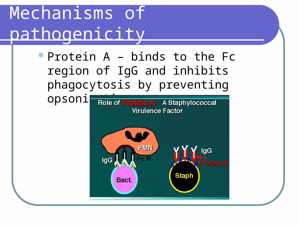
Protein A – binds to the Fc region of IgG and inhibits phagocytosis by preventing opsonization.

Mechanisms of pathogenicity
ToxinsHemolysins – remember that S. aureus is beta
hemolytic. It may produce 4 different hemolysins: ,, , and . Except for hemolysin,they all lyse leukocytes and other tissue cells as well as RBCs.
toxin, in particular, may produce extensive tissue damage.
hemolysin is known as the hot-cold lysin because its hemolytic activity is enhanced when 370 C incubation is followed by 40 C incubation.
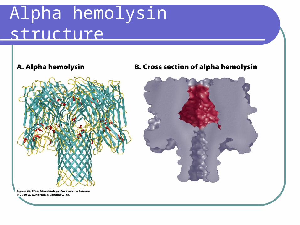
Alpha hemolysin structure
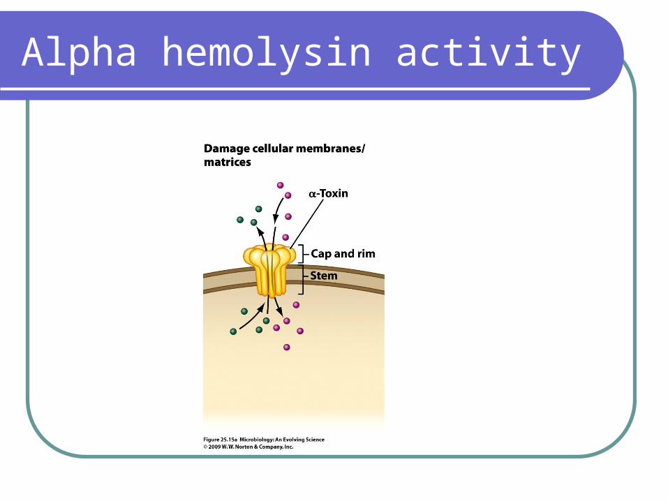
Alpha hemolysin activity
Mechanisms of pathogenicity
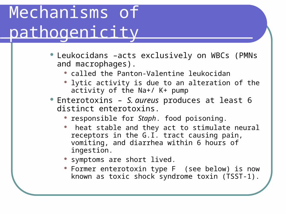
Leukocidans –acts exclusively on WBCs (PMNs and macrophages).
called the Panton-Valentine leukocidan lytic activity is due to an alteration of the activity of the Na+/
K+ pump Enterotoxins – S. aureus produces at least 6 distinct
enterotoxins. responsible for Staph. food poisoning. heat stable and they act to stimulate neural receptors in the
G.I. tract causing pain, vomiting, and diarrhea within 6 hours of ingestion.
symptoms are short lived. Former enterotoxin type F (see below) is now known as
toxic shock syndrome toxin (TSST-1).
Mechanisms of pathogenicity
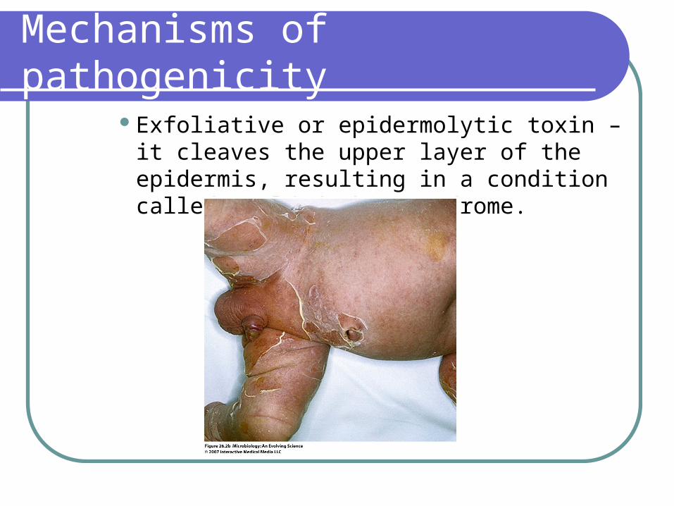
Exfoliative or epidermolytic toxin – it cleaves the upper layer of the epidermis, resulting in a condition called scalded skin syndrome.

Mechanisms of pathogenicity
TSST– 1 - is pyrogenic (fever causing) due to IL-1
induction causes erythroderma (red skin) Causes enhanced susceptibility to endotoxin
shock. Many of the effects of enterotoxins,
exfoliative toxin and TSST-1 are due to their action as a superantigens.

Mechanisms of pathogenicity
What is a superantigen? It causes non-specific stimulation of T cells leading to cytokine release and inflammation which leads to fever, hypertension and shock.
Mechanisms of pathogenicity
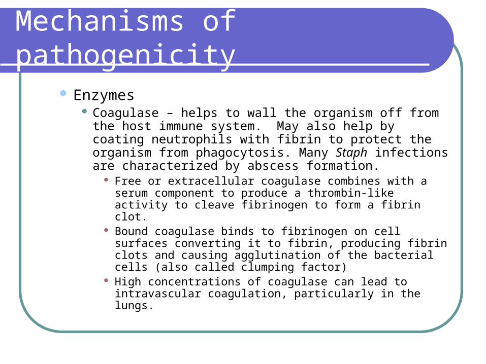
Enzymes Coagulase – helps to wall the organism off from the host
immune system. May also help by coating neutrophils with fibrin to protect the organism from phagocytosis. Many Staph infections are characterized by abscess formation.
Free or extracellular coagulase combines with a serum component to produce a thrombin-like activity to cleave fibrinogen to form a fibrin clot.
Bound coagulase binds to fibrinogen on cell surfaces converting it to fibrin, producing fibrin clots and causing agglutination of the bacterial cells (also called clumping factor)
High concentrations of coagulase can lead to intravascular coagulation, particularly in the lungs.
Mechanisms of pathogenicity
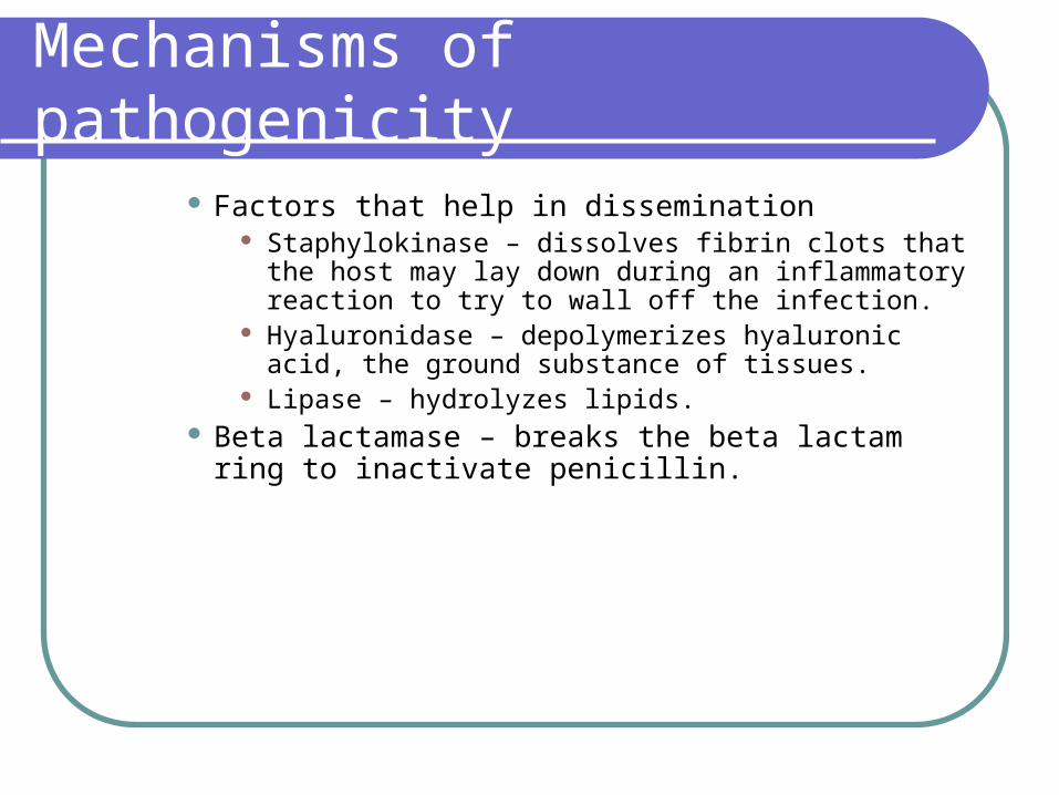
Factors that help in dissemination Staphylokinase – dissolves fibrin clots that the host may lay
down during an inflammatory reaction to try to wall off the infection.
Hyaluronidase – depolymerizes hyaluronic acid, the ground substance of tissues.
Lipase – hydrolyzes lipids. Beta lactamase – breaks the beta lactam ring to inactivate
penicillin.
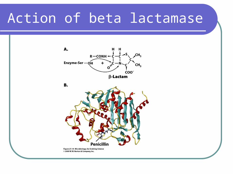
Action of beta lactamase
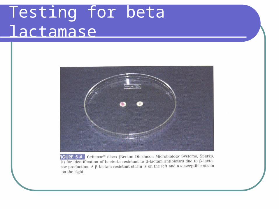
Testing for beta lactamase

Mechanisms of pathogenicity
DNAse – degrades accumulated inflammatory exudate DNA from leukocyte disintegration helping the organism to spread (DNA is very viscous making dissemination more difficult)
Slime production – this is an extracellular glycoconjugate that helps the organism to adhere to smooth surfaces and is produced by CNS as well as S. aureus.
This is important in allowing colonization of indwelling catheters, a major problem in hospitalized patients.
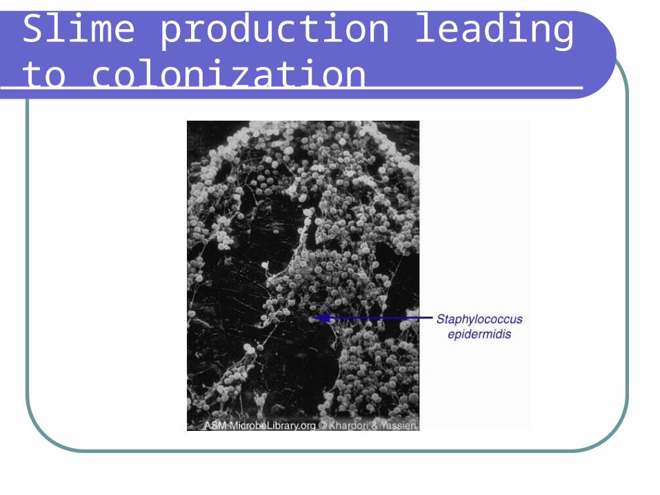
Slime production leading to colonization
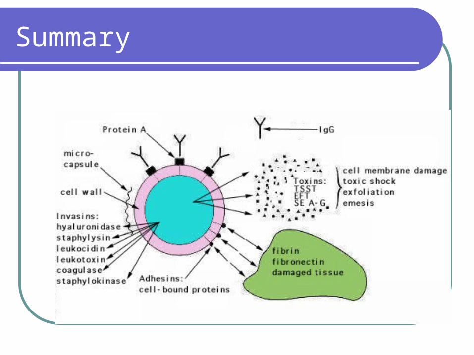
Summary
Micrococcaceae
Clinical significance – Staph are ubiquitous and found as normal flora (NF) of man and other animals.
CNS strains, usually S. epidermidis, are part of the NF of the skin
S. aureus is part of the NF of the nasopharynx in 10-40% of the population. The percentage is higher in hospitalized patients.
They are opportunistic (S. epidermidis) or facultative (S. aureus) pathogens.
Clinical significance
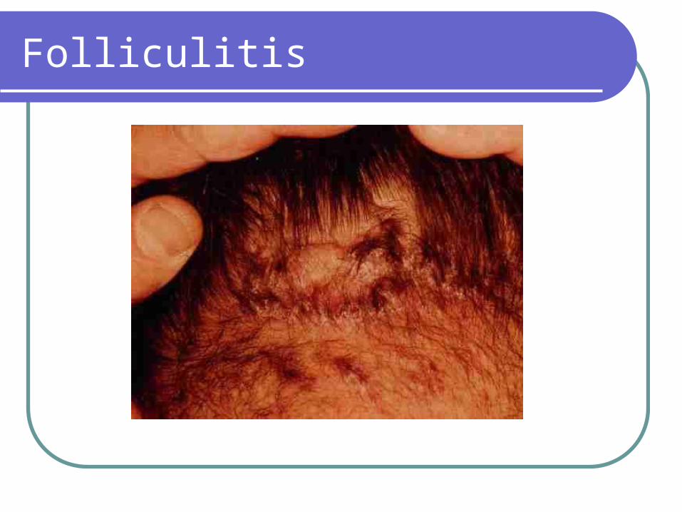
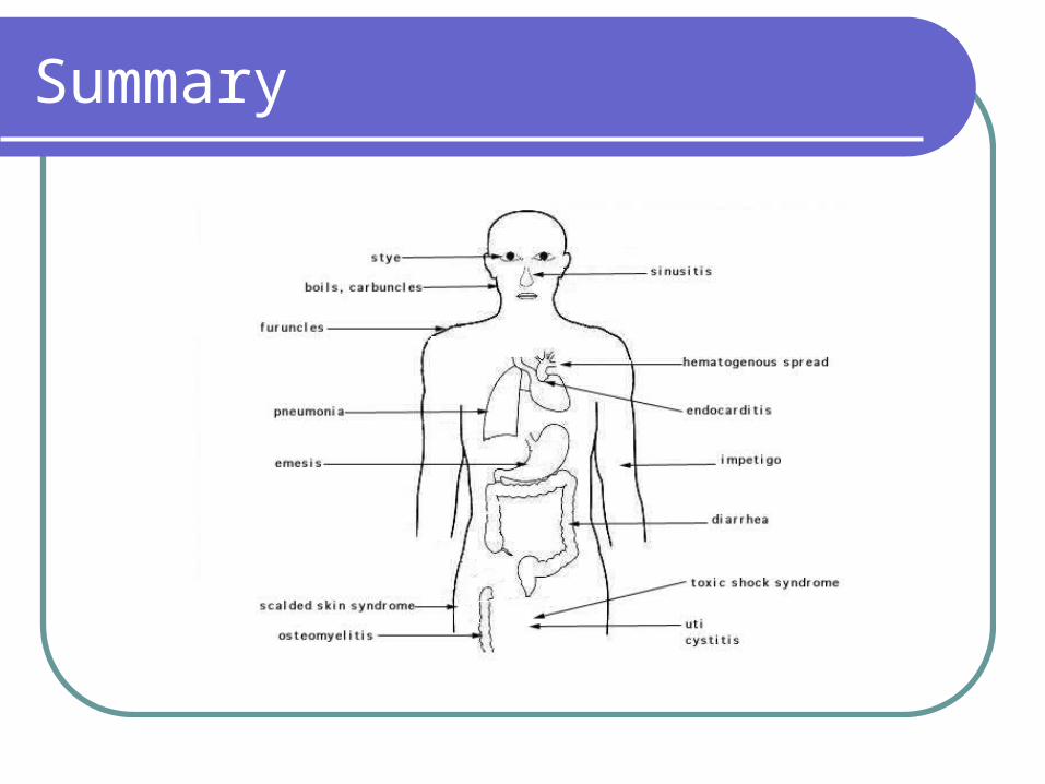
Invasive infectionsS. aureus can cause localized infections in
nearly any area of the body. Local skin infections are the most common type of infection. Suppuration (pus production) is a hallmark of these infections.
Folliculitis is an infection of a hair follicle. If the hair follicle is an eyelash, the infection is commonly called a sty.
Folliculitis
Invasive infections
Furuncle or boil – when folliculitis spreads to involve subcutaneous tissue.
Carbuncle – is a series of interconnected furuncles Dissemination – when the organism spreads throughout the
body to cause: Bacteremia Septicemia with lymphangitis Osteomyelitis Pneumonia Meningitis Endocarditis – acute or subacute (this can occur
following a simple tooth extraction)
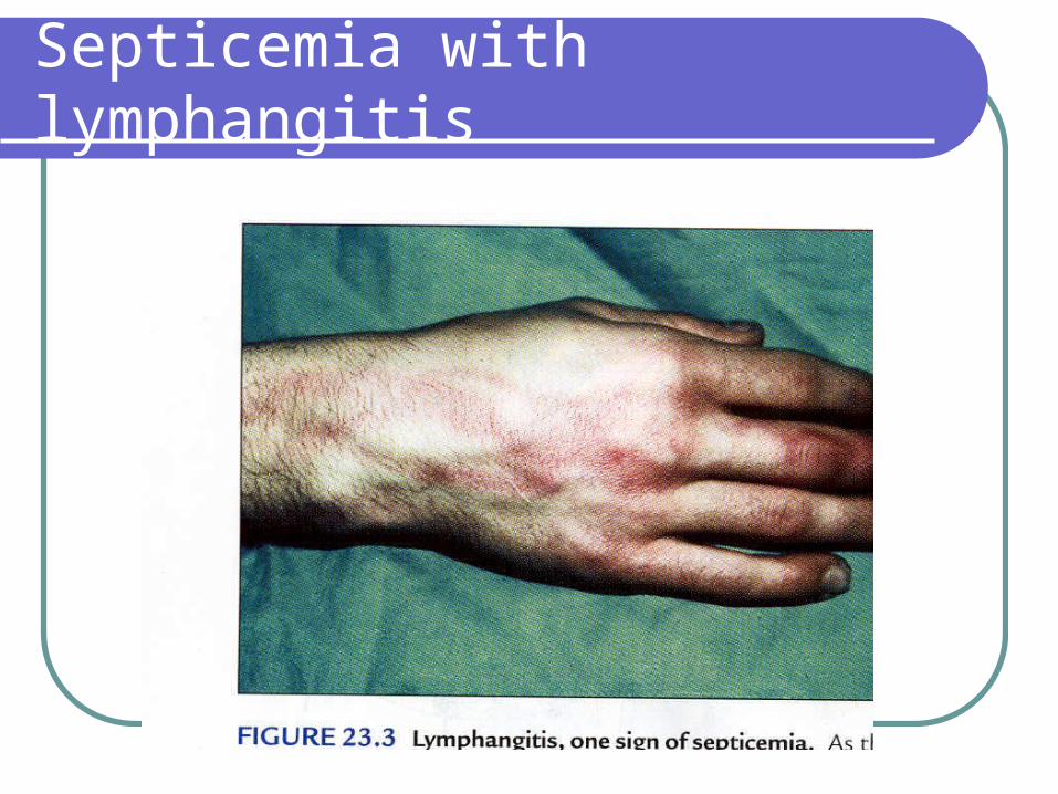
Septicemia with lymphangitis
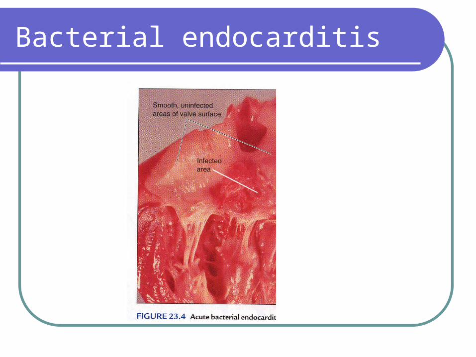
Bacterial endocarditis
Clinical significance
Staph aureus and Staph saprophyticus can cause urinary tract infections.
Staph saprophyticus is the second most common cause of urinary tract infections in sexually active young women.
Toxigenic diseases Food poisoning due to a heat stable enterotoxin. More
common in foods with mayonnaise or custards. Scalded skin syndrome – due to exfoliative toxin which
initially causes a red rash followed by a peeling away of the skin in sheaths.
peeling usually occurs 2 times, but heals without scarring. is more common in infants and young children.
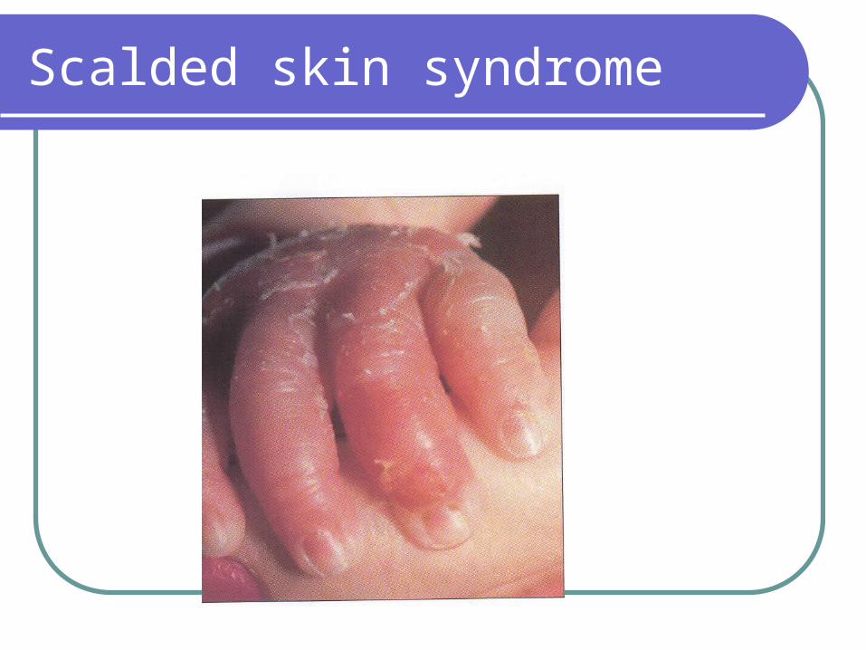
Scalded skin syndrome
Toxigenic diseases
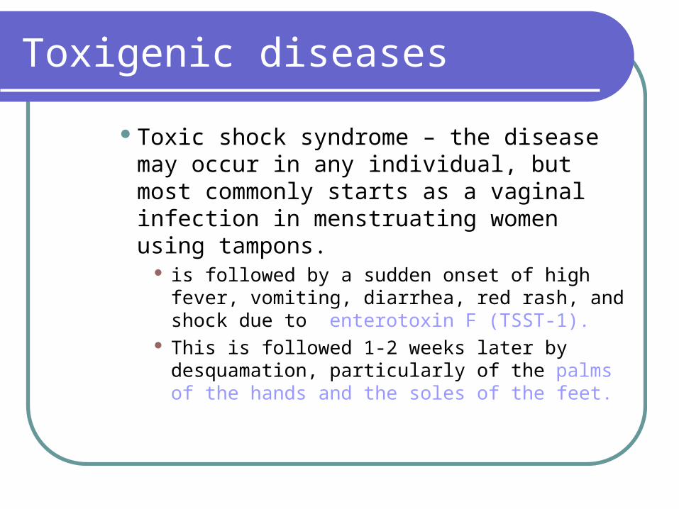
Toxic shock syndrome – the disease may occur in any individual, but most commonly starts as a vaginal infection in menstruating women using tampons.
is followed by a sudden onset of high fever, vomiting, diarrhea, red rash, and shock due to enterotoxin F (TSST-1).
This is followed 1-2 weeks later by desquamation, particularly of the palms of the hands and the soles of the feet.
Summary
Micrococcaceae
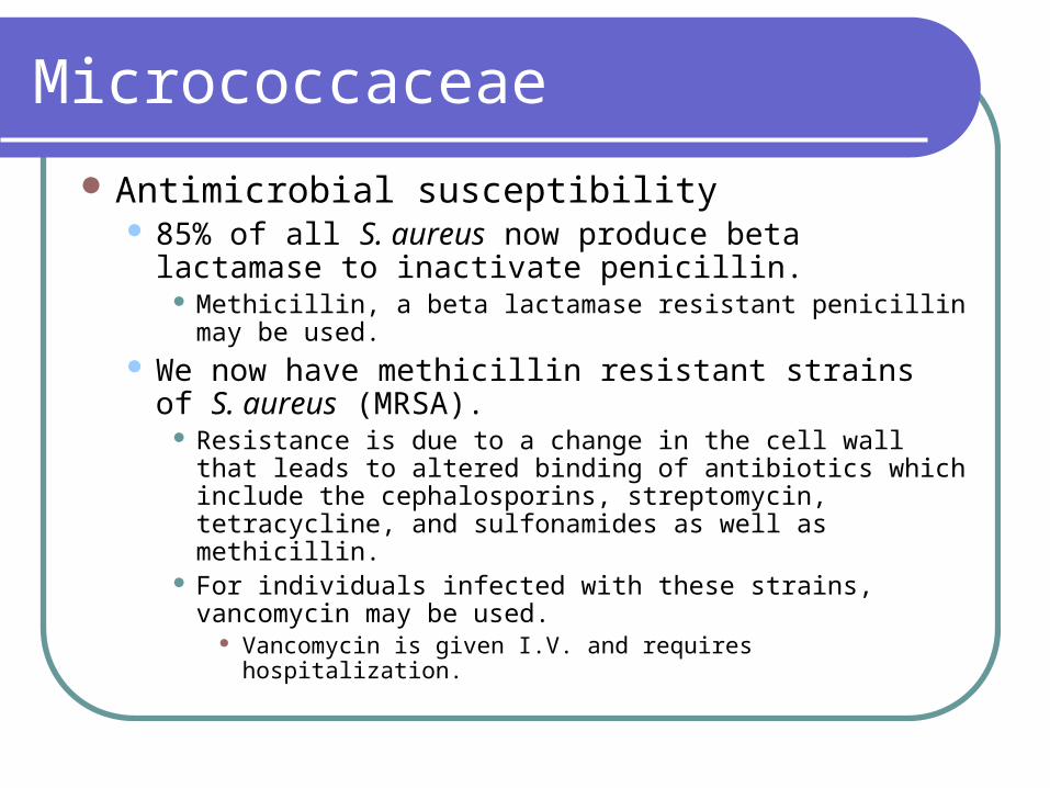
Antimicrobial susceptibility 85% of all S. aureus now produce beta lactamase
to inactivate penicillin. Methicillin, a beta lactamase resistant penicillin may be
used. We now have methicillin resistant strains of S.
aureus (MRSA). Resistance is due to a change in the cell wall that leads to
altered binding of antibiotics which include the cephalosporins, streptomycin, tetracycline, and sulfonamides as well as methicillin.
For individuals infected with these strains, vancomycin may be used.
Vancomycin is given I.V. and requires hospitalization.
Micrococcaceae
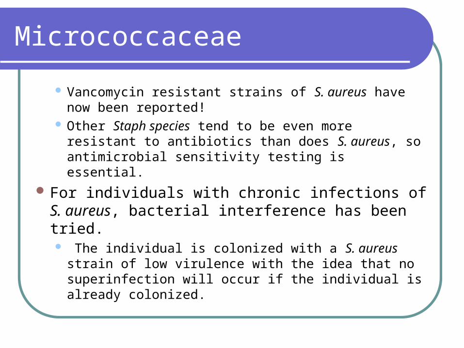
Vancomycin resistant strains of S. aureus have now been reported!
Other Staph species tend to be even more resistant to antibiotics than does S. aureus, so antimicrobial sensitivity testing is essential.
For individuals with chronic infections of S. aureus, bacterial interference has been tried. The individual is colonized with a S. aureus strain
of low virulence with the idea that no superinfection will occur if the individual is already colonized.