microperimetry handbook - haag-streit diagnostics · low tension glaucoma page 30 pilocytic optic...
TRANSCRIPT

Macular Integrity Assessment
Microperimetry Handbook
First Edition

IntroductionMicroperimetry also known as fundus-related perimetry, correlates retinal morphology and function. It combines fundus imaging, retinal sensitivity mapping and fixation analysis in one examination and has been used over a decade as a powerful tool to detect, describe and follow-up pathologies affecting the macular area. Its great advantage is the ability to record and control a patient’s fixation activity while measuring visual field, hence eliminating errors caused by fixation losses.
The purpose of this MAIA Handbook is to show Eye Care professionals some examples of MAIA microperimetry capabilities during the analysis of retinal function in different clinical cases from retina practice to low vision centers.
Index
4 Basic Concepts Related to MAIA Microperimetry
Retinal Imaging Page 4
Analysis of Retinal Sensitivity Page 4
Decibel Scale ( dB ) Page 4
Projection Strategy Page 5
Macular Integrity Index Page 5
Analysis of Fixation and the PRL Page 5
Fixation Location (PRL) Page 5
PRL_initial & PRL_final Page 6
Fixation Stability Page 6
The PRL Training Page 6
7 The MAIA Follow-Up Examination
8 How to Read Printouts
10 Clinical Cases
Age Related Macular Degeneration Page 12 - 19
Diabetic Macular Edema Page 20 - 23
Vitreo-Macular Traction Page 24
Epiretinal Membrane Page 26
Macular Hole Page 28
Low Tension Glaucoma Page 30
Pilocytic Optic Nerve Page 32
Chlorochine Maculopathy Page 34
Plaquenil Toxicity Page 36 - 39
Pre Cataract Surgery Evaluation Page 40
PRL Analysis and Rehabilitation Page 42 - 51
52 Authors Biography Page 52 - 54
2 3Macular Integrity Assessment
Macular Integrity Assessment

Basic Concepts Related to MAIA Microperimetry
MAIA is the 3rd generation of microperimetry instruments and the first easy to use device of its kind. MAIA microperimeter technology combines 3 different techniques in the analysis of the retinal function: a) Retinal Imaging, b) Analysis of retinal sensitivity and c) Analysis of fixation capabilities.
Retinal Imaging
In MAIA, the retinal image is created by means of a Scanning Laser Ophthalmoscope (SLO). The SLO is a confocal technology widely used in the analysis of retinal morphology thanks to the high-resolution image quality.
It is a non mydriatic instrument and does not require a flash to image the retina. Images can be obtained even in the presence of media opacity such as mild cataract.
Image of the MAIA Scanning Laser Ophthalmoscope (SLO).
Analysis of Retinal Sensitivity
Microperimietry, similarly to standard automated perimetry (SAP), measures retinal sensitivity as the minimum light intensity that patients can perceive when spots of light stimulate specific areas of the retina. The examination can be customized with different number of stimuli covering a variable field of vision. The standard MAIA examination covers a 10° diameter area with 37 measurement points. In MAIA, light stimuli are created by a white LED and projected directly onto the retina surface. The stimuli size are Goldmann III, background luminance is 4 asb and maximum luminance is 1000 asb, with a 36 decibels (dB) dynamic range.
Image of the standard MAIA sensitivity grid map.
Decibel Scale (dB)
In perimetry, the stimuli luminance is measured in apostilbs (asb). An apostilb is an absolute unit of luminance and is equal to 0.3183 candela/m2. The decibel scale is a relative scale which depends on the maximum intensity that perimetry instruments can emit. It is an inverted logarithmic scale where zero decibels is set as the brightest stimulus that the perimeter can produce. The decibel scale is not standardized because the maximal luminance varies between instruments. The decibel value range is
calculated between the minimum and the maximum intensity level of the projected stimuli. Therefore physicians shall be careful when comparing results in decibels from different instruments with different maximum intensity of stimuli projection. The decibels scale is color-coded according to the MAIA normative studies where “green” represent normal values, “yellow” suspect, “red” abnormal and “black” represents scotoma as shown in the graphic below.
Projection Strategy
In order to measure the minimum retinal sensitivity over a specific area, the Goldmann III light stimuli may be projected on the same spot several times at different light intensities following a “projection strategy”. MAIA can work with 3 different projection strategies (software version 1.7.0, January 2013); the full threshold 4-2, the 4 Levels Fixed (4-LF) and the Scotoma-Finder (SF).
Maia 4-2 follows the perimetric standard. It changes the light intensity in 4dB steps until there is a change from not seen to seen (or from seen to not seen). Then the intensity changes in 2db steps until the stimulus is not seen again. The standard MAIA examination using 4-2 strategy has an average duration of 5.5 minutes.
The 4-Levels-Fixed (4-LF) is a supra-threshold strategy designed to have a fast assessment of retinal sensitivity on patients with known pathologies. It projects 4 different stimuli intensities: 25 dB, 15 dB, 5 dB, and 0 dB. With this strategy, Maia provides an initial assessment of “good”, “medium”, “bad” or “scotomatous” retinal sensitivity. The standard MAIA examination using the 4-LF has an average duration of 2.5 minutes.
The Scotoma Finder (SF) is a supra-threshold strategy designed for patients with severely affected central vision and provides information concerning progression of the “blind” area. SF strategy projects only 0dB stimuli. The examination using the SF strategy has an average duration of 1.5 minutes.
Analysis of Fixation and the Preferred Retinal Locus (PRL)
Fixation Location (PRL)
Fixation is the process of attempting to “look at” a selected visual target and consists of optically aligning a functional area of the retina to that target.
In normal subjects the retinal area used for fixation is the fovea, whereas when pathology affects the central retina, fixation degrades and patients develop a condition known as eccentric viewing and use extra-foveal regions. In general, the retinal area used to attempt fixation is known as the Preferred Retinal Locus (PRL).
MAIA provides accurate and objective information regarding retinal location and stability of a patient’s fixation. Such parameters are assessed by tracking eye movements 25 times / sec and by plotting the resulting distribution over the SLO image.Each movement is represented by a point in the distribution. The overall cloud of points describes the PRL.
Image of the MAIA dB color scale
Macular Integrity Index
The Macular integrity index is a numerical value that describes the likelihood that a patient’s responses are normal, suspect or abnormal when compared to age-adjusted normative data. It does not represent the severity of the disease process. Higher numbers suggest a greater likelihood of abnormal findings, while lower values suggest a greater likelihood
of normal findings. There is no direct relationship between the average threshold value (dB) and the macular integrity index. In fact it is possible for the average threshold to be normal while the macular integrity index is abnormal. This index is only present in examinations performed with the standard MAIA stimuli grid and the 4-2 projection strategy.
Example of abnormal macular Integrity Index with normal averaged dB values
4 5Macular Integrity Assessment
Macular Integrity Assessment

The MAIA Follow-Up Examination
The MAIA Follow-up examination repeats the baseline test by accurately re-measuring the same anatomical locations. It allows precise functional monitoring, even in cases where the retina morphology has changed due to pathology progression.The time line report shows sensitivity and fixation changes in a differential color grid sensitivity map and a time line graphic.
The color code of the differential grid map is the following:
• Increased sensitivity • Unchanged sensitivity• Sensitivity decrease of 2 dB• Sensitivity decrease of more than 2 dB
The PRL Training
MAIA microperimeter employs auditory and visual bio-feedback signals as eccentric viewing therapy (PRL Training). It is used to train Low Vision patients with central scotoma and unstable fixation, to use
a specific retinal location with better functional characteristics. The purpose of the PRL training is to help low vision patients in the better use of residual vision by increasing fixation stability.
Fixation Stability
Fixation stability is measured in 2 different ways:
• Calculating the percentage of fixation points (%) located within a distance of 1° and 2° respectively (P1 and P2).
The classification of stability is based on the following criteria:
1. If more than 75% of the fixation points are located within P1, the fixation is classified as “stable”.
2. If less than 75% of the fixation points are located within P1, but more than 75% of the fixation points are located within P2, the fixation is classified as “relatively unstable”.
3. If less than 75% are located within P2, the fixation is classified as “unstable”.
• Calculating the area of an ellipse which encompasses the cloud of fixation points for a given proportion based on standard deviations of the horizontal and vertical eye positions during the fixation attempt.
The analysis of fixation is performed in every MAIA test independently of the stimuli grid selected. Image of fixation analysis with
P1, P2 and the Bivariate Contour Ellipse Area.
PRL_initial (PRLi) and PRL_final (PRLf)
MAIA identifies 2 main PRL reference points calculated as the barycenter of the cloud of fixation points known as PRL_initial and PRL_final. The first one is found after the initial ten seconds of the examination, when patients make their highest effort to hold a steady fixation.
The PRL_initial defines the center of the MAIA stimuli grid. The second one is found at the end of the MAIA examination and it serves as the reference point to calculate fixation stability. Patients with stable fixation will present both PRLs in the same anatomical location, while bigger distance among PRL’s will determine more unstable fixation conditions and less visual acuity.
Image of PRL_initial and PRL_final.
6 7Macular Integrity Assessment
Macular Integrity Assessment

Macular Integrity Assessment
How to Read Printouts
MAIA contains a reference database for the quantitative comparison of retinal sensitivity to the corresponding normal ranges. MAIA provides a very detailed printout that encompasses all collected information.
1
2
5
8
3
4
6
7
13
12
11
10
9
1 Clinic name
2 Patient info
3 Examined eye
4 SLO image of fundus
5 Examination Info
6 Interpolated sensitivity map over full SLO image
7 Fixation plot over zoomed SLO image and PRL identification
8 Bivariate Contour Ellipse Area indices
9 Sensitivity values (dB) and PRL over zoomed SLO image
10 Color coded Macular Integrity index
11 Color coded Average Threshold
12 Histogram of Threshold values (grey) compared with normal distribution (green)
13 Fixation graph describing amplitude of eye movements vs. time
Legend
9Macular Integrity Assessment8Macular Integrity Assessment

Clinical CasesMAIA is a perimeter designed to analyze the macular area. There is a wide range of diseases affecting the macula leading to loss of central vision reducing quality of life.
The following clinical cases are collected from a set of retina specialists, who have found in MAIA a useful tool to measure, monitor and rehabilitate central vision in patients with very different retinal pathologies.
Macular Integrity Assessment
10 11Macular Integrity Assessment
Macular Integrity Assessment
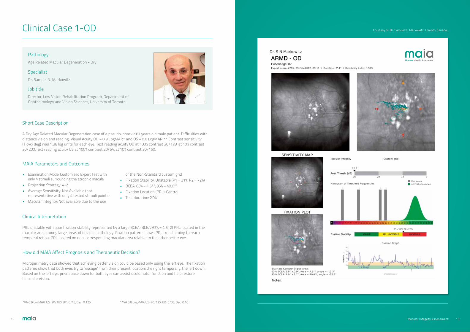
Clinical Case 1-OD
Short Case Description
A Dry Age Related Macular Degeneration case of a pseudo-phackic 87 years old male patient. Difficulties with distance vision and reading. Visual Acuity OD = 0.9 LogMAR* and OS = 0.8 LogMAR.** Contrast sensitivity (1 cyc/deg) was 1.38 log units for each eye. Text reading acuity OD at 100% contrast 20/128, at 10% contrast 20/200.Text reading acuity OS at 100% contrast 20/64, at 10% contrast 20/160.
Clinical Interpretation
PRL unstable with poor fixation stability represented by a large BCEA (BCEA: 63% = 4.5°2) PRL located in the macular area among large areas of obvious pathology. Fixation pattern shows PRL trend aiming to reach temporal retina. PRL located on non-corresponding macular area relative to the other better eye.
How did MAIA Affect Prognosis and Therapeutic Decision?
Microperimetry data showed that achieving better vision could be based only using the left eye. The fixation patterns show that both eyes try to “escape” from their present location: the right temporally, the left down. Based on the left eye, prism base down for both eyes can assist oculomotor function and help restore binocular vision.
MAIA Parameters and Outcomes
• Examination Mode: Customized Expert Test with only 4 stimuli surrounding the atrophic macula• Projection Strategy: 4-2• Average Sensitivity: Not Available (not representative with only 4 tested stimuli points)• Macular Integrity: Not available due to the use
of the Non-Standard custom grid• Fixation Stability: Unstable (P1 = 31%, P2 = 72%)• BCEA: 63% = 4.5°2, 95% = 40.6°2
• Fixation Location (PRL): Central• Test duration: 2’04”
PathologyAge Related Macular Degeneration - Dry
SpecialistDr. Samuel N. Markowitz
Job title Director, Low Vision Rehabilitation Program, Department of Ophthalmology and Vision Sciences, University of Toronto.
Macular Integrity Assessment
*VA 0.9 LogMAR: US=20/160, UK=6/48, Dec=0.125 **VA 0.8 LogMAR: US=20/125, UK=6/38, Dec=0.16
Courtesy of: Dr. Samuel N. Markowitz, Toronto, Canada.
13Macular Integrity Assessment12Macular Integrity Assessment

Clinical Case 1-OS
Short Case Description
Left eye of an 87 year-old pseudo-phakic patient with dry age-related macular degeneration. Difficulties with distance vision and reading. Visual Acuity was OD = 0.9 LogMAR* OS = 0.8 LogMAR**. Contrast sensitivity (1 cyc/deg) was 1.38 log units for each eye. Text reading acuity OD at 100% contrast 20/128, at 10% contrast 20/200.Text reading acuity OS at 100% contrast 20/64, at 10% contrast 20/160.
Clinical Interpretation
PRL stable with good fixation stability represented by a smaller BCEA than the other eye. PRL located in an unfavourable location, between the macular scar and the optic disc, below the original fovea. PRL located on non-corresponding macular area relative to the other poorer eye.
How did MAIA Affect Prognosis and Therapeutic Decision?
Microperimetry data showed that achieving better vision could be based on the left eye. The fixation patterns show that both eyes try to “escape” from their present location, the right temporally, the left down. Based on the left eye, prism base down for both eyes can assist oculomotor function and help restore binocular vision.
MAIA Parameters and Outcomes
• Customized Expert Examination with only 4 stimuli surrounding the atrophic macula • Projection Strategy: 4-2• Average Sensitivity: Not Available (not representative with only 4 tested stimuli points)• Macular Integrity: Not available due to the use
of the Non-Standard custom grid• Fixation Stability: Stable (P1 = 83%, P2 = 93%)• BCEA: 63% = 1.1°2, 95% = 9.7°2
• Fixation Location (PRL): Central-Inferior• Test duration: 2’01”
PathologyAge Related Macular Degeneration - Dry
SpecialistDr. Samuel N. Markowitz
Job title Director, Low Vision Rehabilitation Program, Department of Ophthalmology and Vision Sciences, University of Toronto.
Macular Integrity Assessment
*VA 0.9 LogMAR: US=20/160, UK=6/48, Dec=0.125 **VA 0.8 LogMAR: US=20/125, UK=6/38, Dec=0.16
Courtesy of: Dr. Samuel N. Markowitz, Toronto, Canada.
15Macular Integrity Assessment14Macular Integrity Assessment

Clinical Case 2
Short Case Description
Left eye of an 83 year-old patient with bilateral atrophic age related macular degeneration and cataract. The patient complained of worsening of visual acuity. Best corrected visual acuity was 0.6 LogMAR*.
Clinical Interpretation
This patient’s fixation is located in the fovea, among the atrophic areas. A customized sensitivity map was used in this case consisting in 68 stimuli points covering central 20° of the retina. The sensitivity map shows the presence of relative scotomas in whole examined area with few absolute scotomas, outside the fixation area. The average threshold value is 14.4 dB (stimulus dynamic range varies between 0 dB and 36 dB).
How did MAIA Affect Prognosis and Therapeutic Decision?
MAIA documented stable fixation, outside the atrophic areas. Moreover, absolute scotomas did not cover fixation area. Therefore, decrease in visual acuity is probably due to cataract increase. Cataract surgery was proposed to the patient.
MAIA Parameters and Outcomes
• Examination Mode: 10-2° Expert Test (68 points covering central 20°)• Projection Strategy: 4-2• Average Sensitivity: 14.4 dB• Macular Integrity: Not available due to the use of the Non-Standard custom grid
• Fixation Stability: Stable (P1 = 86%, P2 = 94%)• BCEA: 63% = 1.6°2, 95% = 14.6°2
• Fixation Location (PRL): Central• Test duration: 9’51”
PathologyAge Related Macular Degeneration - Atrophic
SpecialistDr. Stela Vujosevic
Job title Medical Director and R&D Director of the The International Microperimetry Reading Centre, Padova, Italy
Macular Integrity Assessment
*VA 0.6 LogMAR: US=20/80, UK=6/24, Dec=0.25
Courtesy of: Dr. Stela Vujosevic, Padova, Italy.
17Macular Integrity Assessment16Macular Integrity Assessment

Clinical Case 3
Short Case Description
Right eye of a female 77 year-old patient. ARMD diagnosed is associated with the presence of retinal atrophy and loss of central vision. Best corrected visual acuity 1.1 LogMAR.*
Clinical Interpretation
The PRL of this patient is shifted temporal adjacent of the atrophic area. Fixation is unstable (P1=36dB). There is an overall reduced of sensitivity (average = 3.7 dB). Nevertheless there is a large scotomatic area, the sensitivity on the temporal side of the PRL reaches up to 20dB. The actual cornerstone of evaluation of dry ARMD consists of visual acuity measurement and evaluation by Amsler grid, although the specificity and reproducibility of these tests is somewhat limited.
In this case, Microperimetry was requested to analyze retinal sensitivity and fixation and to outline microperimetric rehabilitation.
How did MAIA Affect Prognosis and Therapeutic Decision?
After evaluating the MAIA results, the patient was strongly dedicated to microperimetric Biofeedback training to recover fixation stability in order to get driving license renewal and obtained the minimum visual acuity requested to obtain driving licence (0.7 LogMAR.**)
MAIA Parameters and Outcomes
• Examination Mode: Standard Expert Test (37 points covering central 10°)• Projection Strategy: 4-2• Average Sensitivity: 3.7 dB• Macular Integrity: 100
• Fixation Stability: Unstable (P1 = 36%, P2 = 75%)• BCEA: 63% = 4.9°2, 95% = 44.2°2
• Fixation Location (PRL): Central-Temporal• Test duration: 6’44”
PathologyAge-related macular degeneration - Atrophic
SpecialistDr. Fabio Mazzolani
Job title Low Vision and Retina Consultant
Macular Integrity Assessment
**VA 0.7 LogMAR: US=20/100, UK=6/30, Dec=0.20
Courtesy of: Dr Mazzolani, Dr Lovisolo, Quattroelle Eye Clinic, Milan Italy.
19Macular Integrity Assessment18Macular Integrity Assessment

Clinical Case 4
Short Case Description
Left eye of a 52 year-old diabetic patient (diabetes mellitus type 2) with recent onset exudative macular edema involving the center of the fovea. The patient was asymptomatic with best corrected visual acuity 0.0 LogMAR.*
Clinical Interpretation
Patient presents central and stable fixation (localized in the fovea). The MAIA outcome shows presence of relative scotomas located in the superior region of the examined area corresponding mainly to hard exudates and retinal hemorrhages. Even though the average threshold value is 27.2 dB, which may be considered within the normative range, the Macular Integrity index shows 64.2, which falls within the abnormal macular function in MAIA microperimeter and it is related to the localized low sensitivity points.
How did MAIA Affect Prognosis and Therapeutic Decision?
MAIA documented an initial and localized decrease in central retinal sensitivity despite preserved visual acuity. This data was important in explaining to the patient that his visual function was already reduced and therefore he needed a local treatment and better metabolic control. The treatment proposed to the patient was a subthreshold micropulse diode laser of the macula.
MAIA Parameters and Outcomes
• Examination Mode: Standard Expert Test (37 points covering central 10°)• Projection Strategy: 4-2• Average Sensitivity: 27.2 dB• Macular Integrity: 64.2
• Fixation Stability: Stable (P1 = 98%, P2 = 100%)• BCEA: 63% = 0.2°2, 95% = 2.2°2
• Fixation Location (PRL): Central• Test duration: 4’41”
Macular Integrity Assessment
*VA 0.0 LogMAR: US=20/20, UK=6/6, Dec=1.0
Courtesy of: Dr. Stela Vujosevic, Padova, Italy.
PathologyDiabetic Macular Edema
SpecialistDr. Stela Vujosevic
Job title Medical Director and R&D Director of the The International Microperimetry Reading Centre, Padova, Italy.
21Macular Integrity Assessment20Macular Integrity Assessment

Clinical Case 5
Short Case Description
Right eye of a 58 year-old male patient This patient’s first diagnosis was 20 years ago and during this period, he underwent the following different treatments: Focal grid laser, Intravitreal antiVEGF and Corticosteroid, Posterior vitrectomy and epiretinal peeling. OCT and Angiographic Fluorescein Leakage show the same patterns regardless of different treatments. Moreover, Visual Acuity is steadily 0.5 LogMAR*.
Clinical Interpretation
The PRL of this patient is central, although fixation is relatively unstable. Sensitivity is lower on the perifoveal area. Intravitreal therapy improves retinal function, quantified not only by visual acuity, but also by mean retinal sensitivity and fixation stability, as assessed by scanning laser ophthalmoscope microperimetry.
How did MAIA Affect Prognosis and Therapeutic Decision?
Measurement of retinal sensitivity may facilitate evaluation of the effectiveness of intravitreal treatment in patients with diabetic macular edema. No changes in VA or OCT findings do not always match the real disease activity and the necessity of treatment. Patients with stable Visual Acuity and OCT may still have deteriorating retinal sensitivity. This is usually a late manifestation and may indicate subclinical activity.
MAIA Parameters and Outcomes
• Examination Mode: Standard Expert Test (37 points covering central 10°)• Projection Strategy: 4-2• Average Sensitivity: 20.4 dB• Macular Integrity: 100
• Fixation Stability: Unstable (P1 = 49%, P2 = 86%)• BCEA: 63% = 3.7°2, 95% = 33.5°2
• Fixation Location (PRL): Central• Test duration: 5’38”
Macular Integrity Assessment
*VA 0.5 LogMAR: US=20/63, UK=6/19, Dec=0.32
Courtesy of: Dr Mazzolani, Dr Lovisolo -Quattroelle Eye Clinic, Milan Italy.
PathologyDiabetic Macular Edema
SpecialistDr. Fabio Mazzolani
Job title Low Vision and Retina Consultant
23Macular Integrity Assessment22Macular Integrity Assessment
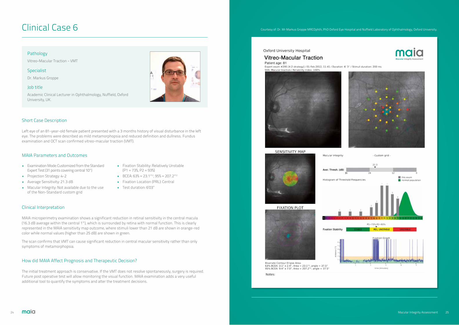
Clinical Case 6
Short Case Description
Left eye of an 81-year-old female patient presented with a 3 months history of visual disturbance in the left eye. The problems were described as mild metamorphopsia and reduced definition and dullness. Fundus examination and OCT scan confirmed vitreo-macular traction (VMT).
Clinical Interpretation
MAIA microperimetry examination shows a significant reduction in retinal sensitivity in the central macula (16.3 dB average within the central 1°), which is surrounded by retina with normal function. This is clearly represented in the MAIA sensitivity map outcome, where stimuli lower than 21 dB are shown in orange-red color while normal values (higher than 25 dB) are shown in green.
The scan confirms that VMT can cause significant reduction in central macular sensitivity rather than only symptoms of metamorphopsia.
How did MAIA Affect Prognosis and Therapeutic Decision?
The initial treatment approach is conservative. If the VMT does not resolve spontaneously, surgery is required. Future post operative test will allow monitoring the visual function. MAIA examination adds a very useful additional tool to quantify the symptoms and alter the treatment decisions.
MAIA Parameters and Outcomes
• Examination Mode: Customized from the Standard Expert Test (31 points covering central 10°)• Projection Strategy: 4-2• Average Sensitivity: 21.3 dB• Macular Integrity: Not available due to the use of the Non-Standard custom grid
• Fixation Stability: Relatively Unstable (P1 = 73%, P2 = 93%)• BCEA: 63% = 23.1°2, 95% = 207.2°2
• Fixation Location (PRL): Central• Test duration: 6’03”
PathologyVitreo-Macular Traction - VMT
SpecialistDr. Markus Groppe
Job title Academic Clinical Lecturer in Ophthalmology, Nuffield, Oxford University, UK.
Macular Integrity Assessment
Courtesy of: Dr. Mr Markus Groppe MRCOphth, PhD Oxford Eye Hospital and Nuffield Laboratory of Ophthalmology, Oxford University.
25Macular Integrity Assessment24Macular Integrity Assessment

Clinical Case 7
Short Case Description
Right eye of a female 68 year-old patient diagnosed is with Epiretinal Membrane. Best corrected visual acuity was 0.3 LogMAR.*
Clinical Interpretation
A relatively diffuse sensitivity reduction was present. PRL is located centrally with relatively unstable fixation. The high quality SLO image available in the MAIA allows a precise correlation between retinal sensitivity and topographic pathology location. Traction lines are also visible.
How did MAIA Affect Prognosis and Therapeutic Decision?
The location and stability of fixation using microperimetry has to be determined because the fixation characteristics are not correlated to quantitative measures on OCT and are useful parameters to suggest visual outcome. Visual acuity strictly depends on fixation stability and location. Moreover, in our experience absolute and relative scotomas may develop postoperatively over the areas where the retinal surface is gently scratched to peel off an overlying membrane. For this reason, the assessment of retinal sensitivity, fixation location and overall retinal functional behaviour are extremely important to determine.
MAIA Parameters and Outcomes
• Examination Mode: 6 degrees Expert Test (37 points covering central 6°)• Projection Strategy: 4-2• Average Sensitivity: 26.4 dB• Macular Integrity: not available due to the use
of the Non-Standard custom grid• Fixation Stability: Unstable (P1 = 62%, P2 = 86%)• BCEA: 63% = 3.8°2, 95% = 34.0°2
• Fixation Location (PRL): Central• Test duration: 5’03”
PathologyEpiretinal Membrane
SpecialistDr. Fabio Mazzolani
Job title Low Vision and Retina Consultant
Macular Integrity Assessment
Courtesy of: Dr Mazzolani, Dr Lovisolo, Quattroelle Eye Clinic, Milan Italy.
*VA 0.3 LogMAR: US=10/40, UK=6/12, Dec=0.5
27Macular Integrity Assessment26Macular Integrity Assessment
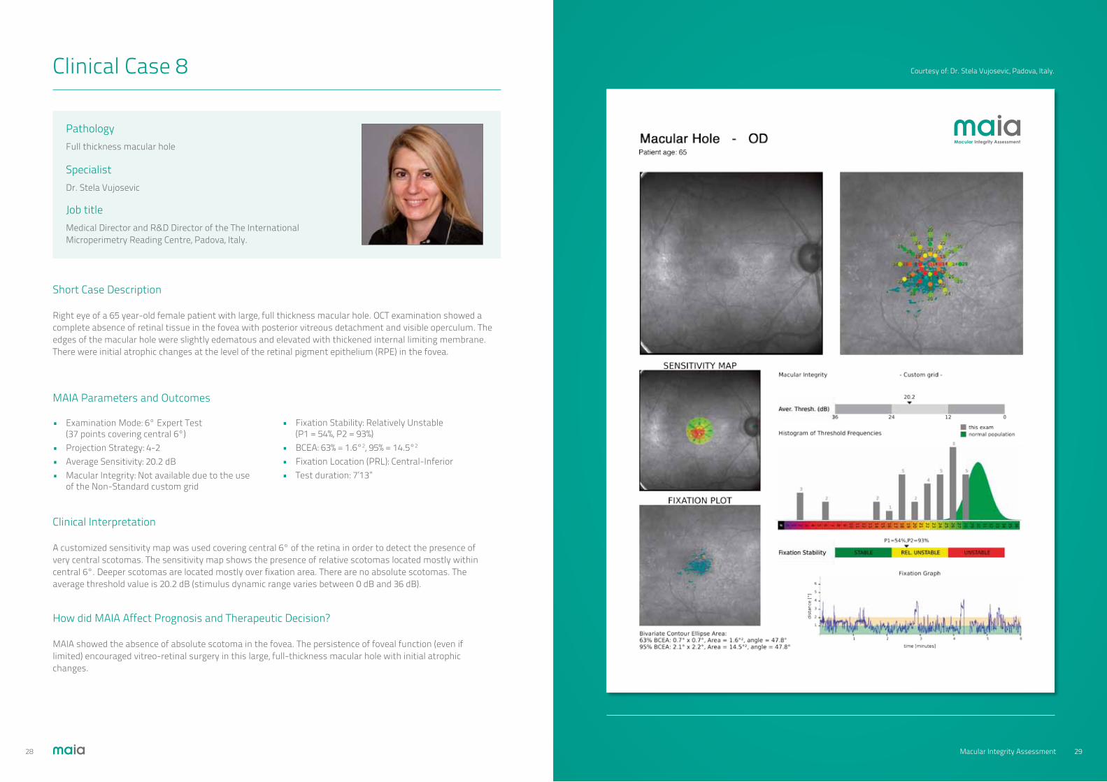
Clinical Case 8
Short Case Description
Right eye of a 65 year-old female patient with large, full thickness macular hole. OCT examination showed a complete absence of retinal tissue in the fovea with posterior vitreous detachment and visible operculum. The edges of the macular hole were slightly edematous and elevated with thickened internal limiting membrane. There were initial atrophic changes at the level of the retinal pigment epithelium (RPE) in the fovea.
Clinical Interpretation
A customized sensitivity map was used covering central 6° of the retina in order to detect the presence of very central scotomas. The sensitivity map shows the presence of relative scotomas located mostly within central 6°. Deeper scotomas are located mostly over fixation area. There are no absolute scotomas. The average threshold value is 20.2 dB (stimulus dynamic range varies between 0 dB and 36 dB).
How did MAIA Affect Prognosis and Therapeutic Decision?
MAIA showed the absence of absolute scotoma in the fovea. The persistence of foveal function (even if limited) encouraged vitreo-retinal surgery in this large, full-thickness macular hole with initial atrophic changes.
MAIA Parameters and Outcomes
• Examination Mode: 6° Expert Test (37 points covering central 6°)• Projection Strategy: 4-2• Average Sensitivity: 20.2 dB• Macular Integrity: Not available due to the use of the Non-Standard custom grid
• Fixation Stability: Relatively Unstable (P1 = 54%, P2 = 93%)• BCEA: 63% = 1.6°2, 95% = 14.5°2 • Fixation Location (PRL): Central-Inferior• Test duration: 7’13”
PathologyFull thickness macular hole
SpecialistDr. Stela Vujosevic
Job title Medical Director and R&D Director of the The International Microperimetry Reading Centre, Padova, Italy.
Macular Integrity Assessment
Courtesy of: Dr. Stela Vujosevic, Padova, Italy.
29Macular Integrity Assessment28Macular Integrity Assessment
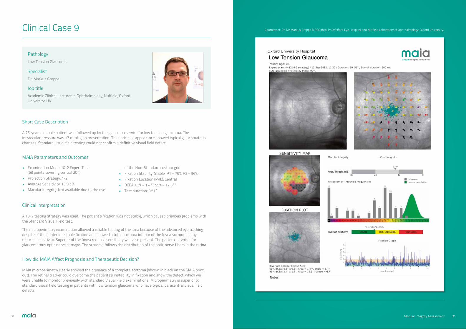
Clinical Case 9
Short Case Description
A 76-year-old male patient was followed up by the glaucoma service for low tension glaucoma. The intraocular pressure was 17 mmHg on presentation. The optic disc appearance showed typical glaucomatous changes. Standard visual field testing could not confirm a definitive visual field defect.
Clinical Interpretation
A 10-2 testing strategy was used. The patient’s fixation was not stable, which caused previous problems with the Standard Visual Field test.
The microperimetry examination allowed a reliable testing of the area because of the advanced eye tracking despite of the borderline stable fixation and showed a total scotoma inferior of the fovea surrounded by reduced sensitivity. Superior of the fovea reduced sensitivity was also present. The pattern is typical for glaucomatous optic nerve damage. The scotoma follows the distribution of the optic nerve fibers in the retina.
How did MAIA Affect Prognosis and Therapeutic Decision?
MAIA microperimetry clearly showed the presence of a complete scotoma (shown in black on the MAIA print out). The retinal tracker could overcome the patients’s instability in fixation and show the defect, which we were unable to monitor previously with standard Visual Field examinations. Microperimetry is superior to standard visual field testing in patients with low tension glaucoma who have typical paracentral visual field defects.
MAIA Parameters and Outcomes
• Examination Mode: 10-2 Expert Test (68 points covering central 20°)• Projection Strategy: 4-2• Average Sensitivity: 13.9 dB• Macular Integrity: Not available due to the use
of the Non-Standard custom grid• Fixation Stability: Stable (P1 = 76%, P2 = 96%)• Fixation Location (PRL): Central• BCEA: 63% = 1.4°2, 95% = 12.3°2
• Test duration: 9’51”
PathologyLow Tension Glaucoma
SpecialistDr. Markus Groppe
Job title Academic Clinical Lecturer in Ophthalmology, Nuffield, Oxford University, UK.
Macular Integrity Assessment
Courtesy of: Dr. Mr Markus Groppe MRCOphth, PhD Oxford Eye Hospital and Nuffield Laboratory of Ophthalmology, Oxford University.
31Macular Integrity Assessment30Macular Integrity Assessment

Clinical Case 10
Patient age: 27
Short Case Description
Left eye of a 27-year-old female patient presented a 1 month history of reduced visual acuity (LogMAR = 0.7)*. Full eye examination, Visual Fields and OCT imaging did not show any abnormality. MAIA microperimetry showed a central defined scotoma. Subsequent MRI imaging suggested a pilocytic astrocytoma of the left optic nerve.
Clinical Interpretation
We used the MAIA 10-2 testing grid map to assess the retina function over a wide central area. The patient has relatively stable fixation (Fixation index P1=76%) considering her reduced visual acuity. The microperimetry examination shows a small central scotoma. The retina sensitivity is reduced or not present (shown in MAIA by the black sensitivity points) in an area from the optic nerve to the fovea. The pattern is highly suggestive of an underlying optic nerve pathology, because the scotoma follows the distribution of the inferior part of the papillar-macular bundle.
How did MAIA Affect Prognosis and Therapeutic Decision?
The MAIA examination confirmed the presence of a scotoma, which we did not achieve with a standard Visual Field test. The distribution of the scotoma is typical for an optic nerve problem, which led to the MR imaging and finally to the diagnosis. MAIA will allow monitoring of the lesion, which can be compared in the follow-up MAIA differential maps.
MAIA Parameters and Outcomes
• Examination Mode: 10-2 Expert Test (68 points covering central 20°)• Projection Strategy: 4-2• Average Sensitivity: 19.7 dB• Macular Integrity: Not available due to the use
of the Non-Standard custom grid• Fixation Stability: Stable (P1 = 76%, P2 = 92%)• BCEA: 63% = 1.3°2, 95% = 11.9°2 • Fixation Location (PRL): Central• Test duration: 12’18”
PathologyPilocytic Optic Nerve
SpecialistDr. Markus Groppe
Job title Academic Clinical Lecturer in Ophthalmology, Nuffield, Oxford University, UK.
Macular Integrity Assessment
Courtesy of: Dr. Mr Markus Groppe MRCOphth, PhD Oxford Eye Hospital and Nuffield Laboratory of Ophthalmology, Oxford University.
*VA 0.7 LogMAR: US=20/100, UK=6/30, Dec=0.20
33Macular Integrity Assessment32Macular Integrity Assessment

Clinical Case 11
Short Case Description
Left eye of a 75 year-old patient who has been treated with oral Plaquenil since 10 years. Fundus examination showed an initial cellophane maculopathy. The patient was asymptomatic with best corrected visual acuity LogMAR = 0.0*
Clinical Interpretation
Macular sensitivity map shows the presence of shallow relative scotomas in the tested area. Fixation is central (localized in the fovea and in this case easily seen). Although the average threshold value is 25 dB, which may be considered as normal in a 75 year old patient, the Macular Integrity Index shows a value of 93.9 which is a high index of abnormal macular function in MAIA microperimeter.”
How did MAIA Affect Prognosis and Therapeutic Decision?
MAIA documented slight decrease in central retinal sensitivity. These shallow scotomas are localized (without any specific pattern) in the whole examined area. This examination can be considered as normal for this age-group. Therefore, a continuation of chlorochine therapy was advised.
MAIA Parameters and Outcomes
• Examination Mode: Standard Expert Test (37 points covering central 10°)• Projection Strategy: 4-2• Average Sensitivity: 25.2 dB• Macular Integrity: 93.9
• Fixation Stability: Stable (P1 = 95%, P2 = 99%)• BCEA: 63% = 0.4°2, 95% = 3.2°2
• Fixation Location (PRL): Central• Test duration: 5’32”
PathologyChlorochine Maculopathy
SpecialistDr. Stela Vujosevic
Job title Medical Director and R&D Director of the The International Microperimetry Reading Centre, Padova, Italy.
Macular Integrity Assessment
Courtesy of: Dr. Stela Vujosevic, Padova, Italy.
*VA 0.0 LogMAR: US=20/20, UK=6/6, Dec1.0
35Macular Integrity Assessment34Macular Integrity Assessment

Clinical Case 12-OD
Short Case Description
A 71 years old male patient with diagnosed Plaquanil Toxicity. Stopped medication 3 years prior to visit. Patient reported difficulty with reading. Visual Acuity was OD = 0.5 LogMAR* and OS = 0.3 LogMAR**. Text reading acuity OS at 100% contrast was 20/30 and at 10% contrast 20/64.
Clinical Interpretation
The MAIA test clearly showed a central ring scotoma in OS with a functional macular PRL. The average retinal sensitivity in the central 20° of vision was in the mid-range (17.8 dB). PRL was very stable with an excellent Fixation Stability (P1 = 100%, BCEA@63% = 0.1°2).
How did MAIA Affect Prognosis and Therapeutic Decision?
A ring scotoma is a serious problem. One cannot use in this case prism form image relocation outside the scotoma, neither can use higher degrees of magnification. The only impact comes from better illumination (which was advised ) and from selective transmission glasses to reduce glare (Corning CPF 450 were prescribed).
MAIA helped in the precise identification of the ring scotoma and helped in the selection of the visual aids as well as with the patient explanation of the visual condition.
MAIA Parameters and Outcomes
• Examination Mode: 20-2 Expert Test (68 points covering central 20°)• Projection Strategy: 4-2• Average Sensitivity: 17.8 dB• Macular Integrity: Not available due to the use
of the Non-Standard custom grid• Fixation Stability: Stable (P1 = 100%, P2 = 100%)• Fixation Location (PRL): Central• BCEA: 63% = 0.1°2, 95% = 0.7°2
• Test duration: 10’17”
PathologyPlaquenil Toxicity
SpecialistDr. Samuel N. Markowitz
Job title Director, Low Vision Rehabilitation Program, Department of Ophthalmology and Vision Sciences, University of Toronto.
Macular Integrity Assessment
Courtesy of: Dr. Samuel N. Markowitz, Toronto, Canada.
*VA 0.5 LogMAR: US=20/63, UK=6/19, Dec=0.32 **VA 0.3 LogMAR: US=20/40, UK=6/12, Dec=0.5
37Macular Integrity Assessment36Macular Integrity Assessment

Clinical Case 12-OS
Short Case Description
OD of the above case. Male 71 diagnosed Plaquanil Toxicity. Stopped medication 3 years prior to visit. Difficulty with reading. Visual Acuity was OD = 0.5 LogMAR and OS = 0.3 LogMAR. Text reading acuity OS at 100% contrast was 20/30 and at 10% contrast 20/64.
Clinical Interpretation
The MAIA test identified well the central scotoma (marked with black stimuli). The PRL is present in the supero nasal area above from the fovea centralis where the scotoma is located. The PRL is shows poor fixation stability (P1 = 30%, P2 = 72%) which is confirmed with the large BCEA of 5.5°2. The PRL is located on a non-corresponding macular area relative to the other better eye.
How did MAIA Affect Prognosis and Therapeutic Decision?
Non-corresponding retinal area for the PRL with unstable fixation is a poor prognosis. Therapeutic considerations are based solely on the better fellow eye.
MAIA Parameters and Outcomes
• Examination Mode: 20-2 Expert Test (68 points covering central 20°)• Projection Strategy: 4-2• Average Sensitivity: 17.3 dB• Macular Integrity: Not available due to the use
of the Non-Standard custom grid• Fixation Stability: Stable (P1 = 30%, P2 = 72%)• Fixation Location (PRL): Para-central, Supero-nasal• BCEA: 63% = 5.5°2, 95% = 49.7°2
• Test duration: 9’33”
PathologyPlaquenil Toxicity
SpecialistDr. Samuel N. Markowitz
Job title Director, Low Vision Rehabilitation Program, Department of Ophthalmology and Vision Sciences, University of Toronto.
Macular Integrity Assessment
Courtesy of: Dr. Samuel N. Markowitz, Toronto, Canada.
*VA 0.5 LogMAR: US=20/63, UK=6/19, Dec=0.32 **VA 0.3 LogMAR: US=20/40, UK=6/12, Dec=0.5
39Macular Integrity Assessment38Macular Integrity Assessment

Clinical Case 13
Short Case Description
Right eye of a 46 year-old female patient. This is a pre Cataract Surgery evaluation. The right eye with a dense corticonuclear cataract, showed a previously undiagnosed juxtafoveal scar due to Toxoplasmosis infection. The infection occurred in the first months of life and left a retinal sequelae. BCVA: 1.0 LogMAR.*
Clinical Interpretation
Fixation of this patient is absolutely stable with central PRL corresponding to foveal area meaning good foveal function. The cataract affects little the quality of Scanning Laser Ophthalmoscopy (foggy effect), although it does not alter the retinal sensitivity detection.
Nevertheless the average sensitivity is high (27.d dB), the Macular Integrity Index is bad (99.1) due to the localized reduced sensitivity corresponding to the scar which does not affect foveal function.
How did MAIA Affect Prognosis and Therapeutic Decision?
Thanks to Microperimetry, retinal sensitivity evaluation is easily carried out, the precise visual impairment due to Toxoplasmosis retinal scar is made and a precise prognosis for visual acuity after cataract surgery is possible.
MAIA Parameters and Outcomes
• Examination Mode: Standard Expert Test (37 points covering central 10°)• Projection Strategy: 4-2• Average Sensitivity: 27.2 dB• Macular Integrity: 99.1
• Fixation Stability: Stable (P1 = 100%, P2 = 100%)• BCEA: 63% = 0.2°2, 95% = 1.7°2
• Fixation Location (PRL): Central• Test duration: 5’00”
PathologyPre Cataract Surgery evaluation in previously undiagnosed Toxoplasmosis infection
SpecialistDr. Fabio Mazzolani
Job title Low Vision and Retina Consultant
Macular Integrity Assessment
Courtesy of: Dr Mazzolani, Dr Lovisolo - Quattroelle Eye Clinic. Milan, Italy
*VA 1.0 LogMAR: US=20/200, UK=6/60, Dec=0.10
41Macular Integrity Assessment40Macular Integrity Assessment

Clinical Case 14
Short Case Description
66 years old male patient attended the Low Vision clinic for eccentric vision rehabilitation. Difficulties with distance vision and reading. Visual Acuity = 0.9 LogMAR* in both eyes. Patient declared to have his right eye as the best eye and use it for reading purposes with a magnifier glass.
Clinical Interpretation
The MAIA test is automatically centered over the initial PRL. OD shows a PRL inferior from the fovea, while OS show the PRL is nasal from the fovea. Fixation stability in OS is clearly better than OD. The PRL_initial and PRL_final in OD is separated about 2° while both PRLs in OS are almost in the same locus. The retina sensitivity surrounding the PRL (inner stimuli ring) is also better in OS than OD.
How did MAIA Affect Prognosis and Therapeutic Decision?
Microperimetry data showed that achieving better vision in activities involving fixation such as reading could be improved using the left eye instead of the right eye. Patient was advised to use his left eye for reading and initiate PRL eccentric vision training with MAIA. During 4 weeks’ follow up, patient reported better vision capabilities with OS instead of OD.
MAIA Parameters and Outcomes
• Examination Mode: Standard Expert Test (37 points covering central 10°)• Projection Strategy: 4-2• Average Sensitivity: OD = 16.2; OS = 17.9• Macular Integrity: 100% Abnormal• Fixation Stability: OD Rel. Unstable (P1 = 49%, P2 = 92%);
OS Stable (P1 = 90%, P2 = 100%)• Fixation Location (PRL): OD = Inferior from scotoma; OS = Nasal from scotoma• BCEA: OD: 63% = 2.7°2, 95% = 23.8°2 ; OS: 63% = 0.4°2, 95% = 3.2°2
• Test duration: OD = 5’47”; OS = 5’41”
PathologyEccentric viewing analysis
SpecialistDr. Winfried Amoaku
Job title Assoc Professor/Reader in Ophthalmology and Vis Sci, Hon Consultant Ophthalmologist. University of Nottingham UK.
Macular Integrity Assessment
Courtesy of: Dr. Winfried Amoaku, University of Nottingham, UK.
*VA 0.9 LogMAR: US=20/160, UK=6/48, Dec=0.125
43Macular Integrity Assessment42Macular Integrity Assessment

Clinical Case 15
Short Case Description
A 70 years old female patient with diagnosed Dry Age-related Macular Degeneration, phakic eye with mild nuclear sclerosis OU. OD -1.50+1.00X30, OS -1.50+1.25X60. Experiences large central blind spots with both eyes. Difficulties with distance vision and recognizing faces. Visual Acuity was OD = 1.2 LogMAR* OS = 1.3 LogMAR**. Contrast sensitivit y OD (1 cyc/deg) 1.18 log units.
Clinical Interpretation
MAIA revealed in less than 2 minutes a large scotoma area (stimuli shown in black). PRL unstable with very poor fixation stability represented by a large BCEA (95% = 58.6°2). PRL located temporal to and above the original fovea. Fixation pattern shows PRL trend aiming to reach upper-temporal retina. The other eye (OS) has poorer fixation stability than OD. PRL located on corresponding macular areas (upper quadrants) relative to the other poorer eye.
How did MAIA Affect Prognosis and Therapeutic Decision?
Stabilization and improvement of fixation stability may improve oculomotor skills aiming to utilize PRLs and shift the scotoma out of central vision as well as promote binocular functions. This can positively affect distance visual acuity. Creation of a therapeutic PRL is required on a more favorable retinal location in the better eye.
MAIA Parameters and Outcomes
• Examination Mode: 20° Star Customized Expert Test manually stopped after 2 minutes test• Projection Strategy: 4-2• Average Sensitivity: Not Available (not representative with only 10 tested stimuli points)
• Macular Integrity: Not available due to the use of the Non-Standard custom grid• Fixation Stability: Unstable (P1 = 12%, P2 = 31%)• Fixation Location (PRL): Para-central• BCEA: 63% = 6.5°2, 95% = 58.6°2
• Test duration: 2’02”
PathologyPRL Eccentric viewing analysis before PRL training
SpecialistDr. Samuel N. Markowitz
Job title Director, Low Vision Rehabilitation Program, Department of Ophthalmology and Vision Sciences, University of Toronto.
Macular Integrity Assessment
Courtesy of: Dr. Samuel N. Markowitz, Toronto, Canada.
*1.2 LogMAR: US=20/320, UK=6/95, Dec=0.063 **1.3 LogMAR: US=20/400, UK=6/120, Dec=0.05
45Macular Integrity Assessment44Macular Integrity Assessment
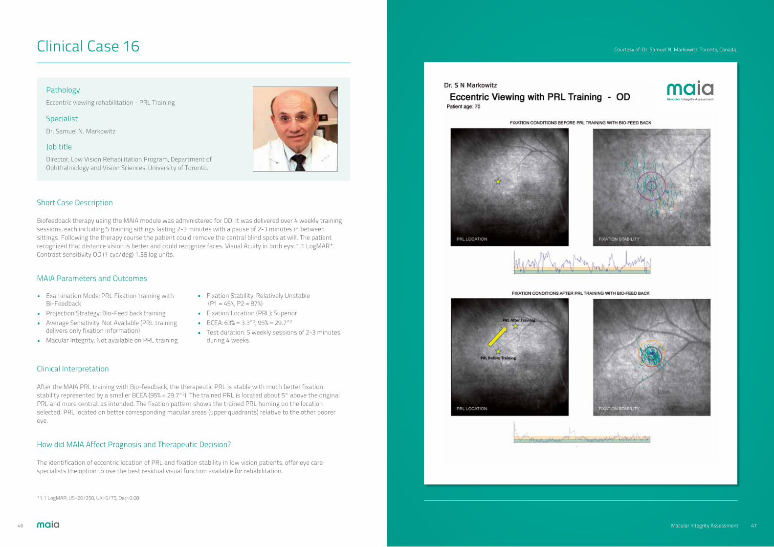
Clinical Case 16
Short Case Description
Biofeedback therapy using the MAIA module was administered for OD. It was delivered over 4 weekly training sessions, each including 5 training sittings lasting 2-3 minutes with a pause of 2-3 minutes in between sittings. Following the therapy course the patient could remove the central blind spots at will. The patient recognized that distance vision is better and could recognize faces. Visual Acuity in both eys: 1.1 LogMAR*. Contrast sensitivity OD (1 cyc/deg) 1.38 log units.
Clinical Interpretation
After the MAIA PRL training with Bio-feedback, the therapeutic PRL is stable with much better fixation stability represented by a smaller BCEA (95% = 29.7°2). The trained PRL is located about 5° above the original PRL and more central, as intended. The fixation pattern shows the trained PRL homing on the location selected. PRL located on better corresponding macular areas (upper quadrants) relative to the other poorer eye.
How did MAIA Affect Prognosis and Therapeutic Decision?
The identification of eccentric location of PRL and fixation stability in low vision patients, offer eye care specialists the option to use the best residual visual function available for rehabilitation.
MAIA Parameters and Outcomes
• Examination Mode: PRL Fixation training with Bi-Feedback• Projection Strategy: Bio-Feed back training• Average Sensitivity: Not Available (PRL training delivers only fixation information)• Macular Integrity: Not available on PRL training
• Fixation Stability: Relatively Unstable (P1 = 45%, P2 = 87%)• Fixation Location (PRL): Superior• BCEA: 63% = 3.3°2, 95% = 29.7°2
• Test duration: 5 weekly sessions of 2-3 minutes during 4 weeks.
PathologyEccentric viewing rehabilitation - PRL Training
SpecialistDr. Samuel N. Markowitz
Job title Director, Low Vision Rehabilitation Program, Department of Ophthalmology and Vision Sciences, University of Toronto.
Macular Integrity Assessment
Courtesy of: Dr. Samuel N. Markowitz, Toronto, Canada.
*1.1 LogMAR: US=20/250, UK=6/75, Dec=0.08
47Macular Integrity Assessment46Macular Integrity Assessment

Clinical Case 17
Short Case Description
A 28 year old female patient with diagnosed Stargardt’s disease performed the Microperimetry test. Patient reported misreading words and disappearance of letters while reading. Visual Acuity was OD = 0.8 LogMAR* and OS = 0.6 LogMAR**.
Clinical Interpretation
A 4LF strategy projection was used to performed a fast macular assessment. The MAIA test clearly showed a central 5° diameter scotoma in both eyes. The OD showed a fixation drift from the left to right of the scotoma demonstrating unstable fixation (P1 = 22%).
How did MAIA Affect Prognosis and Therapeutic Decision?
There are no clinical or surgical aids for this Stargardt’s disease patients who have sizeable central scotoma. MAIA helped explain to the patient the visual problems, and by looking at the graphical output the patient agreed to perform the PRL fixation training in order to learn how to use the peripheral vision more effectively.
MAIA Parameters and Outcomes
• Examination Mode: Standard Expert Test (37 points covering central 10°)• Projection Strategy: 4 Levels Fixed (4LF)• Average Sensitivity: Not available due to the use of the 4LF projection strategy• Macular Integrity: Not available due to the use
of the Non-Standard custom grid• Fixation Stability: Unstable (P1 = 22%, P2 = 70%)• Fixation Location (PRL): Double PRL, temporal and nasal from the lesion• BCEA: 63% = 5.6°2, 95% = 50.6°2
• Test duration: 03’16”
PathologyStargardt’s Disease Eccentric Viewing Analysis
SpecialistWinfried Amoaku
Job title Assoc Professor/Reader in Ophthalmology and Vis Sci, Hon Consultant Ophthalmologist. University of Nottingham UK.
Macular Integrity Assessment
Courtesy of: Dr. Winfried Amoaku, University of Nottingham, UK.
*VA 0.8 LogMAR: US=20/125, UK=6/38, Dec=0.16 **VA 0.6 LogMAR: US=20/80, UK=6/24, Dec=0.25
49Macular Integrity Assessment48Macular Integrity Assessment

Clinical Case 18
Short Case Description
From the above case. A 28 year old female patient with diagnosis of Stargardt’s disease performed the Microperimetry PRL Fixation training with a scope to have better fixation control in the right eye.
Clinical Interpretation
The Fixation graph of the PRL training session with Bio Feedback, showed an important increase of stability. The graph shows most of the fixation within 2° of vision. The high peaks in the fixation graph demonstrate that the patient still moves her gaze to the other size of the scotoma sometimes. The BCEA results shows a narrow ellipse of fixation (BCEA: 63% = 0.9°2) similar to those with central fixation. Due to the constant improvement of fixation stability, more PRL training sessions are suggested to complete the training set of 10 sessions.
How did MAIA Affect Prognosis and Therapeutic Decision?
The MAIA PRL Fixation training with Bio Feedback, helped the patient to understand how to control eye movements and use only one adjacent area of the scotoma (compared both sides previously) more effectively. Before the training, the PRL shift from left to right sides of the scotoma created unstable fixation.
Due to the constant improvement of fixation stability, more PRL training sessions are suggested to complete the training set of 10 sessions.
MAIA Parameters and Outcomes
• Examination Mode: PRL Fixation training with Bio-Feedback• Average Sensitivity: Not Available on PRL training • Macular Integrity: Not available on PRL training
• Fixation Stability: Stable (P1 = 73%, P2 = 98%)• Fixation Location (PRL): Temporal from the lesion• BCEA: 63% = 0.8°2, 95% = 7.6°2
• Test duration: 5 sessions of 10 minutes each session.
PathologyStargardt Disease after Eccentric Viewing - PRL training
SpecialistWinfried Amoaku
Job title Assoc Professor/Reader in Ophthalmology and Vis Sci, Hon Consultant Ophthalmologist. University of Nottingham UK.
Macular Integrity Assessment
Courtesy of: Dr. Winfried Amoaku, University of Nottingham, UK.
51Macular Integrity Assessment50Macular Integrity Assessment

Authors Biography
Dr. Winfried M. Amoaku, FRCS(Ed), FRCOphth, PhDAssociate Professor/Reader in Ophthalmology and Vis Sci, University of Nottingham, U.K. Hon Consultant Ophthalmologist, University Hospital, QMC, Nottingham, U.K.
Dr. Markus Groppe MRCOpth, PhDAcademic Clinical Lecturer in Ophthalmology. The Nuffield Laboratory of Ophthalmology, Oxford University Oxford Eye Hospital, Oxford University Hospitals NHS Trust
Markus Groppe is an Academic Clinical Lecturer at Oxford University. He obtained his medical degree from the University of Münster, Germany. Following this Dr Groppe completed a PhD degree in Ophthalmology at the University of Münster. The thesis focused on the role of nitric oxid in retinal degeneration. He continued his Ophthalmology training in the UK in Birmingham and Oxford and is subspecialising in Medical and Surgical Retina. He is currently Member of Congregation at Oxford University and Honorary Lecturer at the Oxford Eye Hospital and Moorfields Eye Hospital London. His research focuses on treatment of inherited retinal diseases. He is involved in a retinal gene therapy trial for choroideremia and the implantation of retinal implants for restoration of basic vision in blind patients.
Samuel N. Markowitz, M.D., F.R.C.S. (C)Associate Professor of Ophthalmology, Faculty of Medicine, University of Toronto. Director, Low Vision Rehabilitation Program, Department of Ophthalmology and Vision Sciences. University of Toronto
Associate Professor, Department of Ophthalmology, University of Toronto; Active Staff, University Health Network, Toronto Western Hospital; Director, Vision Rehabilitation Program, Department of Ophthalmology, University of Toronto; Section editor: Low Vision Rehabilitation for the Canadian Journal of Ophthalmology; past member of the Vision Rehabilitation Committee of the American Academy of Ophthalmology. Active practice covering all aspects of low vision rehabilitation including pediatric low vision.
Practicing in a multidisciplinary environment which includes occupation therapy, opticianry and vision rehabilitation specialists for enhancement of independent living skills and orientation and mobility. Active research interests in various aspects of low vision rehabilitation.
Past and current involvement covered aspects of low vision rehabilitation such as: accessibility and barriers to low vision rehabilitation, characteristics of scotomata and of preferred retinal loci, identification of residual potential visual acuity, rehabilitation with surgical telescopic magnification, and with prisms towards PRL, residual oculomotor characteristics including stereopsis, fixation location and fixation stability, residual chromatic vision, restitution of vision in older children with amblyopia and field expansion in Stroke, Retinitis Pigmentosa and end stage Glaucoma, microperimetry and residual vision functions, interventions to promote brain plasticity and development of indoor navigation systems for the visually impaired.
Associate Professor/Reader in Ophthalmology and Visual Sciences, University of Nottingham, and Hon. Consultant Ophthalmologist. Teaching lead for ophthalmology in the undergraduate medical course in University of Nottingham. Consultant Ophthalmologist with a special interest in medical retina diseases (including AMD, RVO, diabetic retinopathy) and uveitis. Dr. Amoaku contributes to cataract services, including provision of surgery, in Nottingham. Other (recent/current) responsibilities:
• Ag President, The Royal College of Ophthalmologists, UK Oct 2010 –May 2011• Vice President and Chairman, Scientific Committee, The Royal College of Ophthalmologists, UK 2007-2011.• Chair, The RCOphth Medical Retinal Service Provisions Subcommittee: 2006-2011• Chair, RCOphth Equality and Diversity Committee 2009-2011• Member of Scientific, and Examinations Committees 2001-11• Convener, Elizabeth Thomas Seminar on Macular Diseases, RCOphth: 2003-date• Member, Scientific Advisory Committee of the Macular Disease Society, UK. 2005-date• Scientific Reviewer for several journals, SPARC and Wellcome Trust, FFS.• Editor, Eye News 2005-date
Dr. Fabio Mazzolani MDLow Vision and Retina Consultant
Fabio Mazzolani got his degree in ophthalmology with honors in 2007, his main practice includes diagnosis and treatment of diseases of the posterior segment of the eye (retina and optic nerve) with particular interest in integrating both morpho-functional and visual rehabilitation. Always interested in the integration of diagnostic imaging and functional diagnostics using microperimetry, he developed different procedures to correlate retinal metabolic features to microperimetry.
His medical and surgical experience is based on specialist training courses and internships carried out abroad and in Italy. He is Consultant at various retinal diagnostic services.
52 53Macular Integrity Assessment
Macular Integrity Assessment

Dr. Vujosevic graduated in Medicine and Surgery at the University of Padova in 2000. Residency in Ophthalmology at the Department of Ophthalmology, University of Padova with final certification for the practice of Ophthalmology in 2004.
Fellowship in Medical retina at the Moorfields Eye Hospital, London in 2004. Fellowship at the Reading Centre, Moorfields Eye Hospital in London in 2004. Contract Researcher at the G.B. Bietti Eye Foundation, IRCCS, Rome 2006-2012. Full time Medical Assistant at the Azienda Ospedaliera di Padova since 2012. PhD at the University of Padova in 2013. Assistant Clinical Professor of Ophthalmology, University of Padova.
Medical Director and R&D Director of the International Microperimetry Reading Centre, certified by the European Vision Institute. Member of the European Reading Centre Expert Committe. Member of the Association for the Research in Vision and Ophthalmology, the EURETINA, the EVER, the European Association For The Study of Diabetes (EASDEC), the Italian Retina Society and the Italian Society of Ophthalmologists.
She currently oversees international research projects and carries out clinical activities concerning screening, morphological and functional instrumental diagnoses, laser and intravitreous treatment of degenerative corioretinal pathologies. She performs cataract surgery and intravitreous injections. She has authored numerous papers published in major international ophthalmological journals
Dr. Stela Vujosevic MD, PhDMedical Director and R&D Director of the The International Microperimetry Reading Centre, Padova, Italy.
Macular Integrity Assessment
Marco graduated from La Salle University in Mexico City in 1991 with a MEng Electronics degree. He has worked in the ophthalmic industry since 1993 as biomedical engineer.
In 2006, he joined a group of scientists and developers of ophthalmic instruments in Italy and since then they have created high-end technologies for retina imaging. He is co-founder of CenterVue Italy and holds a consultant position as Chief Scientific Officer.
In 2012, Marco began his PhD in Ophthalmology and Visual Sciences, at the University of Nottingham. His research involves the analysis of functional changes during the progression of pathologies affecting the central retina and the development of eccentric vision therapies with microperimetry techniques.
Marco U. Morales, MEng, PhDcResearch Fellow, PhD Program in Ophthalmology and Visual Science University of Nottingham. Chief Scientific Officer, CenterVue, Italy
54 55Macular Integrity Assessment
Macular Integrity Assessment

REV02 131030
[email protected] www.centervue.com
[email protected] www.centervue.com
Ph: +39 049 7396 147 Fax +39 049 7396 148
Ph: +1 408 988 8404 Fax: +1 408 716 3271
Via San Marco 9H 35129 Padova - Italy
92 Bonaventura Drive, San Jose CA 95134 - USA
Centervue SpA
Centervue Inc.
Macular Integrity Assessment
adim
er.n
et