plaquenil maculopathy dewilde - pacific university · 2017-06-20 · plaquenil)maculopathy) ......
TRANSCRIPT

Plaquenil Maculopathy
Anthony DeWilde OD FAAO
COPE #34898 PS
Event #: 104151
Expiration 6/15/15
Course Description:
Plaquenil maculopathy is an uncommon side effect of the systemic medication, Hydroxychrloroquine. This lecture will guide the optometrist in diagnosis and management of Plaquenil maculopathy. The course will review updated guidelines from the American Academy of Ophthalmology and discuss exciting new research utilizing OCT.
Objectives:
1. Educate about Plaquenil side effects
2. Discuss various tests to detect early plaquenil toxicity
3. Interpretation of OCT in Plaquenil Maculopathy
4. Management of Plaquenil Maculopathy
5. Update on American Academy of Ophthalmology recommendations
Hello and welcome to Pacific University’s online continuing education. My name is Anthony DeWilde and I work at the Kansas City VA Medical Center. What I’m going to talk to you about for the next 50 minutes or so is Plaquenil Maculopathy. I think this is a very important topic for us as optometrists to understand, and it’s one that is often confused in the literature. I’m hoping that in the next 50 minutes I can guide you and set the record straight on some of those misconceptions.
There have been some interesting new updates in the literature and some of the research indicating risk of Plaquenil Maculopathy in even just the last year. I feel that a lot of people who stand up and lecture about Plaquenil Maculopathy or when textbooks are written about this, some of the nuances are left out for the clinician. It seems some of the recommendations are very cut and dry when, in reality, some of the tests we use to diagnose this aren’t 100%

accurate, and leave the clinician without the best guidance. While there are some updated clinical resources we can utilize, such as OCT, we’ll see later on that nothing is perfect and we still need to use our clinical judgment.
I think this is a very important topic for a couple of reasons. As an optometrist, we will probably be getting referrals from primary care physicians and potentially rheumatologists. They’ll be asking us, “Doctor, is this patient still OK to be on this medication? Are there any toxic side effects to their eyes?” because everybody in the medical community is well aware of Plaquenil toxicity affecting the eye. In fact, it’s so common that it’s often printed on the medication labels. In the VA I’m at, it actually says the patient needs an eye exam every 6 months, despite years of nagging the pharmacy and telling the patients that they don’t need an exam every 6 months, we still have that printed on the label.
That leads us to our second big reason this topic is important, and that is fear. Patients will often become fearful of vision loss. They don’t necessarily understand that the vision loss that may occur may be minor, and may not affect their life too much. In fact, the vision loss may be asymptomatic. Whenever someone hears ‘vision loss,’ I think the assumption is that it may be major vision loss or even blindness, so our job is to really understand a lot of these nuances so we can better serve our patients in the referral from their doctor, and to better educate the patients and hopefully relieve some of their fear and be able to guide them in their treatment of the whole self, not just their eye.
I have three big goals today for this discussion. The first is that I want to be able to understand all the diagnostic tests that are available to look for and find Plaquenil Maculopathy. As I alluded to a moment ago, there have been some changes in the literature, and one of the biggest changes is that the American Academy of Ophthalmology (AAO) has changed their recommendations. Back in 2002 after a thorough literature review, they recommended certain things that were changed in 2011. If you haven’t had a chance to read some of that literature, I’ll go over some of that today. The third thing I want to do is discuss some of the newer technology, in particular OCT, and how that relates. Many offices now have an OCT or if they don’t have it, they have one pretty readily available. I’m going to discuss how we interpret that test and how accurate it is.
So let’s start with just a brief amount of history. Back in WWII chloroquine was developed as a safer alternative to general quinine, which is found in Peruvian Bark which we’ve known for a long time helps with malaria. Chloroquine was designed specifically to combat malaria. This is something that is not used in the United States anymore, mostly because malaria is all but eradicated in the US. In other areas, such as Africa, we will see a much higher use of chloroquine, and you may come across that in your clinical practice as we live in an increasing melting pot society where the borders between the US and other countries are not as rigidly

defined, and people come here either for long or short stays who may have had a history of exposure to chloroquine. Thus, it is important to know about it, even if we don’t see a lot of it.
An interesting side effect of chloroquine was that people were noticing their joint pain was improving with this medication. With that, people started using it for other conditions such as lupus and rheumatoid arthritis.
Then what happened was hydroxychloroquine was developed, and it goes by the trade name Plaquenil. We are all familiar with Plaquenil being used for autoimmune diseases, but originally it was formulated as a safer antimalarial to have fewer side effects. Some of the concern about Plaquenil affecting the eyes is a little bit overstated because, in fact, hydroxychloroquine is a safer medication. A lot of those conditions, the ocular problems we were having were with chloroquine, not hydroxychloroquine. Nowadays, hydroxychloroquine is used quite frequently for Lupus, Rheumatoid Arthritis, Sjogren’s and Lyme Arthritis.
Some of the side effects you may encounter with your patients are headache, neurological conditions like anxiety or depression, tinnitus, some GI disturbances, skin disturbances, but really, from our standpoint, the vision changes are the ones we are most concerned about.
Eventually if enough of this medication builds up in the system, it can affect the retina. And, it can affect the retina in a very systematic fashion. It affects the ganglion cell layer first, then moves on to the photoreceptor layer, and then finally at the end disrupting the RPE. I thought this was interesting because when I learned about Plaquenil
Maculopathy I thought for sure that it went the other direction, initially affecting the RPE, which made a sick photoreceptor layer and things broke down from there. However, it’s actually the opposite. Even though this is a bad development, it’s actually good in some ways because we can find some early changes before some of the damage is irreversible. By the time the maculopathy has reached the RPE, the damage is for the most part irreversible.
The atrophy happens in a paracentral fashion, about 5-‐10 degrees away from the center of the fovea. It happens in a bulls-‐eye shape. That is one of the misconceptions, one of the nuances, that lecturers and books on the subject will not actually go into. This doesn’t start off as a bulls-‐eye. It actually starts off as very mild atrophy that is paracentral, and oftentimes it is really difficult to distinguish this atrophy from other types of atrophy, such as age-‐related macular

degeneration (AMD). What happens with this atrophy is that, over time, it will extend in an arcuate fashion and eventually will mimic that bulls-‐eye pattern we have all seen in our textbooks.
What will also happen with Plaquenil Maculopathy is the visual field will mimic the atrophy as well, and we’ll get a paracentral scotoma about 5-‐10 degrees from center of fovea. This leads to irreversible damage and irreversible vision loss. The incredible thing is that even after discontinuing the medication, there can be further progression because the store of the medication in the body is not cleared. There is no definitive time of how long that medication store stays – it will be different from patient to patient. I’ve read as high as 6 months, and I don’t doubt it could even go longer. So the concern with Plaquenil Retinopathy is that the damage can happen and if we don’t find it soon enough, the potential for vision loss can be there, as well as further vision loss once treatment has begun. Our goal, then, is to try to find Plaquenil Maculopathy before it’s too late. That leads to a lot of difficulty, and we will talk about that through the rest of this presentation.
There are several risk factors that have been determined for the development of Plaquenil Maculopathy. For example, being older than 60 is a risk factor. This is a difficult one because the atrophy we see in Plaquenil Maculopathy isn’t necessarily easy to distinguish from other atrophies like AMD. AMD is by far the most common degeneration of the retina, specifically affecting the macula. Thus, when we look at a patient over the age of 60, who is more at risk for AMD and we’re trying to determine if this is AMD or toxicity from the Planquenil.
Which leads us to the next bullet point: is pre-‐existing retinal disease a risk factor, or are these patients not affected? There’s no really good literature on this that says one way or another. I think, theoretically, it makes sense. If a patient comes in with a pre-‐existing retinal disease, such as Stargardt’s or a rod-‐cone dystrophy, or if they already have AMD, we would think a sick retina like that would be more prone to, or at least less resistant to, damage that could happen in the future than one that started off perfectly normal. There’s no real evidence of that, but some will say that at least theoretically, it makes sense. The reason I even bring that up is that it may cause you to want to see these patients a little more frequently than maybe you would otherwise.
If the patient has renal and/or liver disease, that patient is at increased risk because that is where the body clears the medication. If either of those organs are broken down, then the storages of the medication will stay in the body even longer and at higher dosages than someone who can clear medications quicker and better.

It was previously thought that daily dosage was more important than what we think now. Currently we are more interested in cumulative dosage. Back in 2002, in the original AAO recommendations, daily dosage was a very important issue. They were worried that if a patient took too much per body weight, then they could develop toxicity. The guidelines used to be that the patient could take about 200 mg per every 70 lbs of weight. For instance, if someone weighed 140 lbs or more, they could take 400 mg per day. If they were 70 lbs, they could tolerate 200 mg, and if they were 210 lbs, 600 mg. This actually breaks down when we consider where this medication is stored. It’s not stored in fatty tissue – it is stored in the muscles. Thus, when we have a patient who is 5 ft tall and 300 lbs, that’s probably a lot different than someone who is 7 ft tall and 200 or 250 lbs. Those patients will carry their weight very differently – one may have a lot more muscle mass and another much more fatty tissue. We are now more concerned with their height/weight ratio, or BMI. In the past, when we were worried about those daily dosages, we would write down on our chart how much the patient could tolerate. Now it’s much more focused on how long the patient has been on the medication, and at what dosage so we can understand the cumulative dosage.
Again, we will not run into chloroquine much in our practices, but here are the dosages we would be more concerned about and the time frame. If the patient has been taking 460 g total, which comes out to 250 mg/day for over 5 years, that cumulative dose is considered the toxic dose.
Hydroxychloroquine has a higher number, because you are typically taking less of the medication, and the medication itself is less toxic. A patient has to get up to 1000 g before we consider that a toxic dosage. If you do the math on this, that’s 400 mg per day, which is a very typical dose, for 5-‐7 years. If you actually crunch the numbers, 7 years is correct, but the AAO guidelines say 5-‐7 years, and I think that’s reasonable. Especially if the patient is at higher risk, such as they have pre-‐existing retinal disease or renal/liver disease. I say 400 mg/day, but if the patient is only taking 200 mg/day, they are at less risk. If they are on 600 mg/day, they are at higher risk.
In my practice, I almost always see 400 mg/day as 200 mg tablets BID. I have seen 600 mg/day, but I think it’s uncommon, and the patients I’ve seen with this higher dose are Lupus patients, so I wonder if they just require higher dosages sometimes due to having a more progressive form of disease that requires it.
You may wonder what is the risk before 7 years? Before that 7 year mark, there is not much risk. Certainly after the 7 year mark there is a risk. Let’s quantify it. When we look at the

literature where the patients have been on Plaquenil for less than 5 years, 1 out of 1000 of them will develop Plaquenil toxicity in the form of Plaquenil Retinopathy.
After that 5-‐7 year mark, we’re going to see the prevalence increase to 1 patient out of 100. If you’re like me, you look at that number and realize that’s a large increase. But you have to remember that there aren’t a lot of patients on this medication. Let’s take all of the patients in your practice who have Rheumatoid Arthritis (not Osteoarthritis), Lupus, Sjogren’s and Lyme Disease, and you add all of those people up. If we think about how many of them are actually taking Plaquenil, it won’t be many of them. Even fewer of them will have taken Plaquenil for over 7 years. And then we think about all of those people who go on to develop complications, and 1 out of 100 doesn’t seem like much.
When we educate patients, 1/100 seems like a large increase compared to 1/1000. But you also have to consider the patient’s chance of not developing Plaquenil Maculopathy is 99/100. I think most people look at that and realize they are fine with those chances. We can take the patient off of the medication if we need to. As long as we are monitoring our patients closely, and that is key – to monitor them closely, the likelihood of us not finding this in time to prevent major vision loss is pretty slim.
Really quick, I keep switching between Plaquenil Retinopathy and Plaquenil Maculopathy and I apologize for that. Plaquenil Retinopathy is the generic term and it can affect the entire retina. However, it always specifically targets the macula first before going on to the retina, and we almost always diagnose it at this stage before it progresses. I will interchange them a lot, and I mostly mean Plaquenil Maculopathy.
There are two big, broad goals in testing for Hydroxychloroquine toxicity. Ideally, you would like to find it before it is visible on the fundus evaluation. Again it affects the ganglion cell layer, photoreceptor layer, then the RPE. It is likely that you could catch it with some sensitive testing when it is at the ganglion cell or photoreceptor layer that you would not see on the fundus examination.
The second goal is ideally we would like to prevent any irreversible vision loss. The goal is to find it before it’s on the fundus examination so it doesn’t lead to irreversible vision loss. It is possible if we find it on the fundus evaluation and the patient is still symptomatic or has only mild vision loss, that we can prevent major vision loss.
Ideally this testing should be noninvasive, have a really good sensitivity and specificity, be practical,
and have a good cost/benefit ratio. If the test involves needles, or sticking something into the body such as fluorescein angiography, it’s probably not something we want to be doing as a test to find Plaquenil Maculopathy before it manifests in its early stages. Good sensitivity and specificity are needed because we don’t want a lot of false negatives, but we also don’t want a lot of false positives. This test again should be practical and ideally, if the patient is going to be followed every 6 months to a year, we want it to have a good cost/benefit ratio. If the patient is paying for this test but it’s not doing a whole lot for them, that’s not beneficial to everyone involved.
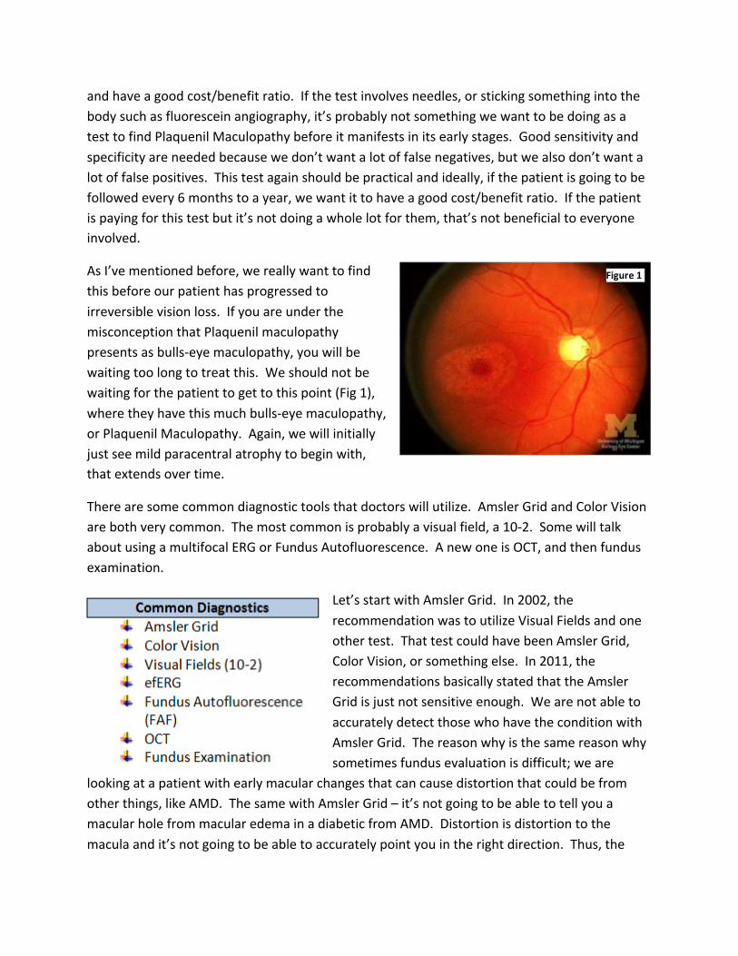
As I’ve mentioned before, we really want to find this before our patient has progressed to irreversible vision loss. If you are under the misconception that Plaquenil maculopathy presents as bulls-‐eye maculopathy, you will be waiting too long to treat this. We should not be waiting for the patient to get to this point (Fig 1), where they have this much bulls-‐eye maculopathy, or Plaquenil Maculopathy. Again, we will initially just see mild paracentral atrophy to begin with, that extends over time.
There are some common diagnostic tools that doctors will utilize. Amsler Grid and Color Vision are both very common. The most common is probably a visual field, a 10-‐2. Some will talk about using a multifocal ERG or Fundus Autofluorescence. A new one is OCT, and then fundus examination.
Let’s start with Amsler Grid. In 2002, the recommendation was to utilize Visual Fields and one other test. That test could have been Amsler Grid, Color Vision, or something else. In 2011, the recommendations basically stated that the Amsler Grid is just not sensitive enough. We are not able to accurately detect those who have the condition with Amsler Grid. The reason why is the same reason why sometimes fundus evaluation is difficult; we are
looking at a patient with early macular changes that can cause distortion that could be from other things, like AMD. The same with Amsler Grid – it’s not going to be able to tell you a macular hole from macular edema in a diabetic from AMD. Distortion is distortion to the macula and it’s not going to be able to accurately point you in the right direction. Thus, the
Figure 1

best way to look for some of the early changes may be a combination of a fundus examination in combination with other tests. According to the AAO, those other tests should not include the Amsler Grid.
Nor should it include color vision testing. Again, this test is not specific to hydroxychloroquine toxicity. There are many other macular and optic nerve diseases that lead to color vision deficits and none of the color tests point to specific hydroxychloroquine deficits.
The AAO is not recommending the following. Fundus photography is not recommended because it’s hard to follow a patient over time, and in an ideal world we are finding this before it shows up in the fundus. Now, I would argue that it’s important to have some sort of fundus photography, especially if they have some sort of pre-‐existing condition, so you can see the changes over time and have it photo documented. That is very nice to have. Others would argue that you need a baseline photo, and that’s fine, but if you mark in the chart that everything looks normal, you should trust yourself in that, and not require photographic evidence. I do think if there are some changes, sometimes they may be hard to describe, and over time you can look for subtle changes over time in photography. I do think, however, that we have better equipment than just simply photography.
Time Domain OCT is not sensitive enough. It’s recommended to do the Spectral Domain OCT.
Fluorescein Angiography is not recommended, partly because it’s a little bit overkill. Plus, we are not really looking for leakage from any sort of vascular insufficiency and so forth. Fluorescein Angiography is really getting at the wrong thing.
Full-‐Field ERG is not recommended, either. The thing about Full-‐Field ERG is it’s just too tedious to be checking the whole retina. It’s labor-‐intensive, time-‐intensive and is overkill – way more information than we need.
We’ve already talked about Amsler Grid and Color Vision.
Just to clarify, Electro-‐Oculogram is not what we are going to be looking for. We want an Electro-‐Retinogram.
Let’s talk about Multi-‐Focal ERG. Some people will recommend this as a way to look for early changes that we can find before we get to seeing changes on the retina. mfERG measures bio-‐electrical potentials. We will put a contact lens on the eye, with a wire attached to the instrument. The patient looks at a visual field and we’re measuring the response to what the

patient sees. What’s nice about this is that the test is objective – we don’t need to rely on the patient to respond to a visual field & indicating that they see something. It’s nice because it’s reliable and specifically targets the area we are concerned about – the macula.
The difficulty though, is the interpretation. When I’ve had mfERG’s done, the report comes back to me and it’s already been interpreted, but honestly, as optometrists we are not well-‐versed with the ERG. I apologize if you are familiar and this is a great tool for you, keep utilizing it. I, however, have some concerns with the interpretation. It’s very difficult to tell the early subtle changes that could be from other macular changes. Again, that’s going to be a recurrent theme throughout my lecture today. It’s so hard because we’re trying to find Plaquenil Maculopathy before it’s affecting the retinal tissue enough for us to see it. There’s no gold standard for any of these tests that we can compare mfERG against, so we’re kind of relying on clinical intuition here. That can sometimes lead us astray in conditions that aren’t overly common. We are much more likely to over-‐call a diagnosis or over-‐diagnose the condition, which is a concern. If the patient really needs their medication, we may be taking them off of a medication that they really need, and we’re doing a disservice to the patient. It’s in our best interests to keep in mind that these tests are often very difficult to interpret because you look at the results, which could be a little bit off, but then when we look at the macula, it is pristine. Or we look and it looks like macular degeneration and we’re stuck saying ‘Well, my evidence isn’t very good for taking this patient off of this medication.’ We’re stuck between a rock and a hard place: it looks abnormal, but the patient really benefits from their medication.
I will elaborate on that in a moment, but I want to introduce to you the fact that these tests are not 100% accurate, and often times they leave us a little bit confused. Also, it’s expensive to have this done, and especially if we’re going to have it done once a year, it’s just a lot for the patient. It’s a big hassle, too. We may think to ourselves that yes, it’s a hassle but if we can catch this early, and prevent the patient from having vision loss, isn’t it worth it? In a moment, we will discuss if it is or is not worth it.
The other thing is accessibility. Where I am, in Kansas City, we have the University of Kansas and their medical center has an ERG, but not everybody is lucky enough to be next to a medical center like that, where they have access to a lot of different diagnostic tests and someone to tell them the results of said tests. I was giving this lecture recently in a rural area and I asked how many doctors had an ERG nearby and no one in the room of about 50 doctors raised their hand. I’m worried that if we are pushing ERG, it’s just really not available. If we are having the patient drive an hour or two for this test, I think that’s overkill. It’s just a bit too much for a test that’s not incredibly accurate.

Let’s go back to the hassle part. We are measuring biological potentials. If we look at this patient in Fig 2, the patient looks incredibly uncomfortable to me. This test takes a very long time – up to an hour, just for the multifocal ERG. They have a contact lens on their eye, and often need anesthetic just to keep it on. This leads to increased risk of corneal abrasions and in turn corneal ulcers. It’s uncomfortable – patients often complain of a foreign body sensation when the contact lens is on, and even after it’s been removed. It’s not a very good test because it’s costly, not accessible, uncomfortable and we just don’t get very good results with it.
Fundus Autofluorescence (Fig 3) is another tool that we can use. With Fundus Autofluorescence (FAF) we use the exciter filter within a Fluorescein Angiography camera to auto fluoresce the retina. This can be used in other conditions, such as seeing drusen in macular degeneration or drusen in the optic nerve head. This is actually a very valuable tool if you’re looking at optic nerve edema and are questioning if it’s caused by optic disc drusen.
Figure 2 -‐ mfERG

We see in this series of photographs from top left to bottom right that there is increasing damage in a bulls-‐eye fashion. We see increasing dimness in that paracentral area, and that’s what we are looking for in FAF. In the upper left, that’s a normal-‐looking retina. These are different patients at different stages of Plaquenil Maculopathy. Over the progression of the disease you’ll see even worse looking maculas than these.
If you are like me, I have a hard time distinguishing in the middle row on the left, if those maculas are normal or not. It looks very similar to the ones at the top of the figure. I personally do not have much experience with FAF so I have to ask ‘is this normal or abnormal?’ We can look at the bottom ones and those are very obvious problems that anybody could identify. In the bottom cases, we wouldn’t even need FAF; if we just looked in our slit lamp with our 90D or 78D lens, we could identify the problem. This is not helping us identify the problem before we could normally, or before damage has already occurred in the retina. It’s not a good test for our needs and, as optometrists, we are not well-‐versed in this test.
The nice thing about FAF is that it’s objective and we can view changes over time, since we are photo-‐documenting it. Again, however, going back to interpretation, it’s not easy to interpret these changes. Also, this is not readily available, unless you already have a Fluorescein Angiography camera in your office. This is not something we are just going to run out and buy
Figure 3: FAF

to have access to an exciter filter. I’ve asked around about purchasing a separate exciter filter to buy to add on to this, and it’s not available yet. For most optometrists and ophthalmologists this test is just not feasible.
Visual fields are a mainstay for a lot of offices in evaluating Plaquenil Maculopathy. For years, in the pre-‐OCT days, this was the go-‐to. It’s not recommended to do a 24-‐2 or 30-‐2 because there are just not enough central points to accurately find that paracentral atrophy early enough. The recommendation is to do 10-‐2 white-‐on-‐white (no blue-‐yellow).Let’s look at some
results.
In Figure 4, on the top left, that is early Plaquenil changes. The top right is the same thing. If you’re like me, I’m looking at that and I think that could be many things, including a pretty normal visual field. I’m not looking and saying I’m really concerned about that. These top results could be from the patient having a bad day, they missed a few clicks, they fell asleep (which happens at the VA quite often), it could be other macular changes. This does not pop out as Plaquenil Maculopathy to me, nor would it make me confident enough to take the patient off of their medication, or relay a report to their primary care provider or rheumatologist saying the patient is experiencing eye changes.
Figure 4

Now, on the bottom left, there are some very concerning arcuate changes. My guess is that in these bottom two sets, we will also see the maculopathy when we look at the fundus. These bottom two are really bad, especially the bottom right. The bottom right is too far gone – at that point, we would definitely say that this is affecting the patient and we should take them off of the medication, or at least have a very serious conversation about discontinuing their medication. There we can move forward, but the big concern with these is the rate of false positives. Are we going to call the top two results abnormal or a bad day? This is where I would urge caution and repeat that visual field.
My big concern with visual fields is that there are a lot of false positives. When we have a condition as uncommon as Plaquenil Toxicity, we run the risk of even more false positives. Say if we had something that was really common, our number of false positives would be significantly less. Thus, we need to consider the sensitivity and specificity of the test. We also have to consider what is the risk of developing toxicity. So let’s go through some numbers and concepts that may be a challenge to grasp but is nice because you can take your time with this pdf and look at the numbers as long as you need to before moving on.
Let’s review the risk again. Before 7 years on the medication, the risk is 1/1000. After 7 years of being on Plaquenil, the risk is 1/100. If we ran the test before the 7 year cutoff, we are more likely to find false positives because very few patients develop this condition. If we ran the test after 7 years, we are less likely to find false positives, but we are still likely to find more false positives than true positives.
Let’s run though sensitivity and specificity again. Sensitivity is the ability of a test to correctly identify those who actually have the condition. In this case, how good is the test at actually finding those who have Plaquenil Toxicity? Specificity is the ability of the test to correctly identify those who do not have the condition. In this case, how good is it at telling us the patient does not have Plaquenil Toxicity. If the sensitivity is low, we will have a lot of false negatives. If the specificity is low, we will get a lot of false positives. We are really concerned with specificity when we have a disease that has a low number of affected patients. Ideally for Plaquenil Maculopathy, we would like to have a very good specificity. If our specificity could be 99%, that would be wonderful. However, anytime we raise the specificity of the test, we also lower the sensitivity. When we lower the sensitivity too much, we make the test useless because we do not detect any affected patients. If we make the sensitivity too high, the specificity can dip, leading to too many false positives, which is equally bad in this condition

because we don’t want to over-‐diagnose this and take patients off of their medication needlessly.
I don’t know the actual specificity for visual fields for Plaquenil Toxicity because, again, we don’t have a gold standard to compare any of our tests to. What I do know is the specificity of visual fields with glaucoma. So let’s assume that the specificity is as good as 90%, which is a pretty good specificity. If we did this test on every single patient who had Plaquenil Maculopathy, we would have 100 times more false positives than we would have true positives before 7 years of medication use. After 7 years, it improves a little bit, but we would still have 10 times more false positives than true positives. So we run a test, get the results, it looks positive, and we say to ourselves that the patient does have Plaquenil Maculopathy based on the visual field, it is 10 times more likely to have given that distinction based on a false positive than a true positive. Now we will potentially take the patient off of the medication when the patient doesn’t actually have the condition and they may benefit more from remaining on their medication than being taken off of it.
The benefit of visual fields is that it’s readily available, inexpensive, and all of us are relatively well versed in their interpretation.
Interpretation could also go in the Con category, because sometimes the findings are subtle and it’s hard for us to really tell. Another Con is that the test is subjective, in that the patient has to click the button and tell us when they see the light, and sometimes patients aren’t very accurate with this. Maybe they are having a bad day, they are tired, or they aren’t well-‐versed or it’s their first time taking the test. The patients often dislike this test – Visual Fields aren’t something my patients jump for joy to have done. If we balance that out, though, with the risk for visual loss, I think most patients understand why we want to have this done. The final Con is the false positives that we just talked about.
When it comes to false positives, I don’t want you to be discouraged, I want you to be able to utilize this test. Just realize that when the test is positive that isn’t 100% accurate. We should weigh the results against what the retina, OCT, and other tests look like, and with how much does this patient need to be on Plaquenil. Often times, Plaquenil is a last-‐ditch effort for someone who doesn’t want to be on steroids and can’t handle other alternative medications who have other big-‐time side effects. Methotrexate, and other medications like it, are often very difficult for patients to tolerate and so we really have to be cautious of that when we are discussing this with them and their provider who referred them to us.

We may think ‘I’m not going to take them off of the medication, I’m just going to refer them to their provider and let them know what’s going on.’ In reality, however, any time you tell their doctor that the patient has vision changes from the Plaquenil, the patient is likely to be taken off of the medication. Let’s face it – no one wants to be the doctor who let the patient stay on the medication despite what the optometrist said, and the patient loses vision. That’s a lawsuit waiting to happen for everybody involved and it’s not in anybody’s best interest. So we want to be very certain that the patient has Plaquenil Toxicity before we pull that trigger.
Spectral Domain OCT (SD-‐OCT) is a very cool newly-‐utilized test for Plaquenil Maculopathy. There’s a really cool study that just came out in the last few years talking about how we can use SD-‐OCT to detect changes caused by Plaquenil Toxicity. I apologize if you’re familiar with SD-‐OCT, but this image (Fig 5) is for those of you who may not be as familiar with it. The top bright red layer on the right side is the nerve fiber layer. Then we will go into the ganglion cell layer, which will be affected first. We may see some thinning there, though it’s often very subtle. Inner plexiform, inner nuclear, outer plexiform and outer nuclear layers are also highlighted for us. The other thing we’re going to look at is the photoreceptor inner segment and outer segment layer – there is a very faint line above the dense red at the bottom of the OCT there. That bottom is the RPE and right above that is the inner segment and outer segment line, which
will be of particular interest to us with the OCT.
What this study found was something called ‘Flying Saucer Sign’, which sounds very cool. (See Fig 6) The Flying Saucer Sign is something that we will see with early Plaquenil changes. There are three distinct changes we will see to the macular region. Look at the top of Figure 6, and
Figure 5
Figure 6

at the very top we will see a normal-‐looking black and white OCT. (Most of us will have colored OCT’s in our office.) We see the divot for the foveal pit, the 2 humps next to it. The bright white band at the bottom is the RPE and then right above it is the inner segment-‐outer segment line of the photoreceptors.
In the bottom half of the page we will see several different things. Number 1 that we notice is that foveal pit has leveled out almost like it got steam-‐rolled and that double hump is gone. What happened is that the double humps are down lower because the ganglion cell layer has atrophied and is thinner, which gives a pseudo-‐dome appearance. That dome is where Marvin the Martian would sit in that flying saucer. Just below that dome we see a little bit of a peak of the inner segment-‐outer segment line. Finally, right where those arrows are, to the right and left of the center of the fovea, are where there is thinning of the inner and outer segment line, and that is in a paracentral fashion. Those are the three things we are going to see in a Flying Saucer Sign on the OCT. We will see the pseudo-‐dome from the 2 humps on either side of the foveal pit atrophying. Then we will see inner segment and outer segment atrophy, as well as that little ‘saucerization’ that appears in the middle there.
Figure 7 shows us some higher-‐resolution examples going from mild atrophy to severe atrophy from the top of the page (mild) to the bottom (severe). The top left of the figure is a very good early example – the foveal pit has flattened, there’s a dome, inner segment-‐outer segment line looks like there’s a little bit of separation, and to either side it looks like there’s a little bit of atrophy.
If you go to the bottom left, not the very bottom but the one just above it, this macula has also lost its foveal pit. It’s hard to see, but that inner-‐outer segment line has peaked a little at the center, and there’s a lot of atrophy paracentrally. Feel free to take your time studying these,
as I think they will be of good use.
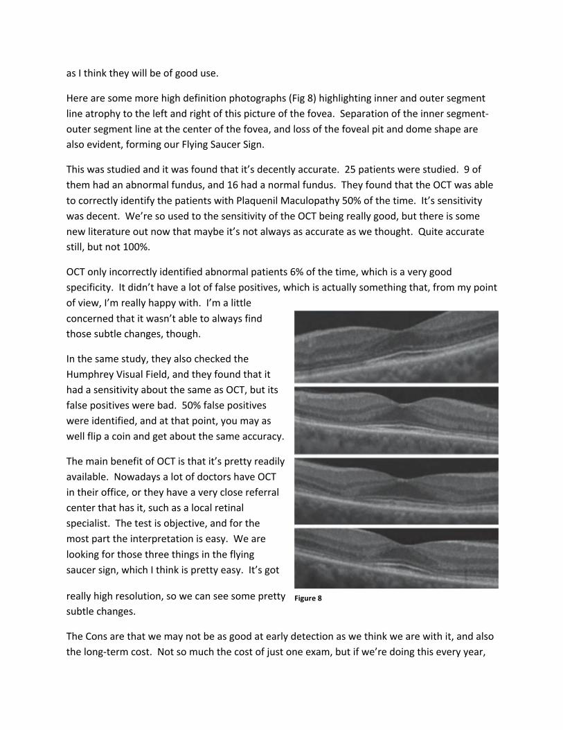
Here are some more high definition photographs (Fig 8) highlighting inner and outer segment line atrophy to the left and right of this picture of the fovea. Separation of the inner segment-‐outer segment line at the center of the fovea, and loss of the foveal pit and dome shape are also evident, forming our Flying Saucer Sign.
This was studied and it was found that it’s decently accurate. 25 patients were studied. 9 of them had an abnormal fundus, and 16 had a normal fundus. They found that the OCT was able to correctly identify the patients with Plaquenil Maculopathy 50% of the time. It’s sensitivity was decent. We’re so used to the sensitivity of the OCT being really good, but there is some new literature out now that maybe it’s not always as accurate as we thought. Quite accurate still, but not 100%.
OCT only incorrectly identified abnormal patients 6% of the time, which is a very good specificity. It didn’t have a lot of false positives, which is actually something that, from my point of view, I’m really happy with. I’m a little concerned that it wasn’t able to always find those subtle changes, though.
In the same study, they also checked the Humphrey Visual Field, and they found that it had a sensitivity about the same as OCT, but its false positives were bad. 50% false positives were identified, and at that point, you may as well flip a coin and get about the same accuracy.
The main benefit of OCT is that it’s pretty readily available. Nowadays a lot of doctors have OCT in their office, or they have a very close referral center that has it, such as a local retinal specialist. The test is objective, and for the most part the interpretation is easy. We are looking for those three things in the flying saucer sign, which I think is pretty easy. It’s got
really high resolution, so we can see some pretty subtle changes.
The Cons are that we may not be as good at early detection as we think we are with it, and also the long-‐term cost. Not so much the cost of just one exam, but if we’re doing this every year,
Figure 7
Figure 8

again we need to keep that in mind. Yes, the patient may have insurance, but you and I are paying that cost with our insurance premiums, and everybody is paying for that so we should be judicious with our use of this test.
Let’s go over the changes from the AAO’s 2002 recommendations. I’ve already talked about this a little bit. Back in 2002, a patient was considered at risk if they had taken greater than 6.5 mg/kg/day, and again that balanced out to 70 lbs body weight for every 200 mg of Plaquenil. It was recommended to test color vision, along with a 10-‐2 visual field or Amsler Grid.
Now we know that color vision and Amsler Grid are not accurate and, in fact, may be misleading. We now know that a cumulative dose of 1000g is much more concerning than the amount the patient takes every day. We should consider the patient’s height to weight ratio, as well, as a risk factor. Now it is recommended to do 10-‐2 combined with one of the following: OCT, Fundus Autofluorescence (FAF), or multifocal ERG. In my practice, I do a 10-‐2 combined with an OCT. I don’t do FAF or mfERG, even though FAF is readily available at my office.
The recommendation is to do a baseline examination. If the patient is low-‐risk, then test them again after 5 years. If the patient is high risk, then test them yearly. Now, this could get misinterpreted. The AAO is not recommending that we do an exam today and send the patient a reminder card in 5 years. Most of these people are going to have other things we will also want to look for, whether it is refractive error changes, cataracts, glaucoma, or macular degeneration; if we need to follow the patient yearly for other conditions, follow them yearly. I believe the AAO is saying that we don’t need to do a visual field every year to check for Plaquenil Maculopathy, we don’t have to do an OCT every year. Our likelihood of finding a false positive is way more likely than finding a true positive before the 5-‐7 year mark. After that 7 years, these patients are at less risk of having a false positive with our testing, so it’s better to monitor them more closely. Just remember, even after 7 years, the patient is still at a high risk for a false positive, so keep that in mind.
My recommendation would be to do an initial fundus examination, and potentially do baseline photography if that’s something you want. I don’t do anything again specifically for Plaquenil Maculopathy for 7 years. When I was giving this lecture recently, it was almost implied that, ‘That’s the VA way – here’s how we do it in the real world…’ It’s fine to criticize the VA and how I do it there, but honestly, I don’t think that I’m insulated at the VA in this recommendation. I think that if the AAO is recommending this, then I think it should be followed by

ophthalmologists and, frankly, optometrists because we are going to be referring to the ophthalmologists for care of some of these patients.
The other thing is that there is no guidance from the American Optometric Association (AOA) as to what we should do for these patients in their updated 2012 recommendations. Thus we are going to have to use ophthalmology’s guidance on this, along with our clinical knowledge. It’s important to keep in mind those false positives because again, we’re going to get to a point where a patient may need that medication to keep them from being in constant pain. We will be the ones telling their primary doctor to take them off of that medication, for what could potentially be a false positive. Thus, I personally do not recommend doing the visual field every year before 7 years. You may have a philosophical difference from mine, and that’s OK. If you want to do a visual field every year to get the patient well-‐versed in the test, and to get a pattern for them, that’s fine. My argument against that is that we are all familiar with what a normal visual field looks like. Thus, maybe start the patient on annual visual fields at 5 years, and see if there are any macular changes that maybe the patient already has, and we can monitor changes from there. But if you’re looking and the patient has a normal macula and a normal nerve, I don’t think we’re going to see visual field changes unless there is a neurological problem. I don’t really see that there’s an absolute necessity in doing that test regularly.
The problem is, once we are finished with the test, we have a few different outcomes. Let’s say the visual field and OCT are both positive. I think we can look at that patient and be pretty confident that they have some toxicity, and we will want to have a serious conversation with them and their doctor. If the visual field is positive but the OCT is negative, I think we are looking at a false positive. If the visual field is negative but the OCT positive, I’d want to follow that up over time, carefully. If the visual field and the OCT are both negative, I think we can safely take the fish off the hook and follow them like we normally would.
Again, I have some frustrations with this. As I mentioned at the beginning, a lot of people will talk of this condition with complete confidence: “This is what you do, and when you see changes, refer and take them off of the medication.” I don’t think it’s that easy, because we have a lot of false positives and there’s no gold standard to compare these tests to. The trouble is that we are often the authority, and we are telling these patients to stop the medication. We are telling their rheumatologist and/or primary care doctor that the patient has this problem and we may be telling them indirectly to take the patient off the medication, even by just reporting the results. With this mindset, I think we are downplaying some of the benefits of Plaquenil. If the patient is 85 years old with very subtle changes that could just be macular

degeneration, and they have rheumatoid arthritis that is debilitating and Plaquenil is the only thing that helps them, we may be doing that patient a disservice. When in reality, they may never suffer from vision changes because those changes can often be so mild and so slow to present.
On the flip side, the patient might be young, and they may have Plaquenil as a first line therapy, and there may be other options for very mild lupus or very mild rheumatoid arthritis. Thus, we have to balance these out on a case-‐by-‐case basis, and that’s why it’s frustrating for me to read in absolutes: “You should take the patient off the medications,” or “When the test is positive, it’s absolutely positive.” We have to keep that in mind and, as clinicians, it’s kind of exciting – we have the opportunity to be doctors here, use our brains, and have good in-‐depth conversations with our patients. We can set their minds at ease. They’re going to bring their medication label in to you, and say, “Doctor, I’m worried about my eyes.” And if they are not, then they may be lying to you or not comfortable to bring that up, in which case it’s our responsibility to bring it up to them: “You may be scared about your vision loss, Mr. Smith, here, let me set your mind at ease – your risk of developing this is very low.” That is where we can be compassionate for our patients and really help them out by understanding this, and understanding those nuances.
Thank you very much for your time in reading this PDF. I hope you’ve learned a lot. If you have any questions, concerns, feedback, feel free to send it to my VA address. Thank you again and I hope you enjoy the rest of your day.
Dr. Anthony DeWilde, OD [email protected]