microrna146 represses endothelial activation by inhibiting
TRANSCRIPT
Research Article TRANSPARENTPROCESS
OPENACCESSMicroRNA‐146 represses endothelial activation
MicroRNA‐146 represses endothelialactivation by inhibiting pro‐inflammatorypathways
Henry S. Cheng1,2,3, Nirojini Sivachandran1,2,3, Andrew Lau1,2,3, Emilie Boudreau1,2,3, Jimmy L. Zhao4,David Baltimore4, Paul Delgado-Olguin5,6, Myron I. Cybulsky1,2,3, Jason E. Fish1,2,3*
Keywords: atherosclerosis; endothelium;
gene regulation; inflammation;
microRNA
DOI 10.1002/emmm.201202318
Received December 01, 2012
Revised April 26, 2013
Accepted April 29, 2013
(1) Toronto General Research Institute, University Heal
Canada
(2) Department of Laboratory Medicine and Pathobi
Toronto, Toronto, Canada
(3) The Heart and Stroke Richard Lewar Centre of Excell
lar Research, Toronto, Canada
(4) Division of Biology, California Institute of Technolog
(5) Department of Physiology & Experimental Medicine,
Children, Toronto, Canada
(6) Department of Molecular Genetics, University o
Canada
*Corresponding author: Tel: þ1 416 581 7496; Fax: þE-mail: [email protected]
� 2013 The Authors. Published by John Wiley and Sons,the terms of the Creative Commons Attribution License (Cin any medium, provided the original work is properly cite
Activation of inflammatory pathways in the endothelium contributes to vascular
diseases, including sepsis and atherosclerosis. We demonstrate that miR-146a
and miR-146b are induced in endothelial cells upon exposure to pro-
inflammatory cytokines. Despite the rapid transcriptional induction of the
miR-146a/b loci, which is in part mediated by EGR-3, miR-146a/b induction is
delayed and sustained compared to the expression of leukocyte adhesion
molecules, and in fact coincides with the down-regulation of inflammatory gene
expression. We demonstrate that miR-146 negatively regulates inflammation.
Over-expression of miR-146a blunts endothelial activation, while knock-down of
miR-146a/b in vitro or deletion ofmiR-146a in mice has the opposite effect. MiR-
146 represses the pro-inflammatory NF-kB pathway as well as the MAP kinase
pathway and downstream EGR transcription factors. Finally, we demonstrate
that HuR, an RNA binding protein that promotes endothelial activation by
suppressing expression of endothelial nitric oxide synthase (eNOS), is a novel
miR-146 target. Thus, we uncover an important negative feedback regulatory
loop that controls pro-inflammatory signalling in endothelial cells that may
impact vascular inflammatory diseases.
INTRODUCTION
The endothelium plays a central role in the pathogenesis ofvascular inflammatory diseases such as sepsis (Aird, 2003) andatherosclerosis (Gimbrone & Garcia‐Cardena, 2012; Pober &Sessa, 2007). During sepsis, massive circulating levels of pro‐
th Network, Toronto,
ology, University of
ence in Cardiovascu-
y, Pasadena, CA
The Hospital for Sick
f Toronto, Toronto,
1 416 581 7484;
Ltd on behalf of EMBO. ThiC BY 3.0), which permits ud.
inflammatory cytokines activate the endothelium and drivepathological vascular permeability and tissue oedema, which leadto acute organ dysfunction (Aird, 2003). Blocking endothelialactivation can limit mortality in mouse models of sepsis (Londonet al, 2010). Endothelial activation also plays a pervasive rolein atherosclerosis, a chronic vascular inflammatory disorder thatis characterized by vessel narrowing, thrombosis and occlusion(Gimbrone & Garcia‐Cardena, 2012; Pober & Sessa, 2007). Cell‐surface expression of leukocyte adhesion molecules suchas vascular cell adhesion molecule‐1 (VCAM‐1), intercellularadhesion molecule‐1 (ICAM‐1) and E‐Selectin, and secretion ofchemokines such as monocyte chemoattractant protein‐1 (MCP‐1), facilitates the binding of circulating monocytes to the bloodvessel wall. Following transmigration into the intima, these cellsmature into inflammatory macrophages, and their secretion ofpro‐inflammatory cytokines further promotes endothelial activa-tion, and serves to drive a feed‐forward loop that perpetuatesleukocyte recruitment (Pober & Sessa, 2007). Identifyingmolecules that negatively regulate inflammatory pathways in
s is an open access article underse, distribution and reproduction
EMBO Mol Med (2013) 5, 1017–1034 1017

Research Article www.embomolmed.orgMicroRNA‐146 represses endothelial activation
1018
the endothelium may provide novel therapeutic targets for thetreatment of acute or chronic vascular inflammatory diseases.
Activation of pro‐inflammatory transcriptional programssuch as the NF‐kB signalling pathway (Gareus et al, 2008;Ruland, 2011; Ye et al, 2008) and the mitogen‐activated proteinkinase (MAPK)/early growth response (EGR) pathway (Hajraet al, 2000; Shin et al, 2009; Wieland et al, 2005; Yan et al, 2000)can cooperatively drive endothelial activation and vascularinflammation. Considering that prolonged or exaggeratedinflammatory responses are detrimental, it is not surprisingthat cellular negative feedback loops act to control the durationand intensity of an inflammatory response (Ruland, 2011). Forexample, activation of the NF‐kB pathway leads to the inductionof IkB proteins, which bind to NF‐kB proteins in the nucleus andexports them to the cytoplasm (Arenzana‐Seisdedos et al, 1995).EGR transcription factors also induce the expression of theirown repressor proteins (Kumbrink et al, 2005). In addition tofeedback regulation at the level of transcription, microRNAshave recently been identified that serve in post‐transcriptionalnegative feedback loops that modulate inflammatory signalling.MicroRNAs bind to the 30 UTRs of target mRNAs and inhibittheir translation and/or stability (Bartel, 2009). MiR‐146a waspreviously found to be transcriptionally induced by NF‐kBin response to activation of innate immune signalling inmonocytes (Taganov et al, 2006). MiR‐146a targets the adaptorproteins TRAF6 and IRAK1/2 (Bhaumik et al, 2008; Houet al, 2009; Nahid et al, 2009; Taganov et al, 2006) and caninhibit activation of the NF‐kB pathway (Bhaumik et al, 2008;Zhao et al, 2011), suggesting that miR‐146a participates in anegative feedback loop to control NF‐kB signalling in mono-cytes. However, the function ofmiR‐146a/b is poorly understoodin endothelial cells.
We previously identified miR‐146a and miR‐146b as beingenriched in endothelial cells isolated from differentiatingembryonic stem cells (Fish et al, 2008). Herein we demonstratethat miR‐146a and miR‐146b are enriched in endothelial cellsin vivo and that they are strongly induced in endothelial cellsin response to pro‐inflammatory cytokines. We also identifya novel transcriptional pathway involving EGR proteinsthat participates in the induction of miR‐146a and miR‐146b.Through delayed induction kinetics, miR‐146a/b appear to actas negative feedback regulators of inflammatory signalling inendothelial cells. MiR‐146 inhibits endothelial activation bydampening the activation of pro‐inflammatory transcriptionalprograms, including the NF‐kB, AP‐1 andMAPK/EGR pathways,likely through regulation of IL‐1b signalling pathway adaptorproteins (i.e., TRAF6, IRAK1/2). In addition, miR‐146 modulatespost‐transcriptional pro‐inflammatory pathways via targetingof the RNA binding protein HuR. We provide evidence thatHuR facilitates endothelial activation by repressing expressionof endothelial nitric oxide synthase (eNOS), a major sourceof nitric oxide, which potently inhibits leukocyte adhesion(Kubes et al, 1991). Thus miR‐146 represses both transcriptionaland post‐transcriptional activation of the inflammatory program.Our results reveal a potent anti‐inflammatory action ofmiR‐146a/b in the endothelium and suggest that therapeuticelevation of this microRNA family may be a useful treatment
� 2013 The Authors. Published by John Wiley and Sons, Ltd on behalf of EMBO.
strategy for vascular inflammatory diseases, including sepsisand atherosclerosis.
RESULTS
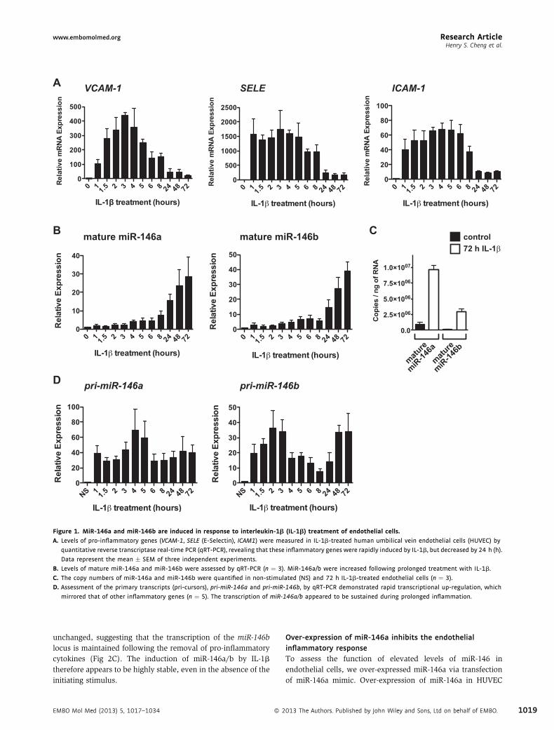
Induction of miR‐146a and miR‐146b (miR‐146a/b) byinflammatory stimuli in endothelial cellsTreatment of human umbilical vein endothelial cells (HUVEC)with the pro‐inflammatory cytokine, IL‐1b, induced the rapidinduction of mRNAs encoding leukocyte adhesion molecules,such as VCAM‐1, E‐Selectin and ICAM1 (Fig 1A). We nextmeasured levels of miR‐146a and miR‐146b. Since miR‐146a andmiR‐146b differ by only two nucleotides near their 30 ends,we designed primers that amplified only miR‐146a or miR‐146b(see Materials and Methods Section). We found that thesemicroRNAs were dramatically induced in response to IL‐1btreatment (Fig 1B). Similar induction of miR‐146a/b wasobserved following tumor necrosis factor‐a (TNF‐a) treatment(Supporting Information Fig S1). MiR‐146a/b levels wereincreased during the late stages of an inflammatory response(i.e., 8–72 h post‐treatment), but levels were only modestlyelevated at early stages (i.e., 1–4 h post‐treatment; Fig 1B,Supporting Information Fig S1). Interestingly, the induction ofmiR‐146a/b coincided with the down‐regulation of adhesionmolecule genes (Fig 1A and B). Quantification of miR‐146a/blevels revealed that miR‐146a was expressed �9‐fold higherthan miR‐146b in unstimulated cells, and �3‐fold higher thanmiR‐146b in IL‐1b‐treated cells (Fig 1C). We next measuredthe transcription of the miR‐146a and miR‐146b genomic loci.MiR‐146a is processed from a two‐exon non‐protein codingmRNA transcript on chromosome 5, and we therefore measuredunspliced pre‐mRNA of this transcript as a surrogate oftranscription [as we have done previously (Fish et al, 2010)].MiR‐146b is processed from a single exon transcript onchromosome 10, and primers were designed to measure thelevels of this transcript. We found that transcription of miR‐146aandmiR‐146bwere rapidly (within 1 h) and dramatically (40‐ and20‐fold, respectively) induced in response to IL‐1b (Fig 1D). Thetranscription of miR‐146a and miR‐146b was sustained for theduration of IL‐1b treatment. This is in contrast to VCAM‐1, SELE(E‐Selectin) and ICAM‐1 mRNA, which were down‐regulatedafter 8 h of IL‐1b treatment. Despite the rapid transcriptionof the miR‐146a and miR‐146b genes, delayed expression ofmature microRNAs implies inefficient or delayed processing ofmiR‐146a/b in cytokine‐stimulated endothelial cells.
MiR‐146a/b expression is sustained following removal ofpro‐inflammatory cytokinesTo determine the stability of the IL‐1b‐mediated induction ofmiR‐146a/b we treated endothelial cells with IL‐1b for 24 h andthen removed the cytokine. In contrast to inflammatory genessuch as VCAM‐1 and SELE, which were rapidly down‐regulatedupon removal of IL‐1b (Fig 2A), miR‐146a/b remained elevatedfor more than 2 days (Fig 2B). MiR‐146b expression wasespecially long‐lived. While the levels of pri‐miR‐146a decreasedfollowing the removal of IL‐1b, levels of pri‐miR‐146b remained
EMBO Mol Med (2013) 5, 1017–1034
A VCAM-1 SELE ICAM-1
B
D pri-miR-146a pri-miR-146b
mature miR-146a mature miR-146b C72 h IL-1βcontrol
mature
miR-14
6amatu
re
miR-14
6b
Figure 1. MiR‐146a and miR‐146b are induced in response to interleukin‐1b (IL‐1b) treatment of endothelial cells.
A. Levels of pro-inflammatory genes (VCAM-1, SELE (E-Selectin), ICAM1) were measured in IL-1b-treated human umbilical vein endothelial cells (HUVEC) by
quantitative reverse transcriptase real-time PCR (qRT-PCR), revealing that these inflammatory genes were rapidly induced by IL-1b, but decreased by 24 h (h).
Data represent the mean � SEM of three independent experiments.
B. Levels of mature miR-146a and miR-146b were assessed by qRT-PCR (n ¼ 3). MiR-146a/b were increased following prolonged treatment with IL-1b.
C. The copy numbers of miR-146a and miR-146b were quantified in non-stimulated (NS) and 72 h IL-1b-treated endothelial cells (n ¼ 3).
D. Assessment of the primary transcripts (pri-cursors), pri-miR-146a and pri-miR-146b, by qRT-PCR demonstrated rapid transcriptional up-regulation, which
mirrored that of other inflammatory genes (n ¼ 5). The transcription of miR-146a/b appeared to be sustained during prolonged inflammation.
Research Articlewww.embomolmed.orgHenry S. Cheng et al.
unchanged, suggesting that the transcription of the miR‐146blocus is maintained following the removal of pro‐inflammatorycytokines (Fig 2C). The induction of miR‐146a/b by IL‐1btherefore appears to be highly stable, even in the absence of theinitiating stimulus.
EMBO Mol Med (2013) 5, 1017–1034 � 2
Over‐expression of miR‐146a inhibits the endothelialinflammatory responseTo assess the function of elevated levels of miR‐146 inendothelial cells, we over‐expressed miR‐146a via transfectionof miR‐146a mimic. Over‐expression of miR‐146a in HUVEC
013 The Authors. Published by John Wiley and Sons, Ltd on behalf of EMBO. 1019

A
B
C
0 24 48 72NS
0 24 48 72NS
0 24 48 72NS
****** ****** ******
*
*** **
Rel
ativ
e m
RN
A Ex
pres
sion
Rel
ativ
e m
iRN
A Ex
pres
sion
Rel
ativ
e pr
i-miR
NA
Expr
essi
on
Figure 2. MiR‐146a and miR‐146b expression is sustained after the
removal of IL‐1b. Endothelial cells were treated with IL-1b for 24 h, after
which IL-1bwas removed. Levels of VCAM-1 and SELE (A), mature miR-146a/b
(B) and pri-miR-146a/b (C) were monitored at various time-points after the
removal of IL-1b by qRT-PCR. While VCAM-1 and SELE rapidly returned to
base-line levels, miR-146a/b levels remained elevated. The transcription of
miR-146a decreased by 24 h after removal of IL-1b, while transcription of
miR-146b was sustained in the absence of IL-1b. Data represents the
mean � SEM of three independent experiments. Statistical analyses were
performed using t-test to compare post-IL-1b removal time-points to 24 h
of IL-1b treatment (i.e., time zero). Significant p-values (from left to right)
in (A) are 0.0004, <0.0001, 0.0004, <0.0001, 0.0006 and 0.0001,
respectively. p-value in (B) is 0.049. p-Values in (C) are 0.012, 0.001 and 0.001,
respectively.
Research Article www.embomolmed.orgMicroRNA‐146 represses endothelial activation
1020 � 2013 The Authors. Published by John Wiley and Sons, Ltd on behalf of EMBO.
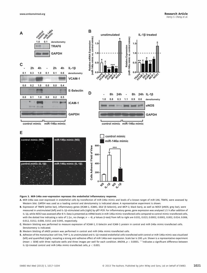
resulted in decreased expression of TRAF6 (Fig 3A), a knowntarget of miR‐146 (Taganov et al, 2006). Next we assessed theexpression of several pro‐inflammatory genes (VCAM‐1, ICAM‐1,SELE andMCP‐1) by qRT‐PCR, and found that the basal levels ofthese mRNAs were suppressed in unstimulated miR‐146a over‐expressing cells (Fig 3B, left). Importantly, miR‐146a over‐expression also dampened the induction of these inflammatorygenes in response to IL‐1b treatment (Fig 3B, right). Nitric oxide(NO) generated by eNOS potently inhibits leukocyte adhesionto the endothelium (Kubes et al, 1991), and eNOS (NOS3) isknown to be transcriptionally (Anderson et al, 2004) and post‐transcriptionally (Yoshizumi et al, 1993) repressed followingtreatment of endothelial cells with pro‐inflammatory cytokines.The level of NOS3 mRNA in unstimulated cells over‐expressingmiR‐146awas elevated (Fig 3B, left). After 8 h of IL‐1b treatment,NOS3 mRNA was decreased by 45.0 � 6.5% (p ¼ 0.004,not shown). Over‐expression of miR‐146a blunted this IL‐1b‐dependent decrease in NOS3 mRNA levels (Fig 3B, right).Western blotting confirmed that the induction of VCAM‐1,E‐Selectin and ICAM‐1 protein was inhibited in miR‐146a over‐expressing cells (Fig 3C), and that the loss of eNOS expressionwas blunted (Fig 3D). Consistent with a reduction in inducibleadhesion molecule expression and an increase in eNOS protein,miR‐146a over‐expression in HUVEC reduced the number ofTHP‐1 monocytes that adhered to IL‐1b‐treated endothelial cells(Fig 3E). Over‐expression of miR‐146a in aortic endothelialcells also inhibited leukocyte adhesion (Supporting InformationFig S2), suggesting that miR‐146a broadly represses endothelialactivation. MiR‐146a therefore inhibits the endothelial inflam-matory response, including the induction of adhesion moleculesand chemoattractants and the loss of eNOS expression.
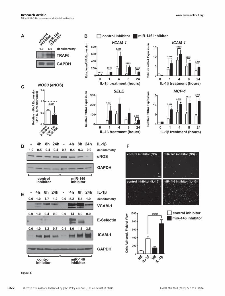
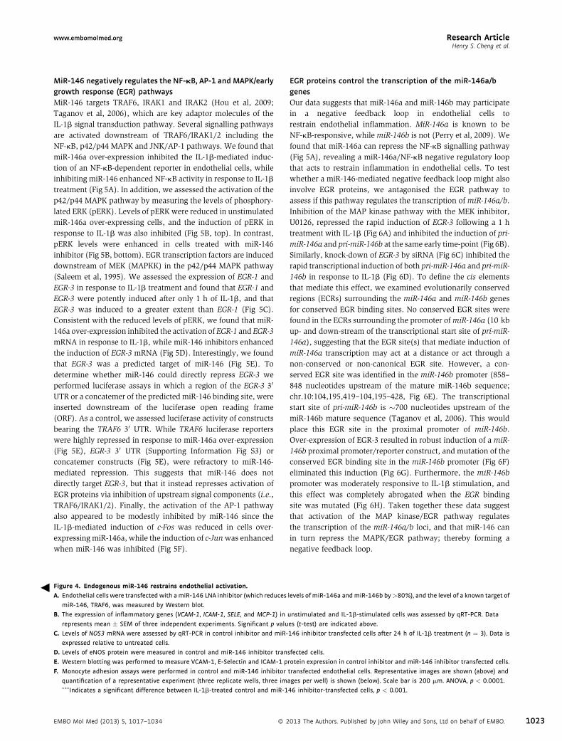
Endogenous miR‐146 inhibits the endothelial inflammatoryresponseWe next utilized a miR‐146 locked‐nucleic acid (LNA) inhibitorto assess the function of endogenous miR‐146 in endothelialcells. In addition to reducing the level of mature miR‐146a by81.7 � 6.5%, this inhibitor also reduced the level of miR‐146bby 92.5 � 2.7% (not shown), likely owing to the limited (twonucleotide) difference in sequence between miR‐146a and miR‐146b. Treatment with miR‐146 inhibitor elevated the level of themiR‐146 target, TRAF6 (Fig 4A). Additionally, miR‐146 inhibitorincreased the basal levels of VCAM‐1 mRNA, and had a potenteffect on the IL‐1b‐inducible expression of VCAM‐1, ICAM‐1,SELE and MCP‐1 (Fig 4B). Endogenous miR‐146 appeared torestrain the intensity as well as the duration of the inflammatoryresponse, since these inflammatory genes remained at elevatedlevels 24 h after IL‐1b treatment (Fig 4B). In addition, thedecrease in eNOS (NOS3) mRNA that was observed after a 24 htreatment with IL‐1b was augmented by miR‐146 inhibitor(Fig 4C). Western blotting confirmed that the loss of eNOSprotein in response to IL‐1b treatment was enhanced bymiR‐146inhibition (Fig 4D) and that IL‐1b‐inducible VCAM‐1, E‐Selectinand ICAM‐1 protein expression was greatly enhanced bymiR‐146 inhibition (Fig 4E). Finally, inhibition of miR‐146 inendothelial cells enhanced the adhesion of THP‐1 monocytesfollowing IL‐1b treatment (Fig 4F).
EMBO Mol Med (2013) 5, 1017–1034
A unstimulated IL-1β treated
C
E
VCAM-1
GAPDH
IL-1β- 2h 4h - 2h 4h
NSIL-1β
control mimicmiR-146a mimiccontrol mimic (NS) miR-146a mimic (NS)
miR-146a mimic (IL-1β)control mimic (IL-1β)
NSIL-1β
control mimic miR-146a mimic
1.00.30.1 0.1 0.1 0.6 densitometry
E-Selectin
ICAM-1
D
B
1.00.20.0 0.0 0.0 0.4
eNOS
GAPDH
control mimic miR-146a mimic
IL-1β- 8h 24h - 8h 24h0.30.81.0 1.1 0.9 0.6 densitometry
TRAF6
GAPDH
contro
l
mimicmiR
-146a
mimic
1.0 0.1 densitometry
1.00.10.0 0.0 0.2 0.5
* ****
*****
*
** * ***
*
*
***
Figure 3. MiR‐146a over‐expression represses the endothelial inflammatory response.
A. MiR-146a was over-expressed in endothelial cells by transfection of miR-146a mimic and levels of a known target of miR-146, TRAF6, were assessed by
Western blot. GAPDH was used as a loading control and densitometry is indicated above. A representative experiment is shown.
B. Expression of TRAF6 (white bar), inflammatory genes (VCAM-1, ICAM1, SELE (E-Selectin), and MCP-1; black bars), as well as NOS3 (eNOS; grey bar), were
measured in unstimulated (left) and IL-1b-stimulated cells (right) by qRT-PCR. For inflammatory genes, gene expression was analysed 1.5 h after addition of
IL-1b, while NOS3was assessed after 8 h. Data is presented as mRNA levels in miR-146a mimic-transfected cells compared to control mimic-transfected cells,
with the dotted line indicating a ratio of 1 (i.e., no change; n ¼ 4). p Values (t-test) from left to right are 0.031, 0.023, 0.0002, 0.0001, 0.002, 0.014, 0.006,
0.012, 0.012, 0.006, 0.011 and 0.045, respectively.
C. Western blotting was performed to measure expression of VCAM-1, E-Selectin and ICAM-1 protein in control and miR-146a mimic-transfected cells.
Densitometry is indicated.
D. Western blotting of eNOS protein was performed in control and miR-146a mimic-transfected cells.
E. Adhesion of the mononuclear cell line, THP-1, to unstimulated and IL-1b-treated endothelial cells transfected with control or miR-146a mimic was visualized
(left) and quantified (right), revealing a strong anti-adhesive effect of miR-146a over-expression. Scale bar is 200 mm. Shown is a representative experiment
(mean � SEM) with three replicate wells and three images per well for each condition. ANOVA, p < 0.0001. ���Indicates a significant difference between
IL-1b-treated control and miR-146a mimic-transfected cells, p < 0.001.
Research Articlewww.embomolmed.orgHenry S. Cheng et al.
EMBO Mol Med (2013) 5, 1017–1034 � 2013 The Authors. Published by John Wiley and Sons, Ltd on behalf of EMBO. 1021
B
C NOS3 (eNOS) IL-1β treatment (hours)0 1 4 8 24
VCAM-1 ICAM-1
SELE
IL-1β treatment (hours)0 1 4 8 24
MCP-1
IL-1β treatment (hours)0 1 4 8 24
IL-1β treatment (hours)0 1 4 8 24
FD
contro
l
inhibitor
miR-14
6
inhibitor
control inhibitor miR-146 inhibitor
NSIL-1β NS
IL-1β
control inhibitor (NS) miR-146 inhibitor (NS)
control inhibitor (IL-1β) miR-146 inhibitor (IL-1β)
IL-1β- 4h 8h
VCAM-1
GAPDH
24h - 4h 8h 24h
controlinhibitor
miR-146inhibitor
control inhibitormiR-146 inhibitor
1.0 1.7 1.2 5.2 5.4 1.90.0 0.0 densitometry
E-Selectin
ICAM-1
A
1.0 0.4 0.0 14 8.9 0.00.0 0.0
1.0 1.2 0.7 1.0 1.6 3.50.0 0.1
TRAF6
GAPDH
contro
l
inhibitor
miR-14
6
inhibitor
IL-1β- 4h 8h 24h - 4h 8h 24h0.5 0.4 0.4 0.4 0.3 0.01.0 0.5 densitometry
eNOS
GAPDH
E
6.01.0 densitometry
controlinhibitor
miR-146inhibitor
0.036
0.012
0.023
0.003
0.006
0.039
0.020
0.009
0.037
0.037
0.019
0.014
0.0190.034
0.015
***
0.019
Figure 4.
Research Article www.embomolmed.orgMicroRNA‐146 represses endothelial activation
1022 � 2013 The Authors. Published by John Wiley and Sons, Ltd on behalf of EMBO. EMBO Mol Med (2013) 5, 1017–1034
3
Research Articlewww.embomolmed.orgHenry S. Cheng et al.
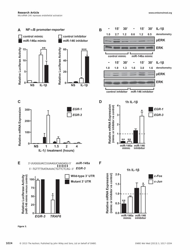
MiR‐146 negatively regulates the NF‐kB, AP‐1 and MAPK/earlygrowth response (EGR) pathwaysMiR‐146 targets TRAF6, IRAK1 and IRAK2 (Hou et al, 2009;Taganov et al, 2006), which are key adaptor molecules of theIL‐1b signal transduction pathway. Several signalling pathwaysare activated downstream of TRAF6/IRAK1/2 including theNF‐kB, p42/p44 MAPK and JNK/AP‐1 pathways. We found thatmiR‐146a over‐expression inhibited the IL‐1b‐mediated induc-tion of an NF‐kB‐dependent reporter in endothelial cells, whileinhibiting miR‐146 enhanced NF‐kB activity in response to IL‐1btreatment (Fig 5A). In addition, we assessed the activation of thep42/p44 MAPK pathway by measuring the levels of phosphory-lated ERK (pERK). Levels of pERKwere reduced in unstimulatedmiR‐146a over‐expressing cells, and the induction of pERK inresponse to IL‐1b was also inhibited (Fig 5B, top). In contrast,pERK levels were enhanced in cells treated with miR‐146inhibitor (Fig 5B, bottom). EGR transcription factors are induceddownstream of MEK (MAPKK) in the p42/p44 MAPK pathway(Saleem et al, 1995). We assessed the expression of EGR‐1 andEGR‐3 in response to IL‐1b treatment and found that EGR‐1 andEGR‐3 were potently induced after only 1 h of IL‐1b, and thatEGR‐3 was induced to a greater extent than EGR‐1 (Fig 5C).Consistent with the reduced levels of pERK, we found that miR‐146a over‐expression inhibited the activation of EGR‐1 and EGR‐3mRNA in response to IL‐1b, while miR‐146 inhibitors enhancedthe induction of EGR‐3 mRNA (Fig 5D). Interestingly, we foundthat EGR‐3 was a predicted target of miR‐146 (Fig 5E). Todetermine whether miR‐146 could directly repress EGR‐3 weperformed luciferase assays in which a region of the EGR‐3 30
UTR or a concatemer of the predicted miR‐146 binding site, wereinserted downstream of the luciferase open reading frame(ORF). As a control, we assessed luciferase activity of constructsbearing the TRAF6 30 UTR. While TRAF6 luciferase reporterswere highly repressed in response to miR‐146a over‐expression(Fig 5E), EGR‐3 30 UTR (Supporting Information Fig S3) orconcatemer constructs (Fig 5E), were refractory to miR‐146‐mediated repression. This suggests that miR‐146 does notdirectly target EGR‐3, but that it instead represses activation ofEGR proteins via inhibition of upstream signal components (i.e.,TRAF6/IRAK1/2). Finally, the activation of the AP‐1 pathwayalso appeared to be modestly inhibited by miR‐146 since theIL‐1b‐mediated induction of c‐Fos was reduced in cells over‐expressing miR‐146a, while the induction of c‐Junwas enhancedwhen miR‐146 was inhibited (Fig 5F).
Figure 4. Endogenous miR‐146 restrains endothelial activation.
A. Endothelial cells were transfected with amiR-146 LNA inhibitor (which reduces
miR-146, TRAF6, was measured by Western blot.
B. The expression of inflammatory genes (VCAM-1, ICAM-1, SELE, and MCP-1) in
represents mean � SEM of three independent experiments. Significant p valu
C. Levels of NOS3 mRNA were assessed by qRT-PCR in control inhibitor and miR-
expressed relative to untreated cells.
D. Levels of eNOS protein were measured in control and miR-146 inhibitor tran
E. Western blotting was performed to measure VCAM-1, E-Selectin and ICAM-1 p
F. Monocyte adhesion assays were performed in control and miR-146 inhibitor
quantification of a representative experiment (three replicate wells, three im���Indicates a significant difference between IL-1b-treated control and miR-1
EMBO Mol Med (2013) 5, 1017–1034 � 2
EGR proteins control the transcription of the miR‐146a/bgenesOur data suggests that miR‐146a and miR‐146b may participatein a negative feedback loop in endothelial cells torestrain endothelial inflammation. MiR‐146a is known to beNF‐kB‐responsive, whilemiR‐146b is not (Perry et al, 2009). Wefound that miR‐146a can repress the NF‐kB signalling pathway(Fig 5A), revealing a miR‐146a/NF‐kB negative regulatory loopthat acts to restrain inflammation in endothelial cells. To testwhether a miR‐146‐mediated negative feedback loop might alsoinvolve EGR proteins, we antagonised the EGR pathway toassess if this pathway regulates the transcription of miR‐146a/b.Inhibition of the MAP kinase pathway with the MEK inhibitor,U0126, repressed the rapid induction of EGR‐3 following a 1 htreatment with IL‐1b (Fig 6A) and inhibited the induction of pri‐miR‐146a and pri‐miR‐146b at the same early time‐point (Fig 6B).Similarly, knock‐down of EGR‐3 by siRNA (Fig 6C) inhibited therapid transcriptional induction of both pri‐miR‐146a and pri‐miR‐146b in response to IL‐1b (Fig 6D). To define the cis elementsthat mediate this effect, we examined evolutionarily conservedregions (ECRs) surrounding the miR‐146a and miR‐146b genesfor conserved EGR binding sites. No conserved EGR sites werefound in the ECRs surrounding the promoter ofmiR‐146a (10 kbup‐ and down‐stream of the transcriptional start site of pri‐miR‐146a), suggesting that the EGR site(s) that mediate induction ofmiR‐146a transcription may act at a distance or act through anon‐conserved or non‐canonical EGR site. However, a con-served EGR site was identified in the miR‐146b promoter (858–848 nucleotides upstream of the mature miR‐146b sequence;chr.10:104,195,419–104,195–428, Fig 6E). The transcriptionalstart site of pri‐miR‐146b is �700 nucleotides upstream of themiR‐146b mature sequence (Taganov et al, 2006). This wouldplace this EGR site in the proximal promoter of miR‐146b.Over‐expression of EGR‐3 resulted in robust induction of a miR‐146b proximal promoter/reporter construct, and mutation of theconserved EGR binding site in the miR‐146b promoter (Fig 6F)eliminated this induction (Fig 6G). Furthermore, the miR‐146bpromoter was moderately responsive to IL‐1b stimulation, andthis effect was completely abrogated when the EGR bindingsite was mutated (Fig 6H). Taken together these data suggestthat activation of the MAP kinase/EGR pathway regulatesthe transcription of the miR‐146a/b loci, and that miR‐146 canin turn repress the MAPK/EGR pathway; thereby forming anegative feedback loop.
levels of miR-146a andmiR-146b by>80%), and the level of a known target of
unstimulated and IL-1b-stimulated cells was assessed by qRT-PCR. Data
es (t-test) are indicated above.
146 inhibitor transfected cells after 24 h of IL-1b treatment (n ¼ 3). Data is
sfected cells.
rotein expression in control inhibitor and miR-146 inhibitor transfected cells.
transfected endothelial cells. Representative images are shown (above) and
ages per well) is shown (below). Scale bar is 200 mm. ANOVA, p < 0.0001.
46 inhibitor-transfected cells, p < 0.001.
013 The Authors. Published by John Wiley and Sons, Ltd on behalf of EMBO. 1023
A
NS IL-1β
control mimicmiR-146a mimic
NF-κB promoter-reporter B
C D
F
pERK
IL-1β-
ERK
control mimic miR-146a mimic
15’
EGR-1
EGR-3
1h IL-1β
control inhibitormiR-146 inhibitor
EGR-1
EGR-3
1h IL-1β
c-Fos
c-Jun
5’--TGTTTTAATAAAACTGTTCTCAG--3’
3’-UUGGGUACCUUAAGUCAAGAGU-5’ miR-146aEGR-3
Wild-type 3’ UTR
Mutant 3’ UTR
EGR-3 TRAF6
E
30’ - 15’ 30’1.0 2.7 1.2 0.6 1.2 0.5 densitometry
1.0 1.9 1.3 1.6 3.0 1.6
IL-1β- 15’ 30’ - 15’ 30’densitometry
control inhibitor miR-146 inhibitor
pERK
ERKRel
ativ
e Lu
cife
rae
Act
ivity
Rel
ativ
e Lu
cife
rae
Act
ivity
NS IL-1β
Rel
ativ
e m
RN
A Ex
pres
sion
** ***
** **
*
***
*
miR-146amimic
miR-146inhibitor
miR-146amimic
miR-146inhibitor
Figure 5.
Research Article www.embomolmed.orgMicroRNA‐146 represses endothelial activation
1024 � 2013 The Authors. Published by John Wiley and Sons, Ltd on behalf of EMBO. EMBO Mol Med (2013) 5, 1017–1034
3
Research Articlewww.embomolmed.orgHenry S. Cheng et al.
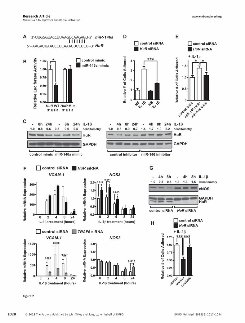
MiR‐146 targets the RNA‐binding protein HuR to controlendothelial activationHuR was previously found to promote endothelial activation inresponse to LPS treatment of endothelial cells by facilitating NF‐kB activation (Rhee et al, 2010). Interestingly, microRNA targetprediction programs (Targetscan and Pictar) suggested that HuRmight be a direct target of miR‐146 (Fig 7A, Fig. S4). Weconfirmed that luciferase constructs containing the HuR 30 UTRcould be repressed by miR‐146a (Fig 7B), and also found thatlevels of HuR mRNA (Supporting Information Fig S5) andprotein (Fig 7C) were suppressed or elevated when miR‐146was over‐expressed or knocked‐down in endothelial cells,respectively. To test the functional importance of HuR inIL‐1b‐mediated endothelial activation, we knocked down HuRand measured the adhesion of THP‐1 cells to endothelial cells.HuR knock‐down inhibited THP‐1 adhesion to IL‐1b treatedendothelial cells (Fig 7D). Additionally, HuR knock‐down alsoinhibited THP‐1 adhesion to TNF‐a treated cells (SupportingInformation Fig S6), suggesting that HuR broadly facilitatesendothelial activation. To assess the contribution of HuR to theenhanced adhesiveness of miR‐146 inhibitor‐treated endothelialcells, we knocked‐down HuR, and were able to block theincrease in THP‐1 adhesion (Fig 7E). Interestingly, VCAM‐1,ICAM‐1, SELE and MCP‐1 contain AU‐rich elements (AREs) intheir 30 UTRs (Supporting Information Fig S7). AREs can conferinstability to transcripts, which is antagonized by HuR binding tothese sites (Fan & Steitz, 1998). We therefore tested whetherHuR could regulate the expression of these inflammatory genes.While VCAM‐1 and MCP‐1 were highly enriched in HuRimmunoprecipitates from IL‐1b‐treated cells (Supporting Infor-mation Fig S8A), HuR knock‐down failed to affect the inductionof VCAM‐1 or MCP‐1 at the mRNA or protein level (Fig 7F,Supporting Information Figs S8B, C, and S9B), suggesting thatthey are not functional targets of HuR. Additionally, we foundthat NF‐kB activity was not altered by HuR knock‐down(Supporting Information Fig S8D), neither was the inductionof EGR‐3 (Supporting Information Fig S9B). This was in contrastto knock‐down of another miR‐146 target, TRAF6, whichdecreased NF‐kB activity (Supporting Information Fig S8D),the induction of adhesion molecules and EGR transcriptionfactors (Fig 7F, Supporting Information Fig S9B). In contrast tothe lack of regulation of VCAM‐1/MCP‐1 by HuR, eNOS mRNA
Figure 5. MiR‐146 inhibits the induction of NF‐kB, MAPK/EGR and AP‐1 path
A. The activity of a NF-kB promoter-luciferase reporter construct was assessed i
inhibitor or miR-146 inhibitor. MiR-146a over-expression reduced IL-1b-induce
activity. Data represents the mean � SEM of three independent experiments. AN
difference between the indicated groups, p < 0.01 and p < 0.001, respective
B. Activation of the MAP kinase pathway was assessed by measuring the levels o
loading control. MiR-146a over-expression inhibited the basal and IL-1b-indu
C. Induction of EGR-1 and EGR-3 in response to IL-1b was assessed by qRT-PCR,
D. MiR-146a over-expression inhibited the IL-1b-mediated induction of EGR-1 and
Significant p values (t-test) from left to right are 0.002, 0.004 and 0.022, res
E. Schematic of a potential miR-146 binding site in the 30 UTR of EGR-3 (top). Lucife
UTR sequences were performed in the presence of control or miR-146a mimi
F. Activation of the JNK/AP-1 pathway was assessed by measuring the induction
expression, while inhibition of miR-146 enhanced c-Jun expression in response
respectively.
EMBO Mol Med (2013) 5, 1017–1034 � 2
and protein levels were elevated in HuR knock‐down cells andeNOS was not down‐regulated in response to IL‐1b treatment ofthese cells (Fig 7F and G). Knock‐down of TRAF6 did not affectthe basal levels of NOS3 mRNA, but did inhibit the down‐regulation of NOS3 in response to IL‐1b (Fig 7F). Finally,inhibition of nitric oxide activity by treatment with L‐NAMEnegated the reduced adhesion in HuR knock‐down cells(Fig 7H), suggesting that HuR regulates endothelial activationby modulation of NO activity. These results suggest that miR‐146 targets TRAF6/IRAK1/2 and HuR, which cooperate tocontrol endothelial activation through distinct pathways. WhileTRAF6/IRAK1/2 affects NF‐kB transcriptional activity andthe induction of leukocyte adhesion molecules and chemo-attractants, HuR affects NO‐dependent leukocyte adhesion.
MiR‐146a knock‐out mice have an exaggerated acute vascularinflammatory responseAssessment of miR‐146a/b expression in blood vessels revealedthat this microRNA family is enriched in the endotheliumcompared to cells in the vascular wall (Fig 8A). To assess the roleof miR‐146a in controlling endothelial activation in vivo, weutilized miR‐146a�/� mice (Boldin et al, 2011). MiR‐146a�/�
mice on a C57/BL6 background are phenotypically normal atbirth, but acquire chronic inflammation, including myeloprolif-eration in the spleen and bone marrow and develop enlargedspleens beginning around 5–6 months of age (Zhao et al, 2011).We therefore utilized young mice (3–4 months of age) for ourexperiments, since they do not appear to have an overtinflammatory phenotype. MiR‐146a was expressed at muchhigher levels than miR‐146b in the heart, and loss of miR‐146adid not affect expression of miR‐146b, suggesting that miR‐146bis likely unable to compensate for loss of miR‐146a (Fig 8B).Additionally, we assessed the expression of several othermicroRNAs that are known to modulate inflammatory signal-ling, and found that these were not appreciably altered inmiR‐146a�/� mice (Supporting Information Fig S10). Similar toour findings using miR‐146 inhibitors in vitro, we found thatlevels of HuR mRNA and protein were increased in the heartsof miR‐146a�/� mice (Fig 8C), suggesting that HuR is also atarget of miR‐146a in vivo. Levels of TRAF6 protein were alsohighly elevated (Fig 8C). To determine the role of miR‐146ain the regulation of an acute vascular inflammatory response,
ways.
n endothelial cells transfected with control mimic, miR-146a mimic, control
d NF-kB-dependent promoter activity, while inhibition of miR-146 enhanced
OVA, p < 0.0001 for mimic and inhibitor data. �� and ��� indicate a significantly.
f phosphorylated ERK (pERK) (p42/p44). Total levels of ERK2 were used as a
ced levels of pERK, while miR-146 inhibitor had the opposite effect.
demonstrating rapid and transient induction (n ¼ 3).
EGR-3, while inhibition of miR-146 enhanced the induction of EGR-3 (n ¼ 3).
pectively.
rase assays utilizing wild-type or seed-mutated EGR-3 concatemer or TRAF6 30
c (p ¼ 0.042, t-test, n ¼ 3).
of c-Fos and c-Jun by qRT-PCR. MiR-146a over-expression reduced c-Fos
to IL-1b (n ¼ 4). Significant p values (t-test) from left to right are 0.005, 0.024,
013 The Authors. Published by John Wiley and Sons, Ltd on behalf of EMBO. 1025
A B
NS 1h IL-1β
EGR-31h IL-1β
D
F
1h IL-1β
cCTCCCACACCt WTEGRdeletion
cC---------------CCt
miR-146b promoter
DMSOU0126
-------TTCCTGGCCCTCCCACACCTTCCTCCTTTCTCAGAAGAGCCAGCATGGGGC human-------TGCCCGGCCCTCCCACACCTTCCTCCTTTCTCAGAAGAGCCAGGATGGGGC cow-------TGCCCAGCCCTCCCACACCTTCCTCCTTTCTCAGAAGAGCCAGGATGGGGC dog
GGCAGCATGCCCGGTCCTCCCACACCTTCCTCCTTTCTCAGAAGAGCCTGCATGG--- mouseGGCAGCATGCCCGGTCCTCCCACACCTTCCTCCTTTCTCAGAAGAGCCTGCATGG--- rat
EGR binding site************* *** ** ** * * ** * ** * * * * * * * * * * * *** *
miR-146b genomic locus:E
C
NS 1h IL-1β
EGR-3
control siRNAEGR-3 siRNA
NSIL-1β
NSIL-1β
miR-146b prom-luciferaseG Hwild-typeEGR deletion
Fold
Indu
ctio
n(E
GR
-3 O
E vs
con
trol
)
wild-ty
peEGR
deletio
n
miR-146b prom-luciferase
Rel
ativ
e m
RN
A Ex
pres
sion
Rel
ativ
e Fo
ld In
duct
ion
(U01
26 v
s D
MSO
)
Rel
ativ
e m
RN
A Ex
pres
sion
Rel
ativ
e Fo
ld In
duct
ion
(EG
R-3
siR
NA
vs c
ontr
ol)
Rel
ativ
e Lu
cife
rase
Act
ivity
***
*
**
***
**
**
** ***
Figure 6. The MAPK/EGR pathway regulates the transcription of miR-146a and miR-146b.
A. Treatment of endothelial cells with the MEK inhibitor, U0126, inhibited the basal expression (t-test, p ¼ 0.0003) and IL-1b-dependent induction (t-test,
p ¼ 0.037) of EGR-3 (n ¼ 3).
B. Induction of pri-miR-146a and pri-miR-146b by IL-1b was reduced in cells pre-treated with the MAP kinase inhibitor, U0126. Data represents the relative
induction of pri-miR-146a/b in cells treated with U0126 compared to cells treated with DMSO (vehicle) (n ¼ 4). p ¼ 0.037 for pri-miR-146a and p ¼ 0.010 for
pri-miR-146b (t-test).
C. EGR-3 knock-down by siRNA transfection reduced the basal (t-test, p < 0.0001) and IL-1b-induced levels (t-test, p ¼ 0.004) of EGR-3 (n ¼ 5).
D. The induction of pri-miR-146a and pri-miR-146b was also reduced in EGR-3 knock-down cells (n ¼ 5). p ¼ 0.023 for pri-miR-146a and p ¼ 0.013 for
pri-miR-146b (t-test).
E. Schematic indicating a potential EGR binding site (shaded area) in the miR-146b promoter. Sequence comparison between various species is indicated.
Asterisks indicate conserved nucleotides across all species.
F. Schematic of deletion of the EGR binding site in the miR-146b promoter.
G. AmiR-146b promoter-luciferase reporter was responsive to EGR-3 over-expression (OE) in bovine aortic endothelial cells (BAEC) and mutation of a conserved
EGR binding site abrogated this responsiveness. Data depicts the fold induction with EGR-3 OE compared to control. Shown is a representative experiment
(n ¼ 3 replicates). p ¼ 0.0017 (t-test).
H. A miR-146b promoter-luciferase reporter was modestly induced in response to IL-1b and this induction was not observed when the EGR site was mutated.
IL-1b was added at concentrations of 10, 20 or 40 ng/mL. Shown is a representative experiment (n ¼ 3 replicates). ANOVA, p ¼ 0.011. � and �� indicate a
significant difference between the indicated groups, p < 0.05, p < 0.01, respectively.
Research Article www.embomolmed.orgMicroRNA‐146 represses endothelial activation
1026 � 2013 The Authors. Published by John Wiley and Sons, Ltd on behalf of EMBO. EMBO Mol Med (2013) 5, 1017–1034
Research Articlewww.embomolmed.orgHenry S. Cheng et al.
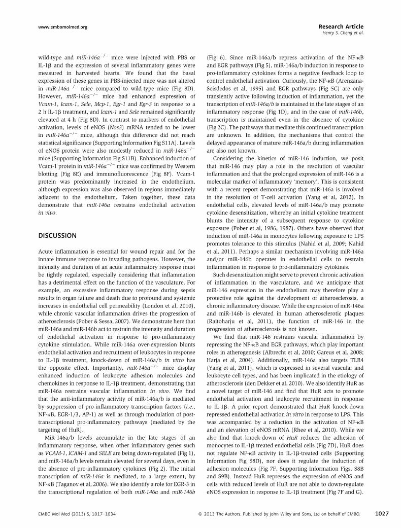
wild‐type and miR‐146a�/� mice were injected with PBS orIL‐1b and the expression of several inflammatory genes weremeasured in harvested hearts. We found that the basalexpression of these genes in PBS‐injected mice was not alteredin miR‐146a�/� mice compared to wild‐type mice (Fig 8D).However, miR‐146a�/� mice had enhanced expression ofVcam‐1, Icam‐1, Sele, Mcp‐1, Egr‐1 and Egr‐3 in response to a2 h IL‐1b treatment, and Icam‐1 and Sele remained significantlyelevated at 4 h (Fig 8D). In contrast to markers of endothelialactivation, levels of eNOS (Nos3) mRNA tended to be lowerin miR‐146a�/� mice, although this difference did not reachstatistical significance (Supporting Information Fig S11A). Levelsof eNOS protein were also modestly reduced in miR‐146a�/�
mice (Supporting Information Fig S11B). Enhanced induction ofVcam‐1 protein inmiR‐146a�/� mice was confirmed byWesternblotting (Fig 8E) and immunofluorescence (Fig 8F). Vcam‐1protein was predominantly increased in the endothelium,although expression was also observed in regions immediatelyadjacent to the endothelium. Taken together, these datademonstrate that miR‐146a restrains endothelial activationin vivo.
DISCUSSION
Acute inflammation is essential for wound repair and for theinnate immune response to invading pathogens. However, theintensity and duration of an acute inflammatory response mustbe tightly regulated, especially considering that inflammationhas a detrimental effect on the function of the vasculature. Forexample, an excessive inflammatory response during sepsisresults in organ failure and death due to profound and systemicincreases in endothelial cell permeability (London et al, 2010),while chronic vascular inflammation drives the progression ofatherosclerosis (Pober & Sessa, 2007). We demonstrate here thatmiR‐146a andmiR‐146b act to restrain the intensity and durationof endothelial activation in response to pro‐inflammatorycytokine stimulation. While miR‐146a over‐expression bluntsendothelial activation and recruitment of leukocytes in responseto IL‐1b treatment, knock‐down of miR‐146a/b in vitro hasthe opposite effect. Importantly, miR‐146a�/� mice displayenhanced induction of leukocyte adhesion molecules andchemokines in response to IL‐1b treatment, demonstrating thatmiR‐146a restrains vascular inflammation in vivo. We findthat the anti‐inflammatory activity of miR‐146a/b is mediatedby suppression of pro‐inflammatory transcription factors (i.e.,NF‐kB, EGR‐1/3, AP‐1) as well as through modulation of post‐transcriptional pro‐inflammatory pathways (mediated by thetargeting of HuR).
MiR‐146a/b levels accumulate in the late stages of aninflammatory response, when other inflammatory genes suchas VCAM‐1, ICAM‐1 and SELE are being down‐regulated (Fig 1),and miR‐146a/b levels remain elevated for several days, even inthe absence of pro‐inflammatory cytokines (Fig 2). The initialtranscription of miR‐146a is mediated, to a large extent, byNF‐kB (Taganov et al, 2006). We also identify a role for EGR‐3 inthe transcriptional regulation of both miR‐146a and miR‐146b
EMBO Mol Med (2013) 5, 1017–1034 � 2
(Fig 6). Since miR‐146a/b repress activation of the NF‐kBand EGR pathways (Fig 5), miR‐146a/b induction in response topro‐inflammatory cytokines forms a negative feedback loop tocontrol endothelial activation. Curiously, the NF‐kB (Arenzana‐Seisdedos et al, 1995) and EGR pathways (Fig 5C) are onlytransiently active following induction of inflammation, yet thetranscription ofmiR‐146a/b is maintained in the late stages of aninflammatory response (Fig 1D), and in the case of miR‐146b,transcription is maintained even in the absence of cytokine(Fig 2C). The pathways that mediate this continued transcriptionare unknown. In addition, the mechanisms that control thedelayed appearance of mature miR‐146a/b during inflammationare also not known.
Considering the kinetics of miR‐146 induction, we positthat miR‐146 may play a role in the resolution of vascularinflammation and that the prolonged expression of miR‐146 is amolecular marker of inflammatory ‘memory’. This is consistentwith a recent report demonstrating that miR‐146a is involvedin the resolution of T‐cell activation (Yang et al, 2012). Inendothelial cells, elevated levels of miR‐146a/b may promotecytokine desensitization, whereby an initial cytokine treatmentblunts the intensity of a subsequent response to cytokineexposure (Pober et al, 1986, 1987). Others have observed thatinduction of miR‐146a in monocytes following exposure to LPSpromotes tolerance to this stimulus (Nahid et al, 2009; Nahidet al, 2011). Perhaps a similar mechanism involving miR‐146aand/or miR‐146b operates in endothelial cells to restraininflammation in response to pro‐inflammatory cytokines.
Such desensitizationmight serve to prevent chronic activationof inflammation in the vasculature, and we anticipate thatmiR‐146 expression in the endothelium may therefore play aprotective role against the development of atherosclerosis, achronic inflammatory disease. While the expression of miR‐146aand miR‐146b is elevated in human atherosclerotic plaques(Raitoharju et al, 2011), the function of miR‐146 in theprogression of atherosclerosis is not known.
We find that miR‐146 restrains vascular inflammation byrepressing the NF‐kB and EGR pathways, which play importantroles in atherogenesis (Albrecht et al, 2010; Gareus et al, 2008;Harja et al, 2004). Additionally, miR‐146a also targets TLR4(Yang et al, 2011), which is expressed in several vascular andleukocyte cell types, and has been implicated in the etiology ofatherosclerosis (den Dekker et al, 2010). We also identify HuR asa novel target of miR‐146 and find that HuR acts to promoteendothelial activation and leukocyte recruitment in responseto IL‐1b. A prior report demonstrated that HuR knock‐downrepressed endothelial activation in vitro in response to LPS. Thiswas accompanied by a reduction in the activation of NF‐kBand an elevation of eNOS mRNA (Rhee et al, 2010). While wealso find that knock‐down of HuR reduces the adhesion ofmonocytes to IL‐1b treated endothelial cells (Fig 7D), HuR doesnot regulate NF‐kB activity in IL‐1b‐treated cells (SupportingInformation Fig S8D), nor does it regulate the induction ofadhesion molecules (Fig 7F, Supporting Information Figs. S8Band S9B). Instead HuR represses the expression of eNOS andcells with reduced levels of HuR are not able to down‐regulateeNOS expression in response to IL‐1b treatment (Fig 7F and G).
013 The Authors. Published by John Wiley and Sons, Ltd on behalf of EMBO. 1027
Rel
ativ
e Lu
cife
rase
Act
ivity
HuR WT3’ UTR
HuR Mut3’ UTR
G
D
control mimicmiR-146a mimic
A
5’--AAGAUUAACCCUCAAAGUUCUCU--3’
3’-UUGGGUACCUUAAGUCAAGAGU-5’ miR-146a
HuR
C
Rel
ativ
e #
of C
ells
Adh
ered
contro
l inhib
miR-14
6 inhib
miR-14
6 inhib
control siRNAHuR siRNA
NSIL-1β
control siRNAHuR siRNA
NSIL-1β
E
F
+ IL-1β
Rel
ativ
e #
of C
ells
Adh
eredB
HuR
IL-1β- 8h 24h - 8h 24h0.51.0 0.5 densitometry0.8 0.6 0.6
GAPDH
control mimic miR-146a mimic
HuR
IL-1β- 4h 8h2.21.0 0.7 densitometry0.6 0.9 1.7
GAPDH
control inhibitor miR-146 inhibitor
1.4 1.924h - 4h 8h 24h
eNOS
GAPDHHuR
control siRNA HuR siRNA
IL-1β- 4h 8h - 4h 8h1.51.0 1.3 densitometry0.8 0.5 1.3
VCAM-1
Rel
ativ
e m
RN
A Ex
pres
sion
0 2 4 8 24
control siRNA HuR siRNA
VCAM-1
Rel
ativ
e m
RN
A Ex
pres
sion
IL-1β treatment (hours)0 2 4 8 24
control siRNA TRAF6 siRNA
NOS3
Rel
ativ
e m
RN
A Ex
pres
sion
IL-1β treatment (hours)0 2 4 8 24
NOS3
Rel
ativ
e m
RN
A Ex
pres
sion
IL-1β treatment (hours)0 2 4 8 24
HIL-1β treatment (hours) control siRNAHuR siRNA
Rel
ativ
e #
of C
ells
Adh
ered
contro
l
contro
l
L-NAME
+ IL-1β
0
1
2
3
4
0
0.5
1.0
1.5* *** * *
0.0170.041
0.005
0.020
0.028
0.027
0.013
*** ***
Figure 7.
Research Article www.embomolmed.orgMicroRNA‐146 represses endothelial activation
1028 � 2013 The Authors. Published by John Wiley and Sons, Ltd on behalf of EMBO. EMBO Mol Med (2013) 5, 1017–1034
3
Research Articlewww.embomolmed.orgHenry S. Cheng et al.
Importantly, eNOS down‐regulation plays a key role inatherogenesis (Knowles et al, 2000; Oemar et al, 1998). Inaddition we show that inhibition of NO activity can rescue thereduced leukocyte adhesion observed in HuR knock‐down cells(Fig 7H). While HuR does not directly bind to NOS3 mRNA,it does bind to a known positive regulator of NOS3 transcription(Lin et al, 2005), KLF2 (Supporting Information Fig S12A),and knock‐down of HuR results in elevated levels of KLF2(Supporting Information Fig S12B). Finally, we find that HuRprotein levels are reduced at the late stages of endothelialactivation (Fig 7C), suggesting that miR‐146 up‐regulation atthis stage may repress HuR, thereby forming a negative feedbackloop. MiR‐146 therefore inhibits endothelial activation bycoordinately repressing the induction of adhesion molecules(through targeting of TRAF6/IRAK1/2) and by promoting theexpression of eNOS, an inhibitor of leukocyte adhesion (throughtargeting of HuR) (Supporting Information Fig S13).
From recent discoveries it appears that a microRNA networkacts in endothelial cells to restrain inflammation (Fish &Cybulsky, 2012). For example, miR‐10a levels are decreasedin regions of the mouse aorta that are susceptible to thedevelopment of atherosclerosis (Fang et al, 2010). MiR‐10arepresses NF‐kB activity by targetingMAP kinase kinase kinase 7(MAP3K7, also known as TAK1) and b‐transducin repeat‐containing gene (b‐TRC), which mediate IkB degradation(Fang et al, 2010). Additionally, TNF‐a up‐regulates miR‐31and miR‐17‐5p, which directly repress the adhesion moleculegenes SELE and ICAM1, respectively (Suarez et al, 2010). Morerecently, miR‐181b was found to repress the expression ofimportin‐a3, which is required for the nuclear import of NF‐kBproteins (Sun et al, 2012). Over‐expression of miR‐181b in thevasculature inhibits the expression of NF‐kB‐dependent genesand protects mice from sepsis (Sun et al, 2012). The existenceof several microRNAs that converge on the NF‐kB pathwaysuggests that tight control of this pathway is crucial for themaintenance of vascular homeostasis. Our findings have addedmiR‐146a and miR‐146b to this microRNA‐mediated NF‐kBregulatory network in the endothelium (Supporting InformationFig S13). In addition to regulating the NF‐kB pathway, miR‐146
Figure 7. HuR, a novel miR‐146 target, controls endothelial activation by reg
A. Schematic of a potential miR-146 binding site in the 30 UTR of HuR.
B. Luciferase assays utilizing wild-type (WT) or seed-mutated (Mut) HuR 30 UTR(mean � SEM, p ¼ 0.008, t-test, n ¼ 4).
C. HuR protein levels were quantified by Western blot in cells transfected with
D. The adhesion of THP-1 cells to vehicle or IL-1b treated cells transfected with
representative experiment is shown (three replicate wells, three images per wel
IL-1b-treated HuR knock-down cells, p < 0.001.
E. THP-1 adhesion assays were performed with endothelial cells transfected wit
reduced the elevated adhesion of THP-1 to endothelial cells transfected with m
three images per well). ANOVA ¼ 0.016. �Indicates a significant difference be
F. Knock-down of HuR (above) or TRAF6 (below) was performed and the induction
qRT-PCR. Expression of other inflammatory genes is indicated in Supporting Inf
contrast to TRAF6 knock-down, which strongly inhibited VCAM-1 induction. H
mean � SEM of three independent experiments. Significant p values (t-test)
G. Levels of eNOS protein were elevated in HuR knock-down cells, and eNOS wa
H. The nitric oxide inhibitor, L-NAME, negated the reduced THP-1 adhesion obse
replicate wells, three images per well). ANOVA, p < 0.0001. ���Indicates a sig
EMBO Mol Med (2013) 5, 1017–1034 � 2
also controls activation of the EGR and AP‐1 pathways, whichare known to drive inflammatory gene expression (De Caterinaet al, 2010; Hajra et al, 2000), and miR‐146 directly targets HuR,which promotes endothelial activation by antagonizing eNOSexpression. This implies that miR‐146 may have an even broaderanti‐inflammatory role than miR‐10a, miR‐31, miR‐17‐5p ormiR‐181b. Our findings suggest that strategies to enhancemiR‐146a or miR‐146b in the vasculature may be therapeuticallyuseful to dampen the endothelial response to inflammatorycytokines, and may potentially be used to shut off the reiterativeinflammatory loop that drives atherogenesis or to quell thevascular damage associated with cytokine storm in the settingof sepsis.
MATERIALS AND METHODS
Reagents usedRecombinant human IL‐1b and TNF‐a were from Invitrogen, and were
used at a concentration of 10 ng/mL. Mouse recombinant IL‐1b was
from R&D Systems. The MAP kinase inhibitor, UO126, was from Sigma–
Aldrich and was dissolved in DMSO and used at a concentration of
10 mM. L‐NAME was purchased from Sigma–Aldrich and was used at a
concentration of 0.1 mM.
Cell culture and treatmentsHuman umbilical vein endothelial cells (HUVEC) and media (Endo-
thelial Cell Medium with 5% FBS and Endothelial Cell Growth
Supplement) were purchased from ScienCell. Bovine aortic endothelial
cells (BAEC) and media were purchased from Lonza. Cells were used at
passages 3–8. HeLa‐S3 and THP‐1 cells were purchased from ATCC.
HeLa cells were maintained in DMEM with 10% FBS and THP‐1 cells
were maintained in RPMI1640 with L‐glutamine and 0.05 nM
b‐mercaptoethanol and 10% FBS.
Monocyte adhesion assayTHP‐1 cells were labelled with CellTracker™ Green (Invitrogen)
immediately prior to the experiment. HUVEC were cultured to
confluence in 12‐well plates and were treated with IL‐1b for 4 h.
ulating eNOS expression.
sequences were performed in the presence of control or miR-146a mimic
control or miR-146a mimic (left) or control or miR-146 inhibitor (right).
control or HuR siRNAs revealed that HuR promotes endothelial activation. A
l). ANOVA, p < 0.0001. ���Indicates a significant decrease in THP-1 adhesion in
h control or miR-146 inhibitor and control or HuR siRNA. HuR knock-down
iR-146 inhibitor. A representative experiment is shown (three replicate wells,
tween indicated groups, p < 0.05.
of adhesion molecules (typified by VCAM-1) and eNOS (NOS3) was assessed by
ormation Fig S9B. HuR knock-down did not reduce the induction of VCAM-1, in
owever, HuR knock-down significantly elevated levels of NOS3. Shown is the
are indicated above.
s not down-regulated in HuR knock-down cells treated with IL-1b.
rved in HuR knock-down cells. A representative experiment is shown (three
nificant difference between groups, p < 0.001.
013 The Authors. Published by John Wiley and Sons, Ltd on behalf of EMBO. 1029

D
C
miR-146a-/-wild-typeVcam-1 Icam-1Sele
Mcp-1
NS 2h IL-1β
HuR
WT KO
Rel
ativ
e m
RN
A Ex
pres
sion
4h IL-1β Rel
ativ
e m
RN
A Ex
pres
sion
NS 2h IL-1β 4h IL-1β Rel
ativ
e m
RN
A Ex
pres
sion
NS 2h IL-1β 4h IL-1β
Rel
ativ
e m
RN
A Ex
pres
sion
NS 2h IL-1β 4h IL-1β
Egr-1
Rel
ativ
e m
RN
A Ex
pres
sion
Rel
ativ
e m
RN
A Ex
pres
sion
Egr-3
NS 2h IL-1β 4h IL-1β NS 2h IL-1β 4h IL-1β
Rel
ativ
e m
RN
A Ex
pres
sion2.5x106
2.0x10⁶
1.5x10⁶
1.0x10⁶
0.5x10⁶Cop
ies/
ng o
f RN
A
miR-146a miR-146b
BA
E F
miR-12
6
miR-14
6a
miR-14
6b
vessel wallendothelium
Rel
ativ
e m
iRN
A Ex
pres
sion
wild-typemiR-146a-/-
wild-ty
pe
miR-14
6a-/-
HuR
Actin
wild-ty
pe
wild-ty
pe
miR-14
6a-/-
miR-14
6a-/-
Vcam-1
Actin
IL-1β (2 h)
+ IL-1β (4 h)
wild-type
Lumen
Lumen
Traf6
1.0 2.7
1.0 2.5
1.0 0.7 1.3 2.6
PBS
Vcam-1 Pecam-1
miR-146a-/-
0.025
0.0090.031
0.0380.050
0.012
0.014
0.049
0.0470.029 0.039
Figure 8.
Research Article www.embomolmed.orgMicroRNA‐146 represses endothelial activation
1030 � 2013 The Authors. Published by John Wiley and Sons, Ltd on behalf of EMBO. EMBO Mol Med (2013) 5, 1017–1034
3
Research Articlewww.embomolmed.orgHenry S. Cheng et al.
Labeled THP‐1 cells (105) were then added to each well for 90 min and
unbound cells were removed by washing with PBS. For experiments
using L‐NAME, cells were treated with IL‐1b and 0.1 mM L‐NAME for
4 h, and THP‐1 cells were allowed to adhere for 15 min. Adherent cells
were fixed with 4% paraformaldehyde and imaged using a Leica
Microsystems inverted fluorescent microscope (Model #DMIL) with an
Olympus DP71 camera. Adherent THP‐1 cells were quantified in three
random fields of view per well using ImageJ. Triplicate wells were
analysed for each experiment.
TransfectionHUVEC were transfected at �50% confluency with control or miR‐
146a mimics (20 nM, Dharmacon), or non‐targeting control, EGR-3,
HuR or TRAF6 siRNAs (Silencer Select s4544, 4390843, s4610
or s14389, respectively, 40 nM, Invitrogen) and analysed after 24–
72 h. For inhibitor experiments, HUVEC were transfected at �90%
confluency with control or miR‐146a locked‐nucleic acid (LNA)
inhibitors (20 nM, Power Inhibitors, Exiqon) and analysed 48–72 h
later. All HUVEC transfections were performed using RNAiMax
(Invitrogen). HeLa cells were transfected with plasmids and microRNA
mimics using Lipofectamine 2000 (Invitrogen; see Supporting Informa-
tion for details).
Bioinformatic analysis of miR-146a and miR-146b proximalpromoter regionsThe genomic regions surrounding the miR-146a and miR-146b
transcriptional start sites were assessed for the presence of
Evolutionary Conserved Regions (ECRs) using ECR Browser (http://
ecrbrowser.dcode.org/), and rVista (http://rvista.dcode.org/) was used
to identify conserved transcription factor binding sites.
Luciferase assays and cloningSee Supporting Information for details.
Gene expression analysisRNAwas isolated using Trizol (Invitrogen), reverse transcribed using the
High‐Capacity cDNA Reverse Transcription kit (Applied Biosystems), and
quantitative reverse‐transcriptase PCR (qRT‐PCR) was performed as
described previously (Fish et al, 2010). For analysis of pri-miR-146a and
pri-miR-146b, RNAwas treated with DNase I (Ambion) to remove traces
of genomic DNA. Real‐time PCR was conducted in triplicate using a
Roche Lightcycler 480® with Roche 480 Probes Master Mix or LC 480
Figure 8. miR-146a�/� mice demonstrate enhanced endothelial activation fo
A. Endothelial cells and cells in the vessel wall were isolated from the descending a
cells) andmiR-146a/b weremeasured by qRT-PCR. Expression was normalized t
vessel wall (n ¼ 4). Significant p values (t-test) are indicated above.
B. Levels of miR-146a and miR-146b were quantified by qRT-PCR in hearts from
miR-146a was >6-fold higher than miR-146b and miR-146b expression was
C. Expression of HuR mRNA was elevated in the hearts of miR-146a�/� mice as
elevated levels of HuR and Traf6 (right).
D. Wild-type andmiR-146a�/�mice (3–4 months of age, n ¼ 4) were injected with
or 4 h. Expression of inflammatory genes was assessed by qRT-PCR. While basa
the induction of Vcam-1, Icam-1, Sele, Mcp-1, Egr-1 and Egr-3 was enhanced
Significant p values (t-test) are indicated above.
E. Expression of Vcam-1 protein was elevated after a 2 h IL-1b treatment in mi
F. Localization of Vcam-1 expression was assessed by immunofluorescence, revea
adjacent to the endothelium of miR-146a�/� mice treated with IL-1b for 4 h
EMBO Mol Med (2013) 5, 1017–1034 � 2
SYBR Green I Master (Roche) for Taqman® and Sybr green chemistries,
respectively. Data was normalized to Tata box binding protein (TBP)
or glyceraldehyde 3‐phosphate dehydrogenase (GAPDH) using the
Delta‐Delta Ct method. The primers used are indicated in Supporting
Information Table SI.
MiR‐146a and U6 were reverse‐transcribed using the Taqman®
MicroRNA Reverse Transcription kit (Applied Biosystems) and
analysed using Taqman Primer sets (Applied Biosystems). The miR‐
146a primer set did not cross react with miR‐146b (<1% cross
reactivity). Since the miR‐146b primer set from Applied Biosystems
cross‐reacted with miR‐146a, we used the miScript system (Qiagen)
for analysis of miR‐146b. MiScript primers for miR‐146b only
partially cross‐reacted with miR‐146a (<20% cross reactivity).
To quantify the number of copies of miR‐146a and miR‐146b,
comparison was made to a standard curve generated by reverse
transcribing a known amount of miR‐146a or miR‐146b mimic
(Dharmacon). The MiScript system was also used for the analysis of
other microRNAs (miR‐10a, miR‐17, miR‐31, miR‐155 and miR‐181b)
in wild‐type and miR-146a�/� hearts. Expression was normalized to
miR‐126 in these experiments.
HuR immunoprecipitationHUVECwere harvested and lysed in RIPA buffer (Santa Cruz) containing
protease inhibitors and 100 U/mL RNAse OUT (Invitrogen). Protein–
RNA complexes were isolated from 1.75 mg of total clarified protein
with 5 mg of either HuR antibody (Santa Cruz, G‐8) or V5 antibody
(Invitrogen) using 60 mL protein A/G beads (Santa Cruz) by rotation at
4°C for 4 h. Beads were washed 3� in RIPA buffer and resuspended in
1 mL Trizol (Invitrogen), followed by RNA isolation.
Western blottingWestern blotting was performed as described (Fish et al, 2008). For
analysis of pERK, HUVEC were serum starved overnight (in basal
medium containing 0.1% FBS) prior to stimulation with IL‐1b (20 ng/
mL). The following antibodies were used: phospho‐ERK (p42/44Thr202/
Tyr204, Cell Signaling, 9101), ERK2 (Santa Cruz, C‐14), E‐Selectin (Santa
Cruz, H‐300), ICAM‐1 (Santa Cruz, G‐5), TRAF6 (Santa Cruz, D‐10),
eNOS [Santa Cruz, C‐20, generously provided by P. Marsden (University
of Toronto)], VCAM‐1 (for human samples; Santa Cruz, E‐10), Vcam‐1
(for mouse samples; R&D Systems, AF643), HuR (for human samples;
Santa Cruz, G‐8), HuR (for mouse samples; Santa Cruz, 3A2), GAPDH
(Santa Cruz, 0411), Actin (Sigma, A2066) and Vinculin (Santa Cruz,
llowing IL‐1b treatment.
orta of wild-typemice, and expression of miR-126 (as a control for endothelial
o U6. MiR-146awas significantly enriched in the endothelium compared to the
wild-type and miR-146a�/� mice (3–4 months of age, n ¼ 3). Expression of
not affected by loss of miR-146a.
assessed by qRT-PCR (left, p ¼ 0.031, t-test, n ¼ 3). Western blot revealed
PBS or 125 ng of IL-1b by tail vein injection and hearts were harvested after 2
l levels of these genes were unchanged in unstimulated mice (PBS injection),
at 2 h in IL-1b treated mice, and Sele and Icam-1 were still elevated at 4 h.
R-146a�/� mice compared to wild-type mice.
ling an enhancement of Vcam-1 expression in the endothelium and in puncta
. Scale bars ¼ 20 mm.
013 The Authors. Published by John Wiley and Sons, Ltd on behalf of EMBO. 1031
The paper explained
PROBLEM:
Inflammation plays a vital role in acute and chronic diseases of
the vasculature, including sepsis and atherosclerosis, respec-
tively. Therapies that directly repress vascular inflammation are
expected to impede the development of these diseases. However,
current therapies are not able to specifically suppress inflam-
matory signalling in the vasculature.
RESULTS:
We find that the miR-146 microRNA family is induced by pro-
inflammatory cytokines and acts to inhibit vascular inflamma-
tion by repressing the expression of leukocyte adhesion
molecules on the surface of endothelial cells, a process known as
endothelial activation. MiR-146 accomplishes this by repressing
both transcriptional and post-transcriptional pro-inflammatory
pathways in endothelial cells.
IMPACT:
Our findings suggest that therapies that augment miR-146
expression in the vascular endothelium may be protective
against the development of inflammatory vascular diseases. As
approaches for enhancing microRNA expression in vivo continue
to improve, it may be feasible to target vascular inflammation by
modulating the expression of miR-146 in the endothelium.
Additionally, it is possible that alterations in the levels of miR-
146 may predispose individuals to the development of vascular
inflammatory diseases.
Research Article www.embomolmed.orgMicroRNA‐146 represses endothelial activation
1032
H‐300). HRP‐conjugated secondary antibodies were from Cell
Signaling or Santa Cruz, and blots were developed using SuperSignal
West Pico Chemiluminescence Substrate (Pierce).
Enzyme‐linked immunosorbent assay (ELISA)MCP‐1 protein was quantified in supernatants using a Quantikine
ELISA kit from R&D Systems, according to the manufacturer’s
recommendations.
Mouse experimentsAll animal protocols were approved by the Animal Care Committee at
the University Health Network (Toronto). Adult (3–4 months) wild‐type
and miR-146a�/� mice (on a C57/BL6 background) were injected with
100 mL of PBS or 125 ng of recombinant mouse IL‐1b (in PBS) by
intravascular injection. Hearts (including a portion of the ascending
aorta) were harvested at 2 h or 4 h post‐injection and processed for
RNA or protein analysis. For analysis of microRNA expression in the
endothelium, endothelial cells were isolated from the vessel wall using
a modified protocol (Jongstra‐Bilen et al, 2006). Briefly, descending
thoracic aortae were dissected, adipose tissue was removed, and aortae
were pinned en face in ice‐cold PBS containing 1 mM aurintricarbox-
ylic acid (Sigma).Tissues were treated with 5U DNase I (Fermentas) and
Liberase TM (1:100 in Ca2þ/Mg2þ‐containing PBS, Roche) for 8 min at
37°C. Intimal cells were visualized by overlaying 0.1 mM fluoresbrite
polystyrene microspheres (Polysciences). Intimal cells were scraped
gently with a 30G needle and harvested directly into RNA extraction
buffer (RNAqueous‐micro kit, Invitrogen). Endothelium‐depleted vessel
wall tissue was homogenized in RNA extraction buffer.
ImmunostainingCryosections were stained as described (Delgado‐Olguin et al, 2012).
Primary antibodies were: FITC‐Pecam‐1 (1:200) (BD Biosciences) and
Vcam‐1 (1:100) (Proteintech). Vcam‐1was detected by incubation with
Alexa Fluor 647 Goat Anti‐Rabbit (Invitrogen). Sections were imaged
using an Eclipse Ni‐U Nikon microscope and processed using NIS‐
Elements Imaging Software.
� 2013 The Authors. Published by John Wiley and Sons, Ltd on behalf of EMBO.
Statistical analysisUnless otherwise indicated, data represent the mean of at least three
independent experiments and error bars represent the standard error
of themean. Pair‐wise comparisons weremade using a Student’s t‐test.
Comparison of three or more groups was performed using a 1‐way
analysis of variance (ANOVA) with Newman–Keuls post hoc test. A
p‐value of 0.05 or less was considered to be statistically significant.
In all figures �, �� and ��� represent a p‐value of �0.05, �0.01 and
�0.001, respectively.
Author contributionsHSC and NS designed and performed experiments, and analyseddata. EB, AL, PDO and JLZ performed experiments. DBsupervised JLZ and provided reagents. MIC supervised ALand designed experiments. JEF designed and performedexperiments, analysed data and wrote the manuscript. Allauthors approved the final manuscript.
AcknowledgementsWe thank D. Srivastava (Gladstone Institute of CardiovascularDisease) for providing reagents and mentorship, and E.Flemington (Tulane University Health Sciences Center), andJ.D. Powell (John Hopkins) for providing reagents. We thankJ. Wythe (Gladstone Institute of Cardiovascular Disease) forcritical comments on the manuscript. Research in the laboratoryof JEF was supported by a Heart and Stroke Foundationof Ontario Grant‐in‐aid (NA 7282) and the Canadian Institutes ofHealth Research (OCN‐126570). Research in the laboratory ofMC is supported by grants from the Heart and Stroke Foundationof Ontario and the Canadian Institutes of Health Research. PDOis supported by Operational Funding from the Hospital for SickChildren Research Institute. JEF is the recipient of a NewInvestigator Award from the Heart and Stroke Foundation ofCanada and an Early Researcher Award from the Ontario
EMBO Mol Med (2013) 5, 1017–1034
Research Articlewww.embomolmed.orgHenry S. Cheng et al.
Ministry of Economic Development and Innovation, and hasreceived funding from the Leaders Opportunity Fund from theCanadian Foundation for Innovation.
Supporting Information is available at EMBO MolecularMedicine online.
The authors declare that they have no conflict of interest.
ReferencesAird WC (2003) The role of the endothelium in severe sepsis and multiple
organ dysfunction syndrome. Blood 101: 3765-3777.
Albrecht C, Preusch MR, Hofmann G, Morris-Rosenfeld S, Blessing E, Rosenfeld
ME, Katus HA, Bea F, (2010) Egr-1 deficiency in bone marrow-derived cells
reduces atherosclerotic lesion formation in a hyperlipidaemic mouse
model. Cardiovasc Res 86: 321-329.
Anderson HD, Rahmutula D, Gardner DG (2004) Tumor necrosis factor-alpha
inhibits endothelial nitric-oxide synthase gene promoter activity in bovine
aortic endothelial cells. J Biol Chem 279: 963-969.
Arenzana-Seisdedos F, Thompson J, Rodriguez MS, Bachelerie F, Thomas D,
Hay RT (1995) Inducible nuclear expression of newly synthesized I kappa B
alpha negatively regulates DNA-binding and transcriptional activities of
NF-kappa B. Mol Cell Biol 15: 2689-2696.
Bartel DP (2009) MicroRNAs: target recognition and regulatory functions. Cell
136: 215-233.
Bhaumik D, Scott GK, Schokrpur S, Patil CK, Campisi J, Benz CC (2008)
Expression of microRNA-146 suppresses NF-kappaB activity with reduction
of metastatic potential in breast cancer cells. Oncogene 27: 5643-5647.
Boldin MP, Taganov KD, Rao DS, Yang L, Zhao JL, Kalwani M, Garcia-Flores Y,
Luong M, Devrekanli A, Xu J, et al (2011) miR-146a is a significant brake on
autoimmunity, myeloproliferation, and cancer in mice. J Exp Med 208:
1189-1201.
De Caterina R, Massaro M, Scoditti E, Annunziata Carluccio M (2010)
Pharmacological modulation of vascular inflammation in atherothrom-
bosis. Ann N Y Acad Sci 1207: 23-31.
De Caterina R, Massaro M, Scoditti E, Annunziata Carluccio M (2010)
Pharmacological modulation of vascular inflammation in atherothrom-
bosis. Ann N Y Acad Sci 1207: 23-31.
Delgado-Olguin P, Huang Y, Li X, Christodoulou D, Seidman CE, Seidman JG,
Tarakhovsky A, Bruneau BG, (2012) Epigenetic repression of cardiac
progenitor gene expression by Ezh2 is required for postnatal cardiac
homeostasis. Nat Genet 44: 343-347.
den Dekker WK, Cheng C, Pasterkamp G, Duckers HJ (2010) Toll like receptor 4
in atherosclerosis and plaque destabilization. Atherosclerosis 209: 314-
320.
Fan XC, Steitz JA (1998) Overexpression of HuR, a nuclear-cytoplasmic
shuttling protein, increases the in vivo stability of ARE-containing mRNAs.
EMBO J 17: 3448-3460.
Fang Y, Shi C, Manduchi E, Civelek M, Davies PF (2010) MicroRNA-10a
regulation of proinflammatory phenotype in athero-susceptible endothe-
lium in vivo and in vitro. Proc Natl Acad Sci USA 107: 13450-13455.
Fish JE, Cybulsky MI (2012) Taming endothelial activation with a microRNA. J
Clin Invest 122: 1967-1970.
Fish JE, Santoro MM, Morton SU, Yu S, Yeh RF, Wythe JD, Ivey KN, Bruneau BG,
Stainier DY, Srivastava D (2008) miR-126 regulates angiogenic signaling
and vascular integrity. Dev Cell 15: 272-284.
Fish JE, Yan MS, Matouk CC, St Bernard R, Ho JJ, Gavryushova A, Srivastava D,
Marsden PA (2010) Hypoxic repression of endothelial nitric-oxide synthase
transcription is coupled with eviction of promoter histones. J Biol Chem
285: 810-826.
Gareus R, Kotsaki E, Xanthoulea S, van der Made I, Gijbels MJ, Kardakaris R,
Polykratis A, Kollias G, deWintherMP, Pasparakis M (2008) Endothelial cell-
EMBO Mol Med (2013) 5, 1017–1034 � 2
specific NF-kappaB inhibition protects mice from atherosclerosis. Cell
Metab 8: 372-383.
Gimbrone MA, Jr, Garcia-Cardena G (2012) Vascular endothelium,
hemodynamics, and the pathobiology of atherosclerosis. Cardiovasc Pathol
22: 9-15.
Hajra L, Evans AI, Chen M, Hyduk SJ, Collins T, Cybulsky MI (2000) The NF-
kappa B signal transduction pathway in aortic endothelial cells is primed
for activation in regions predisposed to atherosclerotic lesion formation.
Proc Natl Acad Sci USA 97: 9052-9057.
Harja E, Bucciarelli LG, Lu Y, Stern DM, Zou YS, Schmidt AM, Yan SF (2004)
Early growth response-1 promotes atherogenesis: mice deficient in early
growth response-1 and apolipoprotein E display decreased atherosclerosis
and vascular inflammation. Circ Res 94: 333-339.
Hou J, Wang P, Lin L, Liu X, Ma F, An H, Wang Z, Cao X (2009) MicroRNA-146a
feedback inhibits RIG-I-dependent type I IFN production inmacrophages by
targeting TRAF6, IRAK1, and IRAK2. J Immunol 183: 2150-2158.
Jongstra-Bilen J, Haidari M, Zhu SN, Chen M, Guha D, Cybulsky MI (2006) Low-
grade chronic inflammation in regions of the normal mouse arterial intima
predisposed to atherosclerosis. J Exp Med 203: 2073-2083.
Knowles JW, Reddick RL, Jennette JC, Shesely EG, Smithies O, Maeda N (2000)
Enhanced atherosclerosis and kidney dysfunction in eNOS(�/�)Apoe(�/�)
mice are ameliorated by enalapril treatment. J Clin Invest 105: 451-458.
Kubes P, Suzuki M, Granger DN (1991) Nitric oxide: an endogenous modulator
of leukocyte adhesion. Proc Natl Acad Sci USA 88: 4651-4655.
Kumbrink J, Gerlinger M, Johnson JP (2005) Egr-1 induces the expression of its
corepressor nab2 by activation of the nab2 promoter thereby establishing a
negative feedback loop. J Biol Chem 280: 42785-42793.
Lin Z, Kumar A, SenBanerjee S, Staniszewski K, Parmar K, Vaughan DE,
Gimbrone MA, Jr, Balasubramanian V, Garcia-Cardena G, Jain MK (2005)
Kruppel-like factor 2 (KLF2) regulates endothelial thrombotic function. Circ
Res 96: e48-e57.
Nahid MA, Pauley KM, Satoh M, Chan EK (2009) miR-146a is critical for
endotoxin-induced tolerance: implication in innate immunity. J Biol Chem
284: 34590-34599.
Nahid MA, Pauley KM, Satoh M, Chan EK (2009) miR-146a is critical for
endotoxin-induced tolerance: implication in innate immunity. J Biol Chem
284: 34590-34599.
Nahid MA, Satoh M, Chan EK (2011) Mechanistic role of microRNA-146a in
endotoxin-induced differential cross-regulation of TLR signaling. J Immunol
186: 1723-1734.
Oemar BS, Tschudi MR, Godoy N, Brovkovich V, Malinski T, Luscher TF (1998)
Reduced endothelial nitric oxide synthase expression and production in
human atherosclerosis. Circulation 97: 2494-2498.
Perry MM, Williams AE, Tsitsiou E, Larner-Svensson HM, Lindsay MA (2009)
Divergent intracellular pathways regulate interleukin-1beta-induced miR-
146a and miR-146b expression and chemokine release in human alveolar
epithelial cells. FEBS Lett 583: 3349-3355.
Pober JS, Bevilacqua MP, Mendrick DL, Lapierre LA, Fiers W, Gimbrone MA, Jr
(1986) Two distinct monokines, interleukin 1 and tumor necrosis factor,
each independently induce biosynthesis and transient expression of the
same antigen on the surface of cultured human vascular endothelial cells. J
Immunol 136: 1680-1687.
Pober JS, Lapierre LA, Stolpen AH, Brock TA, Springer TA, Fiers W, Bevilacqua
MP, Mendrick DL, Gimbrone MA, Jr (1987) Activation of cultured human
endothelial cells by recombinant lymphotoxin: comparison with tumor
necrosis factor and interleukin 1 species. J Immunol 138: 3319-3324.
Pober JS, Sessa WC (2007) Evolving functions of endothelial cells in
inflammation. Nat Rev Immunol 7: 803-815.
Raitoharju E, Lyytikainen LP, Levula M, Oksala N, Mennander A, Tarkka M,
Klopp N, Illig T, Kahonen M, Karhunen PJ, et al (2011) miR-21, miR-210,
miR-34a, and miR-146a/b are up-regulated in human atherosclerotic
plaques in the Tampere Vascular Study. Atherosclerosis 219: 211-217.
Rhee WJ, Ni CW, Zheng Z, Chang K, Jo H, Bao G (2010) HuR regulates the
expression of stress-sensitive genes and mediates inflammatory response
013 The Authors. Published by John Wiley and Sons, Ltd on behalf of EMBO. 1033

Research Article www.embomolmed.orgMicroRNA‐146 represses endothelial activation
1034
in human umbilical vein endothelial cells. Proc Natl Acad Sci USA 107:
6858-6863.
Ruland J (2011) Return to homeostasis: downregulation of NF-kappaB
responses. Nat Immunol 12: 709-714.
Saleem A, Yuan ZM, Taneja N, Rubin E, Kufe DW, Kharbanda SM (1995)
Activation of serine/threonine protein kinases and early growth response 1
gene expression by tumor necrosis factor in human myeloid leukemia cells.
J Immunol 154: 4150-4156.
Shin IS, Kim JM, Kim KL, Jang SY, Jeon ES, Choi SH, Kim DK, Suh W, Kim YW
(2009) Early growth response factor-1 is associated with intraluminal
thrombus formation in human abdominal aortic aneurysm. J Am Coll
Cardiol 53: 792-799.
Suarez Y, Wang C, Manes TD, Pober JS (2010) Cutting edge: TNF-induced
microRNAs regulate TNF-induced expression of E-selectin and intercellular
adhesion molecule-1 on human endothelial cells: feedback control of
inflammation. J Immunol 184: 21-25.
Sun X, Icli B, Wara AK, Belkin N, He S, Kobzik L, Hunninghake GM, Vera MP,
Blackwell TS, Baron RM, et al (2012) MicroRNA-181b regulates
NF-kappaB-mediated vascular inflammation. J Clin Invest 122: 1973-
1990.
Taganov KD, Boldin MP, Chang KJ, Baltimore D (2006) NF-kappaB-dependent
induction of microRNA miR-146, an inhibitor targeted to signaling
proteins of innate immune responses. Proc Natl Acad Sci USA 103: 12481-
12486.
� 2013 The Authors. Published by John Wiley and Sons, Ltd on behalf of EMBO.
Wieland GD, Nehmann N, Muller D, Eibel H, Siebenlist U, Suhnel J, Zipfel PF,
Skerka C (2005) Early growth response proteins EGR-4 and EGR-3 interact
with immune inflammatory mediators NF-kappaB p50 and p65. J Cell Sci
118: 3203-3212.
Yan SF, Fujita T, Lu J, Okada K, Shan Zou Y, Mackman N, Pinsky DJ, Stern DM
(2000) Egr-1, a master switch coordinating upregulation of divergent gene
families underlying ischemic stress. Nat Med 6: 1355-1361.
Yang L, Boldin MP, Yu Y, Liu CS, Ea CK, Ramakrishnan P, Taganov KD, Zhao JL,
Baltimore D (2012) miR-146a controls the resolution of T cell responses in
mice. J Exp Med 209: 1655-1670.
Yang K, He YS, Wang XQ, Lu L, Chen QJ, Liu J, Sun Z, Shen WF (2011) MiR-146a
inhibits oxidized low-density lipoprotein-induced lipid accumulation and
inflammatory response via targeting toll-like receptor 4. FEBS Lett 585:
854-860.
Ye X, Ding J, Zhou X, Chen G, Liu SF (2008) Divergent roles of endothelial NF-
kappaB in multiple organ injury and bacterial clearance in mouse models of
sepsis. J Exp Med 205: 1303-1315.
Yoshizumi M, Perrella MA, Burnett JC, Jr, Lee ME (1993) Tumor necrosis factor
downregulates an endothelial nitric oxide synthase mRNA by shortening its
half-life. Circ Res 73: 205-209.
Zhao JL, Rao DS, Boldin MP, Taganov KD, O’Connell RM, Baltimore D (2011)
NF-kappaB dysregulation in microRNA-146a-deficient mice drives the
development of myeloid malignancies. Proc Natl Acad Sci USA 108: 9184-
9189.
EMBO Mol Med (2013) 5, 1017–1034