microstructure driven tractography in the human brain · · 2018-05-10microstructure driven...
TRANSCRIPT
HAL Id: hal-01408727https://hal.archives-ouvertes.fr/hal-01408727
Submitted on 5 Dec 2016
HAL is a multi-disciplinary open accessarchive for the deposit and dissemination of sci-entific research documents, whether they are pub-lished or not. The documents may come fromteaching and research institutions in France orabroad, or from public or private research centers.
L’archive ouverte pluridisciplinaire HAL, estdestinée au dépôt et à la diffusion de documentsscientifiques de niveau recherche, publiés ou non,émanant des établissements d’enseignement et derecherche français ou étrangers, des laboratoirespublics ou privés.
Microstructure driven tractography in the human brainGabriel Girard, Alessandro Daducci, Kevin Whittingstall, Rachid Deriche,
Demian Wassermann, Maxime Descoteaux
To cite this version:Gabriel Girard, Alessandro Daducci, Kevin Whittingstall, Rachid Deriche, Demian Wassermann, etal.. Microstructure driven tractography in the human brain . Organization for Human Brain Mapping(OHBM)., Jun 2016, Geneva, Switzerland. 2016, Proceedings of: Human Brain Mapping (HBM).<hal-01408727>
Microstructuredriven tractography in the human brain
Submission Number:
1364
Submission Type:
Abstract Submission
Authors:
Gabriel Girard1, Alessandro Daducci2, Kevin Whittingstall1, Rachid Deriche3, Demian Wassermann3, Maxime Descoteaux1
Institutions:
1Université de Sherbrooke, Sherbrooke, Canada, 2École polytechnique fédérale de Lausanne, Lausanne, Switzerland, 3Inria, SophiaAntipolis, France
Introduction:
Diffusion-weighted (DW) magnetic resonance imaging (MRI) tractography has become the tool of choice to probe the human brain'swhite matter (WM) in vivo. However, the relationship between the resulting streamlines and underlying WM microstructurecharacteristics, such as axon diameter, remains poorly understood. In this work, we reconstruct human brain fascicles using a newapproach to trace WM fascicles while simultaneously characterizing the apparent distribution of axon diameters within the fascicle. Thisprovides the mean to estimate the microstructure characteristics of fascicles while improving their reconstruction in complex tissueconfigurations.
Methods:
We used the deterministic tractography algorithm AxTract [Girard et al., 2015] to simultaneously trace axonal fascicles and estimate theiraxon diameter characteristics. The main hypothesis driving AxTract is that the mean diameter of the axons composing a fascicle variesslowly along its pathway [Aboitiz et al., 1992; Debanne et al., 2011]. AxTract locally selects the peak of the fiber Orientation DistributionFunction (ODF) that better follows microstructural information estimated in previous propagation directions. The microstructuralinformation is estimated using the ActiveAx model [Alexander et al., 2010] generalized to multiple fiber populations per voxel [Auria etal., 2015], implemented in the efficient Accelerated Microstructure Imaging via Convex Optimization [Daducci et al., 2015] (AMICO)framework. AxTract estimates, in each fiber ODF peak directions, the mean diameter associated to signal fitted cylinder responsefunctions: the axon diameter index [Alexander et al., 2010; Dyrby et al., 2012; Panagiotaki et al., 2012; Daducci et al., 2015; Auria et al.,2015]. The axon diameter index is then used in the selection of the next propagation direction.
We report results on suject mgh_1001 of the Human Connectome Project MGH adult diffusion dataset [Setsompop et al., 2013]. Thediffusion acquisition scheme consists of 552 volumes with b-values up to 10,000s/mm2, (δ=12.9ms, Δ=21.8ms). The diffusion data wasacquired at 1.5mm isotropic voxel size using a Spin-echo EPI sequence (TR/TE=8800/57 ms). We used the provided pre-processed DWIscorrected for motion and EDDY currents. Fiber ODFs were obtained from spherical deconvolution on a single b-value shell of 3000s/mm2
using Dipy. A T1-weighted 1mm isotropic resolution3D MPRAGE (TR/TE/TI 2530/1.15/1100 ms) image was also acquired. The T1-weightedimage was parcellated using FreeSurfer. Five streamlines were initiated per voxel of the WM volume. Fascicles were obtained using theTractQuerier software [Wassermann et al., 2013]. We report the axon diameter index estimated at each segment of the tracking processand the median along each streamline.
Results:
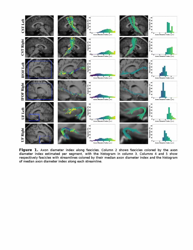
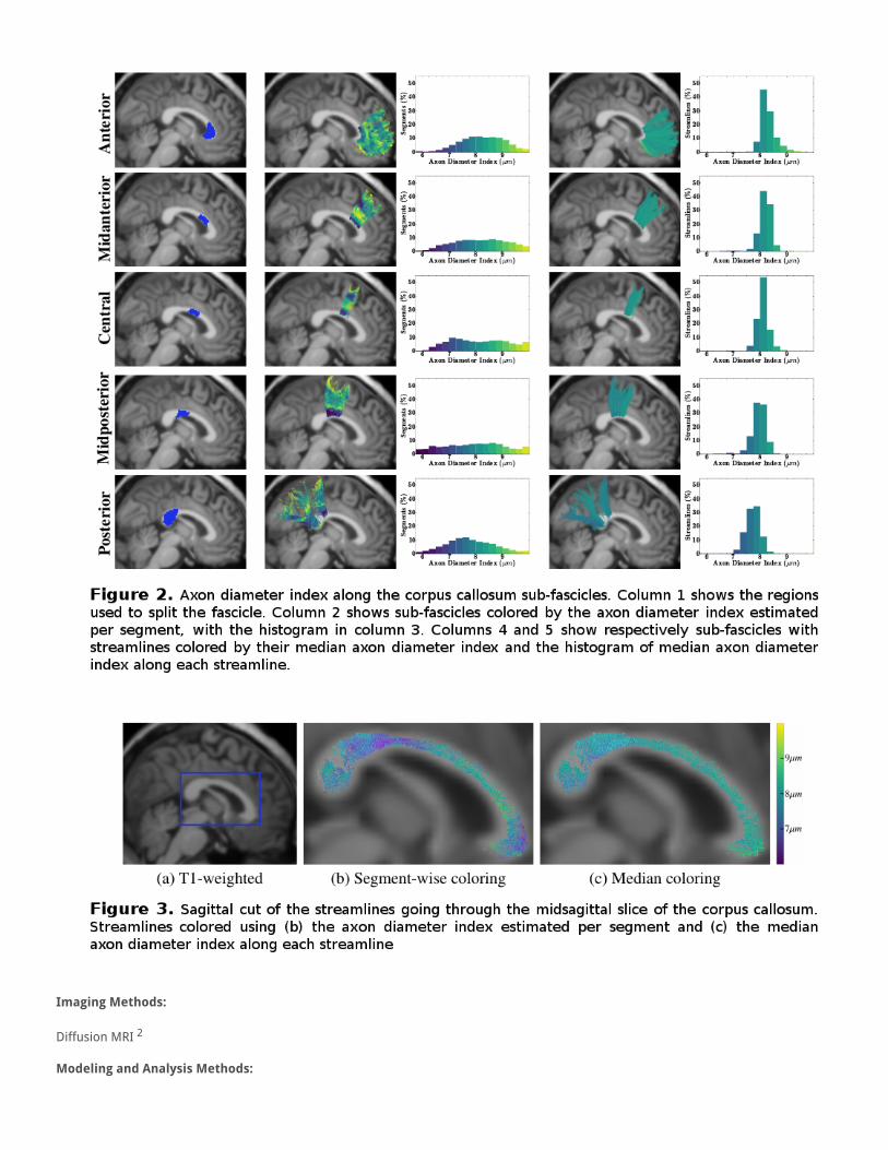
Figures 1 shows the axon diameter index estimation along three WM fascicles: the corticospinal tract (CST), the inferior fronto-occipitalfasciculus (IFOF) and the uncinate fasciculus (UF). Figure 2 shows the same information for the superior part of the corpus callosum (CC).The CC is split in 5 sub-fascicles using the FreeSurfer parcellation. Finally, a midsagittal cut of the same CC is shown in Figure 3. Thehighest axon diameter index (green) can be observed in the central part of the CC using segment-wise estimation.
Conclusions:
AxTract enables the characterization of the axon diameter index along WM fascicles in-vivo. Axon diameter index estimated with AMICOare similar in fascicles of both hemispheres (see Figure 1) and seems to be spatially coherent both on segments and on the medianalong streamlines (see Figures 1 and 2). The axon diameter index observed in the CC (see Figure 3) follows the trend observed inhistology [Aboitiz et al., 1992], with lower values in the splenium and genu, and higher value in the body of the CC. However, this isvisible only in the midsagittal slice of the CC. Further investigation is needed to understand this effect.
Imaging Methods:
Diffusion MRI 2
Modeling and Analysis Methods:

Diffusion MRI Modeling and Analysis 1
Keywords:
MRITractographyWhite MatterWHITE MATTER IMAGING - DTI, HARDI, DSI, ETCOther - Microstructure
1|2Indicates the priority used for review
Would you accept an oral presentation if your abstract is selected for an oral session?
Yes
I would be willing to discuss my abstract with members of the press should my abstract be marked newsworthy:
Yes
Please indicate below if your study was a "resting state" or "task-activation” study.
Other
Healthy subjects only or patients (note that patient studies may also involve healthy subjects):
Healthy subjects
Internal Review Board (IRB) or Animal Use and Care Committee (AUCC) Approval. Please indicate approval below. Please note:Failure to have IRB or AUCC approval, if applicable will lead to automatic rejection of abstract.
Not applicable
Please indicate which methods were used in your research:
Structural MRIDiffusion MRI
For human MRI, what field strength scanner do you use?
3.0T
Which processing packages did you use for your study?
FSLFree SurferOther, Please list - Dipy, TractQuerier
Provide references in author date format
Aboitiz, F., (1992), 'Fiber composition of the human corpus callosum', Brain Research, 598(1-2), pp. 143–153.Alexander, D. C., (2010), 'Orientationally invariant indices of axon diameter and density from diffusion MRI', NeuroImage, 52(4), pp.1374–1389.Auría, A. R., (2015), 'Accelerated Microstructure Imaging via Convex Optimisation for regions with multiple fibres (AMICOx)', In IEEEInternational Conference on Image Processing, Québec, Canada.Daducci, A., (2015), 'Accelerated Microstructure Imaging via Convex Optimization (AMICO) from diffusion MRI data', NeuroImage,105, pp. 32–44.Debanne, D., (2011), 'Axon physiology', Physiological Reviews, 91(2), pp. 555–602.Dyrby, T. B., (2012), 'Contrast and stability of the axon diameter index from microstructure imaging with diffusion MRI', MagneticResonance in Medicine, 721(2013), pp. 711–721.Girard, G., (2015), 'AxTract: Microstructure-Driven Tractography Based on the Ensemble Average Propagator', In InformationProcessing in Medical Imaging. Isle of Sky, Scotland, pp. 675–686.Panagiotaki, E., (2012), 'Compartment models of the diffusion MR signal in brain white matter: A taxonomy and comparison',NeuroImage, 59(3), pp. 2241–2254.Setsompop, K., (2013), 'Pushing the limits of in vivo diffusion MRI for the Human Connectome Project', NeuroImage, pp. 220–233.Wassermann, D., (2013), 'On describing human white matter anatomy: the white matter query language', In Medical ImageComputing and Computer-Assisted Intervention, Nagoya Japan, 16(Pt 1), pp. 647–654.
