white matter maturation of normal human fetal brain. an in ... · an in vivo diffusion tensor...
TRANSCRIPT

White matter maturation of normal human fetal brain.An in vivo diffusion tensor tractography studyEmilie Zanin1,3, Jean-Philippe Ranjeva1, Sylviane Confort-Gouny1, Maxime Guye1, Daniele Denis3,Patrick J. Cozzone1 & Nadine Girard1,2
1Centre de Resonance Magnetique Biologique et Medicale UMR CNRS 6612, Faculte de Medecine de Marseille, Universite de la Mediterranee,Aix-Marseille II, France
2Service de Neuroradiologie Diagnostique et Interventionelle, Centre hospitalo-universitaire de la Timone, Assistance Publique des Hopitaux deMarseille, France
3Service d’Ophtalmologie, Centre hospitalo-universitaire Nord, Assistance Publique des Hopitaux de Marseille, France
KeywordsCorpus callosum, corticospinal tracts, diffusiontractography, fetal development, human,in utero, visual pathways, white mattermaturation.
CorrespondenceJean-Philippe Ranjeva, CRMBM UMR CNRS6612, 27 Bvd Jean Moulin, F 13385 Marseille,France. Tel: +33-4-91-38-49-61; Fax:+33-4-91-25-65-39; E-mail:[email protected]
Received: 16 June 2011; Revised: 8 July 2011;Accepted: 1 August 2011
doi: 10.1002/brb3.17
Abstract
We demonstrate for the first time the ability to determine in vivo and in uterothe transitions between the main stages of white matter (WM) maturation innormal human fetuses using magnetic resonance diffusion tensor imaging (DTI)tractography. Biophysical characteristics of water motion are used as an indirectprobe to evaluate progression of the tissue matrix organization in cortico-spinaltracts (CSTs), optic radiations (OR), and corpus callosum (CC) in 17 normal humanfetuses explored between 23 and 38 weeks of gestation (GW) and selected strictlyon minimal motion artifacts. Nonlinear polynomial (third order) curve fittings ofnormalized longitudinal and radial water diffusivities (Z-scores) as a function ofage identify three different phases of maturation with specific dynamics for eachWM bundle type. These phases may correspond to distinct cellular events such asaxonal organization, myelination gliosis, and myelination, previously reported byother groups on post-mortem fetuses using immunostaining methods. Accordingto the DTI parameter dynamics, we suggest that myelination (phase 3) appearsearly in the CSTs, followed by the OR and by the CC, respectively. DTI tractographyprovides access to a better understanding of fetal WM maturation.
Introduction
Current knowledge on fetal white matter (WM) maturationcomes from post-mortem pathological studies (Gilles 1983;Brody et al. 1987). These studies have mainly focused on themyelination, the last step of WM maturation. Myelination isreported as a nonlinear complex phenomenon progressingwith a spatio-temporal course specific to each species. In hu-mans, it begins at the second half of gestation and can evolveuntil the age of 20 for structures such as the corpus callo-sum (CC) (Kinney et al. 1988). Recent histological advancesin immunostaining methods have allowed a better descrip-tion of the cascade of cellular events characterizing the earlyphases (before myelination) of human fetal WM maturationon post-mortem samples (Back et al. 2002). These observa-tions confirm the existence of a premyelinating phase corre-sponding to the appearance of abundant “myelination glia,”composed by oligodendrocyte (OL) precursors and imma-
ture OL, as an essential step prior to the myelination process(Back et al. 2002).
Although prenatal ultrasound and conventional T1- andT2-weighted MRI bring crucial information on the braindevelopment of human fetuses in utero (Girard et al. 1995),the early cellular events involved in WM maturation are notyet accessible by these techniques.
The unique opportunity to detect such microscopic phe-nomena in utero relies on the characterization of watermolecule motions restricted by cerebral tissue using mag-netic resonance diffusion-weighted imaging (DWI). Basedon published reports that have used in vitro and in vivoneurological model systems, Beaulieu (2002) and Song et al.(2002) have proposed to associate the microstructural orga-nization of WM tracts with water diffusion characteristics.Apparent diffusion coefficient (ADC) reflects the probabilityof displacement of a water molecule (modeled by a sphere)characterized by Brownian motion within a tissue supposed
c© 2011 The Authors. Published by Wiley Periodicals, Inc. This is an open access article under the terms of the CreativeCommons Attribution Non-Commercial License, which permits use, distribution and reproduction in any medium, providedthe original work is properly cited and is not used for commercial purposes.
95
In Vivo DTI Tractography in Human Fetuses E. Zanin et al.
to be isotropic. To date, ADC variations on fetal WM are usedto detect the initiation of myelination processes before con-ventional T1 and T2 images (Prayer and Prayer 2003; Righiniet al. 2003; Schneider et al. 2007). However, ADC alone can-not detect the first stage of WM maturation or differentiatethe successive stage described by histology. Diffusion tensorimaging (DTI) represents a new breakthrough in the analysisof WM maturation by modeling water molecule displace-ment by an ellipse oriented along the main direction of tissuestructure (Mori and Zhang 2006). In anisotropic tissue suchas WM, DTI provides in addition to ADC, information aboutthe anisotropy of water diffusion reflecting a particular cel-lular arrangement of the structure, through parameters suchas fractional anisotropy (FA), longitudinal (λ//), and radial(λ⊥) diffusivities (Song et al. 2002). It also gives access to themain direction of water diffusion within a given voxel. Whencombined, this information can be used to estimate three-dimensional trajectories of WM bundles by tractography al-gorithms. However, imaging fetuses in utero remains an im-portant technical challenge, especially for motion-sensitiveexaminations such as DTI. Bui et al. (2006) were the first tomeasure in utero the diffusion tensor in the fetal WM be-tween 31 GW and 37 GW in a series of 24 fetuses selectedbased on the absence of motion artifact (50% of cases)).They assessed ADC and FA on restrictive regions of interest(ROIs). Kasprian et al. are the only ones who have used DTIand three-dimensional tractography in living non sedatedhuman fetus in utero (Kasprian et al. 2008). The success-ful reconstruction in only 40% of examined fetuses and theabsence of significant correlation between DTI parametersand gestational age illustrate that in utero DTI is extremelychallenging, limited by many sources of errors and artifacts(Kasprian et al. 2008). Few teams are actively working onmotion correction to improve robustness of this technique(Rousseau et al. 2005, 2006; Jiang et al. 2007, 2009) but newimprovements are still required.
By characterizing in utero the relative variations of lon-gitudinal (λ//) and radial (λ⊥) water diffusivities on entireindividual WM bundles during gestation in very well-selectedfetal DTI acquisitions with minimal motion, we aimed at de-termine noninvasively the major key periods and transitionsof tissue organization for the cortico-spinal tract (CST), theOR, and the CC in normal human fetuses.
Materials and Methods
Subjects
Cerebral magnetic resonance examinations were performedfor clinical purpose at our institution after selection of pa-tients by the multidisciplinary fetal medical team. The indi-cations of the fetal magnetic resonance imaging (MRI) explo-rations were pregnancies at risk of brain damage, suspicionof brain malformation on ultrasound scans, and presence of
maternal and/or family history of brain development disor-ders. Gestational age was determined by a previous sonog-raphy at 12 postovulatory weeks. Fetuses were selected whenconventional MRI examinations were normal based on thereport of a neuroradiologist expert in fetal MRI (NG) (ab-sence of anatomical malformation, absence of WM or graymatter lesions) and when they were considered normal atbirth by pediatric neurologist. Of the 141 brain fetus DTIacquisitions, 61 fulfilled these criteria.
Imaging in the presence of subject motion has been anongoing challenge for MRI, especially for motion sensitiveexaminations such as DTI. In utero fetal DTI is an extremecase vulnerable to the mother’s respiration and fetal mo-tion artifacts. Consequently, among 61 normal cerebral fetalMRI with DTI sequence, only 17 (28%) were selected for thestudy based on the absence of motion corruption on coronal,sagittal, and axial views of b = 0 images evaluated by twoindependent readers (EZ, NG) and the sufficient quality ofthe FA color-coded directionality map (color coherence ofthe major bundles) and ADC maps (Fig. 1). Discordant caseswere finally rejected by consensus. The mean gestational agewas 32 ± 4 weeks of gestation (range, 23–38 weeks). The co-hort was constituted by fetuses at gestation ages of 23 GW(1),24 GW(1), 27 GW(1), 28 GW(2), 30 GW(1), 32 GW(1), 33GW(3), 34 GW(2), 35 GW(2), 36 GW(1), 37 GW(1), and 38GW(1).
Image acquisition
MR images were taken with 1.5 T MR scanner (Magne-tom Symphony Siemens, Erlangen, Germany) using a phasedarray coil with four anterior elements wrapped aroundthe mother’s abdomen and two to three posterior spinalelements.
Conventional fetal MRI were acquired using T2-weightedsingle-shot sequences (HASTE, TE/TR: 137 ms/1680 ms; BW220 Hz/pixel, 21 contiguous slices, 3.5 mm thickness, matrix:358 × 512, FOV: 380 mm) acquired in three orthogonalplanes oriented along the fetal brain, and both axial andcoronal gradient echo T1-weighted sequence (Flash TE/TR:3.3 ms/493 ms, BW 260 Hz/pixel, 19 slices, 4 mm thickness,matrix: 154 × 256, FOV: 350 mm).
DTI was acquired with mother sedation (Flunitrazepam,Rohypnol
R©, Roche, Basel, Switzerland) in a dorsal decubitus
position using a single-shot echo-planar spin-echo sequencewith the following parameters: TE/TR: 105 ms/8900 ms, 50contiguous slices, 2.2 mm thickness, matrix: 128 × 128; FOV:256 × 256 mm2. Diffusion gradients were encoded in 12directions with a b values of 1000 s/mm2 and an additionalimage with no diffusion gradient (b = 0 s/mm2). Three setsof DTI data were acquired for average and the total DTIacquisition time was 5 min 47 sec. To improve the signal-to-noise ratio (SNR) for the DTI images, the three separate sets
96 c© 2011 The Authors. Published by Wiley Periodicals, Inc.
E. Zanin et al. In Vivo DTI Tractography in Human Fetuses
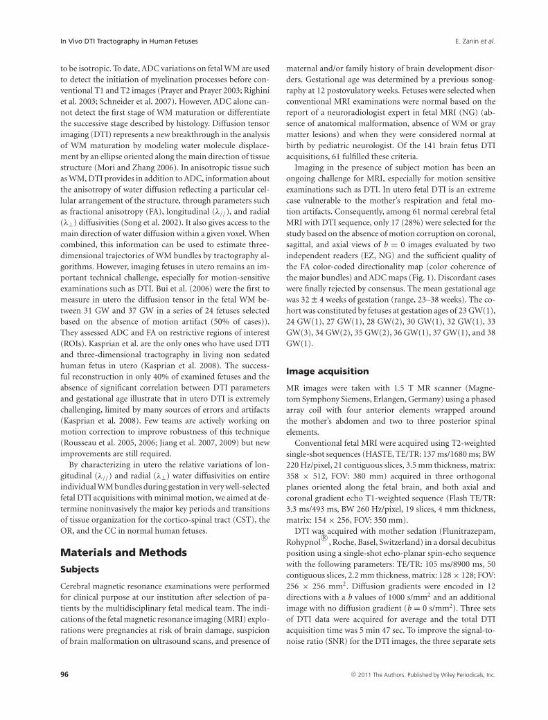
Figure 1. Example of in utero DTI acquisition slice positioning and resulting FA color-coded directionality map. (A and B) The displays of in uteroacquisitions performed in the axial plane relative to the fetus head. The quality of the resulting DTI images was assessed according to the gooddefinition of large white matter (WM) bundles such as corpus callosum (CC) (green arrows) on the reconstructed FA color-coded directionality mapsas in (C and D), examples of high-quality DTI acquisitions included in the study. Image in (E) represents DTI of poor quality excluded from analysis.Note that the spatial reference varies according to the orientation of the fetus head relative to the scanner orientation. This induces changes in thecolor directionality code usually observed in adults.
of images were averaged automatically by the “Neuro3D taskcard” (Siemens Syngo). We determined the SNR according toKasprian et al. (2008). The SNR is defined as the mean valueof the signal of a circumscribed WM region of the fetal brainon diffusion unweighted (b = 0 s/mm2) images divided bythe standard deviation determined in the same region.
A b value of 1000 s/mm2 was used in fetal DTI as Huanget al. (2009). This value was adapted to high ADC valuesexpected at the fetal WM in order to the formula 1.1 perADC that have been thought to provide the best contrast-to-noise ratio (Dudink et al. 2008).
Data postprocessing
Diffusion tensor parameters
DTI calculation and postprocessing were performed aftertransferring diffusion-weighted images to a Siemens off-lineworkstation, using the “DTI task Card” software, and thefour classical parameters were computed: the longitudinaldiffusivity (λ// or λ1), the radial diffusivity λ⊥ = (λ2+λ3)/2,the ADC, and the FA.
Definition of seed regions and tractography
Seed regions for DTI tractography were determined basedon anatomical landmarks observed on the ADC and FAcolor-coded directionality maps (Figs. 2 and 3). Using the“Neuro3D task card” (Siemens Syngo), each axes of the MPR(multi-planar reconstruction) was rotated (double oblique)to get axial, coronal, and sagittal planes in the anatomical ref-erential for each fetus. ADC maps provided a good contrast,and the overlay of color-coded FA helped to depict coherentfiber pathways nearby the ROIs by indicating fiber directionand degree of anisotropy. However, because the fetus headin utero is randomly positioned relative to the scanner refer-ential, the color code used in this study did not correspondto the regular color code obtained in adults with standardpositioning (red: right–left, green: antero–posterior, blue:supero–inferior) (Pajevic and Pierpaoli 1999). The “Neuro3DDTI task card” did not allow to apply tensor rotation to keepthis conventional color code.
The WM bundles selected for analysis in this study in-cluded CST (an example of projection tracts), OR (anexample of association tracts), and CC (an example of
c© 2011 The Authors. Published by Wiley Periodicals, Inc. 97
In Vivo DTI Tractography in Human Fetuses E. Zanin et al.
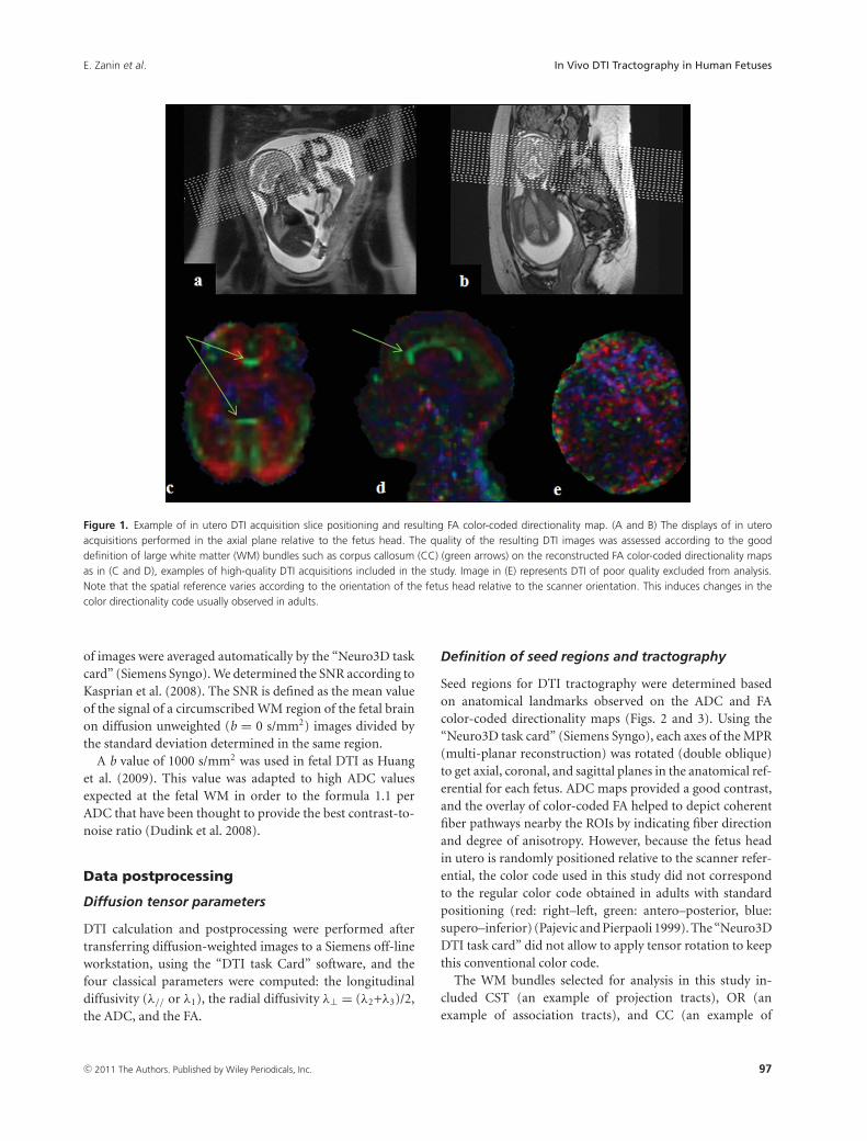
Figure 2. Diffusion tensor tractography method used to depict cortico-spinal tract (CST), optic radiations (OR), and CC bundles. Regions-of-interest(ROIs) were delineated based on anatomical landmarks observed on ADC and FA color-coded directionality maps. (A) For the CST observed in blue onthe image in the left superior quadrant, tractography algorithm (Runge Kutta order 4) was initiated from two ROI located in the posterior limb of theinternal capsule (PCI) (purple ROI) and the cerebral peduncles (PC) (green ROI). (B) For OR observed in red in the image on the left inferior quadrant,the two ROI were defined on coronal slices located on the anterior (ant) and medial (med) parts of OR. (C) Corpus callosum (CC) observed in greenon right quadrant was divided in three subparts: the genu (CCg), the body (CCb), and the splenium (CCs). A single ROI approach was used for thetractography, which was defined from each subpart on the mid-sagittal slice (MSS).
commissural tracts). Because previous study has reporteda different sequence of myelination according to the subpartsof the CC, its analysis was divided into three parts includ-ing genu, body, and splenium (Kinney et al. 1988). Thesethree parts were defined according to Catani et al. (2002)from well-individualized anatomical landmarks on the mid-sagittal slice (MSS) (passing through the median line and inthe midline of the CC), frequently used in MRI: an anteriorportion, the genu (rounded and rolled up in the bottom andahead around the anterior pole of diencephalons), a mid-dle portion, the body (overhanging the septum lucidum andthe area of the roof of the third ventricle), and a posteriorportion, the splenium (rounded and the bulkiest).
The main observer (EZ) performed all the ROIs and trac-tography twice using a two-ROI analysis for CST and OR
(Catani et al. 2002) and single ROIs placed on different levelsof the MSS for the different parts of the CC (Fig. 2).
Reconstructed tracts were validated by the main observer(EZ) according to anatomical landmarks observed on ADCmaps and b = 0 images projected on the different views(axial, sagittal, and coronal) and data from the literature onpost-mortem fetal tractography (Catani et al. 2002; Huanget al. 2009; Vasung et al. 2010). The CSTs were defined as thefibers passing through the cerebral peduncles and the poste-rior limb of the internal capsule (Fig. 2), the OR as the fibersconnecting the lateral geniculate nucleus and the occipitalpole (Fig. 2). Fibers tracts generated from the genu connectanterior parts of the frontal lobes (mainly pre-frontal) andtheir horseshoe-shaped radiating fibers form the anterior(minor) forceps. The genu contains fibers from orbital,
98 c© 2011 The Authors. Published by Wiley Periodicals, Inc.
E. Zanin et al. In Vivo DTI Tractography in Human Fetuses
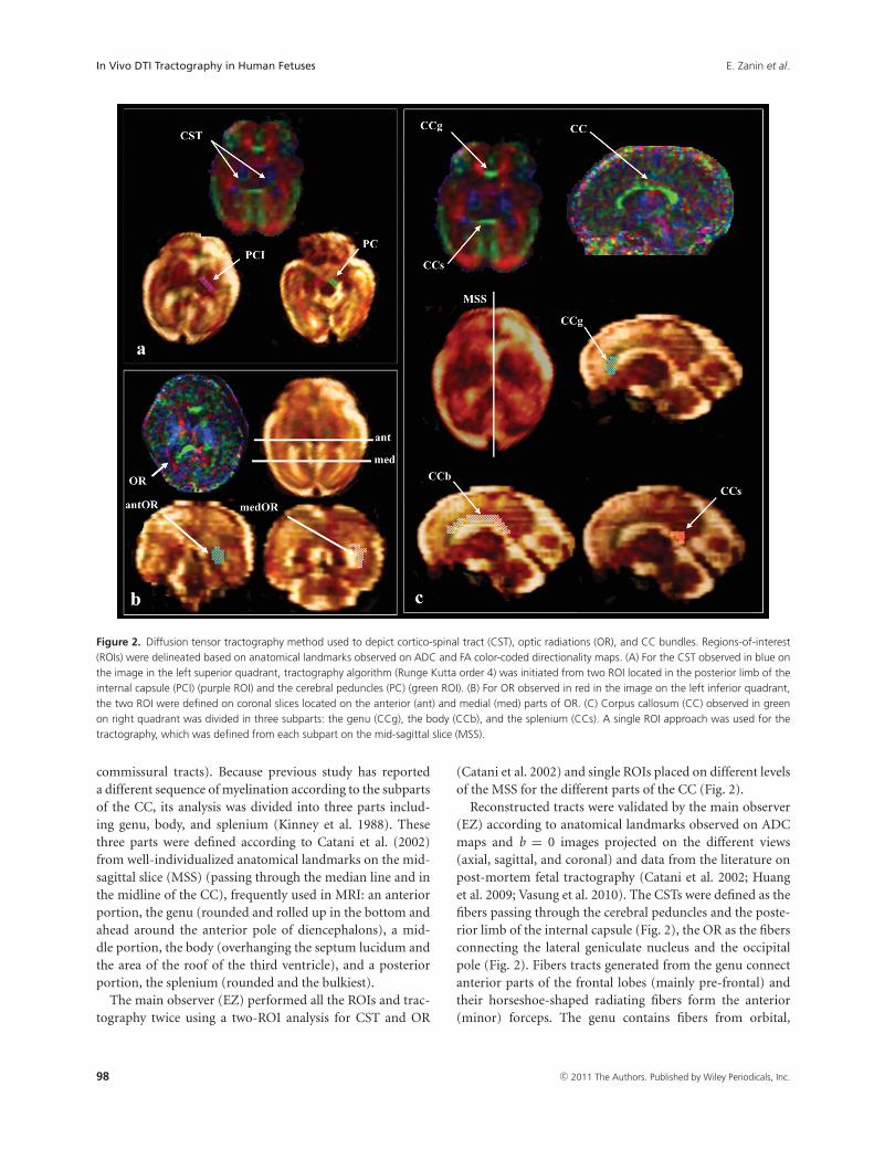
Figure 3. Example of DTI tractography of CST and OR obtained in utero and in vivo for fetal brain. Tractography three-dimensional reconstructionsobtained with the Runge Kutta method (order 4) of the CST at 24 GW (A, superior quadrant) and 27 GW (A, inferior quadrant) and OR at 33 GW (B,superior quadrant) and 34 GW (B, inferior quadrant).
medial, and dorsal frontal cortex. These fibers cross thecorona radiata and converge toward the anterior horn of thelateral ventricle where they form a compact bundle that archesin the genu. The body of CC contains fibers that connect thepremotor and precentral frontal cortex, the parietal lobes,and the temporal lobes. These fibers converge at the posteriorhorn of the lateral ventricle, around which they are shapedlike a cone, before arching medially to cross the midline. Thesplenium connect occipital lobes and make up the posteriormajor forceps (Fig. 2) (Catani et al. 2002). The fiber bundleswere reconstructed using a streamline Runge Kutta algorithm(order 4). A FA threshold of 0.08 (a low value adapted to theimmaturity of the fetal tracts and the intra uterin conditions)was used and a curvature angle smaller than 70◦ (adapted tothe curved geometry of the ORs) was chosen (Yamamoto et al.2007). Two FA thresholds were tested adapted to the imma-turity of the fetal WM: 0.1 and 0.08. Threshold of 0.1 waspreviously used in the literature in postnatal period (Dubois
et al. 2006) and in fetuses in vitro (Vasung et al. 2011). Thebest results in terms of tractography reconstruction were ob-tained with the 0.08 threshold.
Then a second investigator expert in fetal neuroradiology(NG) evaluated each of the reconstructed tracts and validatedthe results. In case of bad scoring by the expert, tracking wasredone once, and finally not considered if not approved by theexpert at that time. The percentage of rejected reconstructedtractography was 4% (5/119).
Diffusion tensor parameters of each bundle
Masks of the reconstructed bundles were applied to the para-metric maps of ADC, FA, λ1, λ2, and λ3 to compute theDTI parameter values of each bundle with SPM 5 (Welcomeinstitute, London). The average values of the ADC, the FA,the λ1 = λ//, λ2, and λ3 for the whole reconstructed bun-dles were obtained for each tract. The λ⊥ were obtained by
c© 2011 The Authors. Published by Wiley Periodicals, Inc. 99

In Vivo DTI Tractography in Human Fetuses E. Zanin et al.
average the λ2 and λ3 values of each reconstructed tracts(www.mricro.com).
Statistical analysis
Statistical analyses were performed using the JMP 2008 soft-ware (SAS Institute Inc., Cary, NC, USA).
Diffusion characteristics of WM tracts independentof gestational age
The means, medians, minima, and maxima of DTI parame-ters (ADC, FA, λ//, λ⊥) measured on each type of bundles forall fetuses were calculated and compared bundles by bundlesusing ANOVA (P <0.05, corrected for multiple comparisons)to assess the differences in diffusion characteristics betweenthe different WM bundles independently of the gestationalage.
Variation of WM tract diffusion characteristicsaccording to gestational age
For each DTI parameter of each WM bundle, Z-scoreswere computed for each subject relative to the mean valuesand standard deviations of the whole population of fetuses.ANOVA was conducted on these diffusion parameters to eval-uate the effect of age and bundle type (statistical thresholdP < 0.05 after False Discovery Rate (FDR) correction).
Variations of diffusion parameters (ADC, FA, λ//, λ⊥) foreach type of WM bundle were modeled as a function ofage first by linear regression (Kasprian et al. 2008) and alsoby polynomial fitting functions (degree 3) (Schneider et al.2007).
Results
Diffusion characteristics of WM tractsindependent of gestational age
The SNR of b0 images measured in the 17 included fetusesresulted in a mean value of 14.07 ± 3.17. All selected tractsstudied were validated by the two experts (EZ, NG) exceptfor five OR (four rights, one left) in four different subjects(age: 27 GW, 34 GW, 34 GW, 35 GW) that were removed forthe subsequent analyses. Thus, 34 CSTs, 29 OR, and 17 CCwere identified and characterized (Figs. 3 and 4).
After verifying that diffusion characteristics were not dif-ferent between left and right bundles for the CST and theOR (P > 0.05, Wilcoxon rank test), left and right diffusionparameters values of each bundle were pooled for subsequentanalyses.
From a structural point of view, each bundle had specificdiffusion tensor characteristics (Fig. 5; Table 1). CST wasthe most organized structure exhibiting significantly lowerADC values compared to OR, genu, body, and splenium ofCC (ANOVA, P < 0.05 corrected for multiple comparisons)
Figure 4. Example of DTI tractography of the CC obtained in utero andin vivo for fetal brain. Tractography three-dimensional reconstructionsobtained with the Runge Kutta method (order 4) of the CC dividedin three subparts: body of the corpus callosum (CCb) at 33 GW (A),splenium of CC at 33 GW (B), and genu of CC at 37 GW (C).
(Fig. 5). CST also showed higher FA values compared toOR, genu, and body of CC (ANOVA, P < 0.05 correctedfor multiple comparisons) (Fig. 5). In contrast, OR appearedas the less-organized structure with significant higher ADCrelative to CST, body, and genu of CC, and significant lowerFA compared to all the others tracts (ANOVA, P < 0.05corrected for multiple comparisons).
Variation of WM tract diffusioncharacteristics according to gestational age
According to the poor linear fitting of diffusion parameterswith age, the polynomial function of degree 3 was chosento model the variation of parameters during gestation. We
100 c© 2011 The Authors. Published by Wiley Periodicals, Inc.
E. Zanin et al. In Vivo DTI Tractography in Human Fetuses
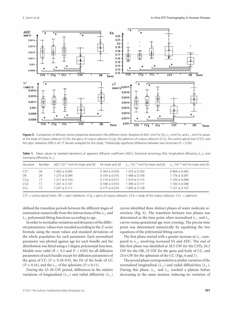
Figure 5. Comparison of diffusion tensor properties observed in the different tracts. Boxplots of ADC (mm2/s), FA, λ// (mm2/s), and λ⊥ (mm2/s) valuesat the body of corpus callosum (CCb), the genu of corpus callosum (CCg), the splenium of corpus callosum (CCs), the cortico-spinal tract (CST), andthe optic radiations (OR) in all 17 fetuses analyzed for this study. *Statistically significant difference between two structures (P < 0.05).
Table 1. Mean values (± standard deviation) of apparent diffusion coefficient (ADC), fractional anisotropy (FA), longitudinal diffusivity (λ//), andtransverse diffusivity (λ⊥)
Structure Number ADC (10−3 mm2/s) mean and SD FA mean and SD λ// (10−3 mm2/s) mean and SD λ⊥ (10−3 mm2/s) mean and SD
CST 34 1.063 ± 0.095 0.183 ± 0.018 1.255 ± 0.103 0.964 ± 0.092OR 29 1.275 ± 0.094 0.154 ± 0.016 1.468 ± 0.104 1.176 ± 0.091CCg 17 1.211 ± 0.104 0.170 ± 0.012 1.414 ± 0.115 1.105 ± 0.098CCb 17 1.201 ± 0.101 0.168 ± 0.014 1.399 ± 0.111 1.100 ± 0.098CCs 17 1.231 ± 0.111 0.177 ± 0.014 1.450 ± 0.128 1.121 ± 0.107
CST = cortico-spinal tracts, OR = optic radiations, CCg = genu of corpus callosum, CCb = body of the corpus callosum, CCs = splenium.
defined the transition periods between the different stages ofmaturation numerically from the intersections of the λ// andλ⊥ polynomial fitting functions according to age.
In order to normalize variations and dynamics of the differ-ent parameters, values were rescaled according to the Z-scoreformula using the mean values and standard deviations ofthe whole population for each parameter. Each normalizedparameter was plotted against age for each bundle and thedistribution was fitted using a 3 degree polynomial function.Models were valid (R > 0.5 and P < 0.05) for all diffusionparameters of each bundle except for diffusion parameters ofthe genu of CC (P = 0.10–0.9), the FA of the body of CC(P = 0.16), and the λ// of the splenium (P = 0.11).
During the 23–38 GW period, differences in the relativevariations of longitudinal (λ//) and radial diffusivity (λ⊥)
curves identified three distinct phases of water molecule re-striction (Fig. 6). The transition between two phases wasdetermined as the time point when normalized λ// and λ⊥curves versus gestational age were crossing. The precise timepoint was determined numerically by equalizing the twoequations of the polynomial fitting curves.
The first phase started with a greater increase in λ// com-pared to λ⊥, involving increased FA and ADC. The end ofthis first phase was identified at 28.5 GW for the CSTs, 26.3GW for the OR, 25 GW for the genu and body of CC, and25.6 GW for the splenium of the CC (Figs. 6 and 7).
The second phase corresponded to a similar variation of thenormalized longitudinal (λ//) and radial diffusivities (λ⊥).During this phase, λ// and λ⊥ reached a plateau beforedecreasing in the same manner, inducing no variation of
c© 2011 The Authors. Published by Wiley Periodicals, Inc. 101

In Vivo DTI Tractography in Human Fetuses E. Zanin et al.
Figure 6. Variation of diffusion parameters during gestation according to the WM structure. Determination of three different maturation phasesaccording to the numerical determination of differential evolutions of the λ// and λ⊥ curves modeled by a 3 degree polynomial function (phase 1 ingreen, phase 2 in orange, and phase 3 in red) and schematic representations of the supposed corresponding histological events displayed in Figure 8.
Figure 7. Putative dynamics of WM maturation derived from polyno-mial fittings of in vivo DTI tractography parameters acquired in utero.CCb = body of corpus callosum, CCg = the genu of corpus callosum,CCs = the splenium of corpus callosum, CST = the cortico-spinal tract,OR = optic radiations, GW = gestational weeks.
ADC before decrease, and stable FA during the whole pe-riod. The end of this second phase (where the two curvesdiverged) was reached at 32.5 GW for the CSTs, 34.8 GW forthe ORs, 35.4 GW for the body of CC, and 35.3 GW for the
splenium of the CC. Conversely, the end of this second periodwas not reached for the genu of CC before 38 GW (Figs. 6and 7).
The third phase corresponded to a faster decrease of thenormalized radial diffusivity (λ⊥) relative to the longitudinaldiffusivity (λ//), reflected by an increase in FA and a slowerdecrease in ADC.
These three different phases of diffusion parameter varia-tions could be observed for all bundles except for the genu ofCC for which the transition between phases 2 and 3 was notobserved before 38 GW. Observed dynamics of maturation ofeach bundle derived from the present study are summarizedin Figure 7.
ANOVA conducted on these data showed global time ef-fects on DTI parameters during gestation (P < 0.002) withsignificant bundle-type effects (P < 0.03) and bundle-type ×age interactions (P < 0.002) for the λ//, λ⊥, ADC, and FA,evidencing a significantly more advanced and faster mat-uration of the CST compared to the genu of CC and theOR. In addition, ANOVA conducted on the subparts of CCevidenced significant global effect (P < 0.02) with a bundle-type effect (P < 0.004) and a bundle-type × age interac-tion (P < 0.05) demonstrating a less-advanced and slower
102 c© 2011 The Authors. Published by Wiley Periodicals, Inc.
E. Zanin et al. In Vivo DTI Tractography in Human Fetuses
maturation of the genu compared to the other subpartsof CC.
Discussion
In vivo DTI tractography of human fetuses
DTI tractography has already been applied to the study ofchildren brain maturation from the early postnatal period(Gilmore et al. 2007; Provenzale et al. 2007; Dubois et al.2008). It has not only shown lower FA and higher ADC val-ues of children’s WM bundles relative to adults, but also linearcorrelations between age and DTI parameters during devel-opment (Gilmore et al. 2007; Provenzale et al. 2007; Duboiset al. 2008). In fetuses, post-mortem DTI detects the mainWM bundles as early as 13 GW (Huang et al. 2009). En-couraging results concerning in utero explorations of livingfetuses have been reported using DTI on limited regions ofinterest without tractography; preventing characterization ofthe entire (or at least a large part) of the WM bundles (Buiet al. 2006).
To our knowledge, only one DTI tractography study hasbeen conducted in utero on living fetuses, demonstratingthe feasibility but also pointing out the numerous techni-cal challenges to overcome in order to obtain robust results(Kasprian et al. 2008). We have also observed that qualityof DTI tractography and derived diffusion parameters arehighly dependent on fetus and mother motions. However, itwas possible to get reliable evolution of diffusion parame-ters during gestation from the examination of 17 fetuses withan optimized acquisition protocol and a drastic selection ofDTI data. The tracking accuracy might also be affected bythe presence of crossing fibers, especially for OR in regionsclosed to the inferior longitudinal fasciculus. However, thetwo ROIs approach chosen to perform DTI tractographylimited trajectory reconstruction errors. Accordingly, ADCvalues determined in our study for the CC and the CSTs wereconsistent with previous results obtained in utero in fetusesusing DTI without tractography (Bui et al. 2006), and valuesof ADC and λ⊥ were logically higher and FA values lowerin the fetuses of our study compared to neonates and adults(Dubois et al. 2008). Differences in mean FA values (0.18 vs.0.28) of the bundles obtained in the present work relative toprevious reports (Kasprian et al. 2008) are logic as far as theFA threshold chosen here to get reliable anatomical recon-structed bundles was lower than in the previous report (0.08vs. 0.15).
Variation of WM tracts diffusioncharacteristics according to gestational age
We have observed three different phases of radial and longitu-dinal diffusivity curve variations as a function of gestationalage for most of the bundles. Accordingly, we have adapted
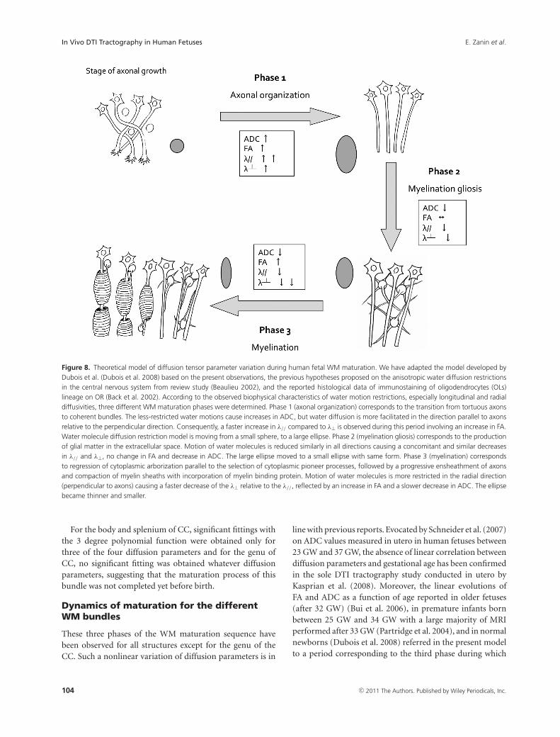
the model developed by Dubois et al. (2008) based on thepresent observations, the previous hypotheses proposed onthe anisotropic water diffusion restrictions in the central ner-vous system from review study (Beaulieu 2002) and the re-ported histological data of immunostaining of OL lineage onOR (Back et al. 2002) (Fig. 8). Our study showed that phase1 (before 26.3 GW for OR) corresponded biophysically toa large increase in longitudinal diffusivity concomitant to aslow increase in radial diffusivity causing significant increasesin ADC and FA. This period coincides with axonal organi-zation, a transition from tortuous axons state to coherentbundles. Indeed, histological data have shown labeled axonswith marker of mature neurofilament (panaxonal neurofil-ament marker SMI 312), presence of late OL progenitors,and absence of myelin-basic-protein (MBP) (mature myelinmarker) (Back et al. 2002). Consequently, the less restrictedwater motions cause increases in ADC, but water diffusion isless hindered in the longitudinal direction parallel to axonsrelative to the perpendicular direction causing an increasein both ADC and FA. Water molecule diffusion restrictionmodel evolves from a small sphere (isotropic tissue), to a largeellipse (anisotropic tissue). Phase 2 (between 26.3 GW and34.8 GW for OR) was characterized by similar and concomi-tant slow decreases in longitudinal and radial diffusivities,causing no change in FA (plateau) and a decrease in ADC.This period corresponds to progressive increase in immatureOL and the emission of cytoplasmic processes randomly ori-ented relative to axons according to histological data fromthe literature (Back et al. 2002). Coming from the samesources, marked expansion in the number of immature OLis observed at 30 GW followed by an emission of “pioneer”processes selectively oriented toward individual axons thatinitiate the axonal contact and then wrap around. This phe-nomenon, known as “myelination gliosis,” corresponds tomaturation of OL progenitors in immature OL followed by alarge production of glial matter randomly oriented relative toaxons in the extracellular space. Such a cascade would causean isotropic reduction of the extracellular space explainingthe observed water diffusion characteristics: the large ellipsemodeling water diffusion moved to a small ellipse with thesame shape. Phase 3 (after 34.8 GW for OR) was related bio-physically to a slow decrease in longitudinal diffusivity andconcomitant fast decrease in radial diffusivity. This periodcorresponds to, according to histological data, regression incytoplasmic arborization parallel to selection of cytoplasmicpioneer processes, followed by progressive ensheathment ofaxons and compaction of myelin sheaths with incorpora-tion of MBP (Back et al. 2002). Such a myelination processcaused a large restriction of water diffusion in the radialdirection (fast decrease in λ⊥), balanced in the longitudinaldirection by disappearance of cytoplasmic arborization (slowdecrease in λ//). The ellipse modeling water diffusion becamethinner.
c© 2011 The Authors. Published by Wiley Periodicals, Inc. 103
In Vivo DTI Tractography in Human Fetuses E. Zanin et al.
Figure 8. Theoretical model of diffusion tensor parameter variation during human fetal WM maturation. We have adapted the model developed byDubois et al. (Dubois et al. 2008) based on the present observations, the previous hypotheses proposed on the anisotropic water diffusion restrictionsin the central nervous system from review study (Beaulieu 2002), and the reported histological data of immunostaining of oligodendrocytes (OLs)lineage on OR (Back et al. 2002). According to the observed biophysical characteristics of water motion restrictions, especially longitudinal and radialdiffusivities, three different WM maturation phases were determined. Phase 1 (axonal organization) corresponds to the transition from tortuous axonsto coherent bundles. The less-restricted water motions cause increases in ADC, but water diffusion is more facilitated in the direction parallel to axonsrelative to the perpendicular direction. Consequently, a faster increase in λ// compared to λ⊥ is observed during this period involving an increase in FA.Water molecule diffusion restriction model is moving from a small sphere, to a large ellipse. Phase 2 (myelination gliosis) corresponds to the productionof glial matter in the extracellular space. Motion of water molecules is reduced similarly in all directions causing a concomitant and similar decreasesin λ// and λ⊥, no change in FA and decrease in ADC. The large ellipse moved to a small ellipse with same form. Phase 3 (myelination) correspondsto regression of cytoplasmic arborization parallel to the selection of cytoplasmic pioneer processes, followed by a progressive ensheathment of axonsand compaction of myelin sheaths with incorporation of myelin binding protein. Motion of water molecules is more restricted in the radial direction(perpendicular to axons) causing a faster decrease of the λ⊥ relative to the λ//, reflected by an increase in FA and a slower decrease in ADC. The ellipsebecame thinner and smaller.
For the body and splenium of CC, significant fittings withthe 3 degree polynomial function were obtained only forthree of the four diffusion parameters and for the genu ofCC, no significant fitting was obtained whatever diffusionparameters, suggesting that the maturation process of thisbundle was not completed yet before birth.
Dynamics of maturation for the differentWM bundles
These three phases of the WM maturation sequence havebeen observed for all structures except for the genu of theCC. Such a nonlinear variation of diffusion parameters is in
line with previous reports. Evocated by Schneider et al. (2007)on ADC values measured in utero in human fetuses between23 GW and 37 GW, the absence of linear correlation betweendiffusion parameters and gestational age has been confirmedin the sole DTI tractography study conducted in utero byKasprian et al. (2008). Moreover, the linear evolutions ofFA and ADC as a function of age reported in older fetuses(after 32 GW) (Bui et al. 2006), in premature infants bornbetween 25 GW and 34 GW with a large majority of MRIperformed after 33 GW (Partridge et al. 2004), and in normalnewborns (Dubois et al. 2008) referred in the present modelto a period corresponding to the third phase during which
104 c© 2011 The Authors. Published by Wiley Periodicals, Inc.
E. Zanin et al. In Vivo DTI Tractography in Human Fetuses
diffusion parameter variations follow a linear model relativeto age.
From a dynamic point of view and in accordance withhistological reports, we observed that myelination (phase 3)could appear early in the CSTs, followed by the OR and bythe CC, respectively (Gilles 1983).
One hypothesis sustaining this particular dynamical orga-nization may rely on the fact that the extent of WM matu-ration can be related to the functionality and excitability ofthe connected cortical areas. For example, the advanced mat-uration of OR during gestation in the absence of exogenousvisual stimulation could be related to the stimulation of thevisual cortex by the pons-geniculate-occipital waves duringthe rapid eye movement sleep that appears in fetuses as earlyas 30 GW (Graven 2008). Indeed, the structural maturationprocess of OR described in DTI coincides with the functionalmaturation of visual pathways evaluated by visually evokedresponse in premature infants (Volpe 2008). The first phaseof our model corresponding to axonal organization (<26.3GW) coincides with the appearance of the first visually evokedresponses previously observed in premature infants (22–24GW). The second phase corresponding to myelination gliosis(26.3–34.8 GW) coincides with evolution of visually evokedresponses to the principal wave forms (32–35 GW). Finally,the third phase corresponding to the myelination coincideswith the appearance of mature visually evoked response (39GW).
Within the CC, maturation is slow and heterogeneous ac-cording to the substructures. The antero-posterior functionaldifferences of the CC are also reflected by differences in mat-uration dynamics with an early maturation of splenium (ini-tiating phase 2 at 25.6 GW and phase 3 at 35.3 GW) andbody (initiating phase 2 at 25 GW and phase 3 at 35.4 GW),and a later maturation of the genu (initiating phase 2 at 25GW continuing until birth). According to the present results,genu remains in the myelination gliosis phase until 38 GW,showing that the third phase of maturation, that is the myeli-nation, may occur entirely after birth. These observationsare consistent with previous data reporting that the greatestvariations of diffusion parameters during the postnatal pe-riod occurred in the CC and especially in the genu (Partridgeet al. 2004; Lobel et al. 2009). Low genu maturation in uterocould be related to the very low cognitive stimulation of thefrontal lobes of fetuses during gestation (Barkovich 2000).
Lastly, it is worth noting that the first half of the myelina-tion gliosis phase (phase 2) characterized by the initiation ofOL progenitor maturation in immature OL within the WMcorresponds to the developmental window of high risk forperiventricular leukomalacia (PVL) (between 23 GW and 32GW) (Back et al. 2001). Indeed, late OL progenitors, knownas the predominant population of OL during this period, aresuspected to be a potential target for injury in PVL (Backet al. 2001).
While promising, this pilot study only provides a proof ofconcept obtained in one highly selected DTI fetal databasewith no or minimal motion artifacts. Generalization ofthis method will require further methodological approachesmostly related to motion corrections to get reliable normativedata in a large cohort of fetuses and to study developmentaldisorders (Rousseau et al. 2005, 2006; Jiang et al. 2007, 2009).
Limitations
Several technical limitations have to be acknowledged thanksto the interpretation of the results obtained in this pilot study.
The major limitation is relative to the fetus motions dur-ing acquisition and the way to handle such a problem. Asdemonstrated by Hayat et al. (2010), there is an importantreduction of fetus motion between 23 GW and 36 GW, thoughattributable to decreased intrauterine space and immobiliza-tion of the fetal head in the maternal pelvis in the cephalic pre-sentation (Kasprian et al. 2008). In this study, 85% of selectedfetuses were in cephalic presentation. Recent very interestingapproaches have been proposed to postprocess DTI data inmoving subjects, by rotating appropriately after image reg-istration the directions of the diffusion-sensitizing gradients(Jiang et al. 2007, 2009). In the present study, we did not usesuch an approach while we decided to select strictly in quitea large sample of data (61 in utero fetuses acquisitions), onlythe exams with very limited or absent motion observed oncoronal, sagittal, and axial views of b = 0 images. Motion wasevaluated by two independent readers (EZ, NG). Only a verysmall number of data survived to this screening (17 out of 61;28%). Accordingly to minimal fetus motion in this dataset,we obtained sufficient SNR (about 14) on b = 0 s/mm2 im-ages to obtain relevant FA and ADC maps as well as coherentfiber pathways reconstructed by tractography. Finally, eachreconstructed tract was evaluated by a second expert in fe-tal neuroradiology (NG) based on anatomical landmarks. Incase of bad scoring, tracking was redone once, and finallynot considered if not approved by the expert at that time.Only a small percentage of tracts were rejected during thissecond selection (4%; five out of 119). Reconstruction of thevalidated selected tracts could be then considered as robust.It is also important to note that between 23 GW and birth,previous histological studies have demonstrated the presenceof the tracts studied in this work (Huang et al. 2009), limitingthe false discovery rate of DTI tractography in this popula-tion whenever the FA threshold used was quite low (0.08).The use of the two-ROI method (Catani et al. 2002) and thecomparison of each reconstructed tracts to anatomical land-marks by an expert in fetal neuroradiology allowed to limitfalse positive tracts for analysis. In the present in utero study,the tractography parameters, especially the FA thresholds,were chosen to account for the immaturity of the nonmyeli-nated fetal tracts. Two FA thresholds were tested for tract
c© 2011 The Authors. Published by Wiley Periodicals, Inc. 105

In Vivo DTI Tractography in Human Fetuses E. Zanin et al.
reconstruction: 0.1 and 0.08, and the best results relative tothe structural connectivity organization were obtained withthe threshold value of 0.08. A threshold value of 0.1 was pre-viously used in the literature in the postnatal period (Duboiset al. 2006) and in post-mortem fetuses (Vasung et al. 2011).It is noteworthy that along the tracts diffusion parametersvary (central FA > peripheral FA). While we have decided tocharacterize the whole tract, it was necessary to take an FAthreshold value sufficiently low relative to the heterogeneityinside the bundles and to immaturity of fetal WM (Gilmoreet al. 2007). The lower FA threshold (FA > 0.08) chosen hererelative to the work of Kasprian et al. (2008) (FA > 0.15) mayalso explain the lower mean FA values obtained here for thereconstructed tracts.
Another point was the choice of the optimal b value. Inthe few previous studies focusing on in vivo fetal brain DTI,various b values have been used ranging from b values of 500s/mm2 (Jiang et al. 2009), 600 s/mm2 (Righini et al. 2003;Kim et al. 2008), 700 s/mm2 (Bui et al. 2006; Kasprian et al.2008) to 1000 s/mm2 (Baldoli et al. 2002). After birth, most ofthe studies have used higher b values (about 1000 s/mm2) inneonates (Righini et al. 2010) and children between 5 and 13years of age (Lebel et al. 2009; Wozniak et al. 2009). In post-mortem fetuses, b value of 1000 s/mm2 has been regularlyused by several groups (Huang et al. 2009; Vasung et al. 2011;Widjaja et al. 2009). We have chosen here a b value of 1000s/mm2 to compare diffusion parameters from the presentfetuses, with data from the literature obtained in neonates,children, and adults. Moreover, this value is consistent withthe usual recommendations giving an optimal b value at 1.1per ADC to provide the best contrast-to-noise ratio (Con-turo et al. 1995; Dudink et al. 2008). In the present study,ADC values fluctuated between 0.9 mm2/sec and 1.4 × 10⁻³
mm2/sec, leading to optimal b values between 800 s/mm2 and1200 s/mm2.
The last major limitation is the absence of direct compar-isons between the maturation stages described in vivo by DTItractography and histological data that cannot be performedhere in the normal human fetuses. However, the correspon-dence in transition onsets of maturation phases between datafrom Back et al. and the present results suggests a good reli-ability of this noninvasive tool to monitor brain maturation(Back et al. 2002). Precision in the transitional periods deter-mined by DTI could suffer from the quality of the polynomialfitting. We did not preclude about the exact determination ofthe different phases but rather a global trend of variations inDTI parameters reflecting physiological changes relative toWM maturation.
Although the harmlessness of MR examination on fetusesduring gestation has been well documented, only the clinicaluse of fetal MRI is ethically justifiable in humans. Reasons toperform fetal MRI are related to a pathological risk for thefetus. Nevertheless, for these fetuses, conventional MRI was
totally normal according to neuropediatric radiology expert(NG).
Finally, a last limitation is relative to the cross-sectionaldesign of the study that does not provide the maturationprocesses at the individual level. However, from an ethicalpoint of view, it appears impossible to obtain longitudinaldata from the same normal fetuses during gestation.
Conclusion
The present study demonstrates the feasibility of in utero DTItractography to evidence different phases of WM maturationand different time courses in the myelination maturationprocesses occurring during gestation in human large WMbundles. DTI appears as a promising tool to investigate non-invasively brain maturation of human fetuses. Nevertheless,significant improvements in sequence design and postpro-cessing are required to allow a real clinical transfer of thispowerful technique to characterize in utero developmentalmaturation and brain disorders.
Acknowledgments
This work is supported by the CNRS.
References
Back, S. A., N. L. Luo, N. S. Borenstein, J. M. Levine, J. J. Volpe,
and H. C. Kinney. 2001. Late oligodendrocyte progenitors
coincide with the developmental window of vulnerability for
human perinatal white matter injury. J Neurosci
21:1302–1312.
Back, S. A., N. L. Luo, N. S. Borenstein, J. J. Volpe, and H. C.
Kinney. 2002. Arrested oligodendrocyte lineage progression
during human cerebral white matter development:
dissociation between the timing of progenitor
differentiation and myelinogenesis. J Neuropathol Exp Neurol
61:197–211.
Baldoli, C., A. Righini, C. Parazzini, G. Scotti, and F. Triulzi.
2002. Demonstration of acute ischemic lesions in the fetal
brain by diffusion magnetic resonance imaging. Ann Neurol
52:243–246.
Barkovich, A. J. 2000. Concepts of myelin and myelination in
neuroradiology. AJNR Am J Neuroradiol 21:1099–1109.
Beaulieu, C. 2002. The basis of anisotropic water diffusion in the
nervous system—a technical review. NMR Biomed
15:435–455.
Brody, B. A., H. C. Kinney, A. S. Kloman, and F. H. Gilles. 1987.
Sequence of central nervous system myelination in human
infancy. I. An autopsy study of myelination. J Neuropathol Exp
Neurol 46:283–301.
Bui, T., J. L. Daire, F. Chalard, I. Zaccaria, C. Alberti, M. Elmaleh,
C. Garel, D. Luton, N. Blanc, and G. Sebag. 2006.
Microstructural development of human brain assessed in utero
by diffusion tensor imaging. Pediatr Radiol 36:1133–1140.
106 c© 2011 The Authors. Published by Wiley Periodicals, Inc.

E. Zanin et al. In Vivo DTI Tractography in Human Fetuses
Catani, M., R. J. Howard, S. Pajevic, and D. K. Jones. 2002.
Virtual in vivo interactive dissection of white matter fasciculi
in the human brain. Neuroimage 17:77–94.
Conturo, T. E., R. C. McKinstry, J. A. Aronovitz, and J. J. Neil.
1995. Diffusion MRI: precision, accuracy and flow effects.
NMR Biomed 8:307–332.
Dubois, J., L. Hertz-Pannier, G. Dehaene-Lambertz, Y. Cointepas,
and D. Le Bihan. 2006. Assessment of the early organization
and maturation of infants’ cerebral white matter fiber bundles:
a feasibility study using quantitative diffusion tensor imaging
and tractography. Neuroimage 30:1121–1132.
Dubois, J., G. Dehaene-Lambertz, M. Perrin, J. F. Mangin, Y.
Cointepas, E. Duchesnay, D. Le Bihan, and L. Hertz-Pannier.
2008. Asynchrony of the early maturation of white matter
bundles in healthy infants: quantitative landmarksrevealed
noninvasively by diffusion tensor imaging. Hum Brain Mapp
29:14–27.
Dudink, J., D. J. Larkman, O. Kapellou, J. P. Boardman, J. M.
Allsop, F. M. Cowan, J. V. Hajnal, A. D. Edwards, M. A.
Rutherford, and S. J. Counsell. 2008. High b-value diffusion
tensor imaging of the neonatal brain at 3T. AJNR Am J
Neuroradiol 29:1966–1972.
Gilles, F. H. 1983. Myelinated tracts: growth patterns. John
Wright PSG Inc., Boston, Bristol, London.
Gilmore, J. H., W. Lin, I. Corouge, Y. S. Vetsa, J. K. Smith, C.
Kang, H. Gu, R. M. Hamer, J. A. Lieberman, and G. Gerig.
2007. Early postnatal development of corpus callosum and
corticospinal white matter assessed with quantitative
tractography. AJNR Am J Neuroradiol 28:1789–1795.
Girard, N., C. Raybaud, and M. Poncet. 1995. In vivo MR study
of brain maturation in normal fetuses. AJNR Am J
Neuroradiol 16:407–413.
Graven, S. B. 2008. Visual development in the human fetus,
infant, and young child. Newborn and Infant Nurs Rev
8:194–201.
Hayat, T. T., A. Nihat, M. Martinez-Biarge, A. McGuinness, J. M.
Allsop, J. V. Hajnal, and M. A. Rutherford. 2010. Optimization
and initial experience of a multisection balanced steady-state
free precession cine sequence for theassessment of fetal
behavior in utero. AJNR Am J Neuroradiol. 32:331–338.
Huang, H., R. Xue, J. Zhang, T. Ren, L. J. Richards, P. Yarowsky,
M. I. Miller, and S. Mori. 2009. Anatomical characterization of
human fetal brain development with diffusion tensor magnetic
resonance imaging. J Neurosci 29:4263–4273.
Jiang, S., H. Xue, A. Glover, M. Rutherford, D. Rueckert, and J. V.
Hajnal. 2007. MRI of moving subjects using multislice
snapshot images with volume reconstruction (SVR):
application to fetal, neonatal, and adult brain studies. IEEE
Trans Med Imaging 26:967–980.
Jiang, S., H. Xue, S. Counsell, M. Anjari, J. Allsop,
M. Rutherford, D. Rueckert, and J. V. Hajnal. 2009. Diffusion
tensor imaging (DTI) of the brain in moving subjects:
application to in-utero fetal and ex-utero studies. Magn Reson
Med 62:645–655.
Kasprian, G., P. C. Brugger, M. Weber, M. Krssak, E. Krampl, C.
Herold, and D. Prayer. 2008. In utero tractography of fetal
white matter development. Neuroimage 43:213–224.
Kim, D. H., S. Chung, D. B. Vigneron, A. J. Barkovich, and O. A.
Glenn. 2008. Diffusion-weighted imaging of the fetal brain in
vivo. Magn Reson Med 59:216–220.
Kinney, H. C., B. A. Brody, A. S. Kloman, and F. H. Gilles. 1988.
Sequence of central nervous system myelination in human
infancy. II. Patterns of myelination in autopsied infants. J
Neuropathol Exp Neurol 47:217–234.
Lebel, C., C. Rasmussen, K. Wyper, G. Andrew, and C. Beaulieu.
2009. Brain microstructure is related to math ability in
children with fetal alcohol spectrum disorder. Alcohol Clin
Exp Res 34:354–363.
Lobel, U., J. Sedlacik, D. Gullmar, W. A. Kaiser, J. R. Reichenbach,
and H. J. Mentzel. 2009. Diffusion tensor imaging: the
normal evolution of ADC, RA, FA, and eigenvalues studied in
multiple anatomical regions of the brain. Neuroradiology
51:253–263.
Mori, S., and J. Zhang. 2006. Principles of diffusion tensor
imaging and its applications to basic neuroscience research.
Neuron 51:527–539.
Pajevic, S., and C. Pierpaoli. 1999. Color schemes to represent the
orientation of anisotropic tissues from diffusion tensor data:
application to white matter fiber tract mapping in the human
brain. Magn Reson Med 42:526–540.
Partridge, S. C., P. Mukherjee, R. G. Henry, S. P. Miller, J. I.
Berman, H. Jin, Y. Lu, O. A. Glenn, D. M. Ferriero, A. J.
Barkovich, et al. 2004. Diffusion tensor imaging: serial
quantitation of white matter tract maturity in premature
newborns. Neuroimage 22:1302–1314.
Prayer, D., and L. Prayer. 2003. Diffusion-weighted magnetic
resonance imaging of cerebral white matter development. Eur J
Radiol 45:235–243.
Provenzale, J. M., L. Liang, D. DeLong, and L. E. White. 2007.
Diffusion tensor imaging assessment of brain white matter
maturation during the first postnatal year. AJR Am J
Roentgenol 189:476–486.
Righini, A., E. Bianchini, C. Parazzini, P. Gementi, L. Ramenghi,
C. Baldoli, U. Nicolini, F. Mosca, and F. Triulzi. 2003. Apparent
diffusion coefficient determination in normal fetal brain: a
prenatal MR imaging study. AJNR Am J Neuroradiol
24:799–804.
Righini, A., C. Doneda, C. Parazzini, F. Arrigoni, U. Matta, and F.
Triulzi. 2010. Diffusion tensor imaging of early changes in
corpus callosum after acute cerebral hemisphere lesions in
newborns. Neuroradiology 52:1025–1035.
Rousseau, F., O. Glenn, B. Iordanova, C. Rodriguez-Carranza, D.
Vigneron, J. Barkovich, and C. Studholme. 2005. A novel
approach to high resolution fetal brain MR imaging. Med
Image Comput Comput Assist Interv 8:548–555.
Rousseau, F., O. A. Glenn, B. Iordanova, C. Rodriguez-Carranza,
D. B. Vigneron, J. A. Barkovich, and C. Studholme. 2006.
Registration-based approach for reconstruction of
c© 2011 The Authors. Published by Wiley Periodicals, Inc. 107
In Vivo DTI Tractography in Human Fetuses E. Zanin et al.
high-resolution in utero fetal MR brain images. Acad Radiol
13:1072–1081.
Schneider, J. F., S. Confort-Gouny, Y. Le Fur, P. Viout, M.
Bennathan, F. Chapon, C. Fogliarini, P. Cozzone,
and N. Girard. 2007. Diffusion-weighted imaging
in normal fetal brain maturation. Eur Radiol
17:2422–2429.
Song, S. K., S. W. Sun, M. J. Ramsbottom, C. Chang, J. Russell,
and A. H. Cross. 2002. Dysmyelination revealed through MRI
as increased radial (but unchanged axial) diffusion of water.
Neuroimage 17:1429–1436.
Vasung, L., H. Huang, N. Jovanov-Milosevic, M. Pletikos, S.
Mori, and I. Kostovic. 2010 . Development of axonal pathways
in the human fetal fronto-limbic brain: histochemical
characterization and diffusion tensor imaging. J Anat
217:400–417.
Vasung, L., N. Jovanov-Milosevic, M. Pletikos, S. Mori, M.
Judas, andI. Kostovic. 2011. Prominent periventricular
fiber system related to ganglionic eminence and striatum
in the human fetal cerebrum. Brain Struct Funct
215:237–253.
Volpe, J. J. 2008. Neurology of the newborn, 5th ed. Philadelphia
Elselvier, Philadelphia.
Widjaja, E., S. Geibprasert, S. Blaser, T. Rayner, and P. Shannon.
2009. Abnormal fetal cerebral laminar organization in
cobblestone complex as seen on post-mortem MRI and DTI.
Pediatr Radiol 39:860–864.
Wozniak, J. R., R. L. Muetzel, B. A. Mueller, C. L. McGee, M. A.
Freerks, E. E. Ward, M. L. Nelson, P. N. Chang, and K. O. Lim.
2009. Microstructural corpus callosum anomalies in children
with prenatal alcohol exposure: an extension of previous
diffusion tensor imaging findings. Alcohol Clin Exp Res
33:1825–1835.
Yamamoto, A., Y. Miki, S. Urayama, Y. Fushimi, T. Okada, T.
Hanakawa, H. Fukuyama, and K. Togashi. 2007. Diffusion
tensor fiber tractography of the optic radiation: analysis with
6-, 12-, 40-, and 81-directional motion-probing gradients, a
preliminary study. AJNR Am J Neuroradiol 28:92–96.
108 c© 2011 The Authors. Published by Wiley Periodicals, Inc.