microtome - frank's hospital...
TRANSCRIPT

Microtome 1
Microtome
An ultramicrotome used in microscopy.
A microtome (from the Greek mikros, meaning "small", and temnein,meaning "to cut") is a tool used to cut extremely thin slices of material,known as sections. Important in science, microtomes are used inmicroscopy, allowing for the preparation of samples for observationunder transmitted light or electron radiation. Microtomes use steel,glass, or diamond blades depending upon the specimen being slicedand the desired thickness of the sections being cut. Steel blades areused to prepare sections of animal or plant tissues for light microscopyhistology. Glass knives are used to slice sections for light microscopyand to slice very thin sections for electron microscopy. Industrial gradediamond knives are used to slice hard materials such as bone, teeth andplant matter for both light microscopy and for electron microscopy. Gem quality diamond knives are used for slicingthin sections for electron microscopy.
Microtomy is a method for the preparation of thin sections for materials such as bones, minerals and teeth, and analternative to electropolishing and ion milling. Microtome sections can be made thin enough to section a human hairacross its breadth, with section thickness between 50 nm and 100 µm.
History
A diagram of a microtome drawn byCummings in 1770.[1]
In the beginnings of light microscope development, sections from plants andanimals were manually prepared using razor blades. It was found that toobserve the structure of the specimen under observation it was important tomake clean reproducible cuts on the order of 100 µm, through which light canbe transmitted. This allowed for the observation of samples using lightmicroscopes in a transmission mode.
One of the first devices for the preparation of such cuts was invented in 1770by George Adams, Jr. (1750–1795) and further developed by AlexanderCummings.[2] The device was hand operated, and the sample held in acylinder and sections created from the top of the sample using a handcrank.[1][3]
In 1835, Andrew Prichard developed a table based model which allowed forthe vibration to be isolated by affixing the device to the table, separating theoperator from the knife.[4]
Occasionally, attribution for the invention of the microtome is given to theanatomist Wilhelm His, Sr. (1865),[5][6] In his Beschreibung eines Mikrotoms(German for Description of a Microtome), Wilhelm wrote:
The apparatus has enabled a precision in work by which I can achievesections that by hand I cannot possibly create. Namely it has enabledthe possibility of achieving unbroken sections of objects in the courseof research.
Other sources further attribute the development to a Czech physiologist Jan Evangelista Purkyně. [7] Several sourcesdescribe the Purkyne model as the first in practical use.[8][9]
Microtome 2
The obscurities in the origins of the microtome are due to the fact that the first microtomes were simply cuttingapparatuses, and the developmental phase of early devices is widely undocumented.At the end of the 1800s, the development of very thin and consistently thin samples by microtomy, together with theselective staining of important cell components or molecules allowed for the visualisation of microscopedetails.[10][11]
Today, the majority of microtomes are a knife-block design with a changeable knife, a specimen holder and anadvancement mechanism. In most devices the cutting of the sample begins by moving the sample over the knife,where the advancement mechanism automatically moves forward such that the next cut for a chosen thickness can bemade. The section thickness is controlled by an adjustment mechanism, allowing for precise control.
Applications
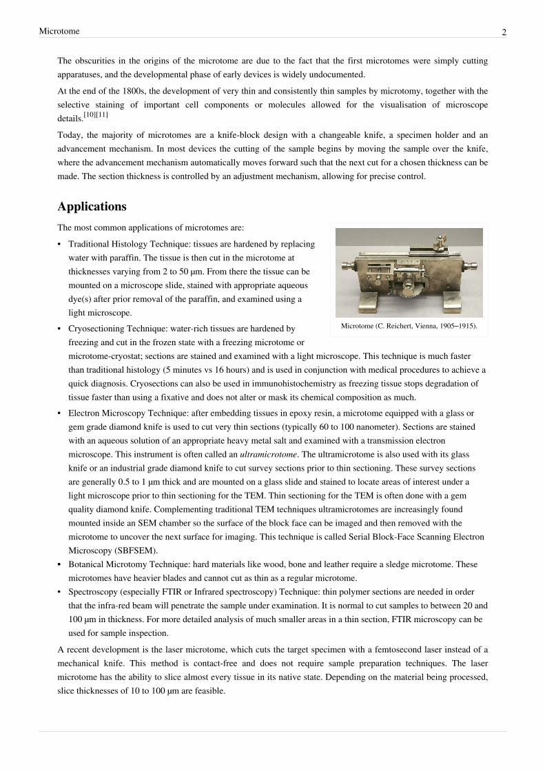
Microtome (C. Reichert, Vienna, 1905–1915).
The most common applications of microtomes are:• Traditional Histology Technique: tissues are hardened by replacing
water with paraffin. The tissue is then cut in the microtome atthicknesses varying from 2 to 50 µm. From there the tissue can bemounted on a microscope slide, stained with appropriate aqueousdye(s) after prior removal of the paraffin, and examined using alight microscope.
• Cryosectioning Technique: water-rich tissues are hardened byfreezing and cut in the frozen state with a freezing microtome ormicrotome-cryostat; sections are stained and examined with a light microscope. This technique is much fasterthan traditional histology (5 minutes vs 16 hours) and is used in conjunction with medical procedures to achieve aquick diagnosis. Cryosections can also be used in immunohistochemistry as freezing tissue stops degradation oftissue faster than using a fixative and does not alter or mask its chemical composition as much.
• Electron Microscopy Technique: after embedding tissues in epoxy resin, a microtome equipped with a glass orgem grade diamond knife is used to cut very thin sections (typically 60 to 100 nanometer). Sections are stainedwith an aqueous solution of an appropriate heavy metal salt and examined with a transmission electronmicroscope. This instrument is often called an ultramicrotome. The ultramicrotome is also used with its glassknife or an industrial grade diamond knife to cut survey sections prior to thin sectioning. These survey sectionsare generally 0.5 to 1 µm thick and are mounted on a glass slide and stained to locate areas of interest under alight microscope prior to thin sectioning for the TEM. Thin sectioning for the TEM is often done with a gemquality diamond knife. Complementing traditional TEM techniques ultramicrotomes are increasingly foundmounted inside an SEM chamber so the surface of the block face can be imaged and then removed with themicrotome to uncover the next surface for imaging. This technique is called Serial Block-Face Scanning ElectronMicroscopy (SBFSEM).
•• Botanical Microtomy Technique: hard materials like wood, bone and leather require a sledge microtome. Thesemicrotomes have heavier blades and cannot cut as thin as a regular microtome.
• Spectroscopy (especially FTIR or Infrared spectroscopy) Technique: thin polymer sections are needed in orderthat the infra-red beam will penetrate the sample under examination. It is normal to cut samples to between 20 and100 µm in thickness. For more detailed analysis of much smaller areas in a thin section, FTIR microscopy can beused for sample inspection.
A recent development is the laser microtome, which cuts the target specimen with a femtosecond laser instead of amechanical knife. This method is contact-free and does not require sample preparation techniques. The lasermicrotome has the ability to slice almost every tissue in its native state. Depending on the material being processed,slice thicknesses of 10 to 100 µm are feasible.

Microtome 3
Microtome types
Sledge microtome
A sled microtome.
A sledge microtome is a device where the sample is placed into a fixedholder (shuttle), which then moves backwards and forwards across aknife. Modern sled microtomes have the sled placed upon a linearbearing, a design that allows for the microtome to readily cut manycoarse sections.[12] By adjusting the angles between the sample and themicrotome knife, the pressure applied to the sample during the cut canbe reduced.[12] Typical applications for this design of microtome are ofthe preparation of large samples, such as those embedded in paraffinfor biological preparations. Typical cut thickness achievable on asledge microtome is between 1 and 60 µm.
Rotary microtome
A rotary microtome of older construction.
This instrument is a common microtome design. This device operateswith a staged rotary action such that the actual cutting is part of therotary motion. In a rotary microtome, the knife is typically fixed in ahorizontal position.[13]
In the figure to the left, the principle of the cut is explained. Throughthe motion of the sample holder, the sample is cut by the knife position1 to position 2), at which point the fresh section remains on the knife.At the highest point of the rotary motion, the sample holder isadvanced by the same thickness as the section that is to be made,allowing for the next section to be made.The flywheel in many microtomes can be operated by hand. This hasthe advantage that a clean cut can be made, as the relatively large massof the flywheel prevents the sample from being stopped during thesample cut. The flywheel in newer models is often integrated inside themicrotome casing. The typical cut thickness for a rotary microtome is between 1 and 60 µm. For hard materials, suchas a sample embedded in a synthetic resin, this design of microtome can allow for good "Semi-thin" sections with athickness of as low as 0.5 µm.

Microtome 4
Cryomicrotome
A cryomicrotome.
For the cutting of frozen samples, many rotary microtomes can beadapted to cut in a liquid nitrogen chamber, in a so-calledcryomicrotome setup. The reduced temperature allows for the hardnessof the sample to be increased, such as by undergoing a glass transition,which allows for the preparation of semi-thin samples.[12] However thesample temperature and the knife temperature must be controlled inorder to optimise the resultant sample thickness
Ultramicrotome
A ribbon of ultrathin sections prepared by roomtemperature ultramicrotomy, floating on water in theboat of a diamond knife used to cut the sections. The
knife blade is the edge at the upper end of the trough ofwater.
An ultramicrotome is a main tool of ultramicrotomy. It can allowfor the preparation of extremely thin sections, with the devicefunctioning in the same manner as a rotational microtome, butwith very tight tolerances on the mechanical construction. As aresult of the careful mechanical construction, the linear thermalexpansion of the mounting is used to provide very fine control ofthe thickness.[12]
These extremely thin cuts are important for use with transmissionelectron microscope (TEM) and Serial Block-Face ScanningElectron Microscopy (SBFSEM), and are sometimes alsoimportant for light-optical microscopy.[13] The typical thickness ofthese cuts is between 40 and 100 nm for transmission electronmicroscopy and often between 30 and 50 nm for SBFSEM.Thicker sections up to 500 nm thick are also taken for specializedTEM applications or for light microscopy survey sections to selectan area for the final thin sections. Diamond knives (preferably) and glass knives are used with ultramicrotomes. Tocollect the sections they are floated on top of a liquid as they are cut and are carefully picked up onto grids suitablefor TEM specimen viewing. The thickness of the section can be estimated by the thin-film interference colors ofreflected light that are seen as a result of the extremely low sample thickness.[14]
Vibrating microtomeThe vibrating microtome operates by cutting using a vibrating blade, allowing the resultant cut to be made with lesspressure than would be required for a stationary blade. The vibrating microtome is usually used for difficultbiological samples.[12] The cut thickness is usually around 30-500 µm for live tissue and 10-500 µm for fixed tissue.
Saw microtomeThe saw microtome is especially for hard materials such as teeth or bones. The microtome of this type has a recessedrotating saw, which slices through the sample. The minimal cut thickness is approximately 30 µm, and can be madefor comparatively large samples.[12]

Microtome 5
Laser microtome
A conceptual diagram of laser microtomeoperation.
The laser microtome is an instrument for contact free slicing.[15] Priorpreparation of the sample through embedding, freezing or chemicalfixation is not required, thereby minimizing the artifacts frompreparation methods. Alternately this design of microtome can also beused for very hard materials, such as bones or teeth as well as someceramics. Dependent upon the properties of the sample material, thethickness achievable is between 10 and 100 µm.
The device operates using a cutting action of an infra-red laser. As thelaser emits a radiation in the near infra-red, in this wavelength regimethe laser can interact with biological materials. Through sharp focusingof the probe within the sample, a focal point of very high intensity, up to TW/cm2, can be achieved. Through thenon-linear interaction of the optical penetration in the focal region a material separation in a process known asphoto-disruption is introduced. By limiting the laser pulse durations to the femtoseconds range, the energy expendedat the target region is precisely controlled, thereby limiting the interaction zone of the cut to under a micrometre.External to this zone the ultra-short beam application time introduces minimal to no thermal damage to theremainder of the sample.
The laser radiation is directed onto a fast scanning mirror based optical system which allows for three dimensionalpositioning of the beam crossover, whilst allowing for beam traversal to the desired region of interest. Thecombination of high power with a high raster rate allows the scanner to cut large areas of sample in a short time. Inthe laser microtome the laser-microdissection of internal areas in tissues, cellular structures, and other types of smallfeatures is also possible.
Microtome knives
A diamond knife blade used for cutting ultrathinsections (typically 70 to 350 nm) for transmission
electron microscopy.
The design of a microtome knife is dependant upon the material andpreparation of the samples, as well as the final sample requirements(e.g. cut thickness and quality).

Microtome 6
Knife design and cut types
Profiles of microtome knives.
Generally, knives are characterized by the profile of the knife blade,which falls under the categories of planar concave, wedge shaped orchisel shaped designs.Planar concave microtome knives are extremely sharp, but are alsovery delicate and are therefore only used with very soft samples.[13]
The wedge profile knives are somewhat more stable and find use inmoderately hard materials, such as in epoxy or cryogenic samplecutting. Finally, the chisel profile with its blunt edge, raises thestability of the knife, whilst requiring significantly more force toachieve the cut.
For ultramicrotomes, glass and diamond knives are required, the cutbreadth of the blade is therefore on the order of a few millimetres andis therefore significantly smaller than for classical microtome knives.Glass knives are usually manufactured by the fracture of glass barsusing special "knife-maker" fracturing devices. Glass knives may beused for initial sample preparations even where diamond knives maybe used for final sectioning. Glass knives usually have small troughs,made with plastic tape, which are filled with water to allow the sampleto float for later collection.[12] Diamond blades may be built into suchan existing trough, allowing for the same collection method.
Sectioning
Prior to cutting by microtomy, biological materials are usually placed in a more rigid fixative, in a process known asembedding. This is achieved by the inflow of a liquid substance around the sample, such as paraffin (wax) or epoxy,which is placed in a mould and later hardened to produce a "block" which is readily cut.The declination is the angle of contact between the sample vertical and knife blade. If the knife blade is at rightangles (declination=90) the cut is made directly using a pressure based mode, and the forces are thereforeproportionally larger. If however the knife is tilted, the relative motion of the knife is increasingly parallel to samplemotion, allowing for a slicing action. This behaviour is very important for large or hard samplesThe inclination of the knife is the angle between the knife face and the sample. For an optimal result, this angle mustbe chosen appropriately. The optimal angle depends upon the knife geometry, the cut speed and many otherparameters. If the angle is adjusted to zero, the knife cut can often become erratic, and a new location of the knifemust be used to smooth this out.If the angle is too large, the sample can crumple and the knife can induce periodic thickness variations in the cut. Byfurther increasing the angle such that it is too large one can damage the knife blade itself.

Microtome 7
References[1] Hill, John (1770). The Construction of Timber, from its early growth; Explained by Microscope, and proven from Experiments, in a great
Variety of Kinds. (http:/ / www. archive. org/ details/ constructiontim00hillgoog). London. pp. 5–11, Plate I. .[2] Quekett, John (1848). A Practical Treatise on the use of the Microscope (http:/ / www. archive. org/ details/ practicaltreatis00quekuoft).
London: Hippolyte Bailliere. pp. 306, Chapter XII (Microtomes and Microtome Knives). .[3] Anonymous (1910). "An eighteenth century Microtome". Journal of the Royal Microscopical Society (Oxford, England: The Royal
Microscopical Society): 779–782.[4] Gilbert Morgan Smith: The Development of Botanical Microtechnique. In: Transactions of the American Microscopical Society 34, Nr. 2.
1915, S. 71–129, ( PDF-Version of the article) (http:/ / scientificobjects. mpiwg-berlin. mpg. de/ scientificobjects/ dms/ResearchNetworkDocuments/ basicdocuments/ V1_Smith--technique1915/ V1_Smith, technique1915. pdf)
[5] "Wilhelm His" (http:/ / www. britannica. com/ EBchecked/ topic/ 266898/ Wilhelm-His). Encyclopædia Britannica Online. EncyclopædiaBritannica. . Retrieved 24. März 2009.
[6] Loukas, Marios; Pamela Clarke, R. Shane Tubbs, Theodoros Kapos and Margit Trotz (2008). "The His family and their contributions tocardiology" (http:/ / linkinghub. elsevier. com/ retrieve/ pii/ S0167527307003907). International Journal of Cardiology (Ireland: Elsevier) 123(2): 75–78. doi:10.1016/j.ijcard.2006.12.070. ISSN 0167-5273. PMID 17433467. . Retrieved 24 März 2009.
[7] "Histology" (http:/ / encarta. msn. com/ encyclopedia_761573144/ histology. html). msn Encarta. . Retrieved 18 March 2009.[8] Detlev Ganten: Handbuch der molekularen Medizin (Handbook of molecular medicine), Springer, ISBN 3-540-64552-7, ( Google-Books
(http:/ / books. google. de/ books?id=DjwuzN4XvLMC& printsec=frontcover#PPA548,M1))[9] Werner Gerabek, Bernhard D. Haage, Gundolf Keil, Wolfgang Wegner (2005): Enzyklopädie Medizingeschichte (Encyclopaedia of medical
history), Walter de Gruyter, ISBN 3-11-015714-4, ( Google-Books (http:/ / books. google. de/ books?id=LLoOUP-y54YC&printsec=frontcover#PPA1203,M1))
[10] Ernst Mayr (2002). [ Google-Books (http:/ / books. google. de/ books?id=Y_HvUDa4OqwC& pg=PA533) Die Entwicklung derbiologischen Gedankenwelt. (The evolution of the biological thought )]. Springer. ISBN 3-540-43213-2. .
[11] Werner Linß, Werner Linb, Jochen Fanghänel: Histologie: Zytologie, allgemeine Histologie, mikroskopische Anatomie. (Histology:Cytology, general Histology, microscopial anatomy) Walter de Gruyter, 1998, ISBN 3-11-014032-2 ( Google-Books (http:/ / books. google.de/ books?id=S1HRxeGOQfMC& pg=PA1& dq=Purkinjes+ mikrotom& as_brr=3))
[12] Gudrun Lang (2006). Histotechnik. Praxislehrbuch für die Biomedizinische Analytik. (Histology : practical textbook for analyticalbiomedicine). Springer, Wien/New York. ISBN 3-211-33141-7.
[13] Klaus Henkel: Das Schneiden mit dem Mikrotom (http:/ / www. mikroskopie-muenchen. de/ cut-mikrotom. html). MikrobiologischeVereinigung München e. V., 2006, accessed 15 February 2009
[14] Peachey Lee D. (1958). "Thin Sections: A study of section thickness and physical distortion produced during microtomy" (http:/ / jcb.rupress. org/ cgi/ reprint/ 4/ 3/ 233. pdf). J. Biophysic. & Biochem. Cytol. 4 (3): 233–242. doi:10.1083/jcb.4.3.233. .
[15] Holger Lubatschowski 2007: Laser Microtomy, WILEY-VCH Verlag GmbH, Biophotonics, S. 49-51, ( PDF (http:/ / www. photonicnet. de/Aktuelles/ partner/ 2007/ 06/ laser_microtomy_optik-photonik_juni_2007. pdf)).

Article Sources and Contributors 8
Article Sources and ContributorsMicrotome Source: http://en.wikipedia.org/w/index.php?oldid=534413767 Contributors: AhMedRMaaty, Ajusted dog, Alexbateman, Antonello41, Arcadian, Belizefan, BillC, Bobblewik,Bobrayner, Brim, Calliopejen1, Chevymontecarlo, Chivesud, Christopher.booth, Daniel Case, DocWatson42, Drnehatyagi, Edgar181, EmmanuelM, Foxd3, FrozenMan, Gits (Neo), Goterpaws,Gurch, Hesperian, HundiVarg, Ivan.Romero, Japanese Searobin, Kkmurray, Lasermedi1, LilHelpa, Miaow Miaow, Ndaener, Nhandler, Nikevich, OldakQuill, Peterlewis, Pietrow, Rjwilmsi,Robert.Baruch, Rustavo, Saperaud, Snek01, Tassedethe, The Thing That Should Not Be, Twinsday, User A1, Valérie75, Verne Equinox, Vniizht, WahreJakob, Walter1954, Xblkx, Ygramul,Zephyris, 45 anonymous edits
Image Sources, Licenses and ContributorsFile:Ultramicrotome 2265 EM GD MB.jpg Source: http://en.wikipedia.org/w/index.php?title=File:Ultramicrotome_2265_EM_GD_MB.jpg License: Creative Commons Attribution-Sharealike2.0 Contributors: Leica Microsystems, GmbHFile:Cummings 1774 Microtome.jpg Source: http://en.wikipedia.org/w/index.php?title=File:Cummings_1774_Microtome.jpg License: Public Domain Contributors: John Hills 1774, Fileassembled by Mirko Junge from scanns fount at archive.orgFile:Microtome1905.JPG Source: http://en.wikipedia.org/w/index.php?title=File:Microtome1905.JPG License: Creative Commons Attribution-ShareAlike 3.0 Unported Contributors:TamorlanFile:Sledge microtome.jpg Source: http://en.wikipedia.org/w/index.php?title=File:Sledge_microtome.jpg License: Creative Commons Attribution-Sharealike 3.0,2.5,2.0,1.0 Contributors: YvanLindekensFile:Microtome-1.jpg Source: http://en.wikipedia.org/w/index.php?title=File:Microtome-1.jpg License: Creative Commons Attribution-Sharealike 2.5 Contributors: User:snek01File:Cryostat microtome.jpg Source: http://en.wikipedia.org/w/index.php?title=File:Cryostat_microtome.jpg License: Creative Commons Attribution-Sharealike 2.0 Contributors: OrlandoFile:Microtome-ultras.jpg Source: http://en.wikipedia.org/w/index.php?title=File:Microtome-ultras.jpg License: GNU Free Documentation License Contributors: euphras. Original uploaderwas Euphras at de.wikipediaFile:Laser-microtome-schematic.png Source: http://en.wikipedia.org/w/index.php?title=File:Laser-microtome-schematic.png License: Creative Commons Attribution-Sharealike3.0,2.5,2.0,1.0 Contributors: Prinzip_Lasermikrotom.jpg: Holger Lubatschowski derivative work: User A1 (talk)File:Diamond Knife Blade Edge.jpg Source: http://en.wikipedia.org/w/index.php?title=File:Diamond_Knife_Blade_Edge.jpg License: Creative Commons Attribution 3.0 Contributors:Richard Wheeler (Zephyris)File:Microtome-knife-profile.svg Source: http://en.wikipedia.org/w/index.php?title=File:Microtome-knife-profile.svg License: GNU Free Documentation License Contributors: derivativework, Rainer Ziel original author
LicenseCreative Commons Attribution-Share Alike 3.0 Unported//creativecommons.org/licenses/by-sa/3.0/