modeling of flap endonuclease interactions with dna...
TRANSCRIPT
Modeling of Flap Endonuclease Interactions withDNA Substrate
Hatim T. Allawi1*, Michael W. Kaiser1, Alexey V. Onufriev2, Wu-Po Ma1
Andrew E. Brogaard1, David A. Case2, Bruce P. Neri1 andVictor I. Lyamichev1
1Third Wave Technologies, Inc.502 S Rosa Road, Madison, WI53719, USA
2Department of MolecularBiology, The Scripps ResearchInstitute, La Jolla, CA 92037USA
Structure-specific 50 nucleases play an important role in DNA replicationand repair uniquely recognizing an overlap flap DNA substrate and pro-cessing it into a DNA nick. However, in the absence of a high-resolutionstructure of the enzyme/DNA complex, the mechanism underlying thisrecognition and substrate specificity, which is key to the enzyme’s func-tion, remains unclear. Here, we propose a three-dimensional model ofthe structure-specific 50 flap endonuclease from Pyrococcus furiosus in itscomplex with DNA. The model is based on the known X-ray structure ofthe enzyme and a variety of biochemical and molecular dynamics (MD)data utilized in the form of distance restraints between the enzyme andthe DNA. Contacts between the 50 flap endonuclease and the sugar–phos-phate backbone of the overlap flap substrate were identified usingenzyme activity assays on substrates with methylphosphonate or 20-O-methyl substitutions. The enzyme footprint extends two to four base-pairs upstream and eight to nine base-pairs downstream of the cleavagesite, thus covering 10–13 base-pairs of duplex DNA. The footprint dataare consistent with a model in which the substrate is bound in the DNA-binding groove such that the downstream duplex interacts with thehelix-hairpin-helix motif of the enzyme. MD simulations to identifythe substrate orientation in this model are consistent with the results ofthe enzyme activity assays on the methylphosphonate and 20-O-methyl-modified substrates. To further refine the model, 50 flap endonucleasevariants with alanine point substitutions at amino acid residues expectedto contact phosphates in the substrate and one deletion mutant weretested in enzyme activity assays on the methylphosphonate-modified sub-strates. Changes in the enzyme footprint observed for two point mutants,R64A and R94A, and for the deletion mutant in the enzyme’s bA/bB
region, were interpreted as being the result of specific interactions in theenzyme/DNA complex and were used as distance restraints in MD simu-lations. The final structure suggests that the substrate’s 50 flap interactswith the enzyme’s helical arch and that the helix-hairpin-helix motif inter-acts with the template strand in the downstream duplex eight base-pairsfrom the cleavage site. This model suggests specific interactions betweenthe 30 end of the upstream oligonucleotide and the enzyme. The proposedstructure presents the first detailed description of substrate recognition bystructure-specific 50 nucleases.
q 2003 Elsevier Science Ltd. All rights reserved
Keywords: structure-specific 50 nuclease; FEN1; point mutation;methylphosphonate; 20-O-methyl*Corresponding author
Introduction
Structure-specific 50 nucleases have been isolatedfrom different organisms including bacterio-phages,1,2 eubacteria,3 – 5 archaea,6 – 8 yeast9,10 and
0022-2836/03/$ - see front matter q 2003 Elsevier Science Ltd. All rights reserved
E-mail address of the corresponding author:[email protected]
Abbreviations used: HhH, helix-hairpin-helix.
doi:10.1016/S0022-2836(03)00351-6 J. Mol. Biol. (2003) 328, 537–554
mammals.11 – 14 The ubiquitous presence of theseenzymes is explained by their essential role inOkazaki fragment processing15 – 17 and repair ofDNA damage caused by alkylating agents or UVradiation.9,18,19 In Okazaki fragment processing, dis-placement synthesis by DNA polymerases gener-ates branched DNA structures in which theupstream and downstream strands overlap andcompete for the same sequence of the templatestrand. The branched structure can exist inmultiple conformations depending on the positionof the branch point between the upstream anddownstream strands on the shared templatesequence.15,20 The structure-specific 50 nucleasesknown as flap endonucleases (FEN1)12 specificallyrecognize a conformation called the overlap flapor 30 one-nucleotide double-flap structure inwhich the upstream strand, excluding the 30 endnucleotide, is annealed to the template strand, dis-placing the 50 portion of the downstreamstrand.8,21,22 FEN1 cleaves the downstream strandof the overlap flap structure precisely after thefirst base-paired nucleotide, creating a ligatablenick.8,22
The conclusion that the 30 end nucleotide of theupstream strand is not base-paired with the tem-
plate has been put forth from the observation thatany of the four natural bases at this position cansupport efficient cleavage.21 It was suggested thatthe 30 end nucleotide of the upstream strand inter-acts with the enzyme to position the substrate inan optimal orientation for cleavage. The demon-stration that sugar modifications of the 30 endnucleotide inhibit activity of FEN1 enzymes hasfurther supported this hypothesis.8,22 The import-ance of the overlapping 30 end nucleotide of theupstream strand was not originally recognized,and many laboratories characterized FEN1enzymes using a flap structure that included adja-cent upstream and downstream strands annealedto a template, but lacking a gap or overlap.10 FEN1enzymes cleave such a flap substrate inefficiently,producing a majority of products that are notligatable. The existence of these products can beexplained by the formation of alternative struc-tures with bulged nucleotides stabilized by the 50
nuclease in an effort to force the overlap flapstructure.8,21,22
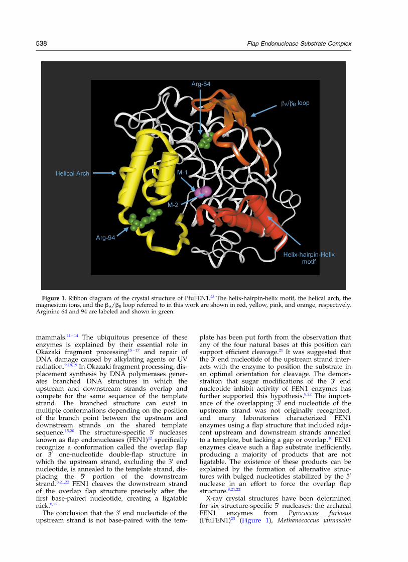
X-ray crystal structures have been determinedfor six structure-specific 50 nucleases: the archaealFEN1 enzymes from Pyrococcus furiosus(PfuFEN1)23 (Figure 1), Methanococcus jannaschii
Figure 1. Ribbon diagram of the crystal structure of PfuFEN1.23 The helix-hairpin-helix motif, the helical arch, themagnesium ions, and the bA/bB loop referred to in this work are shown in red, yellow, pink, and orange, respectively.Arginine 64 and 94 are labeled and shown in green.
538 Flap Endonuclease Substrate Complex
(MjaFEN1)7 and Pyrococcus horikoshii (PhoFEN1);24
the 50 nuclease domain of eubacterial DNApolymerase from Thermus aquaticus (TaqExo);25 the50 –30 exonuclease from bacteriophage T5,26 andthe RNase H enzyme from bacteriophage T4.27
These structures reveal a common a/b topologyand similar structural motifs despite a low aminoacid sequence identity and similarity between thearchaeal, eubacterial and bacteriophage FEN1groups (see, for example, Hosfield et al.23). All ofthese enzymes have been shown to bind twodivalent metal ions which form a complex networkof interactions with highly conserved acidic aminoacids lying at the bottom of a positively chargedcleft. One metal ion is presumably involved incatalysis and the other in the DNA binding.28 Inthe PfuFEN1 structure, the magnesium ion M-1,involved in catalysis, is located in close proximityto the cluster of amino acid residues Asp27,Asp80, Glu152, and Glu154; and the magnesiumion M-2 involved in substrate binding interactswith the cluster of amino acid residues Asp173,Asp175, and Asp236 approximately 5 A fromM-1.
The helical arch is a common structural motifshared by the FEN1 enzymes and was originallyidentified in the T5 50 –30 exonuclease structure.26
The arch is located close to the enzyme’s activesite and forms a flexible loop that can accommo-date single-stranded but not double-strandedDNA. The motif provides structural support forthe hypothesis that the 50 flap of DNA substratethreads through a hole to translocate DNA to theenzyme’s active site. The threading mechanismwas originally proposed to explain biochemicaldata that blocking the free 50 end of the flap with abulky modification or rendering it double-strandedusing a complementary oligonucleotide suppressesthe cleavage efficiency and can even trap FEN1 onthe 50 flap.5,29 While most studies agree on thethreading mechanism, Bambara and his grouphave shown that a variety of bulky flap modifi-cations can be tolerated by human FEN1endonuclease.30
Another common fold shared by the FEN1enzymes is the helix-hairpin-helix (HhH) motiffound in many enzyme families.31,32 This type offold is involved in non-sequence-specific bindingof duplex DNA via interactions with the sugar–phosphate backbone of one of the strands.33,34
Together with the helical arch and network ofamino acids interacting with the M-1 and M-2ions, the HhH motif defines a positively chargedactive-site DNA-binding groove in FEN1. In Pfu-FEN1, the DNA-binding groove is 32 A wide and44 A long, suggesting that it can accommodate a12 base-pair double-stranded DNA.23 Biochemicalanalysis of point mutations at the DNA-bindinggroove of the FEN1 enzymes revealed conservedamino acid residues on the surface of the grooveinvolved in catalysis and substrate binding.24,28,35 – 39
Structural and functional similarity between the50 nucleases suggests a common mechanism for
substrate binding and catalysis for all enzymes inthis family. In the absence of co-crystal or NMRstructures for a 50 nuclease/DNA complex, severalmodels of the complex have been proposed toelucidate the mechanism of substratebinding.7,23,26,40 These models suggest that the sub-strate binds at the active-site DNA-binding groovewith the cleavable phosphodiester linkage close tothe metal ion involved in catalysis and with the 50
flap threading through the helical arch.Methylphosphonate and 20-O-methyl substi-
tutions have proven to be powerful methods foridentifying contacts between nucleic acids andproteins.41 – 46 Methylphosphonate substitutions arealmost isosteric with phosphodiester linkages, butunlike phosphodiester linkages are neutral andtherefore can be used to identify ionic interactionsin protein/substrate complexes without intro-ducing steric clashes with the proteins.41 Methyl-phosphonate linkages have been shown to inducelocal bending in the double-helical DNA axis bythe mechanism of asymmetric phosphate chargeneutralization. However, the bending angleestimated as 3.58 per methylphosphonatesubstitution47 is comparable to the intrinsicsequence-specific DNA bending48 and thermalflexibility of duplex DNA of ,78 per base-pairestimated from its persistence length.49 Substi-tution of a methyl group in place of a non-bridgingoxygen in the phosphodiester linkage at a point ofelectrostatic contact with a protein usuallydecreases the affinity of substrate binding.41 Thisproperty of methylphosphonate modificationsmakes unnecessary, in most cases, the separationof Rp and Sp stereoisomers of chemically intro-duced methylphosphonate linkages and justifiesthe use of their racemic mixtures. 20-O-Methyl sub-stitutions replace the 20 proton in the deoxyribosering with a bulky O-methyl group with two majoroutcomes for duplex DNA structure. First, 20-O-methyl groups change the conformational prefer-ence of ribose from C20-endo to C30-endo sugarpuckering, forcing a local transition from B-formto A-form DNA. Second, they introduce stericclashes at sites of contacts with the proteins.45
Here, we introduced a single methylphospho-nate substitution into each phosphodiester linkageof the overlap flap DNA substrate to map phos-phates interacting with PfuFEN1. Similarly, weintroduced 20-O-methyl substitutions to identifysteric contacts in the PfuFEN1/DNA complex.Using the three-dimensional structure ofPfuFEN123 and a modeled structure of the overlapflap substrate, we performed energy minimization(EM) and molecular dynamics (MD) simulationsto test two alternative structures of the PfuFEN1/DNA complex. The model consistent with themethylphosphonate data was used to identify can-didate amino acid residues contacting phosphatesin the substrate. To confirm the predicted inter-actions, PfuFEN1 variants mutated at theseamino acid residues were tested on the methyl-phosphonate substrates. The confirmed
Flap Endonuclease Substrate Complex 539
interactions were used as restraints in MDsimulations to develop a detailed model of thePfuFEN1/DNA complex.
Results
Substrate for enzyme activity assays
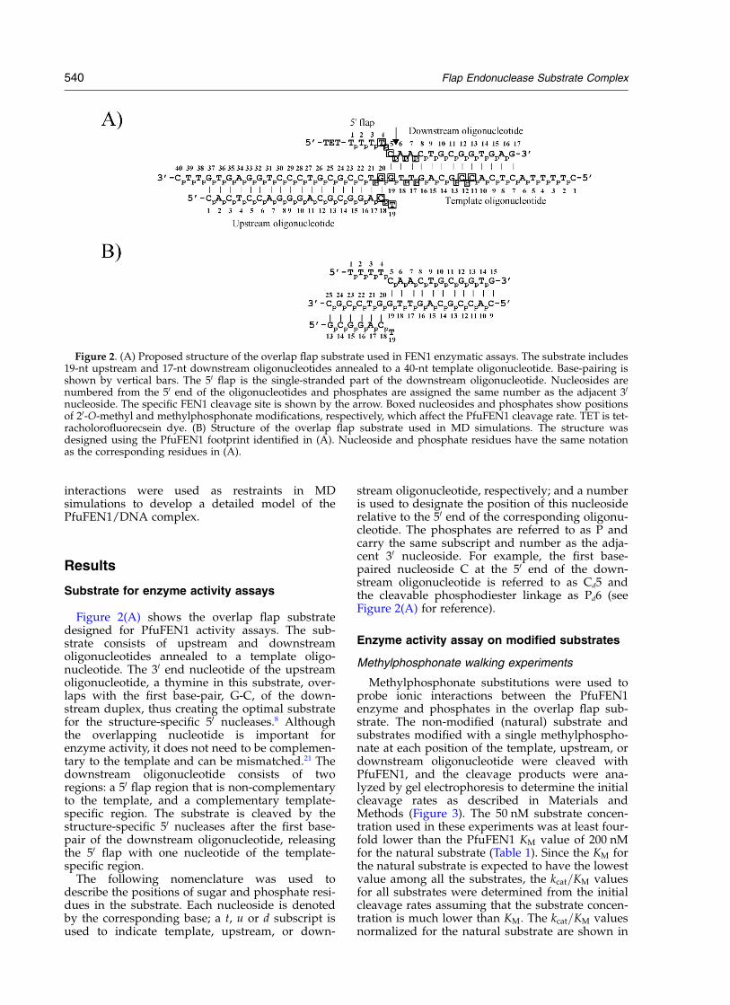
Figure 2(A) shows the overlap flap substratedesigned for PfuFEN1 activity assays. The sub-strate consists of upstream and downstreamoligonucleotides annealed to a template oligo-nucleotide. The 30 end nucleotide of the upstreamoligonucleotide, a thymine in this substrate, over-laps with the first base-pair, G-C, of the down-stream duplex, thus creating the optimal substratefor the structure-specific 50 nucleases.8 Althoughthe overlapping nucleotide is important forenzyme activity, it does not need to be complemen-tary to the template and can be mismatched.21 Thedownstream oligonucleotide consists of tworegions: a 50 flap region that is non-complementaryto the template, and a complementary template-specific region. The substrate is cleaved by thestructure-specific 50 nucleases after the first base-pair of the downstream oligonucleotide, releasingthe 50 flap with one nucleotide of the template-specific region.
The following nomenclature was used todescribe the positions of sugar and phosphate resi-dues in the substrate. Each nucleoside is denotedby the corresponding base; a t, u or d subscript isused to indicate template, upstream, or down-
stream oligonucleotide, respectively; and a numberis used to designate the position of this nucleosiderelative to the 50 end of the corresponding oligonu-cleotide. The phosphates are referred to as P andcarry the same subscript and number as the adja-cent 30 nucleoside. For example, the first base-paired nucleoside C at the 50 end of the down-stream oligonucleotide is referred to as Cd5 andthe cleavable phosphodiester linkage as Pd6 (seeFigure 2(A) for reference).
Enzyme activity assay on modified substrates
Methylphosphonate walking experiments
Methylphosphonate substitutions were used toprobe ionic interactions between the PfuFEN1enzyme and phosphates in the overlap flap sub-strate. The non-modified (natural) substrate andsubstrates modified with a single methylphospho-nate at each position of the template, upstream, ordownstream oligonucleotide were cleaved withPfuFEN1, and the cleavage products were ana-lyzed by gel electrophoresis to determine the initialcleavage rates as described in Materials andMethods (Figure 3). The 50 nM substrate concen-tration used in these experiments was at least four-fold lower than the PfuFEN1 KM value of 200 nMfor the natural substrate (Table 1). Since the KM forthe natural substrate is expected to have the lowestvalue among all the substrates, the kcat=KM valuesfor all substrates were determined from the initialcleavage rates assuming that the substrate concen-tration is much lower than KM: The kcat=KM valuesnormalized for the natural substrate are shown in
Figure 2. (A) Proposed structure of the overlap flap substrate used in FEN1 enzymatic assays. The substrate includes19-nt upstream and 17-nt downstream oligonucleotides annealed to a 40-nt template oligonucleotide. Base-pairing isshown by vertical bars. The 50 flap is the single-stranded part of the downstream oligonucleotide. Nucleosides arenumbered from the 50 end of the oligonucleotides and phosphates are assigned the same number as the adjacent 30
nucleoside. The specific FEN1 cleavage site is shown by the arrow. Boxed nucleosides and phosphates show positionsof 20-O-methyl and methylphosphonate modifications, respectively, which affect the PfuFEN1 cleavage rate. TET is tet-racholorofluorecsein dye. (B) Structure of the overlap flap substrate used in MD simulations. The structure wasdesigned using the PfuFEN1 footprint identified in (A). Nucleoside and phosphate residues have the same notationas the corresponding residues in (A).
540 Flap Endonuclease Substrate Complex
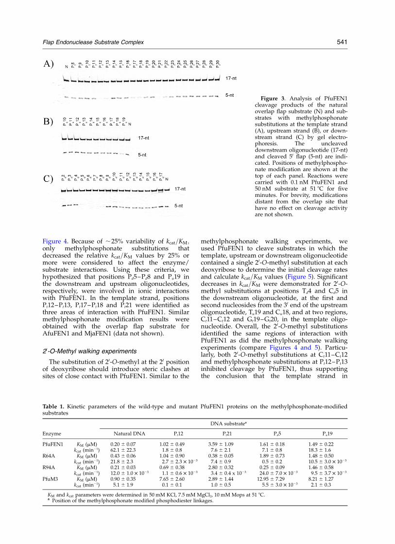
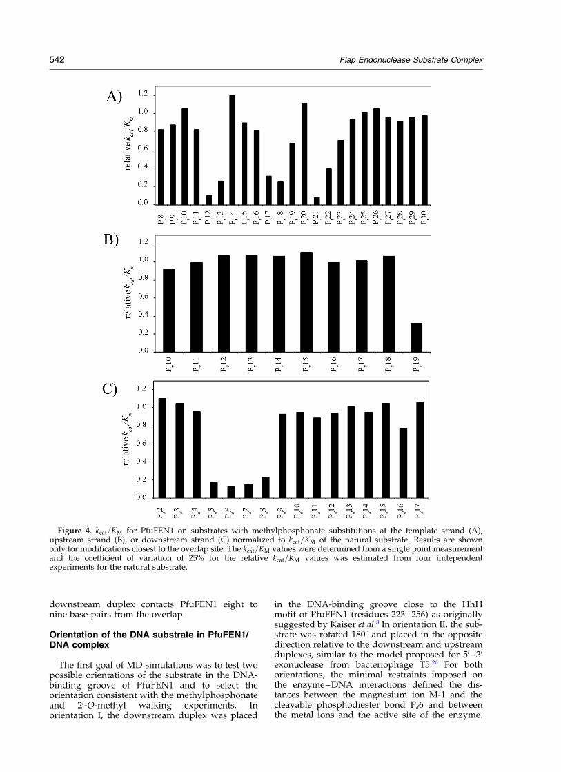
Figure 4. Because of ,25% variability of kcat=KM;only methylphosphonate substitutions thatdecreased the relative kcat=KM values by 25% ormore were considered to affect the enzyme/substrate interactions. Using these criteria, wehypothesized that positions Pd5–Pd8 and Pu19 inthe downstream and upstream oligonucleotides,respectively, were involved in ionic interactionswith PfuFEN1. In the template strand, positionsPt12–Pt13, Pt17–Pt18 and Pt21 were identified asthree areas of interaction with PfuFEN1. Similarmethylphosphonate modification results wereobtained with the overlap flap substrate forAfuFEN1 and MjaFEN1 (data not shown).
20-O-Methyl walking experiments
The substitution of 20-O-methyl at the 20 positionof deoxyribose should introduce steric clashes atsites of close contact with PfuFEN1. Similar to the
methylphosphonate walking experiments, weused PfuFEN1 to cleave substrates in which thetemplate, upstream or downstream oligonucleotidecontained a single 20-O-methyl substitution at eachdeoxyribose to determine the initial cleavage ratesand calculate kcat=KM values (Figure 5). Significantdecreases in kcat=KM were demonstrated for 20-O-methyl substitutions at positions Td4 and Cd5 inthe downstream oligonucleotide, at the first andsecond nucleosides from the 30 end of the upstreamoligonucleotide, Tu19 and Cu18, and at two regions,Ct11–Ct12 and Gt19–Gt20, in the template oligo-nucleotide. Overall, the 20-O-methyl substitutionsidentified the same regions of interaction withPfuFEN1 as did the methylphosphonate walkingexperiments (compare Figures 4 and 5). Particu-larly, both 20-O-methyl substitutions at Ct11–Ct12and methylphosphonate substitutions at Pt12–Pt13inhibited cleavage by PfuFEN1, thus supportingthe conclusion that the template strand in
Table 1. Kinetic parameters of the wild-type and mutant PfuFEN1 proteins on the methylphosphonate-modifiedsubstrates
DNA substratea
Enzyme Natural DNA Pt12 Pt21 Pd5 Pu19
PfuFEN1 KM (mM) 0.20 ^ 0.07 1.02 ^ 0.49 3.59 ^ 1.09 1.61 ^ 0.18 1.49 ^ 0.22kcat (min21) 62.1 ^ 22.3 1.8 ^ 0.8 7.6 ^ 2.1 7.1 ^ 0.8 18.3 ^ 1.6
R64A KM (mM) 0.43 ^ 0.06 1.04 ^ 0.90 0.38 ^ 0.05 1.89 ^ 0.73 1.48 ^ 0.50kcat (min21) 21.8 ^ 2.3 2.7 ^ 2.3 £ 1023 7.4 ^ 0.9 0.5 ^ 0.2 10.5 ^ 3.0 £ 1023
R94A KM (mM) 0.21 ^ 0.03 0.69 ^ 0.38 2.80 ^ 0.32 0.25 ^ 0.09 1.46 ^ 0.58kcat (min21) 12.0 ^ 1.0 £ 1023 1.1 ^ 0.6 £ 1023 3.4 ^ 0.4 x 10²³ 24.0 ^ 7.0 £ 1023 9.5 ^ 3.7 £ 1023
PfuM3 KM (mM) 0.90 ^ 0.35 7.65 ^ 2.60 2.89 ^ 1.44 12.95 ^ 7.29 8.21 ^ 1.27kcat (min21) 5.1 ^ 1.9 0.1 ^ 0.1 1.0 ^ 0.5 5.5 ^ 3.0 £ 1023 2.1 ^ 0.3
KM and kcat parameters were determined in 50 mM KCl, 7.5 mM MgCl2, 10 mM Mops at 51 8C.a Position of the methylphosphonate modified phosphodiester linkages.
Figure 3. Analysis of PfuFEN1cleavage products of the naturaloverlap flap substrate (N) and sub-strates with methylphosphonatesubstitutions at the template strand(A), upstream strand (B), or down-stream strand (C) by gel electro-phoresis. The uncleaveddownstream oligonucleotide (17-nt)and cleaved 50 flap (5-nt) are indi-cated. Positions of methylphospho-nate modification are shown at thetop of each panel. Reactions werecarried with 0.1 nM PfuFEN1 and50 nM substrate at 51 8C for fiveminutes. For brevity, modificationsdistant from the overlap site thathave no effect on cleavage activityare not shown.
Flap Endonuclease Substrate Complex 541
downstream duplex contacts PfuFEN1 eight tonine base-pairs from the overlap.
Orientation of the DNA substrate in PfuFEN1/DNA complex
The first goal of MD simulations was to test twopossible orientations of the substrate in the DNA-binding groove of PfuFEN1 and to select theorientation consistent with the methylphosphonateand 20-O-methyl walking experiments. Inorientation I, the downstream duplex was placed
in the DNA-binding groove close to the HhHmotif of PfuFEN1 (residues 223–256) as originallysuggested by Kaiser et al.8 In orientation II, the sub-strate was rotated 1808 and placed in the oppositedirection relative to the downstream and upstreamduplexes, similar to the model proposed for 50–30
exonuclease from bacteriophage T5.26 For bothorientations, the minimal restraints imposed onthe enzyme–DNA interactions defined the dis-tances between the magnesium ion M-1 and thecleavable phosphodiester bond Pd6 and betweenthe metal ions and the active site of the enzyme.
Figure 4. kcat=KM for PfuFEN1 on substrates with methylphosphonate substitutions at the template strand (A),upstream strand (B), or downstream strand (C) normalized to kcat=KM of the natural substrate. Results are shownonly for modifications closest to the overlap site. The kcat=KM values were determined from a single point measurementand the coefficient of variation of 25% for the relative kcat=KM values was estimated from four independentexperiments for the natural substrate.
542 Flap Endonuclease Substrate Complex

Table 2 summarizes the minimal restraints used inthe MD calculations.
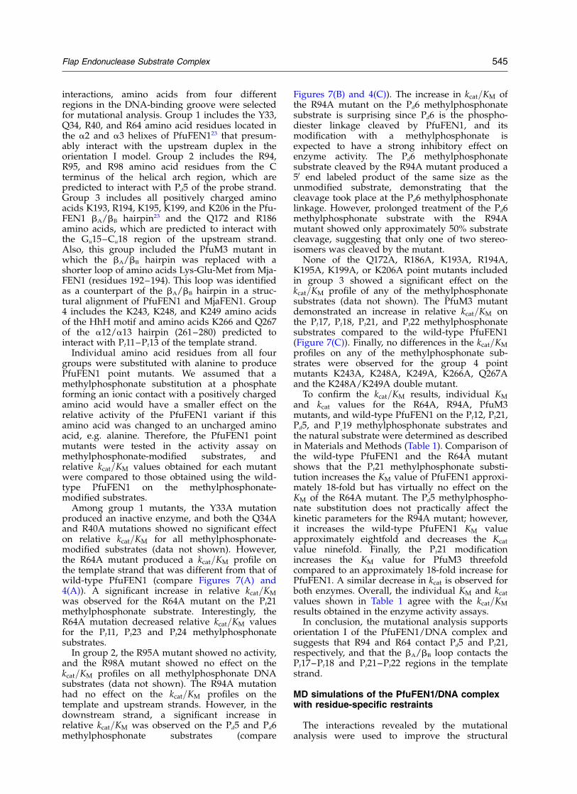
MD simulations predict different structures fororientations I and II (Figure 6). In particular, inorientation I, the template strand (green) in thedownstream duplex interacts with the HhH motif(red) in the Ct11–Ct14 region six to nine base-pairsfrom the overlap. On the other hand, in orientationII, PfuFEN1 enzyme contacts with the templatestrand in the downstream duplex are confined tothe Gt16–Gt19 region. The interactions predictedin orientation I, but not those in orientation II, are
consistent with the results of enzyme activityassays on the methylphosphonate and 20-O-methyl-modified substrates, which suggest thatthe Pt12–Pt13 phosphates and Ct11–Ct12 deoxy-ribose residues are involved in contacts withPfuFEN1.
Mutational analysis of interactions in thePfuFEN1/DNA complex
To provide additional support for orientationI and to identify specific PfuFEN1/DNA
Figure 5. kcat=KM values for PfuFEN1 on substrates with 20-O-methyl substitutions at the template strand (A),upstream strand (B), or downstream strand (C) normalized to kcat=KM of the natural substrate. Results are shownonly for modifications closest to the overlap site. The kcat=KM values were determined from a single point measurementand the coefficient of variation of 25% for the relative kcat=KM values was estimated from four independentexperiments for the natural substrate.
Flap Endonuclease Substrate Complex 543
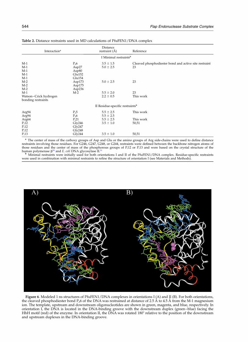
Table 2. Distance restraints used in MD calculations of PfuFEN1/DNA complex
Interactiona
Distancerestraint (A) Reference
I Minimal restraintsb
M-1 Pd6 3.5 ^ 1.5 Cleaved phosphodiester bond and active site restraintM-1 Asp27 5.0 ^ 2.5 23M-1 Asp80M-1 Glu152M-1 Glu154M-2 Asp173 5.0 ^ 2.5 23M-2 Asp175M-2 Asp236M-1 M-2 5.5 ^ 2.0 23Watson–Crick hydrogenbonding restraints
2.2 ^ 0.5 This work
II Residue-specific restraintsb
Arg94 Pd5 5.5 ^ 2.5 This workArg94 Pd6 5.5 ^ 2.5Arg64 Pt21 5.5 ^ 2.5 This workPt12 Gly246 3.5 ^ 1.0 50,51Pt12 Gly247Pt12 Gly248Pt13 Gly244 3.5 ^ 1.0 50,51
a The center of mass of the carboxy groups of Asp and Glu or the amino groups of Arg side-chains were used to define distancerestraints involving those residues. For G246, G247, G248, or G244, restraints were defined between the backbone nitrogen atoms ofthose residues and the center of mass of the phosphorous groups of Pt12 or Pt13 and were based on the crystal structure of thehuman polymerase b50 and E. coli DNA glycosylase II.51
b Minimal restraints were initially used for both orientations I and II of the PfuFEN1/DNA complex. Residue-specific restraintswere used in combination with minimal restraints to refine the structure of orientation I (see Materials and Methods).
Figure 6. Modeled 1 ns structures of PfuFEN1/DNA complexes in orientations I (A) and II (B). For both orientations,the cleaved phosphodiester bond Pd6 of the DNA was restrained at distance of 2.5 A to 4.5 A from the M-1 magnesiumion. The template, upstream and downstream oligonucleotides are shown in green, magenta, and blue, respectively. Inorientation I, the DNA is located in the DNA-binding groove with the downstream duplex (green–blue) facing theHhH motif (red) of the enzyme. In orientation II, the DNA was rotated 1808 relative to the position of the downstreamand upstream duplexes in the DNA-binding groove.
544 Flap Endonuclease Substrate Complex
interactions, amino acids from four differentregions in the DNA-binding groove were selectedfor mutational analysis. Group 1 includes the Y33,Q34, R40, and R64 amino acid residues located inthe a2 and a3 helixes of PfuFEN123 that presum-ably interact with the upstream duplex in theorientation I model. Group 2 includes the R94,R95, and R98 amino acid residues from the Cterminus of the helical arch region, which arepredicted to interact with Pd5 of the probe strand.Group 3 includes all positively charged aminoacids K193, R194, K195, K199, and K206 in the Pfu-FEN1 bA/bB hairpin23 and the Q172 and R186amino acids, which are predicted to interact withthe Gu15–Cu18 region of the upstream strand.Also, this group included the PfuM3 mutant inwhich the bA/bB hairpin was replaced with ashorter loop of amino acids Lys-Glu-Met from Mja-FEN1 (residues 192–194). This loop was identifiedas a counterpart of the bA/bB hairpin in a struc-tural alignment of PfuFEN1 and MjaFEN1. Group4 includes the K243, K248, and K249 amino acidsof the HhH motif and amino acids K266 and Q267of the a12/a13 hairpin (261–280) predicted tointeract with Pt11–Pt13 of the template strand.
Individual amino acid residues from all fourgroups were substituted with alanine to producePfuFEN1 point mutants. We assumed that amethylphosphonate substitution at a phosphateforming an ionic contact with a positively chargedamino acid would have a smaller effect on therelative activity of the PfuFEN1 variant if thisamino acid was changed to an uncharged aminoacid, e.g. alanine. Therefore, the PfuFEN1 pointmutants were tested in the activity assay onmethylphosphonate-modified substrates, andrelative kcat=KM values obtained for each mutantwere compared to those obtained using the wild-type PfuFEN1 on the methylphosphonate-modified substrates.
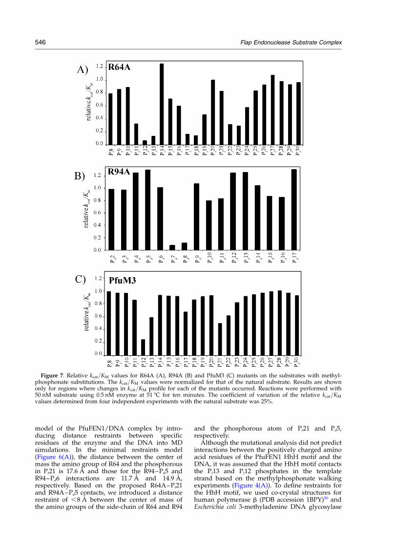
Among group 1 mutants, the Y33A mutationproduced an inactive enzyme, and both the Q34Aand R40A mutations showed no significant effecton relative kcat=KM for all methylphosphonate-modified substrates (data not shown). However,the R64A mutant produced a kcat=KM profile onthe template strand that was different from that ofwild-type PfuFEN1 (compare Figures 7(A) and4(A)). A significant increase in relative kcat=KM
was observed for the R64A mutant on the Pt21methylphosphonate substrate. Interestingly, theR64A mutation decreased relative kcat=KM valuesfor the Pt11, Pt23 and Pt24 methylphosphonatesubstrates.
In group 2, the R95A mutant showed no activity,and the R98A mutant showed no effect on thekcat=KM profiles on all methylphosphonate DNAsubstrates (data not shown). The R94A mutationhad no effect on the kcat=KM profiles on thetemplate and upstream strands. However, in thedownstream strand, a significant increase inrelative kcat=KM was observed on the Pd5 and Pd6methylphosphonate substrates (compare
Figures 7(B) and 4(C)). The increase in kcat=KM ofthe R94A mutant on the Pd6 methylphosphonatesubstrate is surprising since Pd6 is the phospho-diester linkage cleaved by PfuFEN1, and itsmodification with a methylphosphonate isexpected to have a strong inhibitory effect onenzyme activity. The Pd6 methylphosphonatesubstrate cleaved by the R94A mutant produced a50 end labeled product of the same size as theunmodified substrate, demonstrating that thecleavage took place at the Pd6 methylphosphonatelinkage. However, prolonged treatment of the Pd6methylphosphonate substrate with the R94Amutant showed only approximately 50% substratecleavage, suggesting that only one of two stereo-isomers was cleaved by the mutant.
None of the Q172A, R186A, K193A, R194A,K195A, K199A, or K206A point mutants includedin group 3 showed a significant effect on thekcat=KM profile of any of the methylphosphonatesubstrates (data not shown). The PfuM3 mutantdemonstrated an increase in relative kcat=KM onthe Pt17, Pt18, Pt21, and Pt22 methylphosphonatesubstrates compared to the wild-type PfuFEN1(Figure 7(C)). Finally, no differences in the kcat=KM
profiles on any of the methylphosphonate sub-strates were observed for the group 4 pointmutants K243A, K248A, K249A, K266A, Q267Aand the K248A/K249A double mutant.
To confirm the kcat=KM results, individual KM
and kcat values for the R64A, R94A, PfuM3mutants, and wild-type PfuFEN1 on the Pt12, Pt21,Pd5, and P
u19 methylphosphonate substrates and
the natural substrate were determined as describedin Materials and Methods (Table 1). Comparison ofthe wild-type PfuFEN1 and the R64A mutantshows that the Pt21 methylphosphonate substi-tution increases the KM value of PfuFEN1 approxi-mately 18-fold but has virtually no effect on theKM of the R64A mutant. The Pd5 methylphospho-nate substitution does not practically affect thekinetic parameters for the R94A mutant; however,it increases the wild-type PfuFEN1 KM valueapproximately eightfold and decreases the Kcat
value ninefold. Finally, the Pt21 modificationincreases the KM value for PfuM3 threefoldcompared to an approximately 18-fold increase forPfuFEN1. A similar decrease in kcat is observed forboth enzymes. Overall, the individual KM and kcat
values shown in Table 1 agree with the kcat=KM
results obtained in the enzyme activity assays.In conclusion, the mutational analysis supports
orientation I of the PfuFEN1/DNA complex andsuggests that R94 and R64 contact Pd5 and Pt21,respectively, and that the bA/bB loop contacts thePt17–Pt18 and Pt21–Pt22 regions in the templatestrand.
MD simulations of the PfuFEN1/DNA complexwith residue-specific restraints
The interactions revealed by the mutationalanalysis were used to improve the structural
Flap Endonuclease Substrate Complex 545
model of the PfuFEN1/DNA complex by intro-ducing distance restraints between specificresidues of the enzyme and the DNA into MDsimulations. In the minimal restraints model(Figure 6(A)), the distance between the center ofmass the amino group of R64 and the phosphorousin Pt21 is 17.6 A and those for the R94–Pd5 andR94–Pd6 interactions are 11.7 A and 14.9 A,respectively. Based on the proposed R64A–Pt21and R94A–Pd5 contacts, we introduced a distancerestraint of ,8 A between the center of mass ofthe amino groups of the side-chain of R64 and R94
and the phosphorous atom of Pt21 and Pd5,respectively.
Although the mutational analysis did not predictinteractions between the positively charged aminoacid residues of the PfuFEN1 HhH motif and theDNA, it was assumed that the HhH motif contactsthe Pt13 and Pt12 phosphates in the templatestrand based on the methylphosphonate walkingexperiments (Figure 4(A)). To define restraints forthe HhH motif, we used co-crystal structures forhuman polymerase b (PDB accession 1BPY)50 andEscherichia coli 3-methyladenine DNA glycosylase
Figure 7. Relative kcat=KM values for R64A (A), R94A (B) and PfuM3 (C) mutants on the substrates with methyl-phosphonate substitutions. The kcat=KM values were normalized for that of the natural substrate. Results are shownonly for regions where changes in kcat=KM profile for each of the mutants occurred. Reactions were performed with50 nM substrate using 0.5 nM enzyme at 51 8C for ten minutes. The coefficient of variation of the relative kcat=KM
values determined from four independent experiments with the natural substrate was 25%.
546 Flap Endonuclease Substrate Complex
II (1DIZ)51 that demonstrate interactions of thecognate HhH motifs with double-stranded DNAsubstrates. The HhH interactions in these struc-tures occur through hydrogen bonds of ,3.3 Abetween oxygen atoms of two adjacent DNA phos-phates and the backbone nitrogen atoms of theG105, G107, 109, and A110 residues in polymeraseb and the G214, G216, and T219 residues inglycosylase. The corresponding residues in thePfuFEN1 HhH motif, G244, G246, K248, and K249,were identified by structure-based sequencealignment.32 In the minimal restraints model, thedistances between the backbone nitrogen atoms ofG244, G246, K248, and K249 and the nearestoxygen of Pt13 or Pt12 are 10.9, 15.6, 18.2, and18.2 A, respectively. To introduce hydrogen bondinteractions observed in the co-crystals, thedistances between the G244, G246, K248, and K249backbone nitrogen atoms and a center of mass ofphosphorus and two non-bridging oxygen atomsof Pt13 and Pt12 were restrained to 3.5(^1.0) A.Table 2 summarizes the residue-specific restraintsused in the MD calculations.
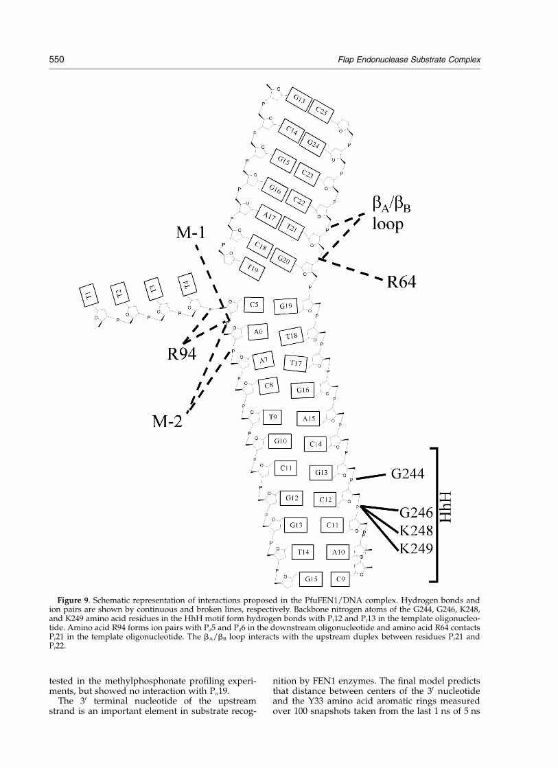
Figure 8 shows a 5 ns MD structure of the Pfu-FEN1/DNA complex using residue-specificrestraints. In the residue-specific model, the sub-strate moved deeper in the active-site DNA-bind-ing groove compared with the minimal restraintsmodel. For instance, the distance between thecenter of mass of the arginine amino groups andphosphorous in R64–Pt21, R94–Pd5, and R94–Pd6pairs is decreased to 3.8, 3.8, and 4.3 A,respectively. The DNA also moved closer to theHhH motif to form hydrogen bonds O2–Pt13–G244, O2–Pt12–G246, O1–Pt12–K248, andO1–Pt12–K249 of 2.77, 3.7, 3.3, and 3.0 A, respect-ively. In addition, positively charged residues ofthe HhH motif demonstrate interactions with Pd8in the downstream strand and with the majorgroove of the downstream duplex in the Ct11–Ct13 region. The upstream and downstreamduplexes are in the B-form conformation and forman angle of approximately 408 between duplexaxes at the overlap. The M-1 magnesium ion inter-acts with the cleavable phosphodiester bond Pd6and the M-2 ion, which is not bound by anyrestraints, interacts with the Pd6 and Pd7 phos-phates. Interactions predicted by the final structureare summarized in Figure 9.
Discussion
Here, we have modeled a three-dimensionalstructure of the PfuFEN1 structure-specific 50
nuclease in its complex with DNA using MD simu-lations guided by enzyme–DNA distancerestraints derived from experimental data. Regionsof the overlap flap substrate involved in specificcontacts with PfuFEN1 were identified usingmethylphosphonate and 20-O-methyl walkingexperiments. Most contacts were observed close tothe cleavage site, however, an additional region of
both electrostatic and steric interactions was identi-fied on the template oligonucleotide at positionsPt12, Pt13, Ct11, and Ct12 (Figure 2(A)). The asym-metry of the footprint, which extends nine base-pairs into the downstream duplex and only twobase-pairs into the upstream duplex, agrees wellwith analyses of enzyme activity on overlap flapsubstrates of different length8 and phosphate ethyl-ation interference experiments with a flap substrateand the 50 nuclease domain of DNA polymerase Ifrom E. coli.52
The PfuFEN1 footprint was used to determinethe orientation of the substrate in the DNA-bindinggroove. Originally proposed models for the 50
nuclease/DNA complex suggested that theupstream duplex of the flap substrate interactswith the HhH motif of the 50 nucleaseenzymes.7,26,53 However, based on the minimal sub-strate length requirements, Kaiser et al.8 proposedthat the HhH motif interacts with the downstreamduplex of the overlap flap substrate. A similarmodel was also proposed by Dervan et al.40 fromkinetic and binding characteristics of T5 50 nucleasemutants using hairpin-like substrates containingonly a downstream duplex. MD simulations usingonly minimal restraints specifying distancebetween the cleavable linkage Pd6 and M-1 mag-nesium ion showed that only orientation I, inwhich the downstream duplex interacts with theHhH motif, was consistent with the asymmetricpattern of the PfuFEN1 footprint and wasproposed as a minimal restraints model of thePfuFEN1/DNA complex (Figure 6(A)).
Binding of DNA substrates often induces signifi-cant conformational changes in protein structure.For example, comparison of the apo and DNAcomplex structures of the Klenow fragment ofE. coli DNA polymerase I demonstrates that thethumb region of the protein moves as much as12 A to accommodate the DNA.54 There is no indi-cation of induced conformational changes in theminimal restraints model, and it is unlikely thatsuch changes, which may occur at a microsecondor even millisecond time range,55 would beobserved in nanosecond-long MD models. Theimportance of these considerations can be illus-trated by the nature of the interactions betweenthe HhH motif and the downstream duplex pre-dicted by the minimal restraints model to occurthrough ionic contacts between the side-chains ofK248 and K249 and the Pt12–Pt13 phosphates.These interactions contradict the co-crystal struc-tures of human polymerase b50 and E. coli 3-methyladenine DNA glycosylase II,51 demonstrat-ing that the HhH motif interacts with double-stranded DNA by forming hydrogen bondsbetween the backbone nitrogen atoms ofconserved HhH residues and oxygen atoms oftwo adjacent DNA phosphates. The contactssuggested from the co-crystal structures wouldrequire that the downstream duplex in the minimalrestraints model move closer to the PfuFEN1 HhHmotif and form specific hydrogen bonds with
Flap Endonuclease Substrate Complex 547
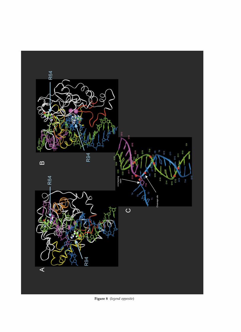
Figure 8 (legend opposite)
backbone nitrogen atoms of G244, G246, K248, andK249.
To experimentally determine specific inter-actions in the PfuFEN1/DNA complex, we usedenzyme activity assays with mutant PfuFEN1enzymes on methylphosphonate-containing sub-strates. The basis for this approach has beenpreviously proven for specific steric interactionsbetween the MS2 bacteriophage coat protein andits cognate RNA hairpin.56 The methylphosphonateprofiling of the PfuFEN1 mutants identifiedspecific ionic contacts between R94 and Pd5 andbetween R64 and Pt21. A similar approach waspreviously used to identify specific contactsbetween the 50 nuclease domain of DNA polymer-ase I from E. coli and a flap substrate52 with methyl-phosphonate substitutions studied in thedownstream strand at positions corresponding toPd3, Pd4, Pd5 and Pd7. The authors observed asimilar inhibitory effect on the cleavage rate of sub-strate modified at positions analogous to Pd5 andPd7 and, based on mutational analysis, suggestedthat these phosphates interact with Y77, K78, andR81 and R20 of the E. coli 50 nuclease domain,respectively. Alignment of the E. coli 50 nucleasedomains and TaqExo sequences followed by three-dimensional structure alignment of TaqExo andPfuFEN1 suggests that Y77, K78, R81, and R20 ofthe E. coli 50 nuclease domain correspond to R94,R95, R98, and Q34 of PfuFEN1, respectively. Herewe show that R94 contacts Pd5, but the R95Amutation results in an inactive enzyme and boththe Q34A and R98A mutations have no significanteffect on the methylphosphonate profiling. Thesedifferences can be explained by a misalignment ofthe E. coli 50 nuclease domain and PfuFEN1 due tolow sequence similarity and differences in thebinding characteristics of the overlap flap and flapsubstrates.
Introduction of the residue-specific restraintsmoved the substrate deeper in the DNA-bindinggroove compared to the minimal restraints model.Superposition of enzyme structures in the minimaland final models shows that the Pt13 and Pt12phosphates in the downstream duplex move by,20 A and the upstream duplex moves by 17 Aon average. The tighter contacts between the DNAand enzyme became possible because of confor-mational changes in the enzyme structure: the Ca
atoms of the bA/bB loop (192–206) moved by 11 Aon average with a maximum distance of 16.5 Aobserved for P197. The final PfuFEN1/DNA
model was challenged to explain differencesbetween the methylphosphonate and 20-O-methylPfuFEN1 footprints. The latter identifies stericinteractions that have not been used to specify thedistance restraints in MD simulations. In thedownstream strand, these differences are mani-fested by an effect of the Pd6, Pd7, and Pd8 methyl-phosphonate substitutions on kcat=KM and the lackof such an effect for the Ad6 and Ad7 20-O-methylsubstitutions (Figure 2(A)). The modeled structureexplains this pattern by the existence of contactsbetween Pd6–Pd8 and the magnesium ions andK248 whereas the Ad6 and Ad7 nucleosides areexposed in solution outside the DNA-bindinggroove and do not contact PfuFEN1. Conversely,the Gt20 and Gt19 20-O-methyl modifications, butnot the Pt20 and Pt19 methylphosphonate modifi-cations, affect kcat=KM: Again, the model predictsthat the Gt20 and Gt19 nucleosides face the DNA-binding groove, and the 20-O-methyl substitutionsat these positions are likely to cause a steric clashwith PfuFEN1. To the contrary, no specific inter-actions are predicted for Pt20 and Pt19.
There are several interactions suggested from thePfuFEN1 footprints that cannot be explained by thefinal model. For example, methylphosphonatesubstitutions at Pt17 and Pt18 decrease kcat=KM
(Figure 4(A)); however, no contacts involvingthese phosphates are predicted in the model.Potential candidates for these contacts include agroup of positively charged lysine residues 87, 89,93 and arginine residues 94, 95, 98, 106 in thehelical arch region; however, the methylphospho-nate profiling does not support this hypothesis forthe tested amino acid residues, R94 and R98, andthe R95A mutation has produced an inactiveenzyme. Interaction between the helical arch andthe Pt17 and Pt18 phosphates might be onlypossible as a result of large substrate-induced con-formational changes in the helical arch, such aspreviously suggested for human FEN1 fromFourier transform infrared spectroscopy demon-strating an increase in helicity of the helical archregion upon DNA binding.57 Some interactionssuggested in the model and supported by PfuFEN1footprint data still need to be confirmed by muta-tional analysis. For example, according to themodel, Pu19 forms ion pairs of 2.9 A, 3.0 A, and3.9 A with K104, K111, and R40, respectively, andthe methylphosphonate substitution at Pu19decreases kcat=KM value of PfuFEN1 (Figure 4(B)).Only one of these amino acid residues, R40, was
Figure 8. 5 ns MD model of the PfuFEN1/DNA complex with residue-specific restraints. (A) A view showing theoverlap flap substrate bound in the DNA-binding groove of PfuFEN1. The HhH motif, bA/bB hairpin, helical archand magnesium ions are shown in red, orange, yellow and pink, respectively. Arginine residues 94 and 64 are dis-played with a space-filled model. The template, upstream and downstream oligonucleotides are shown in green,magenta, and blue, respectively. (B) 90 degree rotated view of the model shown in (A). (C) A cartoon description ofthe substrate structure in the model (adopted from the view (A)). The 30 end overlapping base of the upstream oligonu-cleotide is shown by arrows. The phosphodiester linkages Pd5, Pd6, Pt12, Pt13, Pt17, Pt18, Pt21, and Pu19 are shown withasterisks.
Flap Endonuclease Substrate Complex 549
tested in the methylphosphonate profiling experi-ments, but showed no interaction with Pu19.
The 30 terminal nucleotide of the upstreamstrand is an important element in substrate recog-
nition by FEN1 enzymes. The final model predictsthat distance between centers of the 30 nucleotideand the Y33 amino acid aromatic rings measuredover 100 snapshots taken from the last 1 ns of 5 ns
Figure 9. Schematic representation of interactions proposed in the PfuFEN1/DNA complex. Hydrogen bonds andion pairs are shown by continuous and broken lines, respectively. Backbone nitrogen atoms of the G244, G246, K248,and K249 amino acid residues in the HhH motif form hydrogen bonds with Pt12 and Pt13 in the template oligonucleo-tide. Amino acid R94 forms ion pairs with Pd5 and Pd6 in the downstream oligonucleotide and amino acid R64 contactsPt21 in the template oligonucleotide. The bA/bB loop interacts with the upstream duplex between residues Pt21 andPt22.
550 Flap Endonuclease Substrate Complex
MD simulation is ,6(^2) A. This distance corre-sponds to a maximum of distance distribution foraromatic amino acid pairs in X-ray crystal struc-tures of non-homologous proteins58 suggestingstacking interactions between the 30 nucleotideand the Y33 residue. In support of this conclusion,Y33 is highly conserved in the FEN1 family, andsubstitution of this amino acid with alanine or glu-tamic acid inactivates PfuFEN1, but its substitutionwith another aromatic amino acid, phenylalanine,reduces PfuFEN1 activity only by 50% (data notshown). The exact position of the 50 flap, whichaccording to the threading model should be withinthe helical arch of PfuFEN1,5,29 is not defined in thefinal model. During the dynamics the flap movesin and out of the helical arch due to the high flexi-bility of both the helical arch and the 50 flap, thusmaking it difficult to interpret its specific contacts.It is possible that conformational changessuggested for the helical arch should be includedin the model to determine specific interactions ofthe 50 flap and the 30 terminal nucleotide of theupstream strand.
Significant progress in understanding the mech-anism of lagging strand synthesis in eukaryotesled to a model describing the synthesis as a seriesof highly coordinated steps performed by theOkazaki fragment processing complex, whichincludes FEN1.59,60 It is thought that the synchro-nized product release in one step and its bindingas a substrate in the next step is governed by sub-strate specificity of the proteins constituting thecomplex. FEN1 plays a critical role in the transitionfrom the strand displacement step performed byeukaryotic polymerase d to the 50 flap removalstep and in the passing of the nick substrate toDNA ligase I. The first detailed model ofPfuFEN1/DNA complex proposed here is animportant step in understanding the organizationof Okazaki fragment processing complex anddynamics of complex rearrangement during thelagging strand maturation. Given the consistencyof our model with methylphosphonate and 20-O-methyl walking results, we believe that it providesa correct general orientation of the overlap flapsubstrate in the active-site DNA-binding groove ofPfuFEN1 and defines likely contacts in the Pfu-FEN1/DNA complex. The modeled PfuFEN1/DNA structure may be further improved by incor-porating new experimental data.
Materials and Methods
DNA synthesis and purification
DNA oligonucleotides were synthesized with anExpedite 8909 synthesizer (PerSeptive Biosystems) usingstandard phosphoramidite chemistry. All phosphorami-dites including tetracholorofluorecsein (TET) dye, 20-O-methyl, and methylphosphonate phosphoramiditeswere purchased from Glen Research, Sterling, VA. Syn-thesis and deprotection were performed according to
the vendor’s recommended procedures and furtherpurification was performed by electrophoresis on a 20%(w/v) denaturing acrylamide gel as described.8 Methyl-phosphonate oligonucleotides were used withoutseparation of Rp and Sp isomers. The purity of all oligo-nucleotides used in enzyme activity assays was analyzedby capillary electrophoresis on a P/ACE MDQ CE sys-tem (Beckman) with mPAGE-5 capillaries (Agilent Tech-nologies, Palo Alto, CA). Oligonucleotide concentrationswere determined from the absorption at 260 nm usingextinction coefficients for nucleosides and dinucleotidemonophosphates.61
Mutagenesis
The Y33A, Q34A, R40A, R64A, R94A, R95A, R98A,Q172A, R186A, K193A, R194A, K195A, K199A, K206A,K243A, K248A, K249A, K266A, and Q267A mutantswere prepared by site-directed mutagenesis with a Quik-change kit (Stratagene) using 30 cycles of PCR amplifica-tion on the pTrc99a-PfuFEN1 plasmid DNA8 with200 nM mutagenic primers. After Dpn I digestion theamplified DNA was transformed into E. coli DH5a byelectroporation. The presence of desired mutations wasverified by sequencing. The PfuM3 deletion variant wasprepared by replacing amino acid residues GKRKLPGKNVYVEIK of PfuFEN1 at position 192–206 with aminoacids KEM of MjaFEN1 (192–194) using recombinantPCR. The 50 half of the PfuM3 gene was amplified with50-TGTGGAATTGTGAGCGG (pTrcFwd) and 50-CTCTGGCATCTCCTTTGTTATTGTTAAGTTTCTAAC primersand the 30 half was amplified with 50-ACAAAGGAGATGCCAGAGTTGATAATTTTGGAGGAAG and 50-TAATCTGTATCAGGCTG (pTrcRev) primers. The DNA pro-ducts were purified from a 1% (w/v) agarose gel usinga Qiaquick kit (Qiagen) and used in PCR reaction withthe pTrcFwd and pTrcRev primers. The amplified DNAwas gel-purified and cloned into pTrc99a vector asdescribed.8
Enzyme expression and purification
The plasmids carrying PfuFEN1 variants were trans-formed into E. coli BL21 (Novagen) for expression andpurification of the proteins was performed as described.8
At the final step of purification the proteins weredialyzed and diluted in a buffer containing 50% (v/v)glycerol, 20 mM Tris–HCl (pH 8), 50 mM KCl, 0.5%Tween 20, 0.5% Nonidet P40, 100 mg/mL BSA.
Kinetic parameter measurement and enzymeactivity assays
Kinetic parameters, KM and kcat; for the wild-type andmutant PfuFEN1 enzymes were determined with thesubstrate shown in Figure 2(A). Reactions were preparedby mixing the downstream, upstream and templateoligonucleotides at 5 mM concentration each in a buffercontaining 100 mM Mops, 75 mM MgCl2 and heatingthe sample to 95 8C for one minute and slow cooling toroom temperature. The annealed substrate was stored at220 8C. The kinetics experiments were performed atdifferent substrate concentrations from 10 nM to 800 nMand constant enzyme concentration between 0.1 nM and100 nM, depending on the specific activity of a particularenzyme. Enzyme/substrate mixtures were prepared in100 ml 10 mM Mops (pH 7.5), 50 mM KCl, 7.5 mMMgCl2, 10 ng/ml tRNA on ice, and the reactions were
Flap Endonuclease Substrate Complex 551
started by incubation at 51 8C. Aliquots (10 ml) of eachreaction were then transferred into pre-chilled tubes con-taining 15 ml 95% formamide, 10 mM EDTA, and 0.02%(w/v) methyl violet blue (Sigma-Aldrich) at time pointsof 5, 10, 15, 30, and 60 minutes. Aliquots (5 ml) of thestopped reactions were then analyzed by gel electro-phoresis to obtain initial rates of the cleavage reactionsas described.8 The KM and kcat values were calculatedfrom the initial rates using the Michaelis–Mentenequation. The enzyme activity assay used for fastmeasurement of kcat=KM values for wild-type and mutantPfuFEN1 enzymes was performed in 10 ml 10 mM Mops(pH 7.5), 50 mM KCl, 7.5 mM MgCl2, 10 ng/ml tRNAincluding 0.1–5 nM enzyme and 50 nM natural or modi-fied substrate. The reactions were assembled on ice andthen incubated at 51 8C for periods of time sufficient toreach 5–20% substrate cleavage. The kcat=KM valueswere calculated as the initial cleavage rate divided bythe enzyme and initial substrate concentrations underthe assumption that the initial substrate concentrationwas much lower than KM:
Molecular modeling
PfuFEN1 structure coordinates used in MD simu-lations were obtained from the PDB database, accessionnumber 1B4323 and coordinates for the magnesium ionswere provided by J. A. Tainer (personal communi-cations). To reduce computation time, the C terminusresidues 290–340 of PfuFEN1 were omitted. MD simu-lations were preformed with AMBER 6.0 software.62,63
A 2 fs time-step was employed and SHAKE was usedfor bonds involving hydrogen atoms. The generalizedBorn (GB) solvation model was used with a non-bondedinteractions cut-off distance of either 8 A or 12 A and0.2 M monovalent salt concentration.64 – 66 A 1 ns MDrequired ,six days of computation on a cluster of fourcomputers with dual 1.5 GHz Athlon (AMD) processorsand Linux Mandrake 8.1 operating system. A B-formDNA duplex of the overlap flap substrate (Figure 2(B))was generated using the Biopolymer software (InsightII-Accelrys, Inc.) subjected to 100 steps of gradient EMfollowed by 250 ps of dynamics. To assemble the initialcomplex, the substrate was manually placed in theactive-site DNA-binding groove of PfuFEN1 with anaverage distance of 3–15 A from the enzyme with thecleavable phosphodiester linkage facing the M-1 andM-2 metal ions. The MD protocol used here includedEM, simulated annealing (SA), and production stages.During the EM stage, the initial PfuFEN1/DNA complexassembly was subjected to 200 steps of conjugate gradi-ent EM to relax energetically unfavorable interactions.Following the EM stage, an SA stage was performedduring which the system was gradually heated from 0 Kto 500 K for 6 ps using a heat bath time coupling constantof 2 ps, then the system was slowly cooled to 300 K overa period of 14 ps while gradually decreasing the heatbath constant to 0.5 ps. During the SA stage, all enzymeresidues except the two magnesium ions, the helicalarch (residues 75–127), the bA/bB loop (184–210), andthe HhH motif (223–256) were constrained to theircrystallographic positions using a harmonic potentialforce constant kc of 5 kcal/mol/A2. Additionally, DNAresidues except Au17–Tu19, Td1–Ad7, and Tt17–Tt21were restrained using a flat-well harmonic potential of2.25(^0.50) A for Watson–Crick hydrogen bonds anda ¼ 265(^60)8, b ¼ 2150(^45)8, g ¼ 40(^45)8,d ¼ 120(^90)8, e ¼ 170(^60)8, z ¼ 270(^90)8, andx ¼ 2125(^90)8 for dihedral angels. Finally, flat-well
harmonic minimal restraints were used to define a dis-tance of 3.50(^1.50) A between the M-1 ion and thecleavable phosphodiester linkage Pd6 and a distance of5.0(^2.50) A between the magnesium ions, M-1 andM-2, and the active site residues (Table 2). During theSA stage, the restraint penalties were gradually intro-duced by increasing their harmonic potential force con-stant kr from 0 to 15 kcal/mol per A2 over the 6 psheating steps and then to 20 kcal/mol per A2 over the14 ps cooling steps to allow for slow relaxation and pre-vent abnormal changes in the enzyme or DNA structure.The production stage with minimal restraints was per-formed using a temperature coupling constant of 1.0 ps,kc of 1 kcal/mol per A2 and kr of 20 kcal/mol per A2 at300 K for 1 ns. “Residue-specific” restraints included theminimal restraints and an additional set of distancerestraints defining specific contacts between the sub-strate and the enzyme (see Results). Using the final struc-ture of the 1 ns minimal restraints trajectory as startingcoordinates, MD simulations with residue-specificrestraints were performed with 20 ps SA followed by1 ns production stage (see above) and 4 ns of additionalsimulation during which the enzyme constraints wereremoved for all residues. The final structure was sub-jected to 500 steps of conjugate gradient EM. Cutoff dis-tances of either 8 A or 12 A for non-bonded interactionswere used for two initial 1 ns production dynamics withresidue-specific restraints. Both cut-off distances pro-duced similar structures with RMSD for heavy atoms of,1.2 A. To speed up computation, the 8 A cut-off dis-tance was used for the 4 ns production dynamics withresidue-specific restraints. The average heavy atomsRMSD between consecutive 1 ps structures of 1 ns MDsimulations converged to ,1.5 A RMSD after 400–500 ps.
Acknowledgements
We thank James E. Dahlberg, Olke C. Uhlenbeck,and Laura Heisler for helpful suggestions and criti-cal reading of the manuscript. We also thank JohnA. Tainer and David J. Hosfield for providing themetal coordinates for PfuFEN1.
References
1. Hollingsworth, H. C. & Nossal, N. G. (1991).Bacteriophage T4 encodes an RNase H whichremoves RNA primers made by the T4 DNA replica-tion system in vitro. J. Biol. Chem. 266, 1888–1897.
2. Sayers, J. R. & Eckstein, F. (1990). Properties of over-expressed phage T5 D15 exonuclease. Similaritieswith Escherichia coli DNA polymerase I 50 –30 exo-nuclease. J. Biol. Chem. 265, 18311–18317.
3. Deutscher, M. P. & Kornberg, A. (1969). Enzymaticsynthesis of deoxyribonucleic acid. XXIX. Hydrolysisof deoxyribonucleic acid from the 50 terminus by anexonuclease function of deoxyribonucleic acid poly-merase. J. Biol. Chem. 244, 3029–3037.
4. Klenow, H. & Overgaard-Hansen, K. (1970). Proteo-lytic cleavage of DNA polymerase from Escherichiacoli B into an exonuclease unit and a polymeraseunit. FEBS Letters, 6, 25–27.
5. Lyamichev, V., Brow, M. A. & Dahlberg, J. E. (1993).
552 Flap Endonuclease Substrate Complex
Structure-specific endonucleolytic cleavage ofnucleic acids by eubacterial DNA polymerases.Science, 260, 778–783.
6. Hosfield, D. J., Frank, G., Weng, Y., Tainer, J. A. &Shen, B. (1998). Newly discovered archaebacterialflap endonucleases show a structure-specific mech-anism for DNA substrate binding and catalysisresembling human flap endonuclease-1. J. Biol.Chem. 273, 27154–27161.
7. Hwang, K. Y., Baek, K., Kim, H. Y. & Cho, Y. (1998).The crystal structure of flap endonuclease-1 fromMethanococcus jannaschii. Nature Struct. Biol. 5,707–713.
8. Kaiser, M. W., Lyamicheva, N., Ma, W., Miller, C.,Neri, B., Fors, L. & Lyamichev, V. I. (1999). A com-parison of eubacterial and archaeal structure-specific50-exonucleases. J. Biol. Chem. 274, 21387–21394.
9. Habraken, Y., Sung, P., Prakash, L. & Prakash, S.(1993). Yeast excision repair gene RAD2 encodes asingle-stranded DNA endonuclease. Nature, 366,365–368.
10. Harrington, J. J. & Lieber, M. R. (1994). Functionaldomains within FEN-1 and RAD2 define a family ofstructure-specific endonucleases: implications fornucleotide excision repair. Genes Dev. 8, 1344–1355.
11. Lindahl, T., Gally, J. A. & Edelman, G. M. (1969).Deoxyribonuclease IV a new exonuclease from mam-malian tissues. Proc. Natl Acad. Sci. USA, 62, 597–603.
12. Harrington, J. J. & Lieber, M. R. (1994). The character-ization of a mammalian DNA structure-specificendonuclease. EMBO J. 13, 1235–1246.
13. Murante, R. S., Huang, L., Turchi, J. J. & Bambara,R. A. (1994). The calf 50- to 30-exonuclease is also anendonuclease with both activities dependent onprimers annealed upstream of the point of cleavage.J. Biol. Chem. 269, 1191–1196.
14. O’Donovan, A., Scherly, D., Clarkson, S. G. & Wood,R. D. (1994). Isolation of active recombinant XPGprotein, a human DNA repair endonuclease. J. Biol.Chem. 269, 15965–15968.
15. Lundquist, R. C. & Olivera, B. M. (1982). Transientgeneration of displaced single-stranded DNA duringnick translation. Cell, 31, 53–60.
16. Goulian, M., Richards, S. H., Heard, C. J. & Bigsby,B. M. (1990). Discontinuous DNA synthesis by puri-fied mammalian proteins. J. Biol. Chem. 265,18461–18471. Published erratum appears in J. Biol.Chem, 265, 1990, 22569.
17. Turchi, J. J. & Bambara, R. A. (1993). Completion ofmammalian lagging strand DNA replication usingpurified proteins. J. Biol. Chem. 268, 15136–15141.
18. Murray, J. M., Tavassoli, M., al-Harithy, R., Sheldrick,K. S., Lehmann, A. R., Carr, A. M. & Watts, F. Z.(1994). Structural and functional conservation of thehuman homolog of the Schizosaccharomyces pomberad2 gene, which is required for chromosome segre-gation and recovery from DNA damage. Mol. CellBiol. 14, 4878–4888.
19. O’Donovan, A., Davies, A. A., Moggs, J. G., West,S. C. & Wood, R. D. (1994). XPG endonucleasemakes the 30 incision in human DNA nucleotideexcision repair. Nature, 371, 432–435.
20. Reynaldo, L. P., Vologodskii, A. V., Neri, B. P. &Lyamichev, V. I. (2000). The kinetics of oligonucleo-tide replacements. J. Mol. Biol. 297, 511–520.
21. Lyamichev, V., Brow, M. A., Varvel, V. E. & Dahlberg,J. E. (1999). Comparison of the 50 nuclease activitiesof Taq DNA polymerase and its isolated nucleasedomain. Proc. Natl Acad. Sci. USA, 96, 6143–6148.
22. Kao, H. I., Henricksen, L. A., Liu, Y. & Bambara, R. A.(2002). Cleavage specificity of Saccharomyces cerevisiaeflap endonuclease 1 suggests a double-flap structureas the cellular substrate. J. Biol. Chem. 277,14379–14389.
23. Hosfield, D. J., Mol, C. D., Shen, B. & Tainer, J. A.(1998). Structure of the DNA repair and replicationendonuclease and exonuclease FEN-1: couplingDNA and PCNA binding to FEN-1 activity. Cell, 95,135–146.
24. Matsui, E., Musti, K. V., Abe, J., Yamasaki, K., Matsui,I. & Harata, K. (2002). Molecular structure and novelDNA binding sites located in loops of flap endo-nuclease-1. J. Biol. Chem. 277, 37840–37847.
25. Kim, Y., Eom, S. H., Wang, J., Lee, D. S., Suh, S. W. &Steitz, T. A. (1995). Crystal structure of Thermusaquaticus DNA polymerase. Nature, 376, 612–616.
26. Ceska, T. A., Sayers, J. R., Stier, G. & Suck, D. (1996).A helical arch allowing single-stranded DNA tothread through T5 50-exonuclease. Nature, 382, 90–93.
27. Mueser, T. C., Nossal, N. G. & Hyde, C. C. (1996).Structure of bacteriophage T4 RNase H, a 50 to 30
RNA–DNA and DNA–DNA exonuclease withsequence similarity to the RAD2 family of eukaryoticproteins. Cell, 85, 1101–1112.
28. Shen, B., Nolan, J., Sklar, L. & Park, M. (1996). Essen-tial amino acids for substrate binding and catalysisof human flap endonuclease 1. J. Biol. Chem. 271,9173–9176.
29. Murante, R. S., Rust, L. & Bambara, R. A. (1995). Calf50 to 30 exo/endonuclease must slide from a 50 end ofthe substrate to perform structure-specific cleavage.J. Biol. Chem. 270, 30377–30383.
30. Bornarth, C. J., Ranalli, T. A., Henricksen, L. A.,Wahl, A. F. & Bambara, R. A. (1999). Effect of flapmodifications on human FEN1 cleavage. Biochemistry,38, 13347–13354.
31. Doherty, A. J., Serpell, L. C. & Ponting, C. P. (1996).The helix-hairpin-helix DNA-binding motif: a struc-tural basis for non-sequence-specific recognition ofDNA. Nucl. Acids Res. 24, 2488–2497.
32. Shao, X. & Grishin, N. V. (2000). Common fold inhelix-hairpin-helix proteins. Nucl. Acids Res. 28,2643–2650.
33. Thayer, M. M., Ahern, H., Xing, D., Cunningham,R. P. & Tainer, J. A. (1995). Novel DNA bindingmotifs in the DNA repair enzyme endonuclease IIIcrystal structure. EMBO J. 14, 4108–4120.
34. Pelletier, H., Sawaya, M. R., Wolfle, W., Wilson, S. H.& Kraut, J. (1996). Crystal structures of human DNApolymerase beta complexed with DNA: implicationsfor catalytic mechanism, processivity, and fidelity.Biochemistry, 35, 12742–12761.
35. Shen, B., Nolan, J. P., Sklar, L. A. & Park, M. S. (1997).Functional analysis of point mutations in human flapendonuclease-1 active site. Nucl. Acids Res. 25,3332–3338.
36. Bhagwat, M., Meara, D. & Nossal, N. G. (1997).Identification of residues of T4 RNase H requiredfor catalysis and DNA binding. J. Biol. Chem. 272,28531–28538.
37. Xu, Y., Derbyshire, V., Ng, K., Sun, X. C., Grindley,N. D. & Joyce, C. M. (1997). Biochemical and muta-tional studies of the 50 –30 exonuclease of DNA poly-merase I of Escherichia coli. J. Mol. Biol. 268, 284–302.
38. Garforth, S. J., Ceska, T. A., Suck, D. & Sayers, J. R.(1999). Mutagenesis of conserved lysine residues inbacteriophage T5 50 –30 exonuclease suggests separ-ate mechanisms of endo and exonucleolytic cleavage.
Flap Endonuclease Substrate Complex 553
Proc. Natl Acad. Sci. USA (in process Citation), 96,38–43.
39. Qiu, J., Bimston, D. N., Partikian, A. & Shen, B.(2002). Arginine residues 47 and 70 of human flapendonuclease-1 are involved in DNA substrate inter-actions and cleavage site determination. J. Biol. Chem.277, 24659–24666.
40. Dervan, J. J., Feng, M., Patel, D., Grasby, J. A.,Artymiuk, P. J., Ceska, T. A. & Sayers, J. R. (2002).Interactions of mutant and wild-type flap endo-nucleases with oligonucleotide substrates suggest analternative model of DNA binding. Proc. Natl Acad.Sci. USA, 99, 8542–8547.
41. Dertinger, D. & Uhlenbeck, O. C. (2001). Evaluationof methylphosphonates as analogs for detectingphosphate contacts in RNA–protein complexes.RNA, 7, 622–631.
42. Noble, S. A., Fisher, E. F. & Caruthers, M. H. (1984).Methylphosphonates as probes of protein–nucleicacid interactions. Nucl. Acids Res. 12, 3387–3404.
43. Botfield, M. C. & Weiss, M. A. (1994). Bipartite DNArecognition by the human Oct-2 POU domain:POUs-specific phosphate contacts are analogous tothose of bacteriophage lambda repressor. Biochemistry,33, 2349–2355.
44. Smith, S. A. & McLaughlin, L. W. (1997). Probingcontacts to the DNA backbone in the trp repressor–operator sequence-specific protein–nucleic acidcomplex using diastereomeric methylphosphonateanalogues. Biochemistry, 36, 6046–6058.
45. Hou, Y. M., Zhang, X., Holland, J. A. & Davis, D. R.(2001). An important 20-OH group for an RNA–protein interaction. Nucl. Acids Res. 29, 976–985.
46. Pritchard, C. E., Grasby, J. A., Hamy, F., Zacharek,A. M., Singh, M., Karn, J. & Gait, M. J. (1994).Methylphosphonate mapping of phosphate contactscritical for RNA recognition by the human immuno-deficiency virus tat and rev proteins. Nucl. Acids Res.22, 2592–2600.
47. Tomky, L. A., Strauss-Soukup, J. K., Maher, L. J. &3rd, . (1998). Effects of phosphate neutralization onthe shape of the AP-1 transcription factor bindingsite in duplex DNA. Nucl. Acids Res. 26, 2298–29305.
48. Goodsell, D. S., Kopka, M. L., Cascio, D. & Dickerson,R. E. (1993). Crystal structure of CATGGCCATG andits implications for A-tract bending models. Proc. NatlAcad. Sci. USA, 90, 2930–2934.
49. Cantor, C. R. & Schimmel, P. R. (1980). BiophysicalChemistry Part III: The Behavior of Biological Macro-molecules, W. H. Freeman and company, New Yorkpp. 1036–1037.
50. Sawaya, M. R., Prasad, R., Wilson, S. H., Kraut, J. &Pelletier, H. (1997). Crystal structures of humanDNA polymerase beta complexed with gapped andnicked DNA: evidence for an induced fit mechanism.Biochemistry, 36, 11205–11215.
51. Hollis, T., Ichikawa, Y. & Ellenberger, T. (2000). DNAbending and a flip-out mechanism for base excisionby the helix-hairpin-helix DNA glycosylase,Escherichia coli AlkA. EMBO J. 19, 758–766.
52. Xu, Y., Potapova, O., Leschziner, A. E., Grindley,N. D. & Joyce, C. M. (2001). Contacts between the 50
nuclease of DNA polymerase I and its DNA sub-strate. J. Biol. Chem. 276, 30167–30177.
53. Ceska, T. A. & Sayers, J. R. (1998). Structure-specificDNA cleavage by 50 nucleases. Trends Biochem. Sci.23, 331–336.
54. Beese, L. S., Derbyshire, V. & Steitz, T. A. (1993).Structure of DNA polymerase I Klenow fragmentbound to duplex DNA. Science, 260, 352–355.
55. Katahira, M., Miyanoiri, Y., Enokizono, Y., Matsuda,G., Nagata, T., Ishikawa, F. & Uesugi, S. (2001). Struc-ture of the C-terminal RNA-binding domain ofhnRNP D0 (AUF1), its interactions with RNA andDNA, and change in backbone dynamics upon com-plex formation with DNA. J. Mol. Biol. 311, 973–988.
56. Dertinger, D., Dale, T. & Uhlenbeck, O. C. (2001).Modifying the specificity of an RNA backbonecontact. J. Mol. Biol. 314, 649–654.
57. Kim, C. Y., Park, M. S. & Dyer, R. B. (2001). Humanflap endonuclease-1: conformational change uponbinding to the flap DNA substrate and location ofthe Mg2þ binding site. Biochemistry, 40, 3208–3214.
58. McGaughey, G. B., Gagne, M. & Rappe, A. K. (1998).pi-Stacking interactions. Alive and well in proteins.J. Biol. Chem. 273, 15458–15463.
59. Maga, G., Villani, G., Tillement, V., Stucki, M., Loca-telli, G. A., Frouin, I. et al. (2001). Okazaki fragmentprocessing: modulation of the strand displacementactivity of DNA polymerase delta by the concertedaction of replication protein A, proliferating cellnuclear antigen, and flap endonuclease-1. Proc. NatlAcad. Sci. USA, 98, 14298–14303.
60. Bae, S. H., Bae, K. H., Kim, J. A. & Seo, Y. S. (2001).RPA governs endonuclease switching during proces-sing of Okazaki fragments in eukaryotes. Nature, 412,456–461.
61. Gray, D. M., Hung, S. H. & Johnson, K. H. (1995).Absorption and circular dichroism spectroscopy ofnucleic acid duplexes and triplexes. MethodsEnzymol. 246, 19–34.
62. Case, D. A., Pearlman, D. A., Caldwell, J. C. III,Cheatham, T. E., Ross, W. S., Simmerling, C. L., et al.(1999). AMBER 6, Univerity of California, SanFrancisco.
63. Weiner, S. J., Kollman, P. A., Case, D. A., Singh, U. C.,Ghio, C., Alagona, G. et al. (1984). A new force fieldfor molecular mechanical simulation of nucleic acidsand proteins. J. Am. Chem. Soc. 106, 765–784.
64. Tsui, V. & Case, D. A. (2000). Theory and applicationsof the generalized Born solvation model in macro-molecular simulations. Biopolymers, 56, 275–291.
65. Onufriev, A., Bashford, D. & Case, D. A. (2000).Modification of the generalized born model suitablefor macromolecules. J. Phys. Chem. 104, 3712–3720.
66. Tsui, V. & Case, D. A. (2000). Molecular dynamicssimulations of nucleic acids with a generalized bornsolvation model. J. Am. Chem. Soc. 122, 2489–2498.
Edited by K. Morikawa
(Received 3 January 2003; received in revised form 25 February 2003; accepted 3 March 2003)
554 Flap Endonuclease Substrate Complex