moderation of oxidative stress during experimental small ... · pdf fileanaesthesia 16 3.2.3...
TRANSCRIPT
MODERATION OF OXIDATIVE STRESS
DURING EXPERIMENTAL
SMALL BOWEL AUTOTRANSPLANTATION
Studying the ischemia-reperfusion injury and ischemic preconditioning of the intestine
By
Andrea Ferencz M.D.
Supervisor
Elizabeth Rıth M.D., D.Sc.
University of Pécs, Faculty of Medicine Department of Experimental Surgery
Hungary Pécs 2002
52
KÍSÉRLETE
S SEB
ÉSZET
I INTÉZET

2
Contents
Page
1. Introduction 5-13
1.1. Historical background 5
1.2. Indications for small bowel transplantation 6
1.3. Role of warm ischemia and reperfusion injury in the small bowel 7
1.4. Goals of small bowel preservation 9
1.5. Ischemic preconditioning of the small intestine 11
2. Aims and hypothesis 14
3. The effects of intestinal warm ischemia and reperfusion 15-23
(First series of investigations)
3.1. Insight into the study 15
3.2. Materials and methods 16
3.2.1. Animals 16
3.2.2. Anaesthesia 16
3.2.3. Surgical Procedure 16
3.2.4. Experimental protocol 16
3.2.5. Sampling procedure 17
3.2.6. Biochemical assays 17
3.2.7. Statistical analysis 17
3.3. Results 18
3.3.1. Changes of oxidative stress markers in blood samples 18
3.3.2. Changes of oxidative stress parameters in bowel tissue samples 19
3.4. Conclusions from this study 22
4. Role of cold preservation before intestinal autotransplantation 24-38
(Second series of investigations)
4.1. Insight into the study 24
4.2. Materials and methods 26
4.2.1. Animals 26
4.2.2. Anaesthesia 26
4.2.3. Surgical Procedure 26
4.2.4. Experimental protocol 26
4.2.5. Sampling procedure 28
4.2.6. Biochemical assays 28

3
4.2.7. Demonstration of OFRs production in PMNs isolated from blood 28
4.2.8. Confocal Laser Scanning Microscopy (CLSM) 29
4.2.9. In Situ Detection of Nuclear Fragmentation by Terminal
Deoxynucleotidyl Transferase (TdT) -Mediated dUTP Nick
End-Labelling (TUNEL) 29
4.2.10. Hemodynamic examinations 30
4.2.11. Statistical analysis 30
4.3. Results 31
4.3.1. Changes of oxidative stress markers in blood samples 31
4.3.2. Changes of oxidative stress parameters in bowel tissue samples 32
4.3.3. Visualization of OFRs production by circulating PMNs 35
4.3.4. Detection of nuclear fragmentation by TUNEL technique 36
4.3.5. Hemodynamic parameters 36
4.4. Conclusions from this study 37
5. Small bowel ischemic preconditioning prior to autotransplantation 39-52
(Third series of investigations)
5.1. Insight into the study 39
5.2. Materials and methods 41
5.2.1. Animals 41
5.2.2. Anaesthesia 41
5.2.3. Surgical procedure 41
5.2.4. Ischemic preconditioning protocol 41
5.2.5. Experimental protocol 41
5.2.6. Sampling procedure 42
5.2.7. Biochemical assays 42
5.2.8. Demonstration of OFRs production in PMNs isolated from blood 42
5.2.9. Gel Electrophoretic Mobility Shift Assay (EMSA) 42
5.2.10. TUNEL 43
5.2.11. Hemodynamic examinations 43
5.2.12. Statistical analysis 43
5.3. Results 44
5.3.1. Changes of oxidative stress markers in blood samples 44
5.3.2. Changes of oxidative stress parameters in bowel tissue samples 45
5.3.3. Visualization of OFRs production by circulating PMNs 48

4
5.3.4. Activation of NF-κB after small bowel preconditioning 48
5.3.5. Detection of nuclear fragmentation by TUNEL technique 50
5.3.6. Hemodynamic parameters 50
5.4. Conclusions from this study 51
6. Discussion 53-61
7. Novel findings 62
8. Acknowledgements 63
9. References 64-75
10. Presentations and publications 76-81

5
1. INTRODUCTION
The small bowel has long been considered as a “forbidden” organ to transplant and in fact, is
the last of the splanchnic viscera to have acceded to the clinical area (1). Early clinical
experience was almost universally unsuccessful. Although, in 2002 the 5-year graft survival
of the small bowel transplantation is closed to 65-70 % in some centers, the widespread
application of this procedure is still limited by the relatively high rate of complications (2).
1.1. Historical background
The evolution of the technical procedure first reported in experimental animals by Carrel in
1902, has therefore taken longer than that other solid organ transplants (3). Lillehei et al first
described orthotopic small bowel transplantation of dogs in 1959 (4). In the 1960s and 1970s
several intestinal transplant models were reported in rodents and large animals. Monchik and
Russell developed heterotopic bowel transplantation in a rat model in 1971 (5). Thereby, this
model has been adopted and modified by a number of investigators (6, 7).
Early clinical attempts to transplant small intestine in humans were made between 1960 and
1980 by several groups in USA and Europe (8, 9). All of the patients died of a complication
directly associated with the transplant procedure. The unsatisfactory clinical experience was
mirrored by the experimental results in animals, which clearly demonstrated a need for better
immunosuppression if intestinal transplantation was to succeed. In 1988, Grant et al
performed the first case of successful cadaveric transplantation of small bowel graft used
cyclosporin-based immunosuppressive regiments (10). That same year, Deltz described the
first case of successful intestinal transplantation using a living donor (11). From 1992
introduction of FK506 (tacrolimus) has led to dramatic improvements in outcome following
intestinal and multivisceral transplantation, and a significant increase in a number of
operations performed (12).

6
1.2. Indications for small bowel transplantation
The majority of potential candidates for intestinal transplantation are patients with short-
bowel syndrome. Although, different disease processes can lead to short-bowel syndrome,
both adults and children generally develop this syndrome after extensive intestinal
resection or intestinal malfunction. These patients are generally treated with long-term
parenteral nutrition, which may be associated with septicemia, hyperglycemia, and hepatic
dysfunction. Small intestinal transplantation is an alternative for these patients (13). Common
aetiologies of intestinal failure are shown in Table 1.
Table 1. Indications for small bowel transplantation
1. Visceral Vascular Disease - Superior mesenteric artery thrombosis or embolic occlusion - Superior mesenteric vein thrombosis - Vascular diseases and vasculitis - Traumatic mesenteric transection 2. Primary Intestinal Disease - Crohn’s disease - Malabsorption disease - Radiation enteritis - Unresectable benign tumor of the small bowel (familial polyposis with Gardner’s syndrome) - Necrotizing enterocolitis - Severe secretory diarrhea - Mid-gut volvulus 3. Neuroendocrine Abnormalities - Aganglion syndrome - Progressive motility disorder 4. Trauma 5. Long-term Total Parenteral Nutrition 6. Congenital Abnormalities - Microvillus inclusion disease - Intestinal atresia - Complicated gastroschisis

7
1.3. Role of warm ischemia and reperfusion injury in the small bowel
It is a widely accepted that there is a physiological steady-state established under normal
conditions between the production of oxidants and their neutralization by antioxidants. The
variety of sources of oxygen free radicals (OFRs) during ischemia and reperfusion involves,
among other thing endothelial and epithelial cells with xanthine dehydrogenase/xanthine
oxidase (XD/XO) system and activated polymorphonuclear leukocytes (PMNs). The
susceptibility of a given organ to oxidative stress depends on its antioxidant defense status,
and an overall balance between oxidants and antioxidants is required to maintain cellular
homeostasis. Oxidative lesions result from a disturbance of the sophisticated oxidant-
antioxidant balance and occur when oxidants overwhelm the endogenous antioxidant defense
mechanisms (14).
An intact intestinal mucosa is of vital importance for efficient assimilation of ingested
nutrients, but it also serves as a barrier that limits access of enteric bacteria and other noxious
stimuli to the systemic circulation. Disruption of the mucosal barrier results in the absorption
of nutrients and if the lesion is severe, it can lead to sepsis, multi organ failure (MOF) and
death. There are a number of conditions are associated with a disrupted mucosal barrier, one
condition is warm ischemia followed by reperfusion.
Intestinal cell damage following warm ischemia is a biphasic process. Ischemia initiates the
injury by depriving cells of the energy needed to maintain homeostasis. Reperfusion
exacerbates this damage by triggering an inflammatory reaction involving OFRs and PMNs.
During ischemia and reperfusion (I/R) OFRs can attack any biochemical component of the
cell, inducing the peroxidation of membrane lipids, destruction of carbohydrates, proteins and
DNA-strand scission, thus lead to tissue oxidative injury (15).
The uniquely susceptibility of the bowel to I/R injury is attributed to the high content of
xanthine dehydrogenase. During warm ischemia, XD is converted to xanthine oxidase, a
major source of OFRs generated in postischemic tissue (16). Xanthine oxidase, a
molybdenum containing flavoprotein, is widely distributed among tissues, with the intestinal
mucosa of the small bowel being the richest source. The enzyme is localized primary in the
epithelial and endothelial cells of the mucosal layer with increasing activity from the villus
base to its tip (17). Parks et al demonstrated that XD to XO conversion is a constant and slow

8
process and that only 50% of the XD is converted to xanthine oxidase after 2 h ischemia in rat
bowel model (18). Studies of the enzyme in cultured endothelial cells of artery have revealed
a 5-fold increase in XO form after 45 min anoxia, suggesting that XD to XO conversion also
occurs in microvascular endothelium of bowel (19). Wilkins et al determined if the jejunal
free flaps had ischemia time longer than 1 hour at room temperature the XO activity had
elevated after reperfusion (20).
Another potential source of OFRs in post-ischemic tissues is the PMN leukocytes. PMNs
infiltrate the gut mucosa during the ischemic period, with reperfusion causing a significantly
greater increment in tissue PMNs (21). Activated PMNs can produce OFRs by NADPH
oxidase and release variety of proteolytic enzyme (22). Thus, the actual tissue destruction
occurs by two mechanisms: direct cell injury by OFRs produces in epithelial and endothelial
cells during reperfusion of the intestine and OFR-associated PMNs activation and infiltration
that amplifies the cell and microvasculature damage (23).
Continuous exposure of small intestine to prooxidant challenges has endowed mucosa cells
with efficient antioxidant systems. The intestinal epithelium is the first organ that may be
directly attached by ingested food, drugs, and xenobiotics. Both in normoxic and anoxic
conditions several neutralization and protective systems have been evolved luminal and
cellular exposure of toxic compounds and OFRs. Thiol compounds can protect the cell from
oxidative stress and reduced glutathione (GSH) is the most abundant low molecular mass
cellular thiol. GSH synthesized mainly in liver can undergo transport across membranes, to be
part of a complicated inter-organ transport network. Vincenzini have characterized a Na+-
independent GSH transport system in rabbit intestinal brush-border membrane, and the
presence of a Na+-dependent transport system in basolateral membranes. The presence of
these specific transports at both intestinal cell poles is capable of supplying GSH to plasma
and to other cells (24).
Superoxide dismutase (SOD) is a naturally occurring, highly specific enzyme that plays a
central role in protecting cells and tissues against oxidative stress by destroying OFRs (25). In
1969, McCord and Fridovich first demonstrated that this metalloprotein is a key enzyme that
eliminates free radicals by converting superoxide anions into hydrogen peroxide (26).

9
1.4. Goals of small bowel cold preservation
Small bowel transplantation has progressively improved with modern immunosuppressive
strategies. Another major problem hampering small bowel transplantation is ischemia-
reperfusion injury, which is determined mainly by the quality of organ procurement. Early
graft function and recipient survival largely depend on the duration of cold organ ischemia,
preservation solution and developed oxidative stress. Besides threatening graft function and
graft survival, oxidative injury potentially contributes to the process of acute and chronic
rejection (27). Lillehei et al were the first to successfully preserve and transplant canine small
bowel, but they were not able to prolong ischemic storage of small bowel beyond 5 hours (4).
At present, no consensus exists about the method of choice or preservation solution for a
small bowel graft, nor is it known what maximal ischemia time is tolerable.
Many preservation solutions were developed along with research efforts to find appropriate
solution for small intestine storage. In 1976 Collins developed one of the first solutions (28).
The Eurotransplant Organization modified this Collins solution by taking out magnesium
(29). The basic principle Euro Collins (EC) solution is based on the electrolyte concentration
mimics the intracellular milieu. Hamamoto et al measuring the lipidperoxidation and PMNs
enzyme activity in their model suggested that, EC might be the best preservation fluid for the
canine bowel, as compared to University of Wisconsin solution or Ringer’s lactate (30).
Continuous research resulted in a development of a new preservation solution in 1986. It
contains lactobionat, raffinose, colloid carrier hydroxyethylstarch and OFRs scavengers
(glutathione, allopurinol, adenosine) (31). This solution was also tested prospectively in a
multi-center European trial, where University of Wisconsin (UW) solution has been improved
kidney graft function, extending graft survival (32). At the moment, UW is considered the
standard preservation solution for liver, kidneys and pancreas. Mueller reported that, despite
its success in other abdominal organ preservation, most detrimental for the small bowel is
probably the high viscosity of the UW solution, which permits prolonged reperfusion (33).
Contrarily, Taguchi et al have reported a 24 hours preservation using UW solution with good
graft intestinal functions after 8-10 days (34). Intestinal preservation may render difficulties
that are not encountered in preservation of other organs, and large animal models of
preservation/transplantation are needed to fully explore this area (35).

10
Beyond all aspects of conservation and preservation potencies of all these fluids, it must not
be forgotten that cold ischemia time is a risk factor for organ function. Vascular and luminal
perfusion of the graft alone without cold ischemia leads to mucosal injury in rats that is
completely healed 24 hours after transplantation. After 5 hours of cold ischemia at 4°C using
UW solution, the mucosa of the graft was significantly injured, but 24 hours after
transplantation there was complete healing of the mucosa (36). D’Alessandro has reported
that, clinically the UW fluid has been shown to sustain good results to 16 hours of cold
ischemic time (37). Therefore, small bowel grafts were transplanted as short as possible, at
least within 10 hours (33).
Intestinal I/R injury could damage the mucosal cells to loss of villi and crypts. The
morphological manifestation of mucosal damage has been attributed to cellular necrosis (38).
In the past two decades, apoptosis has been recognized as a major form of cell death distinct
from necrosis. Apoptosis was a term given by Kerr, Wyllie, and Currie to a mechanism of
controlled cell deletion that was an active, inherently programmed phenomenon, which could
be initiated by a variety of environmental stimuli, both physiological and pathological (39).
Recent studies have demonstrated its occurrence in I/R injury to the brain, heart, and kidney
(40, 41, 42). Buttke and Sandrom have proposed that oxidative stress induces apoptosis by
causing DNA damage or direct activation of the genes responsible for apoptosis (43). Shah et
al have been characterized apoptosis as the principle mode of cell death after I/R in cold
storaged of rat small intestine. 24 hours of cold preservation in a modified UW solution
caused few denuded cells of grafts showed apoptosis, this became extensive after 1 hour of
reperfusion (44).

11
1.5. Ischemic preconditioning of the small intestine
Preconditioning is such an endogenous adaptation of the organ and was first described in the
myocardium by Murry et al (45). Ischemic preconditioning (IPC) is the phenomenon of
increased adaptive tolerance to a severe ischemic insult that follows short bouts of non-lethal
ischemia-reperfusion cycles. IPC induces two phases of protection. Classic preconditioning
appears to be an acute and immediate response developing within minutes from the initial
ischemic insult and lasting 2-3 hours. The second wave of protection (delayed
preconditioning, second window of protection: SWOP) becomes apparent 12-24 hours later
and lasts for 3-4 days. The beneficial effects of IPC were first demonstrated in the heart; now
it seems that preconditioning can induce ischemic tolerance in a variety of organs including
the small intestine (46). Nevertheless, the exact cellular and molecular mechanisms
underlying this phenomenon remain to be deciphered in the small bowel.
Although the generation of OFRs during I/R has generally been viewed as a deleterious
phenomenon, mounting evidence indicates that OFRs in small amounts could also serve as
important intracellular signalling molecules in IPC cascades. In preconditioned small intestine
during the I/R periods the increased OFRs along with other triggers (adenosine, acetylcholin,
nitric oxide (NO), endogen opioid peptides) can act as an inducers of phospholipase C (PLC),
which in turn generates diacylglycerol (DAG) (47, 48, 49). DAG may then act as a second
messenger on various isoenzymes of protein kinase C (PKC) (50). It is believed that PKC
activation and translocation in an important denominator in the preconditioning cascade (51).
The activated PKC can lead to downstream opening of mitochondrial ATP-sensitive
potassium channels (KATP) leading to early protection in the small intestine (52). Yang et al
reported that KATP channel opening is the key effector in conferring early protection to the
bowel tissue (53). Another kinase that may be acting either downstream of, or in concert to
PKC, is tyrosine kinase (TyK). This pathway is by TyK phosphorylation, and the activation of
mitogen activated protein kinases (MAP kinases), inducing the activation and translocation of
nuclear factor-κB (NF-κB) to the nucleus stimulating specific gene expression and new
protein synthesis, leading to delayed protection (54) (Figure 1).

12

13
In the normal intestinal mucosa the apoptotic process is a possible role in physiological cell
renewal. Normal physiological processes and pathological stimuli induce apoptosis via
different signal transduction cascades. A new member of the tumor necrosis factor (TNF)
receptor family Apo-3 was detected in several human tissues, including small intestine by
Marsters et al. They suggested that Apo-3 can stimulate NF-κB activity and regulate
apoptosis and multiple signalling functions (55). Accumulating evidence has revealed that
NF-κB has a proapoptotic or antiapoptotic function, depending on cell type and the death
stimuli. The proapoptotic pathway may become activated either by external signals that
trigger receptors on the plasma membrane or by intracellular alteration, such as OFRs in
bowel tissue (56).

14
2. AIMS AND HYPOTHESIS
Extensive research over the years in the field of small bowel transplantation has greatly
extended our understanding of the underlying mechanism of intestinal failure to warm and
cold oxidative injury. Although, many questions are yet to be elucidated, especially with
regard to the ischemic tolerance of the bowel, it remains one of the most powerful
experimental tools in protection that may one day translate into a clinical reality.
In our first series of small bowel investigations, we aimed to determine changes of OFRs
mediated reactions, above all marker of lipidperoxidation malondialdehyde (MDA), endogen
antioxidant scavenger reduced glutathione (GSH) and endogen antioxidant enzyme
superoxide dismutase (SOD) during intestinal warm ischemia and reperfusion.
In our second series of investigations, we set upon examining the optimal and tolerable cold
ischemia time and preservation solution for small bowel tissue. Furthermore we examined the
relationship between prolongation of cold preservation time and reperfusion induced
oxidative stress by biochemical monitoring. In this series we demonstrated the OFRs
production of polymorphonuclear leukocytes and detected internucleosomal DNA strand
breaks generated during apoptosis after cold preservation of the intestine.
In our third series of investigations, we aimed to investigate changes beyond the protective
effect of cold preservation the influence of ischemic preconditioning prior to
autotransplantation. The purposes were to estimate how ischemic preconditioning can
influence oxidative stress parameters in small bowel tissue, and to demonstrate the role of
PMNs in OFRs production. In addition, we attempted to prove the presence and activation of
NF-κB in preconditioned bowels, and to detect DNA strand breaks generated during apoptosis
in preconditioned and autotransplanted small intestinal tissue.

15
3. THE EFFECTS OF INTESTINAL WARM ISCHEMIA AND REPERFUSION
(First series of investigations)
3.1. Insight into the study
The observation that reperfusion following warm intestinal ischemia intestine leads to bowel
tissue dysfunction or injury led to the concept that reperfusion injury mediated by the
formation of reactive oxygen metabolites. Molecular oxygen can be reduced in univalent
steps to generate three oxidant species: superoxide (O2-·), hydrogen peroxide (H2O2), and
hydroxyl radical (OH·). These OFRs are highly reactive with a variety of cellular components.
Membrane-associated polyunsaturated fatty acids are readily attacked by OH· results in the
peroxidation of lipids. Measurement of malondialdehyde is frequently used as an index of
lipid peroxidation (57). Baykal et al detected significantly increased tissue MDA levels
during 2 hours of intestinal warm ischemia and reperfusion (58).
Intestinal cells possess elaborate defense mechanisms (enzymatic and non-enzymatic) to
detoxify OFRs. The presence of glutathione in the intestinal mucosal cells has been reported
and GSH depletion studies have indicated an important function for GSH in the intestinal
epithelium (59, 60). In vitro studies with isolated epithelial cells have shown that glutathione
supplementation can protect these cells from oxidative injury (61).
As previously mentioned one of the key enzymatic mechanism is that SOD catalyses the
dismutation of the superoxide anion (62). In 1981 Granger et al studied in a model of post-
ischemic reperfusion injury that administration of SOD prior to reperfusion prevented the
epithelial necrosis seen after 3 hours of partial warm ischemia in the cat small intestine (16).
The aim of this study was to monitor oxidative stress during different time of warm ischemia
and reperfusion periods. We examined the marker of lipidperoxidation malondialdehyde, the
endogenous antioxidant scavenger reduced glutathione, and endogenous antioxidant enzyme
superoxide dismutase in a large animal model.

16
3.2. Materials and methods
3.2.1. Animals
This study was performed with adult mongrel dogs of either sex (n=20) weighting between
21 and 25 kg. All experiments were in accordance with rules and regulations regarding the use
of animals in medical research. The Committee on Animal Research of Pécs University has
approved the study (1301-7/1999).
3.2.2. Anaesthesia
Animals were fasted for 24 hours prior to the experiments and were premedicated with
Droperidol (1.5 mg/kg), Fentanyl (0.03 mg/kg) and Atropin (1 mg). Anaesthesia was induced
by intravenous administration of Thiopentone-sodium (5-10 mg/kg). General anaesthesia and
ventilation were maintained using a gaseous mixture of nitrous oxide (70%) and oxygen
(30%) in addition to 1.0-1.5% isoflurane. Under sterile conditions the right femoral artery
and vein were prepared and cannulated for systemic arterial pressure monitoring and venous
blood withdrawal respectively. Throughout the procedure standard limb ECG and systemic
blood pressure were monitored continuously. Arterial blood pH, pO2, pCO2 were maintained
within the physiologic range.
3.2.3. Surgical procedures
The abdomen was opened through a mid-line incision. After Na-heparin administration (200
units/kg) the superior mesenteric artery (SMA) was ligated (warm ischemia) for different
times. Following the clamp removal reperfusion was allowed to test the bowel viability.
3.2.4. Experimental protocol
Two warm ischemia groups (group I: GI, group II: GII) were designed to determine the
oxidative injury in blood and bowel tissue samples (Table 2).
Table 2. Warm ischemia groups
Groups Number of cases Warm ischemia time Reperfusion time
GI n=10 1 hour 3 hours
GII n=10 3 hours 1 hour

17
3.2.5. Sampling procedure
To measure the oxidative stress we collected venous blood samples and small bowel
tissue samples according to the same protocol: first after laparotomy (control), then at
the end of warm ischemia, and at the 5th minute (early reperfusion) and also at the 60th
minute (late reperfusion) of the reperfusion periods (Figure 2).
Figure 2. Experimental protocol and sampling procedure in warm ischemia groups
3.2.6. Biochemical assays
Malondialdehyde (MDA) determination in blood samples and in bowel tissue homogenates
using Lipid Peroxidation Assay Kit (Calbiochem, Darmstadt, Germany). This is a
colorimetric assay kit, which is specific for MDA. Final values were given as µM and µM/g
wet tissue. Reduced glutathione (GSH) determination in blood samples and bowel tissue
homogenates using Glutathione Assay Kit (Calbiochem, Darmstadt, Germany). This method
allows transforming GSH into a chromophoric thione with a maximal absorbance at 400 nm.
Values of glutathione were expressed in µM and µM/g wet tissue. Superoxide dismutase
(SOD) determination in blood samples and bowel tissue homogenates using Superoxide
Dismutase Assay Kit (Calbiochem, Darmstadt, Germany). One reagent of the kit underwent
alkaline autoxidation, which was accelerated by SOD. Autoxidation of this reagent yielded a
chromophore, which absorbed maximally at 525 nm. The value of the activity of SOD was
given in IU/ml and IU/g wet tissue.
3.2.7. Statistical analysis
All results are expressed as Mean values ± SEM. Data were analysed with one-way analysis
of variance (ANOVA). The level of significance was set at P< 0.05. Micro Cal Origin (Ver
4.10) program (Microcal Software Inc., Northampton, USA) was used for data evaluation.
Anaesthesia
Warm ischemia
Reperfusion
Blood samples
Tissue samples
1 hour
3 hours
GI
� � �
GII
� � �
�
�
�

18
3.3. Results
3.3.1. Changes of oxidative stress markers in blood samples
Value of lipidperoxidation was increased both in 1 hour and 3 hours warm ischemia groups
in blood samples compare to control. The MDA level was increased more at early phase of
reperfusion, while these changes were decreased in the 60th minutes of reperfusion (Table 3).
Table 3. Changes of peripheral blood MDA in warm ischemia groups
Haemolisatum (µM) Mean±SEM
Samples GI GII Control 89.20±13.40 87.12±11.89 End of warm ischemia 122.24±15.31 139.32±17.80 Early reperfusion 128.86±19.09 144.95±15.98 Late reperfusion 117.96±22.61 131.74±17.52
Furthermore, the content of endogenous scavenger GSH was increased continuously
during 1 hour and 3 hours warm ischemia and subsequent reperfusion period compare to
control values (Table 4).
Table 4. Changes of peripheral blood GSH in warm ischemia groups
Haemolisatum (µM) Mean±SEM
Samples GI GII Control 624.12±26.33 716.92±24.54 End of warm ischemia 732.44±36.41 892.09±34.22 Early reperfusion 752.69±20.09 914.08±41.27 Late reperfusion 811.84±22.89 1043.10±50.23
The endogenous antioxidant enzyme, SOD activity was increased following warm
ischemia and reperfusion in peripheral blood samples (Table 5).
Table 5. Changes of peripheral blood SOD activity in warm ischemia groups
Haemolisatum (IU/ml) Mean±SEM
Samples GI GII Control 412.52±25.66 392.14±36.99 End of warm ischemia 443.08±19.24 455.33±17.44 Early reperfusion 532.91±28.91 576.41±34.99 Late reperfusion 597.16±13.74 609.24±20.37

19
3.3.2. Changes of oxidative stress parameters in bowel tissue samples
Concentration of tissue MDA elevated slightly in GI, and the elevation was significant by the
end of the reperfusion (GI: control 104.28±5.96 µM/g wet tissue, late reperfusion 127.22±
8.70 µM/g wet tissue, P< 0.05 vs. control). Moreover, severe tissue lipidperoxidation was
measurable in GII. During warm ischemia and reperfusion periods MDA level was increased
significantly in all samples compare to control (GI: control 107.18±4.41 µM/g wet tissue,
late reperfusion 171.48±7.32 µM/g wet tissue, P< 0.01 vs. control; Figure 3).
Figure 3. Biochemical measurement of tissue MDA in warm ischemia groups.
At the end of reperfusion in GI mild lipidperoxidation, while in GII severe lipidperoxidation
were measured. Data are presented as Mean±SEM. * P< 0.05 vs. control, ** P< 0.01 vs.
control.
Tis
sue
Mal
ondi
alde
hide
(uM
/g)
0
20
40
60
80
100
120
140
160
180
GI GII
Control
End ischemia
Early reperfusion Late reperfusion
*
** * *

20
The content of tissue GSH decreased slightly following warm ischemia and reperfusion.
However, there was not a significant fall in samples compared to control (GI: control 341.12
±15.35 µM/g wet tissue; late reperfusion 317.18±15.26 µM/g wet tissue; GII: control 338.82
±14.70, late reperfusion 319.19±13.25 µM/g wet tissue; Figure 4).
Figure 4. Biochemical measurement of tissue GSH in warm ischemia groups.
GSH concentration slightly decreased during warm ischemia and subsequent reperfusion.
Data are presented as Mean±SEM.
Control End ischemia
Early reperfusion Late reperfusion
Tis
sue
Red
uced
glu
tath
ione
(uM
/g)
0
100
200
300
GI GII
400

21
In contrast, the endogenous antioxidant enzyme, SOD activity decreased significantly in all
samples in the same period (GI: control 265.33±11.68 IU/g wet tissue, late reperfusion 26.65
±10.31 IU/g wet tissue, P< 0.001; GII: control 273.66±10.45 IU/g wet tissue, late reperfusion
21.86±13.67 IU/g wet tissue, P< 0.01). Furthermore, in some cases its activity was
unmeasurable in GII (Figure 5).
Figure 5. Biochemical measurement of tissue SOD in warm ischemia groups.
The SOD activity decreased dramatically in all samples following warm ischemia and
reperfusion. Data are presented as Mean±SEM. ** P< 0.01 vs. control, *** P< 0.001 vs.
control.
Tis
sue
Sup
erox
ide
dism
utas
e (I
U/g
)
0
50
100
150
200
250
GI GII
Control End ischemia
Early reperfusion
Late reperfusion
** **
*** **
**
***

22
3.4. Conclusions from this study
The first phase of our study aimed at examining the evolved oxidative stress during different
time of warm ischemia and reperfusion periods in mongrel dog models. Both 1 and 3 hours
total warm ischemia and subsequent reperfusion generated lipidperoxidation in peripheral
blood and bowel tissue. Moreover, we measured severe peroxidation of tissue lipids by the
end of 3 hours warm ischemia and reperfusion. Similarly, Otamari and Tagesson
demonstrated mucosal and plasma levels of MDA after reperfusion of ischemic intestine in a
rat model. MDA concentrations in both intestinal mucosa and plasma were increased at 5
minutes after reperfusion (63, 64). Furthermore, Giele et al determined that the critical warm
ischemia time for rat small bowel is 40 minutes. However, the cause of death in rats with a
warm ischemia times greater than 40 minutes was always ischemic haemorrhagic enteropathy
(65). The study published by Slavikova et al described that a 45 minutes period of warm
hypoxia of rat small intestine could be an upper limit for ischemic challenge able to induce
antioxidant defense mechanisms. They are not exceeded with prooxidant action because no
increase in lipid peroxidation either in blood or intestinal tissue samples was observed.
Longer intervals of intestinal warm ischemia induce tissue damage (66).
Park et al suggested that mucosal reconstruction occurred rapidly after 45 minutes and 90
minutes of total warm intestinal ischemia and primarily thought mucosal cell migration (67).
In contrast, Schweizer et al detected the histologically mucosal recovery after 2 hours of
warm ischemia take more than one week (68). Moreover, Beuk et al represented that 60
minutes period of total warm ischemia is fatal during the early reperfusion phase (69). These
observations, along with those described earlier, suggest that OFRs mediate reperfusion-
induced lipidperoxidation and oxidative stress in the small intestine.
Reduced glutathione, an essential component of the cellular defense mechanisms against
OFRs mediated tissue injury, has been used as indicator of oxidative stress in the process of
ischemia-reperfusion. Results from our work indicated that GSH level decreased especially
following 1 and 3 hours of warm ischemia and not decreased more during reperfusion.
Gibson et al applied rat model in which the superior mesenteric artery was clamped for 60
minutes followed by 120 minutes reperfusion. They suggested that reperfusion per se did not
cause a further decrease in the GSH content of the intestine beyond that observed during
ischemia (70). Nakamura et al demonstrated tissue level of GSH decreased due to 1-hour

23
ischemia and it was further lowered after 20 minutes of reperfusion (71). Moreover, Sola et
al showed GSH content was halved in bowel tissue in animals subjected to 90 minutes of
intestinal ischemia followed by 30 minutes of reperfusion (72).
It was observed in our model that both 1 and 3 hours of warm ischemia significantly
decreased the endogenous SOD activity during reperfusion in the intestine. Similar dramatic
decrease detected by Kacmaz et al, who was subjected the enzymatic antioxidant defense
mechanism in rat intestinal tissue (73). Karashima et al examined the kinetics of CuZnSOD
induced 30 minutes warm ischemia and 30 minutes reperfusion using immunohistochemical
procedures. Disappearance of mucosa cells for SOD was measured between 5 and 30 minutes
of reperfusion in canine jejunum (74).

24
4. ROLE OF COLD PRESERVATION BEFORE INTESTINAL
AUTOTRANSPLANTATION
(Second series of investigation)
4.1. Insight into this study
The principles of organ preservation are flushing, cooling, and pharmacological intervention.
The most common procedure for solid organ storage is the perfusion with cold preservation
solutions followed by simple cold storage. Mueller et al subjected the vascular washout, re-
warming, and preservation temperature on grafts and survival in rat model. They described
that preservation should be performed at 4°C or on ice, and grafts should be re-warmed
immediately prior to reperfusion with saline intraperitoneally. Vascular and luminal washouts
after cold storage should be omitted. They suggested that any commercially available solution
could be used for small bowel preservation (33). Zhang et al compared the efficacy of UW,
EC, and lactated Ringer’s (RL) solutions in preserving canine bowel. The worst changes
occurring in the RL solution group, moderate changes in the EC group, and minimal changes
in the UW solution group were observed (75). Thus, the creation of a more optimal
preservation solution for the small intestine still seems desirable.
Next to the preservation solution used, among other factors the cold ischemia time may be
value to minimize preservation and reperfusion injury of the small bowel grafts. Raju et al
reported successfully preserving canine small bowel for 12 and 24 hours by using simple
hypothermic storage (76). For the lack of excellent preservation techniques, small bowel
grafts are transplanted in the clinical settings as soon as possible after harvest. This requires
the delineation of the pathohistology and physiology of intestinal injury as it may occur with
extended preservation intervals (77). Thaler et al demonstrated that UW is superior to EC for
preservation of the small bowel allowing cold storage up to 12 hours (78).
Recent findings suggest that oxidative stress and intracellular OFRs are involved in the
induction of apoptosis (79). Madesh et al examined the possible role of oxidative stress in the
apoptotic process in isolated monkey small intestinal epithelium. They reported that apoptotic
process in these cells were associated with decreased level of GSH due to increased efflux of
this antioxidant from the cells, and significant decrease of SOD activity. Moreover, their data

25
showed that low activity of SOD in cells might be another contributing factor in apoptosis
(56).
Therefore, in this second study we focused on the preservation solutions (EC and UW) and
different cold ischemia (preservation) times to monitor induced oxidative stress. Furthermore,
we demonstrated the OFRs production by PMN leukocytes and detected DNA strand breaks
generated during apoptosis after cold preservation of the intestine.

26
4.2. Materials and methods
4.2.1. Animals
The study was performed with adult mongrel dogs of either sex (n=40, average body weight:
22.3±2.4 kg). All experiments were in accordance with rules and regulations regarding the use
of animals in medical research. The Committee on Animal Research of Pécs University has
been approved the study (1301-7/1999).
4.2.2. Anaesthesia
Animals were anaesthetized as described above (3.2.2.).
4.2.3. Surgical procedures
Total orthotopic intestinal autotransplantation was performed in mongrel dogs. Laparotomy
was performed by a midline incision. After Na-heparin administration (200 units/kg) grafts
were resected from the angle of Treitz to descending colon, and flushed the lumen of the
bowel with 1500 ml normal saline. Grafts were perfused by the superior mesenteric artery
and stored in 4ºC EC or UW solution for different times. After preservation was performed
end-to-end anastomosis between the stumps of mesenteric vessels. Reperfusion periods took
1 hour in all grafts.
4.2.4. Experimental protocol
Four cold ischemia groups (group I: GI, group II: GII, group III: GIII, group IV: GIV) were
established according to different preservation times and preservation solutions (Table 6).
Content of Euro Collins and University of Wisconsin preservation solutions has been shown
in Table 7.
Table 6. Cold ischemia groups
Groups Number
of cases
Preservation solutions Cold ischemia time
(preservation)
Reperfusion
time
GI n=10 Euro Collins 2 hours 1 hour
GII n=10 Euro Collins 3 hours 1 hour
GIII n=10 Euro Collins 6 hours 1 hour
GIV n=10 University of Wisconsin 3 hours 1 hour

27
Table 7. Composition of the preservation solutions
Content (mmol/l) Euro Collins
solution
University of Wisconsin
solution
Glucose 198 -
Lactobionate - 100
Raffinose - 30
Phosphate buffer 100 25
Bicarbonate buffer 10 -
Allopurinol - 1
Adenosine - 5
Glutathione - 3
Hydroxyethylstarch (colloid) - 50 g/L
Na+ 10 30
K+ 115 120
Mg2+ - 5
pH 7.2 7.4
Osmolality (mOsm/L) 355 320
(Adapted from Mülbacher et al: Transplantation Proceedings 1999; 31: 2069-70.)

28
4.2.5. Sampling procedure
To measure the oxidative stress we collected venous blood samples and small bowel tissue
samples according to the same protocol: first after laparotomy (control), then at the end of
cold ischemia (preserved), and at the 5th minute (early reperfusion) and also at the 60th
minute (late reperfusion) of the reperfusion periods (Figure 6).
Figure 6. Experimental protocol and sampling procedure in cold ischemia groups
4.2.6. Biochemical assays
We determined concentration of MDA, GSH and activity of SOD in blood samples and
bowel tissue homogenates as described above (3.2.6.).
4.2.7. Demonstration of OFRs production in PMNs isolated from blood
In GII and GIV venous blood samples were collected from the mesenteric vein after
laparotomy (control) and at the end of the reperfusion period (reperfused). Leukocytes were
harvested from blood. Large drops of blood were immediately placed on pre-cleaned
microscopy slides and incubated in a moist chamber at 36 °C for 2 min. The resulting
coagulum was removed and the slides were rinsed free of nonadherent cells. More than 90%
of the adherent cells were PMNs, with a viability index of >90% using the Trypan Blue
Anaesthesia
Cold ischemia
Reperfusion
Blood samples
Tissue samples
1 hour 3 hours
2 hours 6 hours
GI
� � �
GII
� � �
�
�
�
GIII
� � � �
GIV
� � � �

29
exclusion test. The cells were incubated for 10 min in a cerium-trichloride (CeCl3) solution
(20 mmol/L CeCl3 in lactated Ringer solution), and then the cytochemical reaction was
stopped by methyl alcohol fixation for 2 minutes. The nuclei were stained with propidium
iodide (PI, Sigma, 50 µg/ml), and after being washed the preparation was mounted in
glycerol gelatine (Sigma) supplemented with the anti-fading agent 1.4-diazabicyclo[2.2.2.]-
octane (DABCO, Sigma, 50 mg/ml). Negative control preparations included PMNs without
Ce treatment. Positive control cells were activated for 10 min with 1 µmol/L phorbol-12-
myristate-13-acetate (PMA, Sigma) and CeCl3 (20 mmol/L). For validation of the Ce-
histochemistry were used the method of Telek et al (80).
4.2.8. Confocal Laser Scanning Microscopy (CLSM)
The imaging and semiquantification of leukocyte OFRs production, represented by laser
reflectance signals of Ce-perhydroxide deposits, was performed with a Nikon Eclipse TE-300
inverted microscope attached to an MRC-1024ES confocal system (Bio-Rad, Hertfordshire,
UK) using multichannel detection. The sample was illuminated at 488 and 457 nm with an
argon ion laser. Ce-precipitates were detected using reflectance mode, and images of this
channel were pseudocolored in "glow". Detecting fluorescence above 550 nm imaged the PI-
stained nuclei. The transmission image (in greyscale) was used to show cellular outlines. For
morphological studies, high-resolution (magnification x2500, 100x N.A.: 1.4 oil immersion
objective) images were taken (9 optical sections/cell), followed by pseudo-3D object volume
reconstruction (Laser Sharp software, Bio-Rad, Hertfordshire, UK), and digital superposition
to give a three-layer composite image using Confocal Assistant TM 4.02 software (Todd
Clark Brelje, USA).
4.2.9. In Situ Detection of Nuclear Fragmentation by Terminal Deoxynucleotidyl Transferase
(TdT) -Mediated dUTP Nick End-Labelling (TUNEL)
In GII and GIV small bowel tissue sections were prepared after laparotomy (control) and at
the end of the reperfusion period (reperfused). Apoptotic cell death was determined using In
Situ Cell Death Detection Kit, POD (Roche Molecular Biochemicals, Mannheim, Germany)
according to the manufacturer’s protocol. Activation of the apoptosis-associated
endonuclease results in extensive DNA cleavage, which generates a large number of DNA
strand breaks. The presence of free 3’-OH termini of the strand breaks were detected by
labelling with modified nucleotides (fluorescein-dUTP) in a reaction catalyzed by TdT.

30
Incorporated fluorescein was incubated by anti-fluorescein antibody conjugated with horse-
radish peroxidase (POD). Finally, the sections were incubated with diaminobenzidine-
substrate solution, stained with Methyl Green (Riedel-de Haën, Hannover, Germany) and
analysed under light-microscope (magnification x40). In negative control sections the TdT
was omitted.
4.2.10. Hemodynamic examinations
Mesenteric flow was monitored with Electromagnetic Flowmeter (Nycotron, Norway) and
intestinal mucosal capillary perfusion with Laser Doppler Flowmeter (Moor Instruments Ltd,
Devon, UK) after laparotomy and during reperfusion period.
4.2.11. Statistical analysis
All results are expressed as Mean values ± SEM. Data were analysed with one-way analysis
of variance (ANOVA). The level of significance was set at P< 0.05. Micro Cal Origin (Ver
4.10) program (Microcal Software Inc, Northampton, USA) was used for data evaluation.

31
4.3. Results
4.3.1. Changes of oxidative stress markers in blood samples
During cold preservation and reperfusion the MDA level was increased moderately in all
blood samples compare to control. The elevation was the largest at the early phase of
reperfusion in every group. There was not significantly difference between four groups using
EC, UW solutions and different preservation time (Table 8).
Table 8. Changes of peripheral blood MDA in cold ischemia groups
Haemolisatum (µM) Mean±SEM
Samples GI GII GIII GIV Control 70.16±15.51 79.14±18.14 85.42±10.45 80.23±12.36 End of cold ischemia 79.21±12.44 86.99±17.60 92.62±26.31 86.51±16.32 Early reperfusion 85.25±15.23 102.34±14.16 114.68±10.38 103.15±11.14 Late reperfusion 83.17±16.01 97.29±20.10 109.88±16.15 110.87±19.77
Cold preservation moderated the elevation of peripheral blood GSH concentration compare to
control in cold ischemia groups. The changes were the highest during early phase of
reperfusion and then reduced in all preserved groups (Table 9).
Table 9. Changes of peripheral blood GSH in cold ischemia groups
Haemolisatum (µM) Mean±SEM
Samples GI GII GIII GIV Control 732.94±19.36 782.22±25.23 771.22±18.46 725.36±25.23 End of cold ischemia 752.30±16.82 813.66±24.23 724.21±39.62 762.58±35.24 Early reperfusion 769.47±32.12 1019.39±46.20 800.89±51.99 945.91±56.40 Late reperfusion 758.90±46.33 917.58±35.7 633.56±60.52 825.33±35.44
Mild elevation of SOD activity was observed in blood samples of groups (Table 10).
Table 10. Changes of peripheral blood SOD activity in cold ischemia groups
Haemolisatum (IU/ml) Mean±SEM
Samples GI GII GIII GIV Control 399.92±27.80 435.54±29.23 404.37±22.71 425.02±38.03 End of cold ischemia 402.07±22.66 456.12±27.82 489.30±34.05 436.98±45.78 Early reperfusion 471.07±42.19 486.98±53.79 552.47±48.49 495.61±55.21 Late Reperfusion 551.22±46.65 579.76±22.39 533.78±22.02 458.85±38.29

32
4.3.2. Changes of oxidative stress parameters in bowel tissue samples
Cold preservation in EC and UW solutions moderated bowel tissue lipidperoxidation in cold
ischemia groups (GI: control 103.47±3.98 µM/g wet tissue, late reperfusion 110.12±5.51 µ
M/g wet tissue; GII: control 101.46±4.99 µM/g wet tissue, late reperfusion 113.33±2.65 µ
M/g wet tissue, P< 0.01; GIII: control 109.01±8.32 µM/g wet tissue, late reperfusion 115.17±
4.97 µM/g wet tissue; GIV: control 105.16±4.52 µM/g wet tissue, late reperfusion 119.56±
9.04 µM/g wet tissue). The elevation in EC preserved grafts was significantly by the end of
the reperfusion (Figure 7).
Figure 7. Biochemical measurement of tissue MDA in cold ischemia groups.
Cold preservation moderated tissue lipidperoxidation in cold ischemia groups. Data are
presented as Mean±SEM. ** P< 0.01 vs. control.
Control
End ischemia Early reperfusion
Late reperfusion
Tis
sue
Mal
ondi
alde
hide
(uM
/g)
0
20
40
60
80
100
120
GI GII GIII GIV
**

33
The content of GSH increased in preserved grafts by the end of the reperfusion period (GI:
control 349.67±17.02 µM/g wet tissue, late reperfusion 387.52±10.72 µM/g wet tissue; GII:
control 347.01±19.46 µM/g wet tissue, late reperfusion 402.57±20.83 µM/g wet tissue; GIII:
control 357.64±18.88 µM/g wet tissue, late reperfusion 400.52±15.03 µM/g wet tissue; GIV:
control 363.75±12.79 µM/g wet tissue, late reperfusion 385.03±15.54 µM/g wet tissue).
However, this elevation was not significant compare to control (Figure 8).
Figure 8. Biochemical measurement of tissue GSH in cold ischemia groups.
Concentration of GSH not significantly increased in samples of preserved groups compare to
control. Data are presented as Mean±SEM.
Tis
sue
Red
uced
glu
tath
ione
(uM
/g)
0
100
200
300
400
GI GII GIII GIV
Control
End ischemia
Early reperfusion
Late reperfusion

34
Tissue SOD activity decreased significantly during reperfusion in every preserved graft (GI:
control 280.84±9.44 IU/g wet tissue, late reperfusion 98.59±8.18 IU/g wet tissue, P< 0.01;
GII: control 284.28±10.61 IU/g wet tissue, late reperfusion 60.28±9.49 IU/g wet tissue, P<
0.001; GIII: control 271.77±11.67 IU/g wet tissue, late reperfusion 47.39±8.87 IU/g wet
tissue, P< 0.001; GIV: control 270.22±11.87 IU/g wet tissue, late reperfusion 86.09±10.56
IU/g wet tissue, P< 0.001). This reduction was the largest in tissue preserved for 6 hours
(Figure 9).
Figure 9. Biochemical changes of tissue SOD in cold ischemia groups.
SOD activity reduced markedly during reperfusion compare to control in every preserved
graft. Data are presented as Mean±SEM. * P< 0.05 vs. control, ** P< 0.01 vs. control, *** P<
0.001 vs. control.
Tis
sue
Sup
erox
ide
dism
utas
e (I
U/g
)
0
50
100
150
200
250
300
GI GII GIII GIV
Control
End ischemia
Early reperfusion
Late reperfusion
*
**
* *
***
*
**
***
*
***

35
4.3.3. Visualization of OFRs production by circulating PMNs
Native images of normal living PMNs showed barely detectable reflectance (Figure 10 A),
whereas PMA-induced OFR production resulted in an increase of reflectance signals of
cerium perhydroxide deposits (9 optical sections of cell, Figure 10 B-J). Across the groups
small amount of intracellular OFRs were detected even in the control samples (Figure 10 GII,
GIV, control). The qualitative imaging of reflectance signals of Ce-perhydroxide deposits
showed more dramatically increase during reperfusion both in EC and UW preserved groups.
An apparent release of OFRs was observed, resulting in surprisingly large reflectant deposits
around PMNs (Figure 10 GII, GIV, reperfused).
Figure 10. Cerium histochemistry combined with reflectance Confocal Laser Scanning
Microscopy analysis of OFRs production by circulating PMNs. Isolated PMNs incubated
in vitro with CeCl3 with the nucleus being labelled with fluorescent marker as described in
“Materials and methods” (4.2.7, 4.2.8). The nucleus is demarkated in red and presence of
cerium-perhydroxide precipitates is detected as green fluorescence. Native image of normal
PMNs showing negligible amounts of reflectance (A) (negative control). PMA-induced OFRs
production showed increase of reflectance signals of cerium perhydroxide deposits (B-J)
(positive control, 9 optical sections of the cell). Small amount of OFRs was observed in
control samples (GII, GIV, control). An apparent intra- and extracellular precipitates was
detected during reperfusion in cold ischemia groups (GII, GIV, reperfused) (original
magnification x2500).
GII Reperfused GIV Reperfused GII Control GIV Control
B C D E
F G H I J
A

36
4.3.4. Detection of nuclear fragmentation by TUNEL technique
Both groups preserved in EC and UW, beside normal cells a few DNA-damaged cells were
found in the mucosal layer even in control samples (Figure 11 GII, GIV, control) in the small
intestine. The number of cells suffering DNA strand breaks slightly elevated by the end of
the reperfusion period in cold ischemia groups (Fig 11 GII, GIV, reperfused).
Figure 11. In situ detection of nuclear fragmentation by TUNEL technique in tissue.
To detect DNA damage in autotransplanted small bowel tissue were used an
immunohistochemical assay as described in “Materials and methods” (4.2.9). Normal
mucosal cells are demarkated in green and presence of DNA strand breaks containing cells
are detected as brown color in tissue sections (original magnification x40).
4.3.5. Hemodynamic parameters
Control mesenteric flow was 138.52±6.69 ml/minute, and not decreased less than
129.73±4.01 ml/minute during reperfusion in cold ischemia groups (GI, GII, GIII, GIV). The
intestinal mucosal capillary perfusion was similarly sufficient after laparotomy and during
reperfusion period.
GGIIII RReeppeerrffuusseedd GGIIVV RReeppeerrffuusseedd GGIIII CCoonnttrrooll GGIIVV CCoonnttrrooll

37
4.4. Conclusions from this study
In our second study we have conclusively demonstrated the role of cold preservation of the
canine small bowel prior to autotransplantation. The success of storage we defined by the
development of oxidative stress. We observed that cold ischemia could moderate oxidative
stress parameters both in blood and intestinal tissue in our model. It means that
lipidperoxidation was increased slightly, meanwhile the endogen antioxidant scavenger
capacity, namely GSH content and SOD activity elevated mildly in blood samples.
Furthermore, cold preservation mitigated the biochemical changes also in intestinal tissue.
Thus, our results suggested that small bowel was able to tolerate 2 and 3 hours cold
preservation without serious oxidative injury. In contrast, 6 hours preservation caused more
serious oxidative damage for small bowel, which indicated the largest decrease of SOD
activity. De Oca et al reported that tissue MDA has been raised after prolonged cold
ischemia. The absence of an increase in lipid peroxidation in their study could be explained
by the short period of cold preservation (less than 40 minutes) (81). Similarly, Zhang et al
measured MDA elevation in grafts preserved for 12 hours both in EC and UW solution (82).
In respect of oxidative stress parameters we didn’t find considerable difference between EC
and UW solutions in this models. As a result of other authors, appeared no advantage for
intestinal graft function with the use of the UW solution (83). Previous studies have indicated
that although UW solution contains OFRs scavengers (glutathione and allopurinol), the
stability and effectiveness of these agents have been questioned (84). Zhang et al represented
that small bowel was preserved with UW solution sustain greater damage during preservation
compared with RL and EC solution (82). In contrast, Funovits et al showed that the longest
cold preservation ischemia time has been 10.5 hours, where cooling perfusion is carried out
with UW solution (85). Eventually, Zhang suggested that UW solution might be suboptimal
for small bowel preservation (82).
Direct in vivo histological detection of OFRs is not fully resolved. In our experiments we
applied cerium histochemistry combined with reflectance Confocal Laser Scanning
Microscopy: in which capture of OFRs by cerium atom results in laser-reflectant cerium-
perhydroxide precipitates. This method is a sensitive, reproducible, and spectacular test for
detection of oxidizing species in biological samples. Isolated PMN leukocytes -collected
from mesenteric vein- generated small amount of OFRs even in control specimen.

38
Furthermore, by the end of 1 hour reperfusion large amount of cerium-perhydroxide deposits
were observed both in intracellular and in extracellular areas of PMNs. We supposed these
radicals were produced during 3 hours cold ischemia and subsequent reperfusion. Cicalese et
al demonstrated OFRs generated in the intestinal mucosa were measured using the luminol-
enhanced chemiluminescence’s assay. A significant generation of OFRs was found in the
early phase of reperfusion after 1 or 2 hours cold preservation of the bowel (86). Later, they
detected that as reperfusion commences after cold ischemic intervals of 1, 2, and 4 hours
OFRs are generated in increasing amounts in the mucosa, and simultaneously the mucosa
undergoes infiltration by PMNs (87).
This study has identified apoptosis as the principle mode of cell death after I/R in cold-
storaged small intestine. Spontaneous apoptotic cells were observed in control mucosa layer,
but numbers of TUNEL positive cells slightly elevated by the 1-hour of the reperfusion
period. There was no difference between preservation solutions according to degree of
programmed cell death. Shah et al characterized the role of apoptosis in intestinal I/R, where
small bowel grafts were stored in cold saline or modified UW (mUW) solution for 12 hours.
They have demonstrated the induction of apoptosis by intestinal I/R injury, which begins
within an hour of reperfusion (88). Later on, they have been reported only an occasional villus
tip cell of normal and denuded villus epithelium showed apoptosis, which were stored for 24
hours in a mUW solution. In contrast, widespread apoptosis of crypt and villus epithelial cells
was seen after 1-hour reperfusion (44). Kokudo et al observed villus and crypt apoptosis in
preserved small bowel grafts after oxygenation in Krebs-Henseleit solution. They found that
apoptosis was more frequent after preservation alone then after oxygenation (89).

39
5. SMALL BOWEL ISCHEMIC PRECONDITIONING PRIOR TO
AUTOTRANSPLANTATION
(Third series of investigations)
5.1. Insight into this study
Oxygen free radicals are generated every time there is a bout of warm and cold ischemia
followed by reperfusion. Although, better known for OFRs toxicity, when in large quantities
they overwhelm the endogenous antioxidant systems, recently it has been suggested that at
low concentrations they can modulate functions within the cell. As previously mentioned,
Murry et al was the first to investigate potential role of OFRs as triggers of classic myocardial
preconditioning (45). Ischemic preconditioning refers to a phenomenon in which a tissue
rendered resistant to the deleterious effects of prolonged ischemia and reperfusion by prior
exposure to brief periods of vascular occlusion. This protective effect has been also recently
described in the intestine (90). Tsuruma et al first described small bowel ischemic
preconditioning. They applied 30 minutes first ischemia period and after 4 or 7 days it was
followed 30 minutes second ischemia time (91, 92). By now, this IPC protocol not acceptable
for bowel. In our study we used generally accepted IPC protocol, thus we preconditioned the
intestine occlusion of mesenteric artery by 4 cycles of 5 minutes of ischemia with intermittent
10 minutes reperfusion (4 x 5 IPC).
In 1986, Sen and Baltimore characterized the transcriptional activation mediated by nuclear
factor kappa binding (NF-κB) (93). NF-κB is a major transcription factor which, when
activated, plays a pivotal role in many cellular responses to environmental changes. Both
extracellular and intracellular OFRs would favor the activation of NF-κB. In resting cells, NF-
κB is kept in the cytoplasm by the inhibitor protein IκB. Appropriate stimuli induce selective
IκB phosphorylation, which is then ubiquitinated and targeted for degradation by the
proteasome pathway. Free NF-κB migrates to the nucleus by virtue of its nuclear localization
signal and induces transcription of multiple κB-dependent genes. Then, NF-κB inactivated by
newly synthesized IκB both in the cytoplasm and the nucleus (94). Many of the genes that are
activated in the initiation of apoptosis are target genes of NF-κB (95).
40
In the third series of study we examined the effect of ischemic preconditioning prior to
autotransplantation. The purposes were to estimate how ischemic preconditioning could
influence oxidative stress parameters in small bowel tissue, and to visualize production of
OFRs by PMNs. We attempted to prove the presence and activation of NF-κB in
preconditioned bowel, and to detect DNA strand breaks generated during apoptosis in small
intestine.

41
5.2. Materials and methods
5.2.1. Animals
This study was performed with adult mongrel dogs of either sex (n=30, average body weight:
25.1±1.4 kg). All experiments were in accordance with rules and regulations regarding the use
of animals in medical research. The Committee on Animal Research of Pécs University has
been approved the study (1301-7/1999).
5.2.2. Anaesthesia
Animals were anaesthetized as described above (3.2.2.).
5.2.3. Surgical procedures
Total orthotopic intestinal autotransplantation was performed as described above (4.2.3.).
5.2.4. Ischemic preconditioning protocol
After Na-heparin administration (200 units/kg) we preconditioned the intestine occlusion of
superior mesenteric artery by 4 cycles (1 cycle means 5 minutes ischemia and 10 minutes
reperfusion).
5.2.5. Experimental protocol
Three preconditioned groups (group I: GI, group II: GII, group III: GIII) were established
(Table 11).
Table 11. Preconditioned groups
Groups Number
of cases
Preconditioning Preservation solutions Cold ischemia
time
(preservation)
Reperfusion
time
GI n=10 4 cycles without preservation and
autotransplantation
3 hours
GII n=10 4 cycles Euro Collins 3 hours 1 hour
GIII n=10 4 cycles University of Wisconsin 3 hours 1 hour

42
5.2.6. Sampling procedure
To measure the oxidative stress we collected venous blood samples and small bowel tissue
samples according to the same protocol: first after laparotomy (control: in GI after
laparotomy; in GII and GIII prior to PC), then at the end of cold ischemia (preserved), and at
the 5th minute (early reperfusion) and also at the 60th minute (late reperfusion) of the
reperfusion periods (Figure 12).
Figure 12. Experimental protocol and sampling procedure in preconditioned groups
5.2.7. Biochemical assays
We determined concentration of MDA, GSH and activity of SOD in blood samples and
bowel tissue homogenates as described above (3.2.2.).
5.2.8. Demonstration of OFRs production in PMNs isolated from blood
We determined OFRs production in GII, GIII as described above (4.2.7. and 4.2.8.).
5.2.9. Gel Electrophoretic Mobility Shift Assay (EMSA)
In GI mucosal layer was separated from bowel after laparotomy (control), 15, 30 min and 1,
2, 3 hours after preconditioning. 100 mg tissues were homogenized in 1 ml TE buffer (1.5
mM EDTA, 0.01 M Tris Base, pH 7.4) containing 10 µM PMSF. Nuclei were separated from
cytosol by centrifugation at 1400 xg for 20 min at 4°C, and this separation procedure was
repeated for 3 times. The last pellet was resuspended in 2 volumes of buffer containing 20
mM HEPES pH 7.9, 25% glycerol, 420 mM NaCl, 1.5 mM MgCl2, 0.2 mM EDTA, 0.5 mM
Anaesthesia
Preconditioning
Cold ischemia
Reperfusion
Blood samples
Tissue samples
1 hour
3 hours
�
� �
GII
� � � �
GIII
� � � �
GI

43
DTT, protease inhibitors (Complete Mini, Boehringer Mannheim, Germany) and placed on
ice for 20 min. After 10 sec centrifugation the supernatants were saved, alliquoted and stored.
Protein concentration was determined with the Bio-Rad Protein Assay kit. 5’-end labelling of
oligonucleotides was performed using [γ-32P]-ATP and T4 polynucleotide kinase (Amersham
Biosciences, Little Chalfont, UK) according to the manufacturer’s protocol. 20 µg nuclear
proteins were mixed with 1 µg poly(dl-dC), 100 ng non-specific single-stranded
oligonucleotide and 4 µl buffer containing 10 mM HEPES pH 7.5, 10% glycerol, 1 mM
EDTA, 100 mM NaCl. After 15 min incubation at room temperature the mixtures was
completed with 2 µl, approximately 100 000 cpm of 32P oligonucleotide and then incubated
for another 30 min. DNA-protein complexes were electrophoresed in a 5% non-denaturating
polyacrylamide the gel using a Tris Base, Borate, EDTA buffer system (pH 8.3) for 2.5 h at
200 V. Gel was dried and analysed by a Cyclone Phosphorlmager System (Packard
Instrument Co., Meriden, USA). The specificity of binding interactions was assessed by
competition with an excess of unlabelled double-stranded NF-κB binding oligonucleotide
(Figure 17 S), and excess of unlabelled cAMP responsive element (CRE) oligonucleotide
(Figure 17 N).
5.2.10. TUNEL
We determined DNA strand breaks in GII and GIII as described above (4.2.9.).
5.2.11. Hemodynamic examinations
Mesenteric flow and intestinal mucosal capillary perfusion were monitored after laparotomy
and during reperfusion period as described above (4.2.10.).
5.2.12. Statistical analysis
Results are expressed as Mean values ± SEM. Data were analysed with one-way analysis of
variance (ANOVA). The level of significance was set at P< 0.05. Micro Cal Origin (Ver 4.10)
program (Microcal Software Inc., Northampton, USA) was used for data evaluation.

44
5.3. Results
5.3.1. Changes of oxidative stress markers in blood samples
Ischemic preconditioning more reduced the elevation of tissue lipidperoxidation by the end
of the reperfusion (Table 12).
Table 12. Changes of peripheral blood MDA in preconditioned groups
Haemolisatum (µM) Mean±SEM
Samples GII GIII Control 79.63±16.55 75.04±18.74 End of cold ischemia 82.36±20.12 85.53±12.98 Early reperfusion 76.66±25.66 94.56±13.22 Late reperfusion 84.65±14.52 99.64±12.85
Ischemic preconditioning and cold preservation commonly reduced the elevation of
peripheral blood GSH level during the same period in GII and GIII (Table 13).
Table 13. Changes of peripheral blood GSH in preconditioned groups
Haemolisatum (µM) Mean±SEM
Samples GII GIII Control 675,88±17,96 704,52±22,05 End of ischemia 752,04±30,05 722,65±38,72 Early reperfusion 768,37±26,71 737,01±39,26 Late reperfusion 895,07±18,54 879,87±26,41
During reperfusion peripheral blood SOD activity increased slightly in samples of
preconditioned groups compare to control (Table 14).
Table 14. Changes of peripheral blood SOD activity in preconditioned groups
Haemolisatum (µM) Mean±SEM
Samples GII GIII Control 428.96±24.56 413.39±37.88 End of cold ischemia 441.71±31.09 427.79±29.51 Early reperfusion 432.11±28.06 449.99±15.23 Late reperfusion 486.02±28.46 501.49±37.74
45
5.3.2. Changes of oxidative stress parameters in bowel tissue samples
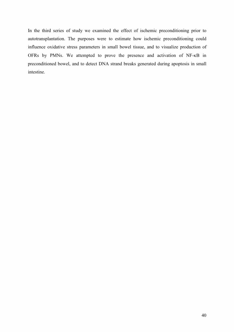
Ischemic preconditioning prior to cold preservation reduced markedly tissue
lipidperoxidation in all samples (GII: control 102.33±4.36 µM/g wet tissue, late reperfusion
98.74±6.17 µM/g wet tissue; GIII: control 104.52±3.82 µM/g wet tissue, late reperfusion
99.12±9.69 µM/g wet tissue). Concentration of MDA was similar to control during cold
preservation and reperfusion periods (Figure 13).
Figure 13. Biochemical changes of tissue MDA in preconditioned groups.
Tissue lipidperoxidation not increased significantly following cold preservation and
reperfusion in all samples. Data are presented as Mean±SEM.
Tis
sue
Mal
ondi
alde
hide
(uM
/g)
0
20
40
60
80
100
GII GIII
Control
End ischemia
Early reperfusion
Late reperfusion

46
However, there was a significant GSH elevation in all samples compared to control in
preconditioned groups, which indicated the activation of endogenous antioxidant
protective system (GII: control 354.45±11.88 µM/g wet tissue, late reperfusion 495.73±
12.38 µM/g wet tissue, P< 0.001; GIII: control 361.12±9.15 µM/g wet tissue, late
reperfusion 489.04±15.18 µM/g wet tissue, P< 0.001; Figure 14).
Figure 14. Biochemical changes of tissue GSH in preconditioned groups.
Concentration of GSH elevated in all samples of preconditioned groups compared to control.
Data are presented as Mean±SEM. * P< 0.05 vs. control, *** P< 0.001 vs. control.
Tis
sue
Red
uced
glu
tath
ione
(uM
/g)
0
100
200
300
400
500
GII GIII
Control End ischemia Early reperfusion
Late reperfusion
*
* ***
* *
***

47
Furthermore, we observed a better preservation of SOD activity in preconditioned groups
(GII: control 282.14±12.95 IU/g wet tissue, late reperfusion 168.45±15.03 IU/g wet tissue,
P< 0.05; GIII: control 275.85±10.99 IU/g wet tissue, late reperfusion 192.62±14.36 IU/g wet
tissue, P< 0.05; Figure 15).
Figure 15. Biochemical changes of tissue SOD in preconditioned groups.
More than 50 % of SOD activity was preserved during examination in preconditioned
groups. Data are presented as Mean±SEM. * P< 0.05 vs. control.
Tis
sue
Sup
erox
ide
dism
utas
e (I
U/g
)
0
50
100
150
200
250
300
GII GIII
Control
End ischemia
Early reperfusion
Late reperfusion
* *
* *
48
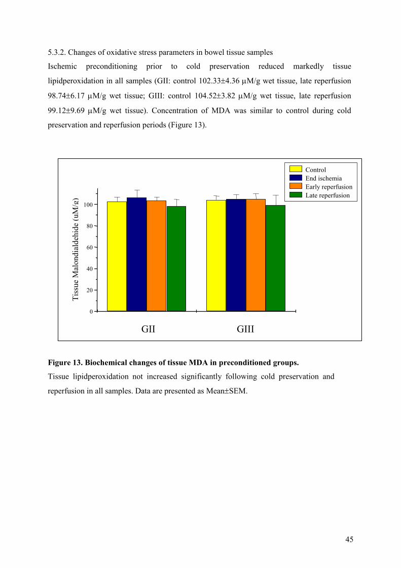
5.3.3. Visualization of OFRs production by circulating PMNs
Small amount of intracellular OFRs was detected in the control samples (Figure 16 GII, GIII,
control). The qualitative imaging of reflectance signals of Ce-perhydroxide deposits slightly
increased inside of PMNs by the end of reperfusion in preconditioned groups (Figure 16 GII,
GIII, reperfused).
Figure 16. Cerium histochemistry combined with reflectance confocal laser scanning
microscopy analysis of OFRs production by circulating PMNs. Isolated PMNs incubated
in vitro with CeCl3 with the nucleus being labelled with fluorescent marker as described in
“Materials and methods” (4.2.7, 4.2.8). The nucleus is demarkated in red and presence of
cerium-perhydroxide precipitates is detected as green fluorescence. Small amount of OFRs
was observed in control samples (GII, GIII, control). Appearance of reflectance signals
slightly elevated by the end of the reperfusion period in preconditioned groups (GII, GIII,
reperfused) (original magnification x2500).
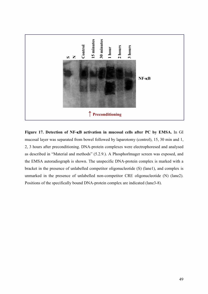
5.3.4. Activation of NF-κB after small bowel preconditioning
To assess the ability of ischemic PC to activate NF-κB in mucosal cells, bowel samples were
collected at different times. We observed that PC induced a time-dependent increase in NF-κ
B DNA binding activity (Figure 17 lane3-8). Both in the control (lane3) and the 15 minutes
(lane4) and 30 minutes (lane5) following preconditioning a low NF-κB activation could be
observed in samples. The NF-κB activation peaked at the 1-hour time-point (lane6), and
decreased by the third hour (lane8) in mucosal cells after small bowel PC. The specificity of
the NF-κB EMSA was assessed using an excess of unlabelled oligonucleotide (S) (Figure 17
lane1), and excess of unlabelled CRE oligonucleotide (N) (Figure 17 lane2).
GIII Reperfused GII Control GII Reperfused GIII Control
49
Figure 17. Detection of NF-κκκκB activation in mucosal cells after PC by EMSA. In GI
mucosal layer was separated from bowel followed by laparotomy (control), 15, 30 min and 1,
2, 3 hours after preconditioning. DNA-protein complexes were electrophoresed and analysed
as described in “Material and methods” (5.2.9.). A Phosphorlmager screen was exposed, and
the EMSA autoradiograph is shown. The unspecific DNA-protein complex is marked with a
bracket in the presence of unlabelled competitor oligonucleotide (S) (lane1), and complex is
unmarked in the presence of unlabelled non-competitor CRE oligonucleotide (N) (lane2).
Positions of the specifically bound DNA-protein complex are indicated (lane3-8).
S
N
Co
ntr
ol
15
min
ute
s 3
0 m
inu
tes
1 h
ou
r
2 h
ou
rs
3 h
ou
rs
NF-κκκκB
↑ Preconditioning

50
5.3.5. Detection of nuclear fragmentation by TUNEL technique
In each preconditioned group a few DNA-damaged cells were found in the mucosal layer in
control samples (Figure 18 GII, GIII, control) in the small intestine. Interestingly, several
TUNEL positive cells in preconditioned groups are indicative of more generalized
programmed-cell death by the end of the reperfusion period (Figure 18 GII, GIII, reperfused).
Figure 18. In situ detection of nuclear fragmentation by TUNEL technique in tissue.
To detect DNA damage in autotransplanted small bowel tissue were used an
immunohistochemical assay as described in “Material and methods” (4.2.9.). Normal mucosal
cells are demarkated in green and presence of DNA strand breaks containing cells are
detected as brown color in tissue sections (original magnification x40).
5.3.6. Hemodynamic parameters
Control mesenteric flow was 135.69±8.48 ml/minute, and not decreased less than
130.20±6.33 ml/minute during reperfusion in preconditioned groups (GI, GII, GIII). The
intestinal mucosal capillary perfusion was similarly sufficient after laparotomy and during
reperfusion period.
GGIIII RReeppeerrffuusseedd GGIIII CCoonnttrrooll GGIIIIII CCoonnttrrooll GGIIIIII RReeppeerrffuusseedd

51
5.4. Conclusions from this study
In the third series of our study investigated the effect of ischemic preconditioning prior to
autotransplantation. The 4 x 5 IPC stimuli conveyed protection against tissue oxidative injury
in the intestine. With respect to MDA, we demonstrated that IPC and cold preservation
commonly reduced tissue lipidperoxidation. Meanwhile, the tissue GSH concentration
elevated, to indicate the activation of endogenous antioxidant protective system. In the case of
preconditioned and later preserved grafts the SOD activity remained to a greater extent,
although it was found to be lower than the control activity. There are no data indicating the
exact mechanism of the maintained activity in the early phase. It can be due to a lower
amount of OFRs produced or to an increase in GSH transport or synthesis. Other investigators
confirmed our results regarding to the IPC groups. Sola et al have also reported the
modification of oxidative stress after IPC with 10 minutes of ischemia and 10 minutes of
reperfusion. Namely, in their models IPC prevented elevation of tissue MDA and significantly
increased concentration of GSH (72). It is known, that brief ischemia in the myocytes can
increase the activity of SOD (96).
The present results are believed to be the first to visualize OFRs production of PMNs after
small bowel ischemic preconditioning. Our results confirmed that IPC can decrease OFRs
release during reperfusion, but low amount of OFRs are necessary for the inductive effect to
begin IPC cascade. Pain et al demonstrated in a heart model that one of the key factor in early
phase of protection opening of that mitochondrial KATP channels by OFRs (97). Similarly,
Jaberansari et al reported the role of OFRs to induce IPC cascade in pig myocardium (96).
Accumulating evidence has revealed that NF-κB is involved in the control of the transcription
of a variety of cellular genes that regulate numerous responses in cells. The involvement of
NF-κB is especially of interest as it is activated by oxidative stress and its activation can be
modulated by antioxidant compounds (98). Our finding showed that 15 and 30 minutes
following IPC stimuli a low NF-κB activation could be observed. But it peaked at the 1-hour
time-point and decreased by the third hour in preconditioned mucosa.
In the present study we demonstrated the connection between IPC prior to bowel
autotransplantation and programmed cell death. Few DNA-damaged cells were found in the

52
mucosa specimens before preconditioning. Interestingly, several cell suffered DNA-damage
were observed after preconditioned and autotransplanted grafts. The role of NF-κB in the
intestinal epithelial cells apoptotic process remains to be established, since this transcription
factor also seems to play a proapoptotic role in cells (94).

53
6. DISCUSSION
The past three decades have seen increasing interest in the role played by oxygen derived free
radicals and oxidants in human disease. Historically, the first documentation of the existence
of oxygen was of great importance to biology and medicine (99, 100). Oxygen is a free
radical, as it possesses two unpaired electrons of parallel spin in an π antibonding orbital.
Gerschman et al proposed, “that oxygen poisoning and radiation injury have at least one
common basis of action, possibly the formation of oxidizing free radicals” (101, 102, 103).
This pioneering idea soon began to capture the imaginations of scientists, thus the role of free
radical reactions in human disease, biology and toxicology has become an area of intense
interest.
In Hungary, the interest in researches of oxygen free radicals (OFRs) has been begun in the
early 1980’ yet. Investigations based on the experience of the role of OFRs in the
pathomechanism of oxidative damage and the antioxidant protection in various organs.
Examinations in animal models and in patients explained the importance of OFRs in acute
liver injury, in Crohn disease and ulcerative colitis, in neonatal retinopathy, in muscular
dystrophy, in gastric diseases and in heart infarct due to the endeavours of Fehér (104),
Blázovics (105), Lakatos (106), Matkovics (107), Mózsik (108), Rıth (109).
Human clinical conditions in which a role for OFRs has been implicated are: ischemia-
reperfusion injury (stroke, myocardial infarction, organ transplantation), cancer, aging,
alcoholism, red blood cell defects (malaria, sickle cell anemia), iron overload (thalassemia,
idiopathic hemochromatosis, Kwashiorkor), kidney (aminoglycoside nephrotoxicity,
autoimmune nephritic syndromes), gastrointestinal tract (free fatty acid-induced pancreatitis,
nonsteroidal anti-inflammatory drug induced gastrointestinal tract lesions, endotoxin liver
injury), inflammatory-immune injury (rheumatoid arthritis, glomerulonephritis), brain
(Parkinson’s disease, neurotoxins, allergic encephalomyelitis), heart and cardiovascular
system (atherosclerosis, alcohol cardiomyopathy), eye (photic retinopathy, ocular
haemorrhage, cataractogenesis, degenerative retinal damage) (110).
Free radicals of importance in living organisms include hydroxyl (OH·), superoxide (O2·-),
nitric oxide (NO·) and peroxyl (RO2·). Hydrogen peroxide (H2O2), hypochlorous acid (HOCl),
singlet oxygen (1O2) and ozone (O3) chemically are not free radicals but can easily lead to free

54
radical reactions in organisms. In the living organisms there are three main source of the
generation of OFRs: mitochondria, xanthine oxidase (XO) system, and polymorphonuclear
leukocytes (PMNs). Under physiological respiration in mitochondria of all cells the electron
transport chain is generally tightly regulated, but some leakage (1-2 %) occurs leading to the
formation of OFRs. Normally the intracellular antioxidant enzyme can neutralize mainly O2·-
radicals as the side-product of terminal oxidant inhibiting the increased lipid peroxidation in
mitochondria.
It is generally considered that endothelium, particularly in the microvasculature, is the
primary cellular sources of OFRs. Levels of XO are normally low in human tissues, but they
may increase after injury such as ischemia and reperfusion (111). In the intestinal tract XO
presences not only in the microvascular endothelial cells but intestinal epithelium also
contains large amounts of the enzyme (112). During ischemia the xanthine dehydrogenase
converted to the xanthine oxidase. At the same time, there is a progressive breakdown of
ATP. This in turn leads to accumulation of the purine metabolites xanthine and hypoxanthine.
XO converts hypoxanthine to xanthine and xanthine to urate, O2 being simultaneously
reduced to both O2·- and H2O2 (113). H2O2 at micromolar levels also seems to be poorly
reactive. However, higher levels of H2O2 can attack several cellular energy-producing
systems. H2O2 also forms the OH· in the presence of transition metal ions and O2 can facilitate
this reaction (114).
PMNs have the function of recognizing foreign particles such as bacteria either because the
bacteria have an unusual surface or, because antibodies have bound to them. An oxidase
enzyme that seems to have specifically evolved to make O2·- exists in the plasma membrane
of PMNs. Although normally inactive, this enzyme is activated when PMNs encounter a
foreign particle. The enzyme takes NADPH from the PMN cytosol, oxidizes it to NADP+ and
passes electron onto O2, hence reducing O2 to O2·- outside the cell (115). Once the foreign
particles have been engulfed into the cytoplasm of the PMNs, other vesicles fuse with the
phagocytic vacuole, emptying into it antibacterial proteins, lysosomal proteases and the
enzyme myeloperoxidase. Myeloperoxidase uses H2O2 produced by dismutation of O2·- to
oxidize chloride ions into HOCl (116). O2·- production is activated when foreign particles
touch the PMN surface and some O2·- escapes into the extracellular fluid. Yet extracellular
fluids have little SOD activity.

55
While a large number of cellular constituents are potentially subject to OFRs attack, a
frequent target is the lipid component of membranes (117). Peroxyl radicals are formed
during lipid oxidation chain reactions, such as the oxidation of polyunsaturated fats and
unsaturated fatty acids. Lipid Peroxidation may be initiated by any species that has sufficient
reactivity to abstract a hydrogen atom from a polyunsaturated fatty acid side chain in
membrane lipids (118, 119, 120). Oxidation of membrane lipids can impair the function of
internal and cell surface membranes and the enzymes and receptors associated with them
(121). Peroxidation of lipids causes cleavage of double bonds, resulting in the formation of
aldehydes, such as malondialdehyde. These aldehydic products are diffusible cytotoxins
(122).
Oxidative damage to DNA appears to occur continuously in vivo, in that low levels
(presumably a steady-state balance between DNA damage and repair) have been detected in
DNA isolated from human cell and tissues. The pattern of damage to the purine and
pyrimidine bases bears the chemical fingerprint of OH· attack, suggesting that OH· formation
occurs within the nucleus in vivo. Hence, a rise in steady-state oxidative DNA damage could
be due to increased damage or decreased repair. Thus, damage to DNA can lead to long-
delayed mutagenic and carcinogenic effects (123).
All cells of organisms are constantly exposed to endogenous or exogenous OFRs. Organisms
have evolved an array of defense endogenous enzymatic and non-enzymatic mechanisms. In
the late 1960’, McCord and Fridovich recognized that SOD was the first example of the
enzyme that uses OFRs as a substrate, catalyzing the dismutation of O2·- into H2O2 and O2 at
diffusion-controlled rates (124, 125, 126, 127, 128). Since then, SOD have come to be
recognized as being nearly ubiquitous in living organisms, an essential component of the
cellular defense against toxic reactive oxygen species generated during the normal biological
reduction of O2 (129). SOD is one of three human enzymes, each named for its active site
metal ions and cellular location. Dimeric CuZnSOD (SOD1) is found in the cell cytoplasm, a
homotetrameric MnSOD (SOD2) is located in the mitochondrial matrix, and a
homotetrameric glycosylated CuZnSOD (SOD3) is confined to the extracellular space (130).
Cellular thiols, because of their relative abundance, stability, and ease of forming reversible
disulfide bounds by a two electrons transfer mechanism, play a very important role in the
biochemistry of metals and in the maintenance of cellular redox homeostasis. Among the

56
most critical sources of cellular reducing power and redox stabilization are cellular thiols,
particularly glutathione (131). GSH (L-γ-glutamyl-L-cysteinyl-glycine) is transported out of
the cell and is present in the extracellular milieu. The compartmentalization and the transport
of GSH are self-regulated and serve to regulate the transport of other molecules. The rate-
limiting step in GSH synthesis within the cell is catalyzed by γ-glutamylcysteine synthetase.
Glutathione peroxidase is a Se-containing enzyme that catalyses the reduction of H2O2 and
the formation of oxidized GSH (GSSG). Glutathione reductase uses NADPH and H+ to
reduce the GSSG back to GSH (132).
GSH plays a critical role in the maintenance of cellular redox status. GSH is sequestered in
the mitochondria and provides the primary defense against OFRs produced by mitochondrial
respiration. The relative redox status of different cellular compartments can be modulated
though variations in the ratio of GSH:GSSG (133). The cytoplasm has much greater reducing
power, while the endoplasmic retikulum presents a much more oxidizing environment,
appropriate for the purpose os protein refolding and the formation of disulfide bonds. GSH is
also actively sequestered within the nucleus where the nuclear concentration of GSH is four-
fold higher than in the cytoplasm (134). The balance of reduced versus oxidized GSH plays a
key role in maintaining active, reduced cellular proteins and is an important determining
factor in metal-induced alterations of the cellular redox status (132).
Oxidative stress is the term referring to the imbalance between generation of OFRs and the
activity of the antioxidant defenses. Clearly this can result either from depletion of the latter
or from excessive formation of OFRs. Cells can often adapt to increased oxidative stress up to
a certain point. However, it is also entirely possible that the oxidative stress becomes
overwhelming, leading to irreversible damage and to cell death (135, 136). The importance of
oxidative stress injury is dependent on the molecular target, the severity of the stress and the
mechanism by which the oxidative stress is imposed.
Early reperfusion is an absolute prerequisite for the survival of ischemic tissue. However,
reperfusion has been referred to as the ‘double edged sword’ because abundant evidence
suggests that reperfusing ischemic tissues carries with it a component of injury known as
‘reperfusion injury’. In the intestinal tissue reperfusion results in OFRs formation via the
endothelial and epithelial cell using mainly the xanthine oxidase pathway (137). The
accumulated hypoxanthine at the tissue of ischemia is quickly oxidized xanthine yielding

57
superoxide anion. OFRs then potentiate villus injury with necrosis of the villus tip, leading to
increased capillary permeability, reduced absorption of nutrients, protein leak into the lumen
and translocation of bacteria, endotoxin and bowel enzymes into the portal circulation (138).
OFRs, in particular the H2O2 coming from activated PMNs, may also stimulate splanchnic
vasoconstriction, which exacerbates the I/R injury (139). Intestinal injury following I/R
occurs in a hierarchical manner. The initial injury is merely functional, but as the extent and
duration of I/R increases, morphologically detectable injury, ranging from superficial changes
at the tip of the villi to transmural infarction (140, 141). One of the sensitive indicators of
mild injury to the intestine is an increase in lipid peroxides and a decrease in defense
mechanisms.
Our findings confirmed that physiological steady-state balance between the production of
lipidperoxides and their neutralization by antioxidants upset following ischemia and
reperfusion. In this study we observed, that OFRs were generated during warm ischemia and
subsequent reperfusion caused a significant elevation in tissue MDA. Meanwhile, the
endogen antioxidant defense systems such as GSH concentration slightly, but not significantly
decreased. Disparately another organs (heart, kidney, liver), principle answer of bowel tissue
to this imbalance was to put into action SOD. The great extent in the reduction of SOD
activity can be explained by the exhaustion of the SOD enzyme carrying out neutralization as
a result of increased O2·- production because of large amount of XO. Meanwhile, SOD could
not to control these processes oxidative injury has been explained. These mechanisms reflect
to susceptibility and specific manner of the intestine against oxidative stress.
Transplantation has become a well-accepted treatment of end-stage disease in many organs
including small intestine. Development of surgical techniques as well as management
strategies for immunological rejection and recurrence of the native disease has occupied
researchers for the past several decades. The emergence of I/R injury as a topic crucial to
outcome of transplant surgery has been not recent problem. The problems of damage by
intense cold ischemia on organs are unique to transplantation. Organs are routinely flushed
with cold preserve solution and quickly cooled. Like warm ischemia, cold ischemia can
disrupt membrane electrical gradients, inhibits Na+-K+-ATP pump activity, leading to
intracellular accumulation of Na+ and Ca2+ and causing increased cellular oedema. In addition
to metabolic derangements, structural abnormalities occur in the setting of cold storage.
Activation of PMNs is another well-recognized mechanism whereby cellular injury results

58
from cold ischemia and reperfusion. In small bowel transplantation, ‘rolling and sticking’ of
leukocytes was significantly increased in the submucosal post-capillary venules following
reperfusion (142).
The possibility of reducing I/R injury by improving the cold preservation is currently
considered being one of the ways to prevent oxidative stress and to restore the morphology
and functionality of the small bowel grafts. Successful small bowel transplantation may
require optimal preservation conditions and minimal reperfusion injury to decrease
postoperative complications. Because most of the studies on the ischemic susceptibility of
small intestine are based on definition the cold ischemia time and optimum preservation fluid
for bowel grafts (78). Zhang et al compared the efficacy of UW, EC and lactated Ringer’s
solutions in preserving canine small bowel. They suggested that, the degree of the changes
varied among the different groups, with the worst change occurring in the lactated Ringer’s
solution group, moderated changes in the EC solution group, and minimal changes in the UW
solution group. Three days after transplantation some of the EC grafts and most of the UW
grafts showed mucosal re-epithelialisation with near-normal villus height (143, 144). In
paradox, Hamamoto and colleagues reported that EC the best preservation fluid for the canine
bowel, as compared to UW or RL (30, 145). Although, UW solution contains glutathione as
OFRs scavenger, Wiconm and Collins first noted that glutathione was unstable and
spontaneously converted to oxidized form during storage of the solution (146). The role of
allopurinol as a XO inhibitor in UW solution and whether this agent is beneficial in an organ-
specific manner are uncertain (147).
In our investigations, using different solutions of which the two most widely accepted ones
were used can perform cold preservation. Regarding to oxidative stress parameters we didn’t
find significant difference between EC and UW solutions. We have found that, cold
preservation slightly moderated tissue lipid peroxidation. However it was surprised that SOD
activity dramatically decreased in spite of low temperature confirmed the key role of XD/XO
conversion and abundant O2·- production, which couldn’t be beneficially modified by cold
preservation. By now, it seems that any commercially available solution could be not used for
small bowel storage. The creation of a more optimal preservation solution for the small
intestine still seems desirable (148).

59
The question arises how else the protection against I/R injuries could be increased. Ischemic
preconditioning (IPC) of tissues has become a successfully and intensely cultivated research
area in the past 15 years. Research has brought a lot of success in the case of myocardium
resulting in clinical application by now, but the results of experiments on ischemic
preconditioning of other tissues (liver, brain, skeletal muscles) are also well known (149).
Ischemic preconditioning is the stress response that occurs during repeated episodes of brief
ischemia and reperfusion, and can render the organ more tolerant to subsequent potential
lethal ischemic injury. Ischemic adaptation occurs in two different steps: an early or
immediate effect (classic IPC) and a late effect (second window of protection) (150, 151). A
number of factors, which were found to regulate early adaptation, have also been found to
play a crucial role in the delayed adaptation to ischemia. The three principal players are
adenosine, ATP-sensitive potassium channels, and protein kinase C (152, 153). The delayed
adaptive protection has mediated by gene expression, transcriptional regulation and
subsequent protein synthesis. These genes include those encoding for proto-ontogenesis (c-
fos, c-myc, c-jun), the heat shock proteins (HSP) (HSP72, HSP70), and antioxidants (SOD,
catalase, glutathione S-transferase), cytokines, transcription factors, immunoreceptors and
growth factors (154, 155).
Initial studies on the small intestine revealed that IPC achieved by a 30-min occlusion of the
mesenteric artery could reduce the ischemic sensitivity of the bowel (91, 92). Actually, only a
few studies have demonstrated the importance of preconditioning prior to cold preservation of
the intestine. Inducing endogenous adaptation may reduce the development of oxidative stress
following cold preservation and it increases viability of the bowel tissue making it more
suitable for a subsequent transplantation (156).
Our results obtained in the bowel homogenates showed that, IPC carried out in 4 cycles prior
to preservation protected the bowel tissue against increased of lipidperoxidation diminishing
characteristic reduction of antioxidant capacity. The tissue level of GSH has significantly
increased in bowel tissue, which refers to protection against oxidative stress of the cells.
Moreover, we observed the best preservation of SOD activity in the preconditioned and later
preserved grafts. There is evidence in the literature that IPC reduces the conversion of XD in
to XO, while the amounts of released OFRs also decrease (72).

60
Until recently, most studies aimed to demonstrate a link between transcriptional factor NF-κB
activation and OFRs. Despite numerous mechanisms of transduction, most inducers of NF-κB
share the ability to induce redox changes. Increased OFRs level can act as chemical inducers
of the expression of specific genes involved in protecting cells against oxidative damage. This
phenomenon, which was first described in bacteria, led to the discovery of regulatory
proteins. In eukaryotes, recent results have demonstrated that H2O2 and lipidperoxides
regulate gene expression (157). This is important for two reasons, firstly, IPC can
significantly decrease the amount of OFRs released during reperfusion, secondly, the extent
of this decrease is only as small as necessary for the inductive effect to begin IPC cascade.
Experiments clearly showed that the overexpression of SOD stimulated the activation of NF-κ
B (158). The addition of N-acetylcysteine, a powerful neutralizer of OFRs inhibits the
activation of NF-κB by H2O2 (159). Recent studies have revealed that the overexpression of
detoxifying enzymes such as catalase or glutathione peroxidase inhibited the activation of NF-
κB (158, 160). Hence, this confirms that the activation of NF-κB is redox modulated.
Based on our results is can be explained that OFRs generating during IPC may induce cellular
signal cascade to activate NF-κB in mucosal cells. Following preconditioning stimuli a time-
dependent activation was observed. These findings is believed to be the first to demonstrate
the activation of transcription factor NF-κB in preconditioned intestinal tissue causing better
preservation of endogenous scavenger capacity of the small bowel mucosa.
The strategy of prevention and treatment of I/R injury includes prevention of cell death. Cell
death accompanies all the most significant periods in the life, including embryogenesis and
development, the adult state, aging and death (161). Based on the morphology of dying cells,
Kerr et al advanced the concept that there exist two distinct types of cell death, pathological
cell death as necrosis and physiological cell death as apoptosis (39). Necrosis occurs as a
catastrophic accident to whole cell, swelling of the cytoplasm and organelles including
mitochondria due to the loss of selective permeability of the cell membrane. This results
inducing an inflammatory reaction in the adjacent viable tissue in response to the released cell
debris. Apoptosis is ultrastucturally seen to involve the chromatin condensation and
margination, cell shrinkage, blebbing, and nuclear fragmentation. The cell breaks up into
several apoptotic bodies, which are phagocytosed by nearby cells without inflammation.

61
Although data in the literature suggest that the role of NF-κB in programmed cell death is
ambivalent, as it has both proapoptotic and antiapoptotic functions, depending on the type of
the cell and the death stimulus (162). Our findings suggest that NF-κB has a proapoptotic
effect during the reperfusion of tissues undergoing IPC prior to cold preservation. It seems
that IPC stimuli increased apoptosis regarding it has beneficial effect against tissue necrosis.
We hypothesize that in the delayed phase of preconditioning by new protein synthesis the
member of apoptotic cells will be decreased. These results can be considered as novel as no
other results describing the apoptosis of preconditioned and subsequently preserved bowel
tissue have been reported so far. However, the precise factors that determine the ability of NF-
κB to regulate apoptotic pathways during I/R period or after ischemic preconditioning in the
intestine are unknown.

62
7. NOVEL FINDINGS
The small bowel was the last splanchnic organ to be transplanted. The difficulty stems from
the fact that small intestine is strongly immunogenic and it is one of the most sensitive tissues
to ischemia-reperfusion injury. To eliminate the immunological complications we applied
several experimental models for studying effects of warm and cold ischemia-reperfusion, and
the influence of ischemic preconditioning prior to cold preservation and autotransplantation.
We demonstrated, that oxidative stress parameters, such as lipid peroxidation and endogenous
antioxidant compound are reliable indicators to monitor the ischemic intestinal damages.
We determined, that commonly applied cold preservation solutions are usable to eliminate the
development of oxidative injury following reperfusion in our autotransplantation model.
We detected, that following warm and cold ischemia changes of superoxide dismutase
activity is the most sensitive indicator in the intestinal reperfusion injury.
First we evaluated the effect of ischemic preconditioning before cold preservation in small
bowel autotransplantation model. Our investigations revealed the protective effect of ischemic
preconditioning on the oxidative stress parameters.
Demonstrating induction of the NF-κB transcription factor in the preconditioned bowel
mucosa we confirmed that IPC stimuli could activate the intracellular signal transduction.
This present study is believed to be the first to demonstrate the early protective effects of
ischemic preconditioning prior to small bowel preservation and autotransplantation. Ischemic
preconditioning of the small intestine may open a new perspective for reducing I/R injury.

63
8. ACKNOWLEDGEMENT
I would like to take this opportunity to thank for the overwhelming support I have received
from my supervisor Professor Elizabeth Rıth in completing this work. Her guidance and
leadership has been a blessing.
I would also like to acknowledge the help and assistance of all the staff at the Department of
Experimental Surgery in Pécs, without whom I would have been lost.
Special thanks to Professor Péter İrs Horváth, Dr. Károly Kalmár-Nagy, Dr. Afsin Tavakoli
of the Department of Surgery, to Professor József Szeberényi, and Dr. Katalin Kiss of the
Department of Molecular Biology for their valuable contribution to my work.

64
9. REFERENCES
1. Pirenne J, Aerts R, Fourneau I, Coosemans W. Present status and future prospects in
abdominal organ transplantation. Acta GastroEnterol Belg 1997; 60: 281-6.
2. Cicalese L, Baum C, Brown M, Sileri P, Smith D, Abcarian H, Benedetti E. Segmental small
bowel transplant from adult living-related donors. Transplant Proc 2001; 33: 1553.
3. Carrel A. La technique operatoire des anastomoses vasculaire et la transplantation des
visceres. Lyon Medico 1902; 98: 859-64.
4. Lillehei RC, Goott B, Miller FA. The physiological response of the small bowel of the dog
to ischemia including prolonged in vitro preservation of the bowel with successful
replacement and survival. Ann Surg 1959; 150: 543-60.
5. Monchik GJ, Russell PS. Transplantation of small bowel in the rat: technical and
immunological considerations. Surgery 1971; 70: 693-702.
6. Kort WJ, Westbroek DL, MacDicken I, Lameijer LD. Orthotopic total small bowel
transplantation in the rat. Eur Surg Res 1973; 5: 81-9.
7. Alican F, Hardy JD, Cayirli M, Varner JE, Moynihan PC, Turner MD, Anas P. Intestinal
transplantation. Am J Surg 1971; 121: 150-9.
8. Fortner JG, Sichuk G, Litwin SD, Beattie EK. Immunological response to an intestinal
allograft with HLA identical donor-recipient. Transplantation 1972; 14: 531-5.
9. Olivier C, Rettori R, Camilleri JP. Interruption of the lymphatic vessels and its
consequences in total homotransplantation of the small intestine and right side of the
colon in man. Lymphology 1972; 5: 24-31.
10. Grant D, Wall W, Mimeault R, Zhong R, Ghent C, Garcia B, Stiller C, Duff J. Successful
small-bowel/liver transplantation. Lancet 1990; 335: 181-4.
11. Deltz E, Schroeder P, Gundlach M, Hansmann ML, Leimenstoll G. Successful clinical
small-bowel transplantation. Transplant Proc 1990; 22: 2501.
12. Todo S, Reyes J, Furukawa H, Abu-Elmagd K, Lee RG, Tzakis A, Rao AS, Starzl TE.
Outcome analysis of 71 clinical intestinal transplantation. Ann Surg 1995; 222: 270-82.
13. Goulet O, Jan D, Brousse N, Revillon Y, Ricour C. Small-intestinal transplantation.
Baillieres Clin Gastroenterol 1997; 11: 573-92.
14. Urban T, Hurbain I, Urban M, Clement A, Housset B. Oxidants and antioxidants.
Biological effects and therapeutic perspectives. Ann Chir 1995; 49: 427-34.
15. de Zwart LL, Meerman JH, Commandeur JN, Vermeulen NP. Biomarkers of free radical
damage applications in experimental animals and in humans. Free Radic Biol Med 1999;

65
26: 202-26.
16. Granger DN, Rutili G, McCord JM. Superoxide radicals in feline intestinal ischemia.
Gastroenterology 1981; 81: 22-29.
17. Granger DN, Hollwarth ME, Parks DA. Ischemia-reperfusion injury: role of oxygen-
derived free radicals. Acta Physiol Scand Suppl 1986; 548: 47-64.
18. Parks DA, Williams TK, Beckman JS. Conversion of xanthine dehydrogenase to oxidase
in ischemic rat intestine: a reevaluation. Am J Physiol 1988; 254: 768-74.
19. Ratych RE, Chuknyiska RS, Bulkley GB. The primary localization of free radical
generation after anoxia/reoxygenation in isolated endothelial cells. Surgery 1987; 102:
122-31.
20. Wilkins EG, Rees RS, Smith D, Cashmer B, Punch J, Till GO, Smith DJ. Identification of
xanthine oxidase activity following reperfusion in human tissue. Ann Plast Surg 1993;
31: 60-5.
21. Grisham MB, Hernandez LA, Granger DN. Xanthine oxidase and neutrophil infiltration
in intestinal ischemia. Am J Physiol 1986; 251: 567-74.
22. Zimmerman BJ, Granger DN. Reperfusion-induced leukocyte infiltration: role of
elastase. Am J Physiol 1990; 259: 390-4.
23. Boros M, Takaichi S, Hatanaka K. Ischemic time-dependent microvascular changes and
reperfusion injury in the rat small intestine. J Surg Res 1995; 59: 311-20.
24. Vincenzini MT, Favilli F, Stio M, Iantomasi T. Intestinal glutathione transport system: a
possible detoxication role. Biochim Biophys Acta 1991; 1073: 571-9.
25. Chavez-Cartaya R, Jamieson NV, Ramirez P, Marin J, Pino-Chavez G. Free radical
scavengers to prevent reperfusion injury following warm liver ischemia. Transplant Proc
1999; 31: 2439-40.
26. McCord JM, Fridovich I, Superoxide dismutase. An enzyme function for erythrocuprein
(hemocuprein). J Biol Chem 1969; 244: 6049-55.
27. Massberg S, Gonzalez AP, Leiderer R, Menger MD, Messmer K. In vivo assessment of
the influence of cold preservation time on microvascular reperfusion injury after
experimental small bowel transplantation. Br J Surg 1998; 85: 127-33.
28. Collins GM, Halasz NA. Forty-eight hour ice storage of kidneys: importance of cation
content. Surgery 1976; 79: 432-5.
29. Eurotransplant Foundation Annual Report 1976.
30. Hamamoto I, Merhav H, Zhu Y, Suzuki M, Fujita S, Murase N, Todo S, Starzl TE. Lipid
peroxidation, brush border, and neutrophil enzyme activity after small bowel

66
preservation: a comparison of preservation solutions. Transplant Proc 1992; 24: 1095.
31. Wahlberg JA, Southard JH, Belzer FO. Development of a cold-storage solution for
pancreas preservation. Cryobiology 1986; 23: 477-82.
32. Ploeg RJ, van Bockel JH, Langendijk PT, Groenewegen M, van der Woude FJ, Persijn
GG, Thorogood J, Hermans J. Effect of preservation solution on results of cadaveric
kidney transplantation. The European Multicentre Study Group. Lancet 1992; 340: 129-
37.
33. Mueller AR, Platz KP, Neuhaus P, Lee KK, Schraut WH. Goals of small bowel
preservation. Transplant Proc 1996; 28: 2633-5.
34. Taguchi T, Zorychta E, Guttman FM. Evaluation of UW solution for preservation of
small intestinal transplants in the rat. Transplantation 1992; 53: 1202-5.
35. Ploeg RJ, D’Alessandro AM. Intestinal transplantation: a clinical update. Scand J
Gastroenterol Suppl 1995; 212: 79-89.
36. Park PO, Wallander J, Tufveson G, Haglund U. Cold ischemic and reperfusion injury in a
model of small bowel transplantation in the rat. Eur Surg Res 1991; 23: 1-8.
37. D’Alessandro AM, Kalayoglu M, Sollinger HW, Pirsch JD, Belzer FO. Liver-intestinal
transplantation: report of a case. Transplant Proc 1992; 24: 1228-9.
38. Banda MA, Granger DN. Mechanism and protection from ischemic intestinal injury.
Transplant Proc 1996; 28: 2595-7.
39. Kerr JF, Wyllie AH, Currie AR. Apoptosis: a basic biological phenomenon with wide-
ranging implications in tissue kinetics. Br J Cancer 1972; 26: 239-57.
40. Li Y, Chopp M, Jiang N, Zhang ZG, Zaloga C. Induction of DNA fragmentation after 10
to 120 minutes of focal cerebral ischemia in rats. Stroke 1995; 26: 1252-8.
41. Gottlieb RA, Gruol DL, Zhu JY, Engler RL. Preconditioning rabbit cardiomyocytes: role
of pH, vacuolar proton ATPase, and apoptosis. J Clin Invest 1996; 97: 2391-8.
42. Schumer M, Colombel MC, Sawczuk IS, Gobe G, Connor J, O'Toole KM, Olsson CA,
Wise GJ, Buttyan R. Morphologic, biochemical, and molecular evidence of apoptosis
during the reperfusion phase after brief periods of renal ischemia. Am J Pathol 1992; 140:
831-8.
43. Buttke TM, Sandstrom PA. Oxidative stress as a mediator of apoptosis. Immunol Today
1994; 15: 7-10.
44. Shah KA, Shurey S, Green CJ. Apoptosis after intestinal ischemia-reperfusion injury.
Transplantation 1997; 64: 1393-7.
45. Murry CE, Jennings RB, Reimer KA. Preconditioning with ischemia: a delay of lethal

67
injury in ischemic myocardium. Circulation 1986; 74: 1124-36.
46. Ishida T, Yarimizu K, Gute D, Korthuis RJ. Mechanisms of ischemic preconditioning.
Shock 1997; 8: 86-94.
47. Davis JM, Gute DC, Jones S, Krsmanovic A, Korthuis RJ. Ischemic preconditioning
prevents postischemic P-selectin expression in the rat small intestine. Am J Physiol 1999;
277: 2476-81.
48. de Oca J. Hotter G, Sola A, Gonzalez R, Rafecas A, Roselló J, Jaurrieta E. Role of nitric
oxide in preconditioning for intestinal transplantation. Transplant Proc 1999; 31: 2573.
49. Zhang Y, Wu YX, Hao YB, Dun Y, Yang SP. Role of endogenous opioid peptides in
protection of ischemic preconditioning in rat small intestine. Life Sci 2001; 68: 1013-19.
50. McCallion K, Wattanasirichaigoon S, Gardiner KR, Fink MP. Ischemic preconditioning
ameliorates ischemia- and reperfusion-induced intestinal epithelial hyperpermeability in
rats. Shock 2000; 14: 429-34.
51. Speechly-Dick ME, Mocanu MM, Yellon DM. Protein kinase C. Its role in ischemic
preconditioning in the rat. Circ Res 1994; 75: 586-90.
52. Gute D, Ishida T, Korthuis R. The beneficial effects of ischemic preconditioning are
abolished by blockade of adenosine A1, but not adenosine A2 receptors. Microcirculation
1996; 3: 88.
53. Yang SP, Hao YB, Wu YX, Dun Y, Shen LH, Zhang Y. Ischemic preconditioning
mediated by activation of KATP channels in rat small intestine. Zhongguo Yao Li Xue
Bao 1999; 20: 341-4.
54. Brennan P, O’Neill LA. Effects of oxidants and antioxidants on nuclear factor κB
activation in three different cell lines: evidence against a universal hypothesis involving
oxygen radicals. Biochim Biophys Acta 1995; 1260: 167-75.
55. Marsters SA, Sheridan JP, Donahue CJ, Pitti RM, Gray CL, Goddard AD, Bauer KD,
Ashkenazi A. Apo-3, a new member of the tumor necrosis factor receptor family,
contains a death domain and activates apoptosis and NF-kappa B. Curr Biol 1996; 6:
1669-76.
56. Madesh M, Benard O, Balasubramanian KA. Apoptotic process in the monkey small
intestinal epithelium: two possible role of oxidative stress. Free Radic Biol Med 1999;
26: 431-8.
57. Otamiri TA. Influence of quinacrine on plasma malondialdehyde after small intestinal
ischemia and reperfusion. Circ Shock 1988; 24: 63-9.
58. Baykal A, Kaynaroglu V, Demirpence E, Kilinc K, Sayek I, Sanac Y. Experimental study

68
of the effect of adrenaline tolerance on intestinal ischemia-reperfusion. Br J Surg 1998;
85: 947-50.
59. Siegers CP, Riemann D, Thies E, Younes M. Glutathione and GSH-dependent enzymes
in the gastointestinal mucosa of the rat. Cancer Lett 1988; 40: 71-6.
60. Martensson J, Jain A, Meister A. Glutathione is required for intestinal function. Proc Natl
Acad Sci USA 1990; 87: 1715-19.
61. Lash LH, Hagen TM, Jones DP. Exogenous glutathione protects intestinal epithelial cells
from oxidative injury. Proc Natl Acad Sci USA 1986; 83: 4641-5.
62. Bannister JV, Bannister WH, Rotilio G. Aspects of the structure, function, and
applications of superoxide dismutase. Crit Rev Biochem 1987; 22: 111-80.
63. Sener G, Akgun U, Satiroglu H, Topaloglu U, Keyer-Uysal M. The effect of
pentoxifylline on intestinal ischemia/reperfusion injury. Fundam Clin Pharmacol 2001;
15: 19-22.
64. Otamiri TA, Tagesson C. Ginkgo biloba extract prevents mucosal damage associated
with small-intestinal ischemia. Scand J Gastroenterol 1989; 24: 666-70.
65. Giele HP, Heel KA, Storrie A, McCauley RD, Hall JC. Warm ischemia time in a model
for small bowel transplantation. Microsurgery 1996; 17: 438-43.
66. Slavíková H, Lojek A, Hamar J, Dusková M, Kubala L, Vondrácek J, Cíz M. Total
antioxidant capacity of serum increased in early but not late period after intestinal
ischemia in rats. Free Radic Biol Med 1998; 25: 9-18.
67. Park PO, Haglund U. Regeneration of small bowel mucosa after intestinal ischemia. Crit
Care Med 1992; 20: 135-9.
68. Schweizer E, Gassel AM, Deltz E, Schroeder P. A comparison of preservation solutions
for small bowel transplantation in the rat. Transplantation 1994; 57: 1406-08.
69. Beuk RJ, Heineman E, Tangelder GJ, Kurvers HA, Bonke HJ, oude Egbrink MG. Effects
of different durations of total warm ischemia of the gut on rat mesenteric
microcirculation. J Surg Res 1997; 73: 14-23.
70. Gibson DD, Brackett DJ, Squires RA, Balla AK, Lerner MR, McCay PB, Pennington LR.
Evidence that the large loss of glutathione observed in ischemia/reperfusion of the small
intestine is not due to oxidation to glutathione disulfide. Free Radic Biol Med 1993; 14:
427-33.
71. Nakamura M, Ozaki M, Fuchinoue S, Teraoka S, Ota K. Ascorbic acid prevents
ischemia-reperfusion injury in the rat small intestine. Transpl Int 1997; 10: 89-95.
72. Sola A, Hotter G, Prats N, Xaus C, Gelpi E, Rosello-Catafau J. Modification of oxidative

69
stress in response to intestinal preconditioning. Transplantation 2000; 69: 767-72.
73. Kacmaz M, Ozturk HS, Karaayvaz M, Guven C, Durak I. Enzymatic antioxidant defence
mechanism in rat intestinal tissue in changed after ischemia-reperfusion. Effects of an
allopurinol plus antioxidant combination. Can J Surg 1999; 42: 427-31.
74. Karashima K, Uchida Y, Tohara K, Kashima K, Yokoyama S, Nakayama I. The kinetics
of cooper-zinc superoxide dismutase in experimentally induced ischemia-reperfusion
injury of the canine jejunum. Surg Today 1995; 25: 343-50.
75. Zhang S, Takeyoshi I, Nomoto M, Zhu Y, Nemoto A, Hamada N, Todo S. Evaluation of
solutions for canine small bowel preservation. Transplant Proc 1996; 28: 2612-3.
76. Raju S, Fujiwara H, Lewin JR, Grogan JB. Twelve-hour and twenty four-hour
preservation of small bowel allografts by simple hypothermia. Transplantation 1988; 45:
290-3.
77. Mueller AR, Platz KP, Nalesnik MA, Langrehr JM, Neuhaus P, Schraut WH. Small
bowel preservation causes basement membrane and endothelial cell injury. Transplant
Proc 1994; 26: 1498-1500.
78. Thaler W, Oberhuber G, Pernthaler H, Margreiter R, Schmid T. Preservation of small
bowel with University of Wisconsin and Euro-Collins solution. Transplant Proc 1993; 25:
1611-12.
79. Madesh M, Anup R, Benard O, Balasubramanian KA. Apoptosis in the monkey small
intestinal epithelium: structural and functional alterations in the mitochondria. Free Radic
Biol Med 1999; 26: 836-43.
80. Telek G, Scoazec JY, Chariot J, Ducroc R, Feldmann G, Rozé C. Cerium-based
histochemical demonstration of oxidative stress in taurocholate-induced acute pancreatitis
in rats: A Confocal Laser Scanning Microscopic study. J Histochem Cytochem 1999; 47:
1201-12.
81. De Oca J, Vallet J, Bettonica C, García A, Montanés T, Castells A, Cuadrado S,
Mercadal M, Fiol C. Influence of liquid preservation on lipid peroxidation and bacterial
permeability after small bowel transplantation. Transplant Proc 1995; 27: 2382-3.
82. Zhang S, Kokudo Y, Nemoto EM, Todo S. Biochemical evidence of mucosal damage of
intestinal grafts during cold preservation in University of Wisconsin, Euro-Collins, and
Lactated Ringer’s solutions. Transplant Proc 1994; 26: 1494-5.
83. Frezza EE, Tzakis A, Fung JJ, Van Thiel DH. Small bowel transplantation: current
progress and clinical application. Hepatogastroenterology 1996; 43: 363-76.
84. Sun SC, Greenstein SM, Schechner RS, Sablay LB, Veith FJ, Tellis VA. Improved small

70
intestinal preservation with additional use of superoxide dismutase to University of
Wisconsin solution. Transplant Proc 1992; 24: 1092-3.
85. Funovits M, Altieri KA, Kovalak JA, Staschak-Chicko SM. Small intestine
transplantation. A nursing perspective. Crit Care Nurs Clin North Am 1993; 5: 203-13.
86. Cicalese L, Caraceni P, Nalesnik MA, Lee KK, Van Thiel DH, Schraut WH. Oxygen free
radical formation is related to mucosal injury during reperfusion after rat small bowel
transplantation. Transplant Proc 1994; 26: 1477.
87. Cicalese L, Caraceni P, Nalesnik MA, Borle AB, Schraut WH. Oxygen free radical
content and neutrophil infiltration are important determinants in mucosal injury after rat
small bowel transplantation. Transplantation 1996; 62: 161-6.
88. Shah KA, Shurey S, Green CJ. Characterization of apoptosis in intestinal ischemia-
reperfusion injury- a light and electron microscopic study. Int J Exp Pathol 1997; 78:
355-63.
89. Kokudo Y, Itoh M, Mori S, Karasawa Y, Okano K, Yachida S, Ishimura K, Wakabayashi
H, Maeba T, Maeta H. Effects of luminal flush on mucosal injury during cold ischemia in
the rat small-bowel. Transplant Proc 1996; 28: 1841-2.
90. Hotter G, Closa D, Prados M, Fernander-Cruz L, Prats N, Gelpi E, Rosello-Catafau J.
Intestinal Preconditioning is mediated by a transient increase in nitric oxide. Biochem
Biophys Res Commun 1996; 222: 27-32.
91. Tsuruma T, Yagihashi A, Matsuno T, Zou XM, Asanuma K, Sasaki K, Hirata K.
Induction of warm ischemic tolerance following preconditioning of the small intestine.
Transplant Proc 1996; 28: 1298-9.
92. Tsuruma T, Yagihashi A, Matsuno T, Zou XM, Asanuma K, Sasaki K, Hirata K. The
Heat-Shock Protein 70 family reduces ischemia/reperfusion injury in small intestine.
Transplant Proc 1996; 28: 2629-30.
93. Sen R, Baltimore D. Multiple nuclear factors interact with the immunoglobulin enhancer
sequences. Cell 1986; 46: 705-16.
94. Jobin C, Sartor RB. The Iκ-B/NF-κB system: a key determinant of mucosal inflammation
and protection. Am J Physiol Cell Physiol 2000; 278: 451-62.
95. Yeh KY, Yeh M, Glass J, Granger DN. Rapid activation of NF-κB and AP-1 and target
gene expression in postischemic rat intestine. Gastroenterology 2000; 118: 525-34.
96. Jaberansari MT, Baxter GF, Muller CA, Latouf SE, Roth E, Opie LH, Yellon DM.
Angiotensin-converting enzyme inhibition enhances a subthreshold stimulus to elicit
delayed preconditioning in pig myocardium. J Am Coll Cardiol 2001; 37: 1996-2001.

71
97. Pain T, Yang XM, Critz SD, Yue Y, Nakano A, Liu GS, Heusch G, Cohen MV, Downey
JM. Opening of mitochondrial K(ATP) channels triggers the preconditioned state by
generating free radicals. Circ Res 2000; 87: 460-6.
98. van den Berg R, Haenen GR, van den Berg H, Bast A. Transcription factor NF-kappaB as
a potential biomarker for oxidative stress. Br J Nutr 2001; 86: 121-7.
99. Gomberg M. An incidence of trivalent carbon trimethyl. J Am Chem Soc 1900; 22: 757-
71.
100. Hey DH, Waters WA. Some organic reactions involving the occurrence of free radicals
in solution. Chemistry Reviews 1937; 21: 169-208.
101. Gerschman R, Gilbert DL, Nye SW, Dwyer P, Fenn WO. Oxygen poisoning and x-
irradiation: a mechanism in common. Science 1954; 119: 623-26.
102. Haugaard N. Cellular mechanisms of oxygen toxicity. Physiol Rev 1968; 48: 311-73.
103. Pryor WA. Free radical biology: xenobiotics, cancer, and aging. Ann N Y Acad Sci
1982; 393: 1-22.
104. Fehér J, Bär-Pollák Zs, Sréter L, Fehér E, Toncsev H. Biochemical markers in carbon-
tetrachloride and galactosamine-induced acute liver injuries. The effects of
dihydroquinoline-type antioxidants. Br J Exp Pathol 1982; 63: 394-400.
105. Blázovics A, Kovacs A, Lugasi A, Hagymasi K, Biro L, Feher J. Antioxidant defense in
erythrocytes and plasma of patients with active and quiescent Crohn disease and
ulcerative colitis: a chemiluminescent study. Clin Chem 1999; 45: 895-6.
106. Lakatos L, Hatvani I, Oroszlán Gy, Karmazsin L, Matkovics B. D-penicillamine in the
prevention of retrolental fibroplasia. Acta Pediatr Acad Sci Hung 1982; 23: 327-35.
107. Matkovics B, László A, Szabó L. A comperative study of superoxide dismutase,
catalase and lipid peroxidation in red blood cells from muscular dystrophy patients and
normal controls. Clin Chim Acta 1982; 118: 289-92.
108. Mózsik Gy, Jávor T, Tóth Gy, Zsoldos T, Tigyi A. Interrelationchips between the
gastric cytoprotective effects of vitamin A and beta-carotene and the gastric superoxide
dismutase activity in rats. Acta Physiol Hung 1984; 64: 315-8.
109. Rıth E, Török B, Zsoldos T, Matkovics B. Lipid peroxidation and scavenger
mechanism in experimentally induced heart infarcts. Basic Res Cardiol 1985; 80: 530-6.
110. Del Maestro RF. An approach to free radicals in medicine and biology. Acta Physiol
Scand Suppl 1980; 492: 153-68.
111. Bulkley GB. Reactive oxygen metabolites and reperfusion injury: aberrant triggering of

72
reticuloendothelial function. Lancet 1994; 344: 934-6.
112. Schoutsen B, De Jong JW, Harmsen E, De Tombe PP, Achterberg PW. Myochardial
xanthine oxidase/dehydrogenase. Biochim Biophys Acta 1983; 762: 519-24.
113. Granger DN. Role of xanthine oxidase and granulocytes in ischemia reperfusion injury.
Am J Physiol 1988; 255: 1269-75.
114. Miller DM, Buettner GR, Aust SD. Transition metals as catalysts of “autoxidation”
reactions. Free Radic Biol Med 1990; 8: 95-108.
115. Klebanoff SJ. Oxygen metabolism and the toxic properties of phagocytes. Ann Intern
Med 1980; 93: 480-9.
116. Weiss SJ. Tissue destruction by neutrophils. N Engl J Med 1989; 320: 365-76.
117. Halliwell B, Chirico S. Lipid peroxidation: its mechanism, measurement and
significance. Am J Clin Nutr 1993; 57: 715-24.
118. Slater TF. Free radicals in medicine. Free Radic Res Commun 1989; 7: 121-388.
119. Dargel R. Lipid peroxidation: a common pathogenic mechanism? Exp Toxicol Pathol
1992; 44: 169-81.
120. Dix TA, Aikens J. Mechanism and biological relevance of lipid peroxidation initiation.
Chem Res Toxicol 1993; 6: 2-18.
121. Kim MS, Akera T. O2 free radicals cause of ischemia-reperfusion injury to cardiac Na+-
K+-ATPase. Am J Physiol 1987; 252: 252-7.
122. Chisolm GM, Morel DW. Lipoprotein oxidation and cytotoxicity: effect of probucol on
streptozotocin-treated rats. Am J Cardiol 1988; 62: 20-6.
123. Frenkel K. Carcinogen-mediated oxidant formation and oxidative DNA damage.
Pharmacol Ther 1992; 53: 127-66.
124. Fridovich I. Superoxide dismutase. An adaptation to paramagnetic gas. J Biol Chem
1989; 264: 7761-4.
125. McCord JM, Fridovich I. The reduction of cytochrome c by milk xanthine oxidase. J
Biol Chem 1968; 243: 5753-60.
126. Michelson AM, McCord JM, Fridovich I. Superoxide and superoxide dismutases.
Academic Press: London 1977.
127. Amstrong D, Sohal RS, Cutler RG, Slater TF. Free radicals in molecular biology, aging
and disease. Raven Press: New York 1984.
128. Kinnula VL, Crapo JD, Raivio KO. Generation and disposal of reactive oxygen
metabolites in the lung. Lab Invest 1995; 73: 3-19.
129. Hassan HM. Biosynthesis and regulation of superoxide dismutases. Free Radic Biol

73
Med 1988; 5: 377-85.
130. Tainer JA, Getzoff ED, Richardson JS, Richardson DC. Structure and mechanism of
cooper, zinc superoxide dismutase. Nature 1983; 306: 284-7.
131. Kalyanaraman B. Thiyl radicals in biological systems: significant or trivial? Portland
Press: London 1995; 55-63.
132. Meister A. Glutathione, ascorbate, and cellular protection. Cancer Res 1994; 54: 1969-
75.
133. Hwang C, Sinskey AJ, Lodish HF. Oxidized redox state of glutathione in the
endoplasmic reticulum. Science 1992; 257: 1469-1502.
134. Bellomo G, Vairetti M, Stivala L, Mirabelli F, Richelmi P, Orrenius S. Demonstration
of nuclear compartalization of glutathione in hepatocytes. Proc Natl Acad Sci USA 1992;
89: 4412-6.
135. Halliwell B. Free radicals and antioxidants: a personal view. Nutr Rev 1994; 52: 253-65.
136. Whittemore ER, Loo DT, Watt JA, Cotman CW. A detailed analysis of hydrogen-
peroxide induced cell death in primary neuronal culture. Neuroscience 1995; 67: 921-32.
137. Kurose I, Granger DN. Evidence implicating xanthine oxidase and neutrophils in
reperfusion-induced microvascular dysfunction. Ann N Y Acad Sci 1994; 723: 158-79.
138. Fiddian-Green RG. Splanchnic ischemia and multiple organ failure in the critically ill.
Ann R Coll Surg Engl 1988; 70: 128-34.
139. Stahl GL, Pan HL, Longhurst JC. Activation of ischemia and reperfusion sensitive
abdominal visceral C-fiber afferents. Role of hydrogen peroxide and the hydroxyl radical.
Circ Res 1993; 72: 1266-75.
140. Chiu CJ, McArdle AH, Brown R, Scott HJ, Gurd FN. Intestinal mucosal lesions in low
flow state. A morphologic, hemodynamic and metabolic reappraisal. Arch Surg 1970;
101: 478-83.
141. Haglund U, Bulkley GB, Granger DN. On the pathophysiology of intestinal ischemic
injury. Clinical review. Acta Chir Scand 1987; 153: 321-4.
142. Parks DA, Granger DN. Contributions of ischemia and reperfusion to mucosal lesion
formation. Am J Physiol 1986; 250: 749-53.
143. Zhang S, Takeyoshi I, Nomoto M, Zhu Y, Nemoto A, Hamada N, Todo S. Evaluation of
solutions for canine small bowel preservation. Tranplant Proc 1996; 28: 2612-3.
144. Takeyoshi I, Zhang S, Nomoto M, Zhu Y, Kokudo Y, Suzuki T, Hamada N, Nemoto A,
Starzl TE, Todo S. Mucosal damage and recovery of the intestine after prolonged
preservation and transplantation in dogs. Transplantation 2001; 71: 1-7.

74
145. de Bruin RW, Heineman E, Marquet RL. Small bowel transplantation: an overview.
Transpl Int 1994; 7: 47-61.
146. Wicomb WE, Collins GM. 24-hour rabbit heart storage with UW solution. Effects of
low-flow perfusion, colloid, and self storage. Transplantation 1989; 48: 6-9.
147. Marshall VC, Jablonski P, Biguzas M, Thomas AC, Walls K, Howden BO. Rat kidney
preservation with UW solution. Transplant Proc 1989; 21: 3783.
148. Mühlbacher F, Langer F, Mittermayer C. Preservation solutions for transplantation.
Transplant Proc 1999; 31: 2069-70.
149. Peralta C, Bartrons R, Serafin A, Blazquez C, Guzman M, Prats N, Xaus C, Cutillas B,
Gelpi E, Rosello-Catafau J. Adenosine monophosphate-activated protein kinase mediates
the protective effects of ischemic preconditioning on hepatic ischemia-reperfusion injury
in the rat. Hepatology 2001; 34: 1164-73.
150. Flack J, Kimura Y, Engelman RM, Rousou JA, Iyengar J, Jones R, Das DK.
Preconditioning the heart by repeated stunning improves myocardial salvage. Circulation
1991; 84: 369-74.
151. Yellon DM, Baxter GF, Garcia-Dorado D, Heusch G, Sumeray MS. Ischemic
preconditioning: present position and future directions. Cardiovasc Res 1998; 37: 21-33.
152. Baxter GF, Marber MS, Patel VC, Yellon DM. Adenosine receptor involvement in a
delayed phase of myocardial protection 24 hours after ischemic preconditioning.
Circulation 1994; 90: 2993-3000.
153. Baxter GF, Goma FM, Yellon DM. Involvement of protein kinase C in the delayed
cytoprotection following sublethal ischemia in rabbit myocardium. Br J Pharmacol 1995;
115: 222-4.
154. Das DK, Moraru II, Maulik N, Engelman RM. Gene expression during myocardial
adaptation to ischemia and reperfusion. Ann N Y Acad Sci 1994; 723: 292-307.
155. Yamashita N, Hoshida S, Nishida M, Igarashi J, Aoki K, Hori M, Kuzuya T, Tada M.
Time course of tolerance to ischemia-reperfusion injury and induction of heat-shock
protein 72 by heat stress in the rat heart. J Mol Cell Cardiol 1997; 29: 1815-21.
156. Sola A, de Oca J, Gonzalez R, Prats N, Rosello-Catafau J, Gelpi E, Jaurrieta E, Hotter
G. Protective effect of ischemic preconditioning on cold preservation and reperfusion
injury associated with rat intestinal transplantation. Ann Surg 2001; 234: 98-106.
157. Collins T. Endothelial nuclear factors κB and the initiation of the atherosclerotic lesion.
Lab Invest 1993; 68: 499-508.
158. Schmidt KN, Amstad P, Cerutti P, Baeuerle PA. The role of hydrogen peroxide and

75
superoxide as messengers in the activation os transcription factor NF-κB. Chem Biol
1995; 2: 13-22.
159. Schreck R, Baeuerle PA. A role for oxygen radicals as second messengers. Trends in
Cell Biology 1991; 1: 39-42.
160. Kretz-Remy C, Mehlen P, Mirault ME, Arrigo AP. Inhibition of IκB-α phosphorylation
and degradation and subsequent NF-κB activation by glutathion peroxidase
overexpression. J Cell Biol 1996; 133: 1083-93.
161. Wyllie AH, Kerr JF, Currie AR. Cell death: the significance of apoptosis. Int Rev Cytol
1980; 68: 251-306.
162. Li L, Rao JN, Bass BL, Wang JY. NF-κB activation and susceptibility to apoptosis after
polyamine depletion in intestinal epithelial cells. Am J Physiol Gastrointest Liver Physiol
2001; 280: 992-1004.

76
10. PRESENTATIONS AND PUBLICATIONS
Publications
1. Ferencz A, Tavakoli A, Kalmár-Nagy K, Szántó Z, Rıth E, Horváth İP. Az oxidatív
stressz monitorozása kísérletesen létrehozott vékonybél autotranszplantációt követıen. Magy Seb 2001; 54: 60-4. 2. Szántó Z, Ferencz A, Kovács F, Horváth İP, Rıth E, Molnár FT. Légcsıpótlás vékonybél
szabadlebennyel állatkísérletes modellben. Magy Seb 2001; 54: 320-4. 3. Ferencz A, Szántó Z, Borsiczky B, Kiss K, Kalmár-Nagy K, Szeberényi J, Horváth İP,
Rıth E. Activation of NF-κB after ischemic preconditioning in small bowel. Proceedings of the 37th Congress of the European Society for Surgical Research (ESSR)
Monduzzi Editore Press: Bologna 2002; 223-6. 4. Szántó Z, Ferencz A, Jancsó G, Boronkai Á, Rıth E, Horváth İP, Molnár FT.
Comparison of different anastomotic techniques in tracheal replacement. Proceedings of the 37th Congress of the European Society for Surgical Research (ESSR)
Monduzzi Editore Press: Bologna 2002; 205-8. 5. Rıth E, Jancsó G, Jaberansari MT, Cserepes B, Borsiczky B, Szántó Z, Ferencz A, Lantos
J. The role of Bradykinin in delayed myocardial adaptation. Proceedings of the 37th Congress of the European Society for Surgical Research (ESSR)
Monduzzi Editore Press: Bologna 2002; 75-80. 6. Pórszász R, Porkoláb Á, Ferencz A, Pataki T, Szilvássy Z, Szolcsányi J. Capsaicin-
induced nonneural vasoconstriction in canine mesenteric arteries. Eur J Pharmacol 2002; 441: 173-5. Impact factor: 2.164 7. Ferencz A, Szántó Z, Borsiczky B, Kiss K, Kalmár-Nagy K, Telek G, Szeberényi J,
Horváth İP, Rıth E. Vékonybél autotranszplantációt megelızı ischemiás prekondícionálás hatása az oxidatív stressz kialakulására. Magy Seb 2002; 55: 331-6.
8. Ferencz A, Szántó Z, Borsiczky B, Kiss K, Kalmár-Nagy K, Szeberényi J, Horváth İP.
Rıth E. The effects of preconditioning on the oxidative stress in small-bowel autotransplantation.
Surgery (accepted for publication) Impact factor: 2.615

77
Abstracts
1. Ferencz A, Rıth E, Késmárky G, Vajda G, Habon L, Lantos J, Jaberansari MT. Evidence
of free radical mediated reactions after coronary angioplasty in humans. Eur Surg Res 1999; 31: 185-6. Impact factor: 0.782 2. Ferencz A, Tavakoli A, Kalmár-Nagy K, Rıth E, Horváth İP. Ischémiás-reperfúziós
változások monitorozása kísérletesen létrehozott vékonybél autotranszplantációt követıen. Magy Seb 1999; 52: 205.
3. Ferencz A, Tavakoli A, Kalmár-Nagy K, Rıth E, Horváth İP. Oxidative stress following
experimental small bowel autotransplantation. Shock 1999; 12: 57. Impact factor: 2.157 4. Ferencz A, Tavakoli A, Szántó Z, Kalmár-Nagy K, Rıth E, Horváth İP. Monitoring of
free radical reactions after intestinal autotransplantation in dogs. Eur Surg Res 2000; 32: 22. Impact factor: 0.782 5. Salló Z, Berzsilla I, Ferencz A, Juhász A, Lantos J, Rıth E. Oxidative stress parameters
are valuable indicators of surgical trauma following laparoscopic or open correcting of duodenal perforation.
Eur Surg Res 2000; 32; 88. Impact factor: 0.782 6. Szántó Z, Papp A, Ferencz A, Molnár FT, Horváth İP. Tracheal replacement with
composite grafts in dogs. Eur Surg Res 2000; 32; 93. Impact factor: 0.782 7. Szántó Z, Papp A, Ferencz A, Molnár FT, Horváth İP, Rıth E. Electrical bio-impedance
imaging in traumatology. Eur Surg Res 2000; 32; 103. Impact factor: 0.782 8. Ferencz A, Tavakoli A, Szántó Z, Kalmár-Nagy K, Rıth E, Horváth İP. Small bowel
preconditioning prior to autotransplantation. Int J Artif Organs 2000; 23: 585. Impact factor: 1.072 9. Szántó Z, Ferencz A, Rıth E, Molnár FT. Tracheal replacement with semisynthetic
composite grafts in dogs. Int J Artif Organs 2000; 23: 557. Impact factor: 1.072 10. Ferencz A, Kalmár-Nagy K, Rıth E, Horváth İP. Szabadgyökös reakciók vizsgálata
meleg és hideg ischémiát követıen kísérletes vékonybél modellen. Folia Hepatologica 2001; 6 (S1): 28.

78
11. Ferencz A, Tavakoli A, Szántó Z, Kalmár-Nagy K, Rıth E, Horváth İP. The effects of preconditioning on the oxidative stress in small bowel autotransplantation. Eur Surg Res 2001; 33: 108.
Impact factor: 0.782
12. Ferencz A, Borsiczky B, Szántó Z, Kiss K, Kalmár-Nagy K, Rıth E, Horváth İP. Vékonybél autotranszplantációt megelızı ischemiás prekondícionálás hatásai. Magy Seb 2001 (S1) 12.
13. Szántó Z, Ferencz A, Rıth E, Horváth İP, Molnár FT. Tracheapótlás során alkalmazott különbözı típusú anasztomózisok összehasonlítása. Magy Seb 2001 (S1) 38.
14. Ferencz A, Borsiczky B, Szántó Z, Kiss K, Szeberényi J, Kalmár-Nagy K, Horváth İP,
Rıth E. Moderating of oxidative stress by ischemic preconditioning: Role of cellular signal mechanisms in small bowel. Eur Surg Res 2002; 34: 16.
Impact factor: 0.782 15. Szántó Z, Ferencz A, Jancsó G, Boronkai Á, Rıth E, Horváth İP, Molnár FT.
Comparison of different anastomotic techniques in tracheal replacement. Eur Surg Res 2002; 34: 96.
Impact factor: 0.782 16. Rıth E, Jaberansari MT, Jancsó G, Borsiczky B, Szántó Z, Ferencz A, Lantos J. The role
of Bradykinin in delayed myocardial adaptation. Eur Surg Res 2002; 34 (S1).
Impact factor: 0.782 17. Ferencz A, Borsiczky B, Szántó Z, Kiss K, Szeberényi J, Kalmár-Nagy K, Horváth İP,
Rıth E. A vékonybél szövetet ért oxidatív stressz csökkentése ischemiás prekondícionálással: a celluláris szignál-mechanizmusok szerepe. Magy Seb 2002; 55: 134.
18. Szántó Z, Boronkai Á, Ferencz A, Jancsó G, Borsiczky B, Rıth E, Horváth İP, Molnár
FT. End-to-end anasztomózis formák összehasonlítása tracheapótlásban. Magy Seb 2002; 55: 133.
Cumulative impact factor: 15.667

79
Complete list of presentations
1. Ferencz A, Rıth E, Gál I. A neutrophil funkciók és gyulladásos mediátorok monitorozása
akut pancreatitisben. Tudományos Diákköri Konferencia, 1997. február 27-március 1, Pécs, Hungary.
2. Ferencz A, Rıth E, Gál I. A neutrophil funkciók és gyulladásos mediátorok monitorozása
akut pancreatitisben. XXIIIrd Országos Tudományos Diákköri Konferencia, 1997. április 10-12, Szeged, Hungary.
3. Ferencz A, Rıth E, Gál I. The role of neutrophils and inflammatory mediators in the
pathomechanism of acute pancreatitis. Vth Free Radical Research Conference, 25-27 August 1997. Gödöllı, Hungary.
4. Ferencz A, Rıth E, Gál I. Monitoring of neutrophil function and inflammation mediators
in acute pancreatitis. VIIIth European Students Conference of The Charité, October 15-18 1997. Berlin, Germany.
5. Ferencz A, Gál I, Lantos J, Rıth E, Oláh M, Hejjel L, Szabó Z. Bursting pressures and
biochemical changes in small-bowel anastomoses following laparoscopic and open surgery. XIVth International Medical Sciences Student Congress, May 6-9 1998. Istambul, Turkey.
6. Ferencz A, Rıth E, Késmárky G, Vajda G, Habon L, Lantos J, Jaberansari MT. Evidence
of free radical mediated reactions after coronary angioplasty in humans. XXXIVth Congress of the European Society for Surgical Research (ESSR), April 21-24 1999. Bern, Switzerland.
7. Ferencz A, Tavakoli A, Kalmár-Nagy K, Rıth E, Horváth İP. Ischémiás-reperfúziós
változások monitorozása kísérletesen létrehozott vékonybél autotranszplantációt követıen. XVII. Magyar Kísérletes Sebészeti Kongresszus, 1999. szeptember 16-18. Szeged, Hungary.
8. Ferencz A, Tavakoli A, Kalmár-Nagy K, Rıth E, Horváth İP. Oxidative stress following
experimental small bowel autotransplantation. VIIth Vienna Shock Forum, November 13-16 1999. Vienna, Austria.
9. Ferencz A, Tavakoli A, Kalmár-Nagy K, Rıth E, Horváth İP. Szabadgyökös reakciók
hatásának vizsgálata kísérletes vékonybél autotranszplantációt követıen. Magyar Transzplantációs Társaság I. Kongresszusa, 1999. december 2-4. Keszthely, Hungary.
10. Ferencz A, Tavakoli A, Szántó Z, Kalmár-Nagy Z, Rıth E, Horváth İP. Monitoring of
free radical reactions after intestinal autotransplantation in dogs. XXXVth Congress of the European Society for Surgical Research (ESSR), June 1-3 2000. Malmö, Sweden.

80
11. Ferencz A, Tavakoli A, Kalmár-Nagy K, Szántó Z, Rıth E, Horváth İP. Autotranszplantált vékonybél ischémiás toleranciájának növelése ischémiás precondícionálással. Magyar Sebésztársaság 55. Kongresszusa, 2000. június 14-17. Gyır, Hungary.
12. Ferencz A, Tavakoli A, Szántó Z, Kalmár-Nagy K, Rıth E, Horváth İP. Small bowel
preconditioning prior to autotransplantation. XXVIIth Congress of the European Society for Artificial Organs (ESAO) in co-operation with the European Society for Engineering and Medicine, August 31-September 2 2000. Lausanne, Switzerland.
13. Ferencz A, Tavakoli A, Kalmár-Nagy K, Szántó Z, Rıth E, Horváth İP.
Autotranszplantált vékonybél ischémiás precondícionálása. Magyar Transzplantációs Társaság II. Kongresszusa, 2000. november 16-18. Sopron, Hungary.
14. Ferencz A, Kalmár-Nagy K, Rıth E, Horváth İP. Szabadgyökös reakciók vizsgálata
meleg és hideg ischémiát követıen kísérletes vékonybél modellen. Magyar Szabadgyök Kutató Társaság I. Kongresszusa, 2001. április 5-7. Pécs, Hungary.
15. Ferencz A, Tavakoli A, Szántó Z, Kalmár-Nagy K, Rıth E, Horváth İP. The effects of
preconditioning on the oxidative stress in small bowel autotransplantation. XXXVIth European Society for Surgical Research (ESSR), June 6-9 2001. Santiago de Compostela, Spain.
16. Ferencz A, Borsiczky B, Szántó Z, Kiss K, Kalmár-Nagy K, Rıth E, Horváth İP.
Vékonybél autotranszplantációt megelızı ischemiás prekondícionálás hatásai. XVIII. Magyar Kísérletes Sebészeti Kongresszus, 2001 augusztus 30-szeptember 1. Pécs, Hungary.
17. Ferencz A, Borsiczky B, Szántó Z, Kiss K, Kalmár-Nagy K, Rıth E, Horváth İP.
Vékonybél autotranszplantációt megelızı ischemiás prekondícionálás hatásai. Magyar Szabadgyök Kutató Társaság II. Kongresszusa, 2001. október 24. Budapest, Hungary.
18. Ferencz A, Szántó Z, Borsiczky B, Kiss K, Rıth E, Horváth İP. Az oxidatív stressz
mérséklése vékonybél autotranszplantációt megelızı prekondícionálással. Magyar Sebész Társaság Dél-Dunántúli Szekció, Fiatal Sebészek Fóruma, 2001. december 1. Pécs, Hungary.
19. Ferencz A, Borsiczky B, Szántó Z, Kiss K, Kalmár-Nagy K, Horváth İP, Rıth E. The
influence of preconditioning prior to small bowel autotransplantation. 63rd Annual meeting of the Society of University Surgeons (SUS), February 13-16 2002. Honolulu, Hawaii USA.
20. Ferencz A, Borsiczky B, Szántó Z, Kiss K, Szeberényi J, Kalmár-Nagy K, Horváth İP,
Rıth E. Moderating of oxidative stress by ischemic preconditioning: Role of cellular signal mechanisms in small bowel. XXXVIIth Congress of the European Society for Surgical Research (ESSR), May 23-25 2002. Szeged, Hungary.

81
21. Ferencz A, Borsiczky B, Szántó Z, Kiss K, Szeberényi J, Kalmár-Nagy K, Horváth İP, Rıth E. A vékonybél szövetet ért oxidatív stressz csökkentése ischemiás prekondícionálással: a celluláris szignál-mechanizmusok szerepe. Magyar Sebésztársaság 56. Kongresszusa, 2002. június 12-14. Budapest, Hungary.