modern phytomorphology, vol. 9, 2016
DESCRIPTION
ÂTRANSCRIPT

MODERNPHYTOMORPHOLOGY
Volume 9 2016
ISSN 2226-3063e-ISSN 2227-9555

M O D E R N P H Y T O M O R P H O L O G YEditor-in-ChiefEditorial AssistantExecutive Editor
Editorial Board
Tasenkevich L.O.Kondratyuk S.Ya.Novikoff A.V.
Berko Yo.M.Budzhak V.V.Bukhtiyarova L.N.Danyluk K.M.Deroin T.Eberwein R.Kalinovych N.O.Klymyshyn O.S.Korzhenevsky V.V.Korzeniak J.Lobachevska О.V.Mamchur Z.I.Mitka J.Odintsova A.V.Ostash B.O.Peruzzi L.Terek O.I.Tiezzi A.Fedorenko V.O.Tsaryk Yo.V.Chernobay Yu.M.Chornej I.I.Shipunov A.Shevchenko S.V.Szczepanek K.
S.Z. Gzhytskyj Lviv National University of Veterinary Medicine and Biotechnologies, Lviv, UkraineYuriy Fedkovich Chernivtsi National University, Chernivtsi, UkraineInstitute for Evolutionary Ecology NASU, Kyiv, UkraineState Natural History Museum NASU, Lviv, UkraineNational Museum of Natural History, Paris, FranceCarinthian Botanic Center, Klagenfurt am Woerthersee, AustriaIvan Franko National University of Lviv, Lviv, UkraineState Natural History Museum NASU, Lviv, UkraineNikitsky Botanical Gardens – National Scientific Centre,Yalta, UkraineInstitute for Nature Conservation PAS, Cracow, PolandInstitute of Ecology of the Carpathians of NAS of Ukraine, Lviv, UkraineIvan Franko National University of Lviv, Lviv, UkraineInstitute of Botany Jagiellonian University, Cracow, PolandIvan Franko National University of Lviv, Lviv, UkraineIvan Franko National University of Lviv, Lviv, UkraineUniversity of Pisa, Pisa, ItalyIvan Franko National University of Lviv, Lviv, UkraineTuscia University, Viterbo, ItalyIvan Franko National University of Lviv, Lviv, UkraineIvan Franko National University of Lviv, Lviv, UkraineState Natural History Museum NASU, Lviv, UkraineYuriy Fedkovich Chernivtsi National University, Chernivtsi, UkraineMinot State University, Minot, USANikitsky Botanical Gardens – National Scientific Centre,Yalta, UkraineInstitute of Botany Jagiellonian University, Cracow, Poland
Approved for publication by Scientific Council of the State Natural History Museum NAS UkraineModern Phytomorphology. – Lviv, 2016. – Vol. 9 – 106 p.Indexed/abstracted in algaeBASE, CABI, CiteFactor, CNKI Scholar, CORE, DOAJ, DRJI, EBSCO, E-journals, EZB, Genamics JournalSeek, Global Impact Factor, Google Scholar, Index Copernicus, IPNI, JIFactor, JournalRate, OAlib, PBN, POL-index, PubAg (Agricola), Ulrichsweb, Vifabio, WorldCat, WorldWideScience.
Technical EditorLayoutDesignCover photo
Novikoff A.V.Novikoff A.V.Novikoff-Supp M.R., Novikoff A.V.Novikoff-Supp M.R.
Ivan Franko National University of Lviv, Lviv, UkraineM.G. Kholodny Institute of Botany NASU, Kyiv, UkraineState Natural History Museum NASU, Lviv, Ukraine
State Natural History Museum NAS Ukraine
www.phytomorphology.org© Modern Phytomorphology
© State Natural History Museum NAS Ukraine
92 0 1 6

Contents
Deroin T., Norman É.M. Notes on the floral anatomy of Deeringothamnus Small (Annonaceae): cortical vascular systems in a chaotic pattern ...................................................... 3
Dinç M., Doğu S. Teucrium pruinosum var. aksarayense var. nov. (Lamiaceae) from Central Anatolia, Turkey .................................................................................................................................. 13
Sołtys-Lelek A., Barabasz-Krasny B., Turis P., Turisová I., Gruszka W. Biometric analysis of interspecific hybrids between Rosa canina L. and Rosa rubiginosa L. (section Caninae DC. em. Christ.) ............................................................... 19
Panda S., Kirtania I. Variation in Rhododendron arboreum Sm. complex (Ericaceae): insights from exomorphology, leaf anatomy and pollen morphology ...................................... 27
Norouzi M., Ghahremaninejad F., Asghar Maassoumi A., Reza Safavi S. Anatomical studies on Scorzonera (Asteraceae) species, subgenera Podospermum and Pseudopodospermum in Iran ............................................................................................................... 51
Skrzypek E., Repka P., Stachurska-Swakoń A., Barabasz-Krasny B., Możdżeń K. Seedlings growth of common sunflower under influence of peppermint extract .................................................................................................................................................... 69
Недуха О.М., Кордюм Є.Л., Шевченко Г.В., Овчаренко Ю.В. Електронно-цитохімічне дослідження Н+-АТФази в коренях кукурудзи .................................................. 75
Nedukha O.M., Kordyum E.L., Shevchenko G.V., Ovcharenko J.V. Electron-cytochemical study of H+-ATPase in maize roots ......................................................................... 75
Щербатюк М., Бабенко Л., Косаківська І. Ультраструктура хлоропластів і фотосинтетичні пігменти плаваючих та занурених вай водної папороті Salvinia natans (L.) All. в онтогенезі ............................................................................................................. 85
Shcherbatiuk M., Babenko L., Kosakivska I. The ultrastructurе of chloroplasts and photosynthetic pigments in floating and submerged leaves of water fern Salvinia natans (L.) All during ontogeny ....................................................................................................... 85
Жигалова С.Л., Футорна О.А. Ультраструктура поверхні насінин видів підроду Xyridion (Tausch) Spach роду Iris L. (Iridaceae) флори України ............................................. 97
Zhygalova S.L., Futorna O.A. The ultrastructure of the seeds surface of the subgenus Xyridion (Tausch) of genus Iris L. (Iridaceae) of the Flora of Ukraine ..................................... 97


ISSN 2226-3063 e-ISSN 2227-9555Modern Phytomorphology 9: 3–12, 2016
© 2016 The Author(s). Published by Modern Phytomorphology. This is an open access article under the Creative Commons BY-NC-ND license (http://creativecommons.org/licenses/by-nc-nd/4.0/)
Introduction
Among the Annonaceae, the genus Deeringothamnus Small is well characterized by its habit (dwarf shrub), diverging from its ally – Northern American too – Asimina Adanson by its diminutive stature, flat or depressed floral receptacle and narrow unsculptured petals (Norman 2003), as well as the petals number (6-12 vs. 6) and stamens number (≤ 25 vs. ≥ 30), after Kral (1960). It comprises two species, D. pulchellus Small and D. rugelii (B.L. Rob.) Small, both are more or less pyrophytic and endemic in open places of Florida, growing in poorly drained sandy soils, in flatwoods. About 27 populations were recognized for D. pulchellus, and 15 for D. rugelii (Norman 2007). A merging of the two species in one with two varieties was even proposed by Ward (2001), in fact based on the erroneous supposition intermediates occur on the field (Norman 2007). Two instances of natural intergeneric hybrids were recognized: A. pygmaea × D. rugelii and A. reticulata × D. pulchellus (Norman 2003), which appear however sterile (Mercer et al. 2016).
As the reproductive biology of these two rare and endangered species (federally listed USFWS 1986) was analysed by the second of us, it was interesting to tackle their floral anatomy, so that comparisons could be made with the related genus Asimina (at present only A. triloba (L.) Dunal has been accurately studied, see Smith 1928).
Material and methods
Two advanced floral buds of each species (D. pulchellus and D. rugelii) were studied, from FAA gatherings (É. Norman s.n., Florida: respectively Orange County, Christmas, IV.2005, and near Smyrna Beach, 15.XI.2004) kept at P (spirit collection) in a mixture of water, ethanol and glycerol (equal volumes). For each species, one bud (Fig. 1 B, E) was dehydrated by t-butanol, then embedded in paraffin (melting point = 60°C) upon the standard schedule (Gerlach 1984). Microtome cross sections were made at a thickness of 15 µm, stained by 0.5% Astrablue (5 mn) and 10% Ziehl’s Fuchsine (5 mn), then dehydrated by acetone and mounted in Eukitt. Flower vasculature was
NOTES ON ThE FLOraL ANATOMy OF Deeringothamnus SMALL (ANNONACEAE): CORTICAL VASCuLAR SySTEMS IN A ChAOTIC PAttERN
Thierry Deroin 1 & Éliane M. Norman 2
Abstract. Floral anatomy of Deeringothamnus Small, an endemic and endangered annonaceous genus from Florida comprising 2 species, was studied. It is mainly distinguished by missing any sclerenchyma at anthesis – apart from anthers – and by a chaotic vasculature exhibiting a partial perianth-stamen cortical vascular system, appearing as an over-evolution of the pattern previously described in the neighbouring Asimina and other paracarpous and pseudosyncarpous genera. Such a pattern is amazingly similar to that previously recognized in the most basal annonaceaous genera, as e.g. Ambavia, and might be considered as a reversion to an ancestral state. These alterations toward decrease are related to the receptacle flatness and lack of pollination chamber, and seem parallel with the dwarf habit of this genus.
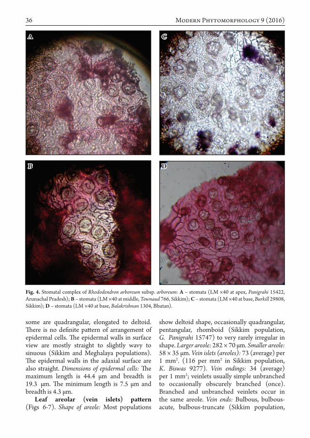
Key words: Asimina, Magnoliales, cortical vascular systems (CVS), floral vasculature, Florida
1 Muséum National d’Histoire Naturelle, Département Systématique et Évolution, UMR 7205, ISYEB, CNRS MNHN UPMC EPHE, CP 39, 57 rue Cuvier, F-75231 Paris cedex 05, France; [email protected] Stetson University, Department of Biology, 421 North Woodland Blvd., DeLand, FL 32720, United States of America; [email protected]

4 Modern Phytomorphology 9 (2016)
reconstituted by drawings of the serial sections using a camera lucida. Mountings were deposited at the slide library (“histothèque”) of P, under the references: Deroin 186 and 187. Moreover, longitudinal hand sections of the other buds were made as a rough check of vascular diagram, and allow a better understanding of carpel morphology.
Results
Pedicel histology (Fig. 2 A, D)
The epidermis is made up by small rounded and papillose cells, including some 2-celled simple hairs, and outlined by a hypodermis, slightly distinct from the cortex, which exhibits 8-12 layers of wider cells, with intercellular spaces and secretory cells, especially in the outer half. No sclereid is recognized at this stage, close to the anthesis. Phloem fibers show thin walls. There are ca. 20 bundles, elliptical in cross section and
2-3 cambial layers are seen in them. Pith cells appear similar to cortical ones. Main differences between the two species occur in the parenchymas (lignified in D. rugelii), and in the arrangement of phloem fibers (rather continuous ring in D. pulchellus; discrete crescent massifs in D. rugelii).
With a diameter of 1000-1250 µm and ca. 20 xylem poles, Deeringothamnus pedicels may be compared with those previously described in Cananga, Isolona, Rollinia or Uvariopsis (Deroin 1997), i.e. in fairly derived genera.
Perianth histology (Fig. 2 B, C)
Sepals are third (D. pulchellus) to half (D. rugelii) the thickness of petals, exhibit a homogeneous parenchyma with scattered secretory cells, and bear simple hairs outside like the pedicel. In petals parenchyma cells decrease in size towards the inner side, which is covered by a papillose epidermis, devoided
Fig. 1. Morphology of the studied Deeringothamnus flowers: A-C – D. pulchellus; D-F – D. rugelii.

5
Fig. 2. Flower histological features in Deeringothamnus pulchellus (A – cross section of pedicel (detail); B – sepal; C – outer petal) and in D. rugelii (D – pedicel). Stained by the combination of aqueous Astrablue 0.5% and Ziehl’s Fuchsine 10%. All scale bars = 100 µm.
Deroin T., Norman É. Notes on the floral anatomy of Deeringothamnus

6 Modern Phytomorphology 9 (2016)
Fig. 3. Ascending transverse sections of the flower of Deeringothamnus pulchellus: A-D – insertion of the bract; E-L – from the pedicel to the gynoecium base level. Abbreviations: B – bract; ls – sepal lateral bundle; ms – sepal median bundle; Pe – outer petal; Pi – inner petal; S – sepal.

7
Fig. 4. Ascending transverse sections of the flower of Deeringothamnus rugelii: A-J – from the pedicel to the receptacle top level. Abbreviations: same as in Fig. 3, with mc – carpel median bundle; mls – sepal mediolateral bundle.
Deroin T., Norman É. Notes on the floral anatomy of Deeringothamnus

8 Modern Phytomorphology 9 (2016)
of any corrugation unlike Asimina (Norman et al. 1992). Secretory cells are located as in the pedicel. In D. rugelii, petals bundles show some phloem fibers.
Anther histology (Fig. 5 C, D)
Epidermis is somewhat thin and discontinuous on the pollen sacs, much thicker
Fig. 5. Histological features in Deeringothamnus pulchellus (A – cross section of flower; B – anther base) and in D. rugelii (C – cross section of flower; D – anther base; E – carpel (second ovule level in ascending order)). Stained by the combination of aqueous Astrablue 0.5% and Ziehl’s Fuchsine 10%. Scale bars: A, C = 1 mm; B, D, E = 100 µm.
9
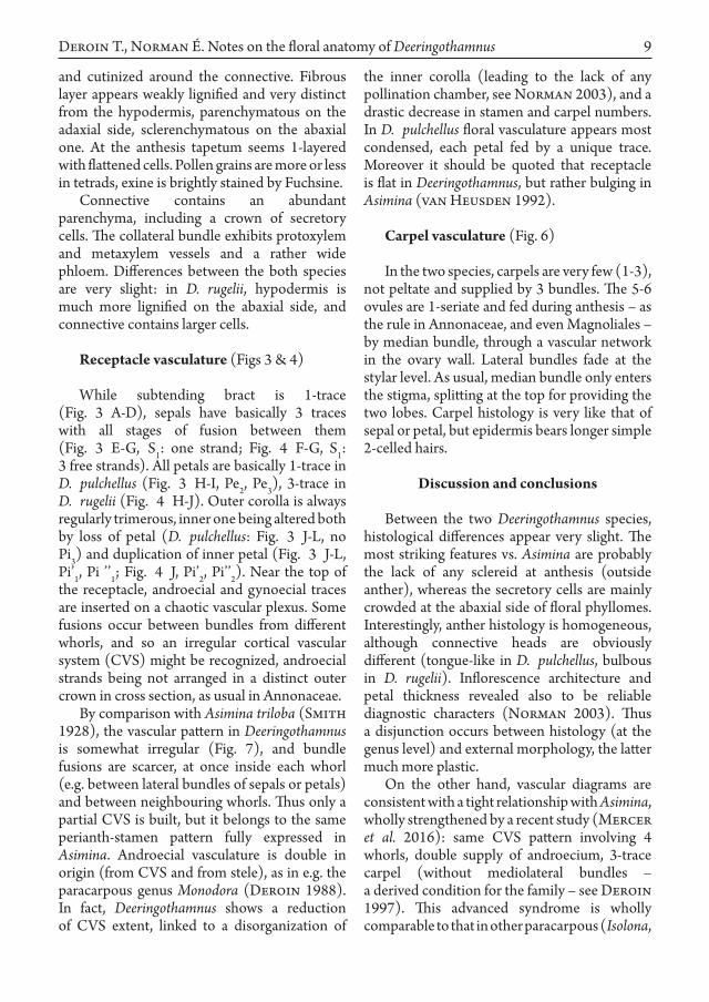
and cutinized around the connective. Fibrous layer appears weakly lignified and very distinct from the hypodermis, parenchymatous on the adaxial side, sclerenchymatous on the abaxial one. At the anthesis tapetum seems 1-layered with flattened cells. Pollen grains are more or less in tetrads, exine is brightly stained by Fuchsine.
Connective contains an abundant parenchyma, including a crown of secretory cells. The collateral bundle exhibits protoxylem and metaxylem vessels and a rather wide phloem. Differences between the both species are very slight: in D. rugelii, hypodermis is much more lignified on the abaxial side, and connective contains larger cells.
Receptacle vasculature (Figs 3 & 4)
While subtending bract is 1-trace (Fig. 3 A-D), sepals have basically 3 traces with all stages of fusion between them (Fig. 3 E-G, S1: one strand; Fig. 4 F-G, S1: 3 free strands). All petals are basically 1-trace in D. pulchellus (Fig. 3 H-I, Pe2, Pe3), 3-trace in D. rugelii (Fig. 4 H-J). Outer corolla is always regularly trimerous, inner one being altered both by loss of petal (D. pulchellus: Fig. 3 J-L, no Pi3) and duplication of inner petal (Fig. 3 J-L, Pi’1, Pi ’’1; Fig. 4 J, Pi’2, Pi’’2). Near the top of the receptacle, androecial and gynoecial traces are inserted on a chaotic vascular plexus. Some fusions occur between bundles from different whorls, and so an irregular cortical vascular system (CVS) might be recognized, androecial strands being not arranged in a distinct outer crown in cross section, as usual in Annonaceae.
By comparison with Asimina triloba (Smith 1928), the vascular pattern in Deeringothamnus is somewhat irregular (Fig. 7), and bundle fusions are scarcer, at once inside each whorl (e.g. between lateral bundles of sepals or petals) and between neighbouring whorls. Thus only a partial CVS is built, but it belongs to the same perianth-stamen pattern fully expressed in Asimina. Androecial vasculature is double in origin (from CVS and from stele), as in e.g. the paracarpous genus Monodora (Deroin 1988). In fact, Deeringothamnus shows a reduction of CVS extent, linked to a disorganization of
the inner corolla (leading to the lack of any pollination chamber, see Norman 2003), and a drastic decrease in stamen and carpel numbers. In D. pulchellus floral vasculature appears most condensed, each petal fed by a unique trace. Moreover it should be quoted that receptacle is flat in Deeringothamnus, but rather bulging in Asimina (van Heusden 1992).
Carpel vasculature (Fig. 6)
In the two species, carpels are very few (1-3), not peltate and supplied by 3 bundles. The 5-6 ovules are 1-seriate and fed during anthesis – as the rule in Annonaceae, and even Magnoliales – by median bundle, through a vascular network in the ovary wall. Lateral bundles fade at the stylar level. As usual, median bundle only enters the stigma, splitting at the top for providing the two lobes. Carpel histology is very like that of sepal or petal, but epidermis bears longer simple 2-celled hairs.
Discussion and conclusions
Between the two Deeringothamnus species, histological differences appear very slight. The most striking features vs. Asimina are probably the lack of any sclereid at anthesis (outside anther), whereas the secretory cells are mainly crowded at the abaxial side of floral phyllomes. Interestingly, anther histology is homogeneous, although connective heads are obviously different (tongue-like in D. pulchellus, bulbous in D. rugelii). Inflorescence architecture and petal thickness revealed also to be reliable diagnostic characters (Norman 2003). Thus a disjunction occurs between histology (at the genus level) and external morphology, the latter much more plastic.
On the other hand, vascular diagrams are consistent with a tight relationship with Asimina, wholly strengthened by a recent study (Mercer et al. 2016): same CVS pattern involving 4 whorls, double supply of androecium, 3-trace carpel (without mediolateral bundles – a derived condition for the family – see Deroin 1997). This advanced syndrome is wholly comparable to that in other paracarpous (Isolona,
Deroin T., Norman É. Notes on the floral anatomy of Deeringothamnus

10 Modern Phytomorphology 9 (2016)
Fig. 6. Carpel architecture of Deeringothamnus pulchellus – longitudinal section with related ascending transverse ones.

11
Monodora, see Deroin & Couvreur 2008) and pseudosyncarpous (Annona, Rollinia) genera, and close phylogenetical relationships are now well supported by molecular analysis (Couvreur et al. 2015: 5). However D. pulchellus differs significantly from D. rugelii by lacking lignified parenchymas, its highly reduced CVS, and the lack of vascular fusion between members of a same whorl. Combining these features with differences in petal thickness and connective head, it seems more appropriate not to merge the two species, as proposed by Ward (2001), although they are undoubtedly very near.
Conversely the vascular architecture of Deeringothamnus flower appears as an over-evolution of the Asimina pattern, its irregular character is reminiscent of that previously described in the East-African genus Sanrafaelia Verdc., in which the flat receptacle exhibits even a trend to inferovary (Deroin 2000), fully expressed in most Xylopia species (van Heusden 1992). Such a chaotic vascular pattern was yet described in the Malagasy endemic Ambavia gerrardii (Baill.) Le Thomas, combined with a wide intra-individual variation in perianth arrangement and lack of any CVS, a relevant feature of the other basal genera in the family (Deroin & Le Thomas 1989). So floral vasculature of the most derived annonaceous genera exemplifies a reversion to an ancestral condition with a recovery of evolutive potentialities.
Acknowledgements
Preliminary results of this study were presented as a poster during the Annonaceae Workshop of the European Union Systematics Association Conference. Leiden, the Netherlands, 7-9 August 2009. We are grateful to Svenja Meinke, Lars Chatrou and Paul Kessler for their kind invitation to this stimulating meeting. We thank too our colleague Emmanuel Côtez (Service des Publications scientifiques du Muséum) for his efficient help in the preparation of the electronic version of the plates.
References
Couvreur T.L.P., Niangadouma R., Sonké B., Sauquet h. 2015. Sirdavidia, an extraordinary new genus of Annonaceae from Gabon. PhytoKeys 46: 1–19.
Deroin T. 1988. Aspects anatomiques et biologiques de la fleur des Annonacées. Unpublished thesis 590. Paris 11 University, Orsay.
Deroin T. 1997. Comparative anatomy of floral pedicels in Annonaceae and Magnoliaceae: bringing out some evolutive trends. Scripta Botanica Belgica 15: 49.
Deroin T. 2000. Floral anatomy of Sanrafaelia Verdc. and its evolutive significance. Annonaceae Newsletters (Utrecht) 13: 36–40.
Deroin T., Couvreur T.L.P. 2008. Floral anatomy. In: Couvreur T.L.P. Revealing the secrets of African Annonaceae. Systematics, evolution and biogeography of the syncarpous genera Isolona and Monodora. PhD thesis Wageningen University, The Netherlands (ISBN 978-90-8504-924-1): 120–124. Wöhrmann Print Service, CPI Group, Zutphen.
Fig. 7. Vascular diagrams of flowers: A – Asimina triloba, after Smith (1928); B – Deeringothamnus pulchellus; C – D. rugelii. Abbreviations: same as in Fig. 3. Sepal bundles stippled, outer petal bundles white, inner petal bundles black, stamen bundles hatched with black tips, carpel bundles stippled with hatched tips, fused bundles (cortical vascular system) crossed.
Deroin T., Norman É. Notes on the floral anatomy of Deeringothamnus

12 Modern Phytomorphology 9 (2016)
Deroin T., Le Thomas A. 1989. Sur la systématique et les potentialités évolutives des Annonacées: cas d’Ambavia gerrardii (Baillon) Le Thomas, espèce endémique de Madagascar. Comptes Rendus de l’Académie des Sciences, Paris, t. 309, Série III: 647–652.
Gerlach D. 1984. Botanische Mikrotechnik. Thieme, Stuttgart.
Kral R. 1960. A revision of Asimina and Deeringothamnus. Brittonia 12: 233–278.
Mercer E., Griffin B., Steele J., Goodrich K.R., Bush C.M. 2016. Phylogenetic relationships of Asimina and Deeringothamnus (Annonaceae) based on morphology, floral scent chemistry, and Inter-Simple Sequence Repeat data. J. Torrey Bot. Soc. 143: 58–68.
Norman E.M. 2003. Reproductive biology of Deeringothamnus rugelii and D. pulchellus (Annonaceae). Taxon 52: 547–555.
Norman E.M. 2007. The “False Pawpaws”. History, biology and conservation of Deeringothamnus. Palmetto (Quaterly J. Florida Native Pl. Soc.) 24: 4–7, 15.
Norman E.M., Rice K., Cochran S. 1992. Reproductive biology of Asimina parviflora (Annonaceae). Bull. Torrey Bot. Club 119: 1–5.
Smith G.h. 1928. Vascular anatomy of ranalian flowers II. Ranunculaceae (cont.), Menispermaceae, Calycanthaceae, Annonaceae. Bot. Gaz. (Crawfordsville) 85: 170–177.
van heusden E.C.h. 1992. Flowers of Annonaceae: morphology, classification and evolution. Blumea Suppl. 7: 1–218.
Ward D.B. 2001. New combinations in the Florida flora. Novon 11: 360–365.

ISSN 2226-3063 e-ISSN 2227-9555Modern Phytomorphology 9: 13–17, 2016
© 2016 The Author(s). Published by Modern Phytomorphology. This is an open access article under the Creative Commons BY-NC-ND license (http://creativecommons.org/licenses/by-nc-nd/4.0/)
Introduction
The genus Teucrium L. is a large and polymorphic genus comprising about 200 species in the world. It has cosmopolitan distribution, mainly in Europe, North Africa and in the temperate parts of Asia (Kästner 1989; Abu-Assab & Cantino 1993). It has been divided into ten sections, identifiable through the calyx shape and the inflorescence structure (McClintock & Epling 1946; Tutin & Wood 1972). These sections are Teucropsis Benth., Teucrium Benth., Chamaedrys (Mill.) Schreb., Polium (Mill.) Schreb., Isotriodon Boiss., Pycnobotrys Benth., Scorodonia (Hill) Schreb., Stachyobotrys Benth., Scordium (Mill.) Benth., and Spinularia Boiss.
In Turkey, the genus Teucrium comprises now a total of 35 species, including the recently added T. chasmophyticum Rech. f. (Dönmez 2006), T. melissoides Boiss. & Hausskn. ex Boiss. (Dönmez et al. 2010), T. aladagense Vural & H. Duman (Vural et al. 2015) and T. sirnakense Ozcan & Dirmenci (Özcan et al. 2015), recently reinstated T. andrusi Post (Dinç et al. 2011), and the long overlooked T. krymense Juz., recorded from Kars (Özhatay & Kültür 2006). According to the infraspecific classification, Turkish flora presently includes 48 taxa (Dönmez 2006; Özhatay & Kültür 2006; Parolly & Eren 2007; Dönmez et al.
2010; Dinç et al. 2011; Vural et al. 2015; Özcan et al. 2015).
Teucrium sect. Teucrium has 30 species world-wide (Navarro & El Oualidì 2000; Parolly & Eren 2007), 11 of them occur in Turkey (Ekim 1982; Duman 2000). Acording to the intraspecific classification, these species includes 13 taxa. In the section, T. orientale, the closest relative of T. pruinosum, divided into 3 varietes in terms of indumentum type and density.
Some Teucrium specimens were collected from Aksaray province of Central Anatolia by the authors. They were identified according to the Flora of Turkey as T. pruinosum. T. pruinosum is mainly characterised by its grey pruinose appearance due to densely canescent indumentum, calyx teeth uncinate at tip and subequal to the tube with conspicious midvein. Our specimens show the characteristics related with the calyx shape. But, they are readily distinguished from the typical T. pruinosum by their subglabrous indumentum (not densely canescent), green appearance of the leaves and stems (not grey pruinose), blue-violet corollas (not light blue), purple pedicels (not grey or bluish), and filaments (not bluish). After the studies on T. pruinosum populations in Central Anatolia, the observations on the syntypes and some other herbarium specimens, and the examinations on the related Floras
teucrium pruinosum VAR. aksarayense VAR. NOV. (LAMIACEAE) FROM CENTraL ANATOLIA, TuRKEy
Muhittin Dinç & Süleyman Doğu *
Abstract. Teucrium pruinosum var. aksarayense M. Dinç & S. Doğu (Lamiaceae), a new variety from Aksaray in Central Anatolia, is described and illustrated. The new variety is similar to the typical one in its calyx teeth uncinate at tip and subequal to the tube with conspicious midvein. It is readily distinguished from var. pruinosum by its general appearance, indumentum, and floral organ pigmentation. The map showing the distributions of the varieties was given.
Key words: Teucrium, Lamiaceae, taxonomy, Turkey
Necmettin Erbakan University, Ahmet Keleşoğlu Faculty of Education, Department of Biology, 42090 Meram, Konya/Turkey; * [email protected]

14 Modern Phytomorphology 9 (2016)
(Rechinger 1964; Mouterde 1966; Feinbrun Dothan 1978; Ekim 1982), we concluded that our specimens represent an undescribed variety included in the species.
Material and methods
Plant materials were collected from the natural populations of T. pruinosum growing in the cenrtal part of Turkey. Collection data is as follows: (as var. pruinosum here) Turkey, C4 Konya, In aridis subsalsis planitiei prope Karaman Lycaoniae ad radices Karadagh, 15.06.1845, Heldreich (syntype, E photo); B5 Kayseri ad Caesaream Cappadociaae, 1107 m, 01.06.1856, Balansa 1069 (syntype, E photo); B5 Kayseri: Yeşilhisar, Güzelöz Köyü civarı, kayalık, step, 1400 m, 06.07.2010, M. Dinç 3303 & S. Doğu; Develi civarı, steppe, 1200 m, 06.07.2010, M. Dinç 3304 & S. Doğu; B5 Aksaray: Hasan Dağı etekleri, Karkın köyü civarı, steppe, 1350 m, 17.07.2010, M. Dinç 3332 & S. Doğu; Demirci Köyü civarı, yol kenarı, 1200 m, 06.07.2010, M. Dinç 3298 & S. Doğu; Aksaray-Niğde sınırı, Kayırlı Kasabası civarı, steppe, 1400 m, M. Dinç 3300 & S. Doğu; B5 Nevşehir: Avanos civarı, 1150 m, steppe, 10.07.2010, M. Dinç 3331 & S. Doğu; (as var. aksarayense here) B5 Aksaray:
Aksaray-Niğde sınırı, Kayırlı Kasabası civarı, steppe, 1400 m, M. Dinç 3299 & S. Doğu; B5 Nevşehir: Avanos civarı, 1150 m, steppe, 10.07.2010, M. Dinç 3330 & S. Doğu, B5 Kayseri: Yeşilhisar, Güzelöz Köyü civarı, kayalık, steppe, 1400 m, 06.07.2010, M. Dinç 3302 & S. Doğu.
The plants collected by the authors were identified using “Flora of Turkey and the east Aegean Islands” (Ekim 1982) and stored in Necmettin Erbakan University, Ahmet Keleşoğlu Faculty of Education, Department of Biology, Konya. Hovewer, with the observations on the syntypes and some other herbarium specimens of T. pruinosum, and the examinations on the related Floras (Rechinger 1964; Mouterde 1966; Feinbrun Dothan 1978), morphological studies on the populations and herbarium specimens showed that some individulas were clearly and persistently differ from typical characteristics of T. pruinosum.
In order to show the differences between the typical and new variety, photo and microphotographs were taken. In order to take microphotographs, the pieces of the stems and leaves of each variety were mounted directly on stubs with double-side adhesive tape and coated with gold. The pieces were examined by SEM and photographed.
Fig. 1. The general appearances and enlarged flowers of the two varietes of Teucrium pruinosum: A, B – var. aksarayense; C, D – var. pruinosum.

15
Results
Teucrium pruinosum var. aksarayense M. Dinç & S. Doğu var. nov. (Figs 1-3).
Type: B5 Aksaray: Hasan Dağı etekleri, Karkın köyü civarı, steppe, 1350 m, 17.07.2010, M. Dinç 3333 & S. Doğu (Holotype: KNYA, Isotypes: GAZI, HUB, Yıldırımlı Herb.)
Diagnosis: var. pruinosum similis sed plantis subglabris (non dense canus), caulis et foliis
viridis (non canus-pruinosus), flos cyaneo-violaceus (non pallidus cyaneus), pedicelli et filamanta purpurei (non canus vel pallidus cyaneus) differt.
Description: Perennial herbs, 30-65 cm, green, subglabrous with very sparse simple, short non-glandular and subsessile clavate-glandular hairs, many-stemmed from an indurate base and a woody root stock. Stems erect or slightly ascending, strongly tetragonal,
Fig. 2. The views of stems and leaves of the two varietes of Teucrium pruinosum: A, C, E – var. aksarayense; B, D, F – var. ruinosum. A, B – the photographs of stems and leaves; C, D – SEM photos of the stems; E, F – SEM photos of the leaves.
Dinç M., Doğu S. Teucrium pruinosum var. aksarayense var. nov. from Central Anatolia

16 Modern Phytomorphology 9 (2016)
rigid, each ending above in a thyrsoid narrow panicle with erect spreading branches. Leaves ovate-lanceolate in outline, ternately 2-3-pinnatisect into green linear rigid lobes with revolute margins, subglabrous with very sparse hairs. Verticillasters 2-flowered; pedicels 1-flowered, purplish, longer than both calyx and floral leaves. Calyx subglabrous, purplish, 4.5-6.0 mm, campanulate; teeth lanceolate, 1.0-1.5 × calyx tube, prominetly 1 veined, uncinate at tip. Corolla blue-violet, 8-12 mm, usually 2-3 × calyx; upper lip elongate, with acute lateral lobes. Stamens long-exserted, filaments purple. Nutlets ovoid, 2.1-3.0 × 0.9 × 1.3 mm, with glandular and nonglandular hairs.
Recommended IuCN threat category: T. pruinosum var. aksarayense has been presently known from four localities in Aksaray and its adjacent provinces. Its estimated area of occupancy is less than 5000 km2 (criterion B). The populations are healthy, but with less than 2500 individuals (criterion C). In addition, the restricted area of the populations implies a high risk of extinction owing to nearness of roads and agricultural areas, subjection to grazing (criterion D). Therefore, T. pruinosum var.
aksarayense should be classified as “Endangered (EN)” based on the criteria of the IUCN Red List Categories (IUCN 2001).
Etymology: The name of the new variety refers to the province in which the variety is collected firstly.
THE IDENTIFICATION KEY OF THE TWO VARIETES OF T. PRUINoSUM
1. Plant canescent with densely simple short non-glandular hairs, grey pruinose, corolla light blue, pedicels grey or bluish, filaments bluish ................................................... var. pruinosum
1*. Plant subglabrous with very sparse simple short non-glandular hairs, green, corolla blue-violet, pedicels and filaments purple .............................................................. var. aksarayense
Discussion
Although the new variety is sympatric for all part of its range with the typical variety, it keeps the diagnostic characters from var. pruinosum. The morphological differences between the two varietes are never subtle
Fig. 3. Distribution map of the two varietes of Teucrium pruinosum: var. pruinosum (■), and var. aksarayense (*).

17
and no intermediate forms exist. Taking into consideration these data, it reveals that the new variety is emerged from Aksaray and its surrounding populations of T. pruinosum by sympatric speciation.
According to the SEM observations, in accordance with general appearances of the two varietes, var. pruinosum has abundant short non-glandular and very sparse subsessile glandular trichomes, while var. aksarayense has very sparse short non-glandular and denser subsessile glandular ones (Fig. 2).
While var. pruinosum has mainly grey pruinose appearance owing to densely canescent indumentum and grows in Syria, Lebanon, Palestine, Iraq, and Turkey (Rechinger 1964; Mouterde 1966; Feinbrun Dothan 1978; Ekim 1982), the new variety has green appearance due to subglabrous indumentum and only occurs in Central Anatolia (Fig. 3). The situation support the opinion that Anatolia is a major speciation centre for Teucrium sect. Teucrium (Parolly et al. 2007).
References
Abu-Assab M.S., Cantino P.D. 1993. Phylogenetic implications of pollen morphology in tribe Ajugeae (Labiatae). Syst. Bot. 18: 100–122.
Dinç M., Doğu S., Bağcı y. 2011. Taxonomic reinstatement of Teucrium andrusi from T. paederotoides based on morphological and anatomical evidences. Nord. J. Bot. 29: 148–158.
Dönmez A.A. 2006. Teucrium chasmophyticum Rech. f. (Lamiaceae): A new record for the Flora of Turkey. Turk. J. Bot. 30: 317–320.
Dönmez A.A., Mutlu B., Özçelik A.D. 2010. Teucrium melissoides Boiss. & Hausskn. ex Boiss. (Lamiaceae): A new record for flora of Turkey. Hacettepe J. Biol. & Chem. 38: 291–294.
Duman h. 2000. Teucrium L. In: Güner A., Özhatay N., Ekim T., Başer K.H.C. (eds), Flora of Turkey and East Aegean Islands (Suppl. II). Vol. 11: 197–198. Edinburgh University Press, Edinburgh, UK.
Ekim T. 1982. Teucrium L. In: Davis P.H. (ed.), Flora of Turkey and the East Aegean Islands. Vol. 7: 53–75. Edinburgh University Press, Edinburgh, UK.
Feinbrun Dothan N. 1978. Flora Palaestina Ericaceae to Compositae. Vol. 3: 101–102. Israel Academy of Sciences and Humanities, Jerusalem.
IUCN 2001. IUCN Red List Categories and Criteria, Version 3.1. Prepared by the IUCN Species Survival Commission.
Kästner A. 1989. Übersicht zur systematischen Gliederung der Gattung Teucrium L. Biocosme Mesogeen (Nice) 6: 63–77.
McClintock E., Epling C. 1946. A revision of Teucrium in the New World, with observations on its variation, geographical distribution and history. Brittonia 5: 491–510.
Mouterde P. 1966. Nouvelle flore du Liban et de la Syrie. Vol. 1: 101–102. Beyrouth-Liban.
Navarro T., El Oualidì J. 2000. Synopsis of Teucrium L. (Labiatae) in the Mediterranean region and surrounding areas. Flora Mediterranea 10: 349–363.
Özcan T., Dirmenci T., Çoşkun F., Akçiçek E., Güner Ö. 2015. A new species of Teucrium sect. Scordium (Lamiaceae) from SE of Turkey. Turk. J. Bot. 39: 310–317.
Özhatay N., Kültür Ş. 2006. Check-list of additional taxa to the Supplement Flora of Turkey III. Turk. J. Bot. 30: 281–316.
Parolly G., Eren Ö. 2007. Contributions to the flora of Turkey. 2. Willdenowia 37: 245–246.
Rechinger K.h. 1964. Flora of Lowland Iraq: 518. Weinheim.
Tutin G., Wood D. 1972. Teucrium L. In: Tutin T.G., Heywood V.H., Burges N.A., Moore D.M., Valentine D.H., Walters S.M., Webb D.A. (eds), Flora Europaea. Vol. 3: 129–135. Cambridge University Press, Cambridge, UK.
Vural M., Duman h., Dirmenci T., Özcan T. 2015. A new species of Teucrium sect. Stachyobotrys (Lamiaceae) from the south of Turkey. Turk. J. Bot. 39: 318–324.
Dinç M., Doğu S. Teucrium pruinosum var. aksarayense var. nov. from Central Anatolia


ISSN 2226-3063 e-ISSN 2227-9555Modern Phytomorphology 9: 19–25, 2016
© 2016 The Author(s). Published by Modern Phytomorphology. This is an open access article under the Creative Commons BY-NC-ND license (http://creativecommons.org/licenses/by-nc-nd/4.0/)
Introduction
Roses have a high ability to form interspecific hybrids, both within one section and between separate sections. It is relatively easy to distinguish intersectional hybrids. Separation of modern hybrids between species of one section requires great caution, especially in the case of the section Caninae DC. em. Christ., which is the most polymorphic group of the Rosa L. It is particularly difficult because their morphological characteristics are the main criterion for distinguishing, wherein none of the morphological characteristics analyzed separately have not a significant diagnostic value in the case of this section (Zieliński 1985).
Rosa canina L. is the most common and most variable species in genus Rosa. It creates transitional forms of species, both within the section of Caninae, as well as with species from other sections. There are known interspecific hybrids of R. canina with the species from the section Caninae – e.g. R. jundzillii Besser, R. tomentosa Sm., R. dumalis Bechst., R. sherardii Davies; from the section
Cinnamomeae DC. – e.g. R. pendulina L.; and from the section Rosa – e.g. R. gallica L. (Zieliński 1987).
Gustafsson (1944) mentioned in his work of yet another hybrid – between R. canina and R. rubiginosa L. The possibility to form this type of hybrid has been also known from later works, regarding genetic testing of species from the entire the section Caninae (inter alia Blackhurst 1947; De Cock 2008; Ritz & Wissemann 2011). However, in the literature relating to this form of hybrid no analysis of morphological traits were found. Therefore, this study is an attempt to establish the inter-relationship between R. canina, R. rubiginosa and their hybrid, based on morphological characteristics, diagnostic for the Caninae section.
Material and methods
The specimens used for morphometric studies of R. canina × R. rubiginosa hybrid and its parental forms were collected in Ukraine (Podolia region) in 2008-2009 on two localities:
BIOMETRIC ANALySIS OF INTERSPECIFIC hyBRIDS BETWEEN rosa canina L. AND rosa rubiginosa L. (SECTION caninae DC. EM. ChRIST.)
Anna Sołtys-Lelek 1, Beata Barabasz-Krasny 2, Peter Turis 3, Ingrid Turisová 4, Wojciech Gruszka 5
Abstract. The article presents the biometric analysis of selected morphological features of interspecies hybrid Rosa canina L. × R. rubiginosa L. This hybrid was the result of spontaneous hybridization between the two species falling into section Caninae DC. em. Christ. So far, it has not been studied in terms of morphological characteristics, in particular with respect to the parental forms.
Key words: Rosa, R. canina, R. rubiginosa, R. canina × R. rubiginosa, Caninae, Rosaceae, morphology, spontaneous hybridization
1 ojców National Park, 32-047 ojców 9, Poland; [email protected] Institute of Biology, Pedagogical University, Podchorążych 2, 30-084 Kraków, Poland; [email protected] Low Tatras National Park, Lazovná 10, 974 01 Banská Bystrica, Slovakia; [email protected] Faculty of Natural Sciences, Matej Bel University, Tajovského 40, 974 01 Banská Bystrica, Slovakia; [email protected] Department of Biology, Morphological and Health Sciences., Faculty of Physical Culture in Gorzów Wlkp., University School of Physical Education in Poznań, Estkowskiego 13, 66-400 Gorzów Wlkp., Poland; [email protected]

20 Modern Phytomorphology 9 (2016)
Medobory Nature Reserve and to the southeast of Sataniv (leg. A. Sołtys-Lelek, Herbarium of the Ojców National Park, Ojców, Poland) and in 2015 in Pila – in North-Western Poland (leg. W. Gruszka, Herbarium of the University School of Physical Education, Gorzow Wielkopolski, Poland).
Several characters were measured, counted, or observed on the leaves and hips (Tab. 1; Fig. 1). Selection of characters was based on previous studies (De Cock et al. 2007, 2008; Mijnsbrugge & Beeckman 2012). The
measurements were made in thirty random samples. For all measured characteristics, arithmetic means and standard deviations were calculated. The differences between the mean values for the analyzed parental forms and their hybrid were tested using non-parametric Kruskal-Wallis test at P ≤ 0.05. The statistical analyses were done by software Statistica 10.0 for Windows.
The systematic approach and the nomenclature are basing on the work of Popek (1996).
Organ Character Abbr. Description
Leaf
Lamina length LL Length of leaflet lamina
Lamina base length LbL Length of basal part of leaflet lamina till largest width
Lamina width LW Largest width of leaflet lamina
Rachis length RL Length of rachis
Leaflet margin serration LmSe Serration of the leaflet margin scored from 1 (single toothed) to 3 (multiple toothed)
Leaflet margin glands LmG Glands on the leaflet margin; from 1 (no glands) to 5 (densely glandular)
Leaflet glands LG Glands on the underside of leaflet (outside the midrib)
Rachis pubescence RPu Pubescence on the rachis; from 1 (no pubescence) to 5 (dense hairiness)
Rachis glands RG Glands on rachis; from 1 (no glands) to 5 (densely glandular)
Lamina shape LS LL/LW
Lamina base shape LbS LbL/LW
Lamina length ratio LLR LL/RL × 100
Lamina base length ratio LbLR LbL/RL × 100
Lamina width ratio LWR LW/RL × 100
Frui
t
Hip length HL Length of hip
Hip base length HbL Basal length of hip till largest width
Hip width HW Largest width of hip
Pedicel length PL Length of hip pedicel
Orificium diameter OD Diameter of orifice
Discus diameter DD Diameter of disc
Discus height DH Height of disc
Pedicel glands PG Glands on pedicel; from 1 (no glands) to 5 (densely glandular)
Fruit length FL HL + PL
Hip shape HS HL/HW
Hip base shape HbS HbL/HW
Hip length ratio HLR HL/FL × 100
Orificium ratio OR OD/DD × 100
Tab. 1. Overview of characters used to describe rose species in this study (according to Mijnsbrugge & Beeckman (2012), modified); Abbr. – abbreviations.

21
Results
A hybrid form of R. canina × R. rubiginosa is characterized by a combination of morphological features of both parental species (Tab. 2). The size and shape of leaves (LL, LbL, LS, LbS) and the glanding of the pedicel (PG) it refers to R. canina. For example, the length of the leaf of the hybrid (LL) is within the range from 15.40 mm to 31.60 mm – 23.60 mm on average, and the lamina base length (LbL) from 6.40 mm to 18.30 mm – 11.00 mm on average. For R. canina LL values are within the range of 18.50 mm to 32.00 mm – 24.62 mm on average, and LbL from 7.20 mm to 14.60 mm – 11.05 mm on average (Tab. 3).
In the case of R. rubiginosa, the hybrid is similar in such features as: length of the rachis (RL), serration of the leaflet margin (LmSe), the diameter of the orifice (OD), hip shape (HS) and the orificium ratio (OR). For example, the length of the rachis (RL) for the hybrid is between 10.40 mm and 39.55 mm – 19.42 mm on average, and for R. rubiginosa from 9.40 to 32.00 mm – 21.13 mm on average. The diameter
of the orifice (OD) for the hybrid is within the range of 1.35 mm to 2.90 mm – average of 1.93 and for R. rubiginosa from 1.25 mm to 2.55 mm – 1.86 mm on average (Tab. 3).
On the other hand, in relation to both parental species, R. canina × R. rubiginosa hybrid shows statistically significant differences with respect to such morphological features as: glands of the leaf margin (LmG), glands on the underside of leaflet (LuG) glands on rachis (RG) and height of a disc (HD). In these cases, the hybrid has intermediate characteristics between parental forms, which are however statistically different from them. For example, the height of disc (HD) for the hybrid is within the range of 0.50 mm to 1.70 mm – 0.86 mm on average, for R. rubiginosa it ranges from 0.00 mm to 0.50 mm – average of 0.03 and for R. canina from 1.50 to 3.30 – an average of 2.16 mm. In the hybrid, this parameter reaches a significantly higher value in relation to R. rubiginosa and in relation to R. canina the value is significantly lower (Tab. 3).
There were no statistically significant differences between these three taxa, with
Fig. 1. Morphological characteristics of leaf (A) and fruit (B, C), that were measured or observed; C – upper part of hip; abbreviations are listed in Tab. 1.
Sołtys-Lelek A. et al. Biometric analysis of hybrids of Rosa canina and R. rubiginosa

22 Modern Phytomorphology 9 (2016)
respect to such morphological features as: the lamina width (LW), the length of the hip (HL), basal length of the hip (HbL), hip width (HW), length of pedicel (PL), diameter of disc (DD), fruit length (FL) (Tab. 3).
Discussion
Until now, information on spontaneous R. canina and R. rubiginosa hybrids have not appeared in the literature too often, even
though the possibility of forming of hybrid forms between these species, carried out by artificial hybridization, was mentioned many times (e.g. Gustafsson 1944; Blackhurst 1947; Wissemann 2006). Perhaps this is due to the fact that the frequency of spontaneous hybridization between the two species is rather low (Ritz & Wissemann 2011), and some authors even looked for the existence of a genetic barrier (Zieliński 1985). Certainly the geographical barrier between these species does
CharacteristicsParental form (1) Hybrid Parental form (2)
R. rubiginosa R. canina × R. rubiginosa R. canina
Shrub 0,5-2(-3) m to 2 (-3) m ca. to 3 m ca.
Type of prickles heteracantha homoioacantha, ± heteracantha homoioacantha
Prickleshooked, ± recurvate, falcate usually mixed with aciculae
and glandular setae
hooked, curved, occasionally mixed with aciculi and glandular setae on flowering short shoot
Petiole densely glandular without glands or ± glandular
Leaflet base usually rounded wedge-shaped or rounded
Leaflet apex acute to obtuse acute
Leaflet shapemostly suborbiculate or broadly oval or broadly
elliptical
elliptical, ovoid, broadly ovoid, roundish
elliptical, ovoid, broadly ovoid, roundish
Leaflet margin serration multiple toothed singe-, double- to multiple toothed
Leaflet margin glands densely stipitate-glandular ± densely stipitate-glandular from no glandular to ± densely stipitate-glandular
Rachis glands densely stipitate-glandular ± densely stipitate-glandular from no glandular to ± glandular
Under side of leaflet densely glandular some leaves no glandular, some ± glandular
without glands, exceptionally ± glandular
Pedicel stipitate-glandular, exceptionally glandless
without glands or occasionally ± stipitate-
glandular
without glands or occasionally ± stipitate-
glandular
Receptacle stipitate-glandular or without glands without glands without glands or
exceptionally ± glandular
Discus plain ± plain or conically convex conically convex
Orifice broadly, > 1 mm diameter narrow, to 1 mm diameter
Rose hip egg shaped, subglobose, broadly ovoid egg shaped, rarely round, ovate
Sepals usually erected irregularly spreaded, partly erected reflexed
Tab. 2. Characteristics of study taxa based on diagnostic features in the sect. Caninae.

23
Tab. 3. Comparison of morphometric characteristics between parental forms and their hybrid (Rosa canina × R. rubiginosa); the average (x) of 30 replicates ± SD; a, b, c (in row) – statistical significance with Kruskal-Wallis test, P≤0.05; abbreviations are listed in Tab. 1.
not exist. Both species have a broad range of occurrence, wherein the acreage of R. rubiginosa is in a range within R. canina (Popek 2007).
Isolation of modern hybrids within the Caninae section is extremely complex, both for the crossing of closely related species, as well as for remote taxa of this section. This concerns especially hybrids derived from R. canina, characterized by a morphological similarity with other species of this section (Zieliński 1985).
In the early nineteenth century, a stabilized hybrid between R. canina and R. rubiginosa was described in the rank of species under the name of R. obtusifolia Desv. Currently, it falls within the form of R. canina var. obtusifolia Desv. (Popek 1996). This form is characterized by leaves which are glandular underneath ± and pinnate leaves folded glandularly, which refers to the characteristics of R. rubiginosa. Kerényi-Nagy (2012) also lists other hybrids between
Characteristics R. canina [mm] R. rubiginosa [mm] R. canina × R. rubiginosa [mm]
x ± SD x ± SD x ± SD
LL 24.62a 3.31 20.23b 4.07 23.60a 4.80
LbL 11.05a 1.85 9.51b 1.99 11.00a 2.65
LW 14.79a 2.36 14.96a 3.46 14.69a 2.61
RL 22.65a 4.99 21.13ab 6.54 19.42b 5.18
LmSe (1-3) 2.17b 0.38 3.00a 0.00 3.00a 0.00
LmG (1-5) 2.07c 0.45 4.93a 0.25 3.60b 0.56
LG (1-5) 1.00c 0.00 4.97a 0.18 2.00b 0.79
Rpu (1-5) 1.10b 0.31 3.97a 0.81 1.23b 0.43
RG (1-5) 1.33c 0.48 4.77a 0.50 3.47b 1.14
LS (LL/LW) 1.68a 0.16 1.37b 0.16 1.61a 0.18
LbS (LbL/LW) 0.75a 0.10 0.64b 0.07 0.74a 0.10
LLR (LL/RL × 100) 112.73ab 22.84 102.09b 26.79 128.24a 30.28
LbLR (LbL/RL × 100) 50.25ab 10.62 47.83b 12.15 58.60a 15.78
LWR (LW/RL × 100) 67.42b 13.84 74.83ab 19.27 78.02a 16.36
HL 14.94a 3.04 13.78a 2.82 13.82a 2.50
HbL 7.25a 2.24 7.02a 1.77 7.10a 1.68
HW 9.65a 1.38 10.47a 1.69 10.35a 1.98
PL 8.83a 3.09 9.89a 2.30 8.49a 2.56
OD 0.96c 0.11 1.86ab 0.32 1.93a 0.46
DD 4.41a 0.45 4.30a 0.54 4.39a 0.48
DH 2.16a 0.42 0.03c 0.13 0.86b 0.48
PG (1-5. 1=0) 1.00c 0.00 4.70a 0.47 1.93bc 1.14
FL (HL + PL) 23.76a 3.90 23.67a 3.96 22.31a 3.81
HS (HL/HW) 1.56a 0.29 1.32b 0.19 1.35b 0.22
HbS (HbL/HW) 0.75a 0.22 0.67a 0.11 0.69a 0.13
HRL (HL/FL × 100) 63.26a 10.15 58.19a 6.50 62.30a 7.66
OR (OD/DD × 100) 22.17c 3.86 43.79ab 8.53 43.84a 9.76
Sołtys-Lelek A. et al. Biometric analysis of hybrids of Rosa canina and R. rubiginosa

24 Modern Phytomorphology 9 (2016)
R. canina and R. rubiginosa in the rank of species. They are: R. squarrosa (Rau) Boreau, R. blondeana Ripart and R. andegavensis Bastard – according to other taxonomic approaches also classified to glandular forms of R. canina (Popek 1996).
The analyzed hybrid displays intermediate characteristics between R. canina and R. rubiginosa, but also specimens closer to one of parental forms or almost indistinguishable from them occur as well (Tabs 2 & 3). It depends on which of the species produced seeds and which only gave pollen. The genetic material given by the maternal specimen makes up to 80% of genotype (Wissemann 2006). The similarity scale of the test hybrid to the parental species primarily concerns with the shape and size of the leaf, which may be more elliptical or oval, as in R. canina or more rounded as in R. rubiginosa. Also, stalks can be glandular, as in R. rubiginosa or without glands as usually is with R. canina.
The essential features that distinguish R. canina × R. rubiginosa hybrid are: glandular in various degree underside of the lamina (whole lamina or only part of it) – usually found only in the case of some leaves, which margins are serrated, quite rich glandular of the axis of leaf and the height of the disk, which is usually ± conical (Fig. 2). However, on one of the flower shoots flowers had clearly conical disk (approx. 2.00 mm in height) and the some of the disk near flat (approx. 0.50 mm in height). The shape of the disk and the size of orificium is one of the most important diagnostic features for different kinds of roses.
Species of Caninae section are of hybrid origin, no doubt, and were formed in the late Tertiary. However, spontaneous interspecific hybrids within this section occur also today, although hybridization of extant species of this section is limited to a large extent by the occurrence of autogamous (Zieliński 1985; Popek 2007).
References
Blackhurst h.T. 1947. Cytogenetic studies on Rosa rubiginosa L. and its hybrids. Doctoral dissertation, A. & M. College of Texas. Texas A. & M. University. Available electronically from http://hdl.handle.net /1969.1/ETD-TAMU-TXA0181161.
De Cock K. 2008. Genetic diversity of wild roses (Rosa spp.) in Europe, with an in-depth morphological study of Flemish populations. PhD, Research Institute for Nature and Forest, Brussels.
De Cock K., Mijnsbrugge K.V., Breyne P., Nybom h., Smulders M.J.M., Van Slycken J., De Riek J. 2007. The diversity of autochthonous roses in Flanders (Belgium) in the view of the European Generose Reference Framework. Acta Hortic. 760: 621–628.
De Cock K., Mijnsbrugge K.V., Breyne P., Van Bockstaele E., Van Slycken J. 2008. Morphological and AFLP-based differentiation within the taxonomical complex section Caninae (subgenus Rosa). Ann. Bot. 102 (5): 685–97.
Gustafsson Ä. 1944. The constitution of the Rosa canina complex. Hereditas 30: 405–428.
Kerényi-Nagy V. 2012. A Történelmi Magyarország területén élő őshonos, idegenhonos és kultúr-reliktum rózsák kismonográfiája. A small monograph of autochton, allochton and cultur-relict roses of the Historical Hungary, NYME Egyetemi Kiadó, Sopron.
Mijnsbrugge K.V., Beeckman h. 2012. Geographically differentiating morphology of genetically similar dogroses: consequences of canina meiosis. Plant Syst. Evol. 298 (9): 1733–1742.
Popek R. 1996. Biosystematyczne studia nad rodzajem Rosa L. w Polsce i krajach ościennych. Prace Monograficzne 218. Wyd. Nauk. WSP, Kraków. (In Polish)
Popek R. 2007. Dziko rosnące róże Europy. Officina Botanica, Kraków. (In Polish)
Ritz C. M., Wissemann V. 2011. Microsatellite analyses of artificial and spontaneous dogrose hybrids reveal the hybridogenic origin of Rosa micrantha by the contribution of unreduced gametes. J. Hered. 102 (2): 217–227.
Wissemann V. 2006. Beauty and the bastards. Intensive hybridization controls the evolution of wild roses. B.I.F. FUTURA 21: 158–163.
Zieliński J. 1985. Studia nad rodzajem Rosa L. – systematyka sekcji Caninae DC. em Christ. Arboretum Kórnickie 30: 3–109. (In Polish)
Zieliński J. 1987. Rodzaj Rosa L. In: Jasiewicz A. (red.), Flora Polski. T. 5. PWN. Warszawa. (In Polish)
Fig. 2. The characteristics of the hybrid Rosa canina × R. rubiginosa. A – serration of the leaflet margin; B – glands on the underside of leaflet; C – part of axis of leaf; D – part of flowering short shoot; E – fruit with no glandular pedicel; F – disc shape and sepals; G – sepals; h – glandular pedicel. Specimen from Herbarium of the University School of Physical Education, Gorzów Wielkopolski, Poland, Piła, leg. W. Gruszka, 2015.
▶

25 Sołtys-Lelek A. et al. Biometric analysis of hybrids of Rosa canina and R. rubiginosa


ISSN 2226-3063 e-ISSN 2227-9555Modern Phytomorphology 9: 27–49, 2016
© 2016 The Author(s). Published by Modern Phytomorphology. This is an open access article under the Creative Commons BY-NC-ND license (http://creativecommons.org/licenses/by-nc-nd/4.0/)
Introduction
Rhododendron arboreum Sm., commonly called as Lali Guras is placed under the subsection Arborea Sleumer, section Ponticum G. Don, subgenus Hymenanthes (Bl.) K. Koch and the genus Rhododendron L. in the family Ericaceae Juss. The species was first described and named by Smith (1805: 9). The genus Rhododendron L. consists of c. 1000 species (Mingyuan et al. 2005; Mabberley 2008). However, Craven et al. (2008: 435-442) reported a range between 600-1000 species in the world as they mentioned “this number range depending upon the breadth of specific variation accepted by individual workers”, of these c. 102 species occur in India by Bhattacharyya & Sanjappa (2014: 9).
R. arboreum Sm. is restricted to a few South Eastern Asian countries viz India, Nepal, Bhutan, Sri Lanka, South Western China, Northern Myanmar, Northern Thailand and Northern Vietnam. In India, the species is distributed in the Himalayas, North Eastern
India and hill tops of South Western Ghats (Tamil Nadu & Kerala).
Detailed investigations of the genus were studied by several workers like Clarke (1882: 493-498), Chamberlain (1982: 328-332), Pradhan & Lachungpa (1990: 65), Long (1991: 372), Chamberlain et al. (1996: 1-184), Kron et al. (2002: 335-423); Mingyuan et al. (2005: 260-455) and Bhattacharyya (2007: 131-138).
Nair & Kothari (1985: 1-7) as well as Paria & Pal (1990: 95-104) described pollen morphology (LM & SEM) of some Indian Ericaceae including a few species of Rhododendron, but they did not study different populations of R. arboreum, rather they studied based on herbarium material from a single collection. Similarly, Vasanthy & Pocock (1987: 213-245) studied pollen tetrads of a few South Indian Ericaceae including R. nilagiricum, but they did not study other subspecies of R. arboreum. Meanwhile, Zhang et al. (2009: 123-138) studied pollen morphology (LM & SEM) of 80 taxa of Rhododendron subgen.
VARIATION IN rhoDoDenDron arboreum SM. COMPLEx (ERICACEAE): INSIGhTS FROM ExOMORPhOLOGy,
LEAF ANATOMy AND POLLEN MORPhOLOGy
Subhasis Panda 1, 2* & Indranil Kirtania 3
Abstract. Rhododendron arboreum Sm., placed under the genus Rhododendron L. in the family Ericaceae Juss. consists of c. 1000 species, of these c. 102 species occur in India. R. arboreum Sm. is restricted to a few South Eastern Asian countries. In India, the species is distributed in the Himalayas, North Eastern India and hill tops of South Western Ghats. Detailed investigations of the genus were studied by several workers but nobody studied variation in R. arboreum complex. A few workers described pollen morphology of Ericaceae including Rhododendron, but they did not study different subspecies of R. arboreum. No detailed investigation on leaf anatomy was also reported. The purpose of the present study is to evaluate the contribution of leaf anatomy and pollen morphology along with herbarium and field based exomorphological data to delimit infraspecific variations in R. arboreum complex.
Key words: Rhododendron arboreum, Ericaceae, variation, exomorphology, leaf anatomy, pollen morphology
1 Angiosperm Taxonomy & Ecology Laboratory, PG Deptt of Botany, Darjeeling Government College, University of North Bengal, Darjeeling-734101, India2 Botany Department, Maulana Azad College, University of Calcutta, Kolkata-700013, India; * [email protected] Taxonomy & Biosystematic Lab, PG Deptt of Botany, Barasat Govt College, Kolkata-700124, India

28 Modern Phytomorphology 9 (2016)
Tsutsusi, but they did not include R. arboreum Sm. Similarly, Sarwar & Takahashi (2013: 185-199) and Park & Song (2010: 663-672) studied pollen grains of 40 taxa of Rhododendron and its closely related genera and pollen morphology of 11 species of Rhododendron in Korea respectively, but they did not include R. arboreum like Zhang et al. (2009). No detailed investigation on leaf anatomy (leaf-stomata, leaf areole patterns, vein endings) were reported. Researchers like Niedenzu (1890: 134-263), Cox (1948: 493-498) and Stevens (1971: 1-53) contributed a little works on leaf-stomata, vein islets and vein endings.
The purpose of the present study is to evaluate the contribution of leaf anatomy (LM) and pollen morphology (both LM & ESEM) along with herbarium and field based exomorphological data to delimit infraspecific variations in R. arboreum complex.
Material and methods
The present work is the outcome of detailed light microscopic (LM; Olympus, Tokyo) as well as environmental scanning electronic microscopic (ESEM; FEI Quanta-200 MK2, Leiden) studies of leaf stomata, leaf areolar pattern (vein islets and vein endings) and pollen morphology of different populations of R. arboreum complex based on Indian (live as well as herbarium materials in CAL, BSIS, ASSAM & Barasat Govt College (BGC) herbaria), Nepal, Bhutan, China, Sri Lanka and Myanmar materials (duplicate herbarium materials in CAL & BSIS). This work was carried out partly in the Taxonomy and Biosystematics Laboratory, Barasat Government College and partly in the Angiosperm Taxonomy & Ecology Laboratory, Darjeeling Government College. All measurements are given in metric system. The dimensions “D”, “(d)” and “2f ” corresponding to the tetrad diameter, diameter of individual pollen grains and colpi lengths respectively were measured according to Oldfield (1959: 37). These pollen measurements are based on at least 10 grains from each specimen.
methodology for stomatal study. Mature leaves were obtained from live specimens
collected during field tour in Arunachal Pradesh and Meghalaya as well as from the herbarium specimens (CAL, ASSAM, BSIS and BGC herbaria). Small cubical pieces (c. 1 cm2) were excised from the base, middle and apical regions of the blade. Several existing methods viz 10% HNO3-boiling for 10 minutes, 5% KOH overnight (12-24 hours) treatment without boiling and with boiling were done. Pieces were ringed in sterilized water until clear. After clearing, pieces were dehydrated in an ethanol series followed by staining with 10% safranin and mounted onto microscope slide in DPX (pieces of basal, middle and apical regions in one slide). The slide was examined under Olympus (Tokyo, Japan) light microscope using ×10, ×40 and ×100 objectives and drawings were made with the help of camera lucida. The descriptive terminology follows Metcalfe & Chalk (1950:1-806), Dilcher (1974: 1-53), Stace (1965: 3-78; 1989: 78-80), Fahn (1997: 168) and Carpenter (2005: 1595-1615).
methodology of leaf clearing for venation study. Entire mature leaves were immersed in 2.5% NaOH solution until clear (closed condition). In the present study, most of the leaves were cleared after 15 days of NaOH treatment. After 7-12 days, these NaOH-treated leaf samples were again immersed in 2.5% NaOH solution for 2-3 days followed by 1 drop chloral hydrate treatment overnight. Leaf samples were then washed in distilled water. After clearing, pieces were dehydrated in an ethanol series followed by staining with 1% safranin and mounted onto microscope slide in DPX (pieces of basal, middle and apical regions in one slide or entire leaf when small size). The descriptive terminology follows Hickey (1973: 17-33) and Dilcher (1974: 1-53).
preparation of pollen slides. The method used in this study was by Erdtman (1952: 1-539; 1969: 486; 1986: 553). The descriptive terminology follows Erdtman (1952, 1969, 1986) and Sarwar et al. (2006: 15-34).
slide preparation for esem. Acetolysed pollen grains (following Erdtman 1952) were prepared for ESEM observation. Pollen grains at least from 10 flowers of each species were acetolysed and studied. Observations were

29
made with FEI Quanta-200 MK2 (Leiden, Netherlands) in the high vacuum mode at an applied voltage of 10 KV. For ESEM, above samples were mounted on the metallic stub using double stick tape.
Results
rhododendron arboreum Sm., Exot. Bot. 1: 9, t. 6, 1805; Hook. f., J. Hort. Soc. London 7: 78, 92, 1852; C.B. Clarke in Hook. f., Fl. Brit. India 3: 465. 1882; Tagg in J.B. Stev., Sp. Rhodod.: 14. 1930; Kanjilal in Kanjilal et al., Fl. Assam 3: 152. 1939; Hara in Hara et al., Enum. Fl. Pl. Nepal 3: 58. 1982; D.F. Chamb., Notes Roy. Bot. Gard. Edinburgh 39 (2): 328. 1982; Ghosh & Samaddar, J. Econ. Taxon. Bot. 13 (1): 206. 1989; Pradhan & Lachungpa, Sikkim-Himalayan Rhododendr.: 75. 1990; Long in Grierson & Long, Fl. Bhutan 2: 372. 1991; Mingyuan et al., in Ruizheng & Chamberlain (eds.), Fl. China 14: 368. 2005; Bhattacharyya, Rev. Gen. Rhododendr. India (Ph.D. thesis): 131. 2007; Bhattacharyya & Sanjappa in Sanjappa & Sastry, Fasc. Fl. India no. 25 (Ericaceae): 87-93. 2014.
The species is variable in respect with indumentums present or absent, lamina surface, margin, apex, flower colour, stamens length, style indumentum.
5 subspecies recognized (Chamberlain 1982).
KEY TO THE SUBSPECIES(based on exomorphology, leaf anatomy and
pollen morphology)
1. Abaxial leaf with a unistrate compacted indumentum, if rarely bistrate, then adaxial surface rufous floccose; stomata variable paracytic, amphiparacytic, brachyparacytic to desmocytic types; stomatal dimensions 16-21 × 12.5-21.5 µm; 300 µm long; tetrads 30.4-45.6 µm in diameter; exine surface reticulate with viscin threads ......................... 2
1*. Abaxial leaf with a fawn to light brown spongy tomentum, always unistrate; stomata variable paracytic, amphiparacytic, brachyparacytic to desmocytic types; stomatal dimensions 15.5-17.5 × 15.5-17.5
µm; leaf areoles up to 176 µm long; tetrads 38.7-46.6 µm in diameter; surface reticulate with viscin threads .......................................... 3
2. Abaxial leaf surface with white to silvery compacted indumentum; stomata brachypara to desmocytic besides para and amphiparacytic; stomatal dimension 16 × 12.3 µm; tetrad 30.4-45.6 µm in diameter ..................................................... 1. subsp. arboreum
2*. Abaxial leaf surface with fawn indumentums; stomata only amphiparacytic; stomatal dimension 21.5 × 21.5 µm; tetrad 38.5-42.6 µm in diameter .......... 2. subsp. cinnamomeum
3. Leaves strongly concave with bullate upper surface .............................. 5. subsp. zeylanicum
3*. Leaves with a ± plane, reticulate or rugose upper surface .................................................... 4
4. Leaf apex rounded, margins revolute, rugulose to rugose; stomata para and amphiparacytic; stomatal dimension 15.5 × 15.5 µm; tetrad 38.7-40.6 µm in diameter ............................................... 4. subsp. nilagiricum
4*. Leaf apex acute, margins not recurved, not rugulose; stomata only paracytic; stomatal dimension 17.5×17.5 µm; tetrad 42.4-46.6 µm in diameter .......... 3. subsp. delavayi
1. subsp. arboreumFigs 1-2.Description based on duplicate herbarium
specimens in CAL, BSIS & ASSAM which include all probable natural habitats of its distribution (Himalayas, North-Eastern India, Nepal, Bhutan, China & Myanmar) as well as live collections from Sikkim, Arunachal Pradesh and Nagaland.
Type. A plate accompanying the protologue, drawn from the plant seen near Srinagar (Kashmir) by Capt. Hardwicke in 1796 (Icono, CAL!). R. puniceum Roxb., Hort. Beng.: 33. 1814 & Fl. India 2: 409. 1832. Type: North India, mountains north of Nohilkhund, Hardwicke s.n. (n.v.). R. windsorii Nutt., Hooker’s J. Bot. Kew.
Panda S., Kirtania I. Variation in Rhododendron arboreum complex

30 Modern Phytomorphology 9 (2016)
Fig. 1. Habit photographs of Rhododendron arboreum subsp. arboreum: A – Salari forest, Arunachal Pradesh (S. Panda 111, BGC); B – Jabrang, Arunachal Pradesh (G. Panigrahi 61878, CAL); C – Lachung, N Sikkim (S. Panda 16, BGC).
A
B C

31
F
D E
Fig. 1. Continued. D – Lachung (J.D. Hooker s.n., CAL); E – Tonglu, Darjeeling (Anderson s.n., CAL); F – Shillong peak, Meghalaya (S. Panda 177, BGC).
Panda S., Kirtania I. Variation in Rhododendron arboreum complex

32 Modern Phytomorphology 9 (2016)
Fig. 2. Habit photographs of Rhododendron arboreum subsp. arboreum: A – Uttaranchal (Strachey & Winterbottom 169, CAL); B – Himachal Pradesh (Lace 1307, CAL); C – Nepal (Scully s.n., CAL); D – Bhutan (Griffith 3487/2, CAL).
A C
B D

33
E
F
Gard. Misc. 5: 357. 1853. Type: Nepal, on the ridges and slopes of Ropprye, 7000-9000 ft, Nuttall s.n. (K, photo!).
Vernacular names. Pullasa (Sanskrit), Brons (Almora), Etok (Bhutia), Zalatni (Burmese), Cheu (Chamba), Burans (Hindi), Bras, Burans (Kumaon), Al-etok-Koong (Lepcha), Guras, Lal-guras, Laliguras, Bhorans, Dotial, Taggu (Nepali), Ardawal, Aru, Broa, Chacheon, Mandal (Punjabi), Baras (Bengali), Chhan, Chiu (Kashmiri), Tin-saw, Dieng-tin-thuin (Khasi).
Description. Lamina (6)8-15 × (2)3-5 cm, usually oblong-lanceolate, apex acute to rarely acuminate (J. Scully 44, Nepal, CAL!), adaxial surface reticulate, abaxial surface compacted, usually white to silvery indumentums; petioles 5-14 mm long. Flowers mostly 33 mm long and 32 mm across, occasionally 40 mm long (K. Biswas 9277, Sikkim, CAL!); pedicels 4-7 mm long, sparsely to densely pubescent. Corolla bright red to carmine, occasionally pink to white. Stamens 10, longer one 16-28 mm long (anther lobes c. 2 mm long in all cases). Ovary 4-7 mm log, style 18-24 mm long, glabrous.
Distribution. India: Himalayas ( Jammu & Kashmir, Himachal Pradesh, Uttarakhand, Sikkim, West Bengal (Darjeeling) and Arunachal Pradesh), North-Eastern States (Meghalaya, Nagaland, Manipur, Mizoram); Nepal; Bhutan; South-Western China.
habitat. Grows in dry as well as moist rocky slope in dense or open forests at altitudes ranging from (800)1500-2800(3400) m in association with Gaultheria fragrantissima, G. seshagiriana, Leucothoe griffithiana and Rhododendron vaccinioides at altitudes ranging in 2200-3200 m.
Flowering. March – May.Fruiting. July – September.Specimens examined in CAL otherwise
mentioned. INDIA: Eastern himalaya: Arunachal Pradesh: West Kameng district: New road from Bomdi-La to Rupa, 2484 m, 14.04.1957, G. Panigrahi 6893; Senge Dzong, ½ mile from Rest house, Kameng F.D., 3231 m, 21.05.1957, R.S. Rao 7656; Duphla hills, 7000 ft, 1874, J.L. Lister s.n., Acc. No. 268259; Salari village, West Kameng district, 24.05.2010, 2200 m, S. Panda 197 (Barasat Govt. College Herbarium). West Bengal (Darjeeling): Senchel, 2250 m, 24/05/1909, I.H. Burkill 32143; Darjeeling, 2700 m, 08.05.1977, J.L. Lister s.n., Acc. no. 268304; Sandakphoo, 3800 m,
Fig. 2. Continued. E – China, Yunnan, (Henry 10983, CAL); F – China, Chumbi valley (Seawright s.n., CAL).
Panda S., Kirtania I. Variation in Rhododendron arboreum complex

34 Modern Phytomorphology 9 (2016)
18.06.1961, Lepcha Jagat 135; 24/11/1996; Sangachelling, 2300 m, 24.05.1909, I.H. Burkill 32143 (BSIS). Sikkim: Phadonchen, 3100 m, 19.05.1950, Dr. K. Bisaws 9175; Lachung, 2750 m, 19.09.1892, G.A. Gammie 10. North-Eastern States: Meghalaya: Shillong peak, 1850 m, 05.04.1959, H. Deka 18301; Khasia hills, 05.04.1894, G.A. Gammie 374; Elephant falls, 1700 m, 23.12.1958, G.K. Deka 14082. Nagaland: Kohima, 5000-5500 ft, 23/04/1886, Dr. D. Prain s.n., acc no. 268263. Manipur: Mao, 2100 m, Feb. 1882, George Watt 6126; Ching Sow, 2100 m, May 1882, G. Watt 5178. Western himalaya: uttaranchal: Near Mussourie, May 1870, G. King s.n., acc. no. 268345; Kedarnath north side, 3800 m, June 1893, J.S. Gamble 24429. himachal Pradesh: Simla, 2300 m, Oct. 1907, A. Meebold 8676. NEPAL: Manichur, 2300 m, 18.03.1961, Dr. P.N. Juwal & Party 133; Chandragiri pass, 2350 m, 03.12.1907, I.H. Burkill 29808; On the way from Chitlong to Sisagan, 03/11/1950, K.S. Srinivasan s.n., acc. no. 44626 (BSIS). BhuTAN: Thachu, 2500 m, 24.08.1963, N.P. Balakrishnan 1304. ChINA: Near Rima, 2450 m, 26.03.1950, F. Kingdonward 19245; Chumbi, 2700 m, April 1909, G.L. Searight 3. MyANMAR: Haka, 2500 m, 05.04.1939, F.G. Dickason 7386; Shan hills, upper Burma, Feb.1892, Abdul Huk 135.
Field notes. The species is a variable from population to population in lamina size and shape, petiole length, flower size, corolla colour varying from blood red, crimson, pink, pinkish-white to white, pedicel indumentums and length, stamens length, pistil length and capsule diameter.
Leaf stomata (Fig. 4). The study of LM (×40, ×100) stomatal architecture includes number, form and arrangement of specialized epidermal cells associated with the stomatal guard cells. Distribution and orientation: Stomata are distributed more or less evenly over the entire abaxial leaf surface in between the veins, but generally not over the finer veins and main veins. Type: The investigated species shows different forms of paracytic stomata, mostly euparacytic to amphiparacytic (Sikkim population, Panigrahi 6385), occasionally brachiparacytic (Himachal population, Burkill 28678) to desmocytic (Arunachal population, acc. no. 268259). Dimensions of stomata: The average dimension is 16 × 12.3 µm in apex, middle and base. The length varies from 12.6 µm to 19.4 µm and breadth – from 10.5 µm to 14.2 µm (19 µm in Sikkim population, Panigrahi 6385). Size of guard cells: The average dimension is 10.6 × 4.4 µm. Size of epidermal cells: The epidermal cells are usually penta- to polygonal, isodiametric to rarely irregular,
Fig. 3. Habit photographs of Rhododendron arboreum: A – var. cinnamomeum (Burkill 32026); B – subsp. delavayi (China, Henry 10983, CAL).
A
B

35
C E
D F
Fig. 3. Continued. C-F – subsp. nilagiricum (in CAL: C – Kerala, Pandurangan 62539; D – Tamil Nadu, 2071; E – Tamil nadu, C.B. Clarke; F – Tamil Nadu, Subramanyam 5540).
Panda S., Kirtania I. Variation in Rhododendron arboreum complex
36 Modern Phytomorphology 9 (2016)
Fig. 4. Stomatal complex of Rhododendron arboreum subsp. arboreum: A – stomata (LM ×40 at apex, Panigrahi 15422, Arunachal Pradesh); B – stomata (LM ×40 at middle, Townaud 766, Sikkim); C – stomata (LM ×40 at base, Burkill 29808, Sikkim); D – stomata (LM ×40 at base, Balakrishnan 1304, Bhutan).
A
B
С
D
some are quadrangular, elongated to deltoid. There is no definite pattern of arrangement of epidermal cells. The epidermal walls in surface view are mostly straight to slightly wavy to sinuous (Sikkim and Meghalaya populations). The epidermal walls in the adaxial surface are also straight. Dimensions of epidermal cells: The maximum length is 44.4 µm and breadth is 19.3 µm. The minimum length is 7.5 µm and breadth is 4.3 µm.
Leaf areolar (vein islets) pattern (Figs 6-7). Shape of areole: Most populations
show deltoid shape, occasionally quadrangular, pentangular, rhomboid (Sikkim population, G. Panigrahi 15747) to very rarely irregular in shape. Larger areole: 282 × 70 µm. Smaller areole: 58 × 35 µm. Vein islets (areoles): 73 (average) per 1 mm2. (116 per mm2 in Sikkim population, K. Biswas 9277). Vein endings: 34 (average) per 1 mm2; veinlets usually simple unbranched to occasionally obscurely branched (once). Branched and unbranched veinlets occur in the same areole. Vein ends: Bulbous, bulbous-acute, bulbous-truncate (Sikkim population,

37
Fig. 5. Stomatal complex of Rhododendron arboreum: A – subsp. cinnamomeum var. cinnamomeum (LM ×40 at base, King’s Collector s.n., Sikkim); B – subsp. delavayi (LM ×40 at apex, Mc Laren 38AA, China); C – subsp. nilagiricum (LM ×40 at apex, Ramamurthy 66376, Kerala); D – subsp. nilagiricum (LM ×40 at apex, Fischer 2530, Tamil Nadu).
A C
B D
G. Panigrahi 15747) to rarely bulbous-rounded (Sikkim population).
Pollen morphology (Figs 9-11). Pollen grains are variable in size. Grains occur mostly in tetrahedral tetrads, occasionally decussate tetrads (Nepal population 23 & Burkill 28678 from Himachal Pradesh), 3-zonocolporate. Tetrad size (D): 30.4-45.6 µm in diameter (30.4 µm in Manipur, 33 µm in Western Himalaya, 34.2 µm in Darjeeling in West Bengal and China, 35.5 µm in Bhutan and Myanmar, 38 µm in Nepal, 43.1 µm in Meghalaya and
Nagaland, 45.6 µm in Sikkim populations), subspheroidal. Individual grain size (d) also variable, 15-31.7 µm in diameter, mostly 19.3 µm. Exine tectate, 2.8-3.9 µm thick, surface reticulate with viscin threads under ESEM, aperture margin granulated and compact. Colpi distinct, 10.8-14.4 µm long, width 2.4-2.8 µm. 2f/D (ratio of colpus length, 2f to tetrad diameter, D): 0.26-0.33 µm, colpus margin distinct, acute to tapering towards ends. Septum thickness 1.7-3.2 µm.
Panda S., Kirtania I. Variation in Rhododendron arboreum complex

38 Modern Phytomorphology 9 (2016)
Fig. 6. Leaf areolar (vein islets) pattern of Rhododendron arboreum subsp. arboreum: A-B – Arunachal Pradesh (LM ×100, ×400); C-E – Sikkim (LM ×50, ×400, and ×100); F – Darjeeling (LM ×50).
A D
B E
C F

39
Fig. 7. Leaf areolar (vein islets) pattern of Rhododendron arboreum subsp. arboreum: A-B – Nagaland (LM ×100, ×400); C – Manipur (LM ×100); D, E – Nepal (LM ×50, ×100); F – Bhutan (LM ×50).
A D
B E
C F
Panda S., Kirtania I. Variation in Rhododendron arboreum complex

40 Modern Phytomorphology 9 (2016)
Fig. 8. Leaf areolar (vein islets) pattern of: A – Rhododendron arboreum subsp. cinnamomeum var. cinnamomeum from Himachal Pradesh (LM ×50); B – R. arboreum subsp. delavayi from China (LM ×100); C-F – R. arboreum subsp. nilagiricum from Kerala (C, D – LM ×100, ×400) and Tamil Nadu (E, F – ×50, ×400).
A D
B E
C F

41
2. subsp. cinnamomeum (Lindley) Tagg in J.B. Stev., Sp. Rhodod.: 17. 1930; D.F. Chamb., Notes Roy. Bot. Gard. Edinburgh 39 (2): 330. 1982; Bhattacharyya, Rev. Gen. Rhododendr. India (Ph.D. thesis): 135. 2007. Bhattacharyya & Sanjappa in Sanjappa & Sastry, Fasc. Fl. India no. 25 (Ericaceae): 91. 2014.
3 varieties recognized (Bhattacharyya & Sanjappa 2014).
KEY TO THE VARIETIES
1. Abaxial leaf unistrate, compacted, fawn or whitish ............................................................... 2
1*. Abaxial leaf bistrate, upper layer loose, floccose, rufous, lower layer compacted, whitish to fawn ........... 2a. var. cinnamomeum
2. Corolla pink to carmine ......... 2b. var. roseum
2*. Corolla white ............................. 2c. var. album
2a. var. cinnamomeum. R. campbelliae Hook. f., Rhododendr. Sikkim-Himalaya: t. 6. 1849.
Fig. 3 A.Type. India, Sikkim Himalaya, 9000-10000 ft,
J.D. Hooker s.n. (CAL!). R. arboreum Sm. subsp. campbelliae (Hook. f.) Tagg in J. B. Stev., Sp. Rhodod.: 15. 1930; Bhattacharyya, Rev. Gen. Rhododendr. India (Ph.D. thesis): 135. 2007.
Vernacular names. Etok (Bhutia); Guras, Lal Guras (Nepali).
Description. Lamina 6-13 × (1.5) 2.5-3.3 cm, usually elliptic-lanceolate to rarely oblong-lanceolate, apex acute, adaxial surface reticulate, abaxial surface with a bistrate indumentums, the upper layer loose and floccose, rufous, lower surfacewhitish to fawn and compacted; petioles 5-8 mm long. Flowers c. 32 mm long and 32 mm across; pedicels 4-8 mm long, sparsely to densely pubescent. Corolla pink to carmine.
Distribution. India: Eastern Himalaya [Sikkim, Darjeeling in West Bengal]; Eastern Nepal; Bhutan; South-Western China.
habitat. Grows in dry as well as moist rocky slope in dense or open forests at altitudes ranging in 2500-3400 m.
Flowering. March – June.Fruiting. September – November.Specimens examined in CAL otherwise mentioned.
INDIA: Eastern himalaya: Sikkim: J.E. Lister s.n., acc. no. 268304; J.D. Hooker s.n., acc. no. 268303; Dr. King s.n., acc. no. 268326. No specimens available from Nepal, Bhutan and China in CAL.
Field notes. The species is a variable from population to population in lamina size and shape, petiole length, flower size, corolla colour.
Leaf stomata (Fig. 5 A, B). The investigated species shows only amphiparacytic type. Dimensions of stomata: The average dimension is 21.05 × 21.05 µm in apex, middle and base. The length varies from 19.3 µm to 22.8 µm and breadth – from 19.3 µm to 22.8 µm. Size of guard cells: The average dimension is 11.5 × 5.8 µm. Size of epidermal cells: The epidermal cells are usually penta- to polygonal, isodiametric to rarely irregular, some are quadrangular. There is no definite pattern of arrangement of epidermal cells. The epidermal walls in surface view are straight. The epidermal walls in the adaxial surface are also straight. Dimensions of epidermal cells: The maximum length is 25.3 µm and breadth is 6.3 µm. The minimum length is 8.5 µm and breadth is 6.3 µm.
Leaf areolar (vein islets) pattern (Fig. 8 A). Shape of areole: Most populations show deltoid shape, occasionally quadrangular and pentangular in shape. Larger areole: 164 × 70 µm. Smaller areole: 58 × 47 µm. Vein islets (areoles): 119 (average) per 1 mm2. Vein endings: 46 (average) per 1 mm2; veinlets simple unbranched occurring inside areole. Vein ends: Bulbous-rounded (Sikkim population).
Pollen morphology (Fig. 12 A, B). Pollen grains occur in tetrahedral tetrads, 3-zonocolporate. Tetrad size (D): 38.5-42.6 µm in diameter, subspheroidal. Individual grain size (d): 25-27.7 µm in diameter. Exine tectate, 3.6-4 µm thick, surface reticulate with viscin threads under ESEM, aperture margin granulated and compact. Colpi distinct, 8.5-15.4 µm long, width 2.4-2.8 µm. 2f/D: 0.24-0.27 µm, colpus margin distinct, acute to tapering towards ends. Septum thickness 1.7-1.9 µm.
Duplicate specimens of var. roseum and var. album were not available for study either
Panda S., Kirtania I. Variation in Rhododendron arboreum complex
42 Modern Phytomorphology 9 (2016)
in herbaria or in field. Therefore, these two varieties are not included in this work.
3. subsp. delavayi (Franchet) D.F. Chamberlain, Notes Roy. Bot. Gard. Edinburgh 37: 328. 1979; D.F. Chamb., Notes Roy. Bot. Gard. Edinburgh 39 (2): 331. 1982; Bhattacharyya, Rev. Gen. Rhododendr. India (Ph.D. thesis): 136. 2007. R. delavayi Franchet, Bull. Soc. Bot. France 33: 231. 1886.
Fig. 3 C, D.Type. China, Yunnan, in monte Calcareo
Houangli-pin, 2500 m, Delavoy 242 (iso. K, photo!).
Vernacular name. Lali gurans (Nepalese of Tenga, Arunachal Pradesh). Bhattacharyya & Sanjappa in Sanjappa & Sastry, Fasc. Fl. India no. 25 (Ericaceae): 92. 2014.
Description. Lamina 8-15 × 2-4 cm, usually elliptic-lanceolate, apex acute, adaxial surface reticulate, abaxial surface with a unistrate spongy, whitish to fawn indumentums; petioles 5-14 mm long. Flowers 32-55 mm (Yunnan population, Henry s.n.) long and 22-40 mm across; pedicels 4-6 (-10 in Yunnan population) mm long, sparsely to densely pubescent. Corolla deep crimson to carmine.
Distribution. India: Eastern Himalaya (Arunachal Pradesh), North-Eastern India (Meghalaya, Nagaland, Manipur, Mizoram); China (Yunnan, Guizhou); Northern Myanmar; Northern Thailand.
habitat. Grows in dry as well as moist rocky slope in dense or open forests at altitudes ranging in 1500-2800 m.
Flowering. Late March – May.Fruiting. September – November.Specimens examined in CAL otherwise
mentioned. INDIA: Eastern himalaya: Arunachal Pradesh: West Kameng district, 16 km from Bomdi-La toward Tenga valley, road side slope, 2150 m, 27.11.2011, S. Panda 212 (BGC). ChINA: Pai-Ching Summit, Mc laren 48AA; Yunnan, 1897, Dr. A. Henry 10983 & 11330; Soshuich mt. Mc Laren 60A; hills west of Lung-fang, 9000-10000 ft, April, 1925, acc. no. 26475.
Field notes. The species is a variable from population to population in lamina size and shape, petiole length, flower size, corolla colour.
Leaf stomata (Fig. 5 C, D). The investigated species shows only paracytic type. Dimensions of stomata: The average dimension is 17.5 ×
17.5 µm in apex, middle and base. The length varies from 15 µm to 20.3 µm and breadth – from 15 µm to 20.3 µm. Size of guard cells: The average dimension is 14.5 × 6.1 µm. Size of epidermal cells: The epidermal cells are usually polygonal to rarely irregular. There is no definite pattern of arrangement of epidermal cells. The epidermal walls in surface view are straight. The epidermal walls in the adaxial surface are also straight. Dimensions of epidermal cells: The maximum length is 21.5 µm and breadth is 6.3 µm. The minimum length is 15 µm and breadth is 6.3 µm.
Leaf areolar (vein islets) pattern (Fig. 8 C, D). Shape of areole: Most populations show quandrangular, pentangular to deltoid in shape. Larger areole: 176 × 141 µm. Smaller areole: 94 × 58 µm. Vein islets (areoles): 67 (average) per 1 mm2. Vein endings: 22 (average) per 1 mm2; veinlets simple unbranched, occurring inside areole. Vein ends: Bulbous-rounded (Sikkim population).
Pollen morphology (Fig. 12 C, D). Pollen grains occur mostly in tetrahedral tetrads, occasionally decussate tetrads (Yunnan population), 3-zonocolporate. Tetrad size (D): 42.4-46.6 µm in diameter, spheroidal. Individual grain size (d): 30-32.7 µm in diameter. Exine tectate, 2-2.8 µm thick, surface reticulate with viscin threads under ESEM, aperture margin granulated and compact. Colpi distinct, 12.8-14 µm long, width 2.4-2.8 µm. 2f/D: 0.26-0.33 µm, colpus margin distinct, acute to tapering towards ends. Septum thickness: 1.7-3.2 µm.
4. subsp. nilagiricum (Zenker) Tagg in J. B. Stev., Sp. Rhodod.: 15. 1930; D.F. Chamb., Notes Roy. Bot. Gard. Edinburgh 39 (2): 331. 1982; Bhattacharyya, Rev. Gen. Rhododendr. India (Ph.D. thesis): 137. 2007. Bhattacharyya & Sanjappa in Sanjappa & Sastry, Fasc. Fl. India no. 25 (Ericaceae): 93. 2014. R. nilagiricum Zenker, Amer. Sci. Nat. Ser. 2 6: 150. 1836.
Fig. 3 E, F.Type. S. India, Saladia, Nilgiris Hills,
Utacamund, Schmidt s.n. (iso. E n.v.).Vernacular names. Alingi, Bili (Tamil),
Badaga, Bili (Kanarese), Kaattu-poovarasu

43
(Malayalam), Pumaram (local inhabitants of Nilgiris).
Description. Lamina 8-12 × 2-3.6 cm, usually oblong, oblong-lanceolate, apex rounded-acute with a short mucro, adaxial surface rugose, abaxial surface with a spongy, yellowish-brown indumentum; petioles 5-10 mm long. Flowers c. 32 mm long and c. 32 mm across; pedicels 4-6 mm long, sparsely to densely pubescent. Corolla carmine. Stamens 10, c. 16 mm long.
Distribution. India: hill tops of Western Ghats (Tamil Nadu, Kerala). Endemic.
habitat. Grows in open rocky slopes, mostly in shola forests at altitudes ranging from 2000-2500 m.
Flowering. February – May.Fruiting. September – November.Specimens examined in CAL otherwise mentioned.
INDIA: Western Ghats: Tamil Nadu: Bison Swamp, Kuyndak, Nilgiri hill, 2250 m, 3.2.1916, C.E.C. Fischer 2530; Naduvattam, Nilgiri hill, 2060 m, 18.01.1961, B.V. Shetty 11920; Edapalli, Nilgiri Dt., 2067 m, 19.10.1956, K.M. Sebastine 991; Pillar rock, Kodaikanal, Madurai dt., 2333 m, 9.3.1958, K. Subramanyam 55; Perumalmalai, Madurai Dt., 12/02/1978, M. Chandrabose 53390. Kerala: Deviculum, 19.02.1957, G.S. Puri 15470; Kodanad-Kotagiri road, 2000 m, 5.1.1957, K. Subramanyam 1925; Coonoor, upper Tiger Shola, 1400 m, 19.01.1957, K.M. Sebastine 2071. Munnar to Bodi road 1800 m, 25.03.1980, K. Ramamurthy 66376.
Field notes. The species is a variable from population to population in lamina size and shape, petiole length, flower size, corolla colour.
Leaf stomata (Fig. 5 E, F). The investigated species shows both paracytic and amphiparacytic types. Dimensions of stomata: The average dimension is 15.5 × 15.5 µm in apex, middle and base. The length varies from 11.4 to 25.3 µm and breadth – from11.4 to 25.3 µm. Size of guard cells: The average dimension is 14.5 × 6.4 µm. Size of epidermal cells: The epidermal cells are polygonal to irregular. There is no definite pattern of arrangement of epidermal cells. The epidermal walls in surface view are straight. The epidermal walls in the adaxial surface are also straight. Dimensions of epidermal cells: The maximum length is 19.4 µm and breadth is 6.3 µm. The minimum length is 12.6 µm and breadth is 6.3 µm.
Leaf areolar (vein islets) pattern (Fig. 8 E, F). Shape of areole: Most populations show quadrangular, pentangular to deltoid in shape (Kerala population (Sebastine 5695,CAL) does not show deltoid shape). Larger areole: from 141 µm (Tamil Nadu population, Fischer 2530) to 235 µm (Kerala population, Sebastine 5695) × 117 (× 235) µm. Smaller areole: 94 × 84 µm. Vein islets (areoles): 46 (average) (Kerala, Sebastine 5695, CAL) to 78 (Tamil Nadu population, Fischer 2530) per 1 mm2. Vein endings: 24 in Tamil Nadu and 30 in Kerala (average) per 1 mm2; veinlets simple unbranched occurring inside areole. Vein ends: Bulbous-rounded to bulbous acute.
Pollen morphology (Fig. 12 E, F). Pollen grains are slightly variable in size. Grains occur in tetrahedral tetrads, 3-zonocolporate. Tetrad size (D): 38.7-40.6 µm in diameter (Tamil Nadu & Kerala populations), spheroidal. Individual grain size (d) also variable, 21.9-30.4 µm in diameter. Exine tectate, 3-3.9 µm thick, surface reticulate with viscin threads under ESEM, aperture margin granulated and compact. Colpi distinct, 12.8-14.4 µm long, width 2.4-2.8 µm. 2f/D: 0.26-0.33 µm, colpus margin distinct, acute to tapering towards ends. Septum thickness: 5-5.07 µm.
5. subsp. zeylanicum (Booth) Tagg in J.B. Stev., Sp. Rhodod.: 16. 1930; D.F. Chamb., Notes Roy. Bot. Gard. Edinburgh 39 (2): 332. 1982. R. zeylanicum Booth, Gard. Chron.: 150. 1850.
Type. Described from a plant cultivated in Sir Charles Lemon’s Garden in Cornwall.
Vernacular names. Ma-ratmal, Asoka (Sinhalese of Sri Lanka – Jayaweera & Senaratna 2006); Alingi, Bili (Tamilese of Sri Lanka – Jayaweera & Senaratna 2006).
Description. Lamina 8-11 × 3-4 cm, usually oblong, oblong-lanceolate, apex blunt-acute, adaxial surface with a strongly impressed veins, bullate, margins strongly recurved, abaxial surface with a spongy, brownish indumentum; petioles 8-10 mm long. Flowers c. 32 mm long and c. 32 mm across; pedicels 4-6 mm long, sparsely to densely pubescent. Corolla carmine.
Panda S., Kirtania I. Variation in Rhododendron arboreum complex

44 Modern Phytomorphology 9 (2016)
A D
B E
C F
Fig. 9. Pollen morphology of Rhododendron arboreum subsp. arboreum: A-D – tetrads of Sikkim population (A – LM ×400; B, C – ESEM; D – part mag. ESEM); E, F – tetrads of Darjeeling population (E – LM ×400; F – ESEM).

45
Fig. 10. Pollen morphology of Rhododendron arboreum subsp. arboreum: A, B – tetrads of Nepal population (A – LM ×400; B – ESEM); C-E – tetrads of Bhutan population (C – LM ×400; D – ESEM; E – ESEM part mag).
A D
B E
C
Panda S., Kirtania I. Variation in Rhododendron arboreum complex

46 Modern Phytomorphology 9 (2016)
Fig. 11. Pollen morphology of Rhododendron arboreum subsp. arboreum: A, B – tetrads of Yunnan population, China (A – LM ×400; B – ESEM); C-F – tetrads of Myanmar population (C – LM ×400; D, E – ESEM of tetrads; F – part of tetrad mag.).
A D
B E
C F

47
Fig. 12. Pollen morphology of Rhododendron arboreum: A-B – tetrads of R. arboreum subsp. subsp. cinnamomeum from Sikkim population (A – LM ×400; B – ESEM); C-F – tetrads of R. arboreum subsp. nilagiricum from Tamil Nadu (C – LM ×1000; D – ESEM), and Kerala (E – LM ×400; F – ESEM) populations.
A D
B E
C F
Panda S., Kirtania I. Variation in Rhododendron arboreum complex

48 Modern Phytomorphology 9 (2016)
Distribution. Sri Lanka (Upland regions).habitat. Grows in open rocky slopes at an
altitude of c. 2300 m.Flowering. April – May.Fruiting. September – November.Specimens examined. SRI LANka: Ceylon:
Upland region, Kurz s.n., acc. no. 268375, veg. (CAL).Field notes. The species is a variable from
population to population in lamina size and shape, petiole length, flower size, corolla colour.
Duplicate materials were not available for leaf anatomical and pollen morphological studies.
Discussion and conclusions
Live and herbarium specimens-based analysis on different populations of R. arboreum complex including its 5 existing subspecies (subsp. arboreum, subsp. cinnamomeum, subsp. delavayi, subsp. nilagiricum and subsp. zeylanicum) and 2 varieties (var. arboreum and var. cinnamomeum) shows that exomorphological variations are qualitative among different infraspecific taxa which are justified as valid taxa in leaf anatomical and pollen morphological data (LM & ESEM).
Subsp. arboreum is more variable in respect to exomorphology, leaf anatomy and pollen morphological data comparing to other subspecies in respect to leaf texture, lamina shape & size, corolla colour, the variable range of stomatal types (from euparacytic, amphiparacytic, brachiparacytic to desmocytic), variable epidermal walls on surface view (straight, slightly wavy to sinuous), longer epidermal cells (up to 44 µm long), largest vein islets (up to 282 µm long), variable vein endings (bulbous-acute, bulbous-truncate to bulbous-rounded) and variable size of pollen tetrads (30.4-45.6 µm in diameter). Among the existing subspecies, subsp. arboreum is more close to subsp. cinnamomeum in respect to leaf texture, amphiparacytic stomata, variable epidermal cells, deltoid vein islets and subspheroidal tetrads in both, and more isolated from subsp. nilagiricum in respect to leaf texture, leaf apex, corolla size, epidermal cells, vein islets shape, vein endings, diameter of pollen tetrads as well as its shape and septum thickness.
Subsp. nilagiricum is more close to subsp. zeylanicum (in respect to exomorphology) and subsp. dilavayi (in respect to leaf anatomy and pollen morphology), but more isolated from subsp. arboreum (in respect to exomorphology, leaf anatomy and pollen already mentioned). Subsp. cinnamomeum and subsp. delavayi are comparatively more closely related in respect to exomorphology, leaf anatomy and pollen morphological data. Thus, the present work shows that exomorphological variations are qualitative among different infraspecific taxa which are justified as valid taxa in leaf anatomical and pollen morphological data.
Acknowledgements
The authors wish to thank Dr. M. Sanjappa, former Director in Botanical Survey of India, to Dr. P.K. Das, former Principal, Barasat Govt College and to Dr. P.C. Lama, Officer-In-Charge, Darjeeling Govt College for providing all kinds of facilities during this work and to the Curators of said Indian Herbaria for their kind permission to consult herbarium specimens.
References
Bhattacharyya D. 2007. Revision of the genus Rhododendron L. (Ericaceae) in India. Ph.D. Thesis: 131–138. Calcutta University, Kolkata.
Bhattacharyya D., Sanjappa M. 2014. Rhododendron L. In: Sanjappa M., Sastry A.R.K. (eds), Fascicle of Flora of India. Fascicle 25. Ericaceae: 9, 87–93. Botanical Survey of India, Kolktata.
Carpenter K.J. 2005. Stomatal architecture and evolution in basal angiosperms. Am. J. Bot. 92 (10): 1595–1615.
Chamberlain D.F. 1982. A revision of Rhododendron II. Subgenus Hymenanthes. Notes Roy. Bot. Gard. Edinburgh 39 (2): 328–332.
Chamberlain D.F., hyam R., Argent G., Fairweather G., Walter K.S. 1996. The genus Rhododendron: Its classification and synonymy: 1–184. Royal Botanic Garden Edinburgh.
Clarke C.B. 1882. Ericaceae. In: Hooker J.D. (ed.), Flora of British India 3: 465. Reeve & Co., London.
Cox h.T. 1948. Studies in the comparative anatomy of the Ericales II: Ericaceae – subfamily Arbutoideae. Am. Midl. Nat. 40 (2): 493–498.

49
Craven L.A., Goetsch L.A., hall B.D., Brown G.K. 2008. Classification of the Vireya group of Rhododendron (Ericaceae). Blumea 53: 435–442.
Dilcher D.L. 1974. Approaches to the identification of angiosperm leaf remains. Bot. Rev. 40 (1): 1–53.
Erdtman G. 1952. Pollen morphology and plant taxonomy I. Angiosperms: 1–539. Almqvist & Wikshell, Stockholm.
Erdtman G. 1969. Handbook of palynology: 486. Munksgaard, Copenhagen.
Erdtman G. 1986. Pollen morphology and plant taxonomy: Angiosperms (an introduction to palynology): 553. E.J. Brill, Leiden.
Fahn A. 1997. Plant anatomy, 4th ed.: 168. Aditya Books (P) Ltd., New Delhi (India).
hickey L.J. 1973. Classification of the architecture of dicotyledonous leaves. Am. J. Bot. 60 (1): 17–33.
Jayaweera D.M., Senaratna L.K. 2006. Medicinal plants (indigenous and exotic) used in Ceylon. Part 2: 185. The National Science Foundation, Colombo, Sri Lanka.
Kron K.A., Judd W.S., Stevens P.F., Crayn D.M., Anderberg A.A., Gadek P.A., Quinn C.J., Luteyn J.L. 2002. Phylogenetic classification of Ericaceae: Molecular and morphological evidence. Bot. Rev. 68 (3): 335–423.
Long D.G. 1991. Ericaceae. In: Grierson A.J.C., Long D.G. (eds), Flora of Bhutan 2 (1): 372. Royal Botanical Garden, Edinburgh.
Mabberley D.J. 2008. Rhododendron. Mabberley’s Plant-Book: A portable dictionary of plants, their classification and uses. Ed. 3: 737. Cambridge University Press, Cambridge, England.
Metcalfe C.R., Chalk L. 1950. Anatomy of Dicotyledonds. Vol. 1: 1–806. Clarendon Press, Oxford.
Mingyuan F., Ruizheng F., Mingyou h.E., Linzhen h.u., hanbi y., Chamber-Lain D.F. 2005. Rhododendron L. In: Ruizheng F., Chamberlain D.F. (eds), Flora of China. 14 (Ericaceae): 260–455. Science Press, Beijing & Missouri Botanical Garden Press, St. Louis.
Nair P.K.K., Kothari S. 1985. Pollen morphology of Indian Heteromerae. Adv. Pollen-Spore Res. 13: 1–7.
Niedenzu F. 1890. Über den anatomischen Bau der Laubblätter der Arbutoideae und Vaccinioideae in Beziehung zu ihrer systematischen Gruppierung und geographischen Verbreitung. Bot. Jahrb. 11: 134–263.
Oldfield F. 1959. The pollen morphology of some of the west European Ericales. Preliminary descriptions and a tentative key to their identification. Pollen Spores 1: 19–48.
Paria N.D., Pal A. 1990. Quantitative and experimental studies on pollen morphology of some Indian Rhododendron (Ericaceae). J. Palynol. 91: 95–104.
Park J., Song u. 2010. Pollen morphology of the genus Rhododendron (Ericaceae) in Korea. J. Corean For. Soc. 99 (5): 663–672.
Pradhan u.C., Lachungpa S.T. 1990. Sikkim-Himalayan rhododendrons: 65. Primulaceae Books, Kalimpong.
Sarwar A.K.M.G., Takahashi h. 2013. Pollen morphology of Rhododendron L. and related genera genera and its taxonomic significance. Bangladesh J. Pl. Taxon. 20 (2): 185–199.
Sarwar A.K.M.G., Ito T., Takahashi h. 2006. An overview of pollen morphology and its relevance to the sectional classification of Vaccinium L. (Ericaceae). Jpn. J. Palynol. 52 (2): 77–96.
Smith J.E. 1805. Rhododendron arboreum. In: Smith J.E. Exotic Botany. Vol. 1: 9. London.
Stace C.A. 1965. Cuticular studies as an aid to plant taxonomy. Bull. Brit. Mus. Bot. 4: 3–78.
Stace C.A. 1989. Plant taxonomy and biosystematics. 2nd ed: 78–80. Edward Arnold.
Stevens P.F. 1971. A classification of the Ericaceae: Subfamilies and tribes. Bot. J. Linn. Soc. 64: 1–53.
Vasanthy G., Pocock S.A.J. 1987. On the pollen tetrads of the South Indian Ericaceae, Gaultheria, Rhododendron & Vaccinium with special reference to R. nilagiricum Zenk. Bull. Jard. Bot. Nat. Belgium 57: 213–245.
Zhang y.-J., Jin x.-F., Ding B.-y., Zhu J.-P. 2009. Pollen morphology of Rhododendron subgen. Tsutsusi and its systematic implications. J. Syst. Evol. 47 (2): 123–138.
Panda S., Kirtania I. Variation in Rhododendron arboreum complex


ISSN 2226-3063 e-ISSN 2227-9555Modern Phytomorphology 9: 51–68, 2016
© 2016 The Author(s). Published by Modern Phytomorphology. This is an open access article under the Creative Commons BY-NC-ND license (http://creativecommons.org/licenses/by-nc-nd/4.0/)
Introduction
Scorzonera L. (Asteraceae) is the largest genus belonging to the subtribe Scorzonerinae Dumort. of the tribe Cichorieae, and is widespread in the more arid regions of Eurasia and Northern Africa (Bremer 1994; Nazarova 1997; Makbul 2006; Coşkunçelebi et al. 2014). It grows mainly in dry areas throughout the Mediterranean region and Central Asia (Rechinger 1977).
The first detailed arrangement of the genus Scorzonera was given by De Candolle (1805). According to the system developed by him, Scorzonera taxa are perennial herbs and shrubs with simple, entire, rarely pinnatifid leaves, phyllaries always deprived of horns, seeds mainly without or with hollow pedicels (Duran & Hamzaŏglu 2004). Then, the genus was revised again by Boissier (1875) and Lipschitz (1935, 1939). Among these revisions, the work of Lipschitz (1939) was more comprehensive (Makbul et al. 2011a).
Currently, Scorzonera is represented by 175 species in the world (Mabberley 2008), and by 55 species in Iran, 17 of which are endemic (Safavi et al. 2013).
This genus consists of several closely related species (Chamberlain 1975). One of the major taxonomic difficulties of the genus is the vast of morphological variation (Bremer 1994) which has not investigated well enough by experts (Koyuncu et al. 2014; Makbul et al. 2011b). Anatomical characters are quite important in the systematics of Scorzonera. Although the classification of Scorzonera seems easy, too vast variation of the genus makes the identification hard (Makbul et al. 2011a, 2011b).
In general, there is little information about the leaf anatomy of this genus. Makbul (2006, 2011a, 2011b) offered anatomical features of some Turkish Scorzonera. However there is no comprehensive anatomical study in Iranian Scorzonera. Therefore, this research was aimed to study of Iranian Scorzonera species belonging
ANATOMICAL STuDIES ON scorzonera (ASTEraCEAE) SPECIES, SuBGENEra poDospermum
AND pseuDopoDospermum IN IraN
Maryam Norouzi 1, Farrokh Ghahremaninejad 1*, Ali Asghar Maassoumi 2, Seyed Reza Safavi 2
Abstract. The genus Scorzonera with approximately 175 species is the largest genus within the Cichorieae tribe. This genus includes 55 species distributed in different parts of Iran, 17 species of which are endemic. Due to morphological similarity, the recognition of species is comparatively difficult, thus the anatomical characters can be taken into consideration. To study anatomical structure, the leaf samples were taken off from herbarium specimens and handmade cross-sections were prepared and stained with methyl green and carmine. Several slides were studied and photographed with light microscope. The results obtained from this research confirm the usefulness of leaf anatomy characters in the identification of most of the species studied. The study of anatomical characters of midrib and lamina of Podospermum and Pseudopodospermum subgenera confirms classification of the series and subgenera of this genus.
Key words: Scorzonera, Asteraceae, Iran, anatomy, leaf
1 Department of Plant Sciences, Faculty of Biological Sciences,, Kharazmi University, Dr. Mofatteh Avenue 43, 15719-14911 Tehran, Iran; * [email protected] Department of Botany, Research Institute of Forests and Rangelands, Agricultural Research Education and Extension organization (AREEo), P.o. Box 13185-116, Tehran, Iran

52 Modern Phytomorphology 9 (2016)
Subgenus Species Voucher specimens (TARI)
Podo
sper
mum
(DC
.) Li
psch
.
S. lachnostegia (Woronow) Lipsch. Azarbaijan province: Arasbaran, mount. of S of Kharil, 2000-2500 m, Assadi & Maassoumi 20268
S. laciniata L. Azarbaijan province: Tabriz, 9 Km Venyar road to Ahar, 1550 m, Maassoumi, Safavi & Ghahremani 82517
S. songorica (Kar. & Kir.) Lipsch. & Vassilcz. Sistan and Baluchestan province: Taftan Mnt., region, Kharestan, 2200 m Mozaffarian 53002
S. luristanica Rech. f. Kermanshah province: Tang-e Dalkushiar W. of Kerend, 1450 m, Wendelbo & Assadi 16764
S. meshhedensis (Rech. f.) Rech. f. Azarbaijan province: 14 km. W of Khoramdareh, 1560 m, Pabo 2718
S. armeniaca (Boiss. & Huet) Boiss. Tehran province: on the road of Qazvin to takestan, 1050 m, Dini & Bazargan 8428
S. cana (C.A.Mey.) Hoffm. Kordestan province: just east of bijar, 1920 m, Wendelbo, Assadi & Shirdelpur 12259
S. meyeri (K.Koch) Lipsch. Semnan province: ca. 20 km. N W of Shahroud, above Nekarman, Khoh-e Shahvar near the top, 3600-3900 m, Assadi & Mozaffarian 40914
S. radicosa Boiss. Azarbijan province: Shahbil. Koh-e Sabalan, 3450 m, Foroughi & Assadi1 13929
S. persepolitana Boiss. Tehran province: 20 km from Saveh, Azgin, 1750 m, Dini & Bazargan 8240
S. kandavanica Rech. f. Mazandaran province: Siahbisheh, Chalus valley, 2300 m, Foroughi 157
Pseu
dopo
dosp
erm
um L
ipsc
h. &
Kra
sch.
S. syriaca Boiss. & C.I. Blanche Bushehr province: 86 km on road from Genaveh to Dogonbadan, 350 m, Runemark & Mozaffarian 27298
S. turkeviczii Krasch. & Lipsch. Kermanshah province: 43 km. N of Kermanshah at right side of the road, above the village Meimoonabad, Parro mount., 1475 m, Fattahi & Lashkarbolooki 339
S. mucida Rech. f., Aellen & Esfand. Tehran province: Firuzkuh to Semnan, Torud village Miantange mount, 2300-2700 m, Safavi 80627
S. leptophylla (DC.) Krasch. & Lipsch. Kerman province: 59 km to Shahr-e Babak from Sirjan, 1800 m, Assadi & Sardabi 42239
S. tunicata Rech. f. & Köie Khorassan province: Birjand, W of Esfezar, village, 2080 m, Massoumi, Safavi & Sohrabi 83350
S. phaeopappa (Boiss.) Boiss. Esfahan province: Kashan, between Abyane and Vazvan around Maravand village, 1420 m, Mozaffarian 74412
S. raddeana C. Winkl. Azarbaijan province: Tabriz, Arpa, Darroci, ca. 6 Km N of Tabriz, 1681 m, Maassoumi, Safavi & Ghahremani 82498
S. stenocephala Boiss. Khorassan province: Torbat-e Heydariyeh to Mashhad, around Robat-e Sefid, 1740 m, Massoumi, Safavi, Sohrabi 83363
S. szovitsii DC. Azarbaijan province: Urmiyeh, Ghassemlu village, 1500 m, Barri 7707
Tab. 1. List of Scorzonera species used in this study.

53
to subgenera Podospermum (DC.) Lipsch. and Pseudopodospermum Lipsch. & Krasch.
Material and methods
In this study, 20 species belonging to 2 subgenera of Scorzonera were selected. The species sampled are listed in Tab. 1. Materials were based on the examination of herbarium specimens from the Institute of Forests and Rangelands herbarium (TARI).
For studying anatomical structure of species, the leaf samples were taken off from herbarium specimens and cross-sections of ⅓ of the middle part of blade were prepared and stained by methyl green and carmine. Appropriate samples were photographed by light photomicroscope.
Results
Results revealed a number of interesting features, which are given in Tabs 2 & 3. It was found that anatomical study might provide useful characters for classifying and distinguishing species in the genus.
According to the Flora of USSR (Bobrov & Tzvelev 1964), members of this genus are set in 6 series. According to morphological features of these series we add some Iranian species to series. S. lachnostegia and S. luristanica were placed in ser. Laciniatae; S. meshhedensis was placed in ser. Armeniacae; S. persepolitana and S. kandavanica were placed in ser. Alpignae; and S. mucida, S. tunicata, S. phaeopappa, S. stenocephala and S. szovitsii were placed in ser. Leptophyllae. Investigation on anatomical features partially confirmed this division.
Series 1. Laciniatae Lipsch. Fl. URSS 29: 717. 1964 (Fig. 1).
Species: S. lachnostegia, S. laciniata, S. songorica, and S. luristanica (Fig. 7).
Morphological features: Biennials with well-developed leafy stems. Achenes cylindrical, glabrous.
Anatomical features: In this series the cross sections are V-shaped. Shape of vascular bundles of midrib is entire. The midrib diameter is ca. 320 to 560 μm. Xylem thickness is larger
than phloem thickness in all the species except S. laciniata. All species have one vascular bundle in midrib and lower collenchyma, however the number of the layers varies among the species.
The lamina thickness is 110-193 μm. Palisade parenchyma of both surfaces consists of 2 layers, sub-straight to crinkle. Spongeous parenchyma in middle part of lamina is short and undulate (except of S. luristanica).
Series 2. armeniacae Lipsch. Fl. URSS 29: 717. 1964 (Fig. 2).
Species: S. armeniaca and S. meshhedensis (Fig. 8).
Morphological features: Perennials. Achenes often hairy, dimorphic – outer achenes broadened and winged, while inner ones narrower and wingless.
Anatomical features: In this series the cross sections are U-shaped. Shape of vascular bundles of midrib is entire to obovate. The midrib diameter is ca. 675 μm. Xylem thickness is larger than phloem thickness in all species. The number of vascular bundles in midrib is different (1 in S. armeniaca and 3 in S. meshhedensis); there is also lower collenchyma, but the number of its layers differs between species.
The lamina thickness is 131-260 μm. Palisade parenchyma of both surfaces consists of 2 layers, substraight to crinkle. Spongeous parenchyma in middle part of lamina is short and undulate.
Series 3. canae Lipsch. Fl. URSS 29: 718. 1964 (Fig. 3).
Species: S. cana (Fig. 9).Morphological features: Perennials.
Growing in foothills and foothill plains. Achenes in lower part truncate and broadened, ”bottle-shaped“; outer achenes wider; inner ones narrow, terete.
Anatomical features: In this series the cross sections are V-shaped. Shape of vascular bundle of midrib is ovate. The midrib diameter is ca. 145 μm. Xylem thickness is larger than phloem thickness. The species have one vascular bundle in midrib and and lower collenchyma is two layered.
The lamina thickness is 358 μm. Palisade parenchyma of lower and upper surfaces consists
Norouzi M. et al. Anatomical studies on Scorzonera in Iran

54 Modern Phytomorphology 9 (2016)
of 2 layers, crinkle. Spongeous parenchyma in middle part of lamina is short and undulate.
Series 4. alpigenae Lipsch. Fl. URSS 29: 719. 1964 (Fig. 4).
= Ser. Alpigenae Grossh. generis Podospermi DC. in Zam. po Sist. i Geogr. Rast. Tbil. 13: 66. 1947.
Species: S. meyeri, S.radicosa, S. persepolitana, and S. kandavanica (Fig. 10).
Morphological features: Forming tussock. Stem scape-like with few reduced leaves. Leaves predominantly basal. Capitula large, 1.5-2.5 cm long (at flowering). Outer involucral bracts usually comiculate. Achenes slender, cylindrical, glabrous.
Anatomical features: In this series the cross sections are U-shaped. Shape of vascular
bundles of midrib is obovate. The midrib diameter is 274-990 μm. Xylem thickness is larger than phloem thickness. The number of vascular bundles in midrib differs (3 in S. meyeri and 1 in S. radicosa); numbers of the layers of lower collenchyma is different between species.
The lamina thickness is ca. 133 to 277 μm. Palisade parenchyma of lower and upper surfaces consists of 2 layers, substraight to crinkle. Spongeous parenchyma in middle part of lamina is irregular.
Series 5. molles Lipsch. Fl. URSS 29: 719. 1964 (Fig. 5).
Species: S. syriaca (Fig. 11).Morphological features: Root tuberous,
near soil surface or deeper. Stems are more
Species Scl Shm Sem Mth (μm)
Xth (μm)
Phth (μm)
Ucth (μm)
Lcth (μm)
Vbn Nlc
S. lachnostegia v e o 561.29 23.5 11 77.06 106.56 1 2-3
S. laciniata v o’ ov 407.86 18.02 27.31 195.67 122.28 1 3
S. songorica v e o 320.53 20.75 10.43 63.57 68.85 1 1
S. luristanica u e o 469.24 33 17.5 114.97 80.68 1 3
S. meshhedensis u i e 697.27 97.61 47.66 105.91 51.82 3 3-4
S. armeniaca u i ob 654.38 31.38 12.8 207.36 149.20 1 2
S. cana v e ov 145.35 32.94 13.26 169.43 76.00 1 1-2
S. meyeri u t ob 732.04 28.15 19.54 427.63 98.53 3 2-3
S.radicosa u i ob 274.30 22 17 104.43 63.49 1 1-2
S. persepolitana v t e 989.45 39.46 13.49 848.42 69.71 5 4-5
S. kandavanica u i ob 569.02 39.36 28.03 601.66 199.38 1 2-3
S. syriaca v e ov 309.18 24.12 18 47.04 30.78 1 2-3
S. turkeviczii em i o 569.29 43.82 23.59 180.31 42.41 5 3-5
S. mucida em i ov 665.57 51.82 33.05 211.64 91.02 1 5
S. leptophylla u e ov 1803.1 31.6 36.29 490.16 94.83 1 5-6
S. tunicata v e ov 509.22 89.96 25.87 249.43 54.42 3 3
S. phaeopappa v i ov 884.89 47.20 29.87 566.32 75.48 5 5
S. raddeana u t ov 605.36 38.65 22.54 111.73 78.45 4 6
S. stenocephala u i ov 834.82 44.58 30.6 249.11 58.61 3 4-5
S. szovitsii em e ov 316.81 29.82 16.28 42.18 35.58 3 3
Tab. 2. Midrib characteristics of Scorzonera species. Scl – shape of cross section of the leaves (em – embowed; u – u-shaped; v – v-shaped); Shm – shape of midrib (e – elliptic; i – irregular; o’ – orbicular to semi elliptic; t – triangular); Sem – shape of vascular bundles of midrib (e – entire; o – orbicular; ob – obovate; ov – ovate;); Mth – midrib thickness; xth – xylem thickness; Phth – phloem thickness; ucth – upper cortex thickness; Lcth – lower cortex thickness; Vbn – vascular bundle number; Nlc – number of layers of lower collenchyma.

55
or less tall, sulcate, and leafy to apex. Ligulate florets yellow.
Anatomical features: In this series shape the cross sections are V-shaped. Shape of vascular bundles of midrib is ovate. The midrib diameter is ca. 309 μm. Xylem thickness is larger than phloem thickness. The species has one vascular bundle in midrib and 2-3 layers of lower collenchyma.
The lamina thickness is ca. 241 μm. Palisade parenchyma of both surfaces consist of 1 layer, substraight. Spongeous parenchyma in middle part of lamina is irregular.
Series 6. Leptophyllae Lipsch. Fl. URSS 29: 719. 1964 (Fig. 6).
Species: S. raddeana, S. szovitsii, S. mucida, S. leptophylla, S. turkeviczii, S. phaeopappa, S. stenocephala (Fig. 12).
Morphological features: Stems scape-like, short, leafy only in lower part, less often with solitary leaves, never densely leafy. Tubers nut-shaped, oval or fusiform, at the soil surface or deeper. Ligulate florets yellow or red.
Anatomical features: In this series the cross sections are from U-shaped to embow shaped. Shape of vascular bundles of midrib is ovate. The midrib diameter is 316-1803 μm.
Tab. 3. Lamina characteristics of Scorzonera species. St – stomata type (1 –hypoamphistomatic; 2 – epiamphistomatic); Lth – lamina thickness; Cth – cuticle thickness; hyth – hypoderm thickness (0 – absent; 1 – present); ueth – upper epidermis thickness; Leth – lower epidermis thickness; Cth – cuticle thickness (ab – abaxial; ad – adaxial); La – leaf anatomy (I – isobilateral one-layered type of palisade parenchyma; I2 – isobilateral two-layered type of palisade parenchyma); Ppln – number of layers of palisade parenchyma (the numbers from left to right show the number of lower and upper layers correspondingly, and the symbols show the comparison size of layers); Spln – number of layers of spongy parenchyma; Ppcwsh – palisade parenchyma cell wall shape (2 – substraight; 3 – crinkle); Spcsh – spongy parenchyma cell shape (1 – short and undulate; 2 – irregular).
Species St Lth (μm)
Hyth Ueth (μm)
Leth (μm)
Cth La Ppln Spln Ppcwsh Spcsh
S. lachnostegia 1 192.69 0 8.28 5.5 ad ≥ab I 1=1 1 3 1
S. laciniata 1 158.51 0 6.99 6.10 ad ≥ab I2 2=2 2 3 1
S. songorica 1 109.77 0 5.18 3.21 ad >ab I2 2=2 2-3 2 1
S. luristanica 1 177 0 6.64 7.08 ab >ad I2 2+2 2-3 3 2
S. meshhedensis 1 260.91 0 5.5 4.5 ab >ad I2 2=2 2-3 2 1
S. armeniaca 1 131.57 0 5.42 6.30 ab ≥ad I2 2>2 2 3 1
S. cana 1 357.94 0 5.09 8.09 ad>ab I2 2=2 3 2 1
S. meyeri 1 217.54 0 5.20 5.54 ab>ad I2 2=2 2-3 2 2
S. radicosa 1 277.07 0 7 4 ab>ad I2 2=2 2-3 2 2
S. persepolitana 2 236.25 0 9.35 6.68 ad>ab I2 2=2 4-5 3 1
S. kandavanica 2 133.01 0 6.18 8.31 ad ≥ab I2 2>2 3-4 2 2
S. syriaca 2 241.56 0 3.01 5.90 ab>ad I 1=1 2 3 2
S. turkeviczii 1 220.23 0 8.27 6.37 ab>ad I 1=1 2-3 3 1
S. mucida 2 316.02 1 4.66 6.04 ad>ab I2 2=2 2 3 2
S. leptophylla 1 358.97 1 6.30 4.13 ad>ab I2 2=2 1 2 2
S. tunicata 2 263.89 0 5.29 4.19 ad≥ab I2 2=2 1 2 2
S. phaeopappa 2 179.66 0 9.90 11.45 ab≥ad I2 2=2 1 2 2
S. raddeana 1 102.16 1 4.26 6.87 ab>ad I2 2=2 1 2 2
S. stenocephala 1 146 0 7.05 7.24 ab>ad I2 2=2 1-2 2 1-2
S. szovitsii 2 100.50 0 4.57 6.49 ab≥ad 2=2 2-3 2 1-2
Norouzi M. et al. Anatomical studies on Scorzonera in Iran

56 Modern Phytomorphology 9 (2016)
Fig. 1. Leaf transversal sections of series Laciniatae (drown from Scorzonera laciniata).
Fig. 3. Leaf transversal sections of series Canae (drown from Scorzonera cana).
Fig. 4. Leaf transversal sections of series Alpigenae (drown from Scorzonera meyeri).
Fig. 5. Leaf transversal sections of series Molles (drown from Scorzonera syriaca).
Fig. 6. Leaf transversal sections of series Leptophyllae (drown from Scorzonera mucida).
Fig. 2. Leaf transversal sections of series Armeniacae (drown from Scorzonera armeniaca).

57
Xylem thickness is larger than phloem thickness (except of S. leptophylla). The number of vascular bundles in midrib differs from 1 to 5. The lower collenchyma was observed in all taxa of this series but the number of its layers differs from 3 to 6.
The lamina thickness is ca. 100-358 μm. Palisade parenchyma of lower and upper surfaces consists of 2 layers; straight, substraight or crinkle. Spongeous parenchyma in middle part of lamina is short, from undulate to irregular.
Midrib. The average thickness of midrib was from 274 to 1803 μm. S. radicosa had the thinnest and S. leptophylla had the thickest midribs.
The midrib is inserted in the central part of lamina. It shows some differences among the species, e.g. is singular in S. lachnostegia, S. laciniata, S. songorica, S. luristanica, S. armeniaca, S. cana, S. meyeri, S. radicosa, S. kandavanica, S. syriaca, S. mucida, S. leptophylla, but usually consists of three bundles in S. tunicata, S. stecocephala, S. szovitsii, S. meshhedensis or five bundles – in S. turkeviczii, S. raddeana, S. phaeopappa and S. persepolitana.
The shape of vascular bundle of midrib is different between the two subgenera. In the subgenus Podospermum it is from orbicular to obovate, while in the subgenus Pseudopodospermum it is mostly obovate.
The distance of the vascular bundle from the lower epidermal layer varies from 30.78 μm in S. syriaca to 199.38 μm in S. kandavanica. The distance of the vascular bundle from the upper epidermal layer ranges from 42.18 μm in S. szovitsii to 848.42 μm in S. persepolitana.
The minimal ratio of xylem length was observed in S. laciniata (18.02 μm) and the maximal was observed in S. meshhedensis (97.61 μm). Phloem length was minimal in S. songorica (10.43 μm) and maximal in S. meshhedensis (47.66 μm).
The species studied only have lower collenchyma. The number of layers of lower collenchyma ranges from a thinnest (1 layer) in S. songorica to a thickest (6 layers) in S. raddeana (Tab. 2).
Lamina. The cross section of the leaves in subgenus Podospermum varies from V-shaped to U-shaped, and in subgenus Pseudopodospermum – mostly from V-shaped to embowed.
Lamina mostly had a regular shape and arrangement. The following layers could be seen from up to down respectively: cuticle layer, upper epidermis, palisade parenchyma, spongy parenchyma, palisade parenchyma, lower epidermis, cuticle layer. There are some exceptions such as the existence of a single lower hypodermis layer (Tab. 3)
The average lamina thickness was from 100 to 358 μm. S. szovitsii had the thinnest leaves, and S. leptophylla had the thickest.
In the species studied, single epidermal layer was observed. Epidermal cells of lower surface are usually larger than those in the upper surface of leaf lamina. The average thickness of the upper epidermal layer was between 3.01 μm (in S. syriaca) and 9.90 μm (in S. phaeopappa); and of lower epidermal layer – between 3.21 μm (in S. songorica) and 11.45 μm (in S. phaeopappa). The shape of epidermal cells on cross sections was mostly rectangular. Cuticle thickness and trichomes density in upper epidermis were more than that of lower epidermis in all of the studied species.
The structure of palisade and spongy mesophylls is important in the genus. Different types of lamina for the species are discussed by Diane et al. (2003). In particular, in bifacial type (B) palisade parenchyma is located only in upper side of lamina, while the rest of the region is filled with spongy parenchyma. Isobilateral one-layered type (I) is characterized by presence of one-layered palisade parenchyma on both sides of lamina. Isobilateral two-layered type (I2) is characterized by 2-layered palisade parenchyma represented on both sides of lamina. Bifacial type was not observed. In most species we have found I2 type of organization of palisade parenchyma. However, in S. lachnostegia, S. syriaca and S. turkeviczii laminas represent I type (Tab. 3).
Norouzi M. et al. Anatomical studies on Scorzonera in Iran

58 Modern Phytomorphology 9 (2016)
Shape of the upper palisade parenchyma cell walls is a significant character too. It is substraight in S. songorica, S. meshhedensis, S. cana, S. meyeri, S. radicosa, S. kandavanica, S. leptophylla, S. tunicata,
S. phaeopappa, S. raddeana, S. stenocephala, and S. szovitsii; and crinkled in S. lachnostegia, S. laciniata, S. luristanica, S. armeniaca, S. persepolitana, S. syriaca, S. turkeviczii, and
Fig. 7. Leaf cross sections of series Laciniatae: a – Scorzonera lachnostegia (a1 – vascular bundle and collenchyma, scale bar = 50 μm; a2 – phloem and xylem, scale bar = 50 μm); b – S. laciniata (b1 – general view, scale bar = 250 μm; b2 – vascular bundle, scale bar = 50 μm; b3 – collenchyma, scale bar = 50 μm)
a2a1
b1
b2
b3

59
Fig. 7. Continued. Leaf cross sections of series Laciniatae: c – Scorzonera songorica (c1 – general view, scale bar = 250 μm; c2 – midrib, scale bar = 50 μm); d – S. luristanica (d1 – general view, scale bar = 250 μm; d2 – midrib, scale bar = 50 μm; d3 – lamina fragment, scale bar = 50 μm).
S. mucida. Spongy parenchyma in most of the species was 2-3-layered with small and irregularly undulated cells, but in S. persepolitana it was 4-5-layered with short and undulate cells.
There was sometimes a distinct layer of hypodermis adjacent to the epidermis. The hypodermis composed either of transparent parenchymatous cells or, more rarely, of
c2c1
d1
d2
d3
Norouzi M. et al. Anatomical studies on Scorzonera in Iran

60 Modern Phytomorphology 9 (2016)
fibrous cells (Metcalfe & Chalk 1957). Hypodermis was found within the lower epidermis in S. mucida, S. leptophylla, and S. raddeana.
Discussion and conclusions
Anatomical characters have a systematic value in many plants (Lersten & Curtis
Fig. 8. Leaf cross sections of series Armeniacae: a – Scorzonera armeniaca (a1 – general view, scale bar = 250 μm; a2 – midrib, scale bar = 50 μm; a3 – lamina fragment, scale bar = 50 μm); b – S. meshhedensis (b1 – general view, scale bar = 250 μm; b2 – vascular bundle and secretory cells, scale bar = 50 μm; b3 – lamina fragment, scale bar = 50 μm).
a1 b1
a2 b2
a3 b3

61
2001), e.g. Epilobium L. (Makbul et al. 2008) and Scrophularia L. (Makbul et al. 2006; Lersten & Curtis 2001; Coşkunçelebi et al. 2012), and may also supply sufficient useful information in Scorzonera (Makbul et al. 2011a, 2011b). As indicated in the above literatures, the presence and distribution of hypodermis, main bundles, collenchyma, mesophyll, as well as surface features are particularly important in the taxa examined. Leaf characters among the morphological traits are generally considered as most important in the delimitation of Scorzonera species (Makbul 2006). According to Makbul (2006) leaf margin shapes vary among the taxa. Chamberlain (1975) also reported that the width of leaf lamina, type of pubescence, margin shape, as well as basal and upper leave features are the most important phenetic characters in the genus Scorzonera (Makbul et al. 2011a). Makbul (2006) also indicated that anatomical properties of root, stem and achenes can also be used in conjunction with morphological ones in Scorzonera (Makbul et al. 2011a). In the
present study, it was found that foliar phenetic differences are generally in accordance with foliar anatomical peculiarities. Makbul et al. (2011a, 2011b) stressed that the distribution of the fibres in Turkish Scorzonera is taxonomically important. Surprisingly, however, fibers were not observed in any of studied Iranian Scorzonera.
Bobrov & Tzvelev (1964) in flora of USSR suggested 6 series for species of subgenera Podospermum and Pseudopodospermum. Our results showed that the anatomical characters are useful in determination of series and subgenera of the genus Scorzonera because species belonging to each series and subgenus show similar features.
References
Bobrov E.G., Tzvelev N.N. (eds.) 1964. Flora of the USSR. Vol. 29, Compositae, Tribe Cichoridae: 26–110. AS USSR, Moscow – Leningrad. (In Russian)
Boissier E.P. 1875. Flora Orientalis. Vol. 3: 151–883. Basileae, Genevae.
Bremer K. 1994. Asteraceae: Cladistics and classification. Timbers Press, Portland.
Fig. 9. Leaf cross sections of series Canae: a – Scorzonera cana (a1 – general view, scale bar = 250 μm; a2 – vascular bundle, secretory cells and collenchyma, scale bar = 50 μm; a3 – lamina, scale bar = 50 μm).
a1
a2
a3
Norouzi M. et al. Anatomical studies on Scorzonera in Iran

62 Modern Phytomorphology 9 (2016)
a1 b1
a2b2
a3 b3
Fig. 10. Leaf cross sections of series Alpignae: a – Scorzonera meyeri (a1 – vascular bundles and central cavity, scale bar = 250 μm; a2 – collenchyma, phloem and xylem, scale bar = 50 μm; a3 – lamina fragment, scale bar = 50 μm); b – S. radicosa (b1 – vascular bundles and central cavity, scale bar = 250 μm; b2 – collenchyma, phloem and xylem, scale bar = 50 μm; b3 – lamina fragment, scale bar = 50 μm).
Chamberlain D.F. 1975. Scorzonera L. In: Davis P.H. (ed.), Flora of Turkey and the East Aegean Islands: 632–657. Edinburgh University Press, Edinburgh.
Coşkunçelebi K., Makbul S. Gültepe M., Okur S., Erdem M. 2014. A conspectus of Scorzonera s.l. in Turkey. Turk. J. Bot. 38: 1–16.
Coşkunçelebi K., Makbul S., Gültepe M., Onat D., Güzel M.E., Okur S. 2012. A new Scorzonera (Asteraceae) species from South Anatolia, Turkey, and its taxonomic position based on molecular data. Turk. J. Bot. 36: 299–310.

63
c1
c2
c3
d1
d2
d3
Fig. 10. Continued. Leaf cross sections of series Alpignae: c – Scorzonera persepolitana (c1 – vascular bundles and central cavity, scale bar = 250 μm; c2 – collenchyma, phloem and xylem, scale bar = 50 μm; c3 – lamina, scale bar = 50 μm); d – S. kandavanica (d1 – vascular bundles and central cavity, scale bar = 250 μm; d2 – collenchyma, phloem and xylem, scale bar = 50 μm; d3 – lamina fragment, scale bar = 50 μm).
Norouzi M. et al. Anatomical studies on Scorzonera in Iran

64 Modern Phytomorphology 9 (2016)
a1 a2
Fig. 11. Leaf cross sections of series Molles: a – Scorzonera syriaca (a1 – general view, scale bar = 250 μm; a2 – collenchyma, secretory cells, phloem and xylem, scale bar = 50 μm).
De Candolle A.P. 1805. Flore française. Tome premier. 61. Chez H. Agasse, Paris.
Diane N., Jacob C., hilger h.h. 2003. Leaf anatomy and foliar trichomes in Heliotropiaceae and their systematic relevance. Flora 198: 468–485.
Duran A., hamzaŏglu E. 2004. A new species of Scorzonera (Asteraceae) from South Anatolia, Turkey. Biologia, Bratislava 59/1: 47–50.
Koyuncu O., yaylaci K., Kuş G. 2014. Taxonomical studies on endemic Scorzonera pygmaea var. pygmaea and var. nutans stat. nov. (Asteraceae) from Turkey. Pak. J. Bot. 46 (2): 555–563.
Lersten N.R., Curtis J.D. 2001. Idioblasts and other unusual internal foliar secretary structures in Scrophulariaceae. Plant Syst. Evol. 227: 63–73.
Lipschitz S.J. 1935. Fragment a monographiae generis Scorzonera. Transactions of the Rubber and Guttapercha Institute, Moscow 1: 1–164. (In Russian).
Lipschitz S.J. 1939. Fragment a monographiae generis Scorzonera. Soc. Nat. Curiosiorum Mosquensis, Moscow 2: 1–165. (In Russian).
Mabberley D.J. 2008. Mabberley’s Plant-book: A portable dictionary of plants, their classifications, and uses. Cambridge University Press.
Makbul S. 2006. Morphological and anatomical features of Scorzonera L. (Asteraceae) taxa distributed in Black Sea Region of Turkey. Dissertation, Karadeniz Technical University, Trabzon, Turkey (in Turkish).
Makbul S., Coşkunçelebi Z. Türkmen Z., Beyazoğlu O. 2006. Morphology and anatomy of Scrophularia L. (Scrophulariaceae) taxa from NE Anatolia. Acta Biol. Crac. Ser. Bot. 48 (1): 33–43.
Makbul S., Türkmen K., Coşkunçelebi K., Beyazoğlu O. 2008. Anatomical and pollen characters in the genus Epilobium L. (Onagraceae) from Northeast Anatolia. Acta Biol Crac. Ser. Bot. 50 (1): 57–67.
Makbul S., Coşkunçelebi K., Beyazoğlu O. 2011a. Notes on the stem anatomy of Scorzonera (Asteraceae) taxa from Northeast Turkey. Phytologia Balcanica 17 (1): 113–121.
Makbul S., Coşkunçelebi K., Tükmen Z., Beyazoglu O. 2011b. Comparison of foliar anatomy of Scorzonera L. (Asteraceae) taxa from North East Anatolia. Pak. J. Bot. 43 (1): 135–155.
Metcalf C.R., Chalk L. 1957. Anatomy of the Dicotyledons. Oxford University Press.
Nazarova E.A. 1997. Karyosystematic investigation of the genus Scorzonera L. s.l. (Lactuceae, Asteraceae). Caryologia 50: 239–261.
Rechinger K.h. 1977. Compositae II, Lactucae, Genus Scorzonera L. In: Rechinger K.H. (ed.), Flora Iranica. Vol. 122: 16–83. Graz.
Safavi S.R., Naseh y., Jafari E., Tavakoli Z., heidarnia N. 2013. Flora of Iran, Asteraceae, Tribe Cichorieae, (No. 77). Research Institute of Forest and Rangelands.

65
Fig. 12. Leaf cross sections of series Leptophyllae: a – Scorzonera mucida (a1 – general view, scale bar = 500 μm; a2 – secretory cells, phloem and xylem, scale bar = 50 μm; a3 – lamina fragment, scale bar = 50 μm); b – S. turkeviczii (b1 – general view, scale bar = 250 μm; b2 – phloem and xylem, scale bar = 50 μm; b3 – lamina fragment, scale bar = 50 μm).
a1
a2
a3
b1
b2
b3
Norouzi M. et al. Anatomical studies on Scorzonera in Iran

66 Modern Phytomorphology 9 (2016)
c1
c2
c3
d1
d2
d3
Fig. 12. Continued. Leaf cross sections of series Leptophyllae: c – Scorzonera leptophylla (c1 – general view, scale bar = 250 μm; c2 – secretory cells, phloem and xylem, scale bar = 50 μm; c3 – hypodermis, scale bar = 50 μm); d – S. stenocephala (d1 – general view, scale bar = 250 μm; d2 – collenchma, secretory cells, phloem and xylem, scale bar = 50 μm; d3 – collenchyma, scale bar = 50 μm).

67
Fig. 13. Leaf cross sections of series Leptophyllae: a – Scorzonera phaeopapa (a1 – general view, scale bar = 250 μm; a2 – phloem and xylem, scale bar = 50 μm; a3 – collenchyma, scale bar = 50 μm); b – S. tunicata (b1 – general view, scale bar = 250 μm; b2 – secretory cells, phloem and xylem, scale bar = 50 μm; b3 – collenchyma, vascular bundle and spongy parenchyma, scale bar = 50 μm).
a1
a2
a3
b1
b2
b3
Norouzi M. et al. Anatomical studies on Scorzonera in Iran

68 Modern Phytomorphology 9 (2016)
c1
c2
c3
d1
d2
d3
Fig. 13. Continued. Leaf cross sections of series Leptophyllae: c – Scorzonera raddeana (c1 – general view, scale bar = 250 μm; c2 – collenchyma, vascular bundle and spongy parenchyma, scale bar = 50 μm; c3 – phloem and xylem, scale bar = 50 μm); d – S. szovitcii (d1 – general view, scale bar = 500 μm; d2 – collenchyma, secretory cells, phloem and xylem, scale bar = 50 μm; d3 – lamina, scale bar = 50 μm).

ISSN 2226-3063 e-ISSN 2227-9555Modern Phytomorphology 9: 69–73, 2016
© 2016 The Author(s). Published by Modern Phytomorphology. This is an open access article under the Creative Commons BY-NC-ND license (http://creativecommons.org/licenses/by-nc-nd/4.0/)
Introduction
Growth and development of plants in natural ecosystems is modified by the physical and chemical processes resulting from the neighborhood of other plants (Sekutowski et al. 2012; Możdżeń & Repka 2014; Możdżeń & Oliwa 2015; Skrzypek et al. 2015a). Plants can react with each other in a positive or negative way, and they can constitute protection against pests and fungal diseases (Finch et al. 2003; Inderjit & Duke 2003). Nowadays, ecology in a common sense is a fashionable trend and a major element in the creation of organic farming, which coming back after years of application of chemical pesticides and fertilizers (Duke et al. 2000). According to Stokłosa (2006) an allelopathic compounds play an important role in the ecological interactions between organisms and allow their use in farming practice.
Peppermint (Mentha ×piperita L.) is one of the many popular aromatic plants. The species easily spreads by stolons (Fig. 1) and needs good fertilization for growth. Therefore, it should not be planted close to other perennials. According to Adaszyńska et al. (2013) mint oil and alcohol extract have different chemical composition. The mint oil is characterized by the highest share of menthol (24.2%), izomenthone (11.6%), isomenthol (7.9%), eucalyptol (6.1%), and menthyl acetate (5.8%). The alcohol mint extract contained menthol (34.4%), isomenthone (17.8%), neoizomentol (7.1%), and precursor of sterols – squalene, phytol, and stigmast-8 (14) -en-3β- ol.
The aim of this study was to investigate the effect of aqueous extracts of peppermint (M. ×piperita) leaves on growth, fresh and dry mass of seedlings and plants of common sunflower (Helianthus annuus L.).
SEEDLINGS GROWTh OF COMMON SuNFLOWER uNDER INFLuENCE OF PEPPERMINT ExTraCT
Edyta Skrzypek 1, Peter Repka 2, Alina Stachurska-Swakoń 3, Beata Barabasz-Krasny 4, Katarzyna Możdżeń 5
Abstract. The aim of this study was to investigate the influence of aqueous extracts from the leaves of peppermint (Mentha ×piperita L.) on the seedlings and plants of common sunflower (Helianthus annuus L.). Different concentration of peppermint leaves extract: 1, 3, 5, 10, 15% was used to examine the growth (length), fresh and dry mass of common sunflower seedlings and plants watered peppermint extracts in phase of growth. We measured the length of seedlings and separated organs of plant: root, hypocotyl, epicotyl, petioles of the first row and remainder of the shoot. Additionally, we determined fresh and dry mass of these organs and cotyledons, leaves of the first order and remainder of the leaves. Depending on the concentration the aqueous extracts showed a stimulatory (low extract concentration) or inhibitory (high extract concentration) effect on growth, fresh and dry mass of common sunflower seedlings and plants.
Key words: allelopathy, fresh and dry mass, morphology, seedlings, Mentha ×piperita, Helianthus annuus
1 Department of Biotechnology, The Franciszek Górski Institute of Plant Physiology Polish 9 Academy of Sciences, Niezapominajek 21, 30-239 Cracow, Poland2 Department of Biology and Ecology, Faculty of Natural Sciences, Matej Bel 11 University, Tajovského 40, SK-974 01 Banská Bystrica, Slovakia3 Institute of Botany, Jagiellonian University, Kopernika 27, 31-501 Cracow, Poland4 Department of Botany, Institute of Biology, Pedagogical University, Podchorążych 2, 30-084 Cracow, Poland5 Department of Plant Physiology, Institute of Biology, Pedagogical University, Podchorążych 2, 30-084 Cracow, Poland; [email protected]

70 Modern Phytomorphology 9 (2016)
Material and methods
Material. We used the dry peppermint leaves (Menthae piperitae folium) from company of Flos (Morsko, Poland) and seeds of common sunflower (H. annuus) from company of PlantiCo (Zielonki, Poland). Aqueous extracts from the peppermint leaves in concentrations of 1, 3, 5, 10, and 15% were prepared according to method described by Skrzypek et al. (2015b).
Plant growth conditions. Sunflower seeds (n = 25) were placed on Petri dishes watered
with aqueous extracts from the peppermint leaves of appropriate concentrations as follow: 1, 2, 3, 5, 10, and 15% and distilled water as control. After 3 days the sunflower seedlings were harvested for measurements. Besides, the seeds germinated on the distilled water were planted in sand and watered extracts from peppermint leaves by 30 days in greenhouse.
Biometric analysis. The caliper was used to measure the length of sunflower seedlings (after 72 h) and separately organs of plant (after 30 days): root, hypocotyl, epicotyl, petioles of first row and the remainder of the shoots.
Fresh and dry mass. For the determination of the fresh mass of seedlings, root, hypocotyl, epicotyl, petioles of first row, first leaves, remainder leaves and remainder of shoot for plants were weighted. To obtain a dry mass of the plant material was frozen at -80 °C and freeze-dried using a lyophilizer, and then weighted.
Statistical analysis. Statistical analysis was made using Statistica for Windows 10 (ANOVA/MANOVA) by Tukey’s test for n = 5, at p ≤ 0.05.
Results and discussion
Many studies pointed out at the potential allelopathic plants in the form of extracts, decoctions (Finch et al. 2003; Gniazdowska & Bogatek 2005; Khan et al. 2005; Różyło & Pałys 2011; Skrzypek et al. 2015b). Aqueous extracts of Chelidonium majus L. with the concentrations of (2%) reduced the length and mass of wheat seedlings (Wrzesińska & Swarcewicz 2006). In our study the length of 72 h H. annuus seedlings, germinated on the distilled water (control test), were similar to the length of the germinated seedlings on the peppermint leaves extracts 1% and 3%. The length of sunflower seedlings decreased by half, with increasing concentration of peppermint extract (Tab. 1).
Fresh mass of seedlings decreased with increasing concentrations of aqueous extracts of peppermint compared to the control. Significant differences in the values of this
Fig. 1. Mentha ×piperita – shoot and inflorescence, single flower and underground stolons.

71
ParameterConcentration of aqueous extracts of M. ×piperita (%)
Control 1 3 5 10 15
Length of seedlings (cm) 2.27a 2.14a 2.00ab 1.66b 1.09c 0.70c
Fresh mass (g) 3.64ab 4.18a 3.58abc 3.53abc 3.06bc 2.82c
Dry mass (g) 1.08a 1.12a 1.04a 1.13a 1.05a 1.15a
Tab. 1. Length, fresh and dry mass of Helianthus annuus seedlings under influence of aqueous extracts from leaves of Mentha ×piperita at various concentrations; the mean ± SE of 5 replicates; the values indicated by different letters (in row) are significantly different at p ≤ 0.05 (Tukey’s test).
parameter were demonstrated at concentration of 15%. Interestingly, there were no significant differences in the dry mass of sunflower seedlings grown in the control and watered with rising extracts of peppermint leaves (Tab. 1).
Length of plant organs differed depending on the peppermint extract concentration and type of organ. In the case of root, the longest root after 30 days had the plants watered with 1% (longer then control) and the shortest: watered with 10% extract. Remainder of the shoot was the longest at the control plants and the shortest with the 10% extract. 10% extracts was the most inhibiting also for length of hypocotyl and epicotyl (Tab. 2).
Fresh and dry mass of sunflower plants differed depending on the peppermint extract concentration. The smallest fresh and dry mass were found at the concentrations of 5% and 10%. Slightly larger fresh mass values had cotyledones, petioles of the first row and first leaves in most of the extracts concentrations, relative to the organs of the control plants (Tabs 3 & 4).
Sekutowski et al. (2012) showed the strongest inhibitory effect on the dry mass and plant height of corn chamomile (Anthemis arvensis L.) and common poppy (Papaver rhoeas L.) after applying 10% extract from coffee grounds. The other two concentrations 2.5% and 5% extracts were characterized by significantly lower efficiency. Skrzypek et al. (2015a, 2015b) showed the negative effect of aqueous extracts of peppermint on germination and photosynthetic activity of common sunflower. It was stated in some studies that allelopathic compounds in low concentrations have a positive effect on the seedlings and
plants growth, and high concentrations of these substances act negatively ( Jankowska et al. 2009; Komorowska et al. 2012).
References
Adaszyńska M., Swarcewicz M., Markowska-Szczupak A., Jadczak D. 2013. Skład chemiczny i właściwości przeciwdrobnoustrojowe olejku eterycznego i ekstraktu z mięty pieprzowej odmiany ‘Asia’. ŻYWNość. Nauka. Technologia. Jakość 2 (87): 116–125. (In Polish)
Duke S.O., Dayan F.E., Romagni J.G., Rimando A.M. 2000. Natural products as sources of herbicides: current status and future trends. Weed Res. 40: 99–111.
Finch S., Billiald h., Collier R.h. 2003. Companion planting – do aromatic plants disrupt host-plant finding by the cabbage root fly and the onion fly more effectively than non-aromatic plants. Entomol. Exp. Appl. 109: 183–195.
Gniazdowska A., Bogatek R. 2005. Allelopathic interactions between plants. Multisite action of allelochemicals. Acta Physiol. Plant. 27: 395–407.
Inderjit, Duke S.O. 2003. Ecophysiological aspects of allelopathy. Planta 217: 529–539.
Jankowska J., Ciepiela G.A., Sosnowski J., Kolczarek R., Jankowski K. 2009. The allelopathic effect of Taraxacum officinale F.G. Wigg on the seeds germination and initial growth of Lolium westerwoldicum R.Br. Acta Agrobot. 62 (2): 207–212.
Khan M.A., Marwat K.B., hassan G., hussain Z. 2005. Bioherbicidal effects of tree extracts on seed germination and growth of crops and weeds. Pakistan J. Weed Sci. Res. 11 (3-4): 89–94.
Komorowska A., Wrzesińska E., Bochyński P. 2012. Allelopathic potential of water extracts from weeds towards plovers of the winter wheat and the rye. Folia Pomeranae Univ. Technol. Stetin. Agric., Aliment., Pisc., Zootech. 296 (23): 43–52.
Możdżeń K., Oliwa J. 2015. The morphological changes of Phaseolus vulgaris L. exposed to the aqueous extracts of the leaves of Juglans regia L. Mod. Phytomorphol. 7: 81–86.
Skrzypek E. et al. Seedlings of common sunflower under influence of peppermint extract

72 Modern Phytomorphology 9 (2016)
Organ
Concentration of aqueous extracts of M. ×piperita (%)
Control 1 3 5 10 15
Length (cm)
Root 15.33b 22.95a 14.24b 10.50c 10.18c 12.45b
Hypocotyl 9.10ab 8.90ab 10.53a 6.03bc 5.53bc 7.14bc
Epicotyl 13.73ab 13.53ab 13.85a 13.53ab 8.40c 11.88b
Petioles of the first row 1.74bc 2.00ab 2.01a 1.67cd 1.72bc 1.41de
Remainder of the shoot 7.80a 4.58b 2.17cd 4.08bc 1.53e 2.55cd
Tab. 2. Length of organs of Helianthus annuus plants watered with aqueous extracts of Mentha ×piperita leaves in phase of growth. The mean ± SE of 5 replicates; values marked with different letters (in row) differ significantly at p ≤ 0.05 (Tukey’s test).
Organ
Concentration of aqueous extracts of M. ×piperita (%)
Control 1 3 5 10 15
Fresh mass (g)
Root 15.19a 14.75a 14.06b 8.09d 7.09e 10.66c
Hypocotyl 5.02b 5.36a 4.19c 2.41d 1.61e 2.33d
Epicotyl 5.07c 6.01a 5.13bc 3.43de 1.86f 3.25e
Cotyledons 0.36d 0.92c 1.50b 1.51b 1.50b 1.64a
Petioles of the first row 0.23c 0.30b 0.36a 0.26bc 0.19d 0.25bc
Leaves of the first order 1.87d 2.49a 2.43b 1.62e 1.35f 2.02c
Remainder of the shoot 2.42a 1.25b 1.17b 1.04c 0.38e 0.77d
Remainder of the leaves 3.33a 2.63b 2.70b 1.85c 1.06e 1.65d
Tab. 3. Fresh mass of organs Helianthus annuus plants watered with aqueous extracts from Mentha ×piperita leaves in phase of growth. The mean ± SE of 5 replicates; the values indicated by different letters (in row) are significantly different at p ≤ 0.05 (Tukey’s test).
Organ
Concentration of aqueous extracts of M. ×piperita (%)
Control 1 3 5 10 15
Dry mass (g)
Root 1.87c 2.95a 2.59b 1.09e 0.75f 1.51d
Hypocotyl 0.45b 0.46ab 0.35c 0.16e 0.12f 0.20d
Epicotyl 0.41b 0.46a 0.37c 0.20d 0.11e 0.23d
Cotyledons 0.21a 0.05a 0.11a 0.08a 0.08a 0.11a
Petioles of the first row 0.20b 0.02c 0.3a 0.02c 0.02c 0.02c
Leaves of the first order 0.21b 0.24a 0.23a 0.14c 0.12c 0.20b
Remainder of the shoot 0.18a 0.11b 0.09c 0.08e 0.03f 0.07d
Remainder of the leaves 0.43a 0.34b 0.33b 0.19cd 0.12d 0.20c
Tab. 4. Dry mass of organs Helianthus annuus plants watered with aqueous extracts from Mentha ×piperita leaves in phase of growth. The mean ± SE of 5 replicates; the values indicated by different letters (in row) are significantly different at p ≤ 0.05 (Tukey’s test).

73
Możdżeń K., Repka P. 2014. Allelopathic influence of aqueous extracts from the leaves Morus alba L. on seed germination and seedling growth of Cucumis sativus L. and Sinapsis alba L. Mod. Phytomorphol. 5: 93–99.
Różyło K., Pałys E. 2011. Ocena potencjału allelopatycznego resztek pożniwnych, samosiewów rzepaku ozimego oraz gleby po jego uprawie. Fragm. Agronom. 28 (2): 79–87. (In Polish)
Sekutowski T., Matysiak K., Kaczmarek S. 2012. Study on the possibility of application of coffee water decoctions for limiting the growth of corn chamomile (Anthemis arvensis) and red poppy (Papaver rhoeas). J. Res. Appl. Agric. Eng. 57 (4): 92–97.
Skrzypek E., Repka P., Stachurska-Swakoń A., Barabasz-Krasny B., Możdżeń K. 2015a. Allelopathic effect of aqueous extracts from the leaves of peppermint (Mentha ×piperita L.) on selected physiological processes of common sunflower (Helianthus annuus L.). Not. Bot. Horti. Agrobot. Cluj Napoca 43 (2): 335–342.
Skrzypek E., Repka P., Stachurska-Swakoń A., Barabasz-Krasny B., Możdżeń K. 2015b. Influence of extracts from peppermint (Mentha ×piperita L.) Hudson) on growth and activity of the PSII sunflower garden (Helianthus annuus L.). Agrobiodiversity for improving nutrition, health and life quality (Proc. of II Int. Sci. Conf., 22-22 August 2015, Nitra): 612–615.
Stokłosa A. 2006. Bioherbicydy i alleloherbicydy w walce z chwastami. Postępy Nauk Rolniczych 6: 41–50. (In Polish)
Wrzesińska E., Swarcewicz M. 2006. Wrażliwość ziarniaków pszenicy na ekstrakt z glistnika jaskółcze ziele. Prog. Plant Prot. 47 (1): 173–176.
Skrzypek E. et al. Seedlings of common sunflower under influence of peppermint extract

ISSN 2226-3063 e-ISSN 2227-9555Modern Phytomorphology 9: 75–84, 2016
© 2016 The Author(s). Published by Modern Phytomorphology. This is an open access article under the Creative Commons BY-NC-ND license (http://creativecommons.org/licenses/by-nc-nd/4.0/)
Вступ
Н+-АТФаза рослинних клітин є Мg2+-залежною і К+-стимулюючою, відіграє важливу роль в біоенергетиці та регуляції росту клітин (Полевой 1986; Palmgren & Harper 1999; Marsomme & Bourty 2000). Цей фермент відноситься до АТФаз Р-типу (тобто фосфорилюються при каталітичному розщепленні АТФ), і виявляється на цитоплазматичній мембрані, тонопласті та ендомембранах рослинних клітин (Depta et al. 1991; Robinson et al. 1996a, 1996b). За рахунок енергії гідролізу АТФ Мg2+-залежна Н+-АТФаза переносить протони із цитоплазми в апопласт чи у вакуоль, створюючи електрохімічний градієнт іонів водню, який є складовою мембранного потенціалу клітини й забезпечує транспорт речовин через цитоплазматичну мембрану, тонопласт чи ендомембрани. Тому цей фермент є одним із головних компонентів протонної сигнальної системи, яка бере участь в адаптивній реакції відповіді клітин на дію різноманітних екзогенних та ендогенних факторів (Serrano 1989; Palmgren
& Harper 1999). Одним із важливих фізіологічних процесів, в яких бере участь Н+-АТФаза плазмалеми, є ріст рослинної клітини розтягом, що активується ауксином (Полевой 1986; Rayle 1973; Luthen & Bottger 1990; Hager 2003; Hager et al. 1991). Активність Н+-АТФази створює умови для росту клітин – збільшення швидкості розтягу клітинної оболонки, а також створення градієнту осмотичних речовин для їхнього входження в клітину (Serrano 1989). У 2009 р. Є. Рудашевська зі співавторами (Рудашевская и др. 2009) показали прямий зв’язок росту клітин епікотилів проростків кукурудзи з активністю Н+-АТФази. Можна припустити наявність прямої залежності інтенсивності росту і клітин коренів саме від активності та локалізації цього ферменту.
Дистальна зона розтягу (ДЗР) коренів є зручною моделлю для таких досліджень, тому що клітини цієї зони є найбільш чутливі до дії ендо- та екзогенних факторів, які можуть впливати на початкові етапи росту клітин розтягом, від якого залежить не тільки ріст кореневої системи, але й ріст надземних органів (Ishikawa & Evans 1993, 1995).
ЕЛЕКтрОННО-цитОхІМІчНЕ дОСЛІдЖЕННя Н+-АтФАзи В КОрЕНях КуКурудзи
Олена М. Недуха *, Єлизавета Л. Кордюм, Галина В. Шевченко, Юлія В. Овчаренко
Анотація. Представлені результати електронно-цитохімічного дослідження локалізації Mg2+-активованої Н+-АТФази в клітинах кори дистальної зони розтягу (ДЗР) коренів 21-ти добових рослин кукурудзи (сорти ‘Переяславський’ та ‘Достаток’), що зростали за різних умов вологості ґрунту (70% та 20%). Електронно-цитохімічним методом доведено участь Н+-АТФази в адаптації проростків кукурудзи до росту в умовах зниженої вологості ґрунту. Виявлено перерозподіл активності Н+-АТФази в мембранах клітин коренів ДЗР рослин кукурудзи, що зростали при 20% вологості ґрунту, у порівнянні із такими у рослин, що зростали при 70% вологості. Ми припускаємо, що виявлені нами відмінності в зниженні площі продуктів цитохімічної реакції на плазмалемі та збільшенні їхнього вмісту на тонопласті вакуолей у клітинах кори коренів за умов посухи сприяють підвищенню тургору клітин, оптимізації їх фізіологічного стану та адаптації рослин до зниження вологості.
Ключові слова: Zea mays, електронно-цитохімічна мікроскопія, корені, посуха ґрунту, локалізація Н+-АТФази
Інститут ботаніки ім. М.Г. Холодного НАН України, вул. Терещенківська, 2, м. Київ, 01601, Україна;* [email protected]
УДК 57.012.4.581.821

76 Modern Phytomorphology 9 (2016)
Тому метою нашої роботи було порівняльне дослідження субклітинної локалізації Н+-АТФази в клітинах кори дистальної зони розтягу коренів проростків кукурудзи, які зростали за різних умов водозабезпечення.
Матеріали і методи досліджень
умови вирощування рослин. Зернівки Zea mays L. (сорти ‘Переяславський’ та посухостійкі ‘Достаток’) попередньо замочували на три доби на фільтрувальному папері у темряві. Субстратом для вирощування рослин було обрано пісок, визначено його повну вологоємність та розраховано необхідну кількість води при відносній вологості піску 70% (контроль) та 20% (експеримент) за стандартною методикою (Сказкин и др. 1953). В контрольних та експериментальних дослідах використовували однаковий за складом пісок. У ємності для вирощування (відро, 5 л) вставляли трубку для поливу та дренажну систему. Рослини зростали у природних умовах під прозорим тентом із 16 годинним світловим днем. Зернівки висаджували у 6 ємностей – 3 контрольні та 3 експериментальні, по 25-28 у кожній. На початок експерименту вологість субстрату була 70% у всіх ємностях. Моніторинг вологості ґрунту проводили через день. Контрольні зразки продовжували поливати та підтримували вологість на рівні 70% протягом 21 доби. Експериментальні зразки не поливали, починаючи із другої доби. Протягом перших 11 діб експерименту вологість ґрунту поступово знижувалась до
20%. Таку 20-ти відсоткову вологість ґрунту підтримували впродовж 10 наступних діб. Середня температура повітря під час досліду (04-25.06.2015 року) становила 23,5 ± 3,2 °С; середня вологість повітря – 45,2 ± 11,7%, повторність дослідів – триразова. Загальна тривалість вирощування рослин – 21 доба.
Електронно-цитохімічне дослідження Н+-АтФази. Об’єкт дослідження – клітини другого та третього зовнішніх шарів кори дистальної зони коренів 21-добових рослин кукурудзи, що зростали за умов 70% та 20% вологості ґрунту. Для цитохімічного дослідження Мg2+-залежної Н+-АТФази використовували електронно-цитохімічний метод Вахштейна-Мейселя в модифікації Н. Беліцер (Belitser et al. 1982). Відрізки апексів коренів довжиною біля 4-6 мм (2 мм апексу та 2 мм зони меристеми і початку зони дистального ростягу) фіксували у суміші 3% глютаральдегіду з 1,5% параформальдегідом на 0,5 М какодилатному буфері, рН 7,2 протягом двох годин при 0 °С. Матеріал промивали в ідентичному буфері 30 хв. та тричі в 25 мМ трис-малеатному буфері протягом 90 хв. при 0 °С. Потім матеріал інкубували при 37 °C протягом 30 хв. у середовищі, яке містило 2 мМ АТФ, 2 мМ MgCl2, 50 мM KCl, 2 мМ Pb(NO3)2 та 25 мM трис-малеатного буферу. Пізніше промивали буфером та дофіксовували 1% розчином OsO4 на какодилатному буфері протягом ночі, обезводжували в серії спиртів та ацетону зростаючої концентрації, заливали у суміш епоксидних смол епон/аралдит за загальноприйнятим в електронній
Номер експерименту
Сорт кукурудзи Вік рослини Відносна вологість ґрунту (%); тривалість росту, доби
Дія посухи: відносна вологість ґрунту (%); тривалість росту, доби
1 ‘Достаток’ 21 доба 70% – 21 доба Посуха відсутня
2 ‘Переяславський’ 21 доба 70% – 21 доба Посуха відсутня
3 ‘Достаток’ 21 доба 70% – 11 діб 20% – 10 діб
4 ‘Переяславський’ 21 доба 70% – 11 діб 20% – 10 діб
табл. 1. Нумерація проростків кукурудзи, що зростали за різних умов вологозабезпечення.Tab. 1. Numeration of maize seedlings grown under conditions of different moisture.

77
мікроскопії протоколом. Контрольний варіант інкубували в середовищі без АТФ, іонів Mg2+ та К+. Для цитохімічних досліджень брали по три корені із трьох рослин контрольних, що зростали при 70% вологості ґрунту, та по три корені із трьох рослин, що зростали за умов зниженої вологості (20%). Ультратонкі зрізи, отримані на ультратомі MT-XL, не контрастували. Вивчення локалізації Н+-АТФази проводили на трансмісійному електронному мікроскопі JEM 1200ЕХ. Кількісну оцінку електронно-щільного продукту реакції, що маркував локалізацію Н+-АТФази, проводили із використанням компютерної програми ImageJ, яка дозволяє диференціювати інтенсивність осаду цитохімічної реакції на мембранах та кількість пікселів у цьому осаді при порівнянні із оточуючим фоном на негативах. Для цього використовували негативи із однаковим збільшенням (×10000). Порогові значення електронної щільності вибирали мануально при кожній кількісній оцінці. Для уникнення помилки (при вимірі фону) виміри проводили в кожній точці по кілька разів, та здійснювали середню оцінку. На Рис. 3 та 4 наведені середні значення площі електронно-щільного осаду в умовних одиницях (×103 пікселів). Кількість проаналізованих негативів у кожному варіанті становила не менше 10.
результати та їх обговорення
За виглядом 21-добові рослини кукурудзи, що зростали за різних умов вологості ґрунту (70% та 20%) не відрізнялися: рослини мали 3-4 листки та розвинену мичкувату кореневу систему. Чотири варіанти рослин кукурудзи, які зростали в різних умовах водопостачання ґрунту (Табл. 1), були використані для електронно-мікроскопічних дослідів.
Електронно-цитохімічний аналіз локалізації Н+-АТФази на мембранах клітин кори ДЗР коренів проростків кукурудзи проводили за наявністю в клітинах електронно-щільного продукту цитохімічної ферментативної реакції – осаду свинцю з
неорганічним фосфатом. У контрольному варіанті при використанні інкубаційного середовища, яке не містило АТФ, чи іонів K та Mg, електронно-щільний осад продукту реакції не виявлявся.
дзр коренів кукурудзи, що зростали за умови 70% вологості ґрунту. В клітинах кори ДЗР коренів двох сортів кукурудзи, що зростали за умов нормального вологого забезпечення, локалізація Н+-АТФази на плазмалемі мала гетерогенний характер: у частини клітин (зовнішнього шару кори) плазмалема характеризувалась наявністю великих скупчень електронно щільних продуктів реакції неправильної форми та розміром від 100 до 200 нм (Рис. 1 А), тоді як у більш внутрішніх шарах кори продукт реакції на плазмалемі виявлявся у вигляді дрібних скупчень чи поодиноких гранул (≈10 нм) преципітату (Рис. 1 Б-В), або ж продукт реакції не виявлявся (Рис. 1 Г, нижня клітина). Преципітат у вигляді дрібних гранул, які формували майже щільний тонкий шар, виявлено на тонопласті центральних вакуоль більшості досліджуваних клітин (Рис. 1 Г, Д). Тонопласт невеликих вакуоль (діаметром до 1 мкм) практично не містив гранул преципітату (Рис. 1 А), в той же час тонопласт вакуоль більших розмірів (>1 мкм) містив продукт реакції у вигляді окремих великих (Рис. 1 Е) або дрібних гранул. Необхідно відмітити наявність преципітату в ядрі (Рис. 1 Б) та зрідка у везикулярних структурах.
дзр коренів проростків кукурудзи за умови посухи (зниженого волого забезпечення). У клітинах ДЗР коренів проростків кукурудзи двох сортів, що зростали 10 діб за умов 20% вологості ґрунту (Табл. 1), локалізація електронно-щільного продукту цитохімічної реакції на Н+-АТФазу виявлена як на плазмалемі, так і на тонопласті. Проте інтенсивність цитохімічної реакції змінилася у клітинах кори коренів обох сортів кукурудзи у порівнянні із відповідними зразками, що зростали при 70% вологості ґрунту. Це виражалося у зниженні щільності
Недуха О.М. та ін. Електронно-цитохімічне дослідження Н+-АТФази в коренях кукурудзи

78 Modern Phytomorphology 9 (2016)
А
В
д
Б
Г
Е
рис. 1. Локалізація Н+-АТФази у клітинах кори коренів рослин кукурудзи, що зростали за умов 70% вологості грунту: А-В – сорт ‘Переяславський’; Г-Е – сорт ‘Достаток’. Стрілками вказано електронно-щільний продукт цитохімічної реакції Н+-АТФазної активності. Реперна мітка = 1 мкм.Fig. 1. The localization of Н+-АТPase in cortex cells of maize plant that was grown at 70% soil moisture: А-В – cv. ‘Perejaslavsky’; Г-Е – cv. ‘Dostatok’. An electron dense precipitate of Н+-АТPase activity is indicated by arrows. Bar = 1 µm.

79
рис. 2. Локалізація Н+-АТФази у клітинах кори коренів рослин кукурудзи, що зростали за умов 20% вологості грунту: А, Б – сорт ‘Переяславський’; В – сорт ‘Достаток’. Стрілками вказано електронно-щільний продукт цитохімічної реакції Н+-АТФазної активності. Реперна мітка = 1 мкм.Fig. 2. The localization of Н+-АТPase in cortex cells of maize plant that was grown at 20% soil moisture: А, Б – cv. ‘Perejaslavsky’; В – cv. ‘Dostatok’. An electron dense precipitate of Н+-АТPase activity is indicated by arrows. Bar = 1 µm.
А
Б
В
Недуха О.М. та ін. Електронно-цитохімічне дослідження Н+-АТФази в коренях кукурудзи

80 Modern Phytomorphology 9 (2016)
продуктуів реакції на плазмалемі (Рис. 2 А, Б) та збільшенні щільості продукту реакції на тонопласті більшості досліджуваних клітин (Рис. 2 А-В). Продукт цитохімічної ракції вкривав мембрану вакуолі майже суцільним електронно-щільним осадом, тоді як на плазмалемі продукти реакції виявлялися зрідка, у вигляді поодиноких гранул. Преципітат реакції також виявлено у везикулярних структурах (Рис. 2 Б).
Кількісний аналіз. Кількісний аналіз, із використанням програми Image J, площі продуктів цитохімічної реакції – електронно-щільних зон, що маркували локалізацію Н+-АТФази, у досліджуваних клітинах кори ДЗР двох сортів кукурудзи показав достовірні зміни за умов зниження вологості ґрунту. За мов посухи площа продукту реакції на плазмалемі у клітинах кори коренів рослин
сорту ‘Достаток’ зменшилася в 2,37 рази, а у сорту ‘Переяславський’ – втричі (Рис. 3). Тоді як на тонопласті клітин кори коренів рослин сорту ‘Достаток’ площа продукту реакції на Н+-АТФази збільшилася утричі, а у сорту ‘Переяславський’ – в 2,4 рази, у порівнянні із площами продукту реакції у клітин коренів, що зростали за умов 70% вологості ґрунту (Рис. 4).
Таким чином, електронно цитохімічне субклітинне дослідження локалізації Н+-АТФази у клітинах зовнішніх шарів кори ДЗР коренів кукурудзи із використанням класичного методу Вахштейна-Мейселя в модифікації Н. Беліцер (Belitser et al. 1982) показало наявність типових електронно щільних гранул свинцю Pb(NO3)2, які, реагуючи із неорганічним фосфатом, формували нерозчинний, як у воді, так і розчинниках (спирті та ацетоні), преципітат
рис. 3. Зміна площі електронно-щільного продукту цитохімічної реакції Н+-АТФазної активності на плазмалемі клітин кори ДЗР коренів двох сортів (‘Переяславський’ та ‘Достаток’) кукурудзи за умов зниження вологості грунту. Площа продукту реакції, на яку припадає 100 пікселів, подана у відносних одиницях Дані отимані із використанням програми Jmage J.
Fig. 3. The change of area of electron dense products of cytochemical reaction shown of Н+-АТPase activity on plasma membrane in cortex cells of DEZ of maize roots (cv. ‘Perejaslavsky’ and cv. ‘Dostatok’), that grown under decrease of soil moisture. The area occupied by reaction product presents by the relative units amount, on which 100 pixels are the share. The data are received by using of ImageJ software.
0
500
1000
1500
2000
2500
3000
3500
4000
70% 20%Площ
апр
одук
туци
тохім
ічно
їреа
кції
(ум
овні
од.)
Вологість грунту
Достаток
Переяславський

81
розміром від 10 до 200 нм. Використання даної моделі (клітин кори дистальної зони розтягу коренів) для дослідження локалізації Н+-АТФази є досить вдалим. Так як клітини ДЗР розміщуються між зоною меристеми та зоною щвидкого росту клітин розтягом, і за даними літератури вже не діляться, а починають ріст повільним розтягом (Ishikawa & Evans 1993, 1995). Саме на таких клітинах кори ми виявили, що на тонопласті невеликих вакуоль менше 1 мкм в діаметрі продукт реакції відсутній, він починає виявлятися на тонопласті лише у вакуолях, які починають збільшуватися (>1 мкм) та на тонопласті великих центральних вакуоль.
Відомо, що Н+-АТФаза бере участь в транспорті осмотичних речовин за умов наявності відповідного градієнту (Serrano 1989). Враховуючи отримані
нами дані та вище зазначені дані літератури, можна висунути припущення, що Н+-АТФаза тонопласту клітин кори ДЗР коренів кукурудзи активувалася лише при підвищенні градієнту осмотичних речовин між цитозолем та вакуолями, які збільшували свій розмір починаючи від 1 мкм.
Наявність преципітату на цитоплазмітичній мембрані більшості досліджуваних клітин ДЗР коренів рослин, що зростали за умов оптимальної (70%) вологості ґрунту свідчить, що цей фермент бере активну участь в рості клітин, перекачуючи протони із цитозолю в апопласт для його підкислення, а значить активації рН-залежних ферментів розпушення та синтезу полісахаридів оболонки, зокрема ксилоглюкан ендотрансглюкозилази, ендо-β-1,4-глюканази та пектинметил-естерази (Sasidharan et al. 2011;
рис. 4. Зміна площі електронно-щільного продукту цитохімічної реакції Н+-АТФазної активності на тонопласті клітин ДЗР коренів двох сортів (‘Переяславський’ та ‘Достаток’) кукурудзи за умов зниження вологості грунту. Площа продукту реакції, на яку припадає 100 пікселів, подана у відносних одиницях. Дані отимані із використанням програми Jmage J.
Fig. 4. The change of area of electron dense products of cytochemical reaction shown of Н+-АТPase activity on tonoplast in cortex cells of DEZ of maize roots (cv. ‘Perejaslavsky’ and cv. ‘Dostatok’), that grown under decrease of soil moisture. The area occupied by reaction product presents by the relative units amount, on which 100 pixels are the share. The data are received by using of ImageJ software.
Недуха О.М. та ін. Електронно-цитохімічне дослідження Н+-АТФази в коренях кукурудзи
0
1000
2000
3000
4000
5000
6000
7000
8000
9000
70% 20%
Площ
а пр
одук
ту ц
итох
іміч
ної р
еакц
ії(у
мов
ні о
д.)
Вологість грунту
Достаток
Переяславський

82 Modern Phytomorphology 9 (2016)
Недуха 2015), які починають інтенсивно синтезуватися при рості клітин розтягом.
Отже, аналіз локалізації Н+-АТФази на мембранах клітин кори ДЗР показав, що незалежно від сорту кукурудзи та від умов зростання рослин, Н+-АТФаза локалізувалася на плазмалемі, тонопласті центральної вакуолі та мембранах везикулярних структур досліджуваних клітин. Проте, інтенсивність цитохімічної реакції була збільшена на тонопласті та знижена на плазмалемі клітин за умов посухи. Рідко трапляються глобули електронно-щільного осаду на плазмалемі клітин за умов посухи, що, очевидно, свідчать про нерівномірний розподіл ферментативної активності на плазмалемі. Тоді як на тонопласті центральної вакуолі за умов посухи розмір та площа преципітату достовірно збільшені майже в усіх клітинах, що, очевидно, свідчить про інтенсивне підкислення вакуолі та підтримання високого тургору за умов посухи. Відсутність преципітату цитохімічної реакції на тонопласті невеликих вакуоль, як в контролі, так і за умов посухи, та посилене маркування тонопласту центральних вакуоль свідчить про суттєві відмінності у функціонуванні невеликих та великих (центральних) вакуоль, аналогічно тому, що описано для лізосом різного розміру у тваринних клітин (Lüllmann-Rauch 2005). Очевидно, в залежності від розміру вакуолі рослинної клітин, функціонування протонної помпи на тонопласті сильно різниться: чим більший розмір вакуолі у клітинах ДЗР коренів кукурудзи, тим вища активність Н+-АТФази.
Враховуючи дані літератури про утворення кластерів ферментних одиниць Н+-АТФази на мембранах (Sondergaard et al. 2004) та результати наших експериментів про збільшення площі продуктів реакції на тонопласті клітин ДЗР проростків за умов посухи, можна припустити, що активність досліджуваного ферменту значно підвищується в досліджуваних клітинах коренів кукурудзи за умов зниження водного забезпечення. Очевидно, що площа продукту цитохімічної реакції визначається активністю роботи
ферменту, тобто інтенсивністю утворення неорганічного фосфату. Отже, значний розмір та величина площі преципітату за умов посухи відображує підвищену гідролітичну активність Н+-АТФази на тонопласті, а можливо, що посилення активності ферменту пов’язано з формуванням кластерів ферментних одиниць (Sondergaard et al. 2004).
Виявлений нами перерозподіл локалізації активності Н+-АТФази в клітинах кори коренів кукурудзи, що зростали за різних умов водозабезпечення можливо пов’язаний із ізоформами цього ферменту, які характерні для певного типу мембран. Схожі дані раніше були описані при використанні імуноцитохімічного методу в клітинах коренів двох ліній гороху: чутливих дo NaCI та нечутливих до засолення (85 mM NaCI) (Olmos & Hellin 1997). Автори допускають існування різних ізоформ Н+-АТФази в різних мембранних структурах у клітинах цих двох ліній гороху. Наявність ізоформ Н+-АТФази виявлено і в колеоптилях кукурудзи (Frías et al. 1996). Ми не виключаємо що в коренях кукурудзи також можуть бути різні ізоформи цього ферменту, а також різні і у двох сортів кукурудзи, які ми вивчали. До того ж відомо, що АТФазна активність на плазмалемі із коренів є видозалежна: так у коренях Lycopersicon esculentum Mill. АТФазна активність сильно понижувалась після обробки проростків сольовим розчином, тоді як у проростків іншого виду – гороху, які були адаптовані до засолення, активність цього ферменту на плазмалемі не відрізнялися від такої у контрольних зразків (Sanchez-Aguayo et al. 1991).
Висновки
1. Електронно-цитохімічним методом у клітинах кори ДЗР коренів кукурудзи двох сортів ‘Переяславський’ та ‘Достаток’ незалежно від умов водозабезечення ґрунту виявлена Н+-АТФаза на цитоплазматичній мембрані, тонопласті центральних вакуолей та ядрі.

83
2. За допомогою програми ImageJ встановлено вплив зниження вологості ґрунту (до 20%) на площу продукту цитохімічної реакції, що маркує локалізацію Н+-АТФази у клітинах кори коренів кукурудзи. Достовірне зниження площі продукту реакції на цитоплазматичній мембрані та збільшення на тонопласті центральних вакуолей, очевидно, сприяє адаптації рослин до ґрунтової посухи.
Використані джерела
Недуха О.М. 2015. Клітинна оболонка рослин і фактори середовища. Альтерпрес, Київ.
[Nedukha O.M. 2015. Plant cell wall and environment. Altpress, Kyiv. (In Ukrainian)]
Полевой В.В. 1986. Роль ауксина в системах регуляции у растений. Наука, Ленинград.
[Polevoy V.V. 1986. The role of auxin in regulation systems of plants. Science, Leningrad. (In Russian)]
рудашевская Е.Л., яковлєв А.Ю., яковлєва О.В., Шишова М.Ф. 2009. Изменение активности Н+-АТФазы плазмалеммы клеток колеоптилей проростков кукурузы разного возраста. Цитология 51: 149–154.
[Rudashevskaya E.L., Yakovlev A.Yu., Yakovleva O.V., Shishova M.F. 2009. The shift in plasma membrane H+-ATPase activity in coleoptile cells within maize seedling development. Tsitologiya 51: 149–154. (In Russian)]
Сказкин Ф.д., Ловчиновская Е.и., Красносельская т.А., Миллер М.С., Аникеев В.В. 1953. Практикум по физиологии растений. Сов. наука, Москва.
[Skazkin F.D., Lovchynovskaya Ye.I., Krasnoselskaya T.A., Miller M.S., Anikeyev V.V. 1953. Practicum on plant physiology. Soviet Science, Moscow. (In Russian)]
Belitser N.V., Zaalishvili G.V., Sytnianskaya N.P. 1982. Ca2+-binding sites and Ca2+-ATPase activity and its reveals by monovalent cations. Physiol. Plant. 54: 112–118.
Depta h., holstein S.E.h., Robinson D.G., Lutzelschwab M., Michalke W. 1991. Membranes markers in highly purified clathrin-coated vesicles from Cucurbita hypocotyls. Planta 183: 434–442.
Frías I., Caldeira M.T., Pérez-Castiñeira J.R., Navarro-Aviñó J.P., Culiañez-Maciá F.A., Kuppinger O., Stransky h., Pagés M., hager A., Serrano R. 1996. A major isoform of the maize plasma membrane H+-ATPase: characterization and induction by auxin in coleoptiles. Plant Cell. 8: 1533–1544.
Ishikawa h., Evans M.L. 1993. The role of the distal elongation zone in the response of maize roots to auxin and gravity. Plant Physiol. 102: 1203–1210.
Ishikawa h., Evans M.L. 1995. Specialized zone of development of roots. Plant Physiol. 109: 725–727.
hager A. 2003. Role of the plasma membrane H+-ATPase in auxin-induced elongation growth: historical and new aspects. J. Plant Res. 116: 483–505.
hager A., Debus G., Edel h.-G., Stransky h., Serrano R. 1991. Auxin induces exocytosis and rapid synthesis of plasma-membrane H+-ATPase. Planta 185: 527–537.
Luthen h., Bottger M. 1990. Reexamination of acid growth theory of auxin action. Plant Physiol. 93: 931–939.
Lüllmann-Rauch R. 2005. History and morphology of lysosome. In: Saftig P. Lysosomes (Online-Ausg. ed.): 1–16. Landes Bioscience, Georgetown, Tex.
Marsomme P., Bourty M. 2000. The plant plasma membrane H+-ATPase: structure, function and regulation. Biochim. Biophys Acta. 1465: 1–16.
Olmos E., hellin E. 1997. Cytochemical localization of ATPase plasma membrane and acid phosphatase by cerium-based method in a salt-adapted cell line of Pisum sativum. J. Exp. Bot. 48: 1529–1535.
Palmgren M.G., harper J.F. 1999. Pumping with plant P-type ATPases. J. Exp. Bot. 50: 883–893.
Rayle D.L. 1973. Auxin induced hydrogen-ion excretion in Avena coleoptiles and its implications. Planta 114: 63–73.
Robinson D.G., haschke h.P., hinz G., hoh B., Maeshima M., Marty F. 1996a. Immunological detection of tonoplast polypeptides in the plasma membrane of pea cotyledons. Planta 198: 95–103.
Robinson D.G., hoppenrath M., Oberbeck K., Luykx P., Ratajczak R. 1996b. Localization of pyrophosphatase and V-ATPase in Chlamydomonas reinhardtii. Bot Act. 111: 108–122.
Sasidharan R., Vosenek L., Pierik R. 2011. Cell wall modifying proteins mediate plant acclimatization to biotic and abiotic stresses. Critical Rev. Plant Sci. 30: 548–562.
Sanchez-Aguayo I., Gonzalez-utor A.L., Medina A. 1991. Cytochemical localization of ATPase activity in salt-treated and salt-free grown Lycopersicon esculentum roots. Plant Physiol. 96: 153–158.
Serrano R. 1989. Structure and function of plasma membrane ATPase. Ann. Rev. Plant Physiol. Plant Mol. Biol. 40: 61–94.
Sondergaard T.E., Schulz A., Palmgren M.G. 2004. Energization of transport processes in plants. Role of the plasma memebrane H+-ATPase. Plant Physiol. 136: 2475–2482.
Недуха О.М. та ін. Електронно-цитохімічне дослідження Н+-АТФази в коренях кукурудзи

84 Modern Phytomorphology 9 (2016)
ELECTRON-CyTOChEMICAL STuDy OF h+-ATPASE IN MAIZE ROOTS
Olena M. Nedukha *, Elizaveta L. Kordyum, Galina V. Shevchenko, Julia V. Ovcharenko
Abstract. The results of electron-cytochemical study of the localization of Mg2+-activated Н+-АТPase in Zea mays cortex cells of distal elongation zone (DEZ) of roots of 21-days old plants (cv. ‘Perejaslavsky’ and cv. ‘Dostatok’) grown under different soil moisture (70% and 20%) are presented. The participation of H+-ATPase in processes of maize adaptation to the conditions of reduced soil moisture was confirmed by electron-cytochemical method. The redistribution of H+-ATPase activity location in the cell membranes of DEZ cells of roots are revealed under influence of lowered soil humidity (to 20%). We proposed that the detected differences in reducing of products area of cytochemical reaction on plasma membrane and increasing of their area on tonoplast of vacuoles in the cells of DEZ of roots under conditions of drought obviously enhance turgor cells for optimize their growth and optimal physiological state and also for plant adaptation to reduce moisture in the soil.
Key words: Zea mays, electron-cytochemical microscopy, roots, soil drought, H+-ATPase location
M.G. Kholodny Institute of Botany NAN Ukraine, Tereschenkivska str. 2, 01004 Kyiv, Ukraine; * [email protected]

ISSN 2226-3063 e-ISSN 2227-9555Modern Phytomorphology 9: 85–95, 2016
© 2016 The Author(s). Published by Modern Phytomorphology. This is an open access article under the Creative Commons BY-NC-ND license (http://creativecommons.org/licenses/by-nc-nd/4.0/)
Вступ
Вивчення структурно-функціональних особливостей різних органів рослин-гідрофітів, які зростають на межі повітряного і водного середовищ важливі для розуміння механізмів адаптації рослин до умов надмірного зволоження. Для багатьох видів таких рослин характерна гетерофілія, яка проявляється у структурно-функціональних відмінностях листових пластинок, тобто у формуванні різних за морфологією та анатомією листків на одній рослині в залежності від комбінації умов існування (Bercu 2004; Недуха 2011а).
Сальвінія плаваюча (Salvinia natans (L.) All.) – однорічна папороть-гідрофіт (Рис. 1) з літньо-зеленим феноритмотипом, спорадично розповсюджена в Україні (Чорна 2001; Дубина та ін. 2003). Рослина існує на межі повітряного й водного середовищ і характеризується відмінною
будовою плаваючих і занурених у воду фотосинтезуючих органів.
S. natans формує занурені ваї досить специфічної анатомічної будови (Bercu 2006), котрі за морфологією більше нагадують корені (Рис. 2), однак чітко встановлено, що вони закладаються апікальною меристемою стебла, як один з трьох листових примордіїв на кожному кільці-мутовці (Рringsheim 1863; Croxdale 1978, 1981). В пізніших роботах вказується, що занурені органи у представників роду Salvinia є фактично видозміненими листками (Lemon & Posluszny 1997; Barthlott et al. 2009). Два листові примордії розвиваються у плаваючі ваї, а третій формує розсічену на багато сегментів коренеподібну структуру (Рис. 2). Повідомлялось, що фотосинтетичний апарат занурених вай S. natans в процесі онтогенезу папороті швидко деградує і вони виконують лише всисну функцію, забезпечуючи
уЛьтрАСтруКтурА хЛОрОПЛАСтІВ І ФОтОСиНтЕтичНІ ПІГМЕНти ПЛАВАЮчих тА зАНурЕНих ВАй ВОдНОї
ПАПОрОтІ saLvinia natans (L.) ALL. В ОНтОГЕНЕзІ
Микола Щербатюк *, Лідія Бабенко, Ірина Косаківська
Анотація. Представлені результати порівняльного аналізу ультраструктури хлоропластів мезофілу і вмісту фотосинтетичних пігментів у плаваючих і занурених у воду ваях однорічної різноспорової папороті-гідрофіта Salvinia natans (L.) All. на різних стадіях онтогенезу. Встановлені чіткі відмінності в ультраструктурній організації фотосинтетичних органел та вмісті пігментів плаваючих і занурених вай. Хлоропласти клітин мезофілу плаваючих вай мають добре розвинену систему тилакоїдних мембран з чисельними гранами і містять багато крохмальних зерен. Показано, що у занурених ваях відбувається формування менших за розміром хлоропластів із незначним вмістом крохмалю у стромі. Зафіксовані також відмінності в кількості та розмірі гранальних комплексів хлоропластів. На стадії формування спорокарпіїв відмічені деструктивні зміни у фотосинтетичних мембранах хлоропластів обох типів вай. Показано, що вміст фотосинтетичних пігментів у плаваючих ваях удвічі, а на окремих стадіях онтогенезу втричі перевищував такий у занурених. У процесі онтогенезу зафіксований ріст вмісту фотосинтетичних пігментів у плаваючих ваях. На стадії формування спорокарпіїв відбувалось зменшення вмісту хлорофілів і каротиноїдів у занурених ваях. Обговорюється зв’язок між виявленими відмінностями і функціональною активністю плаваючих і занурених вай у процесі онтогенезу водної папороті S. natans.
Ключові слова: Salvinia natans, ультраструктура хлоропластів, ваї, онтогенез, фотосинтетичні пігменти
Інститут ботаніки ім. М. Г. Холодного НАН України, вул. Терещенківська, 2, 01601, Київ, Україна; * [email protected]
УДК 57.012.4:581.174:582.394:575.16

86 Modern Phytomorphology 9 (2016)
мінеральне живлення та газообмін із водним середовищем (Холодний 1956). Встановлено, що для підводних листків квіткових рослин-гідрофітів характерним є зниження активності асиміляції вуглекислого газу (Sand-Jensen & Frost-Christensen 1999) і зменшення інтенсивності біосинтезу фотосинтетичних пігментів, порівняно із надводними та плаваючими листками (Ryen 1985; Yang et al. 1987; Некрасова и др. 2003). Вивчення електронно-мікроскопічної будови хлоропластів переважно проводилось у квіткових водних рослин (Недуха 2011б; Клименко 2014), тоді як ультраструктуру фотосинтетичних органел та вміст фотосинтетичних пігментів у плаваючих і занурених ваях папоротей-гідрофітів не досліджували. Тому, метою нашої роботи було дослідити ультраструктурну будову хлоропластів та фонд фотосинтетичних пігментів для пошуку зв’язку між виявленими відмінностями й
функціональною активністю плаваючих і занурених вай у процесі онтогенезу водної папороті S. natans.
Матеріали і методи досліджень
Рослинний матеріал для досліджень збирали впродовж 2015 р. у штучних водоймах Деснянського району м. Києва. Матеріал для визначення вмісту пігментів відбирали в середині червня (перша досліджувана стадія), в другій декаді липня (друга досліджувана стадія) і в середині вересня (третя досліджувана стадія), ультраструктуру хлоропластів вивчали з матеріалу зібраного у першу та третю стадію. Дві перші досліджувані стадії збігаються з активним вегетативним ростом стебла та вай спорофіту S. natans. Зокрема, між першою та другою стадіями довжина стебла збільшувалася удвічі, як і кількість кілець на ньому із яких розвивалися ваї, Також зростали
рис. 1. Загальний вигляд спорофіту Salvinia natans у природних умовах на поверхні стоячої водойми (Міжрічинський регіональний ландшафтний парк).Fig. 1. Salvinia natans in wild nature on the surface of the pond (Mizhrichynskyi Regional Landscape Park).

87
розміри, як плаваючих так і занурених вай. Крім того, у другу досліджувану стадію спостерігалося галуження стебла. Під час третьої стадії відбувалося активне формуванням спорокарпіїв біля основи підводних вай і початком відмирання вегетативної маси.
Для аналізу фотосинтетичних пігментів матеріал зважували, фіксували у рідкому азоті і зберігали при температурі -40 °С. Фотосинтетичні пігменти екстрагували 80% ацетоном і визначали за загальноприйнятим методом (Wellburn 1994). Вимірювання екстинції розчинів проводили на спектрофотометрі Shumadzu UV-1800 (Японія).
Для ультраструктурного дослідження вирізали ділянки тканин розміром 3×1 мм з середньої частини пластинки плаваючих вай між краєм та центральною жилкою і відрізки по 5 мм розсічених сегментів-доль видозмінених занурених вай.
Матеріал фіксували опівдні охолодженим 3% розчином глутарового альдегіду (“Fluka”, Німеччина) на фосфатному буфері, рН 7,2. Постфіксація відбувалася у 1% розчині тетроксиду осмію (“Pelco”, США) на фосфатному буфері. Матеріал зневоднювали серією розчинів етилового спирту зростаючої концентрації й ацетоном і заливали в суміш епоксидних смол – епону з аралдитом згідно з методом Щербатюк та ін. (2015). Для трансмісійної електронної мікроскопії готували ультратонкі зрізи товщиною 400-500 Å на ультрамікротомі LKB-3 (Швеція). Зрізи контрастували розчином цитрату свинцю впродовж 15 хвилин і досліджували в трансмісійному електронному мікроскопі JEM 1230 (“JEOL”, Японія) за прискорюючої напруги 80 кВ. Морфометричний аналіз проводили за допомогою програми ImageJ 1.49 для Windows, використовуючи масштабну лінійку електронно-мікроскопічних
рис. 2. Одна занурена (підводна) видозмінена вая Salvinia natans (зВ) і апікальна брунька стебла з молодими плаваючими ваями (АБ).Fig. 2. The single submerged leaf of Salvinia natans (зВ) and stem apical bud with the young floating leaves (АБ).
Щербатюк М. та ін. Ультраструктура хлоропластів і фотосинтетичні пігменти S. natans

88 Modern Phytomorphology 9 (2016)
зображень. Отримані результати статистично обробляли в програмі Excel стандартного пакету Microsoft Office 2013. Достовірність різниці оцінювали за t-критерієм Ст’юдента, використовуючи 5% рівень значущості (Р≤0,05).
результати та їх обговорення
Вивчення ультраструктури хлоропластів мезофілу плаваючих та занурених вай виявили чіткі відмінності в їхній будові. У період відбору матеріалу, на стадії інтенсивного росту спорофіту, в середині червня хлоропласти клітин мезофілу плаваючих вай мали добре розвинену систему мембран тилакоїдів, які формують численні грани.
Форма хлоропластів була еліпсоподібною, досить видовженою, грани рівномірно розподілялися у стромі, тилакоїди гран у клітинах мезофілу були добре розвинені та щільно прилягали один до одного, їхні межі – достатньо чіткі та електронно-щільні (Рис. 3 А). У стромі хлоропластів плаваючих вай відмічено значну кількість видовжених крохмальних зерен довжиною понад 0,5 мкм і незначну кількість осмiєфiльних структур – пластоглобул.
Хлоропласти клітин паренхіми занурених вай мали майже удвічі менший розмір (Рис. 3 Б). У порівнянні з хлоропластами плаваючих вай, мембрани тилакоїдів занурених вай відзначались дещо вищою електронною щільністю та нечіткими межами. Зрідка
рис. 3. Хлоропласти клітин паренхіми плаваючої (A) та зануреної видозміненої ваї (Б) папороті Salvinia natans у період інтенсивного росту рослини (перша стадія). Збільшення ×15000. Умовні скорочення: Гр – грана; Кр – крохмаль; КС – клітинна стінка; Пл – пластоглобула; Ст – строма.
Fig. 3. The chloroplasts in parenchyma cells of Salvinia natans floating (A) and submerged (Б) leaves at the stage of intensive growth (the first stage). ×15000. Abbreviations: Гр – grana; Кр – starch grain; КС – cell wall; Пл – plastoglobule; Ст – stroma.
А Б

89
відмічено формування невеликих крохмальних зерен та окремих пластоглобул.
Основні ультраструктурні показники хлоропластів клітин мезофілу двох типів вай S. natans представлені в Табл. 1. Характерно, що фотосинтетичні органели плаваючих і занурених вай чітко відрізняються не лише за загальними розмірами, а й за площею гран та кількістю тилакоїдів у одній грані. Також для хлоропластів занурених вай чітко встановлено значне зменшення кількості крохмальних зерен у стромі та їх площі.
Вірогідно, що виявлені відмінності у будові хлоропластів занурених вай обумовлюються постійним затіненням, спричиненим плаваючими ваями. Загалом, подібні відомості отримані для підводних листків інших видів водних рослин (Nielsen 1993; Недуха 2011б; Клименко 2014; Недуха 2015). Однак, на відміну від наземних видів рослин, котрі вегетують в умовах затінення, мембранна система хлоропластів занурених вай S. natans характеризується значно меншою площею. Отримані результати опосередковано
рис. 4. Хлоропласт із частково зруйнованою мембранною системою (А) та хлоропласт з інтактними мембранами (Б) у клітинах паренхіми плаваючої ваї Salvinia natans в період формування спорокарпіїв (третя стадія). Збільшення ×15000. Умовні скорочення: Гр – грана; КС – клітинна стінка; М – мітохондрія; Пл – пластоглобула.
Fig. 4. The chloroplast with partly destroyed membranes (A) and chloroplast with intact membranes (Б) in parenchyma cells of Salvinia natans floating leaf at the stage of sporocarps formation (the third stage). ×15000. Abbreviations: Гр – grana; КС – cell wall; М – mitochondrion; Пл – plastoglobule.
А
Б
Щербатюк М. та ін. Ультраструктура хлоропластів і фотосинтетичні пігменти S. natans

90 Modern Phytomorphology 9 (2016)
вказують на те, що основним продуцентом асимілятів є хлоропласти клітин плаваючих вай, тоді як фотосинтетичні органели занурених у воду вай, вірогідно, виконують допоміжну функцію, забезпечуючи незначну частку продуктів первинного синтезу у вегетуючої в повітряно-водних умовах папороттю S. natans.
У клітинах паренхіми обох типів вай на стадії формування спорокарпіїв відмічено цілий ряд деструктивних змін у будові хлоропластів. Зокрема, зафіксовано руйнацію мембран гранальних тилакоїдів, порушення міжгранальних з’єднань у хлоропластах (Рис. 4 А; Рис. 5 А). У стромі хлоропластів майже не було крохмальних зерен. Крім того, відбувалося набухання зовнішньої мембрани хлоропластів, спостерігалися зміни густини строми і зміни у міжгранальній ламелярній мембранній системі. Характерною особливістю хлоропластів S. natans у цей період є наявність великої кількості пластоглобул у стромі, які розташовуються рядами (Рис. 4 А), часто в периферійній частині хлоропластів плаваючих вай, зберігаючи контакт з ламелами строми. Відомо, що поява великої кількості пластоглобул свідчить про високий рівень окисних процесів у клітинах, а також стресовий стан, зокрема внаслідок дії світла надмірної інтенсивності,
або високої температури (Austin et al. 2006; Bréhélin et al. 2007). В одношаровій ліпідній оболонці пластоглобул присутні ліпідні компоненти тилакоїдних мембран, тоді як у їх ядрі знайдені структурні білки і ферменти. Із пластоглобулами асоціюють цілий ряд жиророзчинних речовин (Spicher & Kessler 2015). Утворення значної кількості пластоглобул на завершальній стадії онтогенезу спорофіту S. natans може бути пов’язане із активним старінням папороті і реутилізацію хлоропластів при завершенні формування спорокарпіїв із мікро- та макроспорами.
Таким чином, у цей період можна констатувати практично повне перетворення частини популяції хлоропластів на структури, які за будовою дещо нагадують хромопласти, і які, частково або повністю втратили здатність до фотосинтезу. Водночас, у клітинах паренхіми плаваючих і занурених вай, зафіксовано наявність (до 40% переглянутих) цілком інтактних хлоропластів (Рис. 4 Б; Рис. 5 Б), що говорить про протікання фотосинтетичного процесу на завершальних стадіях вегетації рослини. Це у свою чергу особливо важливо для дозрівання спорокарпіїв зі спорами.
Подібно до виявленої у клітинах підводних листків Nuphar lutea (L.) Smith взаємодії органел (Клименко 2014), у клітинах
Показник Варіант
Плаваючі ваї Занурені ваї
Середня довжина хлоропластів (довга вісь), мкм 6,59±0,27 3,06±0,33
Середня ширина хлоропластів (коротка вісь), мкм 1,41±0,14 0,92±0,16
Площа перерізу одного хлоропласта, мкм2 5,56±0,51 2,16±0,28
Середня кількість гран на переріз одного хлоропласта 24,50±1,17 18.83±1,78
Середня кількість тилакоїдів у грані 6,54±1,52 4,3±1,04
Середня площа однієї грани, мкм2 0,038±0,011 0,026±0,009
Середня кількість крохмальних зерен на переріз одного хлоропласта 4,11±0,48 0,81±0,48
Площа крохмальних зерен у стромі хлоропласту, мкм2 1,63±0,23 0,14±0,10
Taбл. 1. Ультраструктурні показники хлоропластів клітин мезофілу плаваючих і занурених вай Salvinia natans у першу досліджувану стадію.Tab. 1. The ultrastructural parameters of chloroplasts in mesophyllous cells of Salvinia natans floating and submerged leaves at the first investigated stage.

91
занурених вай S. natans з хлоропластами часто контактують 2-3, а іноді більше мітохондрій округлої або овальної форми з добре розвиненою системою крист (Рис. 5 А). В окремих випадках, ми спостерігали шість контактуючих з хлоропластом мітохондрій.
Успішність адаптації до навколишнього середовища значною мірою залежить від оптимального функціонування асиміляційного апарату, показниками стану якого є вміст і співвідношення фотосинтетичних пігментів. Біохімічний аналіз плаваючих і занурених вай S. natans у
всі три досліджувані стадії показав наявність хлорофілів a і b та каротиноїдів. Вміст фотосинтетичних пігментів у плаваючих ваях удвічі, а на окремих етапах утричі перевищував такий у занурених у воду (Рис. 6 А, Б ). Виявлено, що відношення хлорофілу а до хлорофілу b, як і сума хлорофілів були достовірно меншими у занурених ваях, ніж у плаваючих (Табл. 2). Динаміка вмісту пігментів корелює з даними ультраструктурного дослідження хлоропластів. Зокрема, у хлоропластах занурених вай, формуються грани меншої
рис. 5. Хлоропласти із частково зруйнованою мембранною системою та мітохондрії (А) і хлоропласт з інтактними мембранами (Б) у клітинах паренхіми зануреної видозміненої ваї папороті Salvinia natans в період формування спорокарпіїв (третя стадія). Збільшення ×15000. Умовні скорочення: Гр – грана; КС – клітинна стінка; М – мітохондрія; Пл – пластоглобула.
Fig. 5. The chloroplasts with partly destroyed membranes and mitochondria (A) and chloroplast with intact membranes (Б) in parenchyma cells of Salvinia natans submerged leaf at the stage of sporocarps formation (the third stage). ×15000. Abbreviations: Гр – grana; КС – cell wall; М – mitochondrion; Пл – plastoglobule.
А
Б
Щербатюк М. та ін. Ультраструктура хлоропластів і фотосинтетичні пігменти S. natans

92 Modern Phytomorphology 9 (2016)
площі. У свою чергу, відомо, що основний пул фотосинтетичних пігментів міститься саме в мембранах тилакоїдів гран (Андрианова и Тарчевский 2000; Evert 2007; Кочубей и др. 2014). Ультраструктурний аналіз також виявив меншу кількість фотосинтетичних органел у клітинах мезофілу занурених вай. Проте, через достатньо складну і різну форму клітин паренхіми плаваючих й занурених вай, розрахунок кількості хлоропластів у них не проводився.
Слід відзначити, що плаваючі ваї активно фотосинтезують впродовж усього періоду вегетації, мають яскраво зелене забарвлення, обумовлене високим вмістом хлорофілу a. У дві перші досліджувані стадії занурені ваї мають світле буро-зелене забарвлення, що відповідає низькому вмісту хлорофілу й відносно високій кількості каротиноїдів (Рис. 6 Б). Виявлено значний фонд пігментів у плаваючих ваях на стадії формування спорокарпіїв, а у занурених ваях на стадії інтенсивного росту спорофіту. На стадії формування спорокарпіїв у плаваючих ваях кількість хлорофілу й каротиноїдів зростала, тоді як у занурених – зменшувалась (Рис. 6 А, Б). Характерно, що у порівнянні з листковими пластинками наземних квіткових рослин (Кочубей и др. 2014; Babenko et al. 2014), плаваючі та занурені ваї папороті-гідрофіта S. natans містять значно менше хлорофілів a та b. Невелика
кількість фотосинтетичних пігментів зафіксована для багатьох рослин-гідрофітів і вважається адаптаційним пристосуванням, котре зменшує руйнацію клітин внаслідок фотоокислення (Недуха 2011а).
Проведені дослідження виявили чіткі відмінності структури хлоропластів вай двох типів у S. natans. Зокрема, у хлоропластах плаваючих вай наявна велика кількість гран, що складаються з 4-7 тилакоїдів, а крохмальні зерна формуються значних розмірів. Тоді як ультраструктура фотосинтезуючих органел підводних вай S. natans подібна до такої у підводних листків інших видів водних рослин і, очевидно, зумовлена розширенням світлозбиральних комплексів за рахунок хлорофілу b і каротиноїдів, хоча звичайно не набуває значних структурних адаптаційних пристосувань до умов постійного затінення. Другою особливістю структури хлоропластів занурених вай, яка відрізняє їх від хлоропластів плаваючих, є значно менша кількість крохмалю. Для вай, занурених у воду, характерне зниження кількості хлоропластів із крохмалем майже на порядок, у порівнянні із плаваючими. Це підтверджується даними, де показано достовірне зменшення включення ізотопу вуглецю в крохмаль підводних листків багатьох гідрофітів, порівняно з плаваючими і надводними листками (Некрасова и др. 2003). Ще одна виявлена нами особливість занурених вай S. natans – зниження сумарного вмісту хлорофілів (а+b) у більш ніж два рази й зниження співвідношення хлорофілів а/b, порівняно з показниками плаваючих вай. Відомо, ці ознаки характерні для рослин, що ростуть у затінку за умовах низької освітленості (Некрасова и др. 1998; Evert 2007).
На стадії формування спорокарпіїв з початком періоду відмирання вегетативних органів, мембранна система хлоропластів у клітинах, як плаваючих, так і підводних вай руйнується, з’являється велика кількість пластоглобул у їх стромі. Поряд з цим значна частина популяції хлоропластів зберігає цілком інтактну структуру, забезпечуючи успішне дозрівання генеративних органів рослини.
Орган a+b a/b a+b/кар.
перша досліджувана стадія
плаваючі ваї 0,23 2,9 4,18
занурені ваї 0,1 2,3 5,0
друга досліджувана стадія
плаваючі ваї 0,25 3,1 3,09
занурені ваї 0,15 2,04 5,0
третя досліджувана стадія
плаваючі ваї 0,43 2,6 5,4
занурені ваї 0,09 2 2,25
табл. 2. Співвідношення основних класів пігментів в онтогенезі Salvinia natans.Tab. 2. The ratio of main pigments classes at the different stages of ontogeny Salvinia natans.

93
рис. 6. Пігментний спектр плаваючих (А) і занурених (Б) вай Salvinia natans на різних етапах онтогенезу: 1 – перша стадія; 2 – друга стадія, 3 – третя стадія.Fig. 6. The content of pigments in floating (A) and submerged (B) leaves of Salvinia natans at the different stages of ontogeny: 1 – the first stage; 2 – the second stage; 3 – the third stage.
А
Б
Таким чином, в клітинах мезофілу плаваючих й підводних вай спостерігаються відмінності на рівні фотосинтетичного апарату і вмісту пігментів, що, у свою чергу, спричинює різницю в інтенсивності протікання головного фізіологічного процесу рослин – фотосинтезу. Характер ультраструктури пластид у клітинах плаваючих вай вказує,
що саме вони є основними продуцентами асимілятів. Цю тезу також доводять результати пігментного аналізу. Результати дослідження опосередковано вказують на те, що фотосинтетичні процеси, які протікають у занурених у воду ваях забезпечують дуже незначну частину продуктивності рослини. Натомість, підводні ваї виконують функцію
Щербатюк М. та ін. Ультраструктура хлоропластів і фотосинтетичні пігменти S. natans
94 Modern Phytomorphology 9 (2016)
газообміну з водним середовищем, функцію мінерального живлення, а також стабілізують рослину товщі води.
Використані джерела
Андрианова Ю.Е., тарчевский и.А. 2000. Хлорофилл и продуктивность растений. Наука, Москва.
[Andrianova Yu.E., Tarchevskiy I.A. 2000. Chlorophyll and productivity of plants. Nauka, Moscow. (In Russian)]
дубина д.В., Шеляг-Сосонко Ю.р., Жмуд О.І., дворецький т.В., дзюба т.П. 2003. Дунайський біосферний заповідник. Рослинний світ. Фітосоціоцентр, Київ.
[Dubyna D.В., Shelyag-Sosonko Yu.R., Zhmut O.I., Dvoretsky T.V., Dziuba T.P. 2003. Danube biosphere reserve. Plants. Phytosociocentr, Kyiv. (In Ukrainian)]
Клименко О.М. 2014. Структурно-функціональні аспекти гетерофілії Nuphar lutea (L.) Smith.: Автореф. дис... к-та біол. наук. Київ.
[Klymenko O.M. 2014. Structufal and functional aspects of Nuphar lutea heterophylly (L.) Smith. Thesis of PhD manuscript. Kyiv. (In Ukrainian)]
Кочубей С.М., Бондаренко О.Ю., Шевченко В.В. 2014. Фотосинтез. т. 1. Структурная организация и функциональные особенности световой фазы фотосинтеза. Логос, Киев.
[Kochubey S.M., Bondarenko O.Yu., Shevchenko V.V. 2014. Photosynthesis. Vol. 1. The structure and functional peculiarities of light phase of photosynthesis. Logos, Kiev. (In Russian)]
Недуха О.М. 2011а. Гетерофілія у рослин. Альтерпрес, Київ.
[Nedukha O.M. 2011а. Heterophylly in plants. Alterpress, Kyiv. (In Ukrainian)]
Недуха О.М. 2011б. Ультраструктурна характеристика клітин та аналіз пігментів плаваючих і підводних листків Trapa natans L. Mod. Phytomorphol. 1: 81–84.
[Nedukha O.M. 2011б. The ultrastructural characteristics of cells and pigments of Trapa natans L. floating and submerged leaves. Mod. Phytomorphol. 1: 81–84. (In Ukrainian)]
Недуха О.М. 2015. Aнатомічна будова та особливості ультраструктури хлоропластів листків деяких гідрофітів. Mod. Phytomorphol. 8: 162–168.
[Nedukha O.M. Anatomy of leaves and chloroplasts ultrastructure of some hydrophytes. Mod. Phytomorphol. 8: 162–168. (In Ukrainian)]
Некрасова Г.Ф, ронжина д.А, Коробицына Е.Б. 1998. Формирование фотосинтетического аппарата в период роста погруженного, плавающего и надводного листа гидрофитов. Физиол. раст. 45: 539–548.
[Nekrasova G.F., Ronzhina D.A., Korobitsina E.B. 1998. The photosynthetic system formation during the growth of submerged, floating and overwater leaves of hydrophytes. Plant Physiol. 45: 539–548. (In Russian)]
Некрасова Г.Ф., ронжина д.А., Малева М.Г., Пьянков В.и. 2003. Фотосинтетический метаболизм и активность карбоксилирующих ферментов у надводных, плавающих и погруженных листьев гидрофитов. Физиол. раст. 50: 65–75.
[Nekrasova G.F., Ronzhina D.A., Maleva M.G., Pyankov V.I. 2003. The photosynthetic metabolism and activity of the carboxylation enzymes in overwater, floating and submerged leaves of hydrophytes. Plant Physiol. 50: 65–75. (In Russian)]
холодный Н.Г. 1956. О метаморфозе пластид в волосках подводных листьев у Salvinia natans. В кн: Холодный Н.Г. Избранные труды в 3 т. т. 1. Работы по физиологии растений. Издательство АН УССР, Киев.
[Kholodny N.G. 1956. About metamorphosis of plastids in hairs of Salvinia natans submerged leaves. In: Kholodny N.G. Selected papers in 3 vol. Vol. 1. The papers about the plant physiology. Publishing of the UkrSSR Academy of Sciences. Kiev (In Russian)]
чорна Г.А. 2001. Рослини наших водойм (атлас-довідник). Фітосоціоцентр, Київ.
[Chorna G.A. 2001 The plants of our water reservoirs (guidebook). Phytosociocentr, Kyiv. (In Ukrainian)]
Щербатюк М. М., Бриков В. О., Мартин Г. Г. 2015. Підготовка зразків рослинних тканин для електронної мікроскопії (теоретичні та практичні аспекти). Талком, Київ.
[Shcherbatiuk M. M., Brykov V. O., Martyn G. G. 2015. The preparation of plant tissues for electron microscopy (theoretical and practical aspects). Talkom, Kyiv. (In Ukrainian)]
Austin J. R., Frost E., Vidi P.-A., Kessler F., Staehelin L. A. 2006. Plastoglobules are lipoprotein subcompartments of the chloroplast that are permanently coupled to thylakoid membranes and contain biosynthetic enzymes. Plant Cell 18: 1693–1703.
Babenko L., Kosakivska I., Akimov yu., Klymchuk D., Skaternya T. 2014. Effect of temperature stresses on pigment spectrum, lipoxygenase activity and cell ultrastructure of winter wheat seedling. Genet. Plant Physiol. 4: 117–125.
Barthlott W., Wiersch S., Čolić Z., Koch K. 2009. Classification of trichome types within species of the water fern Salvinia, and ontogeny of the egg-beater trichomes. Botany 87: 830–836.
Bercu R. 2004. Histoanatomy of the leaf of Trapa natans (Trapaceae). Phytol. Balcan. 10: 51–55.
Bercu R. 2006. Anatomical features of the vegetative organs of Salvinia natans (L.) All. (Salviniaceae). USAMVBT Symposium. 5th section: Biology researchers with implications in agriculture: 321–324.

95
Bréhélin C., Kessler F., Van Wijk K. J. 2007. Plastoglobules: versatile lipoprotein particles in plastids. Trends Plant Sci. 12: 260–266.
Croxdale J.G. 1978. Salvinia leaves. I. Origin and early differentiation of floating and submerged leaves. Can. J. Bot. 56: 1982–1991.
Croxdale J.G. 1981. Salvinia leaves. III. Morphogenesis of the submerged leaf. Can. J. Bot. 59: 2065–2072.
Evert R.F. 2007. Esau’s plant anatomy. 3rd edition. Wiley Interscience, Hoboken, New Jersey.
Lemon G.D., Posluszny u. 1997. Shoot morphology and organogenesis of the aquatic floating fern Salvinia molesta D.S. Mitchell, examined with the aid of laser scanning confocal microscopy. Int. J. Plant Sci. 158: 693–703.
Nielsen S.L. 1993. A comparison of aerial and submerged photosynthesis in some Danish amphibious plants. Aquat. Bot. 45: 27–40.
рringsheim N. 1863. Zur Morphologie der Salvinia natans. Jahrbuch für wissenschaftliche Botanik. 3: 484–541.
Ryen F.J. 1985. Isolation and characterization of photosynthetically active cells from submerged and floating leaves of the aquatic macrophyte Potamogeton nodosus Poir. Plant Cell Physiol. 26: 309–315.
Sand-Jensen K., Frost-Christensen h. 1999. Plant growth and photosynthesis in the transition zone between land and stream. Aquat. Bot. 63: 23–35.
Spicher L., Kessler F. 2015. Unexpected roles of plastoglobules (plastid lipid droplets) in vitamin K1 and E metabolism. Curr. opin. Plant Biol. 25:123–129.
Wellburn A. 1994. The spectral determination of chlorophyll a and chlorophyll b, as well as total carotenoids, using various solvents with spectrophotometers of different resolution. J. Plant Physiol. 144: 307–313.
yang J.P., Dengler N.G., horton R .F.1987. Heterophylly in Ranunculus flabellaris: The effect of abscisic acid on leaf anatomy. Ann. Bot. 60: 117–125.
ThE uLTraSTRuCTuRЕ OF ChLOROPLASTS AND PhOTOSyNThETIC PIGMENTS IN FLOATING AND SuBMERGED LEAVES OF WATER FERN saLvinia natans (L.) ALL DuRING ONTOGENy
Mykola Shcherbatiuk *, Lidia Babenko, Iryna Kosakivska
Abstract. The results of the comparative analysis of chloroplast ultrastructure and analysis of photosynthetic pigments content in floating and submerged leaves of water fern Salvinia natans (L.) All. at the different stages of ontogeny are presented. The ultrastructure of photosynthetic organelles and pigments content are significantly different in floating and submerged leaves. The chloroplasts of parenchymal cells of floating leaves have a well-developed membranous system with many grana and contain many starch grains. Submerged leaves were shown to form smaller chloroplasts with low starch content in the stroma. A smaller number and smaller size of grana complexes in chloroplasts were marked, too. Destructive changes in the photosynthetic membranes of chloroplasts in both types of leaves were observed at the stage of sporocarps formation. The content of photosynthetic pigments in the floating leaves was twice higher than in the submerged leaves, and at the certain stages of ontogeny three times higher. During development of the plant, a content of photosynthetic pigments raised up in the floating leaves. At the stage of sporocarps formation, some reduction of chlorophylls and carotenoids content in submerged leaves occurred. In this article, we discuss the relationship between the identified differences and the functional activity of floating and submerged leaves during growth and development of water fern S. natans.
Key words: Salvinia natans, leaf, chloroplasts ultrastructure, ontogenesis, photosynthetic pigments
M.G. Kholodny Institute of Botany of NAS Ukraine, Tereschenkivska str. 2, 01601 Kiev, Ukraine; *[email protected]
Щербатюк М. та ін. Ультраструктура хлоропластів і фотосинтетичні пігменти S. natans


ISSN 2226-3063 e-ISSN 2227-9555Modern Phytomorphology 9: 97–105, 2016
© 2016 The Author(s). Published by Modern Phytomorphology. This is an open access article under the Creative Commons BY-NC-ND license (http://creativecommons.org/licenses/by-nc-nd/4.0/)
Вступ
Рід Iris L. – найбільший за кількістю видів у родині Iridaceae, належить до підродини Iridoideae Eaton. Рід нараховує близько 280 видів, поширених у Північній півкулі, головним чином, на Середньому Сході до Китаю та Японії, а також характеризується значною видовою різноманітністю у Європі та Північній Америці (Goldblatt & Manning 2008). Однією з найголовніших проблем роду на сьогодні є різне розуміння його обсягу. Більшість систематиків на сьогодні приймають систему роду B. Mathew (1989), згідно з якою рід Iris включає в себе шість підродів. Усі «безбороді» іриси у цій системі віднесені до однієї секції Limniris (Tausch) Spach. Проте, ще у 1961 р. Г. Родіоненко вказував на те, що група «безбородих» ірисів є філогенетично давньою і гетерогенною (Родионенко 1961). Гетерогенність даної групи підтверджується багатьма дослідниками (Шнеер 1999; Wilson 2006, 2011; Кравцова и Жинкина 2008; Алексеева 2010; Crespo Villalba 2011). Види «безбородих» ірисів
в процесі довгої пристосувальної еволюції розділились на дві еколого-морфологічні групи, приурочені до посушливих та вологих місць зростання. Група видів, приурочених до посушливих місць, також виявилася гетерогенною, й в результаті багаторічної праці Г. Родіоненко вивів їх із роду Iris в якості окремих родів: ophioris (Y.T. Zhao) Rodionenko (Родионенко 2004), Xyridion (Tausch) Fourr. (Родионенко 2005), Sclerosiphon Nevski та Eremiris (Spach) Rodionenko (Родионенко 2006). Досить велику (біля 50 видів) групу вологолюбних ірисів Г. Родіоненко запропонував віднести до одного роду Limniris (Tausch) Reichenb. (Родионенко 2007) через їх генетичну однорідність.
В Україні зростають представники двох з вищеозначених родів – Xyridion та Limniris. Об’єктами даної статті є сухолюбні види групи Xyridion, яку ми подаємо у більш традиційному розумінні в якості підроду роду Iris s.l. На території України даний підрід представлений чотирма видами і двома підвидами – I. halophila Pall., I. pseudocyperus Schur.,
уЛьтрАСтруКтурА ПОВЕрхНІ НАСІНиН ВидІВ ПІдрОду XyriDion (TAuSCh) SPACh рОду
iris L. (IRIDACEAE) ФЛОри уКрАїНи
С.Л. Жигалова 1 та О.А. Футорна 1, 2
Анотація. Досліджена ультраструктура поверхні насіннєвої шкірки видів підроду Xyridion роду Iris флори України. Досліджені насінини характеризуються як спільними, так і відмінними ознаками. До спільних морфологічних ознак належать: форма та положення рубчика (округлий, невеликий, за положенням – базальний); форма та межі клітин тести (полігональні, їх межі майже не проглядаються); характер потовщення та ступінь звивистості антиклінальних стінок (рівномірно потовщені, прямі). Ці ознаки, на нашу думку, можуть бути додатковими діагностичними на рівні роду. Розрізняються насінини типом кутикули, типом ультраструктури поверхні, формою та розміром насінин. Ці ознаки, на наш погляд, заслуговують на використання в якості діагностичних на видовому рівні. Вперше досліджена ультраструктура насінин I. pontica та I. sintenisii subsp. brandzae.
Ключові слова: Iris, Xyridion, ультраструктура, насінина, СЕМ
1 Інститут ботаніки ім. М.Г. Холодного НАН України, вул. Терещенківська, 2, м. Київ, 01004, Україна; [email protected] Ботанічний сад ім. акад. О.В. Фоміна ННЦ «Інститут біології» Київського національного університету імені Тараса Шевченко, вул. С. Петлюри 1, м. Київ, 01032, Україна; [email protected]
УДК 581.4:582.579.2

98 Modern Phytomorphology 9 (2016)
I. graminea L., I. pontica Zapal., I. spuria L. subsp. musulmanica (Fomin) Takht., I. sintenisii Janka subsp. brandzae (Prodán) D.A. Webb & Chater. Два з них занесені до Червоної книги України: I. pontica має статус «вразливий», I. pseudocyperus – «рідкісний» (Дідух 2009).
Дана робота присвячена дослідженню ультраструктури поверхні насінин цих таксонів. Cтруктурні особливості плодів та насінин мають для систематики і філогенії покритонасінних рослин важливе, часто – першочергове, значення. Безперечним є твердження, що ознаки анатомічної будови насінин та насіннєвих покривів консервативні і, внаслідок еволюційної незворотності, мають важливе діагностичне значення, відіграючи в багатьох випадках вирішальну роль у таксономічних реконструкціях (Netolitzky 1926; Каден и др. 1951; Цингер 1958; Грушвицкий 1961; Каден 1964; Singh 1964; Vaughan 1970; Тихомиров 1972; Меликян 1973, 1988а, 1988б; Stebbins 1976; Василевская и Меликян 1982; Меликян и Немирович-Данченко 1988; Данилова 1996; Кульбаева 1996; Takhajan 1997; Ольшанський 2009, 2012; Перегрим 2014; Перегрим і Вакуленко 2009; Перегрим і Футорна 2013). Необхідність таких досліджень у таксонів родини Iridaceae визначається високою діагностичною цінністю ознак насінин на видовому рівні (Родионенко 1961; Артюшенко 1990; Manning & Goldblatt 1991; Erol et al. 2006; Жигалова і Футорна 2013, 2015; Zhygalova & Futorna 2013; Футорна і Жигалова 2014; Zhygalova et al. 2014). W. Dykes (1913) вперше використав морфологічні ознаки насінин для систематики роду Iris. Г.І. Родіоненко (Родионенко 1961) описав морфологію насінин 120 видів ірисів. У більш пізніх роботах (Dahse 1992; Алексеева и Миронова 2007; Кравцова и Жинкина 2008; Алексеева 2010; Алексеева и др. 2011 та ін.) загалом досліджена морфологія насінин 79 видів роду Iris.
В Україні подібні дослідження практично відсутні. А. Сікура та Й. Сікура дали характеристики плодів та насінин
представників родини Iridaceae, включаючи 10 видів Iris флори України (Сікура і Сікура 2003), проте, на наш погляд, дані результати не є достовірними через некоректний відбір зразків для досліджень (некоректна номенклатура тощо).
Аналіз літератури показав, що серед представників досліджуваного підроду морфологію насінин було вивчено у видів I. halophila та I. graminea, також існують фрагментарні дані по морфології насінин I. musulmanica та I. pseudocyperus. I. pontica та I. sintenisii subsp. brandzae досліджуються нами вперше. Вивчення ультраструктури насінин цих таксонів вкрай необхідно для отримання нових діагностичних ознак з метою їх конструктивного використання в систематиці і філогенії родини Iridaceae. Тому встановлення особливостей ультраструктури поверхні насінин видів підроду Xyridion роду Iris s.l. флори України, виявлення видової специфіки та діагностичної значущості їх ознак на різних таксономічних рівнях було метою нашої роботи.
Матеріали і методи досліджень
Для дослідження був використаний гербарний матеріал, зібраний нами під час експедиційних виїздів, а також відібрані зразки з гербаріїв Інституту ботаніки ім. М.Г. Холодного (KW) та Чернівецького національного університету ім. Ю. Федьковича (CHER). Для дослідження ультраструктури поверхні насінин, матеріал фіксували на латунних столиках і напилювали тонким шаром золота. Ультраструктуру поверхні вивчали за допомогою СЕМ JSM-6060 LA.
Для характеристики ультраструктури насіннєвої шкірки була застосована термінологія W. Stern (1992) та W. Barthlott (1981). Вслід за W. Barthlott (1981), ми розрізняємо первинну та вторинну структури. Первинна структура характеризує макроморфологію насінин і визначається низкою ознак. Найважливіші з них – форма клітин (ізодіаметричні чи видовжені), кривизна

99
периклінальних стінок (випуклі, ввігнуті, прямі), форма та рельєф антиклінальних стінок (прямі, вигнуті, потовщені або без потовщень) та рельєф. Залежно від висоти, розміщення, ступеню злиття та товщини кутикули антиклінальних стінок клітин, межі клітин можуть бути визначені або ні, прямі чи криві (звивисті, хвилясті та ін.), ввігнуті чи пласкі. Вторинна скульптура характеризує мікроморфологію насінин і визначається скульптурою кутикули.
досліджені зразки (подаються за оригінальним текстом етикетки). i. halophila: 1. Мариупольский р-н, Хомутовская целина. 03.08.1934. Е. Карнаух (KW).; 2. Ворошиловградская обл., Краснодонский р-н, с. Подгорное. Солончаковый луг по Донцу на правом берегу. 23.07.1951. М. Котов, Е. Карнаух, Г. Кузнецова (KW).; 3. Миколаївська обл., Вознесенський р-н, Рацинська лісова дача на цілинному степу. 05.07.1949. Ф. Гринь (KW).; 4. Луганська обл., Міловський р-н. Заповідник «Стрілецький степ». Плато. 06.08.1957. Г. Шамринська, О. Дубовик (KW). 5. Одеська обл., Врадієвський р-н, с. Н. Михайлівка, групами на степу. 05.07.1937. С. Бараненко (KW). i. sintenisii subsp. brandzae: 1. Прут-Дністровське межиріччя, Чернівецька обл., Новоселицький р-н, між сс. Ванчинець-Тарасівці, сінокісні луки вздовж залізничної колії. 07.09.2009. О. Волуца, А. Токарюк, О. Дісар (CHER). i. graminea: 1. Чернівецька обл., Новоселицький р-н, с. Ванчиківці. Засолені луки вздовж залізниці, виходить на насип. 26.06.2010. Я.П. Дідух (KW). i. pontica: 1. Днепропетровская обл., Криворожский р-н, Сверьино-Гейковка, каменистая степь на гранитах у леса. 27.06.1953. М. Котов (KW).
результати та їх обговорення
Нижче наводимо морфологічні описи насінин досліджених видів.
iris sintenisii subsp. brandzae. Насінини грушоподібної форми, блискучі, коричневі. Середнього розміру (довжина – 4-5 мм, ширина – 3-4 мм) (Рис. 1). Рубчик округлий,
невеликий, за положенням – базальний. Кутикула гладенька, добре розвинена в усіх досліджених насінин. Клітини тести полігональні, п’яти- або шестикутні, їх межі майже не проглядаються. Периклінальні стінки клітин насіннєвої шкірки дещо увігнуті, гладенькі. Антиклінальні стінки клітин завжди рівномірно потовщені, прямі. Ми визначаємо рельєф насінин досліджуваного виду, як невиразно-комірчастий (Рис. 2).
iris graminea. Насінини D-подібні, блискучі, світло-коричневого або коричневого кольору, зморшкуваті. Середнього розміру (довжина насінин – 3-4 мм, ширина – 2,5-3 мм) (Рис. 3). Рубчик округлий, або каплеподібної форми, з яскраво виразним валиком, невеликий, за положенням – базальний. Кутикула борозенчастого типу (борозенки короткі, не перекриваються), добре розвинена в усіх досліджених насінин. Клітини тести полігональні, п’ятикутні, їх межі чіткі. Периклінальні стінки клітин насіннєвої шкірки пласкі, їх скульптура борозенчаста. Антиклінальні стінки клітин завжди рівномірно потовщені, прямі. Рельєф насінини сітчастий (Рис. 4).
iris halophila. Насінини D-подібні, блискучі, світло-коричневого або коричневого кольору, зморшуваті. Насінини великі (довжина насінин – 5-6 мм, ширина – 3,5-4 мм) (Рис. 5). Рубчик округлий, невеликий, за положенням – базальний (Рис. 9). Кутикула борозенчастого типу, добре розвинена в усіх досліджених насінин. Клітини тести полігональні, їх межі майже не проглядаються. Периклінальні стінки клітин насіннєвої шкірки пласкі, дещо випуклі, скульптура їх гладенька. Антиклінальні стінки клітин не потовщені, прямі. Тип рельєфу варіює від гладенького до фолікулярного (Рис. 6).
iris pontica. Насінини грушоподібної форми, блискучі, світло-коричневі або коричневі. Середнього розміру (довжина – 3,5-4,5 мм, ширина – 2,5-3 мм) (Рис. 7). Рубчик округлий, невеликий, за положенням – базальний. Кутикула зморшкуватого типу, добре розвинена в усіх досліджених насінин. Клітини тести полігональні, їх межі слабко
Жигалова С.Л., Футорна О.А. Ультраструктура поверхні насінин Iris subg. Xyridion

100 Modern Phytomorphology 9 (2016)
рис. 1. СЕМ. Загальний вигляд насінини Iris sintenisii subsp. brandzae.
рис. 2. СЕМ. Ультраструктура поверхні насіннєвої шкірки Iris sintenisii subsp. brandzae.
Fig. 1. SEM. General view of the seed of Iris sintenisii subsp. brandzae.
Fig. 2. SEM. The ultrastructure of Iris sintenisii subsp. brandzae seed surface.
рис. 3. СЕМ. Загальний вигляд насінини Iris graminea.
рис. 5. СЕМ. Загальний вигляд насінини Iris halophila.
рис. 6. СЕМ. Ультраструктура поверхні насіннєвої шкірки Iris halophila.
рис. 4. СЕМ. Ультраструктура поверхні насіннєвої шкірки Iris graminea.
Fig. 3. SEM. General view of the seed of Iris graminea.
Fig. 5. SEM. General view of the seed of Iris halophila. Fig. 6. SEM. The ultrastructure of Iris halophila seed surface.
Fig. 4. SEM. The ultrastructure of Iris graminea seed surface.

101
рис. 7. СЕМ. Загальний вигляд насінини Iris pontica.
рис. 9. СЕМ. Загальний вигляд рубчика Iris halophila.
рис. 8. СЕМ. Ультраструктура поверхні насіннєвої шкірки Iris pontica.
Fig. 7. SEM. General view of the seed of Iris pontica.
Fig. 9. SEM. General view of the hilum of Iris halophila.
Fig. 8. SEM. The ultrastructure of Iris pontica seed surface.
проглядаються. Периклінальні стінки клітин насіннєвої шкірки злегка увігнуті, поверхня зморшкувата. Антиклінальні стінки клітин завжди рівномірно потовщені, прямі. Рельєф насінини комірчастий (Рис. 8).
Крім досліджених нами таксонів, представниками підроду Xyridion, що зростають на території України також є I. pseudocyperus та I. spuria subsp. musulmanica. Güvenç et al. (2005), досліджуючи насінини I. spuria subsp. musulmanica під світловим мікроскопом, встановили, що насінини
великі (14×25 мм), D-подібної форми. Клітини тести полігональні, на поперечному перерізі зовнішні парадермальні стінки клітин гладенькі і тонкі. В центральній частині насінини вони випуклі, на відміну від периферичної ділянки. Поверхня насінин під світловим мікроскопом сітчаста у центрі та хвиляста по периферії насінини. Вчені зазначають, що це пов’язано з тим, що екзотеста щільно прилягає до мезотести лише з двох сторін. Такі особливості виявлені нами також у насінинах I. halophila (колікулярний тип рельєфу, не щільне прилягання екзотести). Ще Г.І. Родіоненко (Родионенко 1961) зазначав, що насінини I. musulmanica (I. spuria subsp. musulmanica) схожі за будовою до I. halophila. Однак, ультраструктуру насінин під скануючим електронним мікроскопом вчені не досліджували.
Макроморфологічна будова насінини I. pseudocyperus описана у «Atlas of seeds and fruits of Central and East-European flora» (Bojňanský & Fargašová 2007), проте цей вид там подається в якості синоніму I. graminea. За даними авторів атласу, насінини представників I. graminea оберненояйцеподібні, великі
Жигалова С.Л., Футорна О.А. Ультраструктура поверхні насінин Iris subg. Xyridion

102 Modern Phytomorphology 9 (2016)
(5-5,6×3,8-4,2 мм). Автори зазначають, що поверхня насінин зморшкувата з добре вираженим рафе. З.Т. Артюшенко (1990) зазначала, що наявність чи відсутність, а також розміри насіннєвого шва мають значення для систематики. Результати наших досліджень I. graminea (описи насінин див. вище) дещо не узгоджуються з результатами словацьких колег, і вказують на те, що насінини представників даного виду не мають рафе. За браком матеріалу, нам не вдалось достеменно дослідити ультраструктуру насінин I. pseudocyperus.
Таким чином, досліджені види підроду Xyridion роду Iris s.l. флори України характеризуються грушоподібною (I. sintenisii subsp. brandzae, I. pontica) або D-подібною (I. graminea, I. halophila) формою насінин. За розміром насінини досліджених зразків середні (I. sintenisii subsp. brandzae, I. pontica, I. graminea) або великі (I. halophila). Рубчик за положенням базальний в усіх досліджених видів, округлої форми. Клітини тести полігональні в усіх досліджених видів. Зовнішні периклінальні стінки пласкі (I. graminea), дещо увігнуті (I. sintenisii subsp. brandzae, I. pontica), випуклі (I. halophila). Антиклінальні стінки потовщені (I. sintenisii subsp. brandzae, I. pontica, I. graminea) або ж ні (I. halophila). Кутикула гладенька (I. sintenisii subsp. brandzae ), борозенчаста (I. graminea, I. halophila), зморшкувата (I. pontica), добре розвинена в усіх досліджених зразків. Рельєф насінин досліджених видів невиразно-комірчастий (I. sintenisii subsp. brandzae) або комірчастий (I. pontica), сітчастий (I. graminea), колікулярний (I. halophila). За мікроморфологічними ознаками найбільш подібними є насінини видів I. sintenisii subsp. brandzae та I. pontica (насінини зморшкуваті, грушоподібної форми, комірчаста ультраструктура).
Висновки
Отже, досліджені нами насінини видів підроду Xyridion роду Iris s.l. флори України характеризуються як спільними,
так і відмінними ознаками. До спільних ознак належать: форма та положення рубчика (округлий, невеликий, за положенням – базальний); тип кутикули (зморшкуватого типу, добре розвинена в усіх досліджених насінин); форма та межі клітин тести (полігональні, їх межі майже не проглядаються); характер потовщення та ступінь звивистості антиклінальних стінок (рівномірно потовщені, прямі). Ці ознаки, на нашу думку, можуть бути додатковими діагностичними на рівні роду. До відмінних ознак насінин належать: тип кутикули, тип ультраструктури поверхні, форма та розміри насінин. Ці ознаки, на наш погляд, заслуговують на використання в якості діагностичних на видовому рівні. Для підтвердження рівня діагностичної значущості ознак ультраструктури насінин у роді Iris s.l. необхідні подальші дослідження інших таксонів роду.
Використані джерела
Алексеева Н.Б. 2010. Морфология семян некоторых видов рода Iris (Iridaceae) в связи с систематикой рода. Ботaн. журн. 3 (95): 345–350.
[Alekseeva N.B. 2010. Morfologija semjan nekotoryh vidov roda Iris (Iridaceae) v svjazi s sistematikoj roda. Russ. Bot. J. 3 (95): 345–350. (In Russian)]
Алексеева Н.Б., Болтенков Е.В., Миронова Л.Н. 2011. Некоторые особенности морфологии семян дальневосточных видов рода Iris (Iridaceae). Ботан. журн. 7 (96): 851–857.
[Alekseeva N.B., Boltenkov E.V., Mironova L.N. 2011. Nekotorye osobennosti morfologii semjan dal’nevostochnyh vidov roda Iris (Iridaceae). Russ. Bot. J. 7 (96): 851–857. (In Russian)]
Алексеева Н.Б., Миронова Л.Н. 2007. Критические заметки о некоторых видах рода Iris (Iridaceae) в Сибири и на Дальнем Востоке России. Ботан. журн. 92 (6): 916–925.
[Alekseeva N.B., Mironova L.N. 2007. Kriticheskie zametki o nekotoryh vidah roda Iris (Iridaceae) v Sibiri i na Dal’nem Vostoke Rossii. Russ. Bot. J. 92 (6): 916–925. (In Russian)]
Артюшенко з.т. 1990. Атлас по описательной морфологии высших растений. Семя: 108–113. Наука, Ленинград.
[Artjushenko Z.T. 1990. Atlas po opisatel’noj morfologii vyschih rastenij. Semia.: 108–113. Nauka, Leningrad. (In Russian)]

103
Василевская В.К., Меликян А.П. 1982. О происхождении и основных направлениях эволюции плодов и семян покрытосеменных. Вестн. Ленингр. ун-та 9: 23–30.
[Vasilevskaja V.K., Melikjan A.P. 1982. O proishozhdenii i osnovnyh napravlenijah jevoljucii plodov i semjan pokrytosemennyh. Proc. Leningrad Univer. 9: 23–30. (In Russian)]
Грушвицкий и.В. 1961. Роль недоразвития зародыша в эволюции цветковых растений. В сб.: 14 Комаровские чтения. АН СССР, Москва – Ленинград.
[Grushvickij I.V. 1961. Rol’ nedorazvitija zarodysha v jevoljuciicvetkovyh rastenij. In: 14th Komarovskie chtenija. Pub. AN SSSR, Moscow – Leningrad (In Russian)]
данилова М.Ф. 1996. Эмбриология цветковых растений. Терминология и концепции. т. 2. Семя.: 649–655. Мир и семья, Санкт-Петербург.
[Danilova M. F. 1996. Embriologija cvetkovyh rastenij. Terminologija i konceptsii. Vol. 2. Semia: 649–655. Mir i sem’ja, Sankt-Peterburg (In Russian)]
дідух я.П. (ред) 2009. Червона книга України. Рослинний світ. Глобалконсалтинг, Київ.
[Didukh Ya.P. (ed.) 2009. Red book of Ukraine. Plant world. Globalconsulting, Kyiv.]
Жигалова С.Л., Футорна О.А. 2013. Особливості мікроморфологічної будови Gladiolus imbricatus L. (Iridaceae Juss.). Mod. Phytomorphol. 3: 273–280.
[Zhygalova S.L., Futorna O.A. 2013. The micromorphological features of Gladiolus imbricatus L. (Iridaceae Juss.). Mod. Phytomorphol. 3: 273–280. (In Ukrainian)]
Жигалова С.Л., Футорна О.А. 2015. Порівняльно-мікроморфологічна характеристика Iris pineticola Klokov та Iris arenaria Waldst. et Kit. (Iridaceae Juss.). Вісник КНУ. Інтродукція та збереження рослинного різноманіття 1 (33): 40–43.
[Zhygalova S.L., Futorna O.A. 2015. Porivnjal’no-mikromorfologichna harakterystyka Iris pineticola Klokov ta Iris arenaria Waldst. et Kit. (Iridaceae Juss.). Proc. Kyiv National Univer. Introduct. Preserv. Biodiv. 1 (33): 40–43. (In Ukrainian)]
Каден h.h. 1964. Основы эволюционной морфологии плодов: Дис... д-ра биол. наук. Москва.
[Kaden N.N. 1964. Osnovy evoljucionnoj morfologii plodov: Diss... dr biol. sci. Moscow (In Russian)]
Каден h.h., Александров В.Г., Коновалов и.Н. 1951. О морфологической сущности костянки и орешка и о природе плода некоторых розоцветных. Вестн. Моск. ун-та. Сер. биол., почв. 5: 143–146.
[Kaden N.N., Aleksandrov V.G., Konovalov I.N. 1951. O morfologicheskoj suschnosti kostjanki i oreshka i o prirode ploda nekotoryh rozocvetnyh. Proc. Moskow Univer. Ser. Biol., Soil. 5: 143–146. (In Russian)]
Кравцова т.и., Жинкина Н.А. 2008. Строение семенной кожуры у представителей рода Iris (Iridaceae). Ботан. журн. 11 (93): 1737–1749.
[Kravtsova T.I., Zhinkina N.A. 2008. Stroenie semennoj kozhury u predstavitelej roda Iris (Iridaceae). Russ. Bot. J. 11 (93): 1737–1749. (In Russian)]
Кульбаева Б. Ж. 1996. Поверхностная структура семян представителей семейства Saxifragaceae. Ботан. журн. 77 (4): 61–68.
[Kul’baeva B.Zh. 1996. Poverhnostnaja struktura semian predstavitelej semejstva Saxifragaceae. Russ. Bot. J. 77 (4): 61–68. (In Russian)]
Меликян А.П. 1973. Сравнительная анатомия семенной кожуры Hamamelidaceae и близких порядков в связи с их систематикой: Автореф. дис... д-ра биол. наук. Ереван.
[Melikjan A.P. 1973. Sravnitel’naja anatomija semennoj kozhury Hamamelidaceae i blizkih porjadkov v svjazi s ih sistematikoj: Avtoref. diss... dr biol. sci. Erevan. (In Russian)]
Меликян А.П. 1988a. Семейство Illiciaceae. Сравнительная анатомия семян. 2. Двудольные. Magnoliidae, Ranunculidae: 48–49. Наука. Ленингр. отд-ние, Ленинград.
[Melikjan A.P. 1988a. Semejstvo Illiciaceae. Sravnitel’naja anatomija semjan. 2. Dvudol’nie. Magnoliidae, Ranunculidae: 48–49. Nauka. Leningrad Dep., Leningrad. (In Russian)]
Меликян А.П. 1988b. Семейство Schisandraceae. Сравнительная анатомия семян. 2. Двудольные. Magnoliidae, Ranunculidae: 49–50. Наука. Ленингр. отд-ние, Ленинград.
[Melikjan A.P. 1988b. Semejstvo Schisandraceae. Sravnitel’naja anatomija semjan. 2. Dvudol’nye. Magnoliidae, Ranunculidae: 49–50. Nauka. Leningrad Dep., Leningrad. (In Russian)]
Меликян А.П., Немирович-данченко E.h. 1988. Семейство Winteraceae. Сравнительная анатомия семян. 2. Двудольные. Magnoliidae, Ranunculidae: 44–47. Наука. Ленингр. отд-ние, Ленинград.
[Melikjan A.P., Nemirovich-Danchenko E.H. 1988. Semejstvo Winteraceae. Sravnitel’naja anatomija semjan. 2. Dvudol’nye. Magnoliidae, Ranunculidae: 44–47. Nauka. Leningrad Dep., Leningrad. (In Russian)]
Ольшанський І.Г. 2009. Морфологічна будова та ультраструктура поверхні насінин видів родини Juncaceae Juss. флори України. Укр. ботан. журн. 66 (2): 179–190.
[Olshansky I.G. 2009. Morphological structure and seed surface ultrastructure of Juncaceae Juss. of species in the flora of Ukraine. Ukr. Bot. J. 66 (2): 179–190. (In Ukrainian)]
Жигалова С.Л., Футорна О.А. Ультраструктура поверхні насінин Iris subg. Xyridion

104 Modern Phytomorphology 9 (2016)
Ольшанський І.Г. 2012. Морфологічна будова й ультраструктура поверхні насінин ожики кримської (Luzula taurica (V.I. Krecz.) Novikov, Juncaceae). Mod. Phytomorphol. 1: 157–160.
[Olshansky I.G. 2012. Morphology and surface ultrastructure of seed in Luzula taurica (V.I. Krecz.) Novikov ( Juncaceae). Mod. Phytomorphol. 1: 157–160. (In Ukrainian)]
Перегрим О.М. 2014. Морфологічні особливості поверхні насінин видів секції Anodontae (Bunge) Maxim. роду Pedicularis L. (Orobanchaceae Vent.) флори Європи. Mod. Phytomorphol. 5: 227–233.
[Peregrym O.M. 2014. Morphological features of seed surface of the section Anodontae (Bunge) Maxim of Pedicularis L. in European flora. Mod. Phytomorphol. 5: 227–233. (In Ukrainian)]
Перегрим О.М., Вакуленко т.Б. 2009. Анатомічна будова та ультраструктура поверхні насінин видів роду Euphrasia L. (Orobanchaceae) флори України. Укр. ботан. журн. 66 (1): 111–117.
[Peregrym O.M., Vakylenko Т.B. 2009. Anatomy and ultrastructure of seeds in species of the genus Euphrasia L. (Orobanchaceae) in the flora of Ukraine. Ukr. Bot. J. 66 (1): 111–117. (In Ukrainian)]
Перегрим О.М., Футорна О.А. 2013. Морфологія насінин видів секції Edentulae Benth. роду Pedicularis L. (Orobanchaceae Vent.) Східної Європи. Mod. Phytomorphol. 3: 249–254.
[Peregrym O.M., Futorna O.A. 2013. Seed mo rphology of section Edentulae Benth. of the genus Pedicularis L. (Orobanchaceae Vent.) in the Eastern Europe. Mod. Phytomorphol. 3: 249–254. (In Ukrainian)]
родионенко Г.и. 1961. Род Ирис – Iris L. В кн.: Вопросы морфологии, биологии, эволюции и систематики. Москва – Ленинград.
[Rodionenko G.I. 1961. Rod Iris – Iris L. In: Voprosi morfologii, biologii, evolutsii I sistematiki. Moscow – Leningrad. (In Russian)]
родионенко Г.и. 2004. ophioiris – новый род семейства Iridaceae. Ботан. журн. 89 (8): 1359–1361.
[Rodionenko G.I. 2004. ophioiris – novyj rod semejstva Iridaceae. Russ. Bot. J. 89 (8): 1359–1361. (In Russian)]
родионенко Г.и. 2005. О независимости рода Xyridion (Iridaceae). Ботан. журн. 90 (1): 55–59.
[Rodionenko G.I. 2005. O nezavisimosti roda Xyridion (Iridaceae). Russ. Bot. J. 90 (1): 55–59. (In Russian)]
родионенко Г.и. 2006. Eremiris – новый род семейства Iridaceae. Ботан. журн. 91 (11): 1707–1712.
[Rodionenko G.I. 2006. Eremiris – novyj rod semejstva Iridaceae. Russ. Bot. J. 91 (11): 1707–1712. (In Russian)]
родионенко Г.и. 2007. О самостоятельности рода Limniris (Iridaceae). Ботан. журн. 92 (4): 547–554.
[Rodionenko G.I. 2007. O samostojatel’nosti roda Limniris (Iridaceae). Russ. Bot. J. 92 (4): 547–554. (In Russian)]
Сікура А.й., Сікура й.й. 2003. Морфологічні особливості плодів і насіння видів родини Iridaceae Lindl. Наук. вісник Ужгород. нац. ун-ту 13: 12–24.
[Sikura A.J., Sikura J.J. 2003. Morfologichni osoblyvosti plodiv i nasinnja vydiv rodyny Iridaceae Lindl. Sci. Proc. Uzhgorod National Univer. 13: 12–24. (In Ukrainian)]
тихомиров В.Н. 1972. Об отражении некоторых особенностей эволюции покрытосеменных в филогенетической системе. Бюл. МОИП. Отд. биол. 77 (з): 73–87.
[Tikhomirov V.N. 1972. Ob otrazhenii nekotoryh osobennostej jevoljucii pokrytosemennyh v filogeneticheskoj sisteme. Bjul. MoIP. otd. Biol. 77 (3): 73–87. (In Russian)]
Футорна О.А., Жигалова С.Л. 2014. Ультраструктура поверхні насінин видів роду Gladiolus L. (Iridaceae Juss.) флори України. Чорноморськ. ботан. журн. 10 (1): 15–25.
[Futorna O.A., Zhygalova S.L. 2014. Ul’trastruktura poverhni nasinyn vydiv rodu Gladiolus L. (Iridaceae Juss.) flory Ukrainy. Chornomors’kiy Bot. J. 10 (1): 15–25. (In Ukrainian)]
цингер Н.В. 1958. Семя, его развитие и физиологические свойства. Изд-во АН СССР, Москва.
[Tsinger N.V. 1958. Semia, jego razvitie i fiziologicheskie svojstva. Pub. AS USSR, Moscow. (In Russian)]
Шнеер В.С.1999. Серотаксономическое исследование рода Iris s. str. (Iridaceae). Ботан. журн. 84 (9): 37–46.
[Shneer V.S. 1999. Serotaksonomicheskoe issledovanie roda Iris s. str. (Iridaceae). Russ. Bot. J. 84 (9): 37–46. (In Russian)]
Barthlott W. 1981. Epidermal and seed surface characters of plants: systematic applicability and some evolutionary aspects. Nord. J. Bot. 1 (3): 345–355.
Bojňanský V., Fargašová A. 2007. Atlas of seeds and fruits of Central and East-European flora. The Carpathian Mountains region: 807–815. Springer.
Crespo Villalba M.B. 2011. Chamaeiris, an earlier name for Xyridion (Iridoideae, Iridaceae). Flora Montiberica 49: 60–71.
Dahse h.-M. 1992. Beiträge zur Samen-Morphologie der Gattung Iris L. Beitr. Phytotaxon 15: 51–67.
Dykes W.R. 1913. The genus Iris. Cambridge.Erol O., Üzen E., Küçüker O. 2006. Preliminary SEM
observations on the seed testa structure of Gladiolus L. species from Turkey. Int. J. Bot. 2 (2): 125–127.
Goldblatt P., Manning J. 2008. The Iris family. Natural history & classification. Timber press.
Güvenç A., Kurucu S., Koyuncu M., Arıhan O., Erdurak C.S. 2005. Investigation on the seeds of Iris spuria l. subsp. musulmanica (Fomin) Takht. (Iridaceae). Turk. J. Pharm. Sci. 2 (3): 125–136.

105
Manning J., Goldblatt P. 1991. Systematic and phylogenetic significance of the seed coat in the shrubby African Iridaceae, Nivenia, Klattia and Witsenia. Bot. J. Linn. Soc. 107: 387–404.
Mathew B. 1989. The Iris. London, B.T. Batsford Ltd.Netolitzky F. 1926. Anatomy der Angiospermen-
Samen. Handbuch der Pflanzenanatonie. Bd. 10. Berlin, Borntraeger.
Singh B. 1964. Development and structure of Angiosprem seed. I. Review of the Indian works. Bull. Nat. Bot. Gdns. India. 89: 1–115.
Stebbins G.L. 1976. Seeds, seedlings, and the origin of angiosperms. In: Beck C.B. (ed.), Origin and early evolution of angiosperms: 300–311. New York, Columbia University Press.
Stern W.T. 1992. Botanical Latin. 4th ed. David Charles, London.
Takhtajan A.I. 1997. Diversity and classification of flowering plants. New York, Columbia University Press.
Vaughan J.G. 1970. The structure and utilization of oil seeds. Chapman & Hall, London.
Wilson C.A. 2006. Patterns of evolution in characters that define Iris subgenera and sections. In: Columbus J.T., Friar E.A., Porter J.M., Prince L.M., Simpson M.G. (eds), Monocots. Comparative biology and evolution. (Vol. 1, excluding Poales). Aliso 22: 425–433.
Wilson C.A. 2011. Subgeneric classification in Iris re-examined using chloroplast sequence data. Taxon 60 (1): 27–35.
Zhygalova S.L., Futorna O.A. 2013. The micromorphology study of Gladiolus L. (Iridaceae Juss.) species in Ukraine. Actual Problems of Botany and Ecology (Proc. Int. Conf. Young Sci., Shcholkine, 18-22 June 2013): 130–131. Phytosociocenter, Kyiv.
Zhygalova S.L., Futorna O.A., Levanets A. 2014. Micromorphological study (ultrastructure of lamina surface, seeds, ultrasculpture of pollen grains) of Gladiolus L. species (Iridaceae Juss.) of Ukrainian flora. Environmental & Socio-Economic Studies 2 (4): 21–27.
ThE uLTraSTRuCTuRE OF ThE SEEDS SuRFACE OF ThE SuBGENuS XyriDion (TAuSCh) OF GENuS iris L. (IRIDACEAE) OF ThE FLOra OF uKraINE
S.L. Zhygalova 1 & O.A Futorna 1, 2
Abstract. The surface ultrastructure of seeds coat of the subgenus Xyridion of the genus Iris L. from Ukrainian flora was examined. The studied seeds are characterized by both common and distinct features. The common features are: the shape and position of hilum (roundish, small, by position – basal); the shape and limits of cells of the test (polygonal, their boundaries are almost not visible); the character of thickening and tortuosity degree of anticlinal cell walls (uniformly thickened, straight). These features, as we suggest, could be additional diagnostic at the genera level. The distinct characteristics are: the type of cuticle; the type of seeds shape, the shape and the size of seeds. We suppose these characteristics as additional diagnostic at the species level. The seeds ultrastructure of I. pontica and I. sintenisii subsp. brandzae has been studied for the first time.
Key words: Iris, Xyridion, ultrastructure, seeds, SEM
1 M.G. Kholodny Institute of Botany of NAS of Ukraine, Tereschenkivska str. 2, 01004 Kyiv, Ukraine; [email protected] o.V. Fomin Botanical Garden, Educational-Scientific Centre «Institute of Biology», National Taras Schevchenko University of Kyiv, Symon Petlyura str. 1, 01032 Kyiv, Ukraine; [email protected]
Жигалова С.Л., Футорна О.А. Ультраструктура поверхні насінин Iris subg. Xyridion

Author index
AAsghar Maassoumi A. .......... 51
BBabenko L. ............................. 85Barabasz-Krasny B. ........ 19, 69
DDeroin T. .................................. 3Dinç M. .................................. 13Doğu S. ................................... 13
FFutorna O.A. ......................... 97
GGhahremaninejad F. ............ 51Gruszka W. ............................ 19
KKirtania I. ............................... 27Kordyum E.L. ....................... 75Kosakivska I. ......................... 85
MMożdżeń K. ........................... 69
NNedukha O.M. ...................... 75Norman É.M. .......................... 3Norouzi M. ............................ 51
OOvcharenko J.V. .................... 75
PPanda S. .................................. 27
RRepka P. .................................. 69Reza Safavi S. ......................... 51
SShcherbatiuk M. ................... 85Shevchenko G.V. ................... 75Skrzypek E. ............................ 69Sołtys-Lelek A. ...................... 19Stachurska-Swakoń A. ......... 69
TTuris P. .................................... 19Turisová I. .............................. 19
ZZhygalova S.L. ....................... 97
ББабенко Л. ............................ 85
ЖЖигалова С.Л. ..................... 97
ККордюм Є.Л. ........................ 75Косаківська І. ....................... 85
ННедуха О.М. ......................... 75
ООвчаренко Ю.В. .................. 75
ФФуторна О.А. ....................... 97
ШШевченко Г.В. ...................... 75
ЩЩербатюк М. ....................... 85

I N f O R M a T I O N f O R a u T H O R s
Modern Phytomorphology is an annual journal (2 volumes are published at the same time per year) with open access policy. It publishes original research articles, reviews and short reports covering plant biology. Modern Phytomorphology is specialized on anatomical, morphological, physiological and taxonomical investigations of plants and fungi in wide sense but also publishes research results from other fields of botany. Some of the issues of journal are closely related to the international conference on plant morphology. Journal prefers manuscripts written in English, but also accepts papers in Russian and Ukrainian.
The manuscript size is unlimited; font – Times New Roman, size – 12 pt., indentation – 1,25 сm, 1,5-spaced, width fitted; margins 2 cm on all four sides.
Text sequence: 1) article title, 2) author’s names, 3) abstract (no less than 100 words) and keywords in the language of main text, 4) institute, postal address, e-mail, 5) main text, 6) references by the following examples, 7) titles for figures and tables in the language of main text and in English. If the language of the main text is different from English, than you also must write in English by 10 pt.: 1) article title, 2) author’s names, 3) abstract (no less than 100 words) and keywords, 4) institute, postal address, e-mail.
References in the text should be quoted as follows. For one author: (Kondracki 1978); for two authors: (Capelletti & Poldini 1984); for three or more authors: (Cieśliński et al. 2009). Also can be quoted as Kondracki (1978: 55-60) or (Kondracki 1978, 1980). All Cyrillic references in References List should be supported by translation or, at least, transliteration into Romanian alphabets (e.g. English).
References examples:Capelletti E., Poldini L. 1984. Seed morphology in some European aconites (Aconitum, Ranunculaceae). Plant Syst. Evol. 145: 193-201.Cieśliński S., Czyżewska K., Fabiszewski J. 2003. Red list of extinct and threatened lichens in Poland. In: K. Czyżewska (ed.), The threat to lichens in Poland. Monogr. Bot. 91: 13-49 (in Polish).Paczoski J. 1927. Aconitum. In: W. Szafer (ed.), Flora Polska. 3: 19-25. PWN, Warszawa-Krakow.Kondracki J. 1978. Karpaty. Wydawnictwo Szkolne i Pedagogiczne,Warszawa.
Pdf of each paper are supplied to each author free of charge.
Editorial board reserves the right to edit and decline the papers which have format errors or do not correspond to the journal thematics. Please note, by sending the paper you accept this statement.
To submit the paper, please use online submission form (https://phytomorphology.org/journal/manuscript-submission/) or send it directly to the editor ([email protected]). Text should be saved as «.doc», «.docx» or «.rtf» files. Images should be given as separate “.jpg” or “.tiff ” files (300 dpi), or they also may be placed at the end of the main file on the separated pages. Tables should be given as separate “.xls” or “.xlsx” files, or they can be placed at the end of the main file on the separate pages too.
https://phytomorphology.org/
