modern work-up and extended resection in perihilar
TRANSCRIPT

REVIEW ARTICLE
Modern work-up and extended resection in perihilarcholangiocarcinoma: the AMC experience
F. Rassam1& E. Roos1 & K. P. van Lienden2
& J. E. van Hooft3 & H. J. Klümpen4& G. van Tienhoven5
& R. J. Bennink2 &
M. R. Engelbrecht2 & A. Schoorlemmer1 & U. H. W. Beuers3 & J. Verheij6 &M. G. Besselink1 &O. R. Busch1& T. M. van Gulik1
Received: 2 August 2017 /Accepted: 15 September 2017 /Published online: 19 January 2018# The Author(s) 2018. This article is an open access publication
AbstractAim Perihilar cholangiocarcinoma (PHC) is a challenging disease and requires aggressive surgical treatment in order to achievecuration. The assessment and work-up of patients with presumed PHC is multidisciplinary, complex and requires extensiveexperience. The aim of this paper is to review current aspects of diagnosis, preoperative work-up and extended resection inpatients with PHC from the perspective of our own institutional experience with this complex tumor.Methods We provided a review of applied modalities in the diagnosis and work-up of PHC according to current literature. Allpatients with presumed PHC in our center between 2000 and 2016 were identified and described. The types of resection, surgicaltechniques and outcomes were analyzed.Results and conclusion Upcoming diagnostic modalities such as Spyglass and combinations of serum biomarkers and molecularmarkers have potential to decrease the rate of misdiagnosis of benign, inflammatory disease. Assessment of liver function withhepatobiliary scintigraphy provides better information on the future remnant liver (FRL) than volume alone. The selective use ofstaging laparoscopy is advisable to avoid futile laparotomies. In patients requiring extended resection, selective preoperativebiliary drainage is mandatory in cholangitis and when FRL is small (< 50%). Preoperative portal vein embolization (PVE) is usedwhen FRL volume is less than 40% and optionally includes the left portal vein branches to segment 4. Associating liver partitionand portal vein ligation for staged hepatectomy (ALPPS) as alternative to PVE is not recommended in PHC. N2 positive lymphnodes preclude long-term survival. The benefit of unconditional en bloc resection of the portal vein bifurcation is uncertain.Along these lines, an aggressive surgical approach encompassing extended liver resection including segment 1, regional lymph-adenectomy and conditional portal venous resection translates into favorable long-term survival.
Keywords Perihilar cholangiocarcinoma . Klatskin tumor . Diagnosis . Staging . Biomarkers . Preoperative assessment .
Hepato-biliary scintigraphy . Biliary drainage . Surgical resection . Postoperative outcome
Introduction
Cholangiocarcinoma accounts for 3% of all gastrointes-tinal malignancies worldwide [1]. The tumors arise from
the epithelium of the biliary tract and may occur in thewhole biliary ductal system. They are sub-classified ac-cording to their location, in intrahepatic, perihilar anddistal cholangiocarcinoma [2]. Each entity comes with
3 Department of Gastroenterology & Hepatology and Tytgat Institutefor Liver and Intestinal Research, Academic Medical Center,Amsterdam, The Netherlands
4 Department of Medical Oncology, Academic Medical Center,Amsterdam, The Netherlands
5 Department of Radiotherapy, Academic Medical Center,Amsterdam, The Netherlands
6 Department of Pathology, Academic Medical Center,Amsterdam, The Netherlands
Langenbeck's Archives of Surgery (2018) 403:289–307https://doi.org/10.1007/s00423-018-1649-2
F. Rassam and E. Roos contributed equally to this work.
* F. [email protected]
1 Department of Surgery, Academic Medical Center,Amsterdam, The Netherlands
2 Department of Radiology and Nuclear Medicine, Academic MedicalCenter, Amsterdam, The Netherlands

a specific set of problems and therefore, managementrequires a tailored approach.
Perihilar cholangiocarcinoma (PHC), also known asKlatskin tumor, is the most frequent biliary tract tumor andaccounts for approximately 60% of all cholangiocarcinoma’s[3]. This tumor originates in the extrahepatic biliary tract prox-imal to the origin of the cystic duct, up until the second-degreebile ducts. PHC can be subdivided according to proximal ex-tent of the tumor into the bile ducts (Bismuth-Corletteclassification) [4] (Fig. 1).
The incidence of cholangiocarcinoma varies widely be-tween regions. In Asian populations and Chili, parasitic infec-tions are strongly associated with PHC, showing a peak inci-dence in Thailand of 87 per 100,000 [3, 5–7]. In Westernpopulations the incidence is considerably lower, 1–2 per100,000, and PHC is mainly associated with primary scleros-ing cholangitis (PSC) [8, 9].
Early symptoms are not specific and patients typically pres-ent with the sequelae of biliary obstruction. When jaundice
finally develops due to local biliary obstruction, patients areoften not resectable anymore, and thus not curable. Up to 65–80% of patients have initially unresectable disease due to ex-tensive hepatic artery and/or portal vein infiltration by tumoror distant metastases at time of presentation [10–13]. Of allpatients who in time undergo a laparotomy, 40–70% ultimate-ly have resectable disease [14–16]. Patients face many obsta-cles during diagnosis and work-up for extended resection.These problems range from confirmation of malignancy tocholestasis and cholangitis due to biliary obstruction, requir-ing biliary drainage.
Oncological outcomes depend heavily on the possibility ofperforming a radical resection. Patients with unresectable dis-ease, receiving palliative chemotherapy with gemcitabine andcisplatin, have an overall median survival of approximately12 months [17, 18]. In contrast, median survival of patientswith an R0 resection is 30–46 months and 5-year survivalrates range from 25 to 40% [19, 20]. The aggressive surgicalapproach necessary to achieve an R0 resection however, is
Fig. 1 Bismuth-Corletteclassification for staging ofperihilar cholangiocarcinoma
290 Langenbecks Arch Surg (2018) 403:289–307

associated with significant postoperative morbidity and mor-tality with reported morbidity rates ranging from 60 to 70%[21] and mortality rates as high as 5–18% [19, 22–24]. It istherefore crucial to optimize patients before exposing them tothis high-risk surgery.
The aim of this review is to elaborate current diagnosis andwork-up and to review the issues of extended resection inpatients presenting with a hilar lesion suspicious of PHC, fromthe perspective of the long-standing experience with this com-plex tumor in our referral center.
The AMC experience; the denominator of patientsreferred with (suspected) PHC
Between 2000 and 2016, a total of 606 patients with lesionssuspicious of PHC have been referred to our center. Patientswere discussed in our HPB oncology multidisciplinary meet-ing, consisting of experienced hepatobiliary surgeons, dedi-cated endoscopists, (interventional, abdominal and nuclear)radiologists, radiotherapists, nurse practitioners, medical on-cologists and pathologists.
A total of 285 (47.0%) patients were deemedunresectable, of which 228 (37.6%) were found to beunresectable at initial presentation on the basis of imag-ing studies. The remaining 57 patients were staged withunresectable disease after diagnostic laparoscopy(Fig. 2). The main reason for unresectability was locallyadvanced disease (n = 104), N2 lymph node metastases(n = 29), liver metastases (n = 27), peritoneal or distant
metastases (n = 68) or unfitness for major resection (n =53) (Table 1).
The remaining 321 (53.0%) patients underwent laparoto-my; 120 (19.8%) patients were deemed unresectable on thebasis of intraoperative findings. Themain reasons were locallyadvanced disease (n = 43), N2 lymph node metastases, (n =39), liver metastases (n = 11), peritoneal or other distant me-tastases (n = 26) or major liver resection precluded by comor-bidities (n = 1) (Table 1).
A total of 201 patients underwent extrahepatic bile ductresection in the majority of cases combined with (extended)liver resection. Of these patients, 66 (32.8%) underwent a lefthemihepatectomy, 8 (4.0%) underwent an extended lefthemihepatectomy, 31 (15.4%) patients underwent a righthemihepatectomy, 51 (25.4%) underwent an extended righthemihepatectomy, 8 (4.0%) patients underwent resection of1 or 2 segments and the remainder of 37 (18.4%) patientsunderwent bile duct resection alone (Table 2).
Based on pathological examination of the resection speci-mens, 170 (84.6%) patients had PHC and 31 (15.4%) hadbenign disease (either unspecified sclerosing cholangitis orIgG4-associated cholangitis) (Fig. 2).
Severe complications (Clavien-Dindo grade 3 or higher)were observed in 93(46.3%) of resected patients. Of all pa-tients who underwent resection, 18 (9.0%) died within the first90 days.
Themedian survival after resection of confirmedmalignan-cy was 52.6 months. The 5-year survival after resection was44.3% (Fig. 3).
Fig. 2 Flow diagram of patientsreferred to the AMC withsuspicion on PHC (2000–2016)
Langenbecks Arch Surg (2018) 403:289–307 291

Pitfalls of diagnosis
Differentiation between malignant and benign disease
In patients with a presumed PHC, it is highly desirable to obtaina definitive diagnosis (Fig. 4). Benign biliary tract strictures aredifficult to differentiate from malignant disease [25–27]. In re-cent years, IgG4-associated cholangitis (IAC) has been identi-fied as a disease entity that may mimic PHC, both clinically ason imaging studies. It belongs to the spectrum of IgG4-relateddisease, a systemic disease which can affect many other organsas well [28–30]. Of all resections for presumed PHCworldwide,8–22% of patients turned out to have a benign disease on mi-croscopical examination of the resection specimen [26].
Endoscop i c techn iques Endoscop i c r e t r og r adecholangiopancreatography (ERCP) combined with brush-cytology for microscopical examination has been the standard
diagnostic modality for years [31–33]. PHC, however, fre-quently shows a submucosal growth pattern resulting in alow sensitivity of brush cytology of 27–56% [31, 34, 35].Various techniques have been investigated to increase sensi-tivity of cytological samples. These include fluorescence insitu hybridization (FISH) which is reported to increase sensi-tivity to 69–93% [36–38]. Mutation analysis has not beenused widely, but seems mainly to increase specificity [39].The use of stiffer bristles or repeated brushings also has notincreased the diagnostic yield of brush cytology [40–42].Other endoscopic techniques have emerged as well. The useof endobiliary forceps biopsy during ERCP resulted in ahigher detection rate ranging from 44 to 89% [43, 44]. Thetechnique however is challenging, especially in more proxi-mal lesions as it is difficult to navigate and position the for-ceps. Consequently, it has not found wide application [44–46].
Cholangioscopy offers direct visualization of biliary stric-tures and seems to improve the diagnostic yield of routinecytology. Percutaneous cholangioscopy requires percutaneousbiliary access and multiple dilatations to allow access of thecholangioscope. Single operator cholangioscopy (Spyglass,Boston scientific, Natick, MA, US) is introduced through aduodenoscope and is used in combination with SpybiteBiopsy Forceps [47, 48]. Using these techniques, the sensitiv-ity in diagnosis of biliary strictures has increased to 74.7%[49]. Cholangioscopy enables targeted biopsies increasingsensitivity and specificity to detect PHC to 66 and 97%, re-spectively, in a meta-analysis. Single operator cholangioscopyseems a useful new step in centers experienced with ERCPand brush cytology.
Alternatively, intraductal ultrasound (IDUS) enables de-tailed imaging of the bile ducts and periductal tissue. IDUS
Table 1 Reasons for unresectability in patients referred with PHC
Reason for unresectability Patients n (%)
Initially unresectable 285
After imaging/laboratory assessment 228
After diagnostic laparoscopy 57
Locally advanced disease 104 (36.5%)
LN metastases 29 (10.2%)
Liver metastases 27 (9.5%)
Peritoneal/distant metastases 68 (23.9%)
Unfit for surgery 53 (18.6%)
Missing 4 (1.4%)
Unresectable during laparotomy 120
Locally advanced disease 43 (35.8%)
LN metastases 39 (32.5%)
Liver metastases 11 (9.2%)
Peritoneal/distant metastases 26 (21.7%)
Unfit for surgery 1 (0.8%)
Table 2 Types of resection undertaken in 201 patients with presumedPHC
Type of resection Patients n (%)
Total number of patients 201
Left hemihepatectomy 66 (32.8%)
Right hemihepatectomy 8 (4.0%)
Extended left hemihepatectomy 31 (15.4%)
Extended right hemihepatectomy 51 (25.4%)
Segmentectomy (≤ 3 Couinaud segments) 8 (4.0%)
Only local excision of hilar bile ducts 37 (18.4%)
Including portal vein resection 30/151 (19.9%), 50 missing
Fig. 3 Overall survival in 170 patients undergoing resection of pathologyproven PHC in the AMC. The 5-year survival rate after resection was44.3%
292 Langenbecks Arch Surg (2018) 403:289–307

has been reported to improve diagnostic accuracy of ERCPfrom 58% to 90% [33, 50]. However, stents that are oftenrequired to drain obstructed bile ducts make the interpretationof IDUS difficult. If this is the case, the use of endoscopicultrasonography (EUS) in combination with fine needle aspi-ration (FNA) may be preferable [50–52]. These techniqueshowever, require specific expertise to reach their maximumpotential and their success rates must be partially attributed tothe experience of their users.
Serum markers The limited ability to reliably acquire tissuesamples has resulted in an ongoing quest for serum bio-markers. The additional use of serum markers to distinguishIAC from PHC has been an area of extensive research. SerumIgG4 levels (ULN = 1.4 mg/ml) have limited diagnostic valuewhen only slightly increased, since up to 15% of patients withPHC have elevated sIgG4 levels as well [53, 54]. Recently anew test has been developed measuring the IgG4/IgG RNAratio. This test distinguishes IAC accurately (94% sensitivity,99% specificity) from PHC and primary sclerosing cholangitis[55]. The value of this test awaits further clinical assessment.
Biomarkers are also needed to monitor patients with anincreased risk of PHC such as in primary sclerosingcholangitis [56]. The conventional serum markers CA19–9and CEA are frequently used in gastrointestinal malignancies.However, the diagnostic value of CA19–9 is debated becauseof its variable sensitivity of 33–93% and specificity of 67–98%. Its use as a prognostic biomarker seems more valuable[57–60]. Furthermore, CA19–9 may be elevated in benignbiliary disease and/or in the presence of cholestasis, impairingits use as a reliable biomarker especially in biliary tumors [61].The same applies for CEAwith a sensitivity of 33–84% andspecificity of 50–88% [58] in pancreato-biliary malignancies.
Staging and resectability
Criteria for the assessment of resectability
Initial imaging is crucial in establishing diagnosis and in de-termining whether a patient is a candidate for resection. Thegoal of curative resection is to achieve negative margins (R0)while preserving sufficient volume and function of remnant
Bili > 30 μmol/L
Benign
Hepatobiliary scin�graphyCT-volumetry
< 40% FRL< 2.7%/min/m2
PVE
Staging laparoscopy Biliary drainage
Unresectable
Follow upHepatectomy + bile duct resec�on
Pallia�ve drainage with metal stents
IAC assessment
BiomarkersCT/MRI/MRCP
Assessment of resectability (Table 3) and staging (Table 4)
Poten�ally respectable
Preopera�ve RTx in drained pa�ents
Laparotomy
Resectable
Suspected PHCFig. 4 Flowchart showing work-up and treatment of patientssuspected of PHC
Langenbecks Arch Surg (2018) 403:289–307 293

liver with adequate portal venous and hepatic arterial bloodsupply. Factors to consider to determine resectability are in-cluded in Table 3 [62].
Unresectability can result from either extensive local disease(including vascular and nodal involvement), presence of distantmetastases or comorbidity of the patient. Local unresectabilitycan be due to involvement of the portal vein and hepatic arteryon the side of the future remnant liver without the possibility ofa vascular reconstruction, extensive bilateral proximal infiltra-tion of the tumor into secondary biliary radicles (segmental bileducts) and/or massive extension of tumor into the liver paren-chyma. Furthermore, extrahepatic metastases including distantlymph node metastases beyond the hepatoduodenal ligament(N2 nodes) are associated with poor survival and in most cen-ters, are considered as unresectable as well.
It should be emphasized that local resectability depends onbiliary anatomy at the liver hilum. The hepatic duct confluenceis defined by the convergence of the right and left hepatic ducts,at which site many anatomic variations exist [63]. In 20% ofcases, the anterior and posterior sectorial branches of the rightductal system drain directly into the main hepatic duct. Thismay give rise to confusion as when a hilar tumor involving theright anterior and posterior sectorial branches in these cases iscombined with segmental involvement on the left side, thetumor is defined by the Bismuth-Corlette classification as typeIV, which in many textbooks is considered unresectable. A typeIV tumor in this situation however does not preclude a radicalresection using an extended left hepatectomy. The same holdstrue for a tumor extending into the right sectorial ducts involv-ing a low inserting segment 4 duct of the left biliary system.Although defined as Bismuth-Corlette type IV, this tumor canof course be radically resected using an extended righthemihepatectomy. Resectability depends on hilar biliary anat-omy and it is therefore important that resectability is assessedby hepatobiliary surgeons with expertise in PHC [64].
Imaging
Imaging plays a decisive role in the diagnosis, staging and as-sessment of resectability. PHC manifests with various
morphological growth patterns that can be recognized on imag-ing to enhance the diagnostic confidence, determine manage-ment and to provide additional information on prognosis.However, imaging can also lead to confusion due to overlappingappearances with other hepatobiliary diseases, including benignlesions. Important conditions to consider are other causes ofbiliary dilatation such as choledocholithiasis, PSC, IAC andbiliary dilatation due to centrally located colorectal metastases[65]. Ultrasound is usually the initial test to evaluate patientswith suspected bile duct obstruction [66, 67], and may provideinformation on the level of obstruction in the biliary tree.
Cross-sectional studies CT and MRI are commonly used invarious combinations with cholangiographic studies, in thediagnosis and preoperative planning of PHC. CTwith iv con-trast offers the opportunity to assess full extension of the tu-mor in detail and determine resectability [68, 69]. If PHC issuspected, imaging is preferably performed before stenting forbiliary drainage, since the images will be obscured by theplastic or metal stent. In general, PHC may be recognized bydilated bile ducts, lack of communication between the left andright first-order bile ducts, crowding of bile ducts, ductal wallthickening and enhancement, and lobar atrophy. In somecases, a solid (mass forming) or papillary mass (intraductalgrowth type) may be seen.
The early arterial and late portal venous phases of a CT-scan aid to assess the relationship between tumor and(branches of) the hepatic artery and portal vein, which is im-portant in determining resectability [70, 71]. Key elements forstaging in imaging are defined in Table 4. According to a
Table 3 Criteria for the assessment of resectability in PHC
Criteria for the assessment of resectability
Presence of (extra) hepatic metastases
Presence of lymph node metastases confined to hepatoduodenal ligament(N1) or lymph node metastases along the common hepatic arteryand/or celiac axis (N2)
Possibility of achieving free ductal margins on the side of the FRL
Involvement of portal vein bifurcation
Involvement of hepatic artery branches
Volume and function of FRL
Table 4 Key elements for staging of PHC
Key elements necessary for staging PHC
Location of primary tumor
Intra- or extrahepatic
Proximal common hepatic duct
Confluence of the left and right hepatic duct
Left or right hepatic duct
Intraductal growth type
Local extension
Segmental duct involvement (including Bismuth-Corlette classification)
Mentioning biliary variant anatomy
Vascular involvement (portal vein and/or hepatic arteries, includingvascular variations and presence of stenosis of celiac axis or mesentericartery)
Lymph nodes
Regional N1; cystic duct, common bile duct, proper hepatic artery andportal vein nodes
Metastatic N2; common hepatic artery, periaortic, pericaval, superiormesenteric or celiac artery nodes
Distant metastasis
Noncontiguous liver, peritoneum, bone, other
294 Langenbecks Arch Surg (2018) 403:289–307

meta-analysis by Ruys et al., sensitivity and specificity of CTwere 89 and 92% for assessment of portal vein involvement(encasement or occlusion are strong evidence), 84 and 93%for hepatic artery involvement and 61 and 88% for lymphnode metastases, respectively [68].
MRI with iv contrast provides an acceptable alternative toCT in the evaluation of PHC. Both CT and MRI have similarstaging accuracy, including that of nodal staging [72]. Theadvantage of MRI is that combined with cholangiography(MRC), it provides anatomical definition of the biliary tree.Whether CTorMRI is used should be based on local expertiseand accessibility to one of these modalities [73].
[18F]-FDGPET-CT has no additional value in the diagnosisand staging of PHC. In the hilar area, it is difficult to distin-guish tumor from concomitant inflammation. Furthermore,for the identification of nodal involvement, [18F]-FDG PET-CT has a sensitivity and specificity of 67 and 68%, respective-ly [74, 75]. Hence, it does not provide additional diagnosticyield in comparison with CT.
Cholangiography MR cholangiography (MRC) combinedwith MRI has comparable staging accuracy with that of CTcombined with direct cholangiography [72]. Alternatively, di-rect cholangiography using ERCP or percutaneoustranshepatic cholangiography (PTC) can also be used. PTCmay be more helpful in assessing the extent of proximal tumorinfiltration. ERCP can also be combined with cytological ortissue sampling, albeit sensitivity and specificity are low (seeabove). A major disadvantage of direct cholangiography is itsinvasiveness, including the risk of inducing infection, pancre-atitis, bleeding, inflammation and pain. Direct cholangiogra-phy for diagnostic purposes is therefore, only rarely per-formed. Especially ERCP entails retrograde contaminationof the obstructed bile ducts with increased risk of cholangitis.Subsequent drainage of the visualized bile ducts using one ormore stents is therefore mandatory. ERCP and PTC are pref-erably used for therapeutic purposes to drain the obstructedbile ducts in the palliative setting or preoperatively, to preparethe patient for resection. In the latter situation, the aim is todrain the biliary system of the future remnant liver while leav-ing the part to be resected alone.
Staging systems
There are many factors associatedwith resectability, prognosisand prediction of long and short-term survival after resectionof PHC [15, 76–79]. The most commonly used staging sys-tems include the American Joint Committee on Cancer(AJCC) staging systemwith incorporated TNM classification,the Bismuth-Corlette system, the Blumgart T-staging system(MSKCC classification) and a classification recently proposedby the International Cholangiocarcinoma Group for the stag-ing of PHC [14, 15, 20, 77, 79–81]. The AJCC staging system
is based on pathology assessment of the resection specimenand is mainly used postoperatively as a prognostic tool. TheBismuth-Corlette classification system, introduced in 1975, isused to describe proximal involvement of tumor into the bileducts [4]. This system is mainly informative to surgeons forplanning of the type of resection, but does not determine re-sectability since other parameters such as distant metastasesand vascular involvement are not included. The Blumgartclassification system takes in addition to bile duct involve-ment, portal vein involvement and lobar atrophy into accountas well [82]. However, since its introduction in 1998, the in-dications for (extended) resections have expanded renderingthe Blumgart system now less applicable. The classificationsystem proposed by the International CholangiocarcinomaGroup for the Staging of PHC takes into account most of thevariables used in the previous systems: suspicious lymphnodes, extent of bile duct involvement, extent of vascularinvolvement, suspected tumor size and lobar atrophy. As inthe other systems, the information is largely descriptive [83].
The staging systems used to date are mainly surgery orient-ed. Each has its merits, but all are limited to the anatomicaldescription of the tumor and are therefore limited in their abilityto predict the likelihood of an R0 resection. Furthermore stag-ing systems have been criticized for having poor predictablequality in different populations [20, 79, 84]. Ideally, a stagingsystem would preoperatively predict the likelihood of resect-able disease along with as well, prognostic value.
Staging laparoscopy
For optimal determination of resectability, patients with po-tentially resectable PHC may undergo staging laparoscopy todetect the presence of occult tumor manifestations. Staginglaparoscopy may detect small liver and/or peritoneal metasta-ses that are undetectable on routine imaging avoiding a futilelaparotomy [84–86, 155]. A thorough inspection of the liver,gallbladder, hepatoduodenal ligament and peritoneum is un-dertaken. The lesser sac is routinely opened and the commonhepatic artery is examined, lymph node station 8 (N2) is thenidentified and biopsied for pathological evaluation. All othersuspicious lesions, based on intraoperative inspection or pre-vious imaging, are biopsied for histopathological analysis.Although not widely used, the combination with laparoscopicultrasound has been reported to increase the yield of the stag-ing procedure to some extent. In a meta-analysis by Coelenet al., which included 832 potentially resectable PHC patients,a pooled sensitivity of 52.2% was found to detectunresectability [14]. Based on our own experience in 273patients undergoing staging laparoscopy for PHC, we devel-oped a risk score that estimates the chance of unresectability.This risk score includes the following factors: tumor size,portal vein involvement, suspected lymph-node metastasesand suspected (extra) hepatic metastases. It showed good
Langenbecks Arch Surg (2018) 403:289–307 295

discrimination between resectable and unresectable disease(AUC 0.77, 0.68–0.86 95% CI) [16].
Assessment of future remnant liver
Liver volumetry
Since extended liver resections are often required, it is criticalto assess the FRL preoperatively where CT-volumetric analy-sis is the standard technique. The segments of the FRL aredelineated on the CT images and the ratio of the remnant liverand the total liver, with subtraction of tumor volume is calcu-lated. This delineation technique gives an indirect measure-ment of the liver function [87, 88].
It is assumed that a FRL-volume of > 25–30% is consid-ered a safe cutoff for patients with healthy liver parenchyma,whereas > 40% is used in patients with compromised liver,like in patients with (post)cholestatic liver that is damagedby longstanding biliary obstruction and possible cholangitis[88–90]. In literature the acceptable minimum volume of FRLin regard with parenchymal disease is variable and controver-sial (10–40%) [91–93]. In PHC, a FRL volume of more than40% is usually considered. A disadvantage of FRL volumetryis that individual patient characteristics are not taken into ac-count and that the delineating technique is prone to error [94,95]. Especially in patients with compromised liver, discrepan-cies have been reported between CT volumetry and postoper-ative outcomes [96] because the quality of the liver parenchy-ma is not taken into consideration [87, 97].
Liver functional tests
Because liver volume does not equal liver function and func-tion is not homogeneously distributed in the liver [97], we relymore on assessment of the function of the FRL, rather than onvolume alone. 99mTc-mebrofenin hepatobiliary scintigraphy(HBS) is a validated quantitative dynamic liver function testfor which mebrofenin, an iminodiacetic (IDA) derivate, isused as a tracer. This agent is mainly taken up by the hepato-cytes and is subsequently excreted in the bile without under-going any biotransformation. The hepatic uptake is mediatedby the same transport mechanisms as that of various endo- andexogenous substances, making it an ideal agent to assess liverfunction. HBS consists of an early dynamic phase, acquireddirectly after intravenous injection of mebrofenin, duringwhich the mebrofenin uptake rate (MUR, %/min) is measured[98]. This corresponds with the total liver function.Immediately afterwards, a SPECT acquisition combined withlow-dose CT is made, falling in the period in whichmebrofenin is accumulated in the liver. The SPECT data pro-vide information on three-dimensional, segmental distributionof function. The low-dose CT is solely used for attenuationcorrection and anatomical mapping [99].
The FRL is delineated on the SPECT images for calcula-tion of the functional share (%). This is then multiplied by thetotal liver function (MUR) to calculate function of specificallythe FRL. This method provides visual and quantitative infor-mation on regional liver function. Functional share of the FRLis corrected for body surface area (BSA, m2) using theMosteller formula, to individualize the results based on theindividual metabolic needs [100]. The current cutoff for a saferesection is a FRL function of at least 2.7%/min/m2 [97].
HBS can be used in patients with normal or impaired qual-ity of liver parenchyma alike using the same cutoff value.MUR has been shown to correlate well with ICG clearance[101]. A limitation of using HBS in patients with PHC is thatthe uptake of bilirubin is competitive with mebrofenin as bothare taken up by the same hepatocyte transporters [102]. Inthese patients, hepatocyte function is likely to be decreasedwhich will be reflected by HBS, with additional underestima-tion due to competition. These receptors are downregulatedduring hyperbilirubinemia, but their expression gradually nor-malizes after drainage [103]. Considering this interaction,HBS should not be performed in patients with high bilirubinlevels (> 30 μmol/L) and is usually postponed until adequatebiliary drainage has been achieved [104].
Preoperative preparation of the patient
Obstructive jaundice and biliary drainage
Patients with PHC usually present with obstructive jaundice.This phenomenon has a negative effect on liver function, in-creases the risk of biliary infection and impairs cellular immu-nity [105]. Preoperative biliary drainage is used to create asafer environment prior to liver surgery. It reduces jaundice,improves liver function and the patient’s condition, at thesame time improving the ability of the liver to regeneratepostoperatively [85, 105–109]. The impact of these effects isparticularly high in patients with an insufficient FLR and pre-operative drainage has shown to improve outcomes especiallyin patients requiring extended resections [108]. On the otherhand, drainage-related complications may be severe and it istherefore advisable to only drain patients with a substantiallyincreased bilirubin and small FLR [85, 110]. Drainage-relatedcomplications such as cholangitis may severely deteriorate apatient’s condition and increase the risk of postoperative mor-bidity and mortality [50, 85, 105, 111]. Preoperativecholangitis is caused by contamination of the biliary tract dur-ing drainage procedures. It is therefore advisable to give pro-phylactic antibiotics previous to any drainage procedure [3,23, 107, 112]. Any episode of cholangitis induced after biliarydecompression should be treated with antibiotics and addi-tional drainage or drain revision if necessary [21].Refractory cholangitis is often caused by incomplete biliarydrainage and requires adequate endoscopic or percutaneous
296 Langenbecks Arch Surg (2018) 403:289–307

stenting of residual, obstructed parts of the biliary tract.Patients should not undergo surgery earlier than that they havefully recovered from cholangitis [21].
The optimal drainage method is still a much-debated topic,in which surgeons tend to favor the percutaneous approach forreasons of direct access to the biliary duct and postoperative useof the intraluminal drain(s) across the hepaticojejunostomy.
Although percutaneous biliary drainage (PTBD) seems as-sociated with higher postoperative morbidity, further prospec-tive studies are needed to better define the optimal mode ofbiliary drainage in PHC [112, 113]. Furthermore, PTBDmightbe complicated by portal vein thrombosis or seeding metasta-sis that may change resectability of the tumor [110, 114]. Fornow, endoscopic biliary drainage (EBD) is still the preferredmethod in most Western countries.
Endo-nasobilairy drainage (ENBD) is the advocated meth-od in many Asian countries. As in PTBD, it provides moreprecise information on the extent of cancer along the bile ducts[115]. Some authors reported less complications and a highsuccess rate of ENBD compared to EBD [116, 117]. However,others reported comparable results with EBD and PTBD[110]. Western centers generally do not perform ENBD be-cause the nasal tubes easily dislocate and from the patients’perspective, are usually less well tolerated. ENBD drains bileexternally via the naso-gastro-duodenal tubes, precluding bileentering the intestinal system and therefore demands bile sup-pletion. This is then only possible via the oral route or a sec-ond gastroduodenal tube.
Several retrospective studies have been performedconcerning the optimal drainage method [118] mainly empha-sizing that each method comes with its own set of complica-tions such as cholangitis, pancreatitis or vascular complica-tions. Until evidence has been presented, EBD remains thereference method in most Western countries [107, 109].
The balance between the benefits and risks of biliary drain-age is fragile and drainage strategies should be optimized inorder to minimalize the risk of intrinsic complications. Due tothese risks, it may be advisable to undertake surgery withoutprior drainage provided there is a surplus of remnant livervolume. Wiggers et al. showed that with a FLR > 50% preop-erative biliary drainage was of no added value [88].
Hence, in patients requiring extended resection, we nowuse selective preoperative biliary drainage of only the futureremnant liver when FRL is small (< 50%) whereas completepreoperative biliary drainage is mandatory in the event of(recent) cholangitis.
Portal vein embolization
If the FRL has not sufficient volume or function to undergo asafe resection, portal vein embolization (PVE) is the standardintervention to increase the functional capacity of the FRL.The local hemodynamic changes proposedly result in a release
of a range of interleukins and growth factors that induce hy-pertrophy of the non-embolized lobe. In our cohort of PHCpatients, the cutoff for proceeding with PVE is a FRL volumeof less than 40% and/or function less than 2.7%/min/m2. In theabsence of cholangitis, the biliary system of the embolizedlobe needs not be drained, since unilateral cholestasis mayeven have a synergistic effect on the hypertrophy responseof the non-embolized lobe. After a period of 3 weeks afterPVE, CT and HBS are repeated and reassessed. We haveshown that functional increase occurs more rapidly than vol-ume, suggesting a shorter waiting time until resection can takeplace [119].
In the series reported by the Nagoya group, PVE showed toimprove the surgical outcomes of PHC [10]. PVE is consid-ered a safe procedure with an overall morbidity rate of 2.2%.Most common complications are hematoma, hemobilia, septiccomplications, backflow of embolization material and throm-bosis in the FRL [120]. Olthof et al. analyzed the incidence ofpostoperative liver failure in a combined series of twoWesterncenters specialized in PHC. A risk score was proposed toselect candidates for PVE based on FRL volume combinedwith jaundice at presentation, preoperative cholangitis andpreoperative bilirubin level > 50 μmol/L [121].
Accelerated tumor growth due to PVE does not seem toinfluence the survival of PHC patients [122, 123]. PVE how-ever, does predetermine the side of the resection and in case ofnew findings that may require a change of strategy, this cannotbe reversed. If the patient becomes unresectable due to diseaseprogression in the waiting time, the atrophy-hypertrophy re-action stabilizes with time and the overall liver volume andfunction remain unchanged. However, the persistence of theatrophied, usually contaminated cholestatic liver lobe can beaccompanied with adverse effects such as liver abscess, com-plicating further palliative treatment of the patient who typi-cally will need repeated treatment with biliary stents [124].
Additional embolization of segment 4 in preparation of ex-tended right hemihepatectomy is an option depending on thetarget increase of FRL volume that needs to be attained. To thisend, the left portal vein branches to segment 4 are occludedalong with embolization of the right portal venous system. Thetechnique is challenging and requires an experienced interven-tional radiologist since access to the left portal venous systemcan give additional risk of injury. Backflow of embolizationmaterial into the left portal venous system can lead to inadver-tent embolization and thrombosis of the portal veins supplyingthe FRL. Alternatively, to decrease these risks, partial emboli-zation of only segment 4a can be performed [120, 156].
Preoperative radiotherapy
There is no general consensus regarding the use of neoadju-vant therapy for PHC. Low-dose preoperative radiotherapy(3 × 3.5 Gy in 3 days prior to resection) was instituted in our
Langenbecks Arch Surg (2018) 403:289–307 297

center in patients with resectable PHC who received preoper-ative drainage to prevent seeding metastases. The increasedrisk of seeding metastases after biliary drainage is an area ofdebate [114, 125] with various outcomes reported in literature[86, 126]. In our cohort, seeding metastasis in up to 20% ofpatients after endoscopic stenting has been observed in thelaparotomy scar or drain tract [86] . This complication wasassociated with tumor cells contained in the bile that inevita-bly contaminated the operative field after bile duct transectionin the course of resection. After using preoperative radiation,no catheter tract recurrences after drainage have been reported[125]. However, there is no evidence for this concept that isuniquely applied in our center [127]. A recent study conductedin two Western specialized centers, did not show an associa-tion of seeding metastases with center or mode of preoperativedrainage, i.e. endoscopic or percutaneous drainage [125].New prospective studies are needed to develop guidelines onthis topic.
Surgical aspects
General considerations
The goal of surgical treatment is to achieve an R0 resection ofthe tumor along with clearance of the regional lymph nodes.Because of the central location of the tumor at the liver hilumand its proximal extension into the segmental bile ducts, com-plete resection requires excision of the extrahepatic biliary ductin combination with extended liver resection. The close relationof the tumor with the right and left portal vein and the hepaticartery branches often demands concomitant vascular resectionsand reconstruction. The Japanese surgeons were the first toshow in the nineties of the previous century, that this aggressiveapproach resulted in improved long-term survival [128]. Stillfollowing these lines, radical resection entails excision of theliver hilum with (extended) hemihepatectomy including seg-ment IV and the caudate lobe, complete lymphadenectomy ofthe hepatoduodenal ligament and excision of the portal veinbifurcation when involved [129]. Additionally, arterial resec-tions are undertaken in order to achieve an R0 resection (Fig. 5).
The type of resection depends on location of the tumor andbiliary anatomy at the hepatic duct confluence, the radial andlongitudinal extent of tumor into the intrahepatic bile ducts andits association with adjacent periductal structures, portal veinand hepatic arteries. When viewing cross-sectional imagingstudies, it is important to perceive the tumor in a three-dimensional fashion. The tumor extends along the right and leftbiliary ducts into the liver and at the same time, in anterior andposterior direction into the ducts of segment 4 and segment 1,respectively. Complete resection therefore should include thecentral sector of the liver along the antero-posterior axis includ-ing the segments 1 and 4 [62]. As the central sector anatomi-cally is part of the left hemi-liver, there is an advantage of a left-
s ided approach compr i s ing an ana tomica l l e f themihepatectomy, whereas coming from the right, an extendedright hemihepatectomy is required to include the central sector.
Intraoperative frozen-section pathological examination ofthe resection margins of the biliary ducts is performed to con-firm radicality at the ductal level. In case of residual tumor inthe resection margin, the level of biliary resection is extendedalthough in our series, survival was worse in these patientscompared to patients who had an initial free margin [108].Some surgeons routinely drain the biliary ducts of the remnantliver after reconstruction using trans-anastomotic tubes. Weusually do not internally drain the hepaticojejunostomy butwhen there are PTC drains in place, these are positionedacross the anastomoses allowing access for possible postoper-ative cholangiography. As recently reported, leaving thedrains open is not advised since the loss of bile negativelyinfluences postoperative regeneration of the liver remnant[130].
Resection of the portal vein bifurcation: unconditionalor on demand
Controversy exists regarding the unconditional, simultaneousen bloc resection of the portal vein bifurcation with the tumor.The hilar vessels run adjacent to the tumor with less than1 mm between tumor and portal vein while perineural infiltra-tion of the tumor along the bile ducts is a common feature ofPHC. Because of this anatomical proximity, a no-touch tech-nique was proposed by the Berlin group in which resectionincludes unconditional excision of the portal vein bifurcationen bloc with tumor excision and hepatectomy. Using this on-cological strategy, dissection of the hepatic hilum is avoidedand the risk of dissemination of tumor cells minimized. Thistechnique however is less feasible in patients requiring lefthepatectomies due to the fact that the right hepatic artery usu-ally crosses the hepatic hilum directly anterior or posterior ofthe tumor, unless there is a displaced right hepatic artery orig-inating from the superior mesenteric artery that runs along theright-lateral margin of the hepatoduodenal ligament. Also, re-construction of the right portal branches with the main stem istechnically more demanding.
Multivariate analysis of the Berlin series of resected PHCshowed that portal vein resection was the only significantfactor to influence patient survival after confirmed R0 resec-tion. The 5-year survival rate of curative liver resection in theirseries was 65%with portal vein resection as compared to 28%without [131]. Other authors advise to only perform portalvein resection when during exploration, the portal venousbifurcation/contralateral portal venous branch is found to beinvaded by tumor.
In our center, we do not advocate the unconditional exci-sion of the portal vein bifurcation, also because portal venousreconstruction has been associated with an increased
298 Langenbecks Arch Surg (2018) 403:289–307

postoperative morbidity rate [88]. Modern preoperative imag-ing techniques now accurately demonstrate portal venous in-volvement. Relying on this information, we only resect theportal vein bifurcation en blocwith the tumor right awaywhenthere is evidence of vascular involvement on CT-scan.Otherwise, the decision to resect the portal vein bifurcationis made intraoperatively. This policy has led to portal veinresection in approximately 20% of our cases (Table 2).Survival analysis of our series showed an overall 5-year sur-vival of 44.3%, which is comparable with the 5-year survivalof 43% reported by the Berlin group in a series of patients thatunderwent R0 resection using unconditional en bloc portalvein resection [132].
Concomitant resection of the portal vein bifurcation withextended hemihepatectomy is followed by end-to-end anasto-mosis of the left portal vein with the main portal venous stem.Complete mobilization of the left portal vein by detaching allside-branches to segments 4 and 1 facilitates reconstruction.As the caliber of the left portal vein usually is much smaller, itis important to bevel the anastomosis after oblique clampingin order to prevent stenosis of the anastomosis. With(extended) left hepatectomy, reconstruction of the right portalbranches with the main stem is technically more demanding.
Concomitant resection of the hepatic artery branch feedingthe remnant liver
Adequate arterial perfusion is crucial to function of the rem-nant liver. The left hepatic artery runs along the medial side ofthe hepatoduodenal ligament and is therefore less at risk for
tumor involvement. The right hepatic artery with its right an-terior and posterior branches however, is frequently infiltratedby tumor. The choice of performing a right or left resection isoften dictated by the side of the liver in which the hepaticartery branches are free. In PHC predominantly involvingthe left liver, (extended) left hemihepatectomy with concom-itant resection of the right hepatic artery is hampered by pre-serving a tumor free, intrahepatic distal stump for arterial re-construction especially when tumor mass is substantial.Microsurgical techniques are usually applied to create a safeanastomosis with the right posterior branch of the right hepaticartery in these cases. Combined arterial resection and recon-struction with portal vein resection is controversial. In theNagoya cohort of resected patients, this challenging techniquewas associated with a mortality of 2% and a 5-year survival of30% for patients with advanced cholangiocarcinoma [133].
Preoperative embolization of the branch of the hepatic ar-tery feeding the future remnant liver and subsequent excisionis not attractive in our view, because the biliary anastomosisdepends on arterial periductal arterial perfusion and loss ofar ter ia l blood supply wil l lead to fai lure of thehepaticojejunostomies. Arterialization of the portal vein canbe used as last resort when arterial perfusion of the remnantliver is sacrificed, however is preferably applied for salvage ofthe remnant liver [134].
Concomitant resection of segment 1
As pointed out above, the caudate lobe is part of the centralantero-posterior axis of the liver and is preferably resected
Fig. 5 Extended resection forPHC should include the centralsector (segment 4) with segment 1along the antero-posterior axis ofthe liver. Depending on thepredominant side of the tumor, aleft (extended) or right extendedhemihepatectomy is chosen for enbloc resection of the hilar area
Langenbecks Arch Surg (2018) 403:289–307 299
en bloc with the tumor and liver hilum. Although the seg-ment 1 bile ducts often drain into the left ductal system, theymay drain into any part of the hepatic duct confluence andthese ducts are frequently involved by tumor as well.Rout ine S1 resec t ion en bloc with (ex tended)hemihepatectomy has therefore been implemented at ourinstitution since 1998 and has increased the rate of R0 re-sections and has resulted in improved survival [135]. Enbloc excision of segment 1 is therefore recommended withresection of PHC.
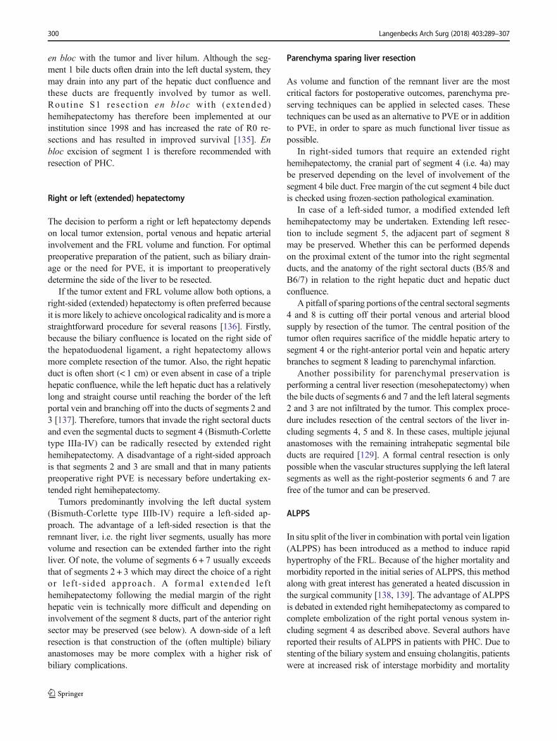
Right or left (extended) hepatectomy
The decision to perform a right or left hepatectomy dependson local tumor extension, portal venous and hepatic arterialinvolvement and the FRL volume and function. For optimalpreoperative preparation of the patient, such as biliary drain-age or the need for PVE, it is important to preoperativelydetermine the side of the liver to be resected.
If the tumor extent and FRL volume allow both options, aright-sided (extended) hepatectomy is often preferred becauseit is more likely to achieve oncological radicality and is more astraightforward procedure for several reasons [136]. Firstly,because the biliary confluence is located on the right side ofthe hepatoduodenal ligament, a right hepatectomy allowsmore complete resection of the tumor. Also, the right hepaticduct is often short (< 1 cm) or even absent in case of a triplehepatic confluence, while the left hepatic duct has a relativelylong and straight course until reaching the border of the leftportal vein and branching off into the ducts of segments 2 and3 [137]. Therefore, tumors that invade the right sectoral ductsand even the segmental ducts to segment 4 (Bismuth-Corlettetype IIIa-IV) can be radically resected by extended righthemihepatectomy. A disadvantage of a right-sided approachis that segments 2 and 3 are small and that in many patientspreoperative right PVE is necessary before undertaking ex-tended right hemihepatectomy.
Tumors predominantly involving the left ductal system(Bismuth-Corlette type IIIb-IV) require a left-sided ap-proach. The advantage of a left-sided resection is that theremnant liver, i.e. the right liver segments, usually has morevolume and resection can be extended farther into the rightliver. Of note, the volume of segments 6 + 7 usually exceedsthat of segments 2 + 3 which may direct the choice of a rightor lef t -s ided approach. A formal extended lef themihepatectomy following the medial margin of the righthepatic vein is technically more difficult and depending oninvolvement of the segment 8 ducts, part of the anterior rightsector may be preserved (see below). A down-side of a leftresection is that construction of the (often multiple) biliaryanastomoses may be more complex with a higher risk ofbiliary complications.
Parenchyma sparing liver resection
As volume and function of the remnant liver are the mostcritical factors for postoperative outcomes, parenchyma pre-serving techniques can be applied in selected cases. Thesetechniques can be used as an alternative to PVE or in additionto PVE, in order to spare as much functional liver tissue aspossible.
In right-sided tumors that require an extended righthemihepatectomy, the cranial part of segment 4 (i.e. 4a) maybe preserved depending on the level of involvement of thesegment 4 bile duct. Free margin of the cut segment 4 bile ductis checked using frozen-section pathological examination.
In case of a left-sided tumor, a modified extended lefthemihepatectomy may be undertaken. Extending left resec-tion to include segment 5, the adjacent part of segment 8may be preserved. Whether this can be performed dependson the proximal extent of the tumor into the right segmentalducts, and the anatomy of the right sectoral ducts (B5/8 andB6/7) in relation to the right hepatic duct and hepatic ductconfluence.
A pitfall of sparing portions of the central sectoral segments4 and 8 is cutting off their portal venous and arterial bloodsupply by resection of the tumor. The central position of thetumor often requires sacrifice of the middle hepatic artery tosegment 4 or the right-anterior portal vein and hepatic arterybranches to segment 8 leading to parenchymal infarction.
Another possibility for parenchymal preservation isperforming a central liver resection (mesohepatectomy) whenthe bile ducts of segments 6 and 7 and the left lateral segments2 and 3 are not infiltrated by the tumor. This complex proce-dure includes resection of the central sectors of the liver in-cluding segments 4, 5 and 8. In these cases, multiple jejunalanastomoses with the remaining intrahepatic segmental bileducts are required [129]. A formal central resection is onlypossible when the vascular structures supplying the left lateralsegments as well as the right-posterior segments 6 and 7 arefree of the tumor and can be preserved.
ALPPS
In situ split of the liver in combinationwith portal vein ligation(ALPPS) has been introduced as a method to induce rapidhypertrophy of the FRL. Because of the higher mortality andmorbidity reported in the initial series of ALPPS, this methodalong with great interest has generated a heated discussion inthe surgical community [138, 139]. The advantage of ALPPSis debated in extended right hemihepatectomy as compared tocomplete embolization of the right portal venous system in-cluding segment 4 as described above. Several authors havereported their results of ALPPS in patients with PHC. Due tostenting of the biliary system and ensuing cholangitis, patientswere at increased risk of interstage morbidity and mortality
300 Langenbecks Arch Surg (2018) 403:289–307

[140, 141]. ALPPS for PHC demonstrated poor outcomeswith 48% perioperative mortality in the ALPPS registry[140]. We therefore for now, do not recommend ALPPS forresection of PHC and rather consider PVE with selective em-bolization of the left portal vein branches to segment 4 foraugmentation of FRL volume in patients requiring extendedright hemihepatectomy.
The extent of lymphadenectomy
Standard lymphadenectomy includes resection of lymphnodes around the extrahepatic bile duct, the portal veinand hepatic artery, as well as the lymphatic channels andnerves contained in the hepatoduodenal ligament. Thenumber of lymph nodes resected is also relevant as lessthan 4 lymph nodes evaluated in the specimen was iden-tified as a poor prognostic factor for time to recurrence[142]. Lymph node metastases that are limited to the he-patic pedicle or the hepatoduodenal ligament (N1) areincluded in the field of resection, but those along thecommon hepatic artery and/or celiac axis (N2) are consid-ered distant metastases. N2-disease has a poor prognosisand disease specific survival of patients with para-aorticlymph node metastasis was similar to M1 patients, sug-gesting that survival is not influenced by the extent oflymph node dissection, but rather by the presence of N2disease [12, 78]. Therefore, we do not recommend routinelymphadenectomy beyond the hepatoduodenal ligament.
Complications
Postoperative morbidity and mortality of patients with PHC issignificant. Reported mortality ranges from 5 to 18% even inhigh volume centers, and morbidity as high as 60–70%, witharound 50% severe complications (Clavien-Dindo grade III orhigher) [21]. Table 5 shows the incidence of the most commoncomplications as reported in literature together with the com-plications recorded in our own series. Risks are particularlyhigh in patients with tumors requiring an extended resection.
Liver failure is a dreaded complication after extensive hepa-tectomy and is a major cause of mortality in patients with PHC[21, 148]. The risk of postoperative liver failure is increaseddue to the combination of intra-operative blood loss, a smallFLR and cholestasis [21, 88, 121]. Reported liver failureranges from 3 to 25% [31, 88, 143, 144]. Biliary leakage fromeither the hepaticojejunal anastomosis or the parenchymal dis-section surface ranges from 6 to 29% [31, 88, 143]. Infectionsrange from 23 to 66% and bleeding complications from 4 to9% [15, 31, 88, 145, 146] (Table 5).
Discussion and future perspectives
The management of perihilar cholangiocarcinoma is complexand requires close multidisciplinary collaboration betweenhepatobiliary surgeons, endoscopists, radiologists, medicaloncologists and pathologists. In this review, we provide asummary of the current diagnosis and work-up in the lightof extended resection and elaborate on future perspectives.
Establishing the diagnosis of PHC is still one of the mostchallenging aspects of the diagnostic work-up. New diagnos-tic endoscopic techniques such as SpyGlass and endoscopicultrasound enablemore precise biopsies, resulting in increasedsensitivity and specificity in diagnosing biliary strictures [33].These techniques will likely decrease the rate of misdiagnosesand bring down the number of futile resections performed forbenign disease. The currently used conventional tumor markerCA19–9 is not particularly sensitive or specific. A combina-tion of different markers seems more useful in the diagnosisand follow-up of PHC. Biomarkers such as CYFRA21-1 andMUC-5 need to be evaluated in larger cohorts to assess itsclinical value. Molecular techniques such as circulatingmiRNA’s and Tumor Educated Platelets (TEP) represent anexciting area with great promise in this field [149, 150]. Fornow, approximately 50% of patients with suspicion on PHCundergo resection without a confirmed tissue diagnosis.
CT-volumetry has traditionally been the golden standardfor assessment of a sufficient FRL. However, not only thequantity but also the quality of the FRL is important whileliver volume does not correlate with liver function. In ourcohort, total and regional (segmental) liver function was pre-operatively evaluated using HBS. This quantitative methodallows measurement of FRL-function and can be used in pa-tients with impaired quality of liver parenchyma using thesame cutoff value. A limitation of using HBS in patients withPHC is that bilirubin induces competitive uptake withmebrofenin as both are taken up by the same hepatocyte trans-porters. In patients with obstructive cholestasis, HBS mayunderestimate liver function when the biliary system is notcompletely drained.
The additional value of staging laparoscopy in the future isquestionable, considering the low yield and further
Table 5 Complications and reported incidence in a selection ofliterature reports including the AMC series
Complication type Incidence literature Incidence AMC
Liver failure 3–25% [31, 88, 143, 144] 19% (29/156*)
Biliary leakage 6–29% [31, 88, 143] 30% (47/156)
Bleeding 4–9% [15, 31, 88, 145, 146] 8% (13/156)
Multi organ failure 1–3% [32, 87, 135] 2% (3/156)
Infections 23–66% [15, 31, 88, 145, 146] 22% (35/156)
Mortality 5–17% [88, 143, 147, 157] 9% (18/201)
*Total cohort: n = 156, missing n = 45
Langenbecks Arch Surg (2018) 403:289–307 301

improvements in accuracy of preoperative imaging tech-niques. Using a risk score allows to predict unresectablePHC at staging laparoscopy in order to make a more selectiveapproach to staging laparoscopy.
Since patients with PHC typically present with obstructivejaundice, decompression of the biliary tract is a much-debatedtopic. For the past, few years it has become clear that drainageof the biliary tract comes with a serious risk of drainage-related complications. Since obstructive jaundice impairs liverregeneration, biliary drainage is still advised in case of a smallFLR. The optimal drainage method has yet to be established.In The Netherlands the DRAINAGE trial is underway to eval-uate outcomes of PTBD vs. EBD in resectable PHC [108,151]. In this multi-center trial with an all-comers design, allpatients with a presumed PHC and cholestasis are randomizedto undergo PTBD or EBD. The study is powered for drainage-related complications and postoperative outcomes. For now,we use selective preoperative, endoscopic biliary drainage ofonly the future remnant liver when FRL is small (< 50%)unless mandated by cholangitis.
The most important prognostic factor for long-term surviv-al of PHC is a margin negative resection of the hilar tumor. Inexperienced hands, even Bismuth-Corlette type IV tumors canbe resected with curative intent. R0 resection requires an ag-gressive surgical approach encompassing hilar resection incombination with extended liver resection, frequently accom-panied with vascular reconstructions. These extended resec-tions are associated with higher morbidity and mortality ratesthan experienced in liver resections without bile duct resec-tion, probably because of the sequelae of obstructive jaundice.Survival after resection is however favorable, with 5-yearoverall survival rates comparable with survival after extendedliver resection for colorectal liver metastases.
PVE is a widely accepted interventional procedure to in-crease FRL volume and function before undertaking majorliver resection. This method of liver augmentation is especial-ly of benefit in patients with PHC who require extended liverresection in predamaged livers. We therefore advocate theliberal use of PVE in patients with PHC in whom the FRL isbelow 40% of total liver volume. It is important to note that toobtain the maximum hypertrophy effect of segments 2 and 3,the side-branches of the left portal vein to segment 4 can beembolized as well. Obviously, selective embolization of thesegment 4 branches requires expertise of the interventionalradiologist as available in specialized centers. Although thefirst successful case of ALPPS was reported by Schlitt in apatient with PHC, the use of ALPPS in PHC as alternative toPVE is not recommended because of the reported high mor-bidity and mortality of the procedure in this category of pa-tients [152].
There are no established strategies regarding the use ofneo-adjuvant therapies in PHC. The only exception is neo-adjuvant chemo-radiation therapy prior to liver transplantation
in a highly selected group of patients [153, 154]. The idea of ashort course of radiation preceding resection was to eradicatefree floating tumor cells in the bile, thus preventing viabletumor cells of contaminating the peritoneal surface. There ishowever no evidence for this concept.
The challenge in the coming years is to reduce morbidityand mortality associated with extended resections for PHC.Optimizing preoperative workup is key to achieve improvedoutcomes after extended resections.
Conclusion
The field of work-up in PHC is changing with the introductionof newer modalities that have emerged over the past fewyears. Upcoming diagnostic modalities and molecular tech-niques might help to decrease the rate of misdiagnosis of be-nign, inflammatory disease. Assessment of liver function withhepatobiliary scintigraphy provides better information on theFRL than volume alone. The selective use of staging laparos-copy is advisable to avoid futile laparotomies. In patients re-quiring extended resection, selective preoperative biliarydrainage is mandatory in cholangitis and when FRL is small(< 50%). Preoperative PVE is used when FRL volume is lessthan 40% and optionally includes the left portal vein branchesto segment 4. ALPPS as alternative to PVE is not recommend-ed in PHC. N2 positive lymph nodes preclude long-term sur-vival. The benefit of unconditional en bloc resection of theportal vein bifurcation is uncertain. Although still associatedwith considerable morbidity and mortality, an aggressive sur-gical approach encompassing extended liver resection includ-ing segment 1, regional lymphadenectomy and conditionalportal venous resection offers the only chance for long-termsurvival.
Authors’ contributions F. Rassam: study conception and design, acquisi-tion of data, analysis and interpretation of data, drafting of manuscript. E.Roos: study conception and design, acquisition of data, analysis andinterpretation of data, drafting of manuscript. K.P. van Lienden: criticalrevision of manuscript. J.E. van Hooft: critical revision of manuscript.H.J. Klümpen: critical revision of manuscript. G. van Tienhoven: criticalrevision of manuscript. R.J. Bennink: critical revision of manuscript.M.R. Engelbrecht: critical revision of manuscript. A. Schoorlemmer: crit-ical revision of manuscript. U.H.W. Beuers: critical revision of manu-script. J. Verheij: critical revision of manuscript. M.G. Besselink: criticalrevision of manuscript. O.R.C. Busch: critical revision of manuscript.T.M. van Gulik: Study conception and design, critical revision ofmanuscript.
Compliance with ethical standards
Conflict of interest The authors declare that they have no conflict ofinterest.
Ethical approval This article does not contain any studies with humanparticipants or animals performed by any of the authors.
302 Langenbecks Arch Surg (2018) 403:289–307
Open Access This article is distributed under the terms of the CreativeCommons At t r ibut ion 4 .0 In te rna t ional License (h t tp : / /creativecommons.org/licenses/by/4.0/), which permits unrestricted use,distribution, and reproduction in any medium, provided you give appro-priate credit to the original author(s) and the source, provide a link to theCreative Commons license, and indicate if changes were made.
References
1. Aljiffry M, Abdulelah A, Walsh M et al (2009) Evidence-basedapproach to cholangiocarcinoma: a systematic review of the cur-rent literature. J Am Coll Surg 208:134–147. https://doi.org/10.1016/j.jamcollsurg.2008.09.007
2. Gatto M, Alvaro D (2010) New insights on cholangiocarcinoma.World J Gastrointest Oncol 2:136–145. https://doi.org/10.4251/wjgo.v2.i3.136
3. Khan SA, Thomas HC, Davidson BR, Taylor-Robinson SD(2005) Cholangiocarcinoma. Lancet (London, England) 366:1303–1314. https://doi.org/10.1016/S0140-6736(05)67530-7
4. Bismuth H, Nakache R, Diamond T (1992) Management strate-gies in resection for hilar cholangiocarcinoma. Ann Surg 215(1):31–38. https://doi.org/10.1097/00000658-199201000-00005
5. Vaeteewoottacharn K, Seubwai W, Bhudhisawasdi V et al (2014)Potential targeted therapy for liver fluke associated cholangiocar-cinoma. J Hepatobiliary Pancreat Sci 21:362–370. https://doi.org/10.1002/jhbp.65
6. Ong CK, Subimerb C, Pairojkul C et al (2012) Exome sequencingof liver flukeg-associated cholangiocarcinoma. Nat Genet 44:690–693. https://doi.org/10.1038/ng.2273
7. Rizvi S, Gores GJ (2013) Pathogenesis, diagnosis, and manage-ment of cholangiocarcinoma. Gastroenterology 145:1215–1229.https://doi.org/10.1053/j.gastro.2013.10.013
8. Sarkar S, Bowlus CL (2016) Primary sclerosing cholangitis: mul-tiple phenotypes, multiple approaches. Clin Liver Dis 20:67–77.https://doi.org/10.1016/j.cld.2015.08.005
9. Boberg KM, Schrumpf E, Bergquist A et al (2000)Cholangiocarcinoma in primary sclerosing cholangitis: K-ras mu-tations and Tp53 dysfunction are implicated in the neoplasticdevelopment. J Hepatol 32:374–380
10. Nagino M, Ebata T, Yokoyama Yet al (2013) Evolution of surgi-cal treatment for perihilar cholangiocarcinoma: a single-center 34-year review of 574 consecutive resections. Ann Surg 258:129–140. https://doi.org/10.1097/SLA.0b013e3182708b57
11. Govil S, Reddy MS, Rela M (2014) Surgical resection techniquesfor locally advanced hilar cholangiocarcinoma. Langenbeck'sArch Surg 399:707–716. https://doi.org/10.1007/s00423-014-1216-4
12. Regimbeau JM, Fuks D, Le Treut YP et al (2011) Surgery for hilarcholangiocarcinoma: a multi-institutional update on practice andoutcome by the AFC-HC study group. J Gastrointest Surg 15:480–488. https://doi.org/10.1007/s11605-011-1414-0
13. Hartog H, Ijzermans JNM, van Gulik TM, Koerkamp BG (2016)Resection of perihilar cholangiocarcinoma. Surg Clin North Am96:247–267. https://doi.org/10.1016/j.suc.2015.12.008
14. Coelen RJS, Ruys AT, Besselink MGH, Busch ORC, van GulikTM (2016) Diagnostic accuracy of staging laparoscopy for detect-ing metastasized or locally advanced perihilar cholangiocarcino-ma: a systematic review and meta-analysis. Surg Endosc 30(10):1–11. https://doi.org/10.1007/s00464-016-4788-y
15. Matsuo K, Rocha FG, Ito K et al (2012) The blumgart preopera-tive staging system for hilar cholangiocarcinoma: analysis of
resectability and outcomes in 380 patients. J Am Coll Surg 215:343–355. https://doi.org/10.1016/j.jamcollsurg.2012.05.025
16. Coelen RJS, Ruys AT, Wiggers JK, Nio CY, Verheij J, Gouma DJ,BesselinkMGH,Busch ORC, vanGulik TM (2016) Developmentof a risk score to predict detection of metastasized or locally ad-vanced perihilar cholangiocarcinoma at staging laparoscopy. AnnSurg Oncol 23(S5):904–910. https://doi.org/10.1245/s10434-016-5531-6
17. Valle J, Wasan H, Palmer DH et al (2010) Cisplatin plusgemcitabine versus gemcitabine for biliary tract cancer. N Engl JMed 4:395–397. https://doi.org/10.1586/egh.10.45
18. Wyluda E, Yee NS (2015) Systemic treatment of advanced biliarytract carcinoma: emerging roles of targeted therapy and molecularprofiling. Clin Cancer Drugs 2:80–86
19. Ito F, Cho CS, Rikkers LF, Weber SM (2009) Hilar cholangiocar-cinoma: current management. Ann Surg 250:210–218. https://doi.org/10.1097/SLA.0b013e3181afe0ab
20. Groot Koerkamp B, Wiggers JK, Gonen M et al (2015) Survivalafter resection of perihilar cholangiocarcinoma-development andexternal validation of a prognostic nomogram. Ann Oncol 26:1930–1935. https://doi.org/10.1093/annonc/mdv279
21. Coelen RJS, Olthof PB, van Dieren S et al (2016) External vali-dation of the estimation of physiologic ability and surgical stress(E-PASS) risk model to predict operative risk in perihilar cholan-giocarcinoma. JAMA Surg 147:26–34. https://doi.org/10.1001/jamasurg.2016.2305
22. Hemming AW, Reed AI, Fujita S et al (2005) Surgical manage-ment of hilar cholangiocarcinoma. Ann Surg 241:693–699-702.https://doi.org/10.1097/01.sla.0000160701.38945.82
23. Anderson JE, Hemming AW, Chang DC et al (2012) Surgicalmanagement trends for cholangiocarcinoma in the USA 1998-2009. J Gastrointest Surg 16:2225–2232. https://doi.org/10.1007/s11605-012-1980-9
24. Nagino M, Ebata T, Yokoyama Yet al (2013) Evolution of surgi-cal treatment for perihilar cholangiocarcinoma: a single-center 34-year review of 574 consecutive resections. Ann Surg 258:129–140. https://doi.org/10.1097/SLA.0b013e3182708b57
25. Maillette de Buy Wenniger LJ, Beuers U (2015) ImmunoglobulinG4-related cholangiopathy: clinical and experimental insights.Curr Opin Gastroenterol 31:252–257. https://doi.org/10.1097/mog.0000000000000170
26. Hubers LM,Maillette de BuyWenniger LJ, Doorenspleet ME et al(2015) IgG4-associated cholangitis: a comprehensive review. ClinRev Allergy Immunol 48:198–206. https://doi.org/10.1007/s12016-014-8430-2
27. Zaydfudim VM, Wang AY, De Lange EE et al (2015) IgG4-associated cholangitis can mimic hilar cholangiocarcinoma. GutLiver 9:556–560. https://doi.org/10.5009/gnl14241
28. Zen Y, Britton D, Mitra V et al (2015) A global proteomic studyidentifies distinct pathological features of IgG4-related and prima-ry sclerosing cholangitis. Histopathology. https://doi.org/10.1111/his.12813
29. Deshpande V, Zen Y, Chan JK et al (2012) Consensus statementon the pathology of IgG4related disease. Mod Pathol 25:11811192www.modernpathology.org. https://doi.org/10.1038/modpathol.2012.72
30. Kamisawa T, Zen Y, Pillai S, Stone JH (2015) IgG4-related dis-ease. Lancet 385:1460–1471. https://doi.org/10.1016/S0140-6736(14)60720-0
31. Kloek JJ, van Deldein OM, Erdogan D et al (2008) Differentiationof malignant and benign proximal bile duct strictures: the diag-nostic dilemma. World J Gastroenterol 14:5032–5038. https://doi.org/10.3748/wjg.14.5032
32. Corvera CU, Blumgart LH, Darvishian F et al (2005) Clinical andpathologic features of proximal biliary strictures masquerading as
Langenbecks Arch Surg (2018) 403:289–307 303

hilar cholangiocarcinoma. J Am Coll Surg 201:862–869. https://doi.org/10.1016/j.jamcollsurg.2005.07.011
33. Victor DW, Sherman S, Karakan T, Khashab MA (2012) Currentendoscopic approach to indeterminate biliary strictures. World JGastroenterol 18:6197–6205. https://doi.org/10.3748/wjg.v18.i43.6197
34. Esnaola NF, Meyer JE, Karachristos A, Maranki JL, Camp ER,Denlinger CS (2016) Evaluation and management of intrahepaticand extrahepatic cholangiocarcinoma. Cancer 122(9):1349–1369.https://doi.org/10.1002/cncr.29692
35. Navaneethan U, Njei B, Lourdusamy Vet al (2016) Comparativeeffectiveness of biliary brush cytology and intraductal biopsy fordetection of malignant biliary strictures: a systematic review andmeta-analysis. Gastrointest Endosc. 1:23–30. https://doi.org/10.1007/s40778-014-0003-z.Genome
36. Barr Fritcher EG, Kipp BR, Halling KC, Clayton AC (2014)FISHing for pancreatobiliary tract malignancy in endoscopicbrushings enhances the sensitivity of routine cytology.Cytopathology 25:288–301. https://doi.org/10.1111/cyt.12170
37. Liew ZH, Loh TJZ, Lim TKH et al (2017) Role of fluorescence insitu hybridization in diagnosing cholangiocarcinoma in indetermi-nate biliary strictures. J Gastroenterol Hepatol. https://doi.org/10.1111/jgh.13824
38. Barr Fritcher EG, Voss JS, Brankley SM et al (2015) An optimizedset of fluorescence in situ hybridization probes for detection ofpancreatobiliary tract cancer in cytology brush samples.Gastroenterology 149:1813–1824e1. https://doi.org/10.1053/j.gastro.2015.08.046
39. Gonda TA (2017) Mutation profile and fluorescence in situ hy-bridization analyses increase detection of malignancies in biliairystrictures. Clin Gastroenterol Hepatol. https://doi.org/10.1016/j.cgh.2016.12.013.This
40. Fogel EL, DeBellis M, McHenry L et al (2006) Effectiveness of anew long cytology brush in the evaluation of malignant biliaryobstruction: a prospective study. Gastrointest Endosc 63:71–77.https://doi.org/10.1016/j.gie.2005.08.039
41. Coté GA, Sherman S (2011) Biliary stricture and negative cytol-ogy: what next? Clin Gastroenterol Hepatol 9:739–743. https://doi.org/10.1016/j.cgh.2011.04.011
42. De Bellis M, Fogel EL, Sherman S et al (2003) Influence of stric-ture dilation and repeat brushing on the cancer detection rate ofbrush cytology in the evaluation of malignant biliary obstruction.Gastrointest Endosc 58:176–182. https://doi.org/10.1067/mge.2003.345
43. Fukuda Y, Tsuyuguchi T, Sakai Yet al (2005) Diagnostic utility ofperoral cholangioscopy for various bile-duct lesions. GastrointestEndosc 62:374–382. https://doi.org/10.1016/j.gie.2005.04.032
44. Kawashima H, Itoh A, Ohno E et al (2012) Transpapillary biliaryforceps biopsy to distinguish benign biliary stricture from malig-nancy: how many tissue samples should be obtained? Dig Endosc24:22–27. https://doi.org/10.1111/j.1443-1661.2012.01253.x
45. Kitajima Y, Ohara H, Nakazawa T et al (2007) Usefulness oftranspapillary bile duct brushing cytology and forceps biopsy forimproved diagnosis in patients with biliary strictures. JGastroenterol Hepatol 22:1615–1620. https://doi.org/10.1111/j.1440-1746.2007.05037.x
46. Lin LF, Siauw CP, Ho KS, Tung JN (2003) Guidewire techniquefor endoscopic transpapillary procurement of bile duct biopsyspecimens without endoscopic sphincterotomy. GastrointestEndosc 58:272–274. https://doi.org/10.1067/mge.2003.329
47. Chen YK, Pleskow DK (2007) SpyGlass single-operator peroralcholangiopancreatoscopy system for the diagnosis and therapy ofbile-duct disorders: a clinical feasibility study (with video){a fig-ure is presented}. Gastrointest Endosc 65:832–841. https://doi.org/10.1016/j.gie.2007.01.025
48. Kurihara T, Yasuda I, Isayama H et al (2016) Diagnostic andtherapeutic single-operator cholangiopancreatoscopy inbiliopancreatic diseases: prospective multicenter study in Japan.World J Gastroenterol 22:1891–1901. https://doi.org/10.3748/wjg.v22.i5.1891
49. Navaneethan U, Hasan M, Lourdusamy V et al (2015) Single-operator cholangioscopy and tarhetted biopsies in the diagnosisof inditerminat ebiliary strictures: a systematic review.Gastrointest Endosc 82:608–614. https://doi.org/10.1007/978-1-4939-2914-6
50. Hara K, Yamao K, Mizuno N, Hijioka S, Imaoka H, Tajika M,Tanaka T, Ishihara M, Okuno N, Hieda N, Yoshida T, Niwa Y(2016) Endoscopic ultrasonography-guided biliary drainage: who,when, which, and how? World J Gastroenterol 22(3):1297–1303.https://doi.org/10.3748/wjg.v22.i3.1297
51. Lee JH, Salem R, Aslanian H et al (2004) Endoscopic ultrasoundand fine-needle aspiration of unexplained bile duct strictures. AmJ Gastroenterol 99:1069–1073. https://doi.org/10.1111/j.1572-0241.2004.30223.x
52. Itoi T, Itokawa F, Uraoka T et al (2013) Novel EUS-guidedgastrojejunostomy technique using a new double-balloon enterictube and lumen-apposing metal stent (with videos). GastrointestEndosc 78:934–939. https://doi.org/10.1016/j.gie.2013.09.025
53. Masaki Y, Kurose N, Yamamoto M, Takahashi H, Saeki T, AzumiA, Nakada S, Matsui S, Origuchi T, Nishiyama S, Yamada K,Kawano M, Hirabayashi A, Fujikawa K, Sugiura T, HorikoshiM, Umeda N, Minato H, Nakamura T, Iwao H, Nakajima A,Miki M, Sakai T, Sawaki T, Kawanami T, Fujita Y, Tanaka M,Fukushima T, Eguchi K, Sugai S, Umehara H (2012) Cutoffvalues of serum IgG4 and histopathological IgG4+ plasma cellsfor diagnosis of patients with IgG4-related disease. Int JRheumatol 2012:0–5. https://doi.org/10.1155/2012/580814
54. Oseini AM, Chaiteerakij R, Shire AM et al (2011) Utility of serumimmunoglobulin G4 in distinguishing immunoglobulin G4-associated cholangitis from cholangiocarcinoma. Hepatology 54:940–948. https://doi.org/10.1002/hep.24487
55. Doorenspleet ME, Hubers LM, Culver EL et al (2016) IgG4+ B-cell receptor clones distinguish IgG4-related disease from primarySclerosing cholangitis and biliary/pancreatic malignancies.Hepatology 64(2):1–49. https://doi.org/10.1002/hep.28568
56. Charatcharoenwitthaya P, Enders FB, Halling KC, Lindor KD(2008) Utility of serum tumor markers, imaging, and biliary cy-tology for detecting cholangiocarcinoma in primary sclerosingcholangitis. Hepatology 48:1106–1117. https://doi.org/10.1002/hep.22441
57. Grunnet M,Mau-SørensenM (2014) Serum tumor markers in bileduct cancer—a review. Biomarkers 19:437–443. https://doi.org/10.3109/1354750X.2014.923048
58. Viterbo D, Gausman V, Gonda T (2016) Diagnostic and therapeu-tic biomarkers in pancreaticobiliary malignancy. World JGastrointest Endosc 8:128–142. https://doi.org/10.4253/wjge.v8.i3.128
59. Liu SL, Song ZF, Hu QG et al (2010) Serum carbohydrate antigen(CA) 19-9 as a prognostic factor in cholangiocarcinoma: a meta-analysis. Front Med China 4:457–462. https://doi.org/10.1007/s11684-010-0240-1
60. Patel AH, Harnois DM,KleeGG et al (2000) The utility of CA 19-9 in the diagnoses of cholangiocarcinoma in patients without pri-mary sclerosing cholangitis. Am J Gastroenterol 95:204–207.https://doi.org/10.1016/S0002-9270(99)00744-3
61. Chapman MH, Sandanayake NS, Andreola F et al (2011)Circulating CYFRA 21-1 is a specific diagnostic and prognosticbiomarker in biliary tract cancer. J Clin Exp Hepatol 1:6–12.https://doi.org/10.1016/S0973-6883(11)60110-2
62. Van Gulik TM, Kloek JJ, Ruys AT et al (2011) Multidisciplinarymanagement of hilar cholangiocarcinoma (Klatskin tumor):
304 Langenbecks Arch Surg (2018) 403:289–307

extended resection is associated with improved survival. Eur JSurg Oncol 37:65–71. https://doi.org/10.1016/j.ejso.2010.11.008
63. Chaib E, Kanas AF, Galvão FHF, D’Albuquerque LAC (2014)Bile duct confluence: anatomic variations and its classification.Surg Radiol Anat 36:105–109. https://doi.org/10.1007/s00276-013-1157-6
64. Van Gulik TM, Dinant S, Busch ORC et al (2007) Original article:new surgical approaches to the Klatskin tumour. AlimentPharmacol Ther 26(Suppl 2):127–132. https://doi.org/10.1111/j.1365-2036.2007.03485.x
65. Wiggers JK, te RieleWW, van Dongen TH et al (2016) Combinedliver and extrahepatic bile duct resection for biliary invasion ofcolorectal metastasis: a case-cohort analysis and systematic re-view. HepatoBiliary Surg Nutr 5(4):350–357. https://doi.org/10.21037/hbsn.2016.05.01
66. Olthof SC, Othman A, Clasen S et al (2016) Imaging of cholan-giocarcinoma. Visc Med 32:402–410. https://doi.org/10.1159/000453009
67. Blackbourne L, Earnhardt R, Sistrom C et al (1994) The sensitiv-ity and role of ultrasound in the evaluation of biliary obstruction.Am Surg 60:683–690
68. Ruys AT, Busch OR, Rauws EA et al (2013) Prognostic impact ofpreoperative imaging parameters on resectability of hilar cholan-giocarcinoma. HPB Surg 2013:657309. https://doi.org/10.1155/2013/657309
69. Engelbrecht MR, Katz SS, Van Gulik TM et al (2015) Imaging ofperihilar cholangiocarcinoma. Am J Roentgenol 204:782–791.https://doi.org/10.2214/AJR.14.12830
70. Mar WA, Shon AM, Lu Yet al (2016) Imaging spectrum of chol-angiocarcinoma: role in diagnosis, staging, and posttreatmentevaluation. Abdom Radiol 41:553–567. https://doi.org/10.1007/s00261-015-0583-9
71. Choi YH, Lee JM, Lee JY et al (2008) Biliary malignancy: valueof arterial, pancreatic, and hepatic phase imaging withmultidetector-row computed tomography. J Comput AssistTomogr 32:362–368. h t tps : / /do i .o rg /10.1097/RCT.0b013e318126c134
72. Hee SP, Lee JM, Choi JY et al (2008) Preoperative evaluation ofb i l e d u c t c a n c e r : M R I c o m b i n e d w i t h M Rcholangiopancreatography versus MDCT with direct cholangiog-raphy. Am J Roentgenol 190:396–405. https://doi.org/10.2214/AJR.07.2310
73. Yeh BM, Liu PS, Soto JA et al (2009) MR imaging and CTof thebiliary tract. Radiographics 29:1669–1688. https://doi.org/10.1148/rg.296095514
74. Ruys AT, Bennink RJ, Van Westreenen HL et al (2011) FDG-positron emission tomography/computed tomography and stan-dardized uptake value in the primary diagnosis and staging of hilarcholangiocarcinoma. HPB 13:256–262. https://doi.org/10.1111/j.1477-2574.2010.00280.x
75. Annunziata S, Pizzuto D, Caldarella C et al (2014) Diagnosticaccuracy of Fluorine-18-Fluorodeoxyglucose positron emissiontomography in the evaluation of the primary tumor in patients withcholangiocarcinoma: a meta-analysis. Eur J Nucl Med MolImaging 41:S488. https://doi.org/10.1007/s00259-014-2901-9
76. Chaiteerakij R, Harmsen WS, Marrero CR et al (2015) A newclinically based staging system for perihilar cholangiocarcinoma.Am J Gastroenterol 2014:1881–1890. https://doi.org/10.1038/ajg.2014.327.A
77. Zaydfudim VM, Clark CJ, Kendrick ML et al (2013) Correlationof staging systems to survival in patients with resected hilar chol-angiocarcinoma. Am J Surg 206:159–165. https://doi.org/10.1016/j.amjsurg.2012.11.020
78. Groot Koerkamp B, Wiggers JK, Gonen M et al (2015) Survivalafter resection of perihilar cholangiocarcinoma-development and
external validation of a prognostic nomogram. Ann Oncol 26:1930–1935. https://doi.org/10.1093/annonc/mdv279
79. Buettner S, van Vugt JLA, Gani F et al (2016) A comparison ofprognostic schemes for perihilar cholangiocarcinoma. JGastrointest Surg 20:1716–1724. https://doi.org/10.1007/s11605-016-3203-2
80. Groot Koerkamp B, Wiggers JK, Allen PJ et al (2014) Americanjoint committee on cancer staging for resected perihilar cholangio-carcinoma: a comparison of the 6th and 7th editions. HPB 16:1074–1082. https://doi.org/10.1111/hpb.12320
81. Deoliveira ML, Schulick RD, Nimura Yet al (2011) New stagingsystem and a registry for perihilar cholangiocarcinoma.Hepatology 53:1363–1371. https://doi.org/10.1002/hep.24227
82. Ding G, YangY, Cao L et al (2015) Amodified Jarnagin-Blumgartclassification better predicts survival for resectable hilar cholan-giocarcinoma. World J Surg Oncol 13:1–9. https://doi.org/10.1186/s12957-015-0526-5
83. Nagino M (2011) Perihilar cholangiocarcinoma: a much neededbut imperfect new staging system. Nat Rev Gastroenterol Hepatol8:252–253. https://doi.org/10.1038/nrgastro.2011.67
84. Steyerberg EW, Vergouwe Y (2014) Towards better clinical pre-diction models: seven steps for development and an ABCD forvalidation. Eur Heart J 35:1925–1931. https://doi.org/10.1093/eurheartj/ehu207
85. Iacono C, Ruzzenente A, Campagnaro T, Bortolasi L,Valdegamberi A, Guglielmi A (2013) Role of preoperative biliarydrainage in jaundiced patients who are candidates forpancreatoduodenectomy or hepatic role of preoperative biliarydrainage in jaundiced patients who arecandidates forpancreatoduodenectomy or hepatic resection: highlights anddrawbacks. Ann Surg 257(2):191–204. https://doi.org/10.1097/SLA.0b013e31826f4b0e
86. Ten Hoopen-Neumann H, Gerhards MF, Van Gulik TM et al(1999) Occurrence of implantation metastases after resection ofklatskin tumors. Dig Surg 16:209–213. https://doi.org/10.1159/000018729
87. Martel G, Cieslak KP, Huang R et al (2015) Comparison of tech-niques for volumetric analysis of the future liver remnant: impli-cations for major hepatic resections. HPB 17:1051–1057. https://doi.org/10.1111/hpb.12480
88. Wiggers JK, Koerkamp BG, Cieslak KP et al (2016)Postoperative mortality after liver resection for perihilarcholangiocarcinoma: development of a risk score and im-portance of biliary drainage of the future liver remnant. JAm Coll Surg 223:321–331.e1. https://doi.org/10.1016/j.jamcollsurg.2016.03.035
89. Hammond JS, Guha IN, Beckingham IJ, Lobo DN (2011)Prediction, prevention and management of postresection liver fail-ure. Br J Surg 98:1188–1200. https://doi.org/10.1002/bjs.7630
90. Van Den Broek E, Dijkstra MJJ, Krijgsman O et al (2015) Highprevalence and clinical relevance of genes affected by chromo-somal breaks in colorectal cancer. PLoS One 10:1–14. https://doi.org/10.1371/journal.pone.0138141
91. Cieslak KP, Bennink RJ, de Graaf Wet al (2016) Measurement ofliver function using hepatobiliary scintigraphy improves risk as-sessment in patients undergoing major liver resection. HPB 18:773–780. https://doi.org/10.1016/j.hpb.2016.06.006
92. Cieslak KP, Runge JH, Heger M et al (2014) New perspectives inthe assessment of future remnant liver. Dig Surg 31:255–268.https://doi.org/10.1159/000364836
93. Shoup M, Gonen M, D’Angelica M et al (2003) Volumetric anal-ysis predicts hepatic dysfunction in patients undergoing majorliver resection. J Gastrointest Surg 7:325–330. https://doi.org/10.1016/S1091-255X(02)00370-0
Langenbecks Arch Surg (2018) 403:289–307 305

94. Vauthey JN, Abdalla EK, Doherty DA et al (2002) Body surfacearea and body weight predict total liver volume in western adults.Liver Transpl 8:233–240. https://doi.org/10.1053/jlts.2002.31654
95. Ribero D, Chun YS, Vauthey JN (2008) Standardized livervolumetry for portal vein embolization. Semin Intervent Radiol25:104–109. https://doi.org/10.1055/s-2008-1076681
96. Dinant S, de Graaf W, Verwer BJ et al (2007) Risk assessment ofposthepatectomy liver failure using hepatobiliary scintigraphy andCT volumetry. J Nucl Med 48:685–692. https://doi.org/10.2967/jnumed.106.038430
97. de Graaf W, van Lienden KP, Dinant S et al (2010) Assessment offuture remnant liver function using hepatobiliary scintigraphy inpatients undergoing major liver resection. J Gastrointest Surg 14:369–378. https://doi.org/10.1007/s11605-009-1085-2
98. Bennink RJ, Dinant S, Erdogan D et al (2004) Preoperative as-sessment of postoperative remnant liver function usinghepatobiliary scintigraphy. J Nucl Med 45:965–971
99. de Graaf W, van Lienden KP, van Gulik TM, Bennink RJ (2010)99mTc-Mebrofenin hepatobiliary scintigraphy with SPECT forthe assessment of hepatic function and liver functional volumebefore partial hepatectomy. J Nucl Med 51:229–236. https://doi.org/10.2967/jnumed.109.069724
100. Du Bois D, Du Bois E (1989) A formula to estimate the approx-imate surface area if height and weight be known. Nutrition 5:303–311
101. Erdogan D, Heijnen BHM, Bennink RJ et al (2004) Preoperativeassessment of liver function: a comparison of 99mTc-Mebrofeninscintigraphy with indocyanine green clearance test. Liver Int 24:117–123. https://doi.org/10.1111/j.1478-3231.2004.0901.x
102. De Graaf W, Häusler S, Heger M et al (2011) Transporters in-volved in the hepatic uptake of 99mTc-mebrofenin and indocya-nine green. J Hepatol 54:738–745. https://doi.org/10.1016/j.jhep.2010.07.047
103. Schaap FG, van der Gaag NA, Gouma DJ, Jansen PLM (2009)High expression of the bile salt-homeostatic hormone fibroblastgrowth factor 19 in the liver of patients with extrahepatic chole-stasis. Hepatology 49:1228–1235. https://doi.org/10.1002/hep.22771
104. Keppler D (2014) Special section on transporters in toxicity anddisease—minireview the roles of MRP2, MRP3, OATP1B1 andOATP1B3 in conjugated hyperbilirubinemia. Drug Metab Dispos42(4):561–565. https://doi.org/10.1124/dmd.113.055772
105. Gaag NA, Van Der KJJ, de Castro SMM (2009) Preoperativebiliary drainage in patients with obstructive jaundice: history andcurrent status. J Gastrointest Surg 13:814–820. https://doi.org/10.1007/s11605-008-0618-4
106. Kennedy TJ, Yopp A, Qin Y et al (2009) Role of preoperativebiliary drainage of liver remnant prior to extended liver resectionfor hilar cholangiocarcinoma. HPB (Oxford) 11:445–451. https://doi.org/10.1111/j.1477-2574.2009.00090.x
107. Al Mahjoub A, Menahem B, Fohlen A et al (2017) Preoperativebiliary drainage in patients with resectable perihilar cholangiocar-cinoma: is percutaneous transhepatic biliary drainage safer andmore effective than endoscopic biliary drainage? A meta-analysis.J Vasc Interv Radiol 28:576–582. https://doi.org/10.1016/j.jvir.2016.12.1218
108. Wiggers JK, Koerkamp BG, Coelen RJ et al (2015) Preoperativebiliary drainage in perihilar cholangiocarcinoma: identifying pa-tients who require percutaneous drainage after failed endoscopicdrainage. Endoscopy 47:1124–1131
109. Farges O, Regimbeau JM, Fuks D et al (2013) MulticentreEuropean study of preoperative biliary drainage for hilar cholan-giocarcinoma. Br J Surg. 100:274–283. https://doi.org/10.1002/bjs.8950
110. Jo JH, Chung MJ, Han DH et al (2017) Best options for preoper-ative biliary drainage in patients with Klatskin tumors. Surg
Endosc Other Interv Tech 31:422–429. https://doi.org/10.1007/s00464-016-4993-8
111. Fang Y, Gurusamy KS, Wang Q et al (2013) Meta-analysis ofrandomized clinical trials on safety and efficacy of biliary drainagebefore surgery for obstructive jaundice. Br J Surg. 100:1589–1596. https://doi.org/10.1002/bjs.9260
112. Celotti A, Solaini L, Montori G et al (2017) Preoperative biliarydrainage in hilar cholangiocarcinoma: systematic review and me-ta-analysis. Eur J Surg Oncol 43:4–11. https://doi.org/10.1016/j.ejso.2017.04.001
113. Wiggers JK, Coelen RJS, Rauws EAJ et al (2015) Preoperativeendoscopic versus percutaneous transhepatic biliary drainage inpotent ia l ly resectable perihi lar cholangiocarcinoma(DRAINAGE trial): design and rationale of a randomized con-trolled trial. Bmc gastroenterol 14:1–8. https://doi.org/10.1186/s12876-015-0251-0
114. Komaya K, Ebata T, Yokoyama Y et al (2017) Verification of theoncologic inferiority of percutaneous biliary drainage to endo-scopic drainage: a propensity score matching analysis of resectableperihilar cholangiocarcinoma. Surgery (United States) 161:394–404. https://doi.org/10.1016/j.surg.2016.08.008
115. Nimura Y (2008) Preoperative biliary drainage before resectionfor cholangiocarcinoma (pro). HPB 10:130–133. https://doi.org/10.1080/13651820801992666
116. Kawakami H, Kuwatani M, Onodera M et al (2011) Endoscopicnasobiliary drainage is themost suitable preoperative biliary drain-age method in the management of patients with hilar cholangio-carcinoma. J Gastroenterol 46:242–248. https://doi.org/10.1007/s00535-010-0298-1
117. Kawashima H, Itoh A, Ohno E et al (2012) Preoperative endo-scopic nasobiliary drainage in 164 consecutive patients withsuspected perihilar cholangiocarcinoma. Ann Surg 257:1. https://doi.org/10.1097/SLA.0b013e318262b2e9
118. Farges O, Regimbeau JM, Fuks D et al (2013) MulticentreEuropean study of preoperative biliary drainage for hilar cholan-giocarcinoma. Br J Surg 100:274–283. https://doi.org/10.1002/bjs.8950
119. De Graaf W, Van Lienden KP, Van Den Esschert JW et al (2011)Increase in future remnant liver function after preoperative portalvein embolization. Br J Surg 98:825–834. https://doi.org/10.1002/bjs.7456
120. Van Gulik TM, Van Den Esschert JW, De Graaf W et al (2009)Controversies in the use of portal vein embolization. Dig Surg 25:436–444. https://doi.org/10.1159/000184735
121. Olthof PB, Wiggers JK, Groot Koerkamp B et al (2017)Postoperative liver failure risk score: identifying patients withresectable perihilar cholangiocarcinoma who can benefit fromportal vein embolization. J Am Coll Surg 225(3):387–394.https://doi.org/10.1016/j.jamcollsurg.2017.06.007
122. Hayashi S, Baba Y, Ueno K et al (2007) Acceleration of primaryliver tumor growth rate in embolized hepatic lobe after portal veinembolization. Acta Radiol 48:721–727. https://doi.org/10.1080/02841850701424514
123. Glantzounis GK, Tokidis E, Basourakos SP et al (2017) The roleof portal vein embolization in the surgical management of primaryhepatobiliary cancers. A systematic review. Eur J Surg Oncol 43:32–41. https://doi.org/10.1016/j.ejso.2016.05.026
124. Huisman F, Cieslak KP, Van Lienden KP, Bennink RJ, Van GulikTM (2017) Liver related complications in unresectable diseaseafter portal vein embolization. Hepatobiliary Surg Nutr 6:379–386.
125. Wiggers JK, Groot Koerkamp B, Coelen RJ et al (2015)Percutaneous preoperative biliary drainage for resectable perihilarcholangiocarcinoma: no association with survival and no increasein seedingmetastases. Ann Surg Oncol 22:1156–1163. https://doi.org/10.1245/s10434-015-4676-z
306 Langenbecks Arch Surg (2018) 403:289–307
126. Heimbach JK, Sanchez W, Rosen CB, Gores GJ (2011) Trans-peritoneal fine needle aspiration biopsy of hilar cholangiocarcino-ma is associated with disease dissemination. HPB 13:356–360.https://doi.org/10.1111/j.1477-2574.2011.00298.x
127. Grendar J, Grendarova P, Sinha R, Dixon E (2014) Neoadjuvanttherapy for downstaging of locally advanced hilar cholangiocarci-noma: a systematic review. HPB 16:297–303. https://doi.org/10.1111/hpb.12150
128. Nishio H, Nagino M, Nimura Y (2005) Surgical management ofhilar cholangiocarcinoma: the Nagoya experience. HPB 7:259–262. https://doi.org/10.1080/13651820500373010
129. Van Gulik TM, Ruys AT, Busch ORC et al (2011) Extent of liverresection for hilar cholangiocarcinoma (klatskin tumor): howmuch is enough? Dig Surg 28:141–147. https://doi.org/10.1159/000323825
130. Olthof PB, Coelen RJS, Wiggers JK et al (2016) External biliarydrainage following major liver resection for perihilar cholangio-carcinoma: impact on development of liver failure and biliaryleakage. HPB 18:1–6. https://doi.org/10.1016/j.hpb.2015.11.007
131. Neuhaus P, Jonas S, Bechstein WO et al (1999) Extended resec-tions for hilar cholangiocarcinoma. Ann Surg 230:808–818 dis-cussion 819
132. Neuhaus P, Thelen A, Jonas S et al (2012) Oncological superiorityof hilar en bloc resection for the treatment of hilar cholangiocar-cinoma. Ann Surg Oncol 19:1602–1608. https://doi.org/10.1245/s10434-011-2077-5
133. Nagino M, Nimura Y, Nishio H et al (2010) Hepatectomy withsimultaneous resection of the portal vein and hepatic artery foradvanced perihilar cholangiocarcinoma: an audit of 50 consecu-tive cases. Ann Surg 252:115–123. https://doi.org/10.1097/SLA.0b013e3181e463a7
134. Bhangui P, Salloum C, Lim C et al (2014) Portal veinarterialization: a salvage procedure for a totally de-arterializedliver. The Paul Brousse Hospital experience. HPB 16:723–738.https://doi.org/10.1111/hpb.12200
135. Dinant S, Gerhards MF, Busch ORC et al (2005) The importanceof complete excision of the caudate lobe in resection of hilar chol-angiocarcinoma. HPB (Oxford) 7:263–267. https://doi.org/10.1080/13651820500372376
136. Uesaka K (2012) Left hepatectomy or left trisectionectomy withresection of the caudate lobe and extrahepatic bile duct for hilarcholangiocarcinoma (with video). J Hepatobiliary Pancreat Sci 19:195–202. https://doi.org/10.1007/s00534-011-0474-6
137. Bismuth H (1982) Surgical anatomy and anatomical surgery of theliver. World J Surg 6:3–9. https://doi.org/10.1007/BF01656368
138. Oldhafer KJ, Stavrou GA, van Gulik TM (2016) ALPPS—wheredo we stand, where do we go? Ann Surg 263:1. https://doi.org/10.1097/SLA.0000000000001633
139. Lang H, de Santibanes E, Clavien PA (2017) Outcome of ALPPSfor perihilar cholangiocarcinoma: case-control analysis includingthe first series from the international ALPPS registry. HPB 19:379–380. https://doi.org/10.1016/j.hpb.2017.01.024
140. Olthof PB, Coelen RJS, Wiggers JK et al (2017) High mortalityafter ALPPS for perihilar cholangiocarcinoma: case-control anal-ysis including the first series from the international ALPPS regis-try. HPB 19:381–387. https://doi.org/10.1016/j.hpb.2016.10.008
141. Li J, Ewald F, Gulati A, Nashan B (2016) Associating liver parti-tion and portal vein ligation for staged hepatectomy: from techni-cal evolution to oncological benefit. World J Gastrointest Surg 8:124–133. https://doi.org/10.4240/wjgs.v8.i2.124
142. Groot Koerkamp B, Wiggers JK, Allen PJ et al (2016) Recurrencerate and pattern of perihilar cholangiocarcinoma after curative
intent resection. J Am Coll Surg 8:583–592. https://doi.org/10.1002/aur.1474.Replication
143. Van Riel WG, Van Golen RF, Reiniers MJ et al (2016) Howmuchischemia can the liver tolerate during resection? HepatobiliarySurg Nutr 5(1):58–71. https://doi.org/10.3978/j.issn.2304-3881.2015.07.05
144. van de Kerkhove MP, de Jong KP, Rijken AM et al (2003) MARStreatment in posthepatectomy liver failure. Liver Int 23:44–51
145. Rocha FG, Matsuo K, Blumgart LH, Jarnagin WR (2010) Hilarcholangiocarcinoma: the Memorial Sloan-Kettering CancerCenter experience. J Hepatobiliary Pancreat Sci 17:490–496.https://doi.org/10.1007/s00534-009-0205-4
146. Jarnagin WR, Fong Y, DeMatteo RP et al (2001) Staging, resect-ability, and outcome in 225 patients with hilar cholangiocarcino-ma. Ann Surg 234:507–517–9. https://doi.org/10.1097/00000658-200110000-00010
147. De Jong MC, Marques H, Clary BM et al (2012) The impact ofportal vein resection on outcomes for hilar cholangiocarcinoma: amulti-institutional analysis of 305 cases. Cancer 118(19):4737–4747. https://doi.org/10.1002/cncr.27492
148. Koch M, Garden OJ, Padbury R et al (2011) Bile leakage afterhepatobiliary and pancreatic surgery: a definition and grading ofseverity by the International Study Group of Liver Surgery.Surgery 149:680–688. https://doi.org/10.1016/j.surg.2010.12.002
149. Correa-Gallego C, Maddalo D, Doussot A et al (2016) Circulatingplasma levels of MicroRNA-21 and MicroRNA-221 are potentialdiagnostic markers for primary intrahepatic cholangiocarcinoma.PLoSOne 11:1–16. https://doi.org/10.1371/journal.pone.0163699
150. Feller SM, Lewitzky M (2016) Hunting for the ultimate liquidcancer biopsy—let the TEP dance begin. Cell Commun Signal14:24. https://doi.org/10.1186/s12964-016-0147-9
151. Dekker AM, Wiggers JK, Coelen RJ et al (2016) Perioperativeblood transfusion is not associated with overall survival or time torecurrence after resection of perihilar cholangiocarcinoma. HPB18:262–270. https://doi.org/10.1016/j.hpb.2015.08.004
152. Schnitzbauer AA, Lang SA, Goessmann H et al (2012) Rightportal vein ligation combined with in situ splitting induces rapidleft lateral liver lobe hypertrophy enabling 2-staged extended righthepatic resection in small-for-size settings. Ann Surg 255:405–414. https://doi.org/10.1097/SLA.0b013e31824856f5
153. Gores GJ, Murad SD, Heimbach JK, Rosen CB (2013) Livertransplantation for perihilar cholangiocarcinoma. Dig Dis 31:126–129. https://doi.org/10.1159/000347207
154. Skipworth JR, Olde Damink SWM, Imber C et al (2011) Reviewarticle: surgical, neo-adjuvant and adjuvant management strategiesin biliary tract cancer. Aliment Pharmacol Ther 34:1063–1078.https://doi.org/10.1111/j.1365-2036.2011.04851.x
155. Bird N, Elmasry M, Jones R et al (2017) Role of staging laparos-copy in the stratification of patients with perihilar cholangiocarci-noma. Br J Surg 104:418–425. https://doi.org/10.1002/bjs.10399
156. Van Lienden KP, Van Den Esschert JW, De Graaf W et al (2013)Portal vein embolization before liver resection: a systematic re-view. Cardiovasc Intervent Radiol 36:25–34. https://doi.org/10.1007/s00270-012-0440-y
157. Dumitrascu T, Brasoveanu V, Stroescu C, Ionescu M, Popescu I(2015) Major hepatectomies for perihilar cholangiocarcinoma:predictors for clinically relevant postoperative complicationsusing the international study Group of Liver Surgery definitions.Asian J Surg 39(2):1–9. https://doi.org/10.1016/j.asjsur.2015.04.007
Langenbecks Arch Surg (2018) 403:289–307 307