molecular characterization of edwardsiella spp. and flavobacterium
TRANSCRIPT

MOLECULAR CHARACTERIZATION OF EDWARDSIELLA SPP. AND
FLAVOBACTERIUM COLUMNARE
Except where reference is made to the work of others, the work described in thisdissertation is my own or was done in collaboration with my advisory committee.
This dissertation does not include proprietary or classified information.
Yinfeng Zhang
Certificate of Approval:
John M. Grizzle Covadonga R. Arias, ChairProfessor Associate ProfessorFisheries and Allied Fisheries and Allied Aquacultures Aquacultures
Craig A. Shoemaker Stuart B. PriceAffiliate Assistant Professor Associate ProfessorFisheries and Allied PathobiologyAquacultures
Kenneth M. Halanych Joe F. PittmanAssociate Professor Interim DeanBiological Sciences Graduate School

MOLECULAR CHARACTERIZATION OF EDWARDSIELLA SPP. AND
FLAVOBACTERIUM COLUMNARE
Yinfeng Zhang
A Dissertation
Submitted to
the Graduate Faculty of
Auburn University
in Partial Fulfillment of the
Requirements for the
Degree of
Doctor of Philosophy
Auburn, AlabamaAugust 4, 2007

iii
MOLECULAR CHARACTERIZATION OF EDWARDSIELLA SPP. AND
FLAVOBACTERIUM COLUMNARE
Yinfeng Zhang
Permission is granted to Auburn University to make copies of this dissertation at its discretion, upon request of individuals or institutions at their expense. The author
reserves all publication rights.
Signature of Author
Date of Graduation

iv
VITA
Yinfeng Zhang, daughter of Liren Zhang and Fengling Wang, was born on April 29,
1979, in Shuangcheng, Hei Long Jiang province, China. She graduated from Zhaolin
High school of Shuangcheng city, Hei Longjiang Province, China in 1998. In the same
year, she attended Dalian Fisheries University, Dalian, Liaoning province, China. In
2002, she received a Bachelor degree in Agricultural Sciences from Dalian Fisheries
University. Upon graduation, she entered Auburn University and became a graduate
research assistant in the Department of Fisheries and Allied Aquacultures. After studying
for two years under the supervision of Dr. Grizzle, she graduated with a Master of
Science degree majoring in fish diseases in 2004. She continued her study in the same
Department pursuing a Doctor of Philosophy degree. During her PhD, she got married
with Mingkang Jiang, a graduate student in the Department of Fisheries and Allied
Aquacultures. They have a baby boy Austin N. Jiang.

v
DISSERTATION ABSTRACT
MOLECULAR CHARACTERIZATION OF EDWARDSIELLA SPP. AND
FLAVOBACTERIUM COLUMNARE
Yinfeng Zhang
Doctor of Philosophy, August 4, 2007(M.S. Auburn University, 2004)
(B.A., Dalian Fisheries University, 2002)
144 Typed Pages
Directed by Covadonga R. Arias
Bacterial diseases are responsible for large economic losses in aquaculture around
the world. Flavobacterium columnare and Edwardsiella spp. negatively impact the
channel catfish industry in southern USA. However, limited biological information is
available for F. columnare and Edwardsiella ictaluri, which has hampered the
development of accurate detection methods. In addition, the mechanisms of virulence of
these pathogens are poorly understood which has prevented us to propose effective
control/treatment methods for them. In the present study, new signature sequences for
these pathogens were identified and evaluated as target candidates in PCR-based
approaches. Unfortunately, multiplex PCR and real-time PCR tests developed in this

vi
study failed to provide the required specificity and sensitivity and could not be
implemented as diagnostic tools. However, an intervening sequence (IVS) was
discovered in E. ictaluri when sequencing the 23S rRNA gene. IVS are seldom found in
bacteria and this is the first time an IVS has been described in the genus Edwardsiella.
This IVS exhibited 97% similarity to the IVS in Salmonella typhimurium. A 23S rRNA
gene-based phylogenetic tree was constructed placing E. ictaluri and E. tarda in context
with other enterobacteria. This tree showed that Edwardsiella spp. are phylogenetically
closer to the genus Erwinia than to the core members of the Enterobacteriaceae family.
A phenotypic and genotypic comparison among F. columnare strains with
different degrees of virulence was carried out in order to identify virulence markers.
Lipopolysaccharide (LPS) and total protein profiles were characterized to illustrate the
phenotypic differences between virulent and avirulent F. columnare strains.
A F. columnare avirulent mutant lacked the high molecular weight bands of the LPS but
showed two low molecular weight proteins that were absent in virulent strains.
Four putative virulence genes were identified (gtf, hemH, norB, trx) by partially
sequencing a shotgun genomic library from a virulent F. columnare strain. Nucleotide
sequences of these genes divided the F. columnare strains analyzed into two populations
that correlated well with previously described genomovars. Expression of these genes
differed among strains under the same conditions. Differential gene expression was also
observed when cells were grown under iron-restricted conditions and in the presence of
catfish skin explants. These results provide new insights into the understanding of
genetics and pathogenesis of F. columnare.

vii
ACKNOWLEDGMENTS
I thank Dr. Arias for her supervision and encouragement. I also want to thank Dr.
Klesius for allowing me to work at the Aquatic Animal Health Research Laboratory
(ARS-USDA, Auburn, AL) and Dr. Shoemaker for his guidance and help during my
research. Thanks also go to Dr. Olivares-Fuster for his technical advice and suggestions.
I want to thank Paige Mumma and Dr. Shelby (Aquatic Animal Health Research
Laboratory ARS-USDA, Auburn, AL) for technical laboratory assistance. Thanks to Dr.
Xu (Aquatic Animal Health Research Laboratory ARS-USDA, Auburn, AL) for helping
with the channel catfish skin explants preparation techniques. Thanks to Drs. John
Grizzle and Jeff Terhune from the Department of Fisheries and Allied Aquacultures,
Auburn University, Bill Hemstreet from the Alabama Fish Farming Center, and Dr. Al
Camus from the Delta Research and Extension Center, Mississippi, for providing and/or
isolating some of the bacteria used in this study. I would like to thank all my committee
members for their suggestions and encouragement. Thanks are also due to family
members Liren Zhang, Fengling Wang, and Mingkang Jiang for their support during the
course of my research.

viii
Journal format used Journal of Aquatic Animal Health
Computer software used Microsoft Word 2001 for Windows XP, Adobe Photoshop 7.0,
Vector NTI® Suite 8, Applied Biosystems Primer Express
software, BioNumerics 4.0, ClustalW, mfold (prediction of
RNA secondary structure (M. Zuker))

ix
TABLE OF CONTENTS
LISTS OF TABLES........................................................................................................... xi
LISTS OF FIGURES ....................................................................................................... xiii
CHAPTER I. INTRODUCTION.........................................................................................1
CHAPTER II. EVALUATION OF REAL-TIME PCR FOR DETECTION OF
EDWARDSIELLA SPP. AND FLAVOBACTERIUM COLUMNARE ......21
Abstract.............................................................................................21Introduction.......................................................................................23Materials and Methods......................................................................25Results...............................................................................................33Discussion.........................................................................................35Tables................................................................................................37Figures ..............................................................................................40
CHAPTER III. IDENTIFICATION AND CHARACTERIZATION OF AN
INTERVENING SEQUENCE WITHIN THE 23S RIBOSOMAL
RNA GENE OF EDWARDSIELLA ICTALURI .......................................41
Abstract.............................................................................................41Introduction.......................................................................................42Materials and Methods......................................................................44Results...............................................................................................48Discussion.........................................................................................51Tables................................................................................................55Figures ..............................................................................................57

x
CHAPTER IV. COMPARISON OF LIPOPOLYSACCHARIDE AND PROTEIN
PROFILES BETWEEN FLAVOBACTERIUM COLUMNARE
STRAINS FROM DIFFERENT GENOMOVARS .................................61
Abstract.............................................................................................61Introduction.......................................................................................62Materials and Methods......................................................................63Results...............................................................................................66Discussion.........................................................................................69Figures ..............................................................................................73
CHAPTER V. IDENTIFICATION, CHARACTERIZATION AND EXPRESSION
PATTERNS OF GTF, HEMH, NORB, AND TRX GENES IN
FLAVOBACTERIUM COLUMNARE ........................................................77
Abstract.............................................................................................77Introduction.......................................................................................79Materials and Methods......................................................................81Results...............................................................................................85Discussion.........................................................................................88Tables................................................................................................92Figures ..............................................................................................96
CHAPTER VI. CONCLUSIONS ......................................................................................99
CUMULATIVE BIBLIOGRAPHY ................................................................................104
xi
LISTS OF TABLES
Table II.1. Isolates used in the PCR detection study ........................................................37
Table II.2. Comparison of real-time PCR, microbial culture and IFA for detection
of E. ictaluri. Tank 1-3 contained 9 challenged fish each. Only 5 fish
in tank 3 were alive at the 14 d post-challenge. Dead fish were not tested.
Non-challenged fish were alive and negative by all detection methods
(data not shown) .............................................................................................39
Table III.1. Strains used in the IVS study..........................................................................55
Table III.2. Comparison of E. ictaluri helix-45 IVS with other
Enterobacteriaceae helix-45 IVSs. Nucleotide sequence
identities over 80% are shown in bold ...........................................................56
Table V.1. Strains of F. columnare used in the study showing genomovar (G)
ascription and standard PCR amplification results of
gtf (glycosyltransferase gene), hemH (ferrochelatase gene),
norB (nitric oxide reductase gene), and trx (thioredoxin gene).
‘+’ represents positive PCR amplification of the gene,
‘-’ represents no amplification when DNA was used as template ...............92
Table V.2. Primers for gene detection and gene expression of F. columnare .................93

xii
Table V.3. RT-PCR results from nine strains of F. columnare.
G represents genomovar ascription. ‘–’represents no expression,
‘w’ represents weak expression, ‘+’ represents expression,
‘++’ represents strong expression. NA, no amplified product
was obtained when genomic DNA was used as template.............................94
Table V.4. F. columnare ALG-00-530 gene expression under standard
conditions, iron-limited conditions, and in the presence of
catfish skin explants. C0 is the baseline control for catfish skin
explant experiment. ‘-’ represents no expression, relative
intensity of the expressed genes is expressed by the number
of ‘+’ symbols ...............................................................................................95

xiii
LISTS OF FIGURES
Figure I.1. Modified Eubacteria domain phylogenetic tree based on 16S rDNA
sequences (Madigan et al. 2003). Numbers reflect percentage of fish
pathogenic bacterial species within each group to total bacterial fish
pathogens (calculated based on the fish bacterial disease list by Austin and
Austin (1999)) .............................................................................................13
Figure II.1. Specificity test of the multiplex PCR. Lane 1, E. ictaluri; lane 2,
E. tarda; lane 3: F. columnare; lane 4, F. psychrophilum; lane 5, F.
johnsoniae,; lane 6, Y. ruckeri; lane 7, S. typhimurium; lane 8,
F. hydatis; lane 9, Escherichia coli. Primers used for top gel (A) were
universal forward primer and EicEta-23S-R. Primers used for bottom
gel (B) were universal forward primer with Fco-23S-R .............................40
Figure III.1. Parsimony tree based on 23S rRNA gene sequences. The tree
shown is a consensus bootstrap tree based on 500 resampled
parsimony trees. Significant (>50) bootstrap values for each branch
are indicated. Species names and GenBank accession numbers are
noted as well. Bar shows 5% sequence divergence.....................................57

xiv
Figure III.2. Proposed secondary structure of IVS from E. ictaluri (A) and S.
typhimurium (B). Secondary structure predictions based on
free-energy minimization. Nucleotide differences in E. ictaluri IVS
compared with S. typhimurium IVS are marked by asterisks. The
proposed conserved site at which helix-45 was replaced by the
IVS is indicated by a horizontal line ...........................................................58
Figure III.3. Agarose gel containing the PCR products from amplification of
23S rRNA gene containing the IVS. Lane M, 50 bp molecular
marker. Lane C, no template DNA; lane 1, Escherichia coli; lane 2,
E. tarda CECT 849; lane 3 to lane 9, E. ictaluri isolates:
CECT 885, EILO, 195, 196, 151, 218 and 219, respectively.
rrl operons containing IVS in helix-45 yielded a 152 bp amplified
product. Operons lacking IVS generated a smaller amplicon of
54 bp. This 54 bp amplified should not be confused with the
primer-dimer band observed in the no-template control .............................59
Figure III.4. rRNAs of E. ictaluri CECT 885 (1), EILO (2), E. tarda CECT 849
(3) and Escherichia coli (4). Total RNA was electrophoresed,
blotted, and stained with methylene blue as described in
Material and Methods..................................................................................60
Figure IV.1. Dendrogram based on AFLP patterns of four strains of
F. columnare (ALG-00-530, FC-RR, ARS-1, and ALG-03-063).
The tree was derived by UPGMA cluster analysis of the AFLP
profiles.........................................................................................................73

xv
Figure IV.2. Silver stained sodium dodecyl sulfate-polyacrylamide gel
electrophoresis profiles of LPS preparation from F. columnare
strains used in this study. Lane 1: molecular standard; lanes 2
through 5, LPS aqueous phase from: ALG-00-530 (2), FC-RR
(3), ARS-1 aqueous (4), and ALG-03-063 (5); lanes 6 through 9,
show LPS phenol phase from ALG-00-530 (6), FC-RR (7), ARS-1
(8) and ALG-03-063 (9) ..............................................................................74
Figure IV.3. Immunoblot using anti-ALG-00-530 serum (A) and anti-FC-RR
serum (B) with LPS preparation. Lane 1: molecular standard;
lanes 2 through 5, LPS aqueous phase from: ALG-00-530 (2),
FC-RR (3), ARS-1 aqueous (4), and ALG-03-063 (5); lanes 6
through 9, show LPS phenol phase from ALG-00-530 (6),
FC-RR (7), ARS-1 (8) and ALG-03-063 (9)...............................................75
Figure IV.4. Western blot analysis of total protein with anti-ALG-00-530
serum (A) and anti-FC-RR serum (B). Lane 1, Molecular standard;
lane 2, ALG-00-530; lane 3, FC-RR; lane 4, ARS-1; lane 5,
ALG-03-063 ................................................................................................76
Figure V.1. Nucleotide sequence alignment of gtf, norB, and trx of
F. columnare from genomovars I and II (GenBank assession
numbers in brackets). 530 stands for ALG-00-530 (genomovar II)
and 49512 is the abbreviation of ATCC 49512 (genomovar I).
Nucleotides that are different between genomovars are indicated
xvi
by shaded/unshaded box. * indicates putative start condon. ‘.’ indicates
missing nucleotides. Y=C+T, R=A+G, and W=A+T .................................96
Figure V.2. hemH, norB, and trx gene expression of F. columnare
strain ALG-00-530 by reverse transcription PCR (RT-PCR).
Expression patterns are shown under iron-limited conditions
in the presence of 2, 2-dipyridyl and in Shieh broth at 0 h,
2 h, 4 h, 6 h, 8 h, and 24 h. 16S rDNA (16S) was used as
an internal control........................................................................................98

1
I. INTRODUCTION
Channel catfish (Ictalurus punctatus Rafinesque) is the most important fish
species commercially cultured in the USA. Total foodsize catfish sales reached $460
million in 2005 (USDA 2005). Alabama, Arkansas, Louisiana, and Mississippi are the
major catfish producing states in the USA, accounting for more than 90% of the total
catfish sales in 2002 with more than 70% of all catfish operations (USDA 2003a).
Channel catfish belongs to the family Ictaluridae within the order of Siluriformes
(Tucker 1985). This species is characterized by having eight barbels around the mouth;
deeply-forked tail; spots on body sides; and soft-rayed fins, although the dorsal and
pectoral fins contain sharp, hard spines (Wellborn 1988). Channel catfish possess several
desirable attributes for commercial production (Tucker 1985). This species is easy to
spawn and usually do not reproduce in culture ponds. Channel catfish is hardy due to its
tolerance to low oxygen, crowding, and its well adaptation to the various culture systems.
It also presents an efficient feed conversion rate and accepts manufactured feed. All
these characteristics make channel catfish a model species for aquaculture production.
The main factor limiting expansion and profitability of the catfish industry is
disease control. Currently, the two most prevalent diseases are: enteric septicemia of
catfish (ESC) caused by Edwardsiella ictaluri and columnaris disease caused by
Flavobacterium columnare (USDA 2003a). Other diseases affecting the catfish industry
are caused by the protozoan Ichthyophthirius multifiliis, the triactinomyxid myxozoan

2
Aurantiactinomyxon ictaluri (proliferative gill disease (PGD)), trematodes, opportunistic
bacterial pathogens, channel catfish virus, channel catfish anemia, and visceral toxicosis
of catfish (USDA 2003a). Infectious disease is a main factor in fish losses. Sixty percent
of foodsize fish operation experienced ESC and 50% experienced columnaris disease. In
fingerling production farms, 50% of losses were caused by ESC and 45% were due to
columnaris disease during 2001-2002 (USDA 2003a; USDA 2003b). Currently,
E. ictaluri and F. columnare are considered the two most important bacterial pathogens
for the channel catfish industry (Wagner et al. 2002). Although E. tarda does not impact
channel catfish aquaculture production as much as E. ictaluri and F. columnare, it was
included in this study for comparison purposes. Both E. tarda and E. ictaluri are thought
to be genetically similar (Hawke et al. 1981); however, while E. ictaluri is a fairly
specific pathogen for catfish, E. tarda presents a broader host range. Therefore, a genetic
characterization of these two species might shed some light on their host-specificity.
Enteric septicemia of catfish
The genus Edwardsiella was first described in 1965, with E. tarda as the type
species (Ewing et al. 1965). A second species was isolated from reptiles and birds, and
was characterized by Grimont et al. (1980) as E. hoshinae. E. ictaluri, the causal agent
of ESC was first isolated in 1976 (Hawke 1979); however, the bacterium was not
characterized and classified until 1979 (Hawke et al. 1981).
Characteristics — The physiological and biochemical characteristics of
E. ictaluri have been described in detail previously (Hawke 1979, 1981; Waltman et al.
1986). E. ictaluri is a short, Gram-negative rod with a dimension of about 0.8 X 3 μm. It
is cytochrome oxidase negative and ferments glucose. E. ictaluri does not produce H2S

3
from triple sugar iron agar and is negative for indole production as well. E. ictaluri is not
able to grow with NaCl concentrations higher than 1.5%. Optimum growth temperature
for E. ictaluri is between 25 ºC and 30 ºC. Regardless of origin, all E. ictaluri isolates
contain plasmids (Speyerer and Boyle 1987). E. ictaluri is more related to E. tarda than
to the other members in the family of Enterobacteriaceae based on DNA-DNA
hybridization (Hawke et al. 1981).
Epidemiology — Enteric septicemia of catfish is a seasonal disease with high
prevalence during May and June, and during September and October. E. ictaluri has a
high infectivity rate in cultured channel catfish as all sizes of fish susceptible to ESC
(Plumb 1999). Naturally occurring infections by E. ictaluri have been reported in
walking catfish (Clarias batrachus Linnaeus) (Kasornchandra et al. 1987), white catfish
(Ameiurus catus Linnaeus) (Hawke et al. 1981) , brown bullhead (Ameiurus nebulosus
Lesueur) (Hawke et al. 1981), freshwater catfish (Pangasius hypophthalmus Sauvage)
(Crumlish et al. 2002), green knifefish (Eigenmannia virescens Valenciennes) (Kent and
Lyons 1982), danio (sind) (Devario devario Hamilton) (Waltman et al. 1985), rainbow
trout (Oncorhynchus mykiss Walbaum) (Keskin et al. 2004), and tadpole madtom
(Noturus gyrinus Mitchill) (Klesius et al. 2003). Experimental infections by injecting E.
ictaluri cells intraperitoneally showed that tilapia (Sarotherodon aureus Steindachner)
was slightly susceptible to E. ictaluri, while golden shiner (Notemigonus crysoleucas
Mitchill), bighead carp (Aristichthys nolilis Richardson), largemouth bass (Micropterus
salmoides Lacepède), and blue catfish (Ictalurus furcatus Valenciennes) were resistant to
this pathogen (Plumb and Sanchez 1983; Wolters et al. 1996).

4
Clinical signs and histopathology — There are two forms of ESC: acute and
chronic. In the acute form, diseased fish suffering from ESC hang with a
head-up-tail-down posture and exhibit spinning swimming behavior. External lesions
caused by E. ictaluri include petechial hemorrhages on skin, pale gills, exophthalmia,
and small cutaneous lesions on the body surface (Hawke 1979). Internally, peritoneal
cavity may contain bloody ascitic fluid, and the intestine may exhibit petechial
hemorrhages (Hawke 1979). Multifocal necrosis of liver and swollen trunk kidney can
be observed (Newton et al. 1989). Microscopically, hepatocytes are swollen and
vacuolated and the exocrine pancrease around hepatic vessels is necrotic (Newton et al.
1989). In the chronic form, olfactory epithelia appear degenerated and granulomatous
inflammation is found in olfactory lamellae. The brain is swollen and ulcerated (Newton
et al. 1989; Morrison and Plumb 1994). An open lesion can develop through the skull
giving the disease its common name ‘hole in the head’ disease.
Diagnosis — Classical fish pathogen identification relies on microbial culture
techniques followed by biochemical characterization of the isolates. E. ictaluri can be
isolated from infected organs by using general culture media such as brain heart infusion
agar (BHI), trypticase soy agar (TSA) (Meyer and Bullock 1973), and blood agar. A
selective medium (E. ictaluri medium or EIM) for the recovery of E. ictaluri was
developed by Shotts and Waltman (1990). Colonies of E. ictaluri are smooth, circular,
and slightly convex (Hawke 1979). An E. ictaluri colony on EIM appears clear and
greenish, which can be distinguished from other Gram-negative bacteria, while the
growth of Gram-positive bacteria is inhibited. Motility at 37C, no indole production in

5
tryptone broth, and no H2S production on triple sugar iron differentiate E. ictaluri from E.
tarda.
Besides culture methods, some alternative techniques for direct detection of E.
ictaluri have been described. Serology methods for detecting E. ictaluri have been
developed by several groups. Enzyme immunosorbent assay (ELISA) has been used to
detect E. ictaluri in decomposing channel catfish (Hanson and Rogers 1989). Indirect
fluorescent antibody (IFA) test has been developed to diagnose E. ictaluri from
artificially infected channel catfish fingerlings (Ainsworth et al. 1986). Though IFA had
90.3% correlation with culture methods, some false-negative results have been reported
(Ainsworth et al. 1986). However, Panangala et al. (2006) described an efficient IFA test
for E. ictaluri with no false negative results. In addition, indirect ELISAs have been
developed to detect catfish serum antibodies against E. ictaluri (Waterstrat et al. 1989;
Klesius et al. 1991).
Fluorescent in situ hybridization (FISH) has also been used for diagnosis of E.
ictaluri. A probe targeting an E. ictaluri plasmid was radiolabeled for detection of this
bacterium in channel catfish. This probe did not hybridize with Escherichia coli,
Aeromonas hydrophila or E. tarda (Speyerer and Boyle 1987). Reid and Boyle (1989)
found that the same plasmid probe hybridized to all the tested E. ictaluri isolates from
channel catfish and non-channel catfish except to an isolate from Maryland recovered
from white catfish (Ameiurus catus Linnaeus).
Polymerase chain reaction (PCR) is another diagnostic method widely used in
clinical diagnosis. Since its description by Mullis and Faloona (1987), PCR has been
proved to be a very useful tool for pathogen detection. Detection by PCR can be very

6
specific, sensitive and does not require pathogen isolation from the sample. PCR is a
method by which nucleic acid sequences can be exponentially amplified in vitro (Mullis
and Faloona 1987). This technique enables researchers to generate millions of copies of a
single DNA molecule in a short period of time by using of a template DNA, DNA
polymerase, nucleotides (A, T, G, C), and primers.
A more advanced PCR technique, named real-time PCR (or quantitative PCR), is
a modification of standard PCR. This technique simultaneously amplifies and quantifies
target DNAs. Therefore, it will not only determine the presence of a specific DNA
sequence but measure the number of copies of DNA. By monitoring fluorescence
intensities in a real-time PCR apparatus, quantification of DNA can be accomplished by
adding double-stranded DNA dyes such as SYBR Green (Yin et al. 2001), or using
fluorescent labelled probes such as TaqMan probes (Heid et al. 1996), hybridization
probes (Dietmaier and Hofstadter 2001), and molecular beacons (Tyagi and Kramer
1996).
The main advantage of real-time PCR over standard PCR is that real-time PCR
allows quantification of the target nucleic acids. In addition, real-time PCR data are
collected during DNA amplification so that contamination risk is reduced. The use of
real-time PCR for fish bacterial pathogen detection could be very useful in monitoring
changes of pathogen loads in the host or the environment. When bacterial loads are low,
accurate quantitative determination of the pathogen can be problematic. The use of
real-time PCR might offer a better diagnostic sensitivity than standard PCR methods.
Real-time PCR might allow accurate detection of fish pathogens under a preclinical stage.

7
Pathogen detection prior to the onset of disease will help to implement preventive
measures and avoid infectious outbreaks.
To date, no standard PCR protocol has been described for E. ictaluri. However, a
real-time PCR protocol targeting a transposon was developed to detect E. ictaluri in fish
samples. This real-time assay was sensitive enough to detect as few as two to three E.
ictaluri cells from mixtures of noninfected catfish blood and E. ictaluri cells (Bilodeau et
al. 2003). Interestingly, transposons are variable genetic elements that a priori are not
considered good targets for PCR detection since they are mobile elements that might not
be present in all E. ictaluri isolates. Loop-mediated isothermal amplification (LAMP),
another nucleic acid amplification method, has been desribed for detection of E. ictaluri
(Yeh et al. 2005) as well. By targeting a putative antigenic gene, eip18, this LAMP assay
could detect as few as 20 colony forming units (CFU). The specificity of this method was
tested and found to be satisfactory (Yeh et al. 2005).
Pathogenesis — Little is known about the virulence mechanisms of E. ictaluri.
Possible virulence factors may include bacterial cell surface material, hemolysin,
lipopolysaccharide (LPS), and hydrolytic enzymes. Virulent isolates of E. ictaluri
contained greater amounts of capsular material and surface proteins and showed higher
chondroitinase activity than avirulent isolates (Stanley et al. 1994). Futher work is needed
to illustrate the role of surface protein and chondroitinase in E. ictaluri virulence.
Although Williams et al. (2003) revealed that an attenuated E. ictaluri strain exhibited
decreased hemolysin activity, hemolysin-deficient strains did not exhibit a reduction in
virulence (Williams and Lawrence 2005). Arias et al. (2003) reported that a

8
rifampicin-mutant of E. ictaluri (RE-33) used as live vaccine, lacked the high molecular
weight bands of the LPS compared with the parent virulent strain; however, RE-33 can
still protect channel catfish against ESC. Compared to RE-33, another E. ictaluri strain
without O-antigen of LPS can not efficiently protect channel catfish against E. ictaluri
(Lawrence and Banes 2005). A recent study showed that a urease gene may play an
important role in the intracellular replication of E. ictaluri in the vacuoles of channel
catfish macrophages (Booth 2005). Further investigation is needed to clarify the role of
urease in E. ictaluri pathogenecity.
Treatment and prevention — The first attempt to control ESC was to feed
diseased fish with antibiotic medicated food (USDA 2003a). Ormetoprim-
sulfadimethoxine, and Aquaflor (Schering-Plough, Kenilworth, NJ) are the approved
drugs for treating ESC in catfish in the USA. Ormetoprim-sulfadimethoxine is known to
inhibit bacterial growth. The effective ingredient in Aquaflor is florfenicol that inhibits
protein synthesis in bacteria (Aquaflor product labeling http://www.aquaflor-usa.com).
However, antibiotic therapy should not be considered the best management practice since
inappropriate use of antibiotics may generate drug resistant bacterial isolates (Hawke et al.
1998).
Vaccination is one of the best options to prevent infectious diseases. Classical
vaccines can be made out of living, attenuated or inactivated microorganisms or purified
macromolecular components derived from them (Abbas and Lichtman 2000). Vaccines
have the benefit to induce protective immunity against microbial pathogens. Vaccination
with inactivated E. ictaluri resulted in no strong acquired immunity to protect channel
catfish against E. ictaluri infection (Thune et al. 1994). As an intracellular pathogen, a

9
modified-live E. ictaluri vaccine may be more effective in inducing acquired immunity in
catfish. A modified-live vaccine developed by Klesius and Shoemaker (1999) is now
commercially available to farmers (AQUAVAC-ESC, Intervet Inc. Millsboro, DE). This
vaccine was developed using a rifampicin-resistant strategy that was previously used to
generate the Brucella abortus live vaccine (RB51) against cattle brucellosis (Schurig et al.
1991). The rifampicin-resistant mutant was derived by repeated passage on nutrient agar
supplemented with rifampicin at varying concentrations. This live E. ictaluri vaccine is
unable to cause ESC but is capable of protecting catfish against ESC (Klesius and
Shoemaker 1999). Channel catfish fry are vaccinated by being immersed in a bath
containing the ESC vaccine (Shoemaker et al. 1999). In 2001-2002, 11.4% of fingerling
operations vaccinated fry against ESC using AQUAVAC-ESC (USDA 2003b).
Edwardsiella tarda septicemia
The disease caused by E. tarda was first discovered in cultured eel (Hoshina
1962). This disease was first called emphysematous putrefactive disease in catfish
(Meyer and Bullock 1973). However, it was referred as Edwardsiellosis by Plumb (1999)
and appears as Edwardsiella tarda septicemia in the AFS-FHS blue book (Hawke 2003).
Characteristics —E. tarda is a small, straight, Gram-negative rod that is 1 μm in
diameter and 2-3 μm in length. It is motile, catalase positive, cytochrome oxidase
negative, lactose negative, and ferments glucose. E. tarda produces indole in tryptone
broth and H2S on triple sugar iron slants.
Epidemiology — E. tarda has a broader host range than E. ictaluri, and it can
infect both freshwater and marine fish species. Plumb (1993) reported a list of fish
species infected by E. tarda such as goldfish (Carassius auratus auratus Linnaeus),

10
common carp (Cyprinus carpio carpio Linnaeus), grass carp (Ctenopharyngodon idella
Valenciennes), and largemouth bass (M. salmoides). Miyazaki (1985) reported the
infection in Japanese flounder (Paralichthys olivaceus Temminck & Schlegel), red
seabream (Chrysophrys major Temminck & Schlegel), nile tilapia (Oreochromis
niloticus niloticus Linnaeus), and Japanese eel (Anguilla japonica Temminck & Schlegel).
E. tarda can also be isolated from other animals such as frogs (Sharma et al. 1974),
reptiles (Sechter et al. 1983), swine (Owens et al. 1974), and humans (Sechter et al. 1983).
The most prominent fish species infected by this organism are Japanese eels (A. japonica)
and channel catfish (I. punctatus) (Meyer and Bullock 1973; Plumb 1999).
Clinical signs — E. tarda septicemia causes different clinical signs in different
fish species. Diseased Japanese eels display petechial hemorrhages on the ventrum, and
the anal region is swollen. Two forms of E. tarda septicemia occur in Japanese eels:
nephric and hepatic (Miyazaki 1985). The nephric form is characterized by necrotic renal
foci that can spread to spleen, liver, gills, stomach, and heart. The hepatic form is
associated with microabscesses in the liver and can spread to other organs. E. tarda
causes small cutaneous lesions, and abscesses can develop as malodorous gas and
necrotized tissue-filled cavities within muscle in channel catfish (Meyer and Bullock
1973).
Diagnosis —Bacteriological or serological methods can be used to identify E.
tarda. Isolation can be achieved on BHI agar or TSA agar. E. tarda forms small, green
colonies with black center on EIM. Contrary to E. ictaluri, E. tarda produces indole,
hydrogen sulphide and reduces methyl red. Standard PCR protocols for E. tarda have
been developed, but they lacked the appropriate sensitivity and/or specificity (Aoki and

11
Hirono 1995; Chen and Lai 1998; Baird et al. 2003). A species-specific fragment cloned
from a shotgun DNA genomic library was targeted to detect E. tarda; however, the
analytical sensitivity of this PCR method was not well described (Aoki and Hirono 1995).
Moreover, the authors failed to compare their PCR-based detection results with standard
culture methods. Chen and Lai (1998) developed a PCR for E. tarda by using the
hemolysin gene as the target, but the specificity of the primers was not tested. Finally,
Baird et al. (2003) used the small ribosomal subunit as a target to identify Edwardsiella
spp., but the assay sensitivity was not determined. Therefore, more effective and reliable
PCR protocols are desirable for detection of E. tarda. Development of PCR protocols for
the detection of E. tarda need to consider that E. ictaluri and E. tarda contain almost
identical 16S rRNA gene sequences (> 99 %) and share a high similarity of 16S-23S
intergenic spacer regions (Panangala et al. 2005). Specific molecular markers are
necessary to differentiate E. tarda from E. ictaluri.
Pathogenesis —several virulence factors are believed to contribute to E. tarda
pathogenesis. These include exotoxins (Ullah and Arai 1983), hemolysin production
(Hirono et al. 1997), ability to resist serum-mediated killing, and invasion of epithelial
cells (Janda et al. 1991). Several virulence genes have been identified in E. tarda by
transposon mutagenesis using a fish infection model. These genes are fimA (fimbrial
protein precursor), gadB (glutamate decarboxylase isozyme), katB (catalase precursor),
pstS (phosphate binding protein), pstC (peripheral membrane protein C), ssrB (secretory
system regulator) (Srinivasa-Rao et al. 2003).
Treatment and prevention— E. tarda infection can be treated with oxytetracycline
antibiotic. However, drug-resistant strains of E. tarda have already been reported (Aoki

12
et al. 1987). A live rifampicin resistant vaccine against E. tarda in fish has been
developed, but it is not commercially available yet (USA Patent 7067122
http://www.freepatentsonline.com/7067122.html).
Columnaris disease
F. columnare belongs to the Cytophaga-Flavobacterium-Bacteroides (CFB) group.
Flavobacteria are phylogenetically distant from other Gram-negative and Gram-positive
pathogenic bacteria (Figure I.2). Flavobacteria account for 13 % of total bacterial fish
pathogens, while most of the fish bacterial pathogens fall within the γ- proteobacteria
group. Experimental methods (i. e. bacteria culturing, protein and DNA extraction, and
genetic manipulation) used with other common pathogens are not suitable for most
Flavobacterium spp. It is possible that virulence mechanisms of Flavobacterium spp.
may also differ from other common fish bacterial pathogens.
The disease caused by F. columnare is called columnaris disease and was first
described by Davis (1922). However, the first successful isolation of the organism was
described about 20 years later (Ordal and Rucker 1944). The causative organism has
suffered several taxonomic reclassifications. It has been referred to as Bacillus
columnaris (Davis 1922), Chondrococcus columnaris (Ordal and Rucker 1944),
Cytophaga columnaris (Garnjobst 1945), Flexibacter columnaris (Leadbetter 1974), and
again Cytophaga columnaris (Reichenbach 1989). Bernardet and Grimont (1989)
maintained the name Flexibacter columnaris by presenting the DNA relatedness to the
type strain of F. columnare. This was the first time this bacterial pathogen was officially
described as a species. This organism was renamed as Flavobacterium columnare in
1996 (Bernardet et al. 1996).

13
Characteristics — The characteristics of F. columnare have been described in
detail by Plumb (1999) and Bernardet et al. (1989). F. columnare is a Gram-negative
rod that is 2-10 μm long and 0.5 μm in diameter, motile by gliding, producing catalase,
producing H2S, hybrolyzing gelatin, and no growth with more than 1% NaCl. Colonies
on agar are spreading and more or less rhizoid. Colony colors range from pale yellow,
greenish yellow, yellow to golden yellow (Griffin 1992). F. columnare presents a G + C
content of about 32 %.
Aquifex
Thermodesulfobacterium
Thermotoga
Green nonsulfurbacteria
Deinococci
Spirochetes
Green sulfur bacteria
13%Flavobacteria
Defferibacter
Cytophaga Verrucomicrobia
Chlamydia
Planctomyces/Pirella
Cyanobacteria
Gram-positive 32%bacteria
Nitrospira
Proteobacteria
High GC
Low GC
εδα
β 2%γ 53%
F. columnare species exhibits variation in serotypes and genotypes. Four
serotypes and one miscellaneous group have been described in the species (Anacker and
Ordal 1959). Additionally, genotypic differences have been reported based on the
analysis of 16S rDNA-RFLP, intergenic spacer region (ISR), amplified fragment length
Figure I.1. Modified Eubacteria domain phylogenetic tree based on 16S rDNA sequences (Madigan et al. 2003). Numbers reflect percentage of fish pathogenic bacterial species within each group to total bacterial fish pathogens (calculated based on the fish bacterial disease list by Austin and Austin (1999)).

14
polymorphism (AFLP), and random amplified polymorphism DNA (RAPD) (Toyama et
al. 1996; Triyanto and Wakabayashi 1999; Arias et al. 2004; Thomas-Jinu and Goodwin
2004).
Epidemiology — F. columnare is distributed worldwide in aquatic environments,
being capable of infecting most freshwater fish species. In general, fish are considered as
the main reservoir for F. columnare. However, this bacterium can be isolated from water
(Rucker et al. 1953), especially during epizootics (McCarthy 1975). Transmission of this
pathogen can occur through water without fish to fish contact (Welker et al. 2005).
Several factors are involved in the spread of the disease. Injured or mechanical abraded
fish are more likely to be infected by F. columnare (Davis 1922; Bader et al. 2003a).
Channel catfish deprived of feed are more susceptible to F. columnare-induced mortality
(Shoemaker et al. 2003a). Water temperature also plays a major role in columnaris
disease epidemiology. Although columnaris disease occurs throughout the whole year,
warm-weather favors F. columnare infection (Davis 1922). Other conditions favoring F.
columnare infection include crowding (Suomalainen et al. 2005), low oxygen (Chen et al.
1982), high ammonia (Chen et al. 1982), and high nitrite (Hanson and Grizzle 1985).
F. columnare often appears associated with other pathogens as mixed infections.
Davis (1922) observed that large quantity of other bacteria present in columnaris lesions
besides F. columnare. Hawke and Thune (1992) showed that out of 53 F. columnare
infectionss, 46 involved E. ictaluri and/or Aeromonas spp. Currently, it is unclear
whether F. columnare is a primary pathogen or an opportunistic one.
Clinical signs — Columnaris disease usually begins as an external infection of the
fins, body surface, or gills. Small lesions can start as areas of pale discoloration at the

15
base of the dorsal fin or occasionally at the base of the pelvic fin, and lead to
deterioration of the fins. Initial skin lesions appear as discrete bluish areas that evolve
into depigmented necrotic lesions. Skin lesions can have yellowish mucoid material
accompanied by mild inflammation. The lesions can rapidly develop to cover a greater
portion of the body (Davis 1922). Lesions on gills are localized as white patches or
yellow-orange-brown necrotic areas depending on the presence of debris in the lesion
(Davis 1922; Plumb 1999). Although F. columnare starts as an external infection, the
skin of the fish may be eroded completely to expose the underlying muscle (Pacha and
Ordal 1970). F. columnare can also become systemic without obvious pathological
changes in fish internal organs (Plumb 1999).
Diagnosis — Columnaris is often diagnosed by typical lesions on the body
surfaces, and the presence of columns or ‘hay stacks’ of bacteria in a wet mount. F.
columnare can be isolated on low nutrient agar plate such as Cytophaga agar (Anacker
and Ordal 1959), Hsu-Shotts agar (Bullock et al. 1986), or Shieh agar (Decostere et al.
1997). A simple five-step method to distinguish F. columnare from the other yellow
pigment producing bacteria was developed by Griffin (1992): (1) ability to grow in the
presence of neomycin sulfate and polymyxin B; (2) colonies rhizoid and yellow
pigmented; (3) production of gelatin degrading enzymes; (4) colonies absorbing Congo
red; and (5) production of an enzyme that degrades chondroitin sulfate A.
Chowdhury and Wakabayashi (1990) described an indirect fluorescent antibody
technique to detect F. columnare in fish and in water. In their study, immunodetection of
F. columnare was more sensitive than plate culture methods. Fatty acid methyl ester
analysis (FAME) is another rapid and reliable method for identification of F. columnare

16
(Shoemaker et al. 2005a). Specific PCR methods have also been developed to identify
putative F. columnare isolates (Toyama et al. 1996; Bader and Shotts 1998; Bader et al.
2003b; Darwish et al. 2004). However, all the PCR methods required either two-rounds
of PCR amplification or restriction analysis after PCR amplification. A new PCR
protocol developed by Welker et al. (2005) exhibited both high specificity and sensitivity.
However, this protocol could not be adapted into real-time PCR due to its large amplicon
size (about 500 bp). Recently, Yeh et al. (2006) developed a LAMP method to detect F.
columnare. The assay can detect as low as 30 pg of genomic DNA and was able to detect
F. columnare from experimentally infected channel catfish.
Pathogenesis — Although columnaris disease has been known for about 80 years
(Davis 1922), little work has addressed the mechanisms of this disease in part due to the
fastidious nature of this bacterium when cultured under laboratory conditions. Most of
the bibliographical references for F. columnare are related to its taxonomic status rather
than to its biological properties. To date, most F. columnare sequences deposited in
GenBank (http://www.ncbi.nlm.nih.gov/Genbank/) correspond to ribosomal genes. Some
non-ribosomal sequences include gliding motility genes (fjo17, fjo23, gldH, gldJ, and
murF), a prolyl oligopeptidase (g4), a major outer membrane protein (momp), an
alginate-O-acetylation protein (algI), and a partial sequence of a metalloprotease-like
gene.
Extracellular protease production in F. columnare has been described by Bertolini
and Rohovec (1992) and Newton et al. (1997). The production of proteases may explain
the necrotizing characteristics of F. columnare infection. F. columnare produces a
chondroitin AC lyase, which can break down polysaccharides of connective tissue
17
(Griffin 1991). A recent study indicated that chondroitin AC lyase activity is related to F.
columnare virulence (Suomalainen et al. 2006). Bacterial adhesion also appears to be
related to virulence of F. columnare (Zaldivar 1985; Decostere et al. 1999a). A lectin-
like carbohydrate-binding substance may be responsible for attachment of F. columnare
to gill (Decostere et al. 1999b). Currently, there is limited information available on F.
columnare genetics and pathogenesis.
Treatment and prevention — Because of the ubiquitous presence of F. columnare
in aquatic environments, it is unrealistic that eradication from fish farms will occur.
Improved water-management has been used to reduce physiological and environmental
stress in fish. Increasing salinity to 1‰ in the culture systems may help to reduce fish
losses (Altinok and Grizzle, 2001). Antibiotic treatment is not effective due to high costs
and drug use restrictions. However, columnaris disease might be prevented through
vaccination. A commercially available rifampicin-resistant live vaccine is now approved
and has just started to be used by farmers (AQUAVAC-COL, Intervet). Our ability to
suggest other health management strategies is limited by the lack of information related
to F. columnare biology.
The development of molecular detection/quantification methods for fish bacterial
pathogens is a basic step needed to study the infection rate and transmission of the
pathogens. Early detection of fish bacterial pathogens is critical to treat the disease at
preclinical stage, and effective treatment methods have to be based on the knowledge of
pathogen’s virulence mechanisms. In this dissertation, PCR-based detection methods
were not successfully developed. However, an intervening sequence (IVS) was identified
from E. ictaluri 23S rDNA. It was the first description in Edwardsiella genus and may

18
serve as a good maker for detection of E. ictaluri. Regarding F. columnare
characterization, phenotypic and genotypic differences were identified within the species.
Phenotypic differences were illustrated by characterizing the lipopolysaccharide (LPS)
and protein profiles in both virulent and avirulent F. columnare strains. Four genes (gtf,
hemH, norB, trx) were, for the first time, identified in F. columnare, and their sequences
divided F. columnare strains into two populations. The dissertation has been arranged
according to published manuscripts followed by a section of overall conclusions.

19
OBJECTIVES
The main objectives of the current study are to develop a multiplex real-time PCR
for simultaneous detection/quantification of E. ictaluri, E. tarda, and F. columnare and to
identify virulence factors in F. columnare. Also, the study aims to evaluate and adapt a
real-time PCR developed for E. ictaluri in our laboratory. Because molecular sequences
are limited for E. ictaluri and F. columnare, ribosomal genes will be the first targets
considered in order to develop a multiplex real-time PCR method. If the available
molecular information does not provide enough differentiation, it will be necessary to
identify new signature sequences in the three bacterial pathogens.
Currently, there is lack of molecular pathogenesis data for F. columnare. The
present study will attempt to identify and characterize virulence factors from both
phenotypic and genotypic aspects. Lipopolysaccharide (LPS) should be first considered
because of its important virulence role in a variety of Gram-negative bacterial species.
Fortunately, a shotgun genomic library of F. columnare was generated recently, from
which some useful information may be extracted. The hypotheses and objectives in the
current study are listed as follows:
Hypothesis 1: Ribosomal RNA genes contain specific signature sequences for E. ictaluri,
E. tarda, and F. columnare that can be used in PCR based diagnosis
Objective 1: Identify specific DNA sequences within 16S rDNA, 23S rDNA, and
16S-23S ISR and develop a real-time PCR and/or a multiplex real-time PCR
(Chapter II and Chapter III)
Hypothesis 2: Differences in virulence among F. columnare strains can be correlated
with different phenotypic and genotypic markers

20
Objective 1: Compare protein and lipopolysaccharide profiles among F.
columnare strains that differ in virulence (Chapter IV)
Objective 2: Identify putative virulence genes from different F. columnare
strains and to characterize the expression of these genes under different
conditions (Chapter V)

21
II. EVALUATION OF REAL-TIME PCR FOR DETECTION OF EDWARDSIELLA SPP.
AND FLAVOBACTERIUM COLUMNARE
Abstract
The objective of this study was to develop a multiplex real-time PCR for the
simultaneous detection and quantification of Edwardsiella ictaluri, E. tarda and
Flavobacterium columnare. Current PCR detection methods for F. columnare and E.
ictaluri were tested in our laboratory for comparison purposes. We found that under our
conditions, the real-time PCR protocol for E. ictaluri described by Bilodeau et al. (2003)
did not provide us with the expected sensitivity. In fact, the sensitivity of this protocol
was lower than classical culture methods. In addition, we failed to adapt the standard F.
columnare PCR protocol developed in our laboratory into a real-time PCR method.
Based on these preliminary data, additional signature sequences were needed to develop
new or improved real-time PCR protocols for the detection of these pathogens. The 16S
and the 23S rRNA gene sequences along with the 16S-23S intergenic spacer region (ISR)
were used to design new primer sets. The ISR did not provide enough variability to
differentiate E. ictaluri from E. tarda. A real-time PCR method was developed for F.
columnare by using primers against the 16S rDNA; however, the specificity of these
primers was not sufficient to discriminate F. columnare from F. aquatile. A multiplex
real-time PCR protocol was developed to simultaneously detect E. ictaluri, E. tarda, and

22
F. columnare by targeting the 23S rDNA gene. Unfortunately, cross-reactivity with non-
target sequences will not allow the use of these primers in an effective PCR protocol.
Keywords: Flavobacterium columnare, Edwardsiella ictaluri, E. tarda, PCR, real-time
PCR

23
Introduction
Edwardsiella ictaluri, E. tarda, and F. columnare are main bacterial pathogens in
aquaculture. In fact, F. columnare and E. ictaluri negatively impact the channel catfish
industry in southeastern USA. Detection and identification of these pathogens are critical
for disease management (Plumb 1999). Among all the detection and identification
methods available to date, polymerase chain reaction (PCR)-based methods have the
highest potential of being fast, specific and sensitive.
Currently, there is only one PCR-based protocol for E. ictaluri detection. This
real-time PCR protocol was developed for E. ictaluri quantification in catfish blood
samples by Bilodeau et al. (2003). A transposon element was targeted in this protocol as
signature sequence, although the transposon type was unknown. As a mobile genetic
entity, such transposon may not be present in all E. ictaluri isolates, raising the question
about the reliability of the protocol. Several PCR protocols have been developed for E.
tarda. Aoki and Hirono (1995) targeted a species-specific fragment DNA of E. tarda
with unknown function. However, the encoding information of this fragment was not
provided. Labelled as a probe, this fragment hybridized only with E. tarda but not with
other related fish pathogens. Primers based on such specific fragment provided desirable
specificity in PCR; however the analytical sensitivity test was not well described.
Additionally, concentrations of PCR reaction components were not given, making it hard
to repeat and confirm their studies. Targeting a hemolysin gene of E. tarda, the PCR
protocol developed by Chen and Lai (1998) amplified the expected amplicon from 40 E.
tarda strains. Unfortunately, the specificity of the primers with other species was not
tested. Baird et al. (2003) described a new PCR protocol using the small ribosomal

24
subunit gene as target to identify Edwardsiella spp. No sensitivity assay was performed
in that study, though. Because of the above reasons, more research is needed to validate
the already developed methods for Edwardsiella spp. detection. Specificity, and
analytical and diagnostic sensitivity need to be established and validated before a PCR-
based method can be applied for molecular diagnosis (Hiney and Smith 1998).
Several PCR detection protocols targetting the 16S rRNA gene have been
described for F. columnare detection (Toyama et al. 1996; Bader and Shotts 1998; Bader
et al. 2003b; Darwish et al. 2004). The developed protocols either lacked sensitivity tests
or needed two-round nested PCR. In addition, a misidentified F. johnsoniae isolate was
thought to be F. columnare and was used in designing F. columnare specific primers,
making these protocols questionable (Toyama et al. 1996; Bader and Shotts 1998; Bader
et al. 2003b). Welker et al. (2005) developed a new protocol to detect F. columnare
based on the intergenic spacer region (ISR) present between the 16S and the 23S rRNA
genes. This new protocol exhibited desirable specificity and sensitivity, being able to
detect as few as 100 F. columnare cells per sample. Unfortunately, the large size of the
amplicon (~500 bp) in this protocol prevented the adaptation of this standard PCR to real-
time PCR. Ideally, the size of the amplicon should be between 80-150 bp when using a
real-time PCR protocol.
PCR-based detection methods do not require pathogen isolation and recovery
from the environment, shortening the time of pathogen detection. Compared to standard
PCR, real-time PCR assays are faster and allow target quantification in a single assay.
Typically, bacterial pathogens are present in natural environments in low numbers.
Therefore, sensitive methods are needed to detect them in their aquatic reservoirs.

25
Moreover, early detection of fish pathogens in the preclinical stage can be critical in
preventing outbreaks. Monitoring the changes of F. columnare load on fish body surface,
internal organs, or the environment may provide important information about this
pathogen’s infection rate and transmission. Welker et al. (2005) reported that F.
columnare can be transmitted through water without direct contact with diseased fish.
The authors also found a good survival of F. columnare in the biofilm from tank walls
four days after inoculating the water with F. columnare cells. It will be interesting to
determine whether F. columnare in biofilm may infect fish and what is the mimimum
bacterial load to cause disease. In addition, F. columnare bacterial cells have the
characteristic of adhering to each other, making it difficult to accurately enumerate by
plate counting method. Real-time PCR, at this point, may provide better sensitivity to
quantify changes of bacterial cell numbers.
The objectives of this study were: (1) to develop a real-time PCR for F.
columnare, and (2) to develop a multiplex real-time PCR protocol for simultaneous
detection of E. ictaluri, E. tarda, and F. columnare.
Materials and Methods
Bacterial isolates, culture conditions and DNA extraction
The type strains of the species E. ictaluri CECT 885, E. tarda CECT 849, and F.
columnare ATCC 23463 as well as other reference strains and clinical and environmental
isolates were used in this study (Table II.1). F. columnare was cultured in modified
Shieh broth (Shoemaker et al. 2005b) at 28 ºC, while Edwardsiella strains were cultured
in brain heart infusion broth (BHI) (Becton, Dickinson and Company, Sparks, MD) at 28

26
ºC. Genomic DNA was extracted using the DNeasy tissue kit (Qiagen, Valencia, CA)
following the manufacturer’s instructions.
Real-time PCR for E. ictaluri detection
We adapted the real-time PCR protocol by Bilodeau et al. (2003) for E. ictaluri in
our laboratory. Probe and primer sequences were identical to those originally described
by Bilodeau et al. (2003). Primers and probe were ordered from Applied Biosystems.
Probe was labelled with 6-carboxyfluorescein (6-FAM) at 5’ end and 6-carboxy-
tetramethyl-rhodamine (TAMRA) at the 3’ end. Some of the reagents and equipment
used in our protocol were different from those described by Bilodeau et al. (2003).
Instead of using 1 X platinum quantitative PCR superMix-UDG (Invitrogen Life
Technologies, Carlsbad, CA) for the real-time PCR reaction mixture, we used Taqman
universal PCR master mix (Applied Biosystems, Foster, CA). The real-time PCR
thermocyler we used was ABI PRISM 7000 Sequence Detection System (Applied
Biosystems) not the iCycler (Bio-Rad Laboratory) used by Bilodeau et al. (2003).
Bilodeau et al. (2003) described two methods for DNA isolation: one to extract DNA
from erythrocytes and a second one to extract DNA from bacterial cells. We followed
their procedure for erythrocyte DNA isolation. In addition, we also tried a commercial
kit specific for blood (MO BIO, Carlsbad, CA) and the general DNeasy tissue kit (Qiagen)
for purifying erythrocyte DNA. We did not try their method for bacterial DNA isolation,
instead, DNeasy tissue kit was used for extraction DNA from bacterial cells, catfish
kidney, and brain.

27
Primers and probe design for real-time PCR of F. columnare
F. columnare 16S rRNA gene sequences were downloaded from Genbank.
Sequences used were (with accession numbers in brackets): F. columnare (AJ491824
AB015480 AY842900 AY747592 AY561521 AY842901 AB016515 AY488506
AY550029 AY488507 AB015481 AY842899 D12659 AY095342 AB023660 AB010952
AB010951 AB078047 AY577821 AY635167 AJ831829 AJ831828 AJ831825 AJ831830
AJ831826 AJ831824 AJ831827), Flexibacter aurantiacus (AB078044 AB078045),
Tenacibaculum maritimum (AB078057), F. psychrophilum (AF090991 AY034478
AB078060 AY577822 AY662493 AY662494), F. aquatile (M62797), F. hydatis
(M58764), F. branchiophilum (D14017). Vector NTI® Suite 8 was used to align all the
sequences. Unique sequence areas were analyzed using the Applied Biosystems Primer
Express software (Applied Biosystems) to design real-time PCR primers and probe. The
probe labeling was the same as above (Fco-16probe: 6FAM-5’-
TTTCTTCGGACGTTTTTCAAGGTGCTGC-TAMRA-3’; primer Fco-16F: 5’-
GGAAACGACAGATTTGGAAACAG-3’; primer Fco-16R: 5’-
GCACGAGCTGACAACAACCA-3’). The optimized PCR reaction included 1 X
Taqman universal PCR master mix (Applied Biosystems), 0.2 μM primers, and 0.25 μM
probe with final volume of 25 µL. Quantified data were obtained by using ABI PRISM
7000 Sequence Detection System (Applied Biosystems). Amplification profile of PCR
was as follows: one cycle of 2 min at 50 ºC, one cycle of 10 min at 95 ºC, and 40 cycles
of 15 s at 95 ºC, 1 min at 60 ºC.

28
Specificity and sensitivity of the primers
Specificity Real-time PCR specificity was tested with bacterial species listed in
Table II.1. For real-time PCR of E. ictaluri, tested bacterial species were: F. columnare
ATCC 49512, F. johnsoniae ATCC 43622, and all the listed isolates of E. ictaluri and E.
tarda. For real-time PCR of F. columnare, tested bacterial species included E. ictaluri
CECT 885, E. tarda CECT 849, Yersinia ruckeri ATCC 29473, and all the listed
Flavobacterium spp. Bacterial DNA was used as template for PCR amplification. PCR
amplification profile was as follows: one cycle of 2 min at 50 ºC, one cycle of 10 min at
95 ºC, and 40 cycles of 15 s at 95 ºC, 1 min at 60 ºC.
Analytical sensitivity Several DNA preparation methods were used for E.
ictaluri real-time PCR. Two strains of E. ictaluri were used: CECT 885 (type strain) and
AL-93-75.
Purified DNA: Purified genomic DNA and plasmid containing the cloned target
transposon from E. ictaluri were 10-fold diluted, respectively. DNA amount ranged from
100 ng to 100 fg. Sensitivity test was performed in triplicate.
Bacterial cells: Sensitivity was also tested by spiking boiled E. ictaluri cells into
the PCR reaction. E. ictaluri CECT 885 and AL-93-75 were cultured in BHI broth
overnight. Ten-fold dilution of the cultures was performed and plate counts were run in
parallel. One hundred microliters of each dilution was boiled at 100 ºC for 5 min
followed by 2 min centrifugation at 200 g. Five microliters supernatant was directly used
as template for the real-time PCR. The sensitivity test was performed in triplicate.
DNA from spiked blood: Further sensitivity tests were conducted by spiking
known numbers of bacterial cells into catfish blood samples followed by DNA extraction.

29
Bacterial culture and dilution procedure was the same as above mentioned. Ten
microliters of freshly obtained catfish blood was mixed with 25 µL of each dilution
followed by DNA extraction, and 50 ng of DNA was subjected to real-time PCR.
Diagnostic sensitivity Channel catfish were challenged with E. ictaluri to
establish the diagnostic sensitivity of our modified PCR approach. The challenge
experiment was conducted by Craig A. Shoemaker (ARS-USDA, Auburn, AL) following
published protocols (Klesius and Shoemaker 1997). Approximate ten-gram channel
catfish were used in the study. The experiment setup included 9 unchallenged control
fish, and 3 replicates of challenged fish in 3 separate tanks (9 fish per tank). Fish were
immersed in 5 X 10⁵ CFU/mL of E. ictaluri AL-93-75 strain in 57 L tank for 30 min.
Water temperature was kept at 25 ºC ± 2 ºC for the duration of the experiment. Blood,
brain, and kidney were sampled at 14d post-challenge for later analysis. DNA was
extracted from kidney, brain, and 10 µL of blood. Template DNA for real-time PCR
included 300 ng of blood DNA, 50 ng of diluted DNA from blood, 50 ng of kidney DNA,
and 50 ng of brain DNA. A standard curve was established with DNA from E. ictaluri
CECT 885. The amount of DNA ranged from 100 ng to 100 fg by serial 10-fold dilution.
As part of a collaborative effort, the above experiment was used to compare the
diagnostic sensitivity between the real-time PCR protocol and a new method developed
by Panangala et al. (2006) using an indirect fluorescent antibody assay (IFA). Samples
for IFA were obtained from experimentally infected fish. Sample smears were made on
glass slides and were air dried and heat fixed. Primary antibody was incubated on the
slides for 45 min followed by rinsing with PBS and air dried. Fluorochrome-conjugated
secondary antibodies were incubated on the slides for 45 min, rinsed, and air dried. Slides

30
were examined with a Nikon Eclipse 800 M epifluorescence microscope. In addition,
samples from infected fish were plated onto E. ictaluri selective medium (EIM). Putative
colonies were confirmed by fatty acid methyl ester analysis (FAME) (Shoemaker et al.
2005a).
23S rRNA gene and ISR amplification, cloning, and sequencing
Amplification Internal spacer region within the ribosomal operon of E. tarda
and E. ictaluri were amplified using two pairs of primers G1/L1 and 16S-14F/23S-1R
(Arias et al. 1995) (G1:5’-GAAGTCGTAACAAGG-3’, L1:5’-CAAGGCATCCACCGT-
3’; 16S-14F: 5’-CTTGTACACACCGCCCGTC-3’, 23S-1R: 5’-
GGGTTTCCCCATTCGGAAATC-3’). The ISR sequences of F. columnare had been
already sequenced in our laboratory (GenBank assession numbers from AY754360 to
AY754388). Welker et al. (2005) developed a standard PCR based on these ISR
sequences; however, adaptation of the primers to real-time PCR failed due to the large
size of the amplicon (~ 500 bp) (Thomas L. Welker, ARS-USDA, Auburn, Al, personal
communication).
The partial 23S rRNA gene of E. ictaluri, E. tarda, and F. columnare were
amplified using universal primers 118V and 1037R (118V:5’-
CCGAATGGGGAAACCCA-3’, 1037R: 5’-CGACAAGGAATTTCGCTAC-3’) (Arias
et al. 1995). For the 3’ end amplification of 23S rRNA gene, another three primers were
used (23-3F: 5’-GGCGGCCGTAACTATAACG-3’, 23-2R: 5’-
AGCCTCACGGTTCATTAGTACC-3’, 23-1R: 5’-GACCGAACTGTCTCACGACG-3’).
23-3F was used as the forward primer for E. ictaluri, E. tarda, and F. columnare. 23-2R
was the reverse primer for E. ictaluri and E. tarda. 23-1R was the reverse primer for F.

31
columnare. Bacterial sequences for designing the three primers were obtained from the
Comparative RNA Web Site (URL:http://www.rna.icmb.utexas.edu/). Sequences used for
comparison were (GenBank accession number in bracket): Escherichia coli (AF053965
AJ278710), Salmonella enterica (U77919), S. bongori (U77927), Citrobacter freundii
(U77928), Klebsiella pneumonia (X87284), Yersinia pestis (NC-004088), Y.
enterocolitica (U77925), Erwinia carotovara (BX950851), Aeromonas hydrophila
(X67946 X87281), Plesiomonas shigeloides (X65487), F. odoratum (M62807),
Flexibacter flexilis (M62806), Chlorobium limicola (M62805), S. typhimurium LT2
(AE008895). The vector NTI® software package (Invitrogen, Carlsbad, CA) was used to
design primers.
All reagents unless otherwise stated were purchased from Promega (Madison,
WI). Amplification reactions were carried out in a final volume of 50 µL containing 2.5
μM MgCl2, 1 X Taq buffer, 0.2 μM of both primers, 0.2 μM of dNTPs, 1.7 unit of Taq
polymerase, and 60 ng of template DNA. The amplification profile was as follows: hot-
start for 10 min at 95°C, 30 cycles of 30 s at 94°C, 45 s at 55°C, and 1.5 min at 72°C.
PCR products were electrophoresed on 1% agarose gel for 30 min at 100 V. The gel was
visualized under ultraviolet light, and the proper bands were cut off and purified using the
Geneclean kit (Q-BIO gene company, Irvine, CA). Purified PCR products were ligated
into pGEM-T easy vector following manufacturer’s instruction.
Cloning and sequencing Fifty microliters of competent Escherichia coli
JM109 cells were used for transformation. Cell transformation was carried out as follows:
cells were incubated for 20 min on ice, then hot shocked for 45 s at 42 °C, and finally
placed on ice for 5 min. The transformed Escherichia coli cells were plated on

32
Luria-Bertani (LB) agar containing ampicillin and IPTG/X-Gal (Invivogen, San Diego,
CA). The plates were incubated at 37 °C for 18 h. Transformants were cultured in LB
broth at 37 °C for 16-18 h. The aurum plasmid mini kit (Bio-Rad, Nercules, CA) was
used to extract plasmids from clones. In order to check the size of the inserted amplicon,
plasmids were restriction digested with EcoRI and resolved through a 1% agarose gel at
100 V for 30 min. Finally, the plasmid DNA was submitted to Auburn University
Genetic Analysis Laboratory (AU-GAL)
(http://www.auburn.edu/research/vpr/aurif/sequencing.htm) for sequencing. This
laboratory uses ABI 3100 Genetic Analyser (Applied Biosystems) for DNA sequencing.
Multiplex PCR
Primers were designed to detect E. ictaluri, E. tarda, and F. columnare
simultaneously. Primers flanking the intervening sequences (IVS) within the 23S rDNA
of E. ictaluri were designed by aligning 23S rDNA sequences of related species.
Comparative RNA Web Site (URL:http://www.rna.icmb.utexas.edu/) was used to obtain
23S rDNA data. These sequences were the same as those used for primer design against
the 3’ end of 23S rDNA above mentioned. The sequence of the universal forward primer
for the three species was 5’-GACAGCYAGGATGYTGGCTT-3’. We based the
specificity of the protocol on the reverse primer sequence. Therefore, two reverse primers
were needed in the reaction. For E. ictaluri and E. tarda amplification, we used EicEta-
23S-R: 5’-CAGCAGCCCTCACAGGC-3’ while the reverse primer for amplifying F.
columnare was Fco-23S-R: 5’-CCAGAAATCCTCACGGAATC-3’. Due to the high
nucleotide similarity between E. ictaluri and E. tarda, we targeted the intervening

33
sequence (IVS) since E. tarda does not present this sequence. Therefore, the species of
Edwardsiella can be differentiated based on amplicon size.
Combinations of the universal forward primer with each specific reverse primer
were tested in the following bacterial species: E. ictaluri CECT 885, E. tarda CECT 849,
F. columnare ATCC 49512, F. psychrophilum ATCC 49418, F. johnsoniae ATCC 43622,
Yersinia ruckeri ATCC 29473, S. typhimurium (clinical isolate), F. hydatis ATCC 29551,
and Escherichia coli K-12.
Results
The target areas for real-time PCR developed in the current study included the
ISR, 16S rDNA, and 23S rDNA. The ISR of E. ictaluri shared high similarity with E.
tarda; therefore, ISR was not suitable for real-time PCR development.
E. ictaluri real-time PCR specificity
The real-time PCR protocol for E. ictaluri in our laboratory modified from
Bilodeau et al. (2003) amplified all 21 isolates of E. ictaluri but did not amplify any of
the E. tarda, F. columnare, and F. johnsoniae strains tested.
E. ictaluri real-time PCR analytical sensitivity
By spiking known amount of DNA into real-time PCR reaction, the modified
real-time PCR for E. ictaluri was able to detect 100 fg by using plasmid DNA containing
the transposon, 100 pg of genomic bacterial DNA, and 4 X 10 6 CFU from boiled
bacterial cells. The real-time PCR failed to detect any sample containing a mixture of E.
ictaluri DNA and catfish erythrocyte DNA regardless of the extraction methods used.

34
E. ictaluri real-time PCR diagnostic sensitivity and comparison with cultural and
immunological methods
Table II.2 displayed the comparison of the real-time PCR results with the results
from microbial culture and immunological methods. Negative control (i.e. non-
challenged fish) samples were negative for E. ictaluri by all methods (data not shown).
Only 23 challenged channel catfish were tested because 4 fish died before sample
collection, and therefore could not be tested by any method. All positive samples by real-
time PCR were also positive by culture methods (EIM). By contrast, real-time PCR did
not amplify some positive samples identified by IFA and by selective medium (EIM).
Real-time PCR failed to detect E. ictaluri in 23 blood samples, and only identified E.
ictaluri from 5 out of 23 kidney samples and 3 out of 23 brain samples. Results obtained
by IFA did not match the results obtained from culture method. Some samples that were
positive by the culture method were negative by IFA method and vice versa.
Specificity of real-time PCR for F. columnare detection
All 32 F. columnare isolates tested yielded the expected positive amplification.
No amplification was observed from F. psychrophilum, F. hydatis, F. johnsoniae, E.
ictaluri, and E. tarda. However, the specific primers designed did amplify F. aquatile
DNA under optimized real-time PCR conditions. To ensure F. aquatile was not
contaminated with F. columnare DNA, F. aquatile cultures recovered from three
different glycerol stock vials were identified by fatty acid methyl ester analysis (FAME)
and confirmed as F. aquatile. No amplification was observed from F. aquatile DNA
when the F. columnare specific primers by Welker et al. (2005) were used. Since

35
cross-reactivity could not be eliminated, no further tests, including sensitivity tests, were
performed with these primers.
Multiplex PCR
The specificity of the multiplex PCR was first tested using a standard PCR
protocol. The combination of forward primer with the Edwardsiella-specific reverse
primer amplified E. ictaluri, E. tarda as well as F. columnare, Y. ruckeri, S. typhimurium
and Escherichia coli DNAs. The combination of forward primer with F. columnare
specific reverse primer amplified F. columnare as well as F. johnsoniae, Y. ruckeri, and S.
typhimurium. Even after the annealing temperature was increased from 51ºC to 58 ºC,
similar results were obtained (Figure II.1). Therefore, this real-time multiplex PCR was
not subjected to further trials.
Discussion
The real-time PCR protocol described by Bilodeau et al. (2003) failed to yield the
expected results in our laboratory. Even though the primers and probe sequence were the
same, the real-time PCR equipment we had available was different. Bilodeau et al. (2003)
used iCycler by Bio-Rad, while we used the ABI PRISM 7000 Sequence Detection
System. To my knowledge, both systems should be compatible. Bilodeau et al. (2003)
reported a very high sensitivity level, since they were able to detect E. ictaluri from all
the blood samples spiked with bacterial cells. However, we could not detect E. ictaluri
from any of the challenged channel catfish blood samples tested, although we tried
different methods for DNA extraction besides the one described by Bilodeau et al. (2003),
including a commercial kit specifically designed for blood. Only less than 20% of kidney
and brain samples were positive by the modified real-time PCR method. Overall, the

36
sensitivity of the E. ictaluri-specific protocol under our conditions was much lower than
expected. On the other hand, when we used the same E. ictaluri specific primers in a
standard PCR reaction, the sensitivity and specificity obtained were within the expected
range (Welker et al. 2005).
The combination of primers and probes designed to specifically amplify F.
columnare also amplified F. aquatile DNA. This result was unexpected since the primers
and probes used presented 11 mismatches with the F. aquatile 23S rDNA sequence.
Several reasons may justify this result; e.g., the specific primers amplified was outside
the target sequences. However, no conclusion could be inferred unless cloning and
sequencing the non-specific band is attempted.
Regarding the multiplex PCR protocol for F. columnare, E. ictaluri, and E. tarda,
the designed primers could not differentiate target species from other non-target species.
Concurrently with our research, Panangala et al. (2006) developed an indirect fluorescent
antibody (IFA) test to simultaneously detect E. ictaluri and F. columnare. Moreover, the
same authors were working on developing a multiplex PCR protocol for F. columnare, E.
ictaluri and A. hydrophila (Panangala et al. 2007). At that point, based on our
unsuccessful data and considering the ongoing research by Dr. Panangala’s group, we
decided to end this project and redirect our research efforts.

37
Table II.1. Isolates used in the PCR detection study.
Species Isolates OriginALG-00-513, ALG-063, ALG-00-527
Greensboro, AL, USA
MS 467, MS 463, MS 465, MS 475
Stoneville, MS, USA
MO-02-23 Lake of the Ozarks, MO, USABZ-05 BrazilATCC 23463 Diseased chinook salmon,
Oncorynchus tshawytscha, Snake River, WA, USA
F. columnare
ATCC 49512 Skin lesion of a brown trout fry, Salmo trutta, Basse-Normandie, France
Fco152, Fco96, Fco107, Fco109, Fco145, Fco147, Fco103, Fco63
Channel catfish, Mobile River, AL, USA
Fco150 Threadfin shad, Mobile River, AL, USA
Fco115, Fco127, Fco113, Fco136, Fco106, Fco131,Fco142, Fco138
Blue catfish, Mobile River, AL, USA
Fco97, Fco102, Fco143, Fco94
Freshwater drum, Mobile River, AL, USA
F.psychrophilum
ATCC 49418 Coho salmon kidney, Oncorhynchuskisutch, WA, USA
F. johnsoniae ATCC17061 Soil, EnglandATCC 43622 Salmonid fish?
F. aquatile ATCC 11947 Deep well in Kent, EnglandF. hydatis ATCC 29551 Gills of diseased salmon, MI,
USAY. ruckeri ATCC 29473 Rainbow trout, Onchorhynchus
mykiss, with redmouth disease, USA
E. ictaluri 151,218, 219,195,196, RE33,AL-02-27, AL-93-75, AL-93-58, AL-02-27, ALG-03-275, ALG-03-278, ALG-03-101, ALG-03-190
Catfish, AL, USA
CECT 885 Catfish, GA, USA(cont.)

38
EILO Catfish, ThailandS94-1051, S94-1034,1696,1963, 1760,
Catfish, MS, USA
E. tarda CECT 849 Human fecesATCC 15947 Human feces
S. typhimurium
Clinical isolate
Escherichia coli
K-12 Human feces
CECT, Spanish Type Culture Collection
ATCC, American Type Culture Collection

39
Table II.2. Comparison of real-time PCR, microbial culture and IFA for detection of E.
ictaluri. Tank 1-3 contained 9 challenged fish each. Only 5 fish in tank 3 were alive at the
14 d post-challenge. Dead fish were not tested. Non-challenged fish were alive and
negative by all detection methods (data not shown).
Blood Kidney BrainTank Fish Culture IFA Real-
timeCulture IFA Real-
timeCulture IFA Real-
time1 1 - - - - - - - - -1 2 - - - + - - - - -1 3 - - - - - - - - -1 4 - - - + - - - + -1 5 + + - + - + + - -1 6 + - - + - + + - +1 7 - - - - - - - - -1 8 + - - + - - + - +1 9 - - - - + - - - -2 1 + - - + - + + + -2 2 + - - - - - - - -2 3 + - - + ? - + + -2 4 - + - + ? - + ? +2 5 - + - - + - - + -2 6 + + - + - - + + -2 7 - + - + + - - + -2 8 - + - - + - - + -2 9 + - - + + + - + -3 1 - - - - - - + - -3 2 - - - + + - - + -3 3 - - - - + - - + -3 4 - - - + - - - + -3 5 + - - + + + + + -T 9 6 0 14 8 5 9 12 3
T: total number of positives
IFA: indirect fluorescence antibody test
?: inconclusive result

40
Figure II.1. Specificity test of the multiplex PCR. M, 50 bp DNA ladder; Lane 1, E.
ictaluri; lane 2, E. tarda; lane 3: F. columnare; lane 4, F. psychrophilum; lane 5, F.
johnsoniae,; lane 6, Y. ruckeri; lane 7, S. typhimurium; lane 8, F. hydatis; lane 9,
Escherichia coli. Primers used for top gel (A) were universal forward primer with EicEta-
23S-R. Primers used for bottom gel (B) were universal forward primer with Fco-23S-R.
M 1 2 3 4 5 6 7 8 9 negative
Primer-dimer
A
B

41
III. IDENTIFICATION AND CHARACTERIZATION OF AN INTERVENING
SEQUENCE WITHIN THE 23S RIBOSOMAL RNA GENE OF EDWARDSIELLA
ICTALURI
Abstract
Comparison of the 23S rRNA gene sequences of Edwardsiella tarda and E.
ictaluri confirmed a close phylogenetic relationship between these two fish pathogen
species and a distant relation with the ‘core’ members of the Enterobacteriaceae family.
Analysis of the rrl gene for the 23S rRNA in E. ictaluri revealed the presence of an
intervening sequence (IVS) in helix-45. This new 98 bp IVS shared 97% nucleotide
identity with Salmonella typhimurium helix-45 IVS. E. ictaluri helix-45 IVS was present
in all E. ictaluri strains analyzed and in at least six rrl operons within each cell.
Fragmentation of 23S rRNA due to IVS excision by RNase III was observed by
methylene blue staining of ribosomal RNA extracted from E. ictaluri isolates. This is the
first report of an IVS in the 23S rRNA gene of the genus Edwardsiella.
Keywords: Edwardsiella, intervening sequences (IVS), rrl genes

42
Introduction
The genus Edwardsiella was first described in 1965 by Ewing et al. to harbor a
collection of 37 strains biochemically distinct from other taxa within the family
Enterobacteriaceae. The proposed type species for the genus was Edwardsiella tarda and
at that time comprised isolates of fecal origin from the USA. At about the same time, a
Japanese group studying bacterial isolates from reptiles identified a distinct yet
homogeneous group, referred to as the Asakusa group, within the Enterobacteriaceae
family (Sakazaki and Murata 1962) which appeared to be very similar to the newly
described E. tarda. It was not until 1980 that a second species, E. hoshinae, was added to
the genus by Grimont et al. (1980) who isolated it from reptiles and birds. Finally, Hawke
et al. (1981) added the last species to the genus, E. ictaluri, described from isolates
recovered from diseased channel catfish (Ictalurus punctatus).
The phylogenetic position of the genus Edwardsiella within the family
Enterobacteriaceae is somewhat unclear since few evolutionary studies including this
genus have been attempted (Ahmad et al. 1990). Nevertheless, DNA-DNA hybridization
data pointed to Serratia as the closest genus to Edwardsiella at 20% similarity (Krieg and
Holt 1984). Other studies based on different signature sequences, such as tRNALeu gene
and 16S rRNA gene, confirmed a distant phylogenetic relationship between Edwardsiella
and core members of the Enterobacteriaceae such as Escherichia coli or Salmonella
(Francino et al. 2003). Phylogenetic comparisons between the three Edwardsiella species
are also scarce. Panangala et al. (2005) studied the interspecies and intraspecies
differences between E. tarda and E. ictaluri based on the 16S-23S rRNA intergenic
spacer region and concluded both species were highly similar (>96% identity). To our

43
knowledge, no formal ribosomal genes-based trees including E. tarda and E. ictaluri
sequences have been published. At the intraspecific level, it is widely accepted that E.
tarda presents a higher degree of intrinsic diversity than E. ictaluri (Klesius et al. 2003;
Panangala et al. 2005). Only by using the high resolution fingerprinting technique AFLP
(Amplified Fragment Length Polymorphism) has it been possible to discriminate among
E. ictaluri isolates (Klesius et al. 2003).
E. ictaluri is the causal agent of enteric septicemia of catfish (ESC) and is
responsible for great economic losses to the catfish industry across the world (Plumb
1999; Wagner et al. 2002). Only in the USA, where channel catfish production accounts
for more than half of total aquaculture products, ESC costs the catfish industry millions
of dollars yearly in direct fish losses (Plumb and Shoemaker 2006). Although a few non-
ictalurid species (e.g. tilapia Oreochromis niloticus, Chinook salmon Oncorhynchus
tshawytscha, and rainbow trout Oncorhynchus mykiss) are susceptible to experimental
infection, E. ictaluri natural host range is mainly restricted to members of the Ictaluridae
family, particularly channel catfish. On the contrary, E. tarda has shown a much broader
host spectrum with the ability to infect fish from different families as well as reptiles and
even humans, though is a minor source of disease in aquaculture. The pathogenic factors
that made E. ictaluri a specialized catfish pathogen while E. tarda retained its broader
spectrum are not known, nor are the phylogenetic relationships between these two
pathogens.
The aim of this work was to expand our knowledge on the phylogenetic
relationships between E. tarda and E. ictaluri by comparing their 23S rRNA gene
sequence and to find new signature sequences for ESC detection. As a result of this study,

44
a new intervening sequence (IVS) in the 23S rRNA of E. ictaluri was identified for the
first time.
Materials and Methods
Bacterial strains and cultivation conditions
Twenty nine E. ictaluri strains, including the type strain CECT 885, along with
five E. tarda strains and Escherichia coli K-12 strain were used in this study (Table III.1).
All strains were grown on brain heart infusion (BHI) (Becton, Dickinson and Company,
Sparks, MD) broth or BHI agar (supplemented with 1.5% agar) at 28°C, except for
Escherichia coli which was incubated at 37°C. Stock cultures were maintained in 15%
glycerol at -70°C, and a single colony was isolated prior to use.
23S rRNA gene amplification, cloning and sequencing
Total bacterial DNA was extracted using the Qiagen DNeasy Tissue kit (Qiagen,
Valencia, CA) following manufacturer’s instructions. Two sets of universal primers 118V
5’-CCGAATGGGGAAACCCA-3’ (positions 112 to 130 in Escherichia coli), 1037R 5’-
CGACAAGGAATTTCGCTAC-3’(positions 1930 to 1948), 23_2F (5’-
GGCGGCCGTAACTATAACG-3’) (positions 1901 to 1919), and 23_2R (5’-
AGCCTCACGGTTCATTAGTACC-3’) (positions 2874-2895), complementary to highly
conserved regions of eubacterial 23S rRNA genes were used to amplify the 23S rRNA
gene from E. ictaluri CECT 885 and EILO and E. tarda CECT 849. PCR conditions were
performed according to Arias et al. (1995). The expected size amplicons (both primer
pair combinations, 118V-1037R and 23_2F-23_2R, yielded similar products ~1,8 kb)
were resolved through standard agarose gel electrophoresis. Amplified products were
purified using the High Pure PCR Product Purification Kit (Roche Diagnostic
45
Corporation, Indianapolis, IN) and cloned into pGEMTEasy (Promega, Madison, WI). To
ensure sequence accuracy, two clones from each strain were sequenced at the Auburn
University Sequencing Core (Auburn, AL). Internal primers were designed to obtain the
full length sequence for each clone. Sequences were trimmed and their similarity to 23S
rRNA genes confirmed by BLAST (Basic Local Alignment Search Tool) search . A
previously unknown intervening sequence (IVS) in the 23S rDNA of E. ictaluri was
identified by comparison with other Enterobacteriaceae 23S rRNA genes.
Intervening sequences PCR amplification
Primers flanking the newly identified IVS were designed, and all strains used in
the study were tested for IVS presence. The IVS primers used were: Eic-IVF-5’-
CTAAACCATGCACCGAAGC-3’ and Eic-IVR-5’-TTACAGAACGCTCCCCTACC-3’.
Fifty nanograms of total DNA was used as template in a PCR reaction containing 0.5 µM
primer Eic-IVF, 0.5 µM primer Eic-IVR, 10 µM deoxynucleotides, 1.5 mM MgCl2, one
unit of Taq DNA polymerase and 1x buffer (Eppendorf, Hamburg, Germany). PCR
conditions were as follows: initial denaturing at 94°C for 5 min; 35 cycles of denaturing
at 94°C for 30 sec, annealing at 50°C for 30 sec and extension at 72°C for 45 sec, with a
final extension step of 10 min at 72°C. Amplified products were electrophoresed, purified,
cloned, and sequenced as described above.
Ribotyping
Analysis of the ribosomal operons present in E. ictaluri was performed
accordingly to Arias et al. (1997a). Briefly, 5 µg of total DNA were digested with BamHI
and KpnI, transferred onto a nylon membrane, and hybridized with a digoxigenin (DIG)
labeled probe. Samples were run in duplicate and hybridized with two ribosomal probes.

46
1037-DIG is a universal probe complementary to a conserved sequence in the 23S rRNA
gene of eubacteria . The entire IVS sequence discovered in E. ictaluri was PCR amplified
using Eic-IVF and Eic-IVR, purified, DIG-labeled with the PCR DIG Probe Synthesis
Kit (Roche), and used as probe (IVS-DIG) for ribotyping. While the 1037-DIG-labelled
probe highlighted the number of ribosomal operons present in both E. ictaluri and E.
tarda, hybridization with IVS-DIG indicated presence of IVS in a given operon.
RNA isolation, Northern blotting, and methylene blue staining
Cells were grown in BHI broth to mid-log phase and RNA extraction was done as
described by Mattatall and Sanderson (1996). Five micrograms of total RNA were
electrophoresed under the same denaturing conditions, transferred to a nylon membrane
and stained with methylene blue.
Data analysis
The GenBank nr/nt database was searched for similar sequences using the
BLASTn algorithm service . Fifty 23S rDNA complete (or near complete) sequences
from other Gammaproteobacteria were downloaded from GenBank and from the
Comparative RNA Web (http://www.rna.icmb.utexas.edu) for comparison with E.
ictaluri and E. tarda 23S rRNA gene sequences. An initial phylogenetic analysis was
performed to verify the phylogenetic position of the genus Edwardsiella within the
Gammaproteobacteria class. Sequences used were (with accession numbers in brackets):
Acinetobacter calcoaceticus (X87280), Aeromonas hydrophila (X67946), A. sobria
(AF508060), Arsenophonus spp. (AY587142), Buchnera aphidicola (NC_004545),
Citrobacter freundii (U77928), E. ictaluri (DQ211094, DQ314205), E. tarda
(DQ211093), Enterobacter aerogenes (AY116918), Erwinia carotovora (BX950851),

47
Escherichia coli (AJ278710, J01695, AB035926, AB035922, U00096), Francisella
tularensis (AY146950), Haemophilus influenza (U32742), Halomonas halmophila
(AJ306879), H. venusta (AJ306887), Hamiltonella defense (AY296733), Klebsiella
pneumoniae (X87284), Leucothrix mucor (X87285), Morganella morganii (AY116909),
Photobacterium damselae (Y18520), Photorhabdus luminescens (NC_005126),
Plesiomonas shigeloides (X65487), Proteus mirabilis (AY116929), P. vulgaris
(AY116927), Pseudomonas aeruginosa (Y00432), P. perfectomarina (L03788), P.
stutzeri (X87289), Regiella insecticolla (AY296734), Salmonella bongori (U77927), S.
enterica (U77919, U77922, U77926, CP000026), S. typhimurium (AE008895,
AE008893), Serratia symbiotica (AY296732), Shewanella oneidensis (AE015456),
Shigella boydii (CP000036), S. flexnerii (NC_004741), Sodalis glossinidius
(NC_007712), unclassified aphid secondary endosymbiont (AF263561), Vibrio cholerae
(AE004157), V. splendidus (AJ294425), V. vulnificus (X87293), Yersinia enterocolitica
(U77925), Y. pestis (AE013984, AE017127), and Y. pseudotuberculosis (NC_006155).
Results of distance analysis in BioNumerics (see below) indicated that Edwardsiella spp.
formed a strongly supported clade (100% bootstrap value) within the family
Enterobacteriaceae. All Enterobacteriaceae sequences clustered together presenting a
robust clade, consistent with previous phylogenetic studies. Further analysis, aimed at
elucidating relationships of E. ictaluri and E. tarda to other lineages of
Enterobacteriaceae, were conducted using 34 sequences representing 25 taxa. In this
analysis we excluded species outside the Enterobacteriaceae family except for Vibrio
choleare, which was designated as the outgroup. Sequences were aligned using
BioNumerics v. 4.0 (Applied Maths, Kortrij, Belgium). Unknown bases were discarded
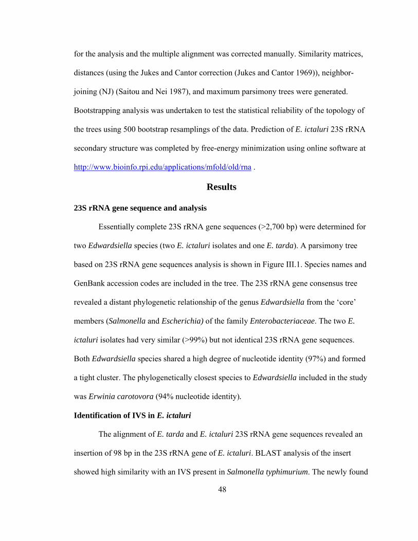
48
for the analysis and the multiple alignment was corrected manually. Similarity matrices,
distances (using the Jukes and Cantor correction (Jukes and Cantor 1969)), neighbor-
joining (NJ) (Saitou and Nei 1987), and maximum parsimony trees were generated.
Bootstrapping analysis was undertaken to test the statistical reliability of the topology of
the trees using 500 bootstrap resamplings of the data. Prediction of E. ictaluri 23S rRNA
secondary structure was completed by free-energy minimization using online software at
http://www.bioinfo.rpi.edu/applications/mfold/old/rna .
Results
23S rRNA gene sequence and analysis
Essentially complete 23S rRNA gene sequences (>2,700 bp) were determined for
two Edwardsiella species (two E. ictaluri isolates and one E. tarda). A parsimony tree
based on 23S rRNA gene sequences analysis is shown in Figure III.1. Species names and
GenBank accession codes are included in the tree. The 23S rRNA gene consensus tree
revealed a distant phylogenetic relationship of the genus Edwardsiella from the ‘core’
members (Salmonella and Escherichia) of the family Enterobacteriaceae. The two E.
ictaluri isolates had very similar (>99%) but not identical 23S rRNA gene sequences.
Both Edwardsiella species shared a high degree of nucleotide identity (97%) and formed
a tight cluster. The phylogenetically closest species to Edwardsiella included in the study
was Erwinia carotovora (94% nucleotide identity).
Identification of IVS in E. ictaluri
The alignment of E. tarda and E. ictaluri 23S rRNA gene sequences revealed an
insertion of 98 bp in the 23S rRNA gene of E. ictaluri. BLAST analysis of the insert
showed high similarity with an IVS present in Salmonella typhimurium. The newly found

49
IVS was present in both E. ictaluri isolates but absent in E. tarda. E. ictaluri IVS was
located in helix-45 of the 23S rRNA gene. Available helix-45 IVS sequences from other
Enterobacteriaceae were downloaded from GenBank and used to construct a neighbor-
joining tree (data not shown) that revealed a high degree of sequence diversity between
Enterobacteriaceae IVSs. S. typhimurium IVS was most similar to E. ictaluri followed
by Yersinia enterocolitica. Specific nucleotide identities are displayed in Table III.2. The
highest percent of nucleotide identity was found between E. ictaluri and S. typhimurium
at 97%, suggesting that both sequences should be grouped under the same IVS family
(Family M) according to Miller et al. (2000). Yersinia bercovieri and Proteus mirabilis
shared 88% nucleotide similarity and could be included under a unique IVS family as
well. Sequence similarities among remaining IVSs ranged from 42% to 86% nucleotide
identity.
When the secondary structure of both E. ictaluri and S. typhimurium was
generated (Figure III.2) only two differences were found between the two structures. E.
ictaluri lacked a small loop present in S. typhimurium due to a single transversion on
position 77, which resulted in a longer stem in this region. The second difference was a
transition on position 35 of the IVS that resulted in no structural change.
Characterization of E. ictaluri IVS
In order to assess the prevalence of IVS within the species E. ictaluri, a collection
of E. ictaluri isolates was screened for IVS presence. Primers flanking the helix-45 IVS
region were designed and used in a PCR assay. Amplified regions containing IVS were
152 bp in length, while 54 bp bands indicated an absence of IVS in helix-45. All twenty
nine E. ictaluri yielded the expected 152 bp amplified product (Figure III.3) while all E.

50
tarda strains lacked the IVS band and only exhibited the 54 bp amplicon. However, all E.
ictaluri strains tested also presented a weak band at 54 bp indicating that at least one rrn
operon in E. ictaluri did not contain IVS. All amplified IVSs were cloned and sequenced.
No differences in nucleotide sequences between the different E. ictaluri isolates tested
were observed (data not shown).
Ribotyping analysis was performed in order to further characterize the rrl operons
in E. ictaluri. Restriction analysis by KpnI and BamHI followed by hybridization with the
universal probe 1037-DIG revealed at least six rrl operons in the E. ictaluri genome (data
not shown). When the IVS-specific probe was used, the same bands were displayed
indicating that all rrl displayed by ribotyping contained the IVS. This result contradicts
the PCR analysis by which at least one rrl operon did not contain IVS. However, bands
highlighted by ribotyping were large in size, ranged from 8 kb to 25 kb, and could
contain more than one rrl operon.
Fragmentation of 23S rRNA due to IVS excision was revealed by methylene blue
staining of ribosomal RNA extracted from E. ictaluri isolates as well as from controls. If
an rrl gene contains an IVS in helix-45 and that IVS is removed during
posttranscriptional processing (without religation of the rRNA), two smaller fragments of
1.2 kb and 1.7 kb result. Figure III. 4 displays the processing of rrn genes in E. ictaluri.
16S and 23S rRNAs were represented by a 2.9 kb and a 1.5 kb band respectively, that
were displayed by all isolates tested including IVS-negative controls. E. ictaluri rRNAs
pattern showed two additional bands of 1.2 kb and 1.7 kb demonstrating fragmentation of
the 23S rRNA in this species.

51
Discussion
Intervening sequences were first described by Burgin et al. (1990) as responsible
for 23S rRNA fragmentation in Salmonella. These intervening sequences, or internal
transcribed spacers, are removed (without splicing) after transcription of ribosomal genes
by the action of RNaseIII, and the resulting 23S rRNA fragments are held together by the
compact structure of ribosomes which remain functional. Since their discovery, IVS have
been described in numerous bacteria including Alphaproteobacteria,
Gammaproteobacteria, and Epsilonproteobacteria as well as in Spirochaeta
(Evguenieva-Hackenberg and Klug 2000). Two characteristics of IVS have yet to be
explained by microbiologists. First, a seemly random and sporadic distribution of IVS
through bacteria has been observed. Second, their function is not known. Presence of
IVSs in ribosomal genes seems not to interfere with cell viability nor appear to confer
any evolutionary advantage. These unique sequences have been well studied within the
family Enterobacteriaceae, where six out of 14 tested genera harbored isolates with IVS
in their 23S rRNA genes (Pronk and Sanderson 2001). Intervening sequences in
Enterobacteriaceae appear at two different sites, helix-25 and helix-45, according to the
proposed secondary structure of the 23S rRNA, and can be grouped in different families
based on nucleotide identity. However, these families (or superfamilies) cannot be
correlated to other hierarchal classifications of Enterobacteriaceae based on taxonomy or
phylogenetics. Horizontal and vertical gene transfer episodes had to be combined in order
to justify IVSs distribution in Enterobacteriaceae (Pabbaraju et al. 2000).
In this work, we have sequenced and characterized the 23S rRNA genes from two
Enterobacteriaceae whose genus, Edwardsiella, has never been the target of ribosomal

52
gene-based phylogenetics studies and is rarely included or compared with other members
of the family Enterobacteriaceae. The analysis of the 23S rRNA of E. ictaluri and E.
tarda confirmed the close relationship between both species which was previously
determined by 16S rRNA analysis (data not shown). The 23S rRNA gene derived
parsimony tree showed the genus Edwardsiella is phylogenetically distant from the ‘core’
members of the family Enterobacteriaceae, such as Salmonella and Escherichia, as
previously reported from DNA:DNA hybridization studies (Krieg and Holt 1984).
Interestingly, E. ictaluri contains an insertion of 98 bp in helix-45 that has high similarity
with IVS described in other Enterobacteriaceae. Sequence analysis of this new IVS
revealed a 97% nucleotide sequence identity with S. typhimurium helix-45 IVS. In fact,
only two nucleotide differences in the IVSs of these two species were found (one
transition and one transversion). Based on this high similarity between E. ictaluri and S.
typhimurium IVSs, both sequences should be included under the same 45 helix-IVS
family M (Miller et al. 2000). Oddly, S. typhimurium IVS has a higher similarity (97%)
with E. ictaluri IVS than with any other helix-45 IVS described to date.
S. typhimurium has seven rrl genes, six of which present helix-45 IVSs of
identical sequence (Mattatall and Sanderson 1996). The number of rrl genes in E. ictaluri
has not been completely defined but, according to our ribotyping data, at least six rrl
genes containing helix-45 IVSs are harbored in this species. When the IVSs from all
thirty E. ictaluri isolates were cloned and sequenced, no differences in sequence between
isolates or clones were noted, suggesting all helix-45 IVSs are identical. However, PCR
results suggest at least one rrl gene in E. ictaluri lacks the 45-helix IVS. These results

53
could be explained by the existence of seven rrl genes in the chromosome, six of which
would contain helix-45 IVS, as in S. typhimurium (Mattatall and Sanderson 1996).
IVSs in members of the seven genera (Salmonella, Yersinia, Proteus, Providencia,
Klebsiella, Citrobacter, and Edwardsiella) of Enterobacteriaceae show some degree of
sequence similarity with each other, but no significant nucleotide identity between
Enterobacteriaceae IVSs and any other sequences, including IVSs from other bacteria
has been detected by BLAST searches (Pronk and Sanderson 2001). However, the
possession of IVS is a rather random phenomenon in this family, since some genera (i.e.
Escherichia and Enterobacter) do not harbor IVSs in their rrl genes. Earlier studies on
nucleotide sequences supported the idea that IVSs must be exchanged between different
genera by lateral transfer. IVSs have terminal inverted repeats flanking the central region,
which is characteristic of a mobile genetic element. These mobile elements could be
transferred from one species to another by phage-mediated transduction, transformation,
or conjugation. However, alternative hypothesis suggests that the progenitor of these
species obtained the IVS from a common ancestor but that subsequent variation between
different species was simplely due to the loss of IVSs by some strains or species.
Edwardsiella could have obtained the IVS prior to species divergence and, while E.
ictaluri still maintains the insertion (in at least six rrn operons), E. tarda could have lost
it.
From an applied point of view, the availability of 16S rRNA gene sequences has
facilitated the development of PCR-based methods in molecular diagnosis. However,
cases of very closely related species have highlighted the need of using other conserved
molecules such as the 23S rDNA (Arias et al. 1995) or the intergenic spacer region (ISR)

54
(Welker et al. 2005) as targets in a PCR detection methods since the 16S rRNA gene
might not provide sufficient genetic variability. This is the case for E. tarda and E.
ictaluri, as both species shared more than 99% identities in their 16S rDNA so that
finding species-specific PCR primers has been an obstacle in developing PCR detection
methods. To date, the only PCR detection method for E. ictaluri (Bilodeau et al. 2003)
targets a mobile element present in the E. ictaluri genome since non specific signature
sequences cannot be found in the 16S rRNA gene. Because of the negative economic
impact caused by these two pathogens in the catfish industry, rapid and specific detection
methods need to be implemented in fish diagnostic laboratories in order to hasten
treatments and control outbreaks. The discovery of a helix-45 IVS sequence in the 23S
rRNA gene of E. ictaluri, which is not present in E. tarda, opens possibilities for using
this newly identified sequence in molecular diagnosis of enteric septicemia of catfish
(ESC). The potential of the E. ictaluri IVS as a PCR-based detection target was described
in chapter II.

55
Table III.1. Strains used in the IVS study.
Species Strain OriginE. ictaluri CECT 885T Catfish, Georgia,
USA195, 196, 151, 218, 219, AL-93-75, AL-02-27, AL-93-58, ALG-03-278, ALG-03-190, ALG-03-161, ALG-03-278,ALG-03-275, ALG-03-277, ALG-03-192
Catfish, Alabama, USA
S94-728, S94-1034, S94-715, S94-703, S94-827, S94-862, S94-872, S94-1051, S94-707,1696, 1760, 1963
Catfish, Mississippi, USA
EILO Catfish, ThailandE. tarda CECT 849T Human feces
1909, Flb, Flb2, 172, Catfish, Alabama, USA
Escherichia coli
K-12 Human feces
T, type strain
CECT, Spanish Type Culture Collection
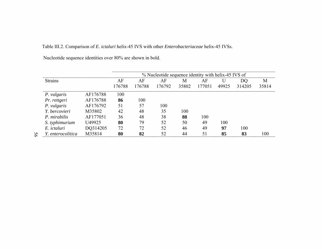
Table III.2. Comparison of E. ictaluri helix-45 IVS with other Enterobacteriaceae helix-45 IVSs.
Nucleotide sequence identities over 80% are shown in bold.
% Nucleotide sequence identity with helix-45 IVS ofStrains AF
176788AF
176788AF
176792M
35802AF
177051U
49925DQ
314205M
35814
P. vulgaris AF176788 100Pr. rettgeri AF176788 86 100P. vulgaris AF176792 51 57 100Y. bercovieri M35802 42 48 35 100P. mirabilis AF177051 36 48 38 88 100S. typhimurium U49925 80 79 52 50 49 100E. ictaluri DQ314205 72 72 52 46 49 97 100Y. enterocolitica M35814 80 82 52 44 51 85 83 100
56
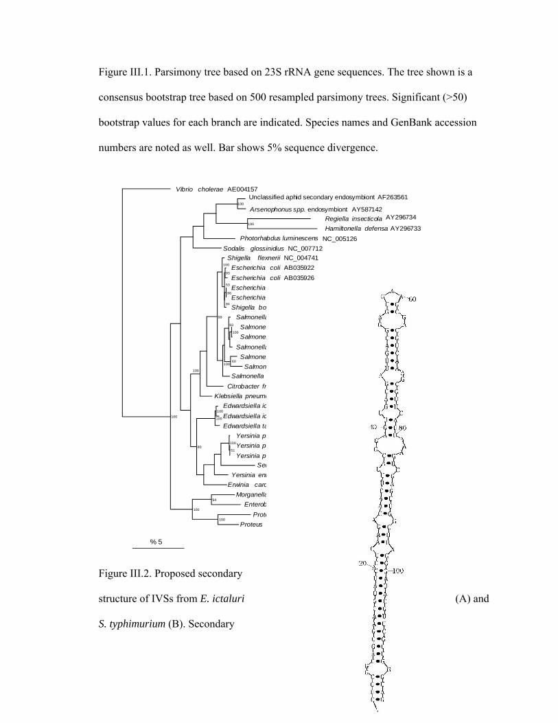
57
Figure III.1. Parsimony tree based on 23S rRNA gene sequences. The tree shown is a
consensus bootstrap tree based on 500 resampled parsimony trees. Significant (>50)
bootstrap values for each branch are indicated. Species names and GenBank accession
numbers are noted as well. Bar shows 5% sequence divergence.
100
100
93
86
84
53
100
100
93
60100
99
100
100
100
51
10080
94
100
100
100
VibrioUnclassified aphid secondary endosymbiont
Arsenophonus spp. endosymbiont
Regiella
Hamiltonella
Photorhabdus
Sodalis
Shigella
Escherichia
Escherichia
Escherichia
Escherichia
Shigella
Salmonella
Salmonella
Salmonella
Salmonella
Salmonella
Salmonella
Salmonella
Citrobacter
Klebsiella
Edwardsiella
Edwardsiella
Edwardsiella
Yersinia
Yersinia
Yersinia
Serratia
Yersinia
Erwinia
Morganella
Enterobacter
Proteus
Proteus
cholerae
insecticola
defensa
luminescens
glossinidius
flexnerii
coli
coli
coli
coli
boydii
enterica
typhimurium
typhimurium
enterica
enterica
bongori
enterica
freundii
pneumoniae
ictaluri
ictaluri
tarda
pseudotuberculosis
pestis
pestis
symbiotica
enterocolitica
carotovora
morganii
aerogenes
vulgaris
mirabilis
AE004157AF263561
AY296734
AY296733
NC_005126
NC_007712
NC_004741
AB035922
AB035926
U00096
J01695
CP000036
U77919
AE008893
AE008895
CP000026
U77926
U77927
U77922
U77928
X87284
DQ211094
DQ314205
DQ211093
NC_006155
AE017127
AE013984
AY296732
U77925
BX950851
% 5
AY587142
AY116909
AY116918
AY116927
AY116929
Figure III.2. Proposed secondary
structure of IVSs from E. ictaluri (A) and
S. typhimurium (B). Secondary

58
structure predictions based on free-energy minimization. Nucleotide differences in E.
ictaluri IVS compared with S. typhimurium IVS are marked by asterisks. The proposed
conserved site at which helix-45 was replaced by the IVS is indicated by a horizontal line.
A B

59
Figure III.3. Agarose gel containing the PCR products from amplification of 23S rRNA
gene containing the IVS. Lane M, 50 bp molecular marker. Lane C, no template DNA;
lane 1, Escherichia coli; lane 2, E. tarda CECT 849; lane 3 to lane 9, E. ictaluri isolates:
CECT 885, EILO, 195, 196, 151, 218 and 219, respectively. rrl operons containing IVS
in helix-45 yielded a 152 bp amplified product. Operons lacking IVS generated a smaller
amplicon of 54 bp. This 54 bp amplified should not be confused with the primer-dimer
band observed in the no-template control.
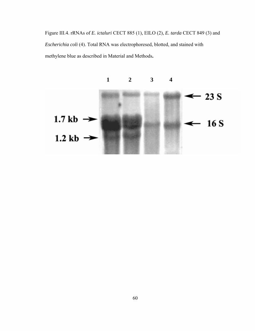
60
Figure III.4. rRNAs of E. ictaluri CECT 885 (1), EILO (2), E. tarda CECT 849 (3) and
Escherichia coli (4). Total RNA was electrophoresed, blotted, and stained with
methylene blue as described in Material and Methods.
1 2 3 4

61
IV. COMPARISON OF LIPOPOLYSACCHARIDE AND PROTEIN PROFILES
BETWEEN FLAVOBACTERIUM COLUMNARE STRAINS FROM DIFFERENT
GENOMOVARS
Abstract
Lipopolysaccharide (LPS) and total protein profiles from four Flavobacterium
columnare isolates were compared. These strains belonged to genetically different
groups and/or presented distinct virulence properties. F. columnare isolates ALG-00-530
and ARS-1 are highly virulent strains that belonged to different genomovars while F.
columnare FC-RR is an attenuated mutant used as a live vaccine against F. columnare.
Strain ALG-03-063 is included in the same genomovar group as FC-RR and presents a
similar genomic fingerprint. Electrophoresis of LPS showed qualitative differences
among the four strains. Further analysis of LPS by immunoblotting revealed that the
avirulent mutant lacks the higher molecular bands in the LPS. Total protein analysis
displayed by immunoblotting showed differences between the strains analyzed although
common bands were present in all the isolates. FC-RR lacked two distinct common
bands (34 kDa and 33 kDa) shared by the other three isolates. Based on the difference of
LPS and total protein profiles, it is possible to discriminate the attenuated mutant FC-RR
from other F. columnare strains.
Keywords: LPS, whole protein profiles, F. columnare, modified live vaccine

62
Introduction
Flavobacterium columnare is the causal agent of columnaris disease, one of the
most important bacterial diseases of freshwater fish species. This bacterium is distributed
world wide in aquatic environments, affecting wild and cultured fish as well as
ornamental fish in aquaria (Austin and Austin 1999). F. columnare is considered the
second most important bacterial pathogen in commercial cultured channel catfish,
Ictalurus punctatus, in the southeastern USA, second only to Edwardsiella ictaluri
(Wagner et al. 2002). Direct losses due to F. columnare are estimated in excess of
millions of dollars per year. Mortality rates of catfish populations in ponds can reach 50%
to 60% and can be as high as 90% in tank-held catfish fingerlings (USDA 2003a, 2003b).
Columnaris disease usually begins as an external infection of fins, body surface,
or gills. The fins become necrotic with grayish to white margins, and initial skin lesions
appear as discrete bluish areas that evolve into depigmented necrotic lesions. Skin
lesions might have yellowish mucoid material accompanied by mild inflammation.
Lesions can develop exclusively on the gills, which usually results in subacute disease
and mortality, as is typically the case in young fish (Plumb 1999).
Due to the ubiquitous presence of F. columnare in aquatic environments,
eradication of the disease in fish farms will not likely occur. Control and treatment of
columnaris have primarily been directed toward the use of improved water-management
practices to reduce physiological and environmental stress in the fish. Recently, a
modified live F. columnare vaccine has been developed using a rifampicin-resistant
strategy (Shoemaker et al. 2005b). This method had been previously used by Montaraz
and Winter (1986) to generate a rough Brucella abortus strain, currently employed as the

63
official vaccine for cattle brucellosis in the USA. The same strategy was used by Klesius
and Shoemaker (1999) to create an E. ictaluri rifampicin-resistant mutant patented as a
modified live vaccine against enteric septicemia of catfish (ESC) (AQUAVAC-ESC,
Intervet, Millsboro, DE). Characterization of B. abortus and E. ictaluri rifampicin-
resistant mutants revealed that lipopolysaccharide (LPS), a main virulence factor for
Gram-negative bacteria, lacked the high molecular bands observed in virulent isolates
(Vemulapalli et al. 1999; Arias et al. 2003).
Flavobacterium columnare belongs to the Cytophaga-Flavobacterium-
Bacteroides (CFB) group and is phylogenetically distant from the better studied
gammaproteobacteria subclass which contains major human and animal pathogens
(including Brucella and Edwardsiella). To date, virulence factors in F. columnare are
poorly characterized. Specifically, the role of LPS in columnaris pathogenicity has not
been explored. The objective of this study was to investigate if the attenuated F.
columnare mutant originated through a rifampicin-resistance strategy presented a
modified LPS and a different total protein profile by comparison with virulent strains.
Materials and Methods
Bacterial isolates and culturing
Four F. columnare strains, ALG-00-530, FC-RR, ARS-1, and ALG-03-063 were
used in this study. Isolate ALG-00-530 was obtained from diseased channel catfish at the
Alabama Fish Farming Center, Greensboro, Alabama. The ARS-1 isolate was recovered
from diseased channel catfish at the Aquatic Animal Health Research Unit, USDA-ARS,
Auburn, Alabama. These two isolates have been demonstrated to be virulent for channel
catfish (Shoemaker et al. 2003a). F. columnare ALG-03-063 was also isolated from

64
diseased channel catfish. FC-RR strain is a rifampicin-resistant mutant, avirulent for
catfish. This attenuated mutant has been licensed as a modified live vaccine by Intervet,
Inc. and it is now undergoing field trials (Shoemaker et al. 2005b) (Patent No.: US
6,881,412 B1).
Genetic fingerprinting
A previous study by Arias et al. (2004) divided F. columnare species into four
well-defined genetic groups. All catfish isolates were grouped into three different
genogroups (I through III; genogroup IV included only tilapia isolates). Strains ALG-00-
530 and ARS-1 belonged to genogroups I and II, respectively; while ALG-03-063
belonged to genogroup III. The avirulent mutant FC-RR was not included in that former
study but was fingerprinted in the present work. Amplified fragment length
polymorphism (AFLP) patterns from FC-RR mutant were obtained as described by Arias
et al. (2004), added to the existing database and analyzed.
Production of polyclonal anti-serum against F. columnare
Twelve channel catfish were randomly divided into three groups of four fish each.
Two groups were used to produce antibodies (Ab) against F. columnare strains (Ab-
ALG-00-530 and Ab-FC-RR) while the third group served as negative control. Antigen
preparation was from cells grown overnight in modified Shieh broth (5.0 g tryptone, 2.0 g
yeast extract, 1.0 mL of 45.6 mM CaCl2 2H2O, 3 mL of 0.36 mM KH2PO4, 10 mL of
0.12 M MgSO4 7H2O, 10 mL of 0.41 mM FeSO4 7H2O, 1 L of distilled water, pH 7.2-
7.4) and quantified by plate count (10 X 108 CFU/mL). Bacterial cells were sonicated for
30 seconds at speed 4 using a Virsornic 600 sonicator (SP Industries Company, Gardiner,
NY). One milliliter of the sonicated suspension was mixed with 1 mL complete Freund’s

65
adjuvant (Sigma, St. Louis, MO). Two hundred microliters of the mixture were
intraperitoneally injected into each fish. Control fish were injected with 200 µL of Shieh
broth. A booster injection was administered 27 days after initial immunization using 200
µL antigen prepared as above (Freund’s incomplete adjuvant (Sigma) was used this time).
Prior to the booster immunization, about 1.5 mL of blood was obtained from the caudal
vasculature of each fish for titer determination (Shoemaker et al. 2003b). Three weeks
after booster immunization, fish were bled again, and this antiserum was used to probe
the western blots (see below). Blood was allowed to clot at room temperature for 1 h and
serum was carefully removed after centrifugation for 5 min at 1,000 g. Sera were stored
at -20 ºC until use. In order to determine the relative titer of the sera, enzyme linked
immunosorbent assays (ELISA) were conducted according to Shoemaker et al. (2003b).
Sonicated cell suspensions were prepared as described above and serial ten-fold dilutions
were used to coat the plates; proper positive and negative controls were included. Serum
from each individual fish was tested against each antigen dilution.
Lipopolysaccharides (LPS) and total protein extraction
The four isolates were cultured in modified Shieh broth for 24 h at 28 ºC. Three
milliliters of broth were centrifuged at 3,000 g for 15 min. Pelleted cells were
resuspended in lysis buffer and proteins were extracted according to Arias et al. (1997b).
Crude LPS was extracted following the phenol-water protocol described by Westphal and
Jann (1965). After lyophilization, the LPS extract was diluted in sample buffer at a final
concentration of 1mg/mL. Aqueous and phenol LPS phases were stored at -80°C until
use.

66
LPS and total protein analysis
Electrophoresis — LPS from both phases were resuspended in sample treatment
buffer and electrophoresed by discontinuous SDS-PAGE on a 4% stacking gel and a 20%
separating gel (Laemmli 1970). Gels were run at 15 mAmp for 90 min. Gels were silver
stained following the instruction of Bio-Rad Silver Stain Kit (Bio-Rad, Hercules, CA).
Total protein electrophoresis followed the procedure described by Arias et al. (1997b).
Coomassie staining was used to visualize the protein bands following standard methods
(Sambrook and Russell 2001).
Western blotting — Protein and LPS samples were analyzed in duplicate. First,
samples were resolved on 12% SDS-PAGE gel, and then transferred to polyvinylidene
difluoride (PVDF) membrane (Bio-Rad) at 100 volts for 1 h. After blotting, one
membrane was incubated with anti-ALG-00-530 serum while the second membrane was
incubated with anti-FC-RR serum. After blocking for 30 min, 1:500 diluted polyclonal
catfish serum was incubated with the membrane overnight followed by a one-hour
incubation with a monoclonal antibody (E-8) specific for channel catfish IgM (1:10)
(Klesius 1990) , and labeled with conjugated goat anti-mouse Ig (1:5,000), followed by
detection with Opti-4CNTM (Bio-Rad).
Results
AFLP analysis
Figure IV.1 shows results of the cluster analysis of the AFLP patterns from the
strains used in the study. Attenuated mutant FC-RR displayed a unique AFLP fingerprint
although clustered among other genogroup III strains. AFLP profile of strain ALG-03-
063 presented the highest percent of similarity with the one displayed by the attenuated

67
mutant. Based on this result, ALG-03-063 was selected for further comparisons as the
most genetically similar strain to FC-RR present in our collection (that currently includes
more than 50 F. columnare isolates; data not shown).
ELISA analysis
All fish immunized with F. columnare yielded positive titers. Control fish
presented a very low cross-reactivity with the sonicated cells. All immunized fish
produced strong immunity against both antigens. Although antisera from immunized fish
reacted with both ALG-00-530 and FC-RR antigens, the corresponding antigen provided
the highest titer (data not shown). Individual fish sera showing the highest titer (≥1/800)
were chosen for immunoblotting analysis (fish no. 3 anti-ALG-00-530 and fish no.1 anti-
FC-RR respectively).
LPS characterization
The results of SDS-PAGE of LPS from F. columnare are shown in Figure IV.2.
The characteristic ladder-like pattern typical of Gram-negative pathogens was not
observed. Only a few bands ranging from 3.5 kDa to 17 kDa were present in all four
strains. Phenol phase extracts contained higher amounts of LPS than the aqueous-
extracted samples. Strain ALG-00-530 showed a few bands in the phenol phase extract
while only one weak band was visualized in the aqueous phase. A similar pattern was
observed with strain ARS-1 although band intensities were stronger. Attenuated mutant
FC-RR shared a similar LPS pattern with ALG-03-063 although FC-RR bands were
slightly smaller.
Bands from F. columnare LPS were visualized much more effectively in
immunoblots. When catfish polyclonal antiserum was used for immunoblotting,

68
important differences between the four strains were revealed (Figure III.3). While all
strains isolated from diseased channel catfish (ALG-00-530, ARS-1, and ALG-03-063)
presented high molecular bands (above 21 kDa) in both phenol and aqueous phases, the
FC-RR mutant exhibited bands below that range. ALG-00-530 and ARS-1 again showed
a similar LPS pattern regardless of antisera or extraction phase used. ALG-03-063
presented the same size bands in the phenol phase as FC-RR but also exhibited higher
molecular weight bands that were absent in the mutant. Interestingly, these results were
consistent regardless of antisera used (anti-ALG-00-530 or anti-FC-RR). However,
significant differences between aqueous and phenol phase samples were found in both
immunoblots.
Total protein analysis
Western blots of total proteins were also analyzed with both sera (Figure IV.4).
The immunoblots indicated that the four strains did contain different antigenic proteins
but also shared some common bands. ARS-1 showed more distinct antigenic protein
bands than any other strain analyzed (Figure IV.4). The strains ALG-00-530, ARS-1 and
ALG-03-063 contained two very distinct bands around 35 kDa. FC-RR mutant lacked
these bands but instead presented two additional bands in the 30 kDa range. Each strain
had very similar banding pattern with anti-ALG-00-530 and anti-FC-RR sera. When
ALG-00-530 serum was used, reactivity against low molecular weight bands present in
the three clinical strains was observed. FC-RR low molecular bands did not react with
anti-ALG-00-530 serum. On the contrary, when anti-FC-RR was used, low molecular
weight proteins in FC-RR were revealed.

69
Discussion
Lipopolysaccharide is a major superstructure of Gram-negative bacteria,
contributes greatly to the structural integrity of the bacteria, and protects them from host
immune defenses. The LPS has been described and well characterized as a virulence
factor in many bacterial species (Salmonella sp. (Makela et al. 1973), Escherichia coli
(Medearis et al. 1968), Shigella flexneri (Rajakumar et al. 1994), Brucella abortus
(Vemulapalli et al. 2000) including fish pathogens such as Vibrio vulnifucus (Amaro et al.
1997), E. ictaluri (Arias et al. 2003), and F. psychrophilum (MacLean et al. 2001).
Attempts to characterize the structure of the LPS in F. columnare were made by
MacLean et al. (2003) and by Vinogradov et al. (2003). Unfortunately, the strain used by
these authors (ATCC 43622) had been originally misidentified as F. columnare but was
actually F. johnsoniae (Darwish et al. 2004; Shoemaker et al. 2005a). Therefore, no
information on F. columnare LPS composition or immunogenic role was available.
In this study, catfish polyclonal antisera against two strains, ALG-00-530 and FC-
RR (attenuated mutant), were generated and used to determine LPS immunogenic
properties between genetically different F. columnare strains. Differences in LPS
between four strains of F. columnare were evident after electrophoresis and silver
staining but even more obvious after western blot analysis. Although a typical LPS
ladder has been reported in other Flavobacterium species (MacLean et al. 2001), our
silver staining of F. columnare LPS analysis revealed only a few bands under 7 kDa.
Immunogenic bands present in the LPS were revealed by immunoblots. However,
there was a major difference in band molecular weight between strains isolated from
diseased channel catfish and the attenuated strain FC-RR. The three strains from diseased
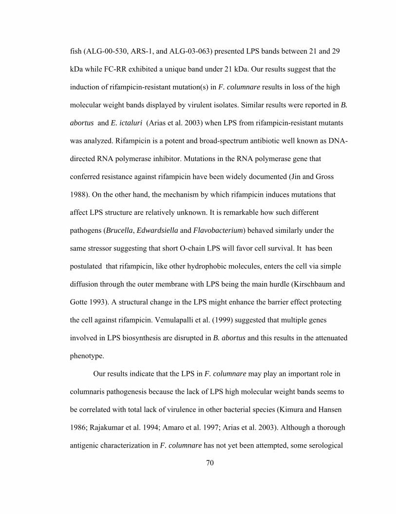
70
fish (ALG-00-530, ARS-1, and ALG-03-063) presented LPS bands between 21 and 29
kDa while FC-RR exhibited a unique band under 21 kDa. Our results suggest that the
induction of rifampicin-resistant mutation(s) in F. columnare results in loss of the high
molecular weight bands displayed by virulent isolates. Similar results were reported in B.
abortus and E. ictaluri (Arias et al. 2003) when LPS from rifampicin-resistant mutants
was analyzed. Rifampicin is a potent and broad-spectrum antibiotic well known as DNA-
directed RNA polymerase inhibitor. Mutations in the RNA polymerase gene that
conferred resistance against rifampicin have been widely documented (Jin and Gross
1988). On the other hand, the mechanism by which rifampicin induces mutations that
affect LPS structure are relatively unknown. It is remarkable how such different
pathogens (Brucella, Edwardsiella and Flavobacterium) behaved similarly under the
same stressor suggesting that short O-chain LPS will favor cell survival. It has been
postulated that rifampicin, like other hydrophobic molecules, enters the cell via simple
diffusion through the outer membrane with LPS being the main hurdle (Kirschbaum and
Gotte 1993). A structural change in the LPS might enhance the barrier effect protecting
the cell against rifampicin. Vemulapalli et al. (1999) suggested that multiple genes
involved in LPS biosynthesis are disrupted in B. abortus and this results in the attenuated
phenotype.
Our results indicate that the LPS in F. columnare may play an important role in
columnaris pathogenesis because the lack of LPS high molecular weight bands seems to
be correlated with total lack of virulence in other bacterial species (Kimura and Hansen
1986; Rajakumar et al. 1994; Amaro et al. 1997; Arias et al. 2003). Although a thorough
antigenic characterization in F. columnare has not yet been attempted, some serological
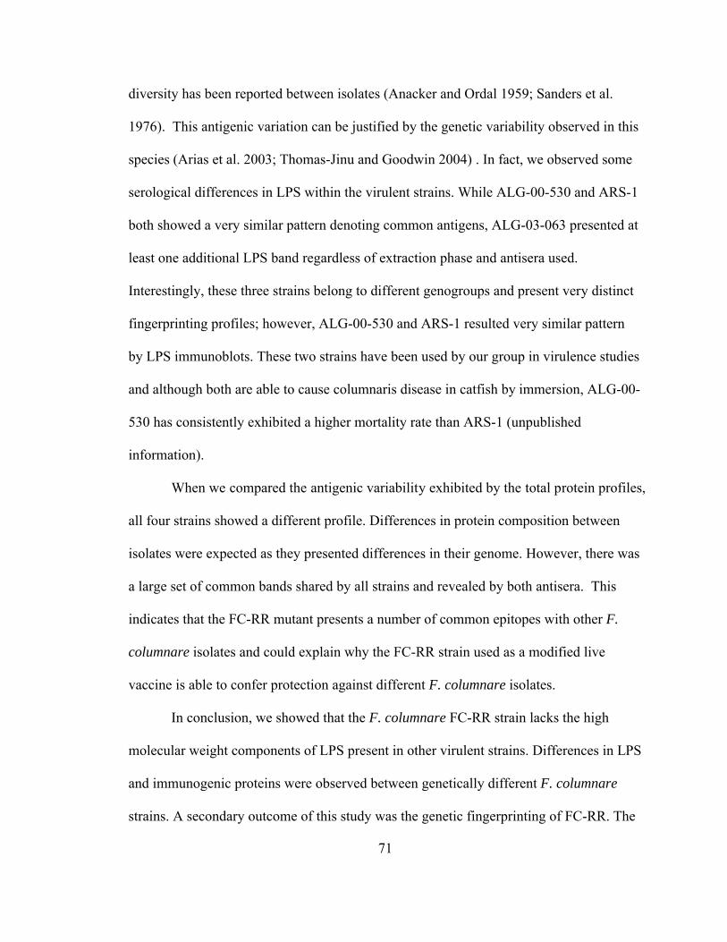
71
diversity has been reported between isolates (Anacker and Ordal 1959; Sanders et al.
1976). This antigenic variation can be justified by the genetic variability observed in this
species (Arias et al. 2003; Thomas-Jinu and Goodwin 2004) . In fact, we observed some
serological differences in LPS within the virulent strains. While ALG-00-530 and ARS-1
both showed a very similar pattern denoting common antigens, ALG-03-063 presented at
least one additional LPS band regardless of extraction phase and antisera used.
Interestingly, these three strains belong to different genogroups and present very distinct
fingerprinting profiles; however, ALG-00-530 and ARS-1 resulted very similar pattern
by LPS immunoblots. These two strains have been used by our group in virulence studies
and although both are able to cause columnaris disease in catfish by immersion, ALG-00-
530 has consistently exhibited a higher mortality rate than ARS-1 (unpublished
information).
When we compared the antigenic variability exhibited by the total protein profiles,
all four strains showed a different profile. Differences in protein composition between
isolates were expected as they presented differences in their genome. However, there was
a large set of common bands shared by all strains and revealed by both antisera. This
indicates that the FC-RR mutant presents a number of common epitopes with other F.
columnare isolates and could explain why the FC-RR strain used as a modified live
vaccine is able to confer protection against different F. columnare isolates.
In conclusion, we showed that the F. columnare FC-RR strain lacks the high
molecular weight components of LPS present in other virulent strains. Differences in LPS
and immunogenic proteins were observed between genetically different F. columnare
strains. A secondary outcome of this study was the genetic fingerprinting of FC-RR. The

72
attenuated mutant presents a unique AFLP profile, not present in our existing database,
which can be used to track the modified live vaccine strain (FC-RR) in the fish farm
environment.

73
Figure IV.1. Dendrogram based on AFLP patterns of four strains of F. columnare (ALG-
00-530, FC-RR, ARS-1, and ALG-03-063). The tree was derived by UPGMA cluster
analysis of the AFLP profiles.
FC-RR
ALG-03-063
ARS-1
ALG-00-530
100
9590858075
Genogroup III
Genogroup II
Genogroup I
% similarity

74
Figure IV.2. Silver stained sodium dodecyl sulfate-polyacrylamide gel electrophoresis
profiles of the LPS preparations from F. columnare strains used in this study. Lane
1,molecular standard; lanes 2 through 5, LPS aqueous phase from ALG-00-530 (2), FC-
RR (3), ARS-1 aqueous (4), and ALG-03-063 (5); lanes 6 through 9 show LPS phenol
phase from ALG-00-530 (6), FC-RR (7), ARS-1 (8) and ALG-03-063 (9).
1 2 3 4 5 6 7 8 9
6.9 kDa21 kDa
29 kDa
75
Figure IV.3. Immunoblot using anti-ALG-00-530 serum (A) and anti-FC-RR serum (B)
with LPS preparation. Lane 1, molecular standard; lanes 2 through 5, LPS aqueous phase
from ALG-00-530 (2), FC-RR (3), ARS-1 aqueous (4), and ALG-03-063 (5); lanes 6
through 9 show LPS phenol phase from ALG-00-530 (6), FC-RR (7), ARS-1 (8) and
ALG-03-063 (9).
21 kDa
6.9 kDa
21 kDa
6.9 kDa
A
B
1 2 3 4 5 6 7 8 9
1 2 3 4 5 6 7 8 9
35.6 kDa
35.6 kDa

76
Figure IV.4. Western blot analysis of total protein with anti-ALG-00-530 serum (A) and
anti-FC-RR serum (B). Lane 1, molecular standard; lane 2, ALG-00-530; lane 3, FC-RR;
lane 4, ARS-1; lane 5, ALG-03-063.
A B
1 2 3 4 5 1 2 3 4 5
35.6 kDa
21 kDa
6.9 kDa
56 kDa
101 kDa
125 kDa
210 kDa
35.6 kDa
21 kDa
6.9 kDa
56 kDa
101 kDa
125 kDa210 kDa

77
V. IDENTIFICATION, CHARACTERIZATION AND EXPRESSION PATTERNS OF
GTF, HEMH, NORB, AND TRX GENES IN FLAVOBACTERIUM COLUMNARE
Abstract
Four genes from a shotgun genomic library of F. columnare virulent strain ALG-
00-530 were identified, characterized, and selected for differential expression analysis
based on sequence similarity to putative virulence genes from related species. These
genes were: glycosyltransferase (gtf), ferrochelatase (hemH), nitric oxide reductase (norB)
and thioredoxin (trx). A collection of 30 F. columnare strains, including strains from
genomovars I and II, were tested for the presence of these genes. Distribution patterns of
gtf, hemH, norB, and trx across the species were not uniform. Nucleotide sequence
variation was observed between genomovars for each gene; however, strains within the
same genomovar shared identical gene sequences.
Nine strains of F. columnare (from both genomovars I and II) were chosen for
gene expression analysis. The expression profile of these genes varied when selected
strains were grown in vitro under identical conditions. ALG-00-530 was chosen for gene
expression comparison under standard growing conditions, iron-limited conditions and in
the presence of skin explants from channel catfish (Ictalurus punctatus). gtf, hemH, norB,
and trx gene expression levels varied when ALG-00-530 was incubated under different
conditions.

78
Keywords: Flavobacterium columnare, gtf, hemH, norB, and trx gene expression, iron-
limited, catfish skin explants, pathogenicity

79
Introduction
Flavobacterium columnare, the etiologic agent of columnaris disease, is a Gram-
negative bacterium that can infect most freshwater fish species. Differences in virulence
are known to exist among F. columnare isolates, resulting in variable mortality of fish
(Pacha and Ordal 1970; Suomalainen et al. 2006). Understanding the pathogenesis of F.
columnare is critical for fish health, but our knowledge of F. columnare virulence factors
is far from being complete.
Only a few genes from F. columnare have been described to date, and most
known sequences from this pathogen correspond to ribosomal genes. Among the non-
ribosomal sequences identified, the chondroitin AC lyase gene in F. columnare has been
previously sequenced and characterized (Xie et al. 2005). The enzyme codified by this
gene is able to degrade acidic polysaccharides, such as hyaluronic acid and chondroitin
sulfates (Griffin 1991; Teska 1993; Stringer-Roth et al. 2002). Differences in chondroitin
AC lyase activity have been observed between F. columnare isolates (Stringer-Roth et al.
2002). Relation of chondroitin AC lyase activity to virulence in F. columnare has been
recently reported (Suomalainen et al. 2006). Genes encoding outer membrane proteins,
such as zinc metalloprotease and prolyl oligopeptidase, have also been characterized in a
virulent F. columnare isolate (Xie et al. 2004). However, association of these two genes
with F. columnare pathogenicity is unclear. In addition, several proteases have been
identified in F. columnare, but their specific roles in columnaris pathogenicity are still
unknown (Bertolini and Rohovec 1992; Newton et al. 1997).
The ability of F. columnare to attach to fish tissues is thought to be a factor
correlated to virulence (Decostere et al. 1999b). Bader et al. (2005) selected for an

80
adhesive-defective F. columnare strain that exhibited reduced virulence to channel
catfish. Unfortunately, no adhesin gene has been identified in F. columnare.
Genetic variability of F. columnare species has been characterized using different
molecular markers (Triyanto and Wakabayashi 1999; Thomas-Jinu and Goodwin 2004;
Arias et al. 2004; Darwish and Ismaiel 2005). Restriction fragment length polymorphism
analysis of the 16S rRNA gene divided F. columnare species into three genomovars
(Triyanto and Wakabayashi 1999). The coexistence of three main genomic groups within
the species (genomovars I, II, and III) has been corroborated by intergenic spacer region
(ISR) sequencing (Darwish and Ismaiel 2005), and single-strand conformation
polymorphism (SSCP) analysis (Olivares-Fuster et al. 2007). To date, most of the
genetic markers used for F. columnare strain typing have relied on ribosomal gene
analysis.
The aim of this work was to identify and characterize putative virulence genes in
this species and to further investigate the genetic diversity of F. columnare using non-
ribosomal markers. To achieve this objective, a partially sequenced shotgun genomic
library constructed from a virulent F. columnare strain was used. Four putative virulence
genes were selected for further analysis. Gene presence and expression patterns were
analyzed across the species. Two in vitro experiments aimed to mimic some of the
environmental conditions F. columnare encounters during infection (limited iron and fish
skin) were used for gene expression analysis.

81
Materials and Methods
Bacterial strains and culture conditions
Thirty strains of F. columnare, representing genomovars I and II, were used in
this study (Table V.1). Cells were cultured in modified Shieh broth (Shoemaker et al.
2005a) at 28 ºC.
Putative virulence gene identification
A shotgun genomic library of the F. columnare virulent strain ALG-00-530 was
constructed. Briefly, F. columnare DNA was extracted and purified following standard
protocols (Sambrook and Russell 2001). Total DNA was partially digested with Sau3A I.
Digested DNA ranging from 1 to 1.25 kb was purified by double size selection and
ligated to the digested pBluescript II (KS-) vector (Stratagene, Cedar Creek, TX) at a
molar ratio of 1:2. The cloning site was BamH I. Library titer was estimated at 35
recombinant clones per microliter. Quality control tests showed 1% or less empty clones
and an expected genomic coverage of 20X based on approximate genome size of 4.1 mb.
Three thousand clones of this library have been sequenced to date at the USDA-ARS
MSA Genomics Laboratory (Stoneville, MS) following standard procedures.
Sequences from the shotgun genomic library were compared with sequences in
the GenBank database available at NCBI (National Center for Biotechnology Information
http://www.ncbi.nlm.nih.gov/) using the BLASTx algorithm (Altschul et al. 1990).
Sequences with more than 35% identity were recorded. Among the identified genes,
four were chosen for further analysis due to their high identity (≥65%) with known genes
described as virulence factors in other bacteria. Open reading frames (ORF) of the four
sequences were identified using Vector NTI® Suite 8 software package (Invitrogen,

82
Carlsbad, CA). ORF were translated into amino acid sequences by using Vector NTI®
Suite 8 and the GenBank database was searched for protein sequences using the BLASTp
tool (Altschul et al. 1990). Protein-protein identity percentage was recorded for each
chosen ORF. Specific primers for each gene were designed using Vector NTI® Suite 8.
Primer sequences for each gene were shown in Table V.2. These primers were tested on
30 strains of F. columnare by PCR. Unless otherwise stated, all PCR reagents were
purchased from Promega (Madison, WI). Each 50 µL PCR reaction included 2.5 μM
MgCl2, 1X buffer, 0.2 μM of both primers, 0.2 μM of dNTPs, 1.7 unit of Taq polymerase,
and 60 ng of DNA template. The PCR amplification profile was 5 min hot start at 95 ºC;
35 cycles of 30 s at 94 ºC, 45 s at 58 ºC, and 1.5 min at 72 ºC; and 10 min at 72 ºC.
DNA, RNA extraction and Reverse transcription PCR (RT-PCR)
Total bacterial DNA was extracted using the Qiagen DNeasy Tissue kit (Qiagen,
Valencia, CA) following the manufacturer’s instructions. Total RNA was extracted from
1.5 mL of bacterial culture using RNeasy plus Mini Kit (Qiagen). Turbo DNA-free kit
(Ambion, Austin, TX) was used to eliminate DNA contamination in RNA samples.
cDNA synthesis and RT-PCR were performed using the Reverse Transcription System
(Promega). One microgram of total RNA was used to synthesize the cDNA in 20 µL of
reaction. cDNA synthesis reaction was diluted to 100 µL, and 10 µL were used as
template for PCR using gene specific primers, as described above.
Cloning and sequencing analysis
PCR products were resolved through standard agarose gel electrophoresis.
Amplified products were purified using the High Pure PCR Product Purification Kit
(Roche Diagnostic Corporation, Indianapolis, IN) and cloned into pGEMTEasy
83
(Promega). To ensure sequence accuracy, two clones from each strain were sequenced at
the Auburn University Sequencing Core (Auburn, AL). Nucleotide sequence alignments
and deduced amino acid sequence alignments were performed by CLUSTAL W multiple
sequence alignment algorithm (Chenna et al. 2003).
Gene expression
Expression of gtf, hemH, norB, trx was tested under standard conditions, iron-
limited conditions, and in the presence of catfish skin explants. Following RNA
extraction and cDNA synthesis, genes were amplified from cDNA by the designed
primers as mentioned above. A 16S rRNA cDNA internal control was amplified with
universal primers UFUL (5’-GCCTAACACATGCAAGTCGA-3’) and URUL (5’-
CGTATTACCGCGGCTGCTGG-3’) (Chen et al. 2004). Positive (genomic DNA) and
negative (total RNA) controls were included for each gene amplification. Amplified
products were electrophoresed at 100 V for 30 min and visualized under UV light with
ethidium bromide added to a 1% gel to a final concentration of 0.5 μg/mL.
Gene expression under standard conditions — Nine strains of F. columnare,
representing genomovars I and II, were cultured in modified Shieh broth at 28 ºC for 20 h.
Bacterial cells were harvested from one and a half milliliters broth culture followed by
RNA extraction and RT-PCR.
ALG-00-530 gene expression under iron-limited conditions — Two iron chelators,
2, 2-dipyridyl and transferrin, were used to remove free iron from the growth medium. 2,
2- dipyridyl was added to 50 mL of Shieh broth to a final concentration of 50 μM.
Preparation of the transferrin solution followed the protocol described by Biosca et al.
(1993). Briefly, human apo-transferrin (Sigma, St. Louis, MO) was dissolved at a

84
concentration of 1 µM in a solution containing 100 μM Tris, 150 μM NaCl, and 50 μM
NaHCO3 (pH 8.0). This solution was sterilized by filtration. Prior to inoculation with F.
columnare, transferrin was added to 50 mL Shieh medium to achieve a final
concentration of 10 μM and incubated at 37ºC for 60 min.
Cultured ALG-00-530 bacterial cells were collected by centrifugation at 2,000 g
for 20 min. Pellets were washed once in PBS and centrifuged again. Cells were
resuspended in approximately 1 mL Shieh broth and then 1/3 mL (of this broth) was
transferred into 50 mL Shieh broth containing 2, 2-dipyridyl, or apo-transferrin. Fifty
milliliters of Shieh broth was inoculated as control. One and a half milliliters of
bacterial cells were collected at time 0 h, 2 h, 4 h, 6 h, 8 h, and 24 h after inoculation.
Cells were centrifuged and pellets were resuspended in 100 µL of RNA later (Qiagen)
and frozen at -80 ºC until used. Reverse transcription PCR was performed as explained
above.
ALG-00-530 gene expression in the presence of catfish skin explants — Skin
explants from channel catfish that tested negative for F. columnare antibody presence
were obtained according to the procedure by Xu and Klesius (2002). One milliliter of
Medium 199 (Sigma) supplemented with fetal bovine serum at 2.5% was added into each
well of a 24-well culture plate (Costar, Cambridge, MA). One cm2 of fish skin explant
was placed onto the bottom of 12 wells. One and a half milliliters of an overnight culture
of ALG-00-530 bacterial cells was collected for the baseline control (C0); one and half
milliliters of the same bacterial culture was added into each of the 24 wells. Wells
without fish skin explants were regarded as controls, while wells containing the fish
explants were referred to as treatments. Bacterial cells were collected from each

85
individual well at 30 min, 1 h, 3 h, 6 h, 10 h, and 24 h from both control and treatment
wells. After centrifugation, bacterial pellets were suspended in RNA later (Ambion,
Austin, TX) and frozen at -80 ºC until used. Total RNA extraction and RT-PCR were
performed as described above.
Results
Putative virulence gene identification
Out of 3,000 clones sequenced from the ALG-00-530 shotgun library, 73
sequences displayed a high nucleotide similarity with known genes present in GenBank
(based on BLASTx search, data not shown). Within this subset, four sequences were
identified from published literature as putative virulence genes and therefore selected for
this study: glycosyltransferase gene (gtf) (GenBank accession number DQ911521),
ferrochelatase gene (hemH) (GenBank accession number DQ911518), nitric oxide
reductase gene (norB) (GenBank accession number DQ911522), and thioredoxin gene
(trx) (GenBank accession number DQ911523). ORFs were translated into amino acid
sequences which were then compared to GenBank sequences using BLASTp. Identity
percentages of ORF based on protein-protein alignment were as follows: Gtf was 81%
identical to F. johnsoniae sequence accession number ZP_01245614, HemH was 63%
identical to F. johnsoniae sequence accession number ZP_01246582, NorB was 77%
identical to an unidentified Flavobacterium species sequence accession number
ZP_01106280, while Trx was 83% identical to F. johnsoniae sequence accession number
ZP_01243853. Based on the PCR results, all strains contained norB and trx genes (Table
V.1). However, no PCR amplification for hemH was observed in eight strains (GA-02-14,

86
ARS-1, BM, HS, GZ, BZ-1, BZ-4, BZ-5), while three strains (ARS-1, BM, HS) failed to
amplify the gtf gene.
Cloning and sequencing analysis
Two F. columnare strains from genomovar I (ATCC 49512 and ALG-03-57) and
genomovar II (ALG-00-530 and MS-02-475) were selected for multi locus sequence
analysis using gtf, hemH, norB, and trx genes. Nearly complete sequences were amplified
from gtf, norB, and trx and used for comparison. Only partial sequence (250 bp out of
626) of hemH was obtained. Alignment of these sequences showed two distinct
populations. Both genomovar I strains shared an identical sequence for all three genes as
did ALG-00-530 and MS-02-475 genomovar II strains (data not shown). However,
nucleotide sequences differed between genomovars (Figure V.1). The variation
percentage observed was 6.9% for gtf, 4.9% for hemH, 3.8% for norB, and 4.3% for trx.
Translation of the nucleotide sequences showed the distinction between genomovars was
carried out at the protein level. Amino acid sequence variation derived from each gene
was not identical between genomovars except for Trx. Gft displayed a 2.2% variation
between both genomovars. HemH protein sequence was 5.1% variable, while NorB
presented only 0.88% sequence variation.
Gene expression
Gene expression under standard conditions — The 16S rRNA gene, used as
internal control, was expressed at any given time in all experiments. Gene expression
patterns of nine strains of F. columnare varied at 20 h post inoculation under standard
growing conditions and are summarized in Table V.3. Gene expression differed not only
between genomovars but also within each genomovar. Genomovar I displayed three

87
expression patterns. Strains GA-02-14 and HS shared an identical expression profile,
showing no expression of both norB and trx gene. Strains BM and ARS-1 also shared a
similar expression profile with no expression of norB but positive expression of trx;
however, the expression of trx gene in BM was stronger than in ARS-1. FC-RR (an
avirulent mutant) displayed a distinct pattern with expression of norB but no expression
of the other three genes. Genomovar II strains showed differences in expression patterns,
as well. Stronger expression of norB and trx was observed in ALG-00-530 compared to
other strains. LSU strain showed weak expression of trx gene, while BZ-1-02 and ALG-
00-527 failed to express any of the tested genes.
ALG-00-530 gene expression under iron-limited conditions and in the presence of
skin explants — Iron-limited conditions did not interfere with F. columnare housekeeping
gene expression (Table V.4). The 16S rRNA gene was uniformly expressed throughout
the experiment. By contrast, gtf did not express at all in the experiment. hemH expression
was only detected when cells were transferred to standard growing conditions for the first
2 h of incubation and after 2 h of incubation in the presence of 2, 2-dipyridyl (Table V.4,
Figure V.2). Overall, transferrin seemed to have little effect on norB and trx gene
expression when compared to standard conditions, although trx transcript levels seemed
lowered when transferrin was incorporated into the medium. 2, 2-dipyridyl had an
immediate repression effect on norB and trx expression, while the 16S rRNA gene was
not affected by this chemical. However, transcript levels for norB and trx went back to
high levels 2 h after transfer to iron-limited conditions, remaining stable until 8 h and 24
h for norB and trx, respectively.

88
When F. columnare cells were incubated in cell culture medium with or without
skin explants, a distinct expression pattern was observed. Little expression of trx was
detected in both controls and skin explants samples. The norB transcript was detected in
both samples at different sampling times, although expression levels never reached those
observed under standard growing conditions. However, expression levels of the 16S
rRNA gene were comparable to those observed under standard conditions. Prior exposure
to the culture medium, F. columnare cells cultivated overnight displayed the same pattern
of expression previously observed under normal growing conditions.
Discussion
F. columnare has been reported to be a morphological (Song et al. 1988; Thomas-
Jinu and Goodwin 2004), serological (Anacker and Ordal 1959), and genomically
(Toyama et al. 1996; Triyanto and Wakabayashi 1999; Thomas-Jinu and Goodwin 2004;
Arias et al. 2004; Darwish and Ismaiel 2005; Schneck and Caslake 2006) diverse species.
According to Bernardet and Grimont (1989), only 78% similarity was found among F.
columnare strains using DNA-DNA hybridization. Triyanto and Wakabayashi (1999)
described three different genomovars among the species based on the analysis of the 16S
rDNA-RFLP. Genomovar ascription has become routine for F. columnare strain
characterization (Triyanto and Wakabayashi 1999; Arias et al. 2004; Olivares-Fuster et al.
2007). However, beyond ribosomal variability, no study has been conducted to
investigate the intra-species variation of F. columnare at the single gene level, mainly
due to the lack of genetic information available for this species.
In the current study, the glycosyltransferase gene (gtf), ferrochelatase gene
(hemH), nitric oxide reductase gene (norB), and thioredoxin gene (trx) have, for the first

89
time, been identified and characterized in F. columnare. These genes presented a high
similarity to homologous genes within the Flavobacterium genus, showing the highest
similarity with F. johnsoniae gene sequences. Although strains within the same
genomovar showed identical gene sequences, there was a 4 to 7% nucleotide sequence
variation observed between genomovars for each gene. Flavobacterium columnare
strains were clearly divided into two groups, and such division was even expressed at the
amino acid level. Lee et al. (1998) suggested that single amino acid substitutions could
change the biological activity of proteins. We found some non-conserved amino acid
substitutions between the genomovar I ALG-00-530 and the genomovar II ATCC 49512
in Gtf, HemH, and NorB protein sequences. Therefore, the activity of these proteins
could differ between F. columnare genomovars.
Our data agreed with the genomovar segregation previously reported for F.
columnare. Genomovars could be identified by sequencing either norB or trx genes. gtf
and hemH cannot be considered good markers, because they were not present in all F.
columnare strains. Some examples have been published of such correlation between
ribosomal gene-based variability and non-ribosomal gene-based variability i.e. F.
psychrophilum (Soule et al. 2005). Non-ribosomal variation divided F. psychrophilum
into two lineages that are associated with different host species. This is not the case for F.
columnare, since channel catfish is susceptible to both F. columnare genomovars.
However, preliminary studies showed genomovar II strains were more virulent to channel
catfish than genomovar I strains (Craig A. Shoemaker, USDA-ARS, Auburn, AL,
personal communication).

90
gtf, hemH, norB, and trx genes characterized in the present study have been
described as virulence factors in other bacterial species (Almiron et al. 2001; Narimatsu
et al. 2004; Bjur et al. 2006; Loisel-Meyer et al. 2006). Our expression data showed that
hemH, norB, and trx genes are weakly or not expressed when cells reached the end of the
log phase approximately 24 h growth; however, they were strongly expressed during the
first few hours following inoculation into fresh Shieh broth culture medium. Gene
expression was weak when cells were transferred to cell culture medium, regardless of
the presence of catfish skin explants. This may be the result of the relative high salinity
in this medium. During the catfish skin explants experiment, bacterial cells were
cultivated in cell culture medium 199 diluted 1:1 with bacterial broth (final salinity was
about 0.45%). This salinity was required to keep the skin cells alive. It has been reported
that increased salinity significantly reduced growth and adhesion ability of F. columnare
(Altinok and Grizzle 2001). These authors reported that the adhesion of F. columnare
was reduced as salinity increased from 0.1% to 0.3%, and from 0.3% to 0.9%. The
general trend observed was a reduction in expression of the four genes analyzed. This
might be due to the elevated salinity levels; however, expression levels of the 16S rRNA
gene seemed to be unaffected.
Both norB and trx gene expression in F. columnare was not inhibited under iron-
limited conditions. Holmes et al. (2005) reported that iron-limited conditions actually
increased the transcript level of thioredoxin in Campylobacter jejuni. Iron-limited
conditions also lead to oxidative stress in Anabaena sp.(Latifi et al. 2005). Additionally,
both norB and trx can defend against oxidative and/or NO-mediated stress (Wieles et al.
1997; Jaegera et al. 2004; Philippot 2005; McGee et al. 2006). As a result, it is possible

91
that F. columnare continues to express both norB and trx in order to reduce oxidative
stress due to iron deficiency or limitation.
In conclusion, our data confirmed the genomic diversity of F. columnare at the
single gene level. Nucleotide sequences of gtf, hemH, norB, and trx of F. columnare
differed between genomovars I and II. Multiple gene expression patterns existed both
between and within genomovars. Although the relation between gene expression pattern
and virulence is unclear, this study is the first to address in vitro gene expression in F.
columnare. Further studies are ongoing in order to confirm the difference in virulence
between genomovars I and II. Nevertheless, and due to the clear genetic division between
genomovars, we strongly recommend the inclusion of more than one genomovar in future
F. columnare studies.

92
Table V.1. Strains of F. columnare used in the study showing genomovar (G) ascription
and standard PCR amplification results of gtf (glycosyltransferase gene), hemH
(ferrochelatase gene), norB (nitric oxide reductase gene), and trx (thioredoxin gene). ‘+’
represents positive PCR amplification of the gene, ‘-’ represents no amplification when
DNA was used as template.
Isolates Fish species Geographic origin G gtf hemH norB trxMS 467 Channel catfish Mississippi, USA I + + + +MS-02-465 Channel catfish Mississippi, USA I + + + +27 Channel catfish Alabama, USA I + + + +IR Common carp Israel I + + + +MS 463 Channel catfish Mississippi, USA I + + + +FC-RR Channel catfish Alabama, USA I + + + +ALG-03-063 Channel catfish Alabama, USA I + + + +ALG-03-57 Channel catfish Alabama, USA I + + + +ATCC 23463 Chinook salmon Washington, USA I + + + +ATCC 49512 Brown trout France I + + + +ALG-03-069 Channel catfish Alabama, USA I + + + +GA-02-14 Rainbow trout Georgia, USA I - - + +GZ Channel catfish Alabama, USA I + - + +ARS-1 Channel catfish Alabama, USA I - - + +BM Channel catfish Alabama, USA I - - + +HS Channel catfish Alabama, USA I - - + +MO-02-23 Largemouth bass Missouri, USA II + + + +ALG-00-530 Channel catfish Alabama, USA II + + + +LSU Channel catfish Louisiana, USA II + + + +ALG-00-513 Channel catfish Alabama, USA II + + + +PT-14-151 Channel catfish Mississippi, USA II + + + +ALG-02-36 Largemouth bass Alabama, USA II + + + +ALG-00-515 Channel catfish Alabama, USA II + + + +ALG-00-527 Channel catfish Alabama, USA II + + + +MS 475 Channel catfish Mississippi, USA II + + + +ALG-00-522 Channel catfish Alabama, USA II + + + +ALG-00-521 Channel catfish Alabama, USA II + + + +BZ-1-02 Nile tilapia Brazil II + - + +BZ-4-02 Nile tilapia Brazil II + - + +BZ-5-02 Nile tilapia Brazil II + - + +

93
Table V.2. Primers for gene detection and gene expression of F. columnare.
Genes Primers
gtf Glytrans-F: 5’-CCAACATTTGGAGGTAGCGG-3’
Glytrans-R: 5’-CCACCATCATCAGTTTAGCAGGT-3’
hemH Ferrochelatase-F: 5’-AAGGAATCATATTGAATACGCGACC-3’
Ferrochelatase-R: 5’-GCCAAAGCTTCCTTTATTGATGTTC-3’
norB Nitric-F: 5’-TGCACGGACATCTAGCATTTTG-3’
Nitric-R: 5’-CCTCATCGTTCGTTTTACCATGTTT-3’
trx Thioredoxin-F: 5’-TGGCATTAGCAATTACAGATGCTAC-3’
Thioredoxin-R: 5’-CCCACTACTTCCCCATTTTGAA-3’

94
Table V.3. RT-PCR results from nine strains of F. columnare. G represents genomovar
ascription. ‘–’represents no expression, ‘w’ represents weak expression, ‘+’ represents
expression, ‘++’ represents strong expression. NA, no amplified product was obtained
when genomic DNA was used as template.
G strains gtf hemH norB trx 16SrRNAI GA-02-14 NA NA - - +++I ARS-1 NA NA - w +++I BM NA NA - + +++I HS NA NA - - +++I FC-RR - - + - +++II BZ-1-02 - NA - - +++II LSU - - - w +++II ALG-00-530 - - ++ ++ +++II ALG-00-527 - - - - +++

95
Table V.4. F. columnare ALG-00-530 gene expression under standard conditions, iron-
limited conditions, and in the presence of catfish skin explants. C0 is the baseline control
for catfish skin explant experiment. ‘-’ represents no expression, relative intensity of the
expressed genes is expressed by the number of ‘+’ symbols.
Time (h) hemH norB trx 16S rRNA gene0 + ++++ ++++ +++2 ++ ++++ ++++ +++4 - +++ + +++6 - +++ ++ ++8 - + - +++
In Shieh broth(normal condition)
24 - +++ +++ +++0 - + + +++2 + ++++ +++ +++4 - +++ ++ ++6 - +++ + ++8 - +++ +++ +++
In the presence of 2,2-dipirydyl(iron-limited condition)
24 - - ++++ +++0 - ++++ + +++2 - ++++ ++ +++4 - +++ ++ +++6 - - - ++8 - + + +++
In the presence of transferrin (iron-limited condition)
24 - + + +++C0 0 - + + +++
0.5 - ++ + +++1 - ++ + +++3 - +++ - +++6 - + - +++10 - + + +++
In the presence of fish skin explants
24 - - - +++0.5 - - - ++1 - +++ - +++3 - ++ - +++6 - + + +++10 - + + +++
Controls for the fish skin explants
24 - - + +++
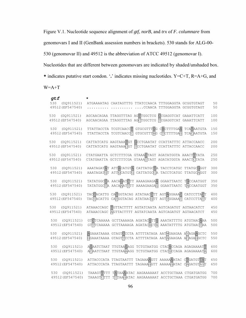
96
Figure V.1. Nucleotide sequence alignment of gtf, norB, and trx of F. columnare from
genomovars I and II (GenBank assession numbers in brackets). 530 stands for ALG-00-
530 (genomovar II) and 49512 is the abbreviation of ATCC 49512 (genomovar I).
Nucleotides that are different between genomovars are indicated by shaded/unshaded box.
* indicates putative start condon. ‘.’ indicates missing nucleotides. Y=C+T, R=A+G, and
W=A+T
gtf *530 (DQ911521) ATGAAAATAG CAATAGTTTG TTATCCAACA TTTGGAGGTA GCGGTGTAGT 5049512(EF547540) .......... .......... ....CCAACA TTTGGAGGTA GCGGTGTAGT 50
530 (DQ911521) AGCAACAGAA TTAGGTTTAG AGTTGGCTCG CCGAGGTCAT GAAATTCATT 100 49512(EF547540) AGCAACAGAA TTAGGTTTAG AGCTGGCTCG TCGAGGTCAT GAAATTCATT 100
530 (DQ911521) TTATTACCTA TCGTCAACCT GTGCGTTTGG CACTTTTGAA TCATAATGTA 15049512(EF547540) TTATTACCTA TCGTCAACCC GTGCGTTTAG CTCTTTTGAG TCACAATGTA 150
530 (DQ911521) CATTATCATG AAGTAAATGT GCCTGAATAT CCATTATTTC ATTACCAACC 20049512(EF547540) CATTATCATG AAGTAAACGT TCCTGAATAT CCATTATTTC ATTACCAACC 200
530 (DQ911521) CTATGAATTA GCTCTTTCGA GTAAACTAGT AGATATGGTA AAACTTTATA 25049512(EF547540) CTATGAATTA GCTCTTTCGA GTAAATTAGT AGATATGGTA AAACTCTATA 250
530 (DQ911521) AAATAGATAT ATTACATGTA CATTATGCTA TACCTCATGC TTATGCGGGT 30049512(EF547540) AAATAGATGT ATTGCATGTG CATTATGCCA TACCTCATGC TTATGCAGGT 300
530 (DQ911521) TATATGGCCA AACAGATCTT AAAAGAAGAA GGAATTAATC TGCCAATGGT 35049512(EF547540) TATATGGCTA AACAAATTTT AAAAGAAGAG GGAATTAATC TACCAATGGT 350
530 (DQ911521) TACCACATTG CACGGTACAG ATATAACCTT AGTAGGAAAC CATCCTTACT 40049512(EF547540) TACTACATTG CATGGTACAG ATATAACGTT AGTTGGAAAT CATCCTTATT 400
530 (DQ911521) ATAAACCAGC TGTTACTTTT AGTATCAATA AGTCAGATGT AGTAACATCT 45049512(EF547540) ATAAACCAGC AGTTACTTTT AGTATCAATA AGTCAGATGT AGTAACATCT 450
530 (DQ911521) GTCTCAAAAA GCTTAAAAGA AGATACGTTG AAATATTTTG ATGTAACAAA 50049512(EF547540) GTTTCAAAAA GCTTAAAAGA AGATACACTA AAATATTTTG ATGTAACTAA 500
530 (DQ911521) AGAAATAAAA GTAGTCCCTA ATTTTATAGA AATWGAAGAA AYAGAAGCTC 55049512(EF547540) GGAAATAAAA GTAGTTCCTA ATTTTATAGA AATAGAAGAA ACAGAGGCTC 550
530 (DQ911521) AGAATCTAAT TTGTAAGAGG TCTGTAATGG CTACACCAGA AGAGAAAATT 60049512(EF547540) ACAATCTAAT TTGTAAAAGG TCTGTAATGG CTACGCCAGA AGAGAAAATA 600
530 (DQ911521) ATTACCCATA TTAGTAATTT TAGAAAAGTT AAAAAAATAC CTGATGTTAT 65049512(EF547540) ATTACCCATA TTAGTAATTT TAGAAAGGTT AAAAAGATAC CAGATGTAGT 650
530 (DQ911521) TAAAGTTTTT TATAAAATAC AAGAAAAAAT ACCTGCTAAA CTGATGATGG 70049512(EF547540) TAAAGTCTTT TTTAAGATAC AAGAAAAAAT ACCTGCTAAA CTGATGATGG 700

97
530 (DQ911521) TGGGTGATGG ACCTGAAAAA GAAAAGGCAG AATGGCTTTG TAGAGAATTA 75049512(EF547540) TGG....... .......... .......... .......... .......... 750
530 (DQ911521) GGTATTGAAG AAAAAGTTAT TTTTTTTGGG AACCAGTCAT GA 792 49512(EF547540) .......... .......... .......... .......... .. 792
norB *530 (EQ911522) ATGCACGGAC ATCTAGCATT TTGGGGAGCA TACGCCATGA TTGTATTAGC 5049512(EF547542) .TGCACGGAC ATCTAGCATT TTGGGGAGCA TACGCCATGA TTGTATTAGC 50
530 (EQ911522) AATTATTAGT TATGCTATAC CTAATCTTAC AGGAAGAAAA AGATATGATT 10049512(EF547542) AATTATTAGT TATGCAATAC CTAATCTTAC AGGAAGAAAA AGATACGATT 100
530 (EQ911522) CAGTTACAGG ACGTATGGCA TTYTGGTTAT CAAATATTGG TATGTTAGGT 15049512(EF547542) CAGTTACAGG ACGTATGGCC TTTTGGTTAT CAAATATTGG TATGTTAGGT 150
530 (EQ911522) ATGACCACCG CTTTTGGGGT AGCTGGAGTA GCTCAAGTAT ATCTTGAAAG 20049512(EF547542) ATGACCACCG CTTTTGGGGT AGCTGGAGTC GCTCAAGTAT ACCTTGAAAG 200
530 (EQ911522) AAAGTTTAAA ATGGAGTTCA TGACCGTTCA GAATGAAATA GCTATTCACT 25049512(EF547542) AAAGTTTAAA ATGGAGTTTA TGACCGTTCA AARTGAAATA GCTATTCATT 250
530 (EQ911522) TTGTAGTATT ACTACTGTGT GCCACATTAT TTACTCTAGG AATYTCTTTA 30049512(EF547542) TTGTAGTACT GCTACTATGT GCCACATTAT TCACTATAGG AATTTCTTTA 300
530 (EQ911522) TACATATACG ATTTTATTAA ACATGGTAAA ACGAACGATG AGGCCATAAT 35049512(EF547542) TACATATACG ATTTTATTAA ACATGGTAAA ACGAACGATG AGG....... 350
530 (EQ911522) TGGCTAA 357 49512(EF547542) ....... 357
trx *530 (DQ911523) ATGGCATTAG CAATTACAGA TGCTACATTT GATGAGGTAG TATTACAATC 5049512(EF547543) .TGGCATTAG CAATTACAGA TGCTACATTT GATGAGGTAG TATTACAATC 50
530 (DQ911523) AGACAAACCT GTTTTAGTGG ATTTTTGGGC GGCTTGGTGT GGACCTTGTC 10049512(EF547543) AGACAAACCA GTTTTAGTGG ATTTTTGGGC AGCTTGGTGC GGGCCTTGTC 100
530 (DQ911523) GTATGGTAGG TCCCGTTATT GATGAAATTG CAACAGAATA TGAAGGAAAA 15049512(EF547543) GTATGGTAGG TCCCGTTATT GACGAAATTG CAACGGAATA TGAAGGAAAA 150
530 (DQ911523) GCAGTAATAG GTAAGGTAGA TGTCGATGCC AATCAAGAAT TTGCTGCAAA 20049512(EF547543) GCAGTAATTG GTAAGGTAGA TGTCGATGCC AATCAAGAGT TTGCTGCAAA 200
530 (DQ911523) GTACGGTGTT CGCAATATTC CTACTGTTTT GATTTTTCAA AATGGGGAAG 25049512(EF547543) GTATGGCGTA CGTAATATTC CTACTGTTTT GATTTTTCAA AATGGGGAAG 250
530 (DQ911523) TAGTGGGACG TCAGGTAGGT GTAGCTCCTA AAGATACTTA TGCTAAAGCA 30049512(EF547543) TAGTG..... .......... .......... .......... .......... 300
530 (DQ911523) ATTGATGCCT TATTGTAA 318 49512(EF547543) .......... ........ 318

98
Figure V. 2. hemH, norB, and trx gene expression of F. columnare strain ALG-00-530 by
reverse transcription PCR (RT-PCR). Expression patterns are shown under iron-limited
conditions in the presence of 2, 2-dipyridyl and in Shieh broth at 0 h, 2 h, 4 h, 6 h, 8 h,
and 24 h. 16S rRNA gene (16S) was used as an internal control.
16S
hemH
trx
norB
0 2 4 6 8 24 0 2 4 6 8 24
Shieh broth Iron-limited
Time (h)

99
VI. CONCLUSIONS
This dissertation focused on molecular characterization of the two main fish
pathogens, E. ictaluri and F. columnare, affecting Alabama aquaculture. The main
outcome of my work is the new genetic information that has been identified,
characterized and made available to the scientific community. My first objective was to
increase the number of known ribosomal sequences from these two pathogens in order to
identify new signature sequences for a PCR-based multiplex protocol. Although this
objective could not be fulfilled due to technical and practical reasons (see Chapter II), I
was able to identify a new IVS in the 23S rRNA gene of E. ictaluri. This is the first
report (Chapter III) of this type of sequence in the genus Edwardsiella. In relation to F.
columnare, an intraspecies characterization allowed me to analyze the avirulent mutant
currently used as a vaccine against columnaris disease. Not only can this mutant now be
tracked using fingerprinting methods, but I correlated its lack of virulence to changes in
the LPS structure (Chapter IV). In addition, by analyzing sequence data from a shotgun
library of F. columnare, I identified and characterized 4 putative virulence genes in this
species. I showed there is a deep division between genomovars I and II at the genomic
level. These results strongly encourage the use of both genomovars in future F.
columnare studies.
In brief, some of the main results from my dissertation can be summarized as
follows:

100
1) An intervening sequence (IVS) is present in the 23S rRNA gene of E. ictaluri but not
in E. tarda
2) E. ictaluri IVS was present in the majority of the rrl operons
3) E. ictaluri IVS shared 97% nucleotide identity with S. typhimurium helix-45 IVS
4) E. ictaluri is closely related to E. tarda based on 23S rDNA analysis; however, both
species are phylogenetically distant from the ‘core’ members within the
Enterobacteriaceae family
5) The modified F. columnare live vaccine FC-RR lacks the higher molecular weight
bands in the lipopolysaccharide (LPS)
6) gtf, hemH, norB, and trx gene sequences divide F. columnare strains into two groups
that correspond with genomovar ascription
7) Expression profiles of genes gtf, hemH, norB, and trx varied among different F.
columnare strains under identical culture conditions
Contribution
The work described in this dissertation opens several venues for future studies.
New phenotypic and genotypic data were generated for E. ictaluri and F. columnare.
Effectively utilizing the data presented in the dissertation may be relevant to E. ictaluri
IVS evolution, LPS structure definition in F. columnare, iron acquisition system in F.
columnare, expression of putative virulence factors in mutant strains of F. columnare,
new virulence genes identification, or evidence collection for defining subspecies in F.
columnare.
E. ictaluri habors an IVS in 23S rRNA gene and such IVS exhibits 97% similarity
to Salmonella typhimurium. As is known, S. typhimurium can infect many

101
mammalian species. Did this IVS get transferred between Salmonella to E.
ictaluri? Why does E. tarda not harbor this IVS, although E. tarda and E. ictaluri
belong to the same genus, and both can infect fish and display similar/identical
sequences in ribosomal gene? The discovery of IVS raises questions about
Edwardsiella spp. evolution.
Lipopolysaccharide has been well characterized as a virulence factor in a variety
of bacterial pathogens. In this work, LPS profiles are different between virulent
and avirulent strains of F. columnare. Additionally, F. columnare does not
display the typical ladder-like pattern of LPS. Further work is needed to show
where the difference is located in the genome and the importance of LPS in F.
columnare pathogenesis. A functional study of LPS will also improve our
understanding of F. columnare pathogenicity.
An iron acquisition system has not been identified in F. columnare. The results of
the dissertation suggested that ferrochelatase, an important iron importing enzyme,
was not involved in iron absorption in F. columnare. Since iron is such an
important element in bacterial growth, survival, and virulence, it is logical that F.
columnare expresses uptake systems that mediate iron acquisition from the
environment and from a catfish.
Putative virulence gene expression in the mutant strain of FC-RR could contribute
to a future research project. FC-RR is a modified live vaccine against F.
columnare. In the current study, we showed that this strain is phenotypically
different from the other virulent strains; however, we did not include this strain in
the putative virulence gene expression study. It may be interesting to characterize

102
gene expression patterns in FC-RR in response to iron-limitation and presence of
fish skin.
Whole genome sequencing of F. columnare is ongoing at Mississippi State
University. However, the strain selected for this project is a genomovar I isolate.
The dissertation discovered that F. columnare genomovar I and II differed in gene
sequences. It seems necessary to sequence strains from both genomovars.
Comparison of whole genomes between the two genomovars should provide some
answers for columnaris pathogenesis and guidelines for vaccine development.
Since a shotgun genomic library of F. columnare is already available, further data
mining could lead to the identification of new virulence genes.
The data presented in the dissertation showed that gtf, hemH, norB, and trx
sequences divided F. columnare strains into two populations which correspond to
genomovars. Based on my data and other information generated by our group, we
see a deep division within the species at the genetic level that is also correlated to
host-species-specificity and virulence properties. These findings point towards a
division of F. columnare into two subspecies. We need more data to support this
hypothesis including DNA:DNA hybridization studies.
Publications
Zhang, Y., and Covadonga R. Arias. 2007. Identification and characterization of
an intervening sequence within the 23S ribosomal RNA genes of
Edwardsiella ictaluri. Systematic and Applied Microbiology 30:93-101.
Zhang, Y., Covadonga R. Arias, Craig A. Shoemaker and Phillip H. Klesius. 2006.
Comparison of lipopolysaccharide and protein profiles between

103
Flavobacterium columnare strains from different genomovars. Journal of
Fish Diseases 29:657-663.
Zhang, Y., Craig A. Shoemaker, Phillip H. Klesius, and Covadonga R. Arias.
Identification and characterization of gtf, norB, and trx gene in
Flavobacterium columnare. Journal of Fish Diseases (under review).

104
CUMULATIVE BIBLIOGRAPHY
Abbas, A. K., and A. H. Lichtman 2000. Cellular and molecular immunology. Saunders,
Philadelphia, PA.
Ahmad, S., W. G. Weisburg, and M. A. Jensen. 1990. Evolution of aromatic amino acid
biosynthesis and application to the fine-tune phylogenetic positioning of enteric
bacteria. Journal of Bacteriology 172:1051-1061.
Ainsworth, A. J., G. Capley, P. Waterstreet, and D. Munson. 1986. Use of monoclonal
antibodies in the indirect fluorescent antibody technique for the diagnosis of
Edwardsiella ictaluri. Journal of Fish Diseases 9:439-444.
Almiron, M., M. Martinez, N. Sanjuan, and R. A. Ugalde. 2001. Ferrochelatase is present
in Brucella abortus and is critical for its intracellular survival and virulence.
Infection and Immunity 69:6225-6230.
Altinok, I., and J. M. Grizzle. 2001. Effects of low salinities on Flavobacterium
columnare infection of euryhaline and freshwater stenohaline fish. Journal of Fish
Diseases 24:361-367.
Altschul, S. F., W. Gish, W. Miller, E. W. Myers, and D. J. Lipman. 1990. Basic local
alignment search tool. Journal of Molecular Biology 215:403-410.

105
Amaro, C., B. Fouz, E. Biosca, E. Marco-Noales, and R. Collado. 1997. The
lipopolysaccharide O side chain of Vibrio vulnificus serogroup E is a virulence
determinant for eels. Infection and Immunity 65:2475-2479.
Anacker, R. L., and E. J. Ordal. 1959. Studies on the myxobacterium Chondrococcus
columnaris I. serological typing. Journal of Bacteriology 78:25-32.
Aoki, T., T. Sakaguchi, and T. Kitao. 1987. Multiple drug-resistant plasmids from
Edwardsiella tarda in eel culture ponds. Nippon Suisan Gakkaish 53:1821-1825.
Aoki, T., and I. Hirono. 1995. Detection of the fish-pathogenic bacteria Edwardsiella
tarda by polymerase chain reaction. Proceedings of the International Symposium
on Biotechnology Applications in Aquaculture, Asian Fisheries Society Special
Publication No.10:135-146.
Arias, C. R., E. Garay, and R. Aznar. 1995. Nested PCR method for rapid and sensitive
detection of Vibrio vulnificus in fish, sediments, and water. Applied and
Environmental Microbiology 61:3476-3478.
Arias, C. R., L. Verdonck, J. Swings, E. Garay, and R. Aznar. 1997a. Intraspecific
differentiation of Vibrio vulnificus biotypes by amplified fragment length
polymorphism and ribotyping. Applied and Environmental Microbiology
63:2600-2606.

106
Arias, C. R., L. Verdonck, J. Swings, R. Aznar, and E. Garay. 1997b. A polyphasic
approach to study the intraspecific diversity amongst Vibrio vulnificus isolates.
Systematic and Applied Microbiology 20:622-633.
Arias, C. R., C. A. Shoemaker, J. J. Evans, and P. H. Klesius. 2003. A comparative study
of Edwardsiella ictaluri parent (EILO) and E. ictaluri rifampicin-mutant (RE-33)
isolates using lipopolysaccharides, outer membrane proteins, fatty acids, Biolog,
API 20E and genomic analyses. Journal of Fish Diseases 26:415-21.
Arias, C. R., T. L. Welker, C. A. Shoemaker, J. W. Abernathy, and P. H. Klesius. 2004.
Genetic fingerprinting of Flavobacterium columnare isolates from cultured fish.
Journal of Applied Microbiology 97:421-428.
Austin, B., and D. A. Austin. 1999. Bacterial fish pathogens: disease of farmed and wild
fish. Springer, New York.
Bader, J. A., and E. J. Shotts. 1998. Identification of Flavobacterium and Flexibacter
species by species-specific polymerase chain reaction primers to the 16S
ribosomal RNA gene. Journal of Aquatic Animal Health 10:311-319.
Bader, J. A., K. E. Nusbaum, and C. A. Shoemaker. 2003a. Comparative challenge model
of Flavobacterium columnare using abraded and unabraded channel catfish,
Ictalurus punctatus (Rafinesque). Journal of Fish Diseases 26:461-7.

107
Bader, J. A., C. A. Shoemaker, and P. H. Klesius. 2003b. Rapid detection of columnaris
disease in channel catfish (Ictalurus punctatus) with a new species-specific 16S
rRNA gene-based PCR primer for Flavobacterium columnare. Journal of
Microbiological Methods 52:209-220.
Bader, J. A., C. A. Shoemaker, and P. H. Klesius. 2005. Production, characterization and
evaluation of virulence of an adhesion defective mutant of Flavobacterium
columnare produced by beta-lactam selection. Letters in Applied Microbiology
40:123-127.
Baird, K. D., H. M. Chikarmane, R. Smolowitz, and K. R.Uhlinger. 2003. Detection of
Edwardsiella infections in Opsanus tau by polymerase chain reaction. Biological
Bulletin 205:235-236.
Bernardet, J. F., and P. A. D. Grimont. 1989. Deoxyribonucleic acid relatedness and
phenotypic characterization of Flexibacter columnaris sp. nov., nom. rev.,
Flexibacter psychrophilus sp. nov., nom. rev., and Flexibacter maritimus
Wakabayashi, Hikida, and Masumura 1986. International Journal of Systematic
Bacteriology 39:346-354.

108
Bernardet, J. F., P. Segers, M. Vancanneyt, F. Berthe, K. Kersters, and P. Vandamme.
1996. Cutting a gordian knot: Emended classification and description of the genus
Flavobacterium, emended description of the family Flavobacteriaceae, and
proposal of Flavobacterium hydatis nom. nov. (Basonym, Cytophaga aquatilis
Strohl and Tait 1978). International Journal of Systematic Bacteriology 46:128-
148.
Bertolini, J. M., and J. S. Rohovec. 1992. Electrophoretic detection of proteases from
different Flexibacter columnaris strains and assessment of their variability.
Diseases of Aquatic Organisms 12:121-128.
Bilodeau, A. L., G. C. Waldbieser, J. S. Terhune, D. J. Wise, and W. R. Wolters. 2003. A
real-time polymerase chain reaction assay of the bacterium Edwardsiella ictaluri
in channel catfish. Journal of Aquatic Animal Health 15:80-86.
Biosca, I. G., H. Llorens, E. Garay, and C. Amaro. 1993. Presence of a capsule in Vibrio
vulnificus biotype 2 and its relationship to virulence for eels. Infection and
Immunity 61:1611-1618.
Bjur, E., S. Eriksson-Ygberg, F. Aslund, and M. Rhen. 2006. Thioredoxin 1 promotes
intracellular replication and virulence of Salmonella enterica serovar
typhimurium. Infection and Immunity 74:5140-5151.
Booth, N. J. 2005. The role of urease in the pathogenesis of Edwardsiella ictaluri.
Doctoral dissertation. Louisiana State University, Baton Rounge, Louisiana, USA.

109
Bullock, G. L., T. C. Hsu, and E. B. Shotts. 1986. Columnaris disease of fishes. Fish
disease leaflet 72. U. S. Fish and Wildlife Service, Washington, D. C.
Burgin, A. B., K. Parodos, D. J. Lane, and N. R. Pace. 1990. The excision of intervening
sequences from Salmonella 23S rRNA. Cell 60:405-414.
Chen, C. R., H. Y. Chung, and G. H. Kuo. 1982. Studies on the pathogenicity of
Flexibacter columnaris I. effect of dissolved oxygen and ammonia on the
pathogenicity of Flexibacter columnaris to eel (Anguilla japonica). CAPD
fisheries serials no. 8 Reports on Fish Disease Research 4:57-61.
Chen, J. D., and S. Y. Lai. 1998. PCR for direct detection of Edwardsiella tarda from
infected fish and environmental water by application of the hemolysin gene.
Zoological Studies 37:169-176.
Chen, Y. C., M. C. Chang, Y. C. Chuang, and C. L. Jeang. 2004. Characterization and
virulence of hemolysin III from Vibrio vulnificus. Current Microbiology 49:175-9.
Chenna, R., H. Sugawara, T. Koike, R. Lopez, T. J. Gibson, D. G. Higgins, and J. D.
Thompson. 2003. Multiple sequence alignment with the clustal series of programs.
Nucleic Acids Research 31:3497-3500.
Chowdhury, B. R., and H. Wakabayashi. 1990. Detection of Flexibacter columnaris from
fish and water using indirect fluorescent antibody technique. Bangladesh Journal
of Microbiology 7:25-35.
Crumlish, M., T. T. Dung, J. F. Turnbull, N. T. N. Ngoc, and H. W. Ferguson. 2002.
Identification of Edwardsiella ictaluri from diseased freshwater catfish,

110
Pangasius hypophthalmus (Sauvage), cultured in the Mekong Delta, Vietnam.
Journal of Fish Diseases 25:733-736.
Darwish, A. M., A. A. Ismaiel, J. C. Newton, and J. Tang. 2004. Identification of
Flavobacterium columnare by a species-specific polymerase chain reaction and
renaming of ATCC43622 strain to Flavobacterium johnsoniae. Molecular and
Cellular Probes 18:421-427.
Darwish, A. M., and A. A. Ismaiel. 2005. Genetic diversity of Flavobacterium columnare
examined by restriction fragment length polymorphism RNA gene and
sequencing of the 16S ribosomal RNA gene and the 16S-23S rDNA spacer.
Molecular and Cellular Probes 19:267-274.
Davis, H. S. 1922. A new bacterial disease of fresh-water fishes. Bulletin of the U. S.
Bureau of Fisheries 38:261-280.
Decostere, A., F. Haesebrouck, and L. A. Devriese. 1997. Shieh medium supplemented
with tobramycin for selective isolation of Flavobacterium columnare (Flexibacter
columnaris) from diseased fish. Journal of Clinical Microbiology 35:322-324.
Decostere, A., F. Haesebrouck, G. Charlier, and R. Ducatelle. 1999a. The association of
Flavobacterium columnare strains of high and low virulence with gill tissue of
black mollies (Poecilia sphenops). Veterinary Microbiology 67:287-298.
Decostere, A., F. Haesebrouck, J. Turnbull, and G. Charlier. 1999b. Influence of water
quality and temperature on adhesion of high and low virulence Flavobacterium
columnare strains to isolated gill arches. Journal of Fish Diseases 22:1-11.

111
Dietmaier, W., and F. Hofstadter. 2001. Detection of microsatellite instability by real
time PCR and hybridization probe melting point analysis. Laboratory
Investigation 81:1453-1456.
Evguenieva-Hackenberg, E., and G. Klug. 2000. RNase III processing of intervening
sequences found in helix 9 of 23S rRNA in the alpha subclass of Proteobacteria.
Journal of Bacteriology 182:4719-4729.
Ewing, W. H., A. C. McWhorter, M. R. Escobar, and A. H. Lubin. 1965. Edwardsiella, a
new genus of Enterobacteriaceae based on a new species, E. tarda. International
Bulletin of Bacteriological Nomenclature and Taxonomy 15:33-38.
Francino, M. P., S. R. Santos, and H. Ochman. 2003. Phylogenetic relationships of
bacteria with special reference to endosymbionts and enteric species. In M.
Dworkin, editor. Prokaryotes: A handbook on the biology of Bacteria:
Ecophysiology, isolation, identification, applications, 3rd edition. Springer-
Verlag, New York.
Garnjobst, L. 1945. Cytophaga columnaris (David) in pure culture: a myxobacterium
pathogenic to fish. Journal of Bacteriology 49:113-128.
Griffin, B. R. 1991. Characteristics of a chondroitin AC lyase produced by Cytophaga
columnaris. Transactions of the American Fisheries Society 120:391-395.
Griffin, B. R. 1992. A simple procedure for identification of Cytophaga columnaris.
Journal of Aquatic Animal Health 4:63-66.

112
Grimont, P. A. D., F. Grimont, C. Richard, and R. Sakazaki. 1980. Edwardsiella
hoshinae, a new species of Enterobacteriaceae. Current Microbiology 4:347-351.
Hanson, L. A., and J. M. Grizzle. 1985. Nitrite induced predisposition of channel catfish
Ictalurus punctatus to bacterial diseases. Progressive Fish Culturist 47:98-101.
Hanson, L. A., and W. A. Rogers. 1989. Enzyme immunoassay identification of
Edwardsiella ictaluri in decomposing channel catfish. Journal of the World
Aquaculture Society 20:279-280.
Hawke, J. P. 1979. A bacterium associated with disease of pond cultured channel catfish,
Ictalurus punctatus. Journal of the Fisheries Research Board of Canada 36:1508-
1512.
Hawke, J. P., A. C. McWhorter, A. G. Steigerwalt, and D. J. Brenner.1981. Edwardsiella
ictaluri sp. nov., the causative agent of enteric septicemia of catfish. International
Journal of Systematic Bacteriology 31:396-400.
Hawke, J. P., and R. L. Thune. 1992. Systemic isolation and antimicrobial susceptibility
of Cytophaga columnaris from commercially reared channel catfish. Journal of
Aquatic Animal Health 4:109-113.
Hawke, J. P., R. M. Durborow, R. L. Thune, and A. C. Camus. 1998. ESC-enteric
septicemia of catfish. Publication No. 477, Southern Regional Aquaculture
Center, Stoneville, Mississippi.
113
Hawke, J. P. 2003. Edwardsiella tarda septicemia. In AFS-FHS (American Fisheries
Society-Fish Health Section). FHS blue book: suggested procedures for the
detection and identification of certain finfish and shellfish pathogens 2006 edition.
AFS-FHS, Bethesda, Maryland.
Heid, C. A., J. Stevens, K. J. Livak, and P. M. Williams. 1996. Real time quantitative
PCR. Genome Research 6:986-994.
Hiney, M. P., and P. R. Smith. 1998. Validation of polymerase chain reaction-based
techniques for proxy detection of bacterial fish pathogens: framework, problems
and possible solutions for environmental applications. Aquaculture 162:41-68.
Hirono, I., N. Tange, and T. Aoki. 1997. Iron-regulated hemolysin gene from
Edwardsiella tarda. Molecular Microbiology 24:851-856.
Holmes, K., F. Mulholland, B. M. Pearson, C. Pin, J. McNicholl-Kennedy, J. M. Ketley,
and J. M. Wells. 2005. Campylobacter jejuni gene expression in response to iron
limitation and the role of Fur. Microbiology 151:243-257.
Hoshina, T. 1962. On a new bacterium, Paracolobactrum anguillimortiferum sp. Bulletin
of the Japanese Society of Scientific Fisheries 28:162-164.
Jaegera, T., H. Buddea, L. Flohe, U. Mengeb, M. Singhb, M. Trujilloc, and R. Radic.
2004. Multiple thioredoxin-mediated routes to detoxify hydroperoxides in
Mycobacterium tuberculosis. Archives of Biochemistry and Biophysics 423:182-
191.
Janda, J. M., S. L. Abbott, S. Kroske-Bystrom, W. K. Cheung, C. Powers, R. P. Kokka,
and K. Tamura. 1991. Pathogenic properties of Edwardsiella species. Journal of
Clinical Microbiology 29:1997-2001.

114
Jin, D. J., and C. A. Gross. 1988. Mapping and sequencing of mutations in the
Escherichia coli rpoB gene that lead to rifampicin resistance. Journal of
Molecular Biology 202:45-58.
Jukes, T. H., and C. R. Cantor. 1969. Evolution of protein molecules. Pages 21-132 in H.
N. Munro, editor. Mamalian protein metabolism. Academic Press, New York.
Kasornchandra, J., W. A. Rogers, and J. A. Plumb. 1987. Edwardsiella ictaluri from
walking catfish, Clarias batrachus L., in Thailand. Journal of Fish Diseases
10:137-138.
Kent, M. L., and J. M. Lyons. 1982. Edwardsiella ictaluri in the green knife fish,
Eigemannia (sic) virescens. Fish Health News: a service to the field of fish health
research 11:ii.
Keskin, O., S. Secer, M. Izgur, S. Turkyilmaz, and R. Mkakosya. 2004. Edwardsiella
ictaluri infection in rainbow trout (Oncorhynchus mykiss). Turkish Journal of
Veterinary and Animal Sciences 28:649-653.
Kimura, A., and E. J. Hansen. 1986. Antigenic and phenotypic variations of Haemophilus
influenzae type b lipopolysaccharide and their relationship to virulence. Infection
and Immunity 51:69-79.
Kirschbaum, T. M., and R. F. Gotte. 1993. Rifampicin resistance of Mu lysogenic strains
by rpoB mutations. Biological Chemistry Hoppe-Seyler 374:657-64.
Klesius, P. H. 1990. Effect of size and temperature on the quantity of immunoglobulin in
channel catfish, Ictalurus punctatus. Veterinary Immunology and
Immunopathology 24:187-95.

115
Klesius, P. H., K. Johnson, R. Durborow, and S. Vinitnantharat. 1991. Development and
evaluation of an ELISA for catfish serum antibody to Edwardsiella ictaluri.
Journal of Aquatic Animal Health 3:94-99.
Klesius, P. H., and C. A. Shoemaker. 1997. Heterologous isolates challenge of channel
catfish, Ictalurus punctatus, immune to Edwardsiella ictaluri. Aquaculture
157:147-155.
Klesius, P. H., and C. A. Shoemaker. 1999. Development and use of modified live
Edwardsiella ictaluri vaccine against enteric septicemia of catfish. Advances in
Veterinary Medicine 41:523-37.
Klesius, P. H., J. Lovy, J. J. Evans, E. Washuta, and C. R. Arias. 2003. Isolation of
Edwardsiella ictaluri from tadpole madtom, Noturus gyrinus in a southwestern
New Jersey River. Journal of Aquatic Animal Health 15:295-301.
Krieg, N. R., and J. G. Holt. 1984. Bergey's Manual of Systematic Bacteriology.
Williams and Wilkins, Baltimore, Maryland.
Laemmli, U. K. 1970. Cleavage of structural proteins during the assembly of the head of
bacteriophage T4. Nature 227:680-685.
Latifi, A., R. Jeanjean, S. Lemeille, M. Havaux, and C. C. Zhang. 2005. Iron starvation
leads to oxidative stress in Anabaena sp. strain PCC 7120. Journal of
Bacteriology 187:6596–6598.
Lawrence, M. L., and M. M. Banes. 2005. Tissue persistence and vaccine efficacy of an
O polysaccharide mutant strain of Edwardsiella ictaluri. Journal of Aquatic
Animal Health 17:228-232.

116
Leadbetter, E. R. 1974. Genus II. Flexibacter Soriano 1945, 92, Lewin 1969, 192 emend.
mut. char. Pages 105-107 in R. E. Buchanan and N. E. Gibbons, editors. Bergey's
manual of determinative bacteriology, 8th edition. Williams and Wilkins,
Baltimore, Maryland.
Lee, C. C., S. P. Craig, and A. E. Eakin. 1998. A single amino acid substitution in the
human and a bacterial hypoxanthine phosphoribosyltransferase modulates
specificity for the binding of guanine. Biochemistry 37:3491-3498.
Loeb, M. 1995. Ferrochelatase activity and protoporphyrin IX utilization in Haemophilus
influenzae. Journal of Bacteriology 177:3613-3615.
Loisel-Meyer, S., M. P. Jimenez de Bagues, E. Basseres, J. Dornand, S. Kohler, J. P.
Liautard, and V. Jubier-Maurin. 2006. Requirement of norD for Brucella suis
virulence in a murine model of in vitro and in vivo infection. Infection and
Immunity 74:1973-1976.
MacLean, L. L., E. Vinogradov, E. M. Crump, M. B. Perry, and W. W. Kay. 2001. The
structure of the lipopolysaccharide O-antigen produced by Flavobacterium
psychrophilum (259-93). European Journal of Biochemistry 268:2710-2716.
MacLean, L. L., M. B. Perry, E. M. Crump, and W. W. Kay. 2003. Structural
characterization of the lipopolysaccharide O-polysaccharide antigen produced by
Flavobacterium columnare ATCC 43622. European Journal of Biochemistry
270:3440-3446.
Madigan, M. T., J. M. Martinko, and J. Parker 2003. Brock biology of microorganisms.
Prentice Hall, Upper Saddle River, New Jersey.

117
Makela, P., V. Valtonen, and M. Valtonen. 1973. Role of O-antigen (lipopolysaccharide)
factors in the virulence of Salmonella. The Journal of Infectious Diseases 128:81-
85.
Mattatall, N. R., and K. E. Sanderson. 1996. Salmonella typhimurium LT2 possesses
three distinct 23S rRNA intervening sequences. Journal of Bacteriology
178:2272-2278.
McCarthy, D. H. 1975. Columnaris disease. Journal of Institute Fisheries Management
6:44-47.
McGee, D. J., S. Kumar, R. J. Viator, J. R. Bolland, J. Ruiz, D. Spadafora, T. L.
Testerman, D. J. Kelly, L. K. Pannell, and H. J. Windle. 2006. Helicobacter pylori
thioredoxin is an arginase chaperone and guardian against oxidative and
nitrosative stresses. The Journal of Biological Chemistry 281:3290-3296.
Medearis, D. N., Jr., B. M. Camitta, and E. C. Heath. 1968. Cell wall composition and
virulence in Escherichia coli. Journal of Experimental Medicine 128:399-414.
Meyer, F. P., and G. L. Bullock. 1973. Edwardsiella tarda a new pathogen of channel
catfish Ictalurus punctatus. Applied Microbiology 25:155-156.
Miller, W., K. Pabbaraju, and K. E. Sanderson. 2000. Fragmentation of 23S rRNA in
strains of Proteus and Providencia results from intervening sequences in the rrn
(rRNA) genes. Journal of Bacteriology 182:1109-1147.
Miyazaki, T. 1985. Comparative histopathology of Edwardsiellosis in fishes. Fish
Pathology 23:219-227.
Montaraz, J. A., and A. J. Winter. 1986. Comparison of living and nonliving vaccines for
Brucella abortus in BALB/c mice. Infection and Immunity 53:245-51.

118
Morrison, E. E., and J. A. Plumb. 1994. Olfactory organ of channel catfish as a site of
experimental Edwardsiella ictaluri infection. Journal of Aquatic Animal Health
6:101-109.
Mullis, K., and F. Faloona. 1987. Specific synthesis of DNA in vitro via a polymerase-
catalyzed chain reaction. Methods in Enzymology 155:335-350.
Narimatsu, M., Y. Noiri, S. Itoh, N. Noguchi, T. Kawahara, and S. Ebisu. 2004. Essential
role for the gtfA gene encoding a putative glycosyltransferase in the adherence of
Porphyromonas gingivalis. Infection and Immunity 72:2698-2702.
Newton, J. C., L. G. Wolfe, J. M. Grizzle, and J. A. Plumb. 1989. Pathology of
experimental enteric septicemia in channel catfish Ictalurus punctatus Rafinesque
following immersion-exposure to Edwardsiella ictaluri. Journal of Fish Diseases
12:335-348.
Newton, J. C., T. M. Wood, and M. M. Hartley. 1997. Isolation and partial
characterization of extracellular proteases produced by isolates of Flavobacterium
columnare derived from channel catfish. Journal of Aquatic Animal Health 9:75-
Olivares-Fuster, O., C. R. Arias, C. A. Shoemaker, and P. H. Klesius 2007 Molecular
typing of Flavobacterium columnare isolates by single-strand conformation
polymorphism analysis (SSCP). FEMS Microbiology Letters. (in press)
Ordal, E. J., and R. R. Rucker. 1944. Pathogenic myxobacteria. Proceedings of the
Society for Experimental Biology and Medicine 56:15-18.
Owens, D. R., S. L. Nelson, and J. B. Addison. 1974. Isolation of Edwardsiella tarda
from swine. Applied and Environmental Microbiology 27:703-705.

119
Pabbaraju, K., W. Miller, and K. E. Sanderson. 2000. Distribution of intervening
sequences in the genes for 23S rRNA and rRNA fragmentation among strains of
the Salmonella reference collection B (SARB) and SARC sets. Journal of
Bacteriology 182:1923-1929.
Pacha, R. E., and E. J. Ordal. 1970. Myxobacterial diseases of salmonids. Pages 243-257
in S. F. Snieszko, editor. A Symposium on Diseases of Fishes and Shellfishes.
American Fisheries Society, Washington, D. C.
Panangala, V. S., V. L. van Santen, C. A. Shoemaker, and P. H. Klesius. 2005. Analysis
of 16S-23S intergenic spacer region of the rRNA operons in Edwardsiella ictaluri
and Edwardsiella tarda isolates from fish. Journal of Applied Microbiology
99:657-669.
Panangala, V. S., R. A. Shelby, C. A. Shoemaker, P. H. Klesius, A. Mitra, and E. E.
Morrison. 2006. Immunofluorescent test for simultaneous detection of
Edwardsiella ictaluri and Flavobacterium columnare. Diseases of Aquatic
Organisms 68:197-207.
Panangala, V., C. A. Shoemaker, V. Van Santan, K. Dybvig, and P. H. Klesius. 2007.
Multiplex-PCR for simultaneous detection of three fish-pathogenic bacteria,
Edwardsiella Ictaluri, Flavobacterium columnare and Aeromonas hydrophila.
Diseases of Aquatic Organisms 74:199-203.
Philippot, L. 2005. Denitrification in pathogenic bacteria: for better or worst? Trends in
Microbiology 13:191-192.
Plumb, J. A., and D. J. Sanchez. 1983. Susceptibility of five species of fish to
Edwardsiella ictaluri. Journal of Fish Diseases 6:261-266.

120
Plumb, J. A. 1993. Edwardsiella septicemia. Pages 60-79 in V. Inglis, R. J. Roberts, and
N. R. Bromage, editors. Bacterial diseases of fish. Halsted Press, New York.
Plumb, J. A. 1999. Health maintenance and principal microbial diseases of cultured fish.
Iowa State University Press, Ames, Iowa.
Plumb, J. A., and C. A. Shoemaker. 2006. Enteric septicemia of catfish in aquaculture
compendium. CAB international, Wallingford, U. K.
Pronk, L. M., and K. E. Sanderson. 2001. Intervening sequences in rrl genes and
fragmentation of 23S rRNA in genera of the family Enterobacteriaceae. Journal
of Bacteriology 183:5782-5787.
Rajakumar, K., B. Jost, C. Sasakawa, N. Okada, M. Yoshikawa, and B. Adler. 1994.
Nucleotide sequence of the rhamnose biosynthetic operon of Shigella flexneri 2a
and role of lipopolysaccharide in virulence. Journal of Bacteriology 176:2362-
2373.
Reichenbach, H. 1989. Genus I. Cytophaga Winogradsky 1929, 577, ALemend. Pages
2015-2046 in J. T. Staley, M. P. Bryant, N. Pfennig, and J. G. Holt, editors.
Bergey's manual of systematic bacteriology. Williams and Wilkins, Baltimore,
Maryland.
Reid, W. S., and J. A. Boyle. 1989. Plasmid homologies in Edwardsiella ictaluri. Applied
and Environmental Microbiology 55:3253-3255.
Rucker, R. R., B. J. Earp, and E. J. Ordal. 1953. Infectious diseases of Pacific salmon.
Transactions of the American Fisheries Society 83:297-312.
Saitou, N., and M. Nei. 1987. The neighbor-joining method: a new method for
reconstructing phylogenetic trees. Molecular Biology and Evolution 4:406-425.

121
Sakazaki, R., and Y. Murata. 1962. The new group of the Enterobacteriaceae, the
Asakusa group. Japanese Journal of Bacteriology 17:616-620.
Sambrook, J., and D. W. Russell. 2001. Molecular cloning: a laboratory manual. Cold
Spring Harbor Laboratory Press, New York.
Sanders, J. E., R. A. Holt, and J. L. Fryer. 1976. Serological comparison of Flexibacter
columnaris isolates using rabbit and rainbow trout (Salmo gairdneri) antisera.
Journal of the Fisheries Research Board of Canada 33:1386-1388.
Schneck, J. L., and L. F. Caslake. 2006. Genetic diversity of Flavobacterium columnare
isolated from fish collected from warm and cold water. Journal of Fish Diseases
29:245-248.
Schurig, G. G., R. M. Roop, T. Bagchi, S. Boyle, D. Buhrman, and N. Sriranganathan.
1991. Biological properties of RB51; a stable rough strain of Brucella abortus.
Veterinary Microbiology 28:171-188.
Sechter, I., M. Shmilovitz, G. Altmann, R. Sellgmann, B. Kretzer, I. Braunstein, and B.
Gericheter. 1983. Edwardsiella tarda isolated in Israel between 1961 and 1980.
Journal of Clinical Microbiology 17:669-67.
Sharma, V. K., Y. K. Kaura, and I. P. Singh. 1974. Frogs as carriers of Salmonella and
Edwardsiella. Antonie Van Leeuwenhoek 40:171-175.
Shoemaker, C. A., P. H. Klesius, and J. M. Bricker. 1999. Efficacy of a modified live
Edwardsiella ictaluri vaccine in channel catfish as young as seven days post
hatch. Aquaculture 176:189-193.
Shoemaker, C. A., P. H. Klesius, C. Lim, and M. Yildirim. 2003a. Feed deprivation of
channel catfish, Ictalurus punctatus (Rafinesque), influences organosomatic

122
indices, chemical composition and susceptibility to Flavobacterium columnare.
Journal of Fish Diseases 26:553-61.
Shoemaker, C. A., R. Shelby, and P. H. Klesius. 2003b. Development of an indirect
ELISA to detect humoral response to Flavobacterium columnare infection of
channel catfish, Ictalurus punctatus (Rafinesque). Journal of Applied Aquaculture
14:43-52.
Shoemaker, C. A., C. R. Arias, P. H. Klesius, and T. L. Welker. 2005a. Technique for
identifying Flavobacterium columnare using whole-cell fatty acid profiles.
Journal of Aquatic Animal Health 17:267-274.
Shoemaker, C. A., P. H. Klesius, and J. Evans. 2005b. Modified live Flavobacterium
columnare against columnaris disease in fish. U. S. patent application 6881412
B1. 2005-04-19.
Shotts, E., and W. Waltman. 1990. A medium for the selective isolation of Edwardsiella
ictaluri. Journal of Wildlife Diseases 26:214-218.
Song, Y. L., J. L. Fryer, and J. S. Rohovec. 1988. Comparison of gliding bacteria isolated
from fish in North America and other areas of the Pacific rim. Fish Pathology
23:197-202.
Soule, M., K. Cain, S. LaFrentz, and D. R. Call. 2005. Combining suppression
subtractive hybridization and microarrays to map the intraspecies phylogeny of
Flavobacterium psychrophilum. Infection and Immunity 73:3799-3802.
Speyerer, P. D., and J. A. Boyle. 1987. The plasmid profile of Edwardsiella ictaluri.
Journal of Fish Diseases 10:461-470.

123
Srinivasa-Rao, P. S., T. M. Lim, and K. Y. Leung. 2003. Functional genomics approach
to the identification of virulence genes involved in Edwardsiella tarda
pathogenesis. Infection and Immunity 71:1343-1351.
Stanley, L. A., J. S. Hudson, T. E. Schwedler, and S. S. Hayasaka. 1994. Extracellular
products associated with virulent and avirulent strains of Edwardsiella ictaluri
from channel catfish. Journal of Aquatic Animal Health 6:36–43.
Stringer-Roth, K. M., W. Yunghans, and L. F. Caslake. 2002. Differences in chondroitin
AC lyase activity of Flavobacterium columnare isolates. Journal of Fish Diseases
25:687-691.
Suomalainen, L. R., M. A. Tiirola, and E. T. Valtonen. 2005. Influence of rearing
conditions on Flavobacterium columnare infection of rainbow trout,
Oncorhynchus mykiss (Walbaum). Journal of Fish Diseases 28:271–277.
Suomalainen, L. R., M. Tiirola, and E. T. Valtonen. 2006. Chondroitin AC lyase activity
is related to virulence of fish pathogenic Flavobacterium columnare. Journal of
Fish Diseases 29:757-763.
Teska, J. D. 1993. Assay to evaluate the reaction kinetics of chondroitin AC lyase
produced by Cytophaga columnaris. Journal of Aquatic Animal Health 5:259-264.
Thomas-Jinu, S., and A. E. Goodwin. 2004. Morphological and genetic characteristics of
Flavobacterium columnare isolates: correlations with virulence in fish. Journal of
Fish Diseases 27:29-35.
Thune, R. L., J. P. Hawke, and M. C. Johnson. 1994. Studies on vaccination of channel
catfish, Ictalurus punctatus, against Edwardsiella ictaluri. Journal of Applied
Aquaculture 3:11-23.

124
Toyama, T., K. Kita-Tsukamoto, and H. Wakabayashi. 1996. Identification of
Flexibacter maritimus, Flavobacterium branchiophilum and Cytophaga
columnaris by PCR targeted 16S ribosomal DNA. Fish Pathology 31:25-31.
Triyanto, and H. Wakabayashi. 1999. Genotypic diversity of strains of Flavobacterium
columnare from diseased fishes. Fish Pathology 34:65-71.
Tucker, C. S. 1985. Channel catfish culture. Elsevier Science Publisher, Amsterdam, the
Netherlands.
Tyagi, S., and F. R. Kramer. 1996. Molecular beacons: probes that fluoresce upon
hybridization. Nature Biotechnology 14:303-308.
Ullah, A., and T. Arai. 1983. Pathological activities of the naturally occurring strains of
Edwardsiella tarda. Fish Pathology 18:65-70.
USDA (United States Department of Agriculture). 2003a. Catfish 2003 Part II: Reference
of foodsize catfish health and production practices in the United States, 2003.
USDA:APHIS:VS,CEAH, National Animal Health Monitoring System, Fort
Collins, Colorado.
USDA (United States Department of Agriculture). 2003b. Catfish 2003 Part I: Reference
of fingerling catfish health and production practices in the United States, 2003.
USDA:APHIS:VS, CEAH, National Animal Health Monitoring System, Fort
Collins, Colorado.
USDA (United States Department of Agriculture). 2005. Census of aquaculture (2005).
Volume 3, USDA: National Agricultural Statistics Service (NASS).
Vemulapalli, R., J. R. McQuiston, G. G. Schurig, N. Sriranganathan, S. M. Halling, and S.
M. Boyle. 1999. Identification of an IS711 element interrupting the wboA gene of

125
Brucella abortus vaccine strain RB51 and a PCR assay to distinguish strain RB51
from other Brucella species and strains. Clinical and Diagnostic Laboratory
Immunology 6:760-764.
Vemulapalli, R., Y. He, L. Buccolo, S. Boyle, N. Sriranganathan, and G. Schurig. 2000.
Complementation of Brucella abortus RB51 with a functional wboA gene results
in O-antigen synthesis and enhanced vaccine efficacy but no change in rough
phenotype and attenuation. Infection and Immunity 68:3927-3932.
Vinogradov, E., M. B. Perry, and W. W. Kay. 2003. The structure of the glycopeptides
from the fish pathogen Flavobacterium columnare. Carbohydrate Research
338:2653-2658.
Wagner, B. A., D. J. Wise, L. H. Khoo, and J. S. Terhune. 2002. The epidemiology of
bacterial diseases in food-size channel catfish. Journal of Aquatic Animal Health
14:263-272.
Waltman, W., E. B. Shotts, and V. S. Blazer. 1985. Recovery of Edwardsiella ictaluri
from danio (Danio devario). Aquaculture 46:63-66.
Waltman, W., E. B. Shotts, and T. C. Hsu. 1986. Biochemical characteristics of
Edwardsiella ictaluri. Applied and Environmental Microbiology 51:101-104.
Waterstrat, P., J. Ainsworth, and G. Capley. 1989. Use of an indirect enzyme-linked
immunosorbent assay ELISA in the detection of channel catfish Ictalurus
punctatus Rafinesque antibodies to Edwardsiella ictaluri. Journal of Fish
Diseases 12:87-94.

126
Welker, T. L., C. A. Shoemaker, C. R. Arias, and P. H. Klesius. 2005. Transmission and
detection of Flavobacterium columnare in channel catfish Ictalurus punctatus.
Diseases of Aquatic Organisms 63:129-138.
Wellborn, T. 1988. Channel catfish life history and biology. Publication No. 180,
Southern Regional Aquaculture Center, Stoneville, Mississippi.
Westphal, O., and K. Jann. 1965. Bacterial lipopolysaccharide extraction with phenol-
water and further application of the procedure. Methods in Carbohydrate
Chemistry 5:83-96.
Wieles, B., T. Ottenhoff, T. Steenwijk, K. Franken, R. de Vries, and J. Langermans. 1997.
Increased intracellular survival of Mycobacterium smegmatis containing the
Mycobacterium leprae thioredoxin-thioredoxin reductase gene. Infection and
Immunity 65:2537-2541.
Williams, M. L., P. Azadi, and M. L. Lawrence. 2003. Comparison of cellular and
extracellular products expressed by virulent and attenuated strains of
Edwardsiella ictaluri. Journal of Aquatic Animal Health 15:264-273.
Williams, M. L., and M. L. Lawrence. 2005. Identification and characterization of a two-
component hemolysin from Edwardsiella ictaluri. Veterinary Microbiology
108:281-289.
Wolters, W. R., D. J. Wise, and P. H. Klesius. 1996. Survival and antibody response of
channel catfish, blue catfish, and channel catfish female X blue catfish male
hybrids after exposure to Edwardsiella ictaluri. Journal of Aquatic Animal Health
8:249-254.

127
Xie, H. X., P. Nie, and B. J. Sun. 2004. Characterization of two membrane-associated
protease genes obtained from screening out-membrane protein genes of
Flavobacterium columnare G4. Journal of Fish Diseases 27:719-729.
Xie, H. X., P. Nie, M. X. Chang, Y. Liu, and W. J. Yao. 2005. Gene cloning and
functional analysis of glycosaminoglycan-degrading enzyme chondroitin AC
lyase from Flavobacterium columnare G(4). Archives of Microbiology 184:49-55.
Xu, D. H., and P. H. Klesius. 2002. Antibody mediated immune response against
Ichthyophthirius multifiliis using excised skin from channel catfish, Ictalurus
punctatus (Rafinesque), immune to Ichthyophthirius. Journal of Fish Diseases
25:299-306.
Yeh, H. Y., C. A. Shoemaker, and P. H. Klesius. 2005. Evaluation of a loop-mediated
isothermal amplification method for rapid detection of channel catfish Ictalurus
punctatus important bacterial pathogen Edwardsiella ictaluri. Journal of
Microbiological Methods 63:36-44.
Yeh, H.-Y., C. A. Shoemaker, and P. H. Klesius. 2006. Sensitive and rapid detection of
Flavobacterium columnare in channel catfish Ictalurus punctatus by a loop-
mediated isothermal amplification method. Journal of Applied Microbiology
100:919-925.
Yin, J. L., N. A. Shackel, A. Zekry, P. H. McGuinness, C. Richards, K. V. Putten, G. W.
McCaughan, J. M. Eris, and G. A. Bishop. 2001. Real-time reverse transcriptase-
polymerase chain reaction (RT-PCR) for measurement of cytokine and growth
factor mRNA expression with fluorogenic probes or SYBR Green I. Immunology
and Cell Biology 79:213-21.

128
Zaldivar, M. 1985. Attachment of the pathogen Flexibacter columnaris to fish cells.
Master's Thesis, Oregon State University, Corvallis, Oregon.